disrupted dorsal neural tube bmp signaling in the cilia
TRANSCRIPT

Disrupted dorsal neural tube BMP signaling in thecilia mutant Arl13bhnn stems from abnormal ShhsignalingVanessa L. Horner, Emory UniversityTamara Caspary, Emory University
Journal Title: Developmental BiologyVolume: Volume 355, Number 1Publisher: Elsevier | 2011-07-01, Pages 43-54Type of Work: Article | Post-print: After Peer ReviewPublisher DOI: 10.1016/j.ydbio.2011.04.019Permanent URL: http://pid.emory.edu/ark:/25593/f0kh8
Final published version: http://dx.doi.org/10.1016/j.ydbio.2011.04.019
Copyright information:© 2011 Elsevier Inc. All rights reserved.This is an Open Access work distributed under the terms of the CreativeCommons Attribution-NonCommercial-NoDerivatives 4.0 International License(http://creativecommons.org/licenses/by-nc-nd/4.0/).
Accessed November 29, 2021 11:45 AM EST

Disrupted dorsal neural tube BMP signaling in the cilia mutantArl13bhnn stems from abnormal Shh signaling
Vanessa L. Horner and Tamara Caspary1
Department of Human Genetics, Emory University School of Medicine, Atlanta, GA 30322, USA
AbstractIn the embryonic neural tube, multiple signaling pathways work in concert to create functionalneuronal circuits in the adult spinal cord. In the ventral neural tube, Sonic hedgehog (Shh) acts asa graded morphogen to specify neurons necessary for movement. In the dorsal neural tube, bonemorphogenetic protein (BMP) and Wnt signals cooperate to specify neurons involved in sensation.Several signaling pathways, including Shh, rely on primary cilia in vertebrates. In this study, weused a mouse mutant with abnormal cilia, Arl13bhnn, to study the relationship between cilia, cellsignaling, and neural tube patterning. Alr13bhnn mutants have abnormal ventral neural tubepatterning due to disrupted Shh signaling; in addition, dorsal patterning defects occur, but thecause of these is unknown. Here we show that the Arl13bhnn dorsal patterning defects result fromabnormal BMP signaling. In addition, we find that Wnt ligands are abnormally expressed inArl13bhnn mutants; surprisingly, however, downstream Wnt signaling is normal. We demonstratethat Arl13b is required non-autonomously for BMP signaling and Wnt ligand expression,indicating that the abnormal Shh signaling environment in Arl13bhnn embryos indirectly causesdorsal defects.
Keywordscilia; dorsal; neural tube; patterning; signaling; Arl13b
IntroductionOur ability to sense and respond to stimuli depends on neural activity in the spinal cord.Broadly speaking, cells in the dorsal half of the spinal cord integrate and relay informationfrom sensory neurons in the periphery to the brain, whereas cells in the ventral half of thespinal cord transmit signals from the brain to muscles for movement. These cellularsubtypes must be positioned correctly within the spinal cord for proper neuronal connectionsto be made. This critical spatial specification is established during embryonic developmentvia signals sent to naïve cells in the neural tube.
The molecular natures of some of the early signals that specify neural cell identity havealready been defined. In the ventral neural tube, Sonic hedgehog (Shh) becomes distributedas a gradient, with the highest concentration of Shh at the ventral-most position, the floor
© 2011 Elsevier Inc. All rights reserved.1Corresponding author: 615 Michael Street, Suite 301, Whitehead Biomedical Research Building, Atlanta, GA 30322 Phone:404-727-9862 Fax: 404-727-3949 [email protected]'s Disclaimer: This is a PDF file of an unedited manuscript that has been accepted for publication. As a service to ourcustomers we are providing this early version of the manuscript. The manuscript will undergo copyediting, typesetting, and review ofthe resulting proof before it is published in its final citable form. Please note that during the production process errors may bediscovered which could affect the content, and all legal disclaimers that apply to the journal pertain.
NIH Public AccessAuthor ManuscriptDev Biol. Author manuscript; available in PMC 2012 July 1.
Published in final edited form as:Dev Biol. 2011 July 1; 355(1): 43–54. doi:10.1016/j.ydbio.2011.04.019.
NIH
-PA Author Manuscript
NIH
-PA Author Manuscript
NIH
-PA Author Manuscript

plate (FP). Shh signaling acts in a concentration- and time-dependent manner to specify fivemolecularly distinct classes of ventral neurons, including motor neurons (MN), and fourtypes of ventral interneurons (V0-V3) (Supplemental Fig. 1A) [reviewed in (Ingham andMcMahon, 2001;Jessell, 2000;Ribes and Briscoe, 2009)].
At the opposite pole of the neural tube, the roof plate (RP) emits signals that specify severalof the dorsal-most interneurons (Lee, et al., 2000). Multiple members of the TGFβ signalingfamily are expressed in the RP, including Bmp4, Bmp6, Bmp7, and Gdf7 [reviewed in (Liuand Niswander, 2005)]. Additionally, two members of the Wnt signaling family areexpressed in the RP, Wnt1 and Wnt3a (Parr, et al., 1993). Members of the TGFβ and Wntfamilies cooperate to establish dorsal progenitor domains 1–3 (Supplemental Fig. 1B).Mouse mutants with a complete loss of BMP signaling in the dorsal neural tube do not formthe Math1 progenitor domain, which in turn is required to specify the first class of dorsalinterneurons (dI1s) (Bermingham, et al., 2001;Gowan, et al., 2001;Wine-Lee, et al., 2004).BMP signaling mutants also show reduced, but not abolished, Wnt expression, indicatingthat BMP signaling normally enhances Wnt expression (Wine-Lee, et al., 2004). Wntsignaling also promotes cell specification in the dorsal neural tube. Mice that are doublymutant for Wnt1 and Wnt3a show a reduction in the number of dI1-dI2s, which appears tostem from lowered Math1 expression and loss of Ngn1 expression (Muroyama, et al., 2002).Wnt signaling may act primarily through the bHLH transcription factor Olig3 to specifydorsal neurons, since Olig3 is essential to maintain proper expression of Math1, Ngn1, andNgn2 (Muller, et al., 2005). Canonical Wnt signaling through β-catenin is sufficient forOlig3 expression, and mutation of Olig3 reduces the number of dI1s and abolishes dI2-dI3s(Muller, et al., 2005;Zechner, et al., 2007).
Interactions among the Shh, BMP, and Wnt signaling pathways are necessary to establishproper patterning of the embryonic neural tube. BMP signaling must be actively repressed inthe ventral neural tube; mice that are mutant for the notochord-derived BMP antagonist,noggin, lack FP cells and motor neurons in the ventral neural tube (McMahon, et al., 1998).Conversely, Shh signaling must be repressed in the dorsal neural tube. Inappropriately activeShh signaling in the dorsal neural tube can repress BMP ligand expression, therebydisrupting BMP signaling and dorsal patterning (Cho, et al., 2008). In chick, Wnt1 andWnt3a directly control the expression of Gli3, the main repressor of Shh signaling in thedorsal neural tube (Alvarez-Medina, et al., 2008). Finally, Gli3 repressor physically interactswith the downstream effectors of both BMP and Wnt signaling, the Smad proteins and β-catenin, respectively (Liu, et al., 1998;Ulloa, et al., 2007). Although these examplesillustrate intimate connections between neural tube signaling pathways, the precisemechanisms underlying their interactions are just beginning to be investigated andunderstood.
In vertebrates, several signaling pathways require primary nonmotile cilia for cellulartransduction, including Shh (Huangfu, et al., 2003), planar cell polarity (PCP) (Jones, et al.,2008;Ross, et al., 2005), and platelet-derived growth factor receptor α (PDGFRα)(Schneider, et al., 2005) [reviewed in (Veland, et al., 2009)]. Mouse embryos lacking thecilia protein Arl13b show left-right axis defects, polydactyly, and ventral neural tubepatterning defects, all of which rely on Shh signaling (Caspary, et al., 2007;Garcia-Garcia, etal., 2005). Arl13b is a small GTPase of the Ras superfamily (Caspary, et al., 2007). A fewmembers of this family are known to function in vesicle trafficking and microtubuledynamics, although the functions of most members of this family remain unknown(Antoshechkin and Han, 2002;Hoyt, et al., 1990;Kahn, et al., 2006;Li, et al., 2004;Radcliffe,et al., 2000;Zhou, et al., 2006). Within the cell, Arl13b localizes predominantly to cilia(Caspary, et al., 2007) and is absent in Arl13bhnn mutants. An examination of cilia inArl13bhnn mutants revealed a defect in the ultrastructure of the axoneme, resulting in cilia
Horner and Caspary Page 2
Dev Biol. Author manuscript; available in PMC 2012 July 1.
NIH
-PA Author Manuscript
NIH
-PA Author Manuscript
NIH
-PA Author Manuscript
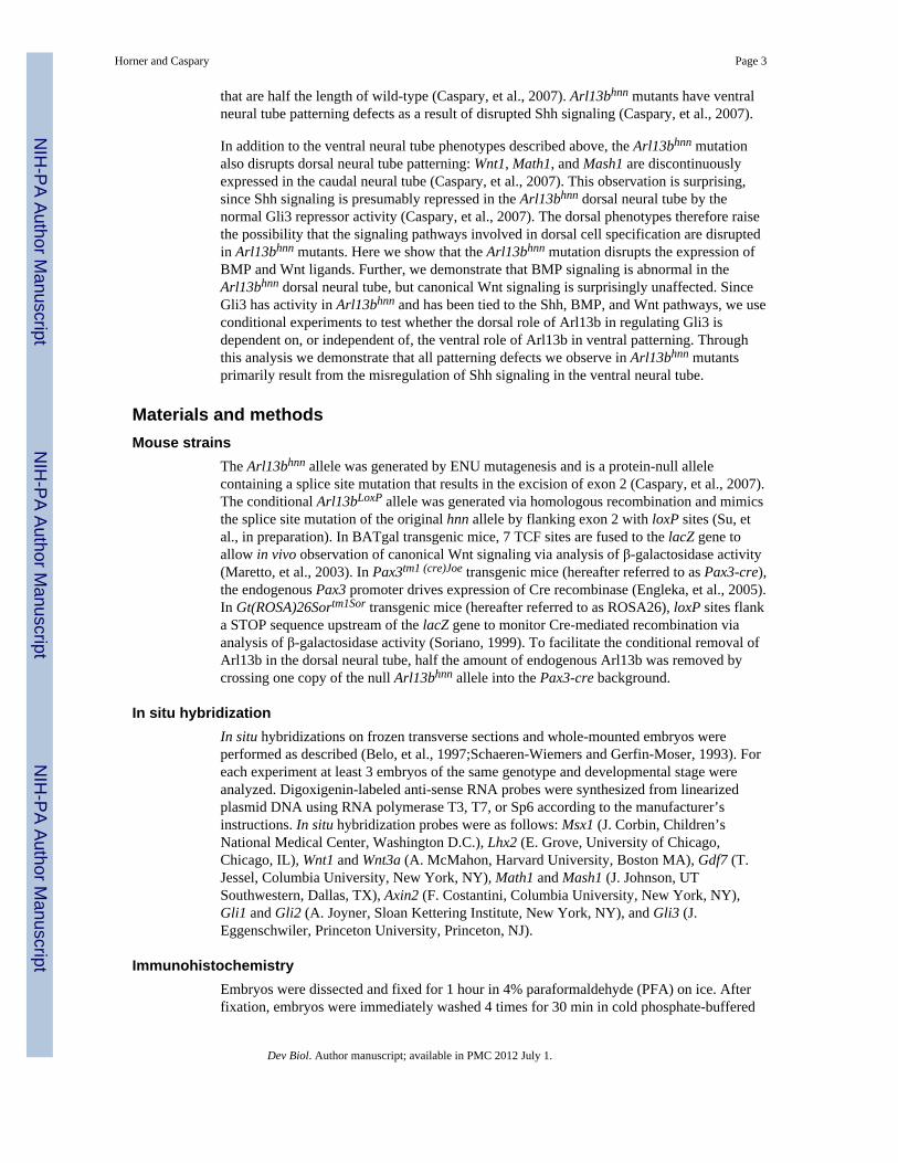
that are half the length of wild-type (Caspary, et al., 2007). Arl13bhnn mutants have ventralneural tube patterning defects as a result of disrupted Shh signaling (Caspary, et al., 2007).
In addition to the ventral neural tube phenotypes described above, the Arl13bhnn mutationalso disrupts dorsal neural tube patterning: Wnt1, Math1, and Mash1 are discontinuouslyexpressed in the caudal neural tube (Caspary, et al., 2007). This observation is surprising,since Shh signaling is presumably repressed in the Arl13bhnn dorsal neural tube by thenormal Gli3 repressor activity (Caspary, et al., 2007). The dorsal phenotypes therefore raisethe possibility that the signaling pathways involved in dorsal cell specification are disruptedin Arl13bhnn mutants. Here we show that the Arl13bhnn mutation disrupts the expression ofBMP and Wnt ligands. Further, we demonstrate that BMP signaling is abnormal in theArl13bhnn dorsal neural tube, but canonical Wnt signaling is surprisingly unaffected. SinceGli3 has activity in Arl13bhnn and has been tied to the Shh, BMP, and Wnt pathways, we useconditional experiments to test whether the dorsal role of Arl13b in regulating Gli3 isdependent on, or independent of, the ventral role of Arl13b in ventral patterning. Throughthis analysis we demonstrate that all patterning defects we observe in Arl13bhnn mutantsprimarily result from the misregulation of Shh signaling in the ventral neural tube.
Materials and methodsMouse strains
The Arl13bhnn allele was generated by ENU mutagenesis and is a protein-null allelecontaining a splice site mutation that results in the excision of exon 2 (Caspary, et al., 2007).The conditional Arl13bLoxP allele was generated via homologous recombination and mimicsthe splice site mutation of the original hnn allele by flanking exon 2 with loxP sites (Su, etal., in preparation). In BATgal transgenic mice, 7 TCF sites are fused to the lacZ gene toallow in vivo observation of canonical Wnt signaling via analysis of β-galactosidase activity(Maretto, et al., 2003). In Pax3tm1 (cre)Joe transgenic mice (hereafter referred to as Pax3-cre),the endogenous Pax3 promoter drives expression of Cre recombinase (Engleka, et al., 2005).In Gt(ROSA)26Sortm1Sor transgenic mice (hereafter referred to as ROSA26), loxP sites flanka STOP sequence upstream of the lacZ gene to monitor Cre-mediated recombination viaanalysis of β-galactosidase activity (Soriano, 1999). To facilitate the conditional removal ofArl13b in the dorsal neural tube, half the amount of endogenous Arl13b was removed bycrossing one copy of the null Arl13bhnn allele into the Pax3-cre background.
In situ hybridizationIn situ hybridizations on frozen transverse sections and whole-mounted embryos wereperformed as described (Belo, et al., 1997;Schaeren-Wiemers and Gerfin-Moser, 1993). Foreach experiment at least 3 embryos of the same genotype and developmental stage wereanalyzed. Digoxigenin-labeled anti-sense RNA probes were synthesized from linearizedplasmid DNA using RNA polymerase T3, T7, or Sp6 according to the manufacturer’sinstructions. In situ hybridization probes were as follows: Msx1 (J. Corbin, Children’sNational Medical Center, Washington D.C.), Lhx2 (E. Grove, University of Chicago,Chicago, IL), Wnt1 and Wnt3a (A. McMahon, Harvard University, Boston MA), Gdf7 (T.Jessel, Columbia University, New York, NY), Math1 and Mash1 (J. Johnson, UTSouthwestern, Dallas, TX), Axin2 (F. Costantini, Columbia University, New York, NY),Gli1 and Gli2 (A. Joyner, Sloan Kettering Institute, New York, NY), and Gli3 (J.Eggenschwiler, Princeton University, Princeton, NJ).
ImmunohistochemistryEmbryos were dissected and fixed for 1 hour in 4% paraformaldehyde (PFA) on ice. Afterfixation, embryos were immediately washed 4 times for 30 min in cold phosphate-buffered
Horner and Caspary Page 3
Dev Biol. Author manuscript; available in PMC 2012 July 1.
NIH
-PA Author Manuscript
NIH
-PA Author Manuscript
NIH
-PA Author Manuscript

saline (PBS) and incubated in 30% sucrose at 4°C overnight. Embryos were washed inOptimal Cutting Temperature (OCT) Compound 3 times for 15 min before embedding inOCT. We obtained 10-12-micron sections on a Leica CM1850 cryostat. Immunostainingwas performed as described (Yamada, et al., 1993). Briefly, slides were washed 3 times for5 min in PBS, and then blocked for 1 h at room temperature in PBS containing 10% heat-inactivated sheep serum and 0.1% triton-X. Slides were incubated with the followingantibodies overnight at 4°C: rabbit polyclonal anti-Arl13b, 1:1500 (Caspary, et al., 2007),mouse monoclonal anti-Cre, 1:500 (Sigma), mouse polyclonal anti-HB9, 1:10(Developmental Studies Hybridoma Bank), rabbit polyclonal anti-Olig2, 1:300 (Chemicon),rabbit polyclonal anti-Olig3, 1:5000, guinea pig anti-Tlx3, 1:10,000, and guinea pig anti-Foxd3, 1:5,000 (gifts from C. Birchmeier, Max Delbruck-Center for Molecular Medicine,Berlin, Germany), mouse monoclonal anti-BrdU, 1:100 (Sigma), and rabbit polyclonalphospho-histone H3, 1:1000 (Millipore). Primary antibody-bound slides were incubated for1–2 h at room temperature with 1:200 Alexa Fluor 488- and 568-conjugated goat anti-mouseand goat anti-rabbit antibodies (Molecular Probes) and 1:6000 Hoechst 33342 (MolecularProbes). Slides were mounted in 80% glycerol and viewed within 24 h.
Bromodeoxyuridine (BrdU)BrdU was injected intraperitoneally into pregnant females on embryonic day 10.5, at a finalconcentration of 50 μg/g body weight. Thirty min after injection, females were sacrificedand embryos were fixed for antibody staining as described above. Prior to the blocking stepof antibody staining, sections were treated for antigen retrieval by covering slides with 10mM sodium citrate and then steaming the slides for 15 min in an Oster steamer.
Staining for β-galactosidase activityStaining for β-galactosidase activity was performed as described in (Nagy, et al., 2003).Briefly, embryos were fixed in 1M Phosphate Buffer containing 0.2% glutaraldehyde for15–30 min at room temperature, and then washed in detergent rinse (Nagy, et al., 2003) 3times for 15–30 min each. To observe β-galactosidase activity, embryos were incubated withstaining solution (Nagy, et al., 2003) containing 0.1 mg/mL X-gal overnight at roomtemperature. To section X-gal-stained embryos, embryos were washed 2 times for 5 min inPBS, fixed in 4% PFA for 2 h at room temperature or overnight at 4°C, re-washed in PBSfor 5 min, incubated in 30% sucrose overnight, and embedded in OCT as above. Slidescontaining 25-50-μM sections were washed 3 times for 5 min in PBS and mounted in 80%glycerol prior to viewing.
MicroscopyImages of whole embryos and neural tube sections were collected with a Leica MZFLIIIstereomicroscope and a Leica DM6000 B upright fluorescence microscope, respectively. Q-capture and Simple PCI software were used to collect images. Adobe Photoshop CS2 wasused to crop and adjust color, brightness, and contrast of images.
Cell counting and statisticsCells in the neural tube were counted using Simple PCI software. A total of 15 neural tubesections from 3 Arl13bhnn embryos were counted, and a total of 12 neural tube sections from3 WT embryos were counted. The number of cells stained with BrdU (S phase) and thenumber of cells stained with phosphohistone H3 (M phase) were normalized to the totalnumber of cells in the neural tube (stained with Hoechst). Statistical differences betweenWT and Arl13bhnn were determined with a 2-tailed Student’s t-test.
Horner and Caspary Page 4
Dev Biol. Author manuscript; available in PMC 2012 July 1.
NIH
-PA Author Manuscript
NIH
-PA Author Manuscript
NIH
-PA Author Manuscript

Results and discussionWnt ligands are abnormally expressed in the Arl13bhnn neural tube, but downstreamcanonical Wnt signaling is normal
Two Wnt ligands are expressed in the neural tube roof plate, Wnt1 and Wnt3a, and theircombined loss results in lowered Math1 expression and loss of Ngn1 expression(Muroyama, et al., 2002;Parr, et al., 1993). Wnt1 is aberrantly expressed in the Arl13bhnn
neural tube; caudal to the forelimb, Wnt1 is expressed in discontinuous patches, with aregion near the hindlimb showing complete loss of expression (Caspary, et al., 2007). Wefound that Wnt3a is also discontinuously expressed and absent in the Arl13bhnn caudalneural tube (Fig. 1A, B). Since both Wnt ligands are lost in the same region, we nextexplored whether canonical Wnt signaling is disrupted in Arl13bhnn mutant embryos.Canonical Wnt signaling can be visualized with tools that detect TCF/LEF transcriptionfactor activity, the mediators of canonical Wnt signaling. In BATgal transgenic mice(Maretto, et al., 2003), seven TCF sites are fused to the lacZ gene to allow in vivoobservation of canonical Wnt signaling via analysis of β-galactosidase activity. To analyzecanonical Wnt signaling in Arl13bhnn mutants, we obtained E9.5, E10.5, and E11.5 wild-type (WT) and Arl13bhnn embryos carrying the BATgal transgene and stained them with X-gal. Canonical Wnt signaling was present at similar levels, places, and times in all WT andArl13bhnn mutants examined, in both whole embryos and neural tube sections at thehindlimb level (Fig. 2). In the WT caudal neural tube, canonical Wnt signaling is observedthroughout most of the dorsal-ventral axis of the neural tube at E9.5 (Yu, et al., 2008), butby E10.5 and E11.5, canonical Wnt signaling is largely restricted to the dorsal-most third ofthe neural tube (Fig 2). In Arl13bhnn mutants, Wnt signaling is likewise observed throughoutthe neural tube at E9.5 and limited to the dorsal neural tube at E10.5 and E11.5 (Fig. 2).
Another global measure of canonical Wnt signaling is expression of Axin2, a directtranscriptional target of canonical Wnt signaling (Jho, et al., 2002). The expression of Axin2in whole embryos and neural tube sections is indistinguishable between WT and Arl13bhnn
mutants (Fig. 1C, D). Moreover, canonical Wnt signaling controls the expression of Olig3,and we see that Olig3 protein is present at the correct time and place in the Arl13bhnn dorsalneural tube (Fig. 1E-H). Since Olig3 is required to specify dI2 and dI3 interneurons (Muller,et al., 2005;Zechner, et al., 2007), we next examined whether these neurons are present inthe Arl13bhnn neural tube using homeodomain transcription factors that are differentiallyexpressed in postmitotic neurons (Helms and Johnson, 2003;Qian, et al., 2002). We foundthat Foxd3+ (dI2) and Tlx3+ (dI3 and dI5) cells are specified in the Arl13bhnn neural tube,although their patterning within the neural tube is not as precisely ordered as in WT (Fig 1E-H). For instance, the separation between dI3 and dI5 neurons is sharply distinct in WT, butappear to be more of a continuum in Arl13bhnn (Fig. 1H).
In addition to cell fate, Wnt signaling controls cell proliferation in the neural tube (Cayusoand Marti, 2005;Dickinson, et al., 1994;Megason and McMahon, 2002). Arl13bhnn mutantsdo not show gross errors in cell proliferation (Caspary, et al., 2007). However, to detect anysubtle changes in cell proliferation, we quantified the number of proliferating cells in WTand Arl13bhnn neural tubes by staining for BrdU, which marks S phase, and phospho-histoneH3, which marks M phase, and normalized to the total number of cells in the neural tube(Fig. 3). We did not observe any difference in the number of cells in S phase (p = 0.29) or Mphase (p = 0.08) in WT and Arl13bhnn neural tubes (Fig. 3C). Taken together, these resultsindicate that, despite abnormal ligand expression, canonical Wnt signaling is unaffected inArl13bhnn mutants.
Horner and Caspary Page 5
Dev Biol. Author manuscript; available in PMC 2012 July 1.
NIH
-PA Author Manuscript
NIH
-PA Author Manuscript
NIH
-PA Author Manuscript

BMP signaling is aberrant in the Arl13bhnn dorsal neural tubeSince Wnt ligand expression is disrupted in the Arl13bhnn roof plate, we next exploredwhether TGFβ ligand expression is similarly disrupted. Several TGFβ ligands are expressedin the roof plate, including Bmp4, Bmp6, Bmp7, and Gdf7. The role of individual TGFβligands in dorsal neural tube patterning has remained elusive, due to functional redundancyor early embryonic lethality (Beppu, et al., 2000;Dudley, et al., 1995;Dudley and Robertson,1997;Luo, et al., 1995;Lyons, et al., 1995;Mishina, et al., 1995;Winnier, et al., 1995;Zhangand Bradley, 1996). The exception is loss of Gdf7, which results in loss of dI1a neurons(Lee, et al., 1998). Since loss of Gdf7 alone is sufficient to cause dorsal neural tubepatterning defects, we used in situ hybridization to examine its expression in WT andArl13bhnn mutant embryos. We observed specific and strong staining in the WT neural tubealong the entire rostral-caudal axis (Fig. 4A, C). Similar to the Wnt ligands, expression ofGdf7 is largely absent and discontinuous in the caudal neural tube of Arl13bhnn mutantembryos (Fig. 4B, D).
To see whether the abnormal Gdf7 expression has downstream effects in Arl13bhnn mutants,we next examined several BMP targets. An established read-out of BMP signaling istranscription of Msx-1, a homeobox transcription factor that acts as an effector of BMPsignaling and whose expression is directly upregulated in response to BMP signaling(Furuta, et al., 1997;Liem, et al., 1997;Liu, et al., 2004). We observe similar levels of Msx-1in the limb buds and pharyngeal arches of WT and Arl13bhnn mutant embryos, but Msx-1expression is discontinuous and largely absent in the caudal neural tube of Arl13bhnn mutantembryos (Fig. 4E-H). Furthermore, BMP signaling is required for the specification of dI1interneurons, and we found that expression of Lhx2, which marks dI1s, is discontinuous inthe caudal neural tube (Fig. 4I-L). Together with the previous observations that Math1,Wnt1, and Wnt3a expression are disrupted, these results indicate that BMP signaling isaberrant in the dorsal neural tube of Arl13bhnn mutants. These results are consistent withother mutants in which BMP signaling is lost, since total elimination of BMP receptors inthe roof plate results in the loss of Math1 and dI1s (Wine-Lee, et al., 2004). Furthermore, thediscontinuous expression of Wnt1 and Wnt3a in the Arl13bhnn roof plate may stem from thedisruption in BMP signaling, since BMP signaling mutants show reduced expression of Wntligands (Wine-Lee, et al., 2004).
Arl13b is required non-autonomously for BMP signaling and Wnt ligand expressionSince interactions between the Shh signaling pathway and dorsal signaling pathways arenecessary to establish proper patterning of the neural tube, it is possible that the disruptionof Shh signaling in the Arl13bhnn neural tube indirectly disrupts BMP signaling and thusdorsal patterning. Alternatively, Arl13b might be directly required in the dorsal neural tubefor BMP signaling and Wnt ligand expression. To distinguish these two possibilities, weused our conditional Arl13b allele, Arl13bLoxP, in which LoxP sites flank the second exon ofArl13b, so that upon Cre-mediated recombination exon 2 is excised, resulting in a mutationthat mimics the null hnn mutation (Su, et al., in preparation). We removed Arl13bexclusively in the dorsal neural tube by crossing Arl13bLoxP mice to mice that express Crerecombinase under the control of the Pax3 promoter (Engleka, et al., 2005). Pax3 isexpressed starting from E8.5, just as neurogenesis begins (Solloway and Robertson, 1999),and becomes restricted to the dorsal-most third of the dorsal neural tube. This cross alsogenerated the appropriate control embryos that carry the Arl13bLoxP conditional allele alone,or the Pax3-cre transgene alone.
Pax3-cre-mediated recombination occurs by E9.5, and Arl13b protein isremoved in the dorsal neural tube beginning at E9.5—To monitor the spatial andtemporal activity of Pax3-cre, we used the ROSA26 reporter, in which a STOP sequence
Horner and Caspary Page 6
Dev Biol. Author manuscript; available in PMC 2012 July 1.
NIH
-PA Author Manuscript
NIH
-PA Author Manuscript
NIH
-PA Author Manuscript

flanked by loxP sites is upstream of the lacZ gene (Soriano, 1999). Upon Cre-mediatedrecombination, β-galactosidase is expressed and can be visualized with X-gal staining. Thus,the ROSA26 reporter shows not only when and where Cre recombinase is expressed, butalso the length of time necessary for recombination to occur. We crossed mice carryingPax3-cre to the ROSA26 reporter line and dissected embryos at E8.5, E9.5, and E10.5. Wedid not detect recombination at E8.5, but at E9.5 and E10.5 β-galactosidase activity wasobserved in the neural tube, midbrain, hindbrain, somites, pharyngeal arches, andfrontonasal prominence (Supplemental Fig. 2). Neural tube sections taken at the forelimband hindlimb levels of X-gal-stained embryos show that recombination occurs only in thedorsal neural tube (Supplemental Fig. 2).
To detect when Arl13b protein is removed from the dorsal neural tube, we crossed micecarrying our Arl13bLoxP conditional allele to mice carrying Pax3-cre and dissected embryosat E9.5, E10.5, and E11.5. Neural tube sections of Arl13bΔPax3-cre and control embryos werestained with antibodies against Arl13b and Cre. At E9.5 Cre recombinase is restricted tocells in or around the roof plate (Fig 5B). Arl13b protein can be visualized in the ventricularzone along most of the dorsal-ventral axis of the neural tube; however, where Cre isexpressed, loss of Arl13b is already apparent (Fig. 5B). In E10.5 and E11.5 neural tubesections at the hindlimb level, Arl13b is absent where Cre is expressed, in the dorsal-mostone third to one half of the neural tube (Fig. 5C-F). In summary, Pax3-cre drives expressionof Cre recombinase at E8.5, recombination occurs in the neural tube by E9.5, and Arl13bprotein is lost in the dorsal neural tube by E9.5-E10.5.
Based on these data and previous studies, we can make predictions regarding the embryonicstage at which we would expect to see a phenotype in our conditional mutants. Todemonstrate that the roof plate is a source of signaling molecules necessary for dorsalinterneuron specification, (Lee, et al., 2000) conditionally ablated the roof plate by drivingthe expression of diphtheria toxin in the roof plate via the Gdf7 promoter. Gdf7 is expressedat E9.5-E10.0 (Lee, et al., 2000). Prior to Gdf7 expression and roof plate formation, Msx1,Wnt1, and Wnt3a are expressed in dorsal midline cells, presumably due to BMP signalingfrom the epidermal ectoderm. When the roof plate was removed at E9.5-E10.0, expressionof Msx1, Wnt1, and Wnt3a was lost by E11.0. In another study, BMP receptors wereremoved in the neural tube at E9.5-E10.0 to completely eliminate BMP signaling (Wine-Lee, et al., 2004). In these mutants, Math1 was expressed at E10.0, but lost by E10.5. Thesestudies demonstrate that BMP signaling is required to maintain expression of dorsalpatterning genes. Since the expression of genes that rely on BMP signaling was disruptedabout 0.5-1 day after BMP signaling was removed in the above studies, we would expect tosee any dorsal phenotypes in Arl13bΔPax3-cre embryos between E10.5-E12.5.
Removing Arl13b exclusively in the dorsal neural tube permits proper Shhsignaling in the ventral neural tube—The ability to distinguish whether loss of Arl13bdirectly or indirectly disrupts dorsal neural tube patterning depends on our prediction thatselectively removing Arl13b in the dorsal neural tube will permit proper Shh signaling in theventral neural tube. To ensure that ventral patterning is normal in Arl13bΔPax3-cre embryos,we dissected Arl13bΔPax3-cre embryos at E10.5 and E11.5 and stained neural tube sectionswith antibodies against HB9 and Olig2, which mark motor neurons (MNs) and progenitorsof motor neurons (pMNs), respectively (Arber, et al., 1999;Novitch, et al., 2001). InArl13bhnn mutant embryos, MNs and pMNs are dramatically expanded from their restricteddomains in the ventral neural tube both ventrally and dorsally (Fig. 5G) (Caspary, et al.,2007). When Arl13b is removed only in the dorsal neural tube, however, we see that thedomains of MNs and pMNs are identical to WT, confirming that Shh signaling and ventralpatterning is normal in Arl13bΔPax3-cre embryos (Fig. 5H-K).
Horner and Caspary Page 7
Dev Biol. Author manuscript; available in PMC 2012 July 1.
NIH
-PA Author Manuscript
NIH
-PA Author Manuscript
NIH
-PA Author Manuscript

Conditional loss of Arl13b results in postnatal death at P0—Arl13bhnn embryoshave several morphological defects, including exencephaly and spina bifida, and they diebetween E13.5-E14.5 (Caspary, et al., 2007). To determine whether Arl13bΔPax3-cre
embryos are viable, we performed timed matings and examined embryos at specific stages,as well as just after birth. We found Arl13bΔPax3-cre embryos have no overt morphologicaldefects and are indistinguishable from WT during embryogenesis (Table 1). However, atbirth most Arl13bΔPax3-cre pups died after an episode of observable gasping. We examinedthe internal organs of 2 of the Arl13bΔPax3-cre mutants just after death and found that thestomachs contained air bubbles, consistent with a breathing problem. We did see oneArl13bΔPax3-cre pup survive, indicating that escapers can occur; however, this mouse wasseverely runted and died by postnatal day 21. Therefore, Arl13bΔPax3-cre mutants die shortlyafter birth at P0, likely due to a breathing problem.
Arl13b is not directly required in the dorsal neural tube for BMP signaling orWnt ligand expression—If the observed Arl13bhnn dorsal patterning defects areindirectly caused by disrupted Shh signaling, then removing Arl13b exclusively in the dorsalneural tube will allow normal dorsal signaling and patterning in the conditionalArl13bΔPax3-cre mutants. Alternatively, if Arl13b is directly required in the dorsal neuraltube, then loss of Arl13b in the dorsal neural tube will result in the dorsal signaling andpatterning defects observed in Arl13bhnn mutants (Figs. 1 and 4). We monitored BMPsignaling via expression of Msx1, Math1, Gdf7, and Lhx2, as described above. In addition,we examined dorsal patterning via expression of Wnt1, Wnt3a, and Mash1.
The expression patterns of Msx1 and Math1 were examined in whole Arl13bΔPax3-cre mutantembryos and their sibling controls from E10.5-E13.5. Unlike Arl13bhnn mutants, whereexpression of these markers is discontinuous in the caudal neural tube, both Msx1 and Math1are expressed continuously and at similar levels in the caudal neural tube of Arl13bΔPax3-cre
mutant embryos and their littermate controls at all stages examined (Fig. 6). There was somevariation between experiments in the posterior boundary of expression (number of somitesfrom the tip of the tail), but there was no consistent correlation between genotype and theposterior limit of expression. In addition, neural tube sections of E11.5 and E12.5 embryostaken at the hindlimb level show that the pattern of Msx1 and Math1 expression along thedorsal-ventral axis of the neural tube is similar in Arl13bΔPax3-cre mutant and controlembryos (Fig. 6). Since Lhx2 marks differentiated dI1s, we examined embryos at slightlylater stages, E12.5-E14.5. In all embryos and at all stages examined, we detected nodifference in the level of staining or pattern of Lhx2 expression between Arl13bΔPax3-cre
mutant and control embryos (Fig. 7).
Since we interpreted the abnormal Msx1, Math1, and Lhx2 expression in Arl13bhnn embryosto result from the lack of Gdf7 ligand, their presence in the conditional mutants predicts thatGdf7 expression should be normal. Indeed, Gdf7 is expressed continuously and at similarlevels in all Arl13bΔPax3-cre mutant and control embryos examined (Fig 8A-D). Finally, weexamined Wnt1, Wnt3a, and Mash1 expression at E11.5 and E12.5 in Arl13bΔPax3-cre
mutant and control embryos. Despite the abnormal expression of these genes in theArl13bhnn dorsal neural tube, we detected no differences in the level or expression pattern ofany of these genes between Arl13bΔPax3-cre mutant and control embryos (Fig. 8E-P).
Taken together, these data demonstrate that cells in the dorsal neural tube do not requireArl13b for TGFβ signaling or Wnt ligand expression, and the Arl13bhnn dorsal neural tubedefects are non-autonomous. Therefore, the dorsal patterning defects observed in Arl13bhnn
embryos are an indirect consequence of the abnormal Shh signaling environment in theseembryos.
Horner and Caspary Page 8
Dev Biol. Author manuscript; available in PMC 2012 July 1.
NIH
-PA Author Manuscript
NIH
-PA Author Manuscript
NIH
-PA Author Manuscript

Subtle changes in Gli activator/Gli repressor ratios could influence BMP signalingShh signaling is mediated at the transcriptional level by three proteins in vertebrates: Gli1,Gli2, and Gli3 (Matise and Joyner, 1999). Gli1 behaves only as a transcriptional activator,while Gli2 and Gli3 can act as both transcriptional activators and repressors (Aza-Blanc, etal., 2000;Jacob and Briscoe, 2003). The graded response of cells in the ventral neural tube toShh is mediated by the ratio of Gli activator (GliA) to Gli repressor (GliR) activity[reviewed in (Ribes and Briscoe, 2009)]. In mouse mutants that completely lack cilia, thereis a loss of both GliA and GliR activity in the neural tube (Huangfu and Anderson,2005;Liu, et al., 2005;May, et al., 2005). However, the unique cilia defect of Arl13bhnn hasunique consequences for Shh signaling. Unlike total-loss-of-cilia mutants, GliR activity isintact in Arl13bhnn mutants, because ventral cell fates are dorsally expanded when GliR isremoved in Arl13bhnn /Gli3 double mutants (Caspary, et al., 2007). At the same time, GliAis not properly modulated in Arl13bhnn mutants, since cells that require the highest or lowestlevels of Shh are not specified; instead, cells that require intermediate GliA activity (i.e.,MNs) are expanded dorsally and ventrally (Caspary, et al., 2007). Thus, the dorsalexpansion of Shh signaling in the Arl13bhnn neural tube corresponds to an increase in theratio of GliA/GliR in lateral and dorsal neural cells (Caspary, et al., 2007).
To directly examine the levels of GliA and GliR in the Arl13bhnn neural tube, we performedin situ hybridization with antisense probes for Gli1 and Gli3 on WT and Arl13bhnn caudalneural tube sections, since Gli1 can only be an activator and Gli3 is the predominantrepressor in the neural tube. Consistent with previous data, we find that Gli3 is properlyexpressed in the Arl13bhnn dorsal neural tube at levels similar to those observed in WT (Fig.9E-H). However, the pattern of Gli1 expression differs between WT and Arl13bhnn embryos:in WT embryos, Gli1 expression is excluded from the dorsal-most cells of the neural tube(Fig. 9A, C), while in Arl13bhnn embryos, expression of Gli1 is shifted dorsally and enrichedin the dorsal neural tube (Fig 9B, D). Together with previous findings, these datademonstrate that the balance of GliA/GliR repressor is shifted in the Arl13bhnn dorsal neuraltube.
The mechanism by which the abnormal Shh signaling environment causes the observeddorsal phenotypes remains unclear, but it is likely related to cross-talk among signalingpathways. The abnormal Gdf7 expression and aberrant BMP signaling in Arl13bhnn mice arereminiscent of the situation in another mouse mutant, where ectopic Gli2 activator preventsexpression of the BMP ligands BMP7 and Gdf7 (Cho, et al., 2008). Like Arl13bhnn, thismutation affects a ciliary protein, Fkbp8, and the mutants have aberrant BMP signaling asmeasured by loss of Math1 and Lhx2 expression and a reduction in Msx1/2 protein levels(Cho, et al., 2008); however, there are important distinctions between the Arl13bhnn andFkbp8 phenotypes. In Fkbp8 mutants, the most ventral cell types requiring the highest levelsof Gli activator (FP and V3 interneurons) are expanded dorsally. Both the dorsal expansionof ventral cell types and disruption of BMP signaling are abolished in Fkbp8/Gli2 doublemutants, indicating that ectopic Gli2 activator is principally responsible for the Fkbp8phenotype (Cho, et al., 2008). In contrast, the phenotype of Arl13bhnn mutants is notconsistent with an increase in the highest levels of Gli2 activator; instead, the dorsal andventral expansion of intermediate cell types (MNs) are consistent with constitutive,intermediate-level activation of Gli (Caspary, et al., 2007). Furthermore, removing the majorGli activator in the Arl13bhnn mutant background (via Arl13bhnn/Gli2 double mutants) doesnot rescue all the Arl13bhnn patterning defects (Caspary, et al., 2007). Specifically, thedorsal expansion of MNs persists in Arl13bhnn/Gli2 doubles, illustrating that ectopicallyactive Gli2 in the Arl13bhnn neural tube is not sufficient to explain the loss of BMP ligandexpression. Since Gli1 was found to be dorsally enriched in Arl13bhnn and Gli1 alsopromotes the differentiation of MNs (Ruiz i Altaba, 1998), the dorsal expansion of MNs inArl13bhnn/Gli2 double mutants is likely due to changes in the position and/or activity of Gli1
Horner and Caspary Page 9
Dev Biol. Author manuscript; available in PMC 2012 July 1.
NIH
-PA Author Manuscript
NIH
-PA Author Manuscript
NIH
-PA Author Manuscript

activator. Thus, one interpretation of these data would posit a new role for Shh signaling inthe modulation of the BMP signaling pathway: namely, that ectopic Gli1 activator activitycan prevent Gdf7 expression and disrupt BMP signaling.
Normal Wnt response in the absence of ligand may reveal sensitivity of β-cateninMost enigmatic is our result showing canonical Wnt signaling occurs normally in theabsence of ligand in the Arl13bhnn neural tube. Since Wnt1 and Wnt3a are expressed in therostral neural tube and in portions of the caudal neural tube, one possibility is that Wntligands from one area diffuse into regions lacking Wnt ligand in the Arl13bhnn caudal neuraltube; however, this seems unlikely, given that Wnts are secreted glycoproteins that arebound by the extracellular matrix and diffuse over a distance of just a few cell diameters(Bradley and Brown, 1990;Gonzalez, et al., 1991;van den Heuvel, et al., 1989). Anotherpossibility is that early Wnt signals from the surface ectoderm are normal, allowing the Wntresponse to be maintained in the dorsal neural tube over time in the absence of ligand, butconditional loss of β-catenin at E9.5, after the neural tube has formed, results in loss ofOlig3 expression by E12.5 (Zechner, et al., 2007), a time point at which we still see normalOlig3 in Arl13bhnn mutants (Fig. 1F, H.) A third possibility is that a downstream signalingcomponent, such as β-catenin, is not properly regulated in Arl13bhnn mutants. Misregulationof β-catenin has been shown to occur in cells that lack cilia (Corbit, et al., 2008) or basalbody proteins (Gerdes, et al., 2007). While it is well established that cilia are required fornon-canonical Wnt signaling (Gerdes, et al., 2007;Jones, et al., 2008;Ross, et al., 2005), thedata surrounding cilia and canonical Wnt signaling remain controversial. Severalcomponents of the canonical Wnt pathway physically localize to cilia, including β-cateninand a negative regulator of canonical Wnt signaling, the APC complex (Corbit, et al., 2008).Further, the inactive, phosphorylated form of β-catenin localizes to the base of the cilium,suggesting that the cilium directly restrains Wnt signaling (Corbit, et al., 2008). Nonetheless,studies that test this hypothesis using mouse mutants or cells derived from mouse mutantshave yielded different results. For instance, cells in culture that either completely lack ciliaor have shortened cilia show hyper-responsive activation of the canonical Wnt pathway inresponse to Wnt3a stimulation in one study (Corbit, et al., 2008), but not in others (Ocbina,et al., 2009). If cilia are required to restrain canonical Wnt signaling, our results might beexplained simply by the short cilia in Arl13bhnn mutants. In this scenario, the short ciliacause active β-catenin to be stabilized in the cytoplasm of Arl13bhnn mutant cells, allowingWnt signaling to occur in the absence of ligand. If this hypothesis is correct, we predict thatin Arl13bΔPax3-cre conditionals, which show normal Wnt ligand expression, we will seehyper-responsive canonical Wnt signaling. To test this hypothesis, we examined Axin2expression in our conditional mutants. At E11.5, Axin2 expression is identical inArl13bΔPax3-cre conditionals and controls, both in whole embryos and neural tube sections(Fig. 10). Even though this result is not consistent with cilia being required to restraincanonical Wnt signaling, there is the possibility of a more indirect relationship, in which theabnormal cilia in Arl13bhnn disrupts non-canonical Wnt (PCP) signaling, which in turnstabilizes β-catenin and affects canonical Wnt signaling (Brembeck, et al., 2004;Schwarz-Romond, et al., 2002).
A final possibility to explain how canonical Wnt signaling could occur in the absence ofligand in the Arl13bhnn neural tube is that, as with the BMP signaling defects, the abnormalShh signaling environment is principally responsible. The physical interaction between Gli3repressor and β-catenin allows us to put forward one speculative model of how Wntsignaling might occur in the absence of both Wnt ligands in the Arl13bhnn dorsal neuraltube. Normally, there is a delicate balance in the dorsal neural tube between the level of Shhsignaling, the amount of Gli3R, and Wnt signaling. Excessive Gli3R inhibits canonical Wntsignaling by binding to the active form of β-catenin (Ulloa, et al., 2007). This situation
Horner and Caspary Page 10
Dev Biol. Author manuscript; available in PMC 2012 July 1.
NIH
-PA Author Manuscript
NIH
-PA Author Manuscript
NIH
-PA Author Manuscript

occurs in Shh null embryos, because most of the available Gli3 is processed to becomeGli3R (Ulloa, et al., 2007). This implies that in WT embryos, Shh signaling in the dorsalneural tube regulates the available Gli3R, so there is sufficient free active β-catenin for Wntsignaling. In Arl13bhnn mutants, increased GliA activity in the dorsal neural tube mighteffectively lower the amount of Gli3R. Although not sufficient to abolish Gli3R activity, thismay tip the balance enough to free some active β-catenin, permitting Wnt signaling or themaintenance of Wnt response in the absence of ligand.
Supplementary MaterialRefer to Web version on PubMed Central for supplementary material.
AcknowledgmentsThis work was supported by NIH grant R01NS056380 to T.C. and a Ruth L. Kirschstein National Research ServiceAward 1F32HD060368-01A2 to V.L.H. We thank all the members of the Caspary lab as well as Cheryl TimmsStrauss for helpful suggestions on the manuscript, and Carmen Birchmeier for Olig3, Foxd3, and Tlx3 antibodies.The HB9 antibody, developed by T. Jessell (Columbia University, New York, NY), was obtained from theDevelopmental Studies Hybridoma Bank developed under the auspices of the NICHD and maintained by TheUniversity of Iowa, Department of Biology, Iowa City, IA 52242.
ReferencesAlvarez-Medina R, Cayuso J, Okubo T, Takada S, Marti E. Wnt canonical pathway restricts graded
Shh/Gli patterning activity through the regulation of Gli3 expression. Development. 2008; 135:237–247. [PubMed: 18057099]
Antoshechkin I, Han M. The C. elegans evl-20 gene is a homolog of the small GTPase ARL2 andregulates cytoskeleton dynamics during cytokinesis and morphogenesis. Dev Cell. 2002; 2:579–591. [PubMed: 12015966]
Arber S, Han B, Mendelsohn M, Smith M, Jessell TM, Sockanathan S. Requirement for the homeoboxgene Hb9 in the consolidation of motor neuron identity. Neuron. 1999; 23:659–674. [PubMed:10482234]
Aza-Blanc P, Lin HY, Ruiz i Altaba A, Kornberg TB. Expression of the vertebrate Gli proteins inDrosophila reveals a distribution of activator and repressor activities. Development. 2000;127:4293–4301. [PubMed: 10976059]
Belo JA, Bouwmeester T, Leyns L, Kertesz N, Gallo M, Follettie M, De Robertis EM. Cerberus-like isa secreted factor with neutralizing activity expressed in the anterior primitive endoderm of themouse gastrula. Mech Dev. 1997; 68:45–57. [PubMed: 9431803]
Beppu H, Kawabata M, Hamamoto T, Chytil A, Minowa O, Noda T, Miyazono K. BMP type IIreceptor is required for gastrulation and early development of mouse embryos. Dev Biol. 2000;221:249–258. [PubMed: 10772805]
Bermingham NA, Hassan BA, Wang VY, Fernandez M, Banfi S, Bellen HJ, Fritzsch B, Zoghbi HY.Proprioceptor pathway development is dependent on Math1. Neuron. 2001; 30:411–422. [PubMed:11395003]
Bradley RS, Brown AM. The proto-oncogene int-1 encodes a secreted protein associated with theextracellular matrix. EMBO J. 1990; 9:1569–1575. [PubMed: 2158444]
Brembeck FH, Schwarz-Romond T, Bakkers J, Wilhelm S, Hammerschmidt M, Birchmeier W.Essential role of BCL9-2 in the switch between beta-catenin's adhesive and transcriptionalfunctions. Genes Dev. 2004; 18:2225–2230. [PubMed: 15371335]
Caspary T, Larkins CE, Anderson KV. The graded response to Sonic Hedgehog depends on ciliaarchitecture. Dev Cell. 2007; 12:767–778. [PubMed: 17488627]
Cayuso J, Marti E. Morphogens in motion: growth control of the neural tube. J Neurobiol. 2005;64:376–387. [PubMed: 16041754]
Cho A, Ko HW, Eggenschwiler JT. FKBP8 cell-autonomously controls neural tube patterning througha Gli2- and Kif3a-dependent mechanism. Dev Biol. 2008; 321:27–39. [PubMed: 18590716]
Horner and Caspary Page 11
Dev Biol. Author manuscript; available in PMC 2012 July 1.
NIH
-PA Author Manuscript
NIH
-PA Author Manuscript
NIH
-PA Author Manuscript

Corbit KC, Shyer AE, Dowdle WE, Gaulden J, Singla V, Chen MH, Chuang PT, Reiter JF. Kif3aconstrains beta-catenin-dependent Wnt signalling through dual ciliary and non-ciliarymechanisms. Nat Cell Biol. 2008; 10:70–76. [PubMed: 18084282]
Dickinson ME, Krumlauf R, McMahon AP. Evidence for a mitogenic effect of Wnt-1 in thedeveloping mammalian central nervous system. Development. 1994; 120:1453–1471. [PubMed:8050356]
Dudley AT, Lyons KM, Robertson EJ. A requirement for bone morphogenetic protein-7 duringdevelopment of the mammalian kidney and eye. Genes Dev. 1995; 9:2795–2807. [PubMed:7590254]
Dudley AT, Robertson EJ. Overlapping expression domains of bone morphogenetic protein familymembers potentially account for limited tissue defects in BMP7 deficient embryos. Dev Dyn.1997; 208:349–362. [PubMed: 9056639]
Engleka KA, Gitler AD, Zhang M, Zhou DD, High FA, Epstein JA. Insertion of Cre into the Pax3locus creates a new allele of Splotch and identifies unexpected Pax3 derivatives. Dev Biol. 2005;280:396–406. [PubMed: 15882581]
Furuta Y, Piston DW, Hogan BL. Bone morphogenetic proteins (BMPs) as regulators of dorsalforebrain development. Development. 1997; 124:2203–2212. [PubMed: 9187146]
Garcia-Garcia MJ, Eggenschwiler JT, Caspary T, Alcorn HL, Wyler MR, Huangfu D, Rakeman AS,Lee JD, Feinberg EH, Timmer JR, Anderson KV. Analysis of mouse embryonic patterning andmorphogenesis by forward genetics. Proc Natl Acad Sci U S A. 2005; 102:5913–5919. [PubMed:15755804]
Gerdes JM, Liu Y, Zaghloul NA, Leitch CC, Lawson SS, Kato M, Beachy PA, Beales PL, DeMartinoGN, Fisher S, Badano JL, Katsanis N. Disruption of the basal body compromises proteasomalfunction and perturbs intracellular Wnt response. Nat Genet. 2007; 39:1350–1360. [PubMed:17906624]
Gonzalez F, Swales L, Bejsovec A, Skaer H, Martinez Arias A. Secretion and movement of winglessprotein in the epidermis of the Drosophila embryo. Mech Dev. 1991; 35:43–54. [PubMed:1720017]
Gowan K, Helms AW, Hunsaker TL, Collisson T, Ebert PJ, Odom R, Johnson JE. Crossinhibitoryactivities of Ngn1 and Math1 allow specification of distinct dorsal interneurons. Neuron. 2001;31:219–232. [PubMed: 11502254]
Helms AW, Johnson JE. Specification of dorsal spinal cord interneurons. Curr Opin Neurobiol. 2003;13:42–49. [PubMed: 12593981]
Hoyt MA, Stearns T, Botstein D. Chromosome instability mutants of Saccharomyces cerevisiae thatare defective in microtubule-mediated processes. Mol Cell Biol. 1990; 10:223–234. [PubMed:2403635]
Huangfu D, Anderson KV. Cilia and Hedgehog responsiveness in the mouse. Proc Natl Acad Sci U SA. 2005; 102:11325–11330. [PubMed: 16061793]
Huangfu D, Liu A, Rakeman AS, Murcia NS, Niswander L, Anderson KV. Hedgehog signalling in themouse requires intraflagellar transport proteins. Nature. 2003; 426:83–87. [PubMed: 14603322]
Ingham PW, McMahon AP. Hedgehog signaling in animal development: paradigms and principles.Genes Dev. 2001; 15:3059–3087. [PubMed: 11731473]
Jacob J, Briscoe J. Gli proteins and the control of spinal-cord patterning. EMBO Rep. 2003; 4:761–765. [PubMed: 12897799]
Jessell TM. Neuronal specification in the spinal cord: inductive signals and transcriptional codes. NatRev Genet. 2000; 1:20–29. [PubMed: 11262869]
Jho EH, Zhang T, Domon C, Joo CK, Freund JN, Costantini F. Wnt/beta-catenin/Tcf signaling inducesthe transcription of Axin2, a negative regulator of the signaling pathway. Mol Cell Biol. 2002;22:1172–1183. [PubMed: 11809808]
Jones C, Roper VC, Foucher I, Qian D, Banizs B, Petit C, Yoder BK, Chen P. Ciliary proteins linkbasal body polarization to planar cell polarity regulation. Nat Genet. 2008; 40:69–77. [PubMed:18066062]
Horner and Caspary Page 12
Dev Biol. Author manuscript; available in PMC 2012 July 1.
NIH
-PA Author Manuscript
NIH
-PA Author Manuscript
NIH
-PA Author Manuscript

Kahn RA, Cherfils J, Elias M, Lovering RC, Munro S, Schurmann A. Nomenclature for the human Arffamily of GTP-binding proteins: ARF, ARL, and SAR proteins. J Cell Biol. 2006; 172:645–650.[PubMed: 16505163]
Lee KJ, Dietrich P, Jessell TM. Genetic ablation reveals that the roof plate is essential for dorsalinterneuron specification. Nature. 2000; 403:734–740. [PubMed: 10693795]
Lee KJ, Mendelsohn M, Jessell TM. Neuronal patterning by BMPs: a requirement for GDF7 in thegeneration of a discrete class of commissural interneurons in the mouse spinal cord. Genes Dev.1998; 12:3394–3407. [PubMed: 9808626]
Li Y, Kelly WG, Logsdon JM Jr, Schurko AM, Harfe BD, Hill-Harfe KL, Kahn RA. Functionalgenomic analysis of the ADP-ribosylation factor family of GTPases: phylogeny among diverseeukaryotes and function in C. elegans. Faseb J. 2004; 18:1834–1850. [PubMed: 15576487]
Liem KF Jr, Tremml G, Jessell TM. A role for the roof plate and its resident TGFbeta-related proteinsin neuronal patterning in the dorsal spinal cord. Cell. 1997; 91:127–138. [PubMed: 9335341]
Liu A, Niswander LA. Bone morphogenetic protein signalling and vertebrate nervous systemdevelopment. Nat Rev Neurosci. 2005; 6:945–954. [PubMed: 16340955]
Liu A, Wang B, Niswander LA. Mouse intraflagellar transport proteins regulate both the activator andrepressor functions of Gli transcription factors. Development. 2005; 132:3103–3111. [PubMed:15930098]
Liu F, Massague J, Ruiz i Altaba A. Carboxy-terminally truncated Gli3 proteins associate with Smads.Nat Genet. 1998; 20:325–326. [PubMed: 9843199]
Liu Y, Helms AW, Johnson JE. Distinct activities of Msx1 and Msx3 in dorsal neural tubedevelopment. Development. 2004; 131:1017–1028. [PubMed: 14973289]
Luo G, Hofmann C, Bronckers AL, Sohocki M, Bradley A, Karsenty G. BMP-7 is an inducer ofnephrogenesis, and is also required for eye development and skeletal patterning. Genes Dev. 1995;9:2808–2820. [PubMed: 7590255]
Lyons KM, Hogan BL, Robertson EJ. Colocalization of BMP 7 and BMP 2 RNAs suggests that thesefactors cooperatively mediate tissue interactions during murine development. Mech Dev. 1995;50:71–83. [PubMed: 7605753]
Maretto S, Cordenonsi M, Dupont S, Braghetta P, Broccoli V, Hassan AB, Volpin D, Bressan GM,Piccolo S. Mapping Wnt/beta-catenin signaling during mouse development and in colorectaltumors. Proc Natl Acad Sci U S A. 2003; 100:3299–3304. [PubMed: 12626757]
Matise MP, Joyner AL. Gli genes in development and cancer. Oncogene. 1999; 18:7852–7859.[PubMed: 10630638]
May SR, Ashique AM, Karlen M, Wang B, Shen Y, Zarbalis K, Reiter J, Ericson J, Peterson AS. Lossof the retrograde motor for IFT disrupts localization of Smo to cilia and prevents the expression ofboth activator and repressor functions of Gli. Dev Biol. 2005; 287:378–389. [PubMed: 16229832]
McMahon JA, Takada S, Zimmerman LB, Fan CM, Harland RM, McMahon AP. Noggin-mediatedantagonism of BMP signaling is required for growth and patterning of the neural tube and somite.Genes Dev. 1998; 12:1438–1452. [PubMed: 9585504]
Megason SG, McMahon AP. A mitogen gradient of dorsal midline Wnts organizes growth in the CNS.Development. 2002; 129:2087–2098. [PubMed: 11959819]
Mishina Y, Suzuki A, Ueno N, Behringer RR. Bmpr encodes a type I bone morphogenetic proteinreceptor that is essential for gastrulation during mouse embryogenesis. Genes Dev. 1995; 9:3027–3037. [PubMed: 8543149]
Muller T, Anlag K, Wildner H, Britsch S, Treier M, Birchmeier C. The bHLH factor Olig3 coordinatesthe specification of dorsal neurons in the spinal cord. Genes Dev. 2005; 19:733–743. [PubMed:15769945]
Muroyama Y, Fujihara M, Ikeya M, Kondoh H, Takada S. Wnt signaling plays an essential role inneuronal specification of the dorsal spinal cord. Genes Dev. 2002; 16:548–553. [PubMed:11877374]
Nagy, A.; Gertsenstein, M.; Vintersten, K.; Behringer, R. Manipulating the Mouse Embryo: ALaboratory Manual. 3. Cold Spring Harbor Laboratory Press; Cold Spring Harbor, New York:2003.
Horner and Caspary Page 13
Dev Biol. Author manuscript; available in PMC 2012 July 1.
NIH
-PA Author Manuscript
NIH
-PA Author Manuscript
NIH
-PA Author Manuscript

Novitch BG, Chen AI, Jessell TM. Coordinate regulation of motor neuron subtype identity and pan-neuronal properties by the bHLH repressor Olig2. Neuron. 2001; 31:773–789. [PubMed:11567616]
Ocbina PJ, Tuson M, Anderson KV. Primary cilia are not required for normal canonical Wnt signalingin the mouse embryo. PLoS One. 2009; 4:e6839. [PubMed: 19718259]
Parr BA, Shea MJ, Vassileva G, McMahon AP. Mouse Wnt genes exhibit discrete domains ofexpression in the early embryonic CNS and limb buds. Development. 1993; 119:247–261.[PubMed: 8275860]
Qian Y, Shirasawa S, Chen CL, Cheng L, Ma Q. Proper development of relay somatic sensory neuronsand D2/D4 interneurons requires homeobox genes Rnx/Tlx-3 and Tlx-1. Genes Dev. 2002;16:1220–1233. [PubMed: 12023301]
Radcliffe PA, Vardy L, Toda T. A conserved small GTP-binding protein Alp41 is essential for thecofactor-dependent biogenesis of microtubules in fission yeast. FEBS Lett. 2000; 468:84–88.[PubMed: 10683446]
Ribes V, Briscoe J. Establishing and interpreting graded Sonic Hedgehog signaling during vertebrateneural tube patterning: the role of negative feedback. Cold Spring Harb Perspect Biol. 2009;1:a002014. [PubMed: 20066087]
Ross AJ, May-Simera H, Eichers ER, Kai M, Hill J, Jagger DJ, Leitch CC, Chapple JP, Munro PM,Fisher S, Tan PL, Phillips HM, Leroux MR, Henderson DJ, Murdoch JN, Copp AJ, Eliot MM,Lupski JR, Kemp DT, Dollfus H, Tada M, Katsanis N, Forge A, Beales PL. Disruption of Bardet-Biedl syndrome ciliary proteins perturbs planar cell polarity in vertebrates. Nat Genet. 2005;37:1135–1140. [PubMed: 16170314]
Ruiz i Altaba A. Combinatorial Gli gene function in floor plate and neuronal inductions by Sonichedgehog. Development. 1998; 125:2203–2212. [PubMed: 9584120]
Schaeren-Wiemers N, Gerfin-Moser A. A single protocol to detect transcripts of various types andexpression levels in neural tissue and cultured cells: in situ hybridization using digoxigenin-labelled cRNA probes. Histochemistry. 1993; 100:431–440. [PubMed: 7512949]
Schneider L, Clement CA, Teilmann SC, Pazour GJ, Hoffmann EK, Satir P, Christensen ST.PDGFRalphaalpha signaling is regulated through the primary cilium in fibroblasts. Curr Biol.2005; 15:1861–1866. [PubMed: 16243034]
Schwarz-Romond T, Asbrand C, Bakkers J, Kuhl M, Schaeffer HJ, Huelsken J, Behrens J,Hammerschmidt M, Birchmeier W. The ankyrin repeat protein Diversin recruits Casein kinaseIepsilon to the beta-catenin degradation complex and acts in both canonical Wnt and Wnt/JNKsignaling. Genes Dev. 2002; 16:2073–2084. [PubMed: 12183362]
Solloway MJ, Robertson EJ. Early embryonic lethality in Bmp5;Bmp7 double mutant mice suggestsfunctional redundancy within the 60A subgroup. Development. 1999; 126:1753–1768. [PubMed:10079236]
Soriano P. Generalized lacZ expression with the ROSA26 Cre reporter strain. Nat Genet. 1999; 21:70–71. [PubMed: 9916792]
Su CY, Hillman MJ, Caspary T. Sonic hedgehog acts first as an instructive morphogen and then as apermissive signal in mammalian neural tube patterning. unpublished data.
Su CY, Peyrot SM, Hillman MJ, Harland RM, Wallingford JB, Caspary T. Conserved developmentalconstraints on Shh as a morphogen in the neural tube. in preparation.
Ulloa F, Itasaki N, Briscoe J. Inhibitory Gli3 activity negatively regulates Wnt/beta-catenin signaling.Curr Biol. 2007; 17:545–550. [PubMed: 17331723]
van den Heuvel M, Nusse R, Johnston P, Lawrence PA. Distribution of the wingless gene product inDrosophila embryos: a protein involved in cell-cell communication. Cell. 1989; 59:739–749.[PubMed: 2582493]
Veland IR, Awan A, Pedersen LB, Yoder BK, Christensen ST. Primary cilia and signaling pathways inmammalian development, health and disease. Nephron Physiol. 2009; 111:p39–53. [PubMed:19276629]
Wine-Lee L, Ahn KJ, Richardson RD, Mishina Y, Lyons KM, Crenshaw EB 3rd. Signaling throughBMP type 1 receptors is required for development of interneuron cell types in the dorsal spinalcord. Development. 2004; 131:5393–5403. [PubMed: 15469980]
Horner and Caspary Page 14
Dev Biol. Author manuscript; available in PMC 2012 July 1.
NIH
-PA Author Manuscript
NIH
-PA Author Manuscript
NIH
-PA Author Manuscript

Winnier G, Blessing M, Labosky PA, Hogan BL. Bone morphogenetic protein-4 is required formesoderm formation and patterning in the mouse. Genes Dev. 1995; 9:2105–2116. [PubMed:7657163]
Yamada T, Pfaff SL, Edlund T, Jessell TM. Control of cell pattern in the neural tube: motor neuroninduction by diffusible factors from notochord and floor plate. Cell. 1993; 73:673–686. [PubMed:8500163]
Yu W, McDonnell K, Taketo MM, Bai CB. Wnt signaling determines ventral spinal cord cell fates in atime-dependent manner. Development. 2008; 135:3687–3696. [PubMed: 18927156]
Zechner D, Muller T, Wende H, Walther I, Taketo MM, Crenshaw EB 3rd, Treier M, Birchmeier W,Birchmeier C. Bmp and Wnt/beta-catenin signals control expression of the transcription factorOlig3 and the specification of spinal cord neurons. Dev Biol. 2007; 303:181–190. [PubMed:17150208]
Zhang H, Bradley A. Mice deficient for BMP2 are nonviable and have defects in amnion/chorion andcardiac development. Development. 1996; 122:2977–2986. [PubMed: 8898212]
Zhou C, Cunningham L, Marcus AI, Li Y, Kahn RA. Arl2 and Arl3 regulate different microtubule-dependent processes. Mol Biol Cell. 2006; 17:2476–2487. [PubMed: 16525022]
Horner and Caspary Page 15
Dev Biol. Author manuscript; available in PMC 2012 July 1.
NIH
-PA Author Manuscript
NIH
-PA Author Manuscript
NIH
-PA Author Manuscript

Research highlights
• Arl13bhnn dorsal patterning defects result from abnormal BMP signaling
• Wnt ligand expression is abnormal, but downstream Wnt signaling is normal inArl13bhnn mutants
• Arl13b is required non-autonomously for BMP signaling and Wnt ligandexpression
• Anomalies in the Gli activator/Gli repressor ratio may account for the Arl13bhnn
dorsal patterning defects
Horner and Caspary Page 16
Dev Biol. Author manuscript; available in PMC 2012 July 1.
NIH
-PA Author Manuscript
NIH
-PA Author Manuscript
NIH
-PA Author Manuscript

Figure 1. Normal Wnt response in the absence of Wnt ligands in Arl13bhnn embryos(A-D) Expression of Wnt3a (A and B) and Axin2 (C and D) in whole E10.5 WT andArl13bhnn embryos. The Arl13bhnn embryos pictured here all exhibit mild to moderate spinabifida in the caudal neural tube. Only Wnt3a is abnormally expressed in Arl13bhnn embryos;it is absent from most of the Arl13bhnn caudal neural tube. This was previously shown forthe other Wnt ligand in the RP, Wnt1 (Caspary, et al., 2007). Scale bars in A-D representapproximately 300 microns.(C’, D’) Caudal neural tube sections showing Axin2 expression in the dorsal neural tube ofE10.5 WT and Arl13bhnn embryos, respectively. Scale bars represent approximately 50microns.(E-H) Comparison of the distribution of Olig3 (green) and neuronal subtypes dI2 (markedby Foxd3, red) and dI3/dI5 (marked by Tlx3, red) in E12.5 WT and Arl13bhnn caudal neuraltube sections. Olig3 is similarly distributed in the caudal neural tube of E12.5 WT andArl13bhnn embryos. Foxd3+ (dI2) and Tlx3+ (dI3/dI5) cells are observed in Arl13bhnn,although the pattern is different from WT. Scale bars represent approximately 50 microns.
Horner and Caspary Page 17
Dev Biol. Author manuscript; available in PMC 2012 July 1.
NIH
-PA Author Manuscript
NIH
-PA Author Manuscript
NIH
-PA Author Manuscript

Figure 2. The BATgal reporter shows equivalent canonical Wnt signaling in WT and Arl13bhnnembryos(A-F) Ventral view of E9.5 (A and B), E10.5 (C and D), and E11.5 (E and F) WT andArl13bhnn embryos carrying the BATgal transgene and stained for lacZ activity.(G-L) Corresponding neural tube sections of the above embryos at the hindlimb level. Scalebars in A-F represent approximately 300 microns. Scale bars in G-L represent approximately50 microns.
Horner and Caspary Page 18
Dev Biol. Author manuscript; available in PMC 2012 July 1.
NIH
-PA Author Manuscript
NIH
-PA Author Manuscript
NIH
-PA Author Manuscript

Figure 3. Equivalent cell proliferation in the neural tube of WT and Arl13bhnn embryos(A-B) E10.5 WT (A) and Arl13bhnn (B) neural tube sections stained with antibodies againstphospho-histone H3 (in red) and BrdU (in green). All cells in the neural tube are stainedwith Hoechst (blue). Scale bars represent approximately 50 microns.(C-D) Quantification of the number of cells staining with phospho-histone H3 (in M phase)and the number of cells staining with BrdU (in S phase). Cells were counted in 12 neuraltube sections from 3 WT embryos and 15 neural tube sections from 3 Arl13bhnn embryos.To account for neural tube size differences, the number of cells in S phase and M phasewere normalized to the total number of cells in the neural tube and expressed as apercentage.
Horner and Caspary Page 19
Dev Biol. Author manuscript; available in PMC 2012 July 1.
NIH
-PA Author Manuscript
NIH
-PA Author Manuscript
NIH
-PA Author Manuscript

Figure 4. BMP signaling is disrupted in Arl13bhnn embryos(A-D) Expression of the BMP ligand Gdf7 is discontinuous/absent in the caudal neural tubeof E10.5 Arl13bhnn embryos. C and D are higher-magnification views of the region ofinterest.(E-L) Expression of the BMP pathway target genes Msx1 (E-H) and Lhx2 (I-L) isdiscontinuous/absent in the caudal neural tube of E10.5 and E12.5 Arl13bhnn embryos,respectively. G, H, K, and L are higher-magnification views of the region of interest. Scalebars represent approximately 300 microns.
Horner and Caspary Page 20
Dev Biol. Author manuscript; available in PMC 2012 July 1.
NIH
-PA Author Manuscript
NIH
-PA Author Manuscript
NIH
-PA Author Manuscript

Figure 5. In Arl13bΔPax3-cre embryos, Arl13b is deleted in the dorsal neural tube, and ventralpatterning is normal(A-F) Caudal neural tube sections of E9.5 (A and B), E10.5 (C and D), and E11.5 (E and F)control and Arl13bΔPax3-cre (CKO) embryos stained with anti-Arl13b (green) and anti-Cre(red). Arl13b is particularly apparent in cells that line the ventricular zone. Control embryoslack Cre recombinase.(G-K) Caudal neural tube sections of E10.5 Arl13bhnn (G), E10.5 control andArl13bΔPax3-cre (H and I), and E11.5 control and Arl13bΔPax3-cre (J and K) embryos stainedwith anti-Olig2 (green) and anti-HB9 (red). Olig2 marks motor neuron progenitors, and HB9marks motor neurons. Scale bars represent approximately 50 microns.
Horner and Caspary Page 21
Dev Biol. Author manuscript; available in PMC 2012 July 1.
NIH
-PA Author Manuscript
NIH
-PA Author Manuscript
NIH
-PA Author Manuscript

Figure 6. Msx1 and Math1 are expressed normally in Arl13bΔPax3-cre embryos(A-P) Expression of Msx1 (A, B, E, F, I, J, M and N) and Math1 (C, D, G, H, K, L, O and P)in whole control (Cntrl) and Arl13bΔPax3-cre (CKO) embryos at E10.5 (A-D), E11.5 (EH),E12.5 (I-L), and E13.5 (M-P). Scale bars represent approximately 300 microns. (Q-X)Neural tube sections of the above embryos, taken at the hindlimb level of E11.5 (Q-T) andE12.5 (U-X) control and Arl13bΔPax3-cre (CKO) embryos. Scale bars representapproximately 50 microns.
Horner and Caspary Page 22
Dev Biol. Author manuscript; available in PMC 2012 July 1.
NIH
-PA Author Manuscript
NIH
-PA Author Manuscript
NIH
-PA Author Manuscript

Figure 7. Lhx2 is expressed normally in Arl13bΔPax3-cre embryos(A-F) Expression of Lhx2 in whole control (Cntrl) and Arl13bΔPax3-cre (CKO) embryos atE12.5 (A and B), E13.5 (C and D), and E14.5 (E and F). Scale bars represent approximately300 microns.
Horner and Caspary Page 23
Dev Biol. Author manuscript; available in PMC 2012 July 1.
NIH
-PA Author Manuscript
NIH
-PA Author Manuscript
NIH
-PA Author Manuscript

Figure 8. Both the BMP and Wnt ligands and the dorsal marker Mash1 are expressed normallyin Arl13bΔPax3-cre embryos(A-D) Gdf7 expression in E11.5 (A and B) and E12.5 (C and D) control (Cntrl) andArl13bΔPax3-cre (CKO) embryos.(E-L) Wnt1 (E-H) and Wnt3a (I-L) expression in E11.5 (E, F, I, J) and E12.5 (G, H, K, L)control (Cntrl) and Arl13bΔPax3-cre (CKO) embryos.(M-N) Mash1 expression in E11.5 (M and N) and E12.5 (O and P) control (Cntrl) andArl13bΔPax3-cre (CKO) embryos. Scale bars represent approximately 300 microns.
Horner and Caspary Page 24
Dev Biol. Author manuscript; available in PMC 2012 July 1.
NIH
-PA Author Manuscript
NIH
-PA Author Manuscript
NIH
-PA Author Manuscript
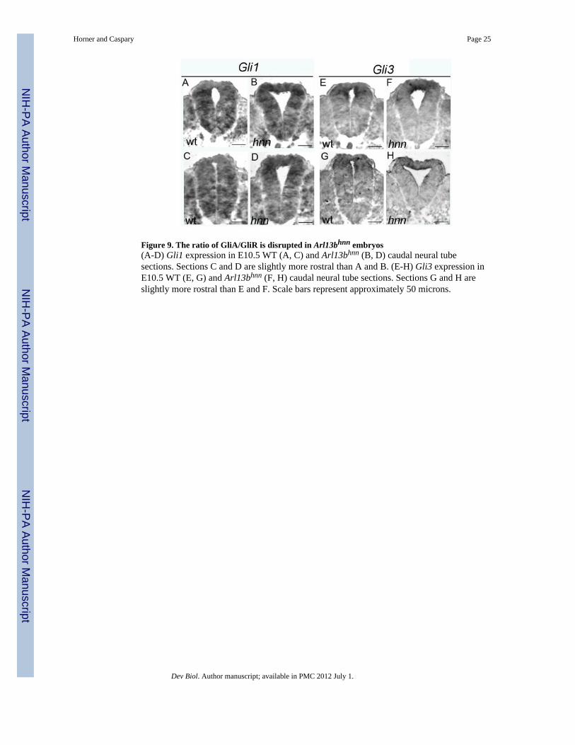
Figure 9. The ratio of GliA/GliR is disrupted in Arl13bhnn embryos(A-D) Gli1 expression in E10.5 WT (A, C) and Arl13bhnn (B, D) caudal neural tubesections. Sections C and D are slightly more rostral than A and B. (E-H) Gli3 expression inE10.5 WT (E, G) and Arl13bhnn (F, H) caudal neural tube sections. Sections G and H areslightly more rostral than E and F. Scale bars represent approximately 50 microns.
Horner and Caspary Page 25
Dev Biol. Author manuscript; available in PMC 2012 July 1.
NIH
-PA Author Manuscript
NIH
-PA Author Manuscript
NIH
-PA Author Manuscript

Figure 10. Axin2 is not overexpressed in Arl13bΔPax3-cre embryos(A and B) Axin2 is expressed at identical levels in E11.5 control (Cntrl) andArl13bΔPax3-cre (CKO) embryos. Scale bars represent approximately 300 microns.(C and D) Neural tube sections taken at the hindlimb level of the above control (Cntrl) andArl13bΔPax3-cre (CKO) embryos. Scale bars represent approximately 50 microns.
Horner and Caspary Page 26
Dev Biol. Author manuscript; available in PMC 2012 July 1.
NIH
-PA Author Manuscript
NIH
-PA Author Manuscript
NIH
-PA Author Manuscript

NIH
-PA Author Manuscript
NIH
-PA Author Manuscript
NIH
-PA Author Manuscript
Horner and Caspary Page 27
Tabl
e 1
Surv
ival
of A
rl13
bΔPa
x3-c
re m
utan
ts a
t var
ious
tim
e po
ints
bef
ore
and
afte
r birt
h. A
rl13
bΔPa
x3-c
re m
utan
ts a
re a
ble
to su
rviv
e in
ute
ro u
p un
til b
irth;
how
ever
, im
med
iate
ly a
fter b
irth
90%
die
from
an
appa
rent
bre
athi
ng p
robl
em.
E14
.5 (a
ll al
ive)
E17
.5 (a
ll al
ive)
E19
.5 (a
ll al
ive)
P0P2
1
Aliv
eD
ead
Aliv
eD
ead
Arl1
3bΔP
ax3-
cre
32
29
1
Con
trol
54
525
325
0
Tota
l8
67
2612
251
Dev Biol. Author manuscript; available in PMC 2012 July 1.