detection and characterization of bartonella strains in australia
TRANSCRIPT

i
DETECTION AND CHARACTERIZATION OF
BARTONELLA SPECIES IN WESTERN AUSTRALIA
Gunn Kaewmongkol, DVM, MSc
School of Veterinary and Biomedical Sciences, Faculty of Health Sciences, Murdoch
University, Perth, Western Australia
This thesis is presented for the degree of Doctor of Philosophy of Murdoch University,
2012

ii
I declare that this thesis is my own account of my research and contains as its main
content work which has not previously been submitted for a degree at any tertiary
education institution.
…………………………………………………….
(Gunn Kaewmongkol)
iii
Abstract
In this study, the prevalence and genetic diversity of Bartonella species in various
arthropod vectors from both wild and domestic animals in Australia were investigated
using nested-polymerase chain reaction (PCR) assays and multilocus sequence analysis
(MLSA). Previous studies on Bartonella species in Australia have been confined to
mammalian hosts, including humans, cats, native rodents and eastern grey kangaroos.
However, little is known about the status of bartonellae in arthropod vectors, which is
essential in understanding the transmission dynamics of the organisms.
To facilitate the investigation, ectoparasites (ticks and fleas) were collected from both
wild and domestic animals from various locations in Australia. All ectoparasites were
screened for Bartonella species using newly designed nested-PCRs targeting the gltA
gene (citrate synthase) and the ribosomal internal transcribed spacer (ITS) region,
developed as part of the present study. Multilocus sequence analysis of the 16S
ribosomal RNA (rRNA), citrate synthase (gltA),cell division protein (ftsZ) and RNA
polymerasebeta-subunit (rpoB) genes and the ribosomal ITS region was applied to
identify and confirm the status of all Bartonella species identified in this study.
Multilocus sequence analysis of the cytochrome oxidase subunit I (COI) and 18S
ribosomal RNA (rRNA) genes of flea vectors harbouring a diversity of Bartonella
species were analysed to characterize the extent of genetic diversity in the flea vectors
and to elucidate vector-parasite associations.
A phylogenetic analysis of the 5 concatenated loci identified 3 novel Bartonella species
in flea vectors from marsupials in Western Australia. Candidatus Bartonella antechini
was detected in fleas (Acanthopsylla jordani) from mardos (Antechinus flavipes - also

iv
called the yellow-footed antechinus). Candidatus Bartonella woyliei was detected in
fleas (Pygiopsylla hilli), from brush-tailed bettongs (Bettongia penicillata– also called
woylies), and Candidatus Bartonella bandicootii was detected in Pygiopsylla tunneyi
fleas from western barred bandicoots (Perameles bougainville). Furthermore, a potential
novel species, Bartonella sp. strain WC2 was detected in ticks (Ixodes australiensis)
from woylies based on the criterion of a genetic similarity of less than 96% of the gltA
locus compared with other validated Bartonella species. In the present study, the
grouping of marsupial-derived Bartonella species confirmed the existence of a
marsupial cluster of Bartonella species in Australia, which appears to have evolved
separately to Bartonella species in other mammals.
The detection of the known zoonotic Bartonella species, B. henselae and B.
clarridgeiae in red foxes and their fleas (Ctenocephalides felis), indicated that red foxes
could be an important reservoir of Bartonella infections for other animals and humans
in the same geographical locality. Bartonella henselae and B. clarridgeiae DNA were
also detected from fleas collected from pet cats in the same area. The genetic
association of these zoonotic Bartonella species detected in wildlife and pet animals has
demonstrated and confirmed the distribution of zoonotic Bartonella species in fleas
from both wild and domestic animals in this region and a possible ecological association
between the animal species.
The genetic clustering of Bartonella species and flea vectors with their Australian fauna
hosts suggests co-evolution of hosts, fleas and Bartonella species in Australia. In
conclusion, the close association between Australian fauna, Australian fleas and
Bartonella species suggests adaptation by Bartonella species to a specific ecological

v
niche, comprised of specific mammalian hosts and specific flea vectors in particular
environments.

vi
Acknowledgments
I am deeply indebted to my supervisors, Professor Stan Fenwick, Associate
Professor Peter Irwin and Professor Una Ryan for all their help and support throughout
my Ph.D. study. Most specially, I would like to thank Professor Stan Fenwick, my
principal supervisor for his enthusiasm, wonderful guidance and his patience to me over
the last three years of my study.
My research could not have been adequately carried out without the devoted and
dedicated help from Associate Professor Peter Irwin, my co-supervisor. This research
was made possible by the most generous support in all laboratory techniques provided
in the Molecular Epidemiology research team, School of Veterinary and Biomedical
Sciences, Murdoch University by its director, Professor Una Ryan, my co-supervisor. I
would especially like to thank her for every available help and valuable suggestions.
The members of this research team gave me the most generous help in everything.
To the members of the Molecular Epidemiology group, I would like to thank Dr.
Linda McInnes, Dr. Rongchang Yung, Josephine Ng and Josh Sweeney for all their
support. I am also thankful for help and friendship from the „trailer trash‟ members, Dr.
Peter Adams, Dr. Louise Pallant, Dr. Michael Banazis, Dr. Yazid Abdad and Dr. Jim
Carro Domingo who have made my study at Murdoch University both enjoyable and
entertaining. I would also like to thank Professor John Edwards who gave me a great
opportunity to pursue my Ph.D. study at Murdoch University. This study was supported
by the grants from the Australian Companion Animal Health Foundation (ACAHF) and
the Morris Animal Foundation (MAF).

vii
Finally, I would like to acknowledge my wife, Dr. Sarawan Kaewmongkol, for
her guidance in advance molecular techniques and especially for her love, support and
patience all the time.
In these acknowledgments, I cannot conclude without describing my respect and
admiration for my father, Dr. Surapol Kaewmongkol, who is my greatest teacher. He
has supported and encouraged me in both my work and my life without complaint and
possibly, with pleasure. My Ph.D. study could not be completed without his support and
encouragement.
Gunn Kaewmongkol
February 2012

viii
Publications
Peer-reviewed publications and conference proceedings
Kaewmongkol, G., Kaewmongkol, S., Owen, H., Fleming, P.A., Adams, P.J., Ryan,
U., Irwin, P.J., Fenwick, S.G. 2011. Candidatus Bartonella antechini: A novel
Bartonella species detected in fleas and ticks from the yellow-footed antechinus
(Antechinus flavipes), an Australian marsupial. Veterinary Microbiology. 149: 517-521.
Kaewmongkol, G., Kaewmongkol, S., Owen, H., Fleming, P. A., Adams, P. J., Ryan,
U., Irwin, P. J., Fenwick, S. G., Burmej, H., Bennett, M. D., and Wayne, A. F. 2011.
Diversity of Bartonella species detected in arthropod vectors from animals in Australia.
Comparative Immunology, Microbiology & Infectious Diseases. 34(5): 411-417.
Kaewmongkol, G., Kaewmongkol, S., Fleming, P. A., Adams, P. J., Ryan, U., Irwin, P.
J., and Fenwick, S. G. 2011. Zoonotic Bartonella species in Fleas and Blood from Red
Foxes in Australia. Vector-Borne and Zoonotic Disease. 11(12): 1549-1553.
Kaewmongkol, G., Kaewmongkol, S., McInnes, L., Burmej, H., Bennett, M. D.,
Adams, P. J., Ryan, U., Irwin, P. J., Fenwick, S. G. 2011. Genetic characterization of
flea-derived Bartonella species from native animals in Australia suggestss host-parasite
co-evolution. Infection, Genetics and Evolution. 11(8): 1868-1872.
Gunn Kaewmongkol, Sarawan Kaewmongkol, Helen Owen, Trish Fleming, Peter J.
Adams, Una Ryan, Peter J. Irwin and Stanley G. Fenwick. Novel Bartonella species
detected in fleas and ticks from Australian marsupials. The Combined Sciences
Biology Meeting 2010 (CBSM 2010), University of Western Autralia 2010 (Oral
presentation).
Gunn Kaewmongkol, Sarawan Kaewmongkol, Halina Burmej, Mark D. Bennett,
Patricia A. Fleming, Peter J. Adams, Adrian F. Wayne, Una Ryan, Peter J. Irwin,
Stanley G. Fenwick. Diversity of Bartonella species detected in arthropod vectors from

ix
animals in Australia. The Wildlife Meeting, the Morris Animal Foundation 2011, New
Orleans, Louisiana, USA. March 10-13, 2011 (Poster presentation).

x
Table of contents
Title page……………………………………………………………………………….. i
Declaration…………………………………………………………………………….. ii
Abstract……………………………………………………………………………….. iii
Acknowledgments…………………………………………………………………….. vi
Table of Contents……………………………………………………………………… x
List of Tables………………………………………………………………………… xiii
List of Figures……………………………………………………………………....... xv
CHAPTER 1. General introduction to Bartonella species………………………….. 1
1.1 Overview and Historical Aspects of Bartonella species...................................... 1
1.2 Arthropod vectors……………………………………………………………..... 5
1.2.1 Bartonella bacilliformis transmitted by sandflies................................................ 6
1.2.2 Bartonella quintana transmission by lice………………………………………. 6
1.2.3 Bartonella species transmitted by fleas………………………………………… 7
1.2.4 Bartonella species transmitted by other arthropods............................................. 8
1.2.5 Bartonella species transmitted by ticks................................................................ 9
1.3 Bartonella species in dogs.................................................................................. 14
1.3.1 Clinical evidence for Bartonella infection in dogs............................................. 17
1.3.2 Treatment of Bartonella infection in dogs......................................................... 21
1.3.3 Epidemiological studies of Bartonella infection in dogs................................... 22
1.4 Diagnosis of Bartonella species in humans, dogs, and cats................................26
1.4.1 Serological assays……………………………………………………………... 26
1.4.2 Culture of genus Bartonella…………………………………………………... 29
1.4.3 PCR assays……………………………………………………………………. 34
1.5 Genetic-based analysis for species identification in genus Bartonella……….. 38

xi
1.6 Bartonella species in wild animals and their ectoparasites................................ 42
1.7 Bartonella species in Australia………………………………………………... 44
1.8 Objectives........................................................................................................... 45
CHAPTER 2. General Materials and Methods…………………………………… 46
2.1 Ectoparasite collection and identification........................................................... 46
2.2 DNA extraction................................................................................................... 53
2.3 PCR detection of Bartonella species.................................................................. 53
2.4 PCR for phylogenetic analysis at additional loci................................................ 55
2.5 Agarose gel electrophoresis and PCR product purification................................ 56
2.6 DNA sequencing and phylogenetic analysis...................................................... 57
CHAPTER 3. Candidatus Bartonella antechini: A novel Bartonella species
detectedin fleas and ticks from the yellow-footed antechinus (Antechinus flavipes),
an Australian marsupial………………………………………………..…………… 61
3.1 Introduction…………………………………………………………………….61
3.2 Materials and Methods………………………………………………………....62
3.3 Results………………………………………………………………………….63
3.4 Discussion……………………………………………………………………...68
CHAPTER 4. Diversity of Bartonella species detected in arthropod vectors from
animals in Australia...................................................................................................... 70
4.1 Introduction…………………………………………………………………… 70
4.2 Materials and Methods………………………………………………………... 72
4.3 Results………………………………………………………………………… 73
4.4 Discussion……………………………………………………………………... 78
xii
CHAPTER 5. Zoonotic Bartonella species in fleas and blood from red foxes in
Australia……………………………………………………………………………… 80
5.1 Introduction…………………………………………………………………… 80
5.2 Materials and Methods………………………………………………………...81
5.3 Results………………………………………………………………………… 85
5.4 Discussion……………………………………………………………………...90
CHAPTER 6. Investigation of Bartonella species infections in pets and their
ectoparasites in Australia……………………………………………………………. 92
6.1 Introduction…………………………………………………………………… 92
6.2 Materials and Methods………………………………………………………... 93
6.3 Results………………………………………………………………………… 97
6.4 Discussion……………………………………………………………………. 100
CHAPTER 7. Genetic characterization of flea-derived Bartonella species from
native animals in Australia reveals host-vector-bacteria co-evolution………….. 103
7.1 Introduction………………………………………………………………….. 103
7.2 Materials and Methods………………………………………………………. 105
7.3 Results……………………………………………………………………….. 108
7.4 Discussion……………………………………………………………………. 113
CHAPTER 8. General discussion…………………………………………………. 116
8.1 Introduction………………………………………………………………….. 116
8.2 Genotypic status of Bartonella species in Australia…………………………. 117
8.3 Co-evolution of Bartonella species, Australian fleas and Australian fauna..... 120
8.4 Future directions for Bartonella research in Australia………………………. 123
REFERENCES........................................................................................................... 128

xiii
List of Tables
Table 1.1 Bartonella species infections in humans.......................................................... 3
Table 1.2 Confirmed vectors for Bartonella spp............................................................ 11
Table 1.3 Suspected vectors for Bartonella spp............................................................. 12
Table 1.4 Bartonella species infections in dogs............................................................. 15
Table 1.5 Antibiotics used in dogs to treat Bartonella infection.................................... 24
Table 1.6 Clinical signs that indicate laboratory testing for Bartonella infection in
dog.................................................................................................................................. 25
Table 1.7 Semisolid media used for Bartonella culture................................................. 33
Table 1.8 PCR assays targeting various genes for Bartonella detection........................ 36
Table 2.1 Numbers and species of ectoparasites collected from mammalian hosts in
Western Australia in the present study........................................................................... 48
Table 2.2 Hosts from which ectoparasites were collected in various locations in Western
Australia.......................................................................................................................... 49
Table 2.3 Oligonucleotide primers used for nested-PCR and single step PCR
amplifications of the 16S rRNA, gltA, ftsZ, rpoB loci and the ribosomal ITS region.... 58
Table 2.4 Genbank accession numbers of Bartonella species used for the concatenated
phylogenetic analysis...................................................................................................... 59
Table 2.5 GenBank accession numbers of Bartonella species discovered in possible
arthropod vectors from animals in Western Australia in the present study.................... 60
Table 3.1 Percentage genetic similarity of the concatenated sequences from Candidatus
Bartonella antechini n. sp. compared with other confirmed Bartonella spp.................. 65
Table 5.1 Number of fleas, pooled flea DNA, blood samples and Bartonella spp.
collected from two locations in Western Australia…………………………………… 84
Table 6.1 Number of samples and animals examined for Bartonella species in
Australia……………………………………………………………………………….. 94

xiv
Table 7.1 Flea species collected from a variety of locations in Western Australia...... 107
Table 7.2 GenBank accession numbers of flea species from animals in Australia...... 110
xv
List of Figures
Fig. 2.1 Map of Western Australia demonstrating sample collection sites including
Dwellingup, Boyup Brook, Katanning, Kendenup and Fitzgerald River National
Park................................................................................................................................. 50
Fig 2.2 Map of the Southern Forest, Western Australia................................................. 51
Fig. 2.3 Map of Dorre and Bernier Islands..................................................................... 52
Fig. 3.1 Neighbor-Joining concatenated phylogenetic tree of the 16S rRNA, gltA, ftsZ,
rpoB, and the ITS region of Australian marsupial isolates and validated species and
subspecies of Bartonella. Percentage bootstrap support (>50%) from 1000
pseudoreplicates is indicated at the left of the supported node...................................... 66
Fig. 3.2 Maximum-Parsimony concatenated phylogenetic tree of the 16S rRNA, gltA,
ftsZ, rpoB, and the ITS region of Australian marsupial isolates (B. australis and
Candidatus B. antechini) and validated species and subspecies of Bartonella.
Percentage bootstrap support (>50%) from 1000 pseudoreplicates is indicated at the left
of the supported node..................................................................................................... 67
Fig. 4.1 Neighbor-Joining phylogenetic tree of the gltA gene of Bartonella species
detected in Australian marsupials and validated species and subspecies of Bartonella.
Percentage bootstrap support (>40%) from 1000 pseudoreplicates is indicated at the left
of the supported node..................................................................................................... 76
Fig. 4.2 Neighbor-Joining concatenated phylogenetic tree of the 16S rRNA, gltA, ftsZ,
rpoB, and the ITS region of Bartonella species detected in Australian marsupials and
validated species and subspecies of Bartonella. Percentage bootstrap support (>60%)
from 1000 pseudoreplicates is indicated at the left of the supported node..................... 77
Fig. 5.1 Red fox carcasses shot by volunteers and farmers as part of the „Red Card for
the Red Fox‟ 2010 culling program coordinated by the Department of Agriculture and
Food, Western Australia................................................................................................. 83
xvi
Fig. 5.2 Neighbor-Joining concatenated phylogenetic tree of the 16S rRNA, gltA, ftsZ,
rpoB, and the ITS region of Bartonella henselae and Bartonella clarridgeiae isolates in
red foxes. Percentage bootstrap support (>45%) from 1000 pseudoreplicates is indicated
at the left of the supported node..................................................................................... 87
Fig. 5.3 Neighbor-Joining phylogenetic tree of the ITS region of Bartonella henselae
isolates. Percentage bootstrap support (>45%) from 1000 pseudoreplicates is indicated
at the left of the supported node..................................................................................... 88
Fig. 5.4 Neighbor-Joining phylogenetic tree of the ITS region of Bartonella
clarridgeiae isolates. Percentage bootstrap support (>45%) from 1000 pseudoreplicates
is indicated at the left of the supported node.................................................................. 89
Fig. 6.1 Aortic valve endocarditis in a dog suspected to have Bartonella infection….. 96
Fig. 6.2 Neighbor-Joining phylogenetic tree of the ITS region of Bartonella
clarridgeiae isolates. Percentage bootstrap support (>40%) from 1000 pseudoreplicates
is indicated at the left of the supported node………………………………………….. 98
Fig. 6.3 Neighbor-Joining phylogenetic tree of the ITS region of Bartonella henselae
isolates. Percentage bootstrap support (>40%) from 1000 pseudoreplicates is indicated
at the left of the supported node………………………………………………………. 99
Fig. 7.1 Neighbor-Joining concatenated phylogenetic tree of the 18S rRNA, and COI
genes of flea species and their associated Bartonella species from marsupials and other
mammals in Australia. Percentage bootstrap support (>40%) from 1000
pseudoreplicates is indicated at the left of the supported node. The tree is rooted using
Calliphora vomitoria (Blue bottle fly) as an outgroup………………………………. 111

xvii
Fig. 7.2 Neighbor-Joining concatenated phylogenetic tree of 16S rRNA, gltA, ftsZ,
rpoB, and the ITS region of Bartonella species showing the separate clustering of
Bartonella spp. from Australian marsupials and B. rattaustraliani and B.
coopersplainsensis from native Australian rodents. Percentage bootstrap support
(>60%) from 1000 pseudoreplicates is indicated at the left of the supported node….. 112
Fig. 8.1 Native host-vector-parasite interactions and non-native interferences........... 122

1
CHAPTER 1
Literature Review
1.1 Overview and Historical Aspects of Bartonella Species
Bacteria of the genus Bartonella are among the most important emerging
pathogens in veterinary and human medicine. Bartonella species are fastidious,
haemotropic, Gram-negative organisms that have been identified in a wide range of
domestic and wild mammals (Breitschwerdt et al., 2000). More than 20 species and
subspecies of the genus Bartonella have been reported, based on either isolation of the
bacteria and/or genetic analysis. Most Bartonella species are zoonotic pathogens and
have been reported to cause human diseases for centuries. Oroya fever or Carrion‟s
disease is one of most common bartonelloses in humans and is caused by Bartonella
bacilliformis. Alberto Leonardo Barton Thompson identified the bacterium in 1905 and
the genus Bartonella is named after him. Prior to this, in 1885, Daniel Alcides Carrion,
a medical student, discovered the two phases of disease (acute and chronic) occurring in
Oroya fever by inoculating himself with blood containing B. bacilliformis from a patient
with a chronic form of the disease. He presented with the recognised signs of Oroya
fever and died after this experiment (Huarcaya et al., 2004).
Trench fever was a very serious disease in humans caused by Bartonella
quintana during World War I. One million soldiers are estimated to have been infected
in that era (Swift, 1920). It has also been suggested that the disease caused by
Bartonella quintana contributed to the demise of the Inca Empire (Huarcaya et al.,
2004).

2
Vectors such as fleas, sand flies, lice, and possibly ticks (Podsiadly et al., 2007;
Sanogo et al., 2003) are the major arthropods that transmit Bartonella species between
the reservoirs and the final hosts, usually by biting, or in other ways, such as
contamination of the skin or mucosal surface with flea faeces in the case of cat-scratch
disease. The affinity and specificity between a variety of vectors and the different
species of bacteria in the genus Bartonella has not been completely elucidated.
In 1992, a new species, Rochalimaea henselae (renamed as Bartonella henselae)
was isolated from the blood of an immunocompromised human patient (Welch et al.,
1992). This report also described the clinical findings and genetic characterization of
other Bartonella species including B. bacilliformis, B. vinsonii and B. quintana, the
latter species, along with B. henselae, are the causative agents of bacillary angiomatosis
(BA). Because of this discovery in 1992, worldwide scientific studies of this genus
commenced and more than one novel Bartonella species has been described each year
for the last decade (Chomel et al., 2004). Although B. henselae was only recently
discovered and named, this pathogen has been detected in the dental pulp of a cat that
lived 800 years ago in France (La et al., 2004). This evidence suggests that these
bacteria may have affected animal and human health for many centuries.

3
Table 1.1 Bartonella species infections in humans
Species Location References Main vector Main reservoirs Human diseases
B. bacilliformis Peru Barton et al., 1909 Sand fly Human Carrion‟s disease
B. quintana Russia Strong et al., 1918 Body louse
(Pediculus humanis)
human Trench fever, CSD, BA,
CA,
Bacteraemia, Endocarditis
B .henselae USA Regnery et al., 1992 Cat flea
(Ctenocephalides felis)
Cat CSD, BA, CA,
Bacteraemia,
Endocarditis, Septicaemia
B. elizabethae USA
Daly et al., 1993 Oriental rat flea
(Xenopsylla cheopis)
Rodent flea
Rat Endocarditis, Retinitis

4
Species Location References Main vector Main reservoirs Human diseases
B. grahamii UK
Birtles et al., 1995 Wild mice
Retinitis
B. washoensis USA Regnery et al., 1995 Flea? California ground squirrel Myocarditis
B. vinsonii berkhoffii USA Kordick et al., 1996 Tick? Coyote
Dog
Endocarditis
B. clarridgeiae USA Lawson et al., 1996 Cat flea Cat CSD
B. alsatica France Heller et al., 1999 Flea? Rabbit Endocarditis
B. vinsonii arupensis USA Welch et al., 1999 Flea or Tick? White-footed mouse Bacteraemia
B. rochalimae Peru/USA Eremeeva et al., 2007
Henn et al., 2009
Flea? Unknown Bacteraemia
CSD - cat-scratch disease; BA - bacillary angiomatosis; CA - chronic adenopathy.

5
1.2 Arthropod vectors
With recent advances in DNA technologies, Bartonella DNA has been detected
in several different vectors in many areas of the world in last 15 years. For example, a
study reported the detection of B. tribocorum and B. vinsonii subspecies vinsonii DNA
from the fleas Xenopsylla cheopis, Ctenophthalmus pseudagyrtes and Sternopsylla
texanus (Reeves et al., 2007). Other papers have reported the detection of B. henselae
DNA in Ixodes ricinus ticks removed from dogs and humans (Sanogo et al., 2003;
Posiadly et al., 2007) and a single tick has been shown to carry Bartonella together with
other pathogens (Halos et al., 2005). The intra-cellular endosymbionts of microbial
communities harboured by ticks, mosquitoes, lice and leeches have been increasingly
studied in the last decade (Benson et al., 2004; Lindh et al., 2005; Reed et al., 2002;
Worthen et al., 2006) and multiple organisms in a single flea have also been studied.
For example, the diversity of bacterial communities was classified in Bartonella
positive fleas to explore their interactions within individual fleas (Jones et al., 2008).
The adaptations of Bartonella species in arthropod vectors may be influenced by other
competitive organisms inside the arthropod vectors (Chomel et al., 2009).
The identification of known and suspected arthropod vectors for Bartonella is
essential for understanding host, vector and pathogen relationships and has been the
subject of increasing study. Due to the ability of this genus to colonise target cells of a
variety of mammalian hosts, there is the possibility that various Bartonella spp. could
be transmitted by a variety of arthropod vectors. A summary of the known and potential
vectors for Bartonella are shown in Tables 1.2 and 1.3 (Billeter et al., 2008). DNA
detection or culture of Bartonella spp. in arthropod vectors does not prove that these
arthropods are true vectors, however. The difference between vector competence and
6
vector potential should be confirmed by experimental studies that demonstrate
transmission between vectors and hosts.
1.2.1 Bartonella bacilliformis transmitted by sandflies
The sandfly (Lutzomyia verrucarum) was believed to be a potential vector for B.
bacilliformis, the agent of Oroya fever, in very early reports. This hypothesis was
supported by the fact that the distribution and feeding habits of Lutzomyia verrucarum
correlated with the spread of Oroya fever in the same area. Experimental transmission
of B. bacilliformis by Lutzomyia verrucarum and other suspected arthropods into
monkeys (Macaca mulatta) was published by Noguchi and Battistini in 1926 (Billeter et
al., 2008). In that study, Lutzomyia spp, ticks, mites, and other arthropods were
collected from known B. bacilliformis endemic areas but B. bacilliformis was only
isolated from the blood of sandfly-inoculated monkeys, without clinical signs. Other
experiments were also performed to demonstrate that B. bacilliformis could be
transmitted via sandflies (Billeter et al., 2008). The identification of B. bacilliformis-like
organisms in many organs was reported to confirm the replication and survival of B.
bacilliformis within the sandfly (Billeter et al., 2008).
1.2.2 Bartonella quintana transmission by lice
Outbreaks and infection with the agent of trench fever, Bartonella
(Rochalimaea) quintana are commonly reported in homeless individuals in France
(Fournier et al., 2002), the Netherlands (Fournier et al., 2002), Tokyo (Sasaki et al.,
2002), rural Andean communities (Raoult et al., 1999), Moscow (Rydkina et al., 1999),

7
various countries in Africa (Fournier et al., 2002), and the San Francisco Bay area,
California (Koehler et al., 1997).
Bartonella quintana was believed to be restricted to at-risk individuals
(homeless and immunocompromised) exposed to the human body louse (Pediculus
humanus humanus) (Fournier et al., 2002). However, B. quintana has been isolated
from non-human research primates (Macaca fascicularis), dogs with endocarditis and
feral farm cats associated with B. quintana infection in a woman, possibly transmitted
by cat bite (Kelly et al., 2006; Breitschwerdt et al., 2007). As B. quintana has been
isolated from various hosts, the mode of transmission of B. quintana is still unclear and
has been questioned. The possibility that other species of arthropod vectors may be
potential vectors for B. quintana needs to be investigated (Billeter et al., 2008). In early
experimental work in Germany, England and the USA, researchers induced trench
fever-like symptoms in human patients by having them ingest infected lice (Swift et al.,
1920). The same researchers also induced B. quintana infection in non-infected patients
by scarification of the skin and intra-subcutaneous injection with infected louse faeces.
Immuno-fluorescent antibodies specific for B. quintana have identified B. quintana in
the louse intestinal lumen and also in louse faeces (Fournier et al., 2001). However,
PCR analysis of eggs and larvae of infected lice indicates that B. quintana cannot be
transmitted by transovarial transmission (Fournier et al., 2001).
1.2.3 Bartonella species transmitted by fleas
The cat flea (Ctenocephalides felis) has been confirmed as a competent vector
for B. henselae infection by the experimental transmission into specific-pathogen-free
(SPF) kittens (Chomel et al., 1996). Five SPF-kittens were fed on by fleas collected

8
from bacteraemic cats. Two weeks post-exposure to fleas, bacteraemia was identified in
four of the five kittens (Chomel et al., 1996). The biological processes of B. henselae
inside flea vectors have been examined in many publications using PCR detection and
culture methods (Higgins et al., 1996; Koehler et al., 1994). Bartonella henselae DNA
has been detected in fleas 3 hours after Ctenocephalides felis were fed with infected cat
blood (Higgins et al., 1996; Koehler et al., 1994). In the flea gut, B. henselae was
detected up to 9 days post feeding. Detection of Bartonella DNA and successful culture
of Bartonella culture has been achieved from flea faeces (Higgins et al., 1996). An
increase in bacterial numbers (CFU/mg) from flea faeces was correlated to an increase
in incubation time (Finkelstien et al., 2002). Transmission of B. henselae to cats by
inoculation with infected flea faeces was successfully performed (Foil et al., 1998).
From these studies, it can be concluded that flea faeces in the environment are a
potential source for Bartonella transmission between hosts. The diversity of flea species
harbouring various species of Bartonella that have been reported worldwide is shown in
Tables 1.2 and 1.3.
1.2.4 Bartonella species transmitted by other arthropods
Bartonella DNA has been identified in various arthropods such as deer keds
(Hippoboscidae sp.) (Dehio et al., 2004), biting flies (Halos et al., 2004), and mites
(Kim et al, 2005; Reeves et al., 2006) indicating that they may be potential vectors for
Bartonella transmission. In one study, Bartonella DNA was detected in three ked
species including the deer ked (Lipoptena cervi), horse flies (Hippobosca equine) and
sheep keds (Melophagus ovinus), collected from both wild and domestic ruminants in
France and Romania (Halos et al., 2004). It has been suggested that the cause of deer
ked dermatitis in humans is due to B. shoenbuchensis localized in the midgut of deer

9
keds (Dehio et al., 2004). Multilocus PCR analysis targeting the riboflavin synthase
alpha chain (ribC), the heat shock protein (groEL) or the citrate synthase (gltA) genes
has been used to detect many species of Bartonella in various species of biting
arthropods such as horn flies (Haematobia spp.), stable flies (Stomoxys spp.), deer flies
(Chrysops spp.), horse flies (Tabanus spp.), bat flies (Trichobius major), Eastern bat
bed bugs (Cimex adjunctus), bat mites (Steatonyssus spp.), and rat mites (Ornithonyssus
bacoti) (Chung et al., 2004; Reeves et al., 2005; 2006; 2007; Kim et al., 2005).
Bartonella DNA has also been detected in non-biting arthropods such as house dust
mites (Dermatophagoides favinae and Dermatophagoides pteronyssinus) and honey-
bees (Apis mellifera capensis) (Valerio et al., 2005; Jeyaprakash et al., 2003). It has
been suggested that high levels of Bartonella lipopolysaccharides in house dust mites
may be the source of mite allergen endotoxin (Valerio et al., 2005).
1.2.5 Bartonella species transmitted by ticks
The role of ticks in the potential transmission of Bartonella species has been
proposed since 1996 (Kruszewska et al., 1995) and Bartonella DNA has been detected
in a wide variety of tick species DNA (Table 1.3). However, to date only one study was
able to culture B. bacilliformis from Ixodes ricinus (Kruszewska et al., 1995) and until
recently there had been no experimental research that demonstrated the infectivity of
Bartonella spp. transmitted by ticks (Angelakis et al., 2010). However, the potential
transmission of B. henselae by Ixodes ricinus ticks was demonstrated by Cotté et al in
2008. Ticks (I. ricinus) were fed with B. henselae infected blood using an artificial
feeding platform made of rabbit skin. This experiment was successful in infecting ticks
and the infections were confirmed by PCR screening. In addition, these infected ticks
were fed on uninfected blood using the same type of artificial feeding platform. Within

10
72 hours, B. henselae was cultured and detected by PCR analysis of previously
uninfected blood (Cotté et al., 2008). The evidence supporting ticks as vectors of
Bartonella spp. was primarily based on historical reports of people exposed to tick bites
prior to the appearance of clinical bartonellosis (Lucey et al, 1992; Zangwill et al.,
1993; Eskow et al., 2001). Therefore, experimental transmission from ticks to live
animals is required to demonstrate the vector competency of ticks for the transmission
of Bartonella species (Angelakis et al., 2010).
11
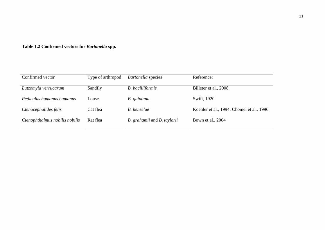
Table 1.2 Confirmed vectors for Bartonella spp.
Confirmed vector Type of arthropod Bartonella species Reference:
Lutzomyia verrucarum Sandfly B. bacilliformis Billeter et al., 2008
Pediculus humanus humanus Louse B. quintana Swift, 1920
Ctenocephalides felis Cat flea B. henselae Koehler et al., 1994; Chomel et al., 1996
Ctenophthalmus nobilis nobilis Rat flea B. grahamii and B. taylorii Bown et al., 2004
12
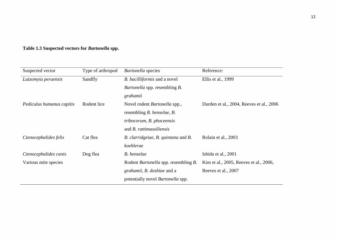
Table 1.3 Suspected vectors for Bartonella spp.
Suspected vector Type of arthropod Bartonella species Reference:
Lutzomyia peruensis Sandfly B. bacilliformis and a novel
Bartonella spp. resembling B.
grahamii
Ellis et al., 1999
Pediculus humanus capitis Rodent lice Novel rodent Bartonella spp.,
resembling B. henselae, B.
tribocorum, B. phoceensis
and B. rattimassiliensis
Durden et al., 2004, Reeves et al., 2006
Ctenocephalides felis Cat flea B. clarridgeiae, B. quintana and B.
koehlerae
Rolain et al., 2003
Ctenocephalides canis Dog flea B. henselae Ishida et al., 2001
Various mite species Rodent Bartonella spp. resembling B.
grahamii, B. doshiae and a
potentially novel Bartonella spp.
Kim et al., 2005, Reeves et al., 2006,
Reeves et al., 2007
13
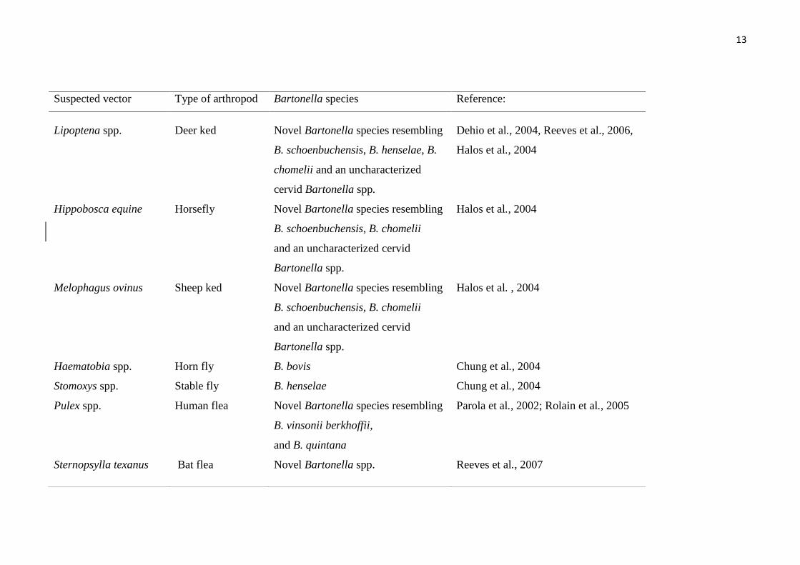
Suspected vector Type of arthropod Bartonella species Reference:
Lipoptena spp. Deer ked Novel Bartonella species resembling
B. schoenbuchensis, B. henselae, B.
chomelii and an uncharacterized
cervid Bartonella spp.
Dehio et al., 2004, Reeves et al., 2006,
Halos et al., 2004
Hippobosca equine Horsefly Novel Bartonella species resembling
B. schoenbuchensis, B. chomelii
and an uncharacterized cervid
Bartonella spp.
Halos et al., 2004
Melophagus ovinus Sheep ked Novel Bartonella species resembling
B. schoenbuchensis, B. chomelii
and an uncharacterized cervid
Bartonella spp.
Halos et al. , 2004
Haematobia spp. Horn fly B. bovis Chung et al., 2004
Stomoxys spp. Stable fly B. henselae Chung et al., 2004
Pulex spp. Human flea Novel Bartonella species resembling
B. vinsonii berkhoffii,
and B. quintana
Parola et al., 2002; Rolain et al., 2005
Sternopsylla texanus Bat flea Novel Bartonella spp. Reeves et al., 2007

14
1.3 Bartonella species in dogs
Bartonella infection has been reported to cause a long term intra-erythrocytic
bacteraemia in both humans and animals (Kordick et al., 1998). Pets represent an
important reservoir for human bartonellosis because most Bartonella spp. infecting pets
are zoonotic (Chomel et al., 2006) (Table 1.1 and 1.4). Cats appear to be the main
reservoir for B. henselae, B. clarridgeiae, and B. koehlerae (Chomel et al., 2004;
Avidor et al., 2004). Dogs can be infected with B. clarridgeiae, B. elizabethae, B.
henselae, B. vinsonii subspecies berkhoffii, B. washoensis, B. quintana, and B.
rochalimae (Boulouis et al., 2005; Kelly et al., 2006; Henn et al., 2008; Breitschwerdt et
al., 1995; 2002; 2008; Kordick et al., 1996; Chomel et al., 2001; 2003). Bartonella
vinsonii subspecies berkhoffii and B. henselae are currently believed to be the most
common Bartonella infections in dogs. A wide variety of clinical signs for Bartonella
infection in dogs have been discussed in the literature, including polyarthritis, cutaneous
vasculitis, endocarditis, myocarditis, epistaxis, peliosis hepatitis and granulomatous
inflammatory diseases (Chomel et al., 2001; 2003; Breitschwerdt et al., 1995; Cockwill
et al., 2007; Cadenas et al., 2008; Henn et al., 2009; Pasavento et al., 2005; Saunders et
al., 2006). The role of dogs as reservoirs of Bartonella spp. is less clear than it is for
cats, because domestic dogs are more likely to be final, clinically-affected hosts.
Because dogs present the same spectrum of clinical signs as in humans, dogs are
potentially an important research model for human infections (Kelly et al., 2006). There
is little research available concerning Bartonella species and genotypes in dogs and in
dog ticks and fleas and little is known about Bartonella infections in Australian dogs
and their ectoparasites.
.

15
Table 1.4 Documented Bartonella species infections in dogs
Species References Location Main vector Main
reservoirs
Major clinical diseases Other clinical
signs
B. henselae Breitschwerdt et al., 2004 USA Cat flea Cat Lymphadenopathy
Immune-mediated
thromcytopaenia
L5-L6 Spinal lesion
Lethargy, weight
loss
B. elizabethae Breitschwerdt et al., 2004 USA Rat flea Rat Weight loss
Mild anaemia
Mild azotaemia
B. washoensis Chomel et al., 2003 USA Unknown Unknown Mitral valve endocarditis Chronic arthritis
B. clarridgeiae Chomel et al., 2001 USA Cat flea Cat endocarditis Mild azotaemia

16
Species References Location Main vector Main
reservoirs
Major clinical diseases Other clinical signs
B. rochalimae Henn et al., 2007 USA
Unknown Gray fox?
Red fox
Aortic valve endocarditis Not reported
B. vinsonii
berkhoffii
Breitschwerdt et al., 1995,
2008
Kordick et al., 1996
Cockwill et al., 2007
Cherry et al., 2009
USA
USA
Canada
Unknown
(Tick?)
Coyote Endocarditis, Myocarditis
Arrhythmia, Uveitis, Choroiditis
Splenomegaly, Polyarthritis
Epistaxis
Pleural effusion,
Abdominal effusion,
Chylothorax

17
1.3.1 Clinical evidence for Bartonella infection in dogs
Endocarditis
In 1992, B. henselae, which causes cat scratch disease (CSD), was identified as
the causative agent of endocarditis in humans (Hadfield et al., 1993). Since then,
Bartonella infection has become recognised as a significant pathogen of culture-
negative endocarditis in humans (Raoult et al., 1996). Most endocarditis cases in
humans are related to infection with B. quintana and B. henselae. In dogs, a newly
documented subspecies, B. vinsonii berkhoffii has been identified from the blood of
dogs with intermittent epistaxis and endocarditis (Breitschwerdt et al., 1995; Kordick et
al., 1996).
The most common pathological finding of bartonellosis in dogs is endocarditis
(Chomel et al, 2001; 2003; Breitschwerdt et al., 1995; Cockwill et al., 2007; Cadenas et
al., 2008; Henn et al., 2008; Kelly et al., 2008; Pasavento et al., 2005). The diagnosis of
endocarditis is made by echocardiography to detect typical vegetative or destructive
lesions on the mitral or aortic valves (Macdonald et al., 2004; Tou et al., 2005; Sykes et
al., 2006).
Infective endocarditis in dogs caused by other organisms, including
Staphylococcus spp., Streptococcus spp., and various Gram-negative bacilli
(MacDonald et al., 2004; Sykes et al., 2006), more commonly result in lesions on the
mitral valve than on the aortic valve compared with Bartonella endocarditis. In contrast,
lesions on the aortic valve have been reported in more than 80 percent of infective
endocarditis cases in dogs caused by Bartonella, with only a few cases of lesions on the
18
mitral valve (Table 1.4). Fourteen cases of Bartonella endocarditis in dogs were
described from 1995 to 2008 (Breitschwerdt et al., 1995; 2008; Henn et al., 2008;
Chomel et al., 2001; 2003). Of these, some endocarditis cases involved the aortic valve,
one case involved both the aortic and mitral valves and the identity of the affected
valves was not reported in one case. Among other presenting signs coincidental with
Bartonella endocarditis in dogs is concurrent arthropathy in >70 percent of dogs
(Breitschwerdt et al 2008). Because most cases of Bartonella endocarditis are in
middle-aged (4-8yr) male dogs of medium to large breeds, the presumptive diagnosis
was chronic orthopaedic abnormalities or chronic arthritis, and infectious causes of joint
problems were not investigated (Breitschwerdt et al., 2008). It is thought that Bartonella
infection of the epithelium is important in the aetiology of joint problems (Chomel et
al., 2003; Kelly et al., 2006). Cardiac murmur is usually auscultated during physical
examination of dogs with endocarditis (Macdonald et al., 2004; Tou et al., 2005; Sykes
et al., 2006).
The consequences of infective endocarditis in dogs include the development of
heart failure and cardiac arrhythmias. Thromboemboli commonly cause regional
infarction of the spleen, kidneys, myocardium, brain, and skeletal muscle (Pesavento et
al., 2005; Sykes et al., 2006). The median survival time for dogs with infective
endocarditis has been reported to be 54 days (Sykes et al., 2006). In some reports, dogs
with endocarditis due to Bartonella spp. died within 2 weeks of diagnosis (Cockwill et
al., 2007), while dogs with endocarditis caused by other bacteria lived for 11 to 12
months (Macdonald et al., 2004). Heart valve replacement operations in dogs with
Bartonella infection may be established in future to increase the survival rate of dogs
infected with Bartonella spp.

19
Granulomatous diseases
The second important clinical sign of Bartonella infection in dogs (after
endocarditis) is granulomatous inflammatory disease. Granulomatous inflammatory
lesions related to Bartonella infection in dogs have been reported in various organs
including spleen, heart, lymph nodes, omentum, liver, kidney, lung, mediastinum, and
salivary glands (Saunders et al., 2006; Gillespie et al., 2003; Pappalardo et al., 2000).
The association between granulomatous diseases and Bartonella spp. was confirmed by
immunofluorescent assays for Bartonella spp. and the detection of Bartonella DNA in
granulomatous tissues. The first reports of granulomatous disease in dogs were
described in a 4-year-old greyhound (dog 1) and an 11-year-old mixed breed dog (dog
2) from North Carolina, USA (Pappalardo et al., 2000). A solitary granulomatous mass
was found in a submandibular lymph node in dog 1 and in the nasal sinus in dog 2. A
history of fever and rapid growth of the mass was reported in dog 1 and a nasal
discharge of 6-weeks duration was reported in dog 2. PCR of the 16S rRNA gene and
PCR-RFLP analysis of the gltA gene identified Bartonella DNA from the
granulomatous masses. Granulomatous hepatitis associated with Bartonella spp. was
reported in two dogs from Philadelphia, Pennsylvania, USA (Gillespie et al., 2003).
Chronically high hepatic enzyme activity and liver dysfunction were characterized in
these cases.

20
Other clinical signs
Other clinical signs associated with Bartonella endocarditis in dogs are shown in
Table 1.4. Epistaxis in dogs associated with Bartonella infection was first reported in
2005 (Breitschwerdt et al., 2005). The pathology of epistaxis in dogs due to Bartonella
infection may not be related to thrombocytopaenia (Breitschwerdt et al., 2005). Two of
three dogs with epistaxis presented with thrombocytopaenia (platelet counts 24,000 and
158000/µl respectively) but one was reported to have thrombocytosis (platelet count
562000/ µl) (Breitschwerdt et al., 2005). One of three cases of epistaxis developed other
clinical signs including coughing, dyspnoea and lethargy 3 months after recovery from
epistaxis. Cardiac murmur was examined and aortic valve endocarditis was confirmed
by echocardiography.
The development of pre-enrichment liquid culture, Bartonella-
Alphaproteobacteria Growth Medium (BAPGM), combined with PCR, has increased
the sensitivity of Bartonella detection from a variety of specimens from dogs suspected
to have Bartonella infection (Duncan et al., 2007; Cadenas et al., 2007; Breitschwerdt et
al., 2007; Cherry et al., 2009). The other clinical associations detected using this
combination of detection methods include pleural effusion, abdominal effusion and
chylothorax. The detection of Bartonella DNA from pre-enrichment media, or directly
from effusion fluid, may not prove that Bartonella infection is the major cause of these
signs, but this combined detection method may reveal a potential infectious cause of
transudate or modified transudate effusion in dogs.

21
1.3.2 Treatment of Bartonella infection in dogs
An appropriate treatment procedure or optimal antibiotic use has not been
determined for Bartonella infection in cats, dogs, or humans. There are many types of
antibiotics which have been used in dogs for treating Bartonella infections, including
doxycycline, tetracycline, azithromycin, enrofloxacin, amoxicillin-clavulanic acid, and
ampicillin (Table 1.5). Antibiotic treatment has been shown to be effective in many
cases of Bartonella infection in dogs. Doxycycline is usually used to treat dogs with
Bartonella infection (Breitschwerdt et al., 1995; 2004; Chomel et al., 2003). Due to the
similarity of clinical signs between canine ehrlichiosis and bartonellosis, the
presumptive diagnosis of ehrlichiosis should be considered in dogs infected with
Bartonella species (Breitschwerdt et al., 1995; 2002; Chomel et al., 2003). Co-
infections with other arthropod-borne pathogens such as Ehrlichia spp. and Babesia
spp. have been reported (Breitschwerdt et al., 2009). Multiple infections with more than
one arthropod-borne pathogen may mislead the diagnosis of canine bartonellosis
because atypical clinical signs may be seen, which may mask more common clinical
signs of canine bartonellosis. In 2009, Breitschwerdt et al. proposed a list of clinical
signs in dogs that indicate a requirement for laboratory testing for Bartonella infection.
The list of differential diagnosis indications are shown in Table 1.6 (Breitschwerdt et
al., 2009). Treatment with doxycycline (10mg/kg, q24h for 3-6 wk period) resulted in
two of three epistaxis cases of Bartonella infection recovering (Breitschwerdt et al.,
2005). Other clinical signs, including septic polyarthritis, fever, lymphadenitis and
weight loss, improved after antibiotic treatment (Breitschwerdt et al., 2004; 2005;
Chomel et al., 2003). However, a poor prognosis is predicted in dogs with endocarditis
as the valvular defect can produce congestive heart failure in the final stage of infection
(Kelly et al., 2006). In addition to doxycycline, other drugs which reach high

22
intracellular concentrations have been proposed for the treatment of bartonellosis,
particularly azithromycin, which has been used as the drug of choice for oral
administration. Fluoroquinolones may also have some effect for treatment of
bartonellosis. However, further research is required to explain the benefit of this
antibiotic (Breitschwerdt et al., 2004). Long-term use of antibiotics (4-6wk) is
recommended in all dogs that have clinical signs associated with Bartonella infection
(Guptill et al., 2003). Currently, the efficacy of different antibiotics for the treatment of
chronic bartonellosis is far from clear. It is not certain whether Bartonella bacteria can
be effectively cleared from the host. The prognosis is variable, but the long-term
prognosis of individuals with endocarditis is poor.
1.3.3 Epidemiological studies of Bartonella infection in dogs
The epidemiologic situation for canine bartonellosis appears to be quite different
between tropical areas and more temperate latitudes. Several studies from tropical areas
have shown a high prevalence of B. v. berkhoffii antibodies, especially in stray dogs, but
very low antibody prevalence has been detected in domestic dogs, especially among
pets in northern latitudes. In sub-Saharan Africa, a seroprevalence of 26% in dogs in
Senegal and up to 65% in native dogs from Sudan has been reported (Chomel et al.,
2004). In North Africa, a study found that 38% of 147 dogs from Morocco were
seropositive for B. v. berkhoffii (Henn et al., 2005). In 113 dogs from Reunion Island, in
the Indian Ocean, a seroprevalence of 18% was reported in stray dogs, but only 3% of
dogs checked at veterinary clinics were seropositive and no dog was bacteraemic
(Muller et al., 2004). In Thailand, the seropositive rate in sick dogs with fever, anaemia
or thrombocytopaenia was 38% for B. v. berkhoffii (Chomel et al., 2004). In contrast,
studies in the United States and Europe reported an overall seroprevalence of <5% in
23
domestic dogs; yet selected dog populations were at higher risk, including rural dogs
and government working dogs (Boulouis et al., 2005). However, the issue of false-
positive results using serological techniques in animals is of concern and determination
of specificity and sensitivity of the serological tests for dogs have not been completely
assessed. In California, B. v. berkhoffii has infrequently been isolated from domestic
dogs or detected by PCR, but coyotes (Canis latrans) may also serve as a reservoir of
this pathogen. The seropositivity of coyotes tested in California was 35%, and
bacteraemia was detected in 28% of coyotes within a highly disease endemic area of
California (Boulouis et al., 2005).

24
Table 1.5 Antibiotics used in dogs to treat Bartonella infection
Species References Antibiotic use
B. henselae Breitschwerdt et al., 2004 Enrofloxacin, ampicillin, doxycycline
B. elizabethae Breitschwerdt et al., 2004 Not reported
B. washoensis Chomel et al., 2003 Doxycycline, enrofloxacin, amoxycillin-
clavulanic acid
B. vinsonii
berkhoffii
Breitschwerdt et al., 1995, 2008
Kordick et al.,1996
Cockwill et al., 2007
Cherry et al., 2009
Doxycycline, enrofloxacin, azithromycin
B. clarridgeiae Chomel et al., 2001 Not reported
B. quintana Kelly et al., 2006 Enrofloxacin, amoxycillin-clavulanic acid
B. rochalimae Henn et al., 2008 Not reported

25
Table 1.6 Clinical signs that indicate laboratory testing for Bartonella infection in dogs
Granulomatous inflammatory lesions
Unexplained reactive lymphadenopathy
Endocarditis
Myocarditis
Polyarthritis
Immune-mediated haemolytic anaemia
Immune-mediated thrombocytopaenia
Eosinophilia
Splenomegaly
Epistaxis
Idiopathic cavitary effusions
Unexplained neurologic disease
Fever of unknown origin
Vasculitis
Chronic hepatitis
26
1.4 Diagnosis of Bartonella species in humans, dogs and cats
1.4.1 Serological assays
In humans, serological tests (mainly immunofluorescence assays such as
immunofluorescent antibody (IFA) testing) are the standard method for the diagnosis of
cat scratch disease (Fournier et al., 2002; Jacomo et al., 2002). An IgG anti-B.henselae
antibody titre of 1:64 is considered a positive result for infection and serological tests
should be re-confirmed at least two to three weeks after the first seropositive result.
Bartonella-associated endocarditis in humans and animals is usually associated with
much higher antibody titres (> 1:800) (Fournier et al., 2002; Jacomo et al., 2002;
Brouqui et al., 2006) and it has been suggested that the Duke criteria (a set of diagnostic
criteria for infectious endocarditis based on a combination of echocardiogram,
laboratory, and physical examination findings) be considered together with the results
of high antibody titre for the diagnosis of bartonellosis (Fournier et al., 2002; Brouqui et
al., 2006). Commercially prepared IFA slides for B. henselae and B. quintana antigens
are available. In 1993, Zangwill et al., estimated the sensitivity and specificity of a B.
henselae-based IFA to be 84% and 96%, respectively. The sensitivity and specificity of
two commercially available IFA tests were studied and it was reported that the tests for
IgG antibodies to B. henselae had higher sensitivities (100% and 85%, respectively)
than specificities (70% and 73%, respectively), although the high sensitivity of the tests
may reflect previous exposure to B. henselae in the non-infected group (Sander et al.,
1998). Limitations of this diagnostic test in humans are the lack of commercial tests for
other Bartonella species such as rodent-borne zoonotic Bartonella spp. and cross-
reactivity with other newly recognised species.

27
Serological tests for cats have low diagnostic value as most cats (particularly
stray cats) are expected to be seropositive for B. henselae (Chomel et al., 1995).
Serological testing is recommended only in kittens or recently adopted cats, because
seronegative cats in most cases will not be bacteraemic, which is a consideration for
immunocompromised persons who wish to adopt any cats. Prior to adoption, an IFA test
to detect antibodies against B. henselae should be performed on the cats. Bacteraemia in
seronegative cats has been reported in a few cases but this is thought to have been due
to antibody cross-reaction with several Bartonella antigens (Chomel et al., 2004). As
shown in one study, serological screening for Bartonella antibodies may not be useful
for the identification of bacteraemic cats (positive predicted value = 46.4%), but the
lack of antibodies to B. henselae was highly predictive of the absence of bacteraemia
(negative predicted value = 89.7%) (Chomel et al., 2004). Because of these limitations
for serological techniques, bacterial isolation or PCR assays are needed to identify
current infection with Bartonella spp.
Similar to humans, diagnosis of Bartonella infection in dogs is mostly based on
the presence of specific antibodies. Serological tests for B. vinsonii berkhoffii and B.
henselae have been developed (Henn et al., 2005; Solano-Gallego et al., 2004).
However, there have been few formal studies to evaluate the sensitivities and
specificities of the serological tests used for the diagnosis of Bartonella infection in cats
and dogs. A seroprevalence investigation of B. henselae and B. quintana among pet cats
in Jordan reported that the sensitivities of B. henselae IgG and B. quintana IgG IFA
tests were 99% and 88% respectively and the specificities were 94% and 90%
respectively (Al-Majali et al., 2004). However, these results require validation because
the true infection status of the cats was not determined. (Al-Majali et al., 2004).

28
Indirect fluorescent antibody (IFA) detection of anti-B. henselae IgG antibodies
has been reported in the serum of healthy people and also in the serum of healthy family
members of cat scratch disease patients (Amerien et al., 1996; Demers et al., 1995;
Regnery et al., 1992; Zangwil et al., 1993). Bartonella henselae-based IFA and Enzyme
Linked Immunosorbent Assay (ELISA) IgM and IgG detection assays were evaluated
and compared with PCR assays (Bergmans et al., 1997). The study group for this report
included patients suspected of having CSD as they showed clinical symptoms of the
disease. The lack of sensitivity of both IFA and ELISA IgG serologies and IFA IgM
serology was reported (Bergmans et al., 1997). However, the IgM ELISA showed high
sensitivity (71%) and correlated with the sensitivity of the PCR assays (86.4%).
Although serological tests, especially IFA and enzyme-linked immunosorbent
assays (ELISA), are useful diagnostic tools in human bartonellosis (Barka et al., 1993;
Dalton et al., 1995; Patnaik et al., 1995), they lack sufficient sensitivity to confirm a
diagnosis. The sensitivity of IFA was compared with PCR detection in lymph nodes
from patients with cat scratch disease (Rolain et al., 2003). Compared to PCR, the
sensitivity of the IFA test, using a cut-off titre of ≥64 for detection of anti-B.henselae
IgG antibodies, was 86.8% and the specificity was 74.1%. Using a cut-off titre of ≥128,
the sensitivity was only 60.5% and the specificity reached 86.2%. The specificity of
serological assays has also been questioned due to cross-reactivity between B. henselae
and other species, including B. quintana, Coxiella burnetii (Dupon et al., 1996; La
Scola et al, 1996; Sander et al., 1998), and Chlamydia species (Maurin et al., 1997).
Western immunoblotting has been developed to reduce the cross–reaction
between Bartonella spp. and other species of bacteria (Houpikian et al., 2003).
Absorption of the sera with cross-reactive species allows Western blot analysis to be a
29
more specific diagnostic tool (Houpkia et al., 2003). Immunofluorescence detection
(IFD) using a mouse monoclonal antibody (MAb) specific for B. henselae has been
performed and compared with PCR assays (Rolain et al., 2003). The patients‟ lymph
nodes were used as the target for PCR and IFD assays. A total of 54 of 200 lymph nodes
were positive by PCR using the ITS region and pap31 gene primers. For the IFD assay,
43 of 200 lymph nodes were positive. The authors proposed that the diagnosis of CSD
should be confirmed using both IFA commercial serology tests and IFD assay (Rolain et
al., 2003).
1.4.2 Culture of genus Bartonella
Traditionally, direct plating onto blood agar was used for Bartonella isolation,
but slow growth and difficulties in culturing Bartonella spp. on blood agar plates have
been reported (Maggi et al., 2005; Maurin et al., 1994). A prolonged incubation period
(~21 days) on blood agar is usually needed (Maggi et al., 2005), although in some cases
extended incubation periods up to 45 days may be required (Maurin et al., 1994). Low
sensitivity is another problem with conventional culture methods (Fournier et al., 2002).
For example, in a study of lymph node biopsy specimens from patients associated with
cat scratch disease, 26 of 47 (55.3%) samples were positive for B. henselae by PCR but
Bartonella-like organisms were only cultured from 13 of 49 lymph node specimens
(26.5%) (Fournier et al., 2002). Bartonella culturing methods, such as directly
inoculating into semi-solid media, liquid suspension, and solid plate culture, have been
confirmed as poor diagnostic tools (Battisti et al., 2008). Culture methods for Bartonella
spp. are generally used to support basic laboratory maintenance for cultivation and
preservation of Bartonella spp. Stored stock cultures can be used for genetic
manipulation (Fournier et al., 2001; Minnick et al., 2003; Battisti et al., 2006), gene

30
regulation (Battisti et al., 2007), proteome analysis (Boonjakuakul et al., 2007), and
genome sequencing (Alsmark et al., 2004).
Several culture media have been developed to improve the growth efficiency of
Bartonella spp. (La Scola et al., 1999; Maggi et al., 2005). The most consistently used
culture media in both human and veterinary medicine diagnosis are three separate semi-
solid formulations (Battisti et al., 2008) including (1) heart infusion broth with 4% (v/v)
sheep blood (HIB-B), (2) chocolate agar containing 1% (w/v) haemoglobin (Koehler et
al., 1992), and (3) Brucella broth supplemented with a variable concentration of hemin
(BB-H) (Table 1.7). Both HIB-B and chocolate agar plates have been used for routine
diagnosis (La Scola et al., 1999; Bai, et al., 2008; Chomel et al., 2001; 2003; Henn et
al., 2007). For culture conditions, a small amount of oxygen is required for Bartonella
growth (microaerophilic culture conditions). Bartonella growth is promoted in the
presence of increased CO2 tension (capnophile). Incubation in a 5% CO2 incubator at
atmospheric O2 is recommended for Bartonella culture. The range of incubation
temperatures for Bartonella culture is 35°C to 37°C. Cultures should be examined
weekly and cultured for up to 4 weeks before a negative result can be assumed (Chomel
et al., 1999; Heller et al., 1998). The colour and size of colonies can vary depending on
different culture media.
A higher efficiency culture procedure was reported using two types of media and
two steps for culture (La Scola et al., 1999). The first step was inoculation of blood
samples into blood culture broth (BACTEC PLUS blood culture broth, Becton
Diagnostic Instrument system, Sparks, Md.). The second step was subculture of blood
culture broth into a shell vial. The sensitivity of this method and various culture
methods were compared with PCR-based assays in endocarditis patients (La Scola et

31
al., 1999). The sensitivity of directly plating samples on agar was 5% for blood sample
culture and 4% for valve biopsy culture. However, the sensitivity of the combination
culture methods described above was 71% when compared with PCR detection. To
enhance Bartonella initial growth in culture broth, the use of haemin, erythrocyte
membranes, or erythrocyte membrane components as a blood substitute has been
proposed (La Scola et al., 1999; Schwartzman et al., 1993). The appropriate haemin
concentration needs to be varied for different Bartonella spp. as excess haemin
concentration can induce slow growth for some strains (Schwartzman et al., 1993).
A novel blood-free liquid isolation media has been reported to promote more
efficient growth of Bartonella spp. prior to subculturing onto conventional blood agar
and PCR detection (Maggi et al., 2005; Duncan et al., 2007). Bartonella-
Alphaproteobacteria Growth Medium (BAPGM) was formulated from insect cells and
DS2 commercial media from Mediatech®
(Herndon, VA) (Maggi et al., 2005).
Optimization of amino acid components added to this liquid media was one of the most
important keys to improving Bartonella growth efficacy (Chenoweth et al., 2004;
Maggi et al., 2005).
The development of the pre-enrichment liquid culture BAPGM, combined with
PCR assays, has increased the sensitivity of Bartonella detection in a variety of
specimens from dogs associated with Bartonella infection (Duncan et al., 2007;
Cadenas et al., 2007; Breitschwerdt et al., 2007; Cherry et al., 2009). This novel
combination method was also used to isolate a new species of Bartonella from human
patients in Thailand (Kosoy et al., 2008). Seven- and 10-day incubations of BAPGM
pre-enrichment cultures kept at 35°C or 36°C were used and recommended for the
combination assay, followed by subculture onto blood agar and PCR detection (Duncan

32
et al., 2007; Kosoy et al., 2008). However, other bacteria can proliferate in BAPGM
which may interfere with the specific growth of Bartonella spp. (Duncan et al., 2007).
33
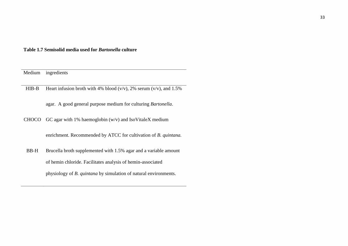
Table 1.7 Semisolid media used for Bartonella culture
Medium ingredients
HIB-B Heart infusion broth with 4% blood (v/v), 2% serum (v/v), and 1.5%
agar. A good general purpose medium for culturing Bartonella.
CHOCO GC agar with 1% haemoglobin (w/v) and IsoVitaleX medium
enrichment. Recommended by ATCC for cultivation of B. quintana.
BB-H Brucella broth supplemented with 1.5% agar and a variable amount
of hemin chloride. Facilitates analysis of hemin-associated
physiology of B. quintana by simulation of natural environments.

34
1.4.3 PCR assays
PCR seems to be the most useful technique for diagnosis of Bartonella infection
in both human medicine and veterinary medicine and the detection assays have been
available for several decades. Many reports describing the advantages of PCR to detect
this pathogen in various specimens‟ types have been published, but there are some
reports that suggest this technique lacks sensitivity (Cherry et al., 2009). This may be
due to high concentrations of immunoglobulin in patient lymph nodes or blood which
may inactivate the PCR reaction resulting in a lack of sensitivity (Al-Soud et al., 2000),
or to low DNA copy numbers of some Bartonella in certain specimens from some hosts.
A variety of rapid PCR detection assays have been developed, including PCR
amplification of the 16S-23S rRNA intergenic region with genus- and species-specific
primer sets (Minnick et al., 1994), species-specific amplification of ftsZ gene sequences
(Kelly et al., 1998; Ehrenborg et al., 2000; Zeaiter et al., 2002), repetitive-element PCR
(Rodriguez-Barradas et al., 1995), restriction fragment length polymorphism (RFLP)
analysis of PCR-amplified 16S rRNA genes (Birtles et al., 1995; Dauga et al., 1996),
RFLP analysis of the PCR-amplified 16S-23S rRNA intergenic region (Matar et al.,
1993; Roux et al., 1995), a single-step PCR-based assay for the differentiation of
Bartonella species using one set of primers to amplify a fragment of the 16S-23S rRNA
intergenic region (Jensen et al., 2000), RFLP analysis of the PCR-amplified citrate
synthase gene (Norman et al., 1995), semi-nested PCR of the citrate synthase gene
(Margolis et al., 2003), sequence analysis of the PCR amplified citrate synthase gene
(Birtles et al., 1996; Joblet et al., 1995) and PCR amplification of the phage-associated
gene (pap31) (Kelly et al., 2006; Maggi et al., 2006).

35
Regardless, for accurate species identification, the method requires multiple
PCR amplification steps of a variety of gene targets or additional sample-processing
steps following the primary PCR amplification. Multi-locus variable number tandem
repeat analysis (VNTR) and multilocus sequence typing (MLST) were developed to
discriminate between isolates of B. henselae in order to trace the source of transmission
between reservoirs, vectors and final hosts (Monteil et al., 2007; Li et al., 2006).

36
Table 1.8 PCR assays targeting various genes for Bartonella detection
Target locus PCR assays Bartonella species reference
16S rRNA RFLP
RFLP
Single step
B. bacilliformis
B. quintana, B. henselae
B. quintana, B. henselae
Birtles et al., 1995
Dauga et al., 1996
Tang et al., 2009
16S-23S rRNA (ITS) RFLP
RFLP
Single step
Single step
B. quintana, B. henselae, B. bacilliformis
B. vinsonii, B. elizabethae, B. quintana, B. henselae
B. vinsonii berkhoffii, B. elizabethae, B. quintana, B. henselae,
B. clarridgeiae, B. bacilliformis
B. vinsonii arupensis, B. tribocorum, B. alsatica, B. koehlerae
B. alsatica, B. doshiae, B. elizabethae, B. grahamii, B.
koehlerae,
B. schoenbuchensis, B. taylorii, B. tribocorum, B. vinsonii
arupensis,
B. vinsonii berkhoffii, B. vinsonii vinsonii
Matar et al., 1993
Roux et al., 1995
Jensen et al., 2000
Houpikian et al.,
2001
Riboflavin synthase (ribC) Single step B. quintana, B. henselae, B. bacilliformis, B. clarridgeiae Bereswill et al.,
1999

37
Target locus PCR assays Bartonella species reference
60 kDa heat shock protein (groEL)
Single step
B. quintana, B. henselae, B. bacilliformis, B. clarridgeiae B.
doshiae,
B. elizabethae B. grahamii, B. vinsonii berkhoffii,
B. vinsonii vinsonii
Marston et al.,
1999
Phage associated (pap31) Single step B. quintana
B. vinsonii berkhoffii
Kelly et al., 2006
Maggi et al., 2006
Cell division protein (ftsZ) Single step
Single step
B. quintana, B. henselae, B. bacilliformis
B. alsatica, B. birtlesii, B. doshiae, B. elizabethae, B. grahamii,
B. koehlerae, B. schoenbuchensis, B. taylorii,
B. tribocorum, B. vinsonii arupensis, B. vinsonii berkhoffii,
B. vinsonii vinsonii, and B. bovis
Kelly et al., 1998
Zeaiter et al., 2002
RNA polymerase beta subunit
(rpoB)
RFLP B. quintana, B. henselae, B. bacilliformis, B. clarridgeiae Renesto et al.,
2001

38
1.5 Genetic-based analysis for species identification in genus Bartonella
The first PCR-based diagnostic test for Bartonella infection (Relman et al.,
1990) designed to amplify Bartonella 16S rRNA gene sequences, but it was discovered
that these primers also amplified 16S rRNA gene fragments of many other
Proteobacteria. PCR amplification and sequencing of the 16S rRNA gene has been
considered to be one of the most functional and informative tools for the genetic
characterization and phylogenetic studies of bacteria (Olsen et al., 1993) and for the
investigation of diseases caused by uncultured or difficult-to-culture bacteria (Sontakke
et al., 2008). However, 16S rRNA sequence analysis has failed to establish a consistent
phylogeny for Bartonella species due to the high degree of similarity of the 16S rRNA
gene (>97%) within the genus Bartonella (La Scola et al., 2003; Birtles et al., 1995;
Brenner et al., 1993; Daly et al., 1993). Therefore, its usefulness as a single assay in the
study of intra-genus or inter-species phylogeny for Bartonella has been limited (Fox et
al., 1980; Olsen et al., 1993; Stackebrandt et al., 1994).
To solve this limitation, sequencing of several housekeeping genes have been
used for phylogenetic studies and also for PCR diagnosis. Many DNA regions and
encoding gene sequences have been used for these genetic studies, including the 16S-
23S rRNA intergenic spacer region (ITS), the citrate synthase gene (gltA), the riboflavin
synthase alpha chain gene (ribC), the heat shock protein gene (groEL), the gene
encoding the pap31 and 35-kDA proteins, the RNA polymerase beta-subunit-encoding
gene (rpoB) and the cell division protein gene (ftsZ) (Table 1.8).
The 16S-23S rRNA intergenic region has proved to be an appropriate target for
analysis of Bartonella species (Jensen et al., 2000; Minnick et al., 1997; Roux et al.,

39
1995). This region contains sufficient sequence diversity to differentiate between
Bartonella species. However, limitations of the ITS region for molecular detection has
been reported (Breitschwerdt et al., 2005), particularly non-specific amplification of
Mesorhizobium species and the fact that the amplicon size can vary even in the same
species of Bartonella due to the presence of highly variable repeat regions within the
ITS (Breitschwerdt et al., 2005). These repeats can vary in size from 15 bp to 45 bp
(Dillon et al., 2002; Houpikian et al., 2001) and therefore the amplicon size alone
should not be used as a reliable diagnostic criterion. This is a serious issue for
diagnostic laboratories that rely on visualisation of gel electrophoresis band size only
and do not sequence the amplified product.
Based on information from the citrate synthase (gltA) gene sequence from
Rickettsia prowazekii, amplification of Bartonella gltA gene was achieved in 1995
(Joblet et al., 1995). The full length of the B. henselae citrate synthase gene was then
cloned and sequenced (Norman et al., 1995). PCR-RFLP assays of the gltA have also
been developed to differentiate Bartonella spp. (Norman et al., 1995; Pappalardo et al.,
2000; Pesavento et al., 2005).
Genes encoding enzymes of the riboflavin biosynthetic pathway are
evolutionarily conserved in bacteria and plants and are absent in humans (Bacher et al.,
1996). This group of genes is one of many excellent targets for the PCR diagnosis and
phylogenetic analysis of Bartonella species (Bereswell et al, 1999). Riboflavin (vitamin
B2) is the precursor of flavin mononucleotide and flavin adenine dinucleotide, which are
both essential cofactors for electron transport functions of proteins involved in the basic
energy metabolism of the cell. Riboflavin is synthesized from GTP, and the
corresponding biosynthetic pathway is present in bacteria, fungi, and plants, but absent

40
in vertebrates. Species-specific primers based on the riboflavin synthesis (ribC) gene
sequence have been designed for the diagnosis of B. bacilliformis, B. clarridgeiae, B.
henselae and B. quintana (Bereswell et al., 1999). This gene was selected because it is
evolutionarily conserved in these bacteria and it is absent in vertebrate hosts (Bacher et
al., 1996).
The groEL gene encodes the 60 kDa heat-shock protein GroEL. The nearly full-
length 1,368 bp fragment was sequenced and used for intra-species phylogenetic
analysis of 8 Bartonella species (Marston et al., 1999). GroEL is one component of the
highly conserved heat-shock chaperonin system of proteins in bacteria (Haake al., 1997;
Segal et al., 1995). This gene has been used for phylogenetic analysis of eubacteria,
because it is ubiquitously distributed among the eubacteria, archaea, and eukaryotic
organelles (Masui et al., 1997). More recently, phylogenetic relationships within the
genus Bartonella have been characterized for another eight species of Bartonella at the
groEL locus (Zeaiter et al., 2002).
The pap31 gene encodes a major protein associated with a phage isolated from
B. henselae and is probably implicated in its pathogenesis (Bowers et al., 1998). A
semi-nested PCR of the pap31 gene (using three primers) has been developed to detect
B. henselae in humans (Zeaiter et al., 2002). In that study, the sensitivity of pap31 semi-
nested PCR was compared with single step PCR of the ITS region (Roux et al., 1995).
The result indicated that the semi-nested PCR of pap31 gene was 1.9 times more
sensitive than PCR of the ITS region.
The RNA polymerase beta-subunit gene (rpoB) sequence has been a useful tool
for the identification of members of the family Enterobacteriaceae (Mollet et al., 1997).
41
Different pairs of primers used to amplify the rpoB gene fragments, based on the
genetic data from Enterobacteriaceae, were tested with Bartonella spp. (Renesto et al.,
2001). Two sets of primers conserved for Bartonella spp. were chosen and tested with
21 bacterial strains unrelated to the genus Bartonella and 13 distinct Bartonella spp. A
PCR-RFLP assay of an 825-bp amplicon from the rpoB gene has been developed to
discriminate between different species of Bartonella (Renesto et al., 2001)
The 75-kDa B. bacilliformis antigen is a homologue of FtsZ, an essential cell
division protein that is strongly conserved among prokaryotes (Padmalayam et al.,
1997). The FtsZ protein plays a significant role in bacterial cell division, and its gene
sequence information has been used to identify three Bartonella species (Kelly et al.,
1998). The C-terminal region is more highly variable than the N-terminal region and is
a useful sequence for sub-typing of intra-species strains of the genus Bartonella
(Maurin et al., 1997).
Until recently, the definition of a new species of Bartonella was based on
polyphasic classification that included both determination of phenotypic characteristics
and DNA-DNA homology (La Scola et al., 2003). However, there were many
limitations with this approach, and in 2003 it was proposed that new species of
Bartonella be defined based on genetic data alone (La Scola et al., 2003) and that
sequence comparison of at least 5 housekeeping-genes was required (Stackebrandt et al,
2002) (as per the recommendations from an ad hoc committee to identify novel species
in bacteriology). La Scola et al., (2003) compared genetic similarity values for various
loci that discriminated Bartonella at the species level, assessed the relative
discriminatory power (DP) of each gene examined and published cut off values to
classify distinct Bartonella species (La Scola et al., 2003). All of the sequences studied,

42
including the gltA, groEL, rpoB, ribC, FtsZ genes and the ITS region, had good
discriminatory power, with DPs ranging from 86.5% to 94.4%, with the exception of a
16S rRNA sequence that had a DP of 99.7%. However, a good DP may not be sufficient
to clearly discriminate between some species of Bartonella. For example, the ITS
region and groEL, ftsZ, ribC gene sequences among B. schoenbuchensis, B. bovis, B.
grahamii and B. taylorii have high homologies ranging from 97.9% to 99.8%. Only two
genes (rpoB and gltA) have a homology less than 96% for these species.
The use of five loci and their relative cut off values has been used recently to
identify a novel species of Bartonella isolated from kangaroos in Australia (B. australis)
and a novel species from a human patient in Thailand (B. tamiae) using 5 different
genes gltA, groEL, rpoB, ribC, ftsZ) and the ITS region (Fournier et al., 2007; Kosoy et
al., 2008)
1.6 Bartonella species in wild animals and their ectoparasites
Recently, new Bartonella species have been reported in wildlife and their
ectoparasites and some of these novel species have been identified as zoonotic
Bartonella species (Chomel et al., 2010). With this has come the recognition that
wildlife may play an important role as a reservoir for infections of domesticated animals
and humans. Bartonella alsatica isolated from wild rabbits in France was reported to
cause endocarditis and lymphadenitis in humans (Raoult et al., 2006; Angelakis et al.,
2008; Jeanclaude et al., 2009). It has also been suggested that Spilopsyllus cuniculi, a
rabbit flea could be a major vector for B. alsatica (Marquez et al., 2009). Bartonella
tamiae was initially isolated from human patients in Thailand (Kosoy et al., 2008) and
then B. tamiae DNA was detected in chigger mites and one tick collected from wild
43
rodents in Thailand (Kabeya et al., 2010). Bartonella washoensis was first isolated from
a human patient with fever and myocarditis (Kosoy et al., 2003). Consequently, B.
washoensis was found in California ground squirrels (Spermophilus beecheyi) with
more than 17% prevalence of infection in the same area (Kosoy et al., 2003). Bartonella
washoensis was also isolated from a dog with endocarditis in the same year and the
identical strain of B. washoensis from the human patients and squirrels was
demonstrated in this dog (Chomel et al., 2003). Bartonella schoenbuchensis was first
isolated from wild roe deer (Capreolus capreolus) in Germany in 2001 and from French
cattle in 2003 (Matsumoto et al., 2008). Bartonella DNA closely related to B.
schoenbuchensis was detected in deer keds (Lipoptena cervi) collected from white-
tailed deer (Odocoileus virginianus) (Reeves et al., 2006) and was shown to localize in
the midgut of this blood-sucking arthropod, which is a cause of deer ked dermatitis in
humans (Dehio et al., 2004). The presence of B. rochalimae as a cause of fever,
splenomegaly, mild anaemia and rash was reported in a human patient (Eremeeva et al.,
2007). This Bartonella sp. was previously described as B. clarridgeiae-like strain in
dogs (MacDonald et al., 2004). Consequently, B. rochalimae was isolated from possible
carnivore reservoir hosts including dogs, grey and red foxes, raccoons and coyotes, as
well as fleas collected from grey foxes (Chomel et al., 2010). The number of zoonotic
Bartonella species identified in wild animals and their ectoparasites has increased
significantly in recent years and the further investigation of reservoirs and arthropod
vectors of Bartonella species is required in order to recognize potential sources for
human infection (Chomel et al., 2010).

44
1.7 Bartonella species in Australia
Zoonotic Bartonella species, including B. henselae and B. clarridgeiae, have
been reported in cats, cat fleas and humans in Australia (Iredell et al., 2003; Barrs et al.,
2010). Bartonella henselae sequence type 1 (ST 1) (or strain Houston-1) has been
proposed as the major strain causing human bartonellosis and is distributed widely in
the cat population in Australia (Iredell et al., 2003; Arvand et al., 2007). Bartonella
henselae ST 1 is also a major strain distributed in cats and humans in North America
(Arvand et al., 2007). Bartonella quintana was isolated from an immunocompromised
human patient in Australia and was diagnosed as the cause of their fever and
bacteraemia (Rathbone et al., 1996). Additionally, four novel Bartonella species have
been reported in non-domestic mammalian hosts in Australia. Bartonella australis was
first isolated from the blood of eastern grey kangaroos (Macropus giganteus) (Fournier
et al., 2007) and was the first report of a Bartonella species isolated from an Australian
marsupial. Another three novel Bartonella species, including B. rattaustraliani, B.
queenslandensis and B. coopersplainsensis, were isolated from the blood of rodents
(Rattus spp., Melomys spp., Uromys spp., respectively) (Gundi et al., 2009). Bartonella
DNA has also been detected in ticks (Ixodes tasmani) collected from koalas
(Phascolarctos cinereus) at the Koala Conservation Centre in Victoria (Vilcins et al.,
2009). However, to date, the relationships between Bartonella species, marsupials and
their arthropod vectors in Australia is still largely unknown.

45
1.8 Objectives
The objectives of the work reported in this thesis were:
1. To develop molecular tools for detection and characterization of Bartonella species.
2. To investigate genetic diversity of Bartonella species in a variety of
animals and their arthropod vectors in Western Australia.
3. To develop molecular tools for intra-species identification or strain identification of
zoonotic Bartonella species in animals in Western Australia.
4. To characterize genetic diversity of arthropod vectors which are infected by
Bartonella species in animals in Western Australia.

46
Chapter 2
General Materials and Methods
2.1 Ectoparasite collection and identification
While this project did not specifically involve the handling and sampling of
marsupials directly to collect ectoparasites, there was significant interaction with other
conservation-related projects in order to acquire these samples. Woylies (Bettongia
penicillata) are currently being trapped for health checks as part of the Woylie
Recovery Programme and to assess the reasons for a significant decline in the Western
Australia (WA) population. The programme is run by the Department of Environment
and Conservation (DEC), with input from staff of Perth Zoo and Murdoch University.
Other marsupials are trapped and examined annually by DEC staff to assess population
and health characteristics. Some marsupials were also under investigation by other
research groups at Murdoch University, for example western barred bandicoots on
Dorre Island. All trapping and removal of ectoparasites (ticks and fleas) was carried out
under strict DEC and Murdoch University Animal Ethics guidelines. In addition, fleas
and blood samples were collected from red foxes in February and March 2010 as part of
the „Red Card for the Red Fox‟ 2010 culling program coordinated by the Department of
Agriculture and Food, WA. The numbers and species of ectoparasites collected from
mammalian hosts in various locations in WA in this study are shown in Tables 2.1 and
2.2. All ectoparasites were stored in 70% ethanol until required for DNA extraction.
Ticks were identified by light microscopy using the STANDARD KEY FOR
AUSTRALIAN TICK IDENTIFICATION (Roberts, 1970). Fleas were also identified

47
using light microscopy with reference to a standard key for Australian fleas (Dunnet and
Mardon, 1974).

48
Table 2.1 Numbers and species of ectoparasites collected from mammalian hosts in
Western Australia in the present study.
Hosts
(Numbers)
Mardos
(36)
Woylies
(62)
Rodents
(2)
Bandicoots
(55)
Red foxes
(164)
Ectoparasites No. of
samples
Ticks
Amblyomma triguttatum 0 8 0 0 0
Ixodes tasmani 0 2 0 0 0
Ixodes myrmecobii 0 3 0 0 0
Ixodes australiensis 0 16 0 0 0
Ixodes spp. 27 24 1 0 0
Fleas
Echidnophaga myrmecobii 0 1 0 0 0
Acanthopsylla jordani 39 0 0 0 0
Pygiopsylla hilli 0 18 0 0 0
Stephanocircus dasyuri 0 2 0 0 0
Stephanocircus pectinipes 0 0 1 0 0
Pygiopsylla tunneyi 0 0 0 25 0
Pulex irritans 0 0 0 1 0
Xenopsylla vexabilis 0 0 0 2 0
Ctenocephalides felis 0 0 0 0 151
Total 66 74 2 28 151

49
Table 2.2 Hosts from which ectoparasites were collected in various locations in
Western Australia.
Host Location
Fig. 2.1, 2.2, 2.3
Year
Woylie
(Bettongia
penicillata)
Rodent
(Rattus fuscipes)
Southwest
Forest
South Coast
Keninup
Balban
Karakamia Sanctuary
Fitzgerald River
National Park
34°2/S, 116°37
/E
34°5/S, 116°35
/E
31°48/S, 116°14
/E
33°56/S, 119°37
/E
2003-2004
Western barred
bandicoot
(Perameles
bougainville)
Bernier
and
Dorre Islands
25°7/S, 113°6
/E
24°51/S, 113°8
/E
2005-2007
Mardo
(Antechinus
flavipes)
Southwest The areas surrounding
the town of
Dwellingup
32°38/S, 116°5
/E 2003-2004
Red fox
(Vulpes vulpes)
Feral cat
(Felis catus)
Rabbit
(Oryctolagus
cuniculus)
Southwest The areas surrounding
the towns of
Katanning
and
Boyup Brook
33°41/S, 117°34
/E
33°50/S, 116°23
/E
2010
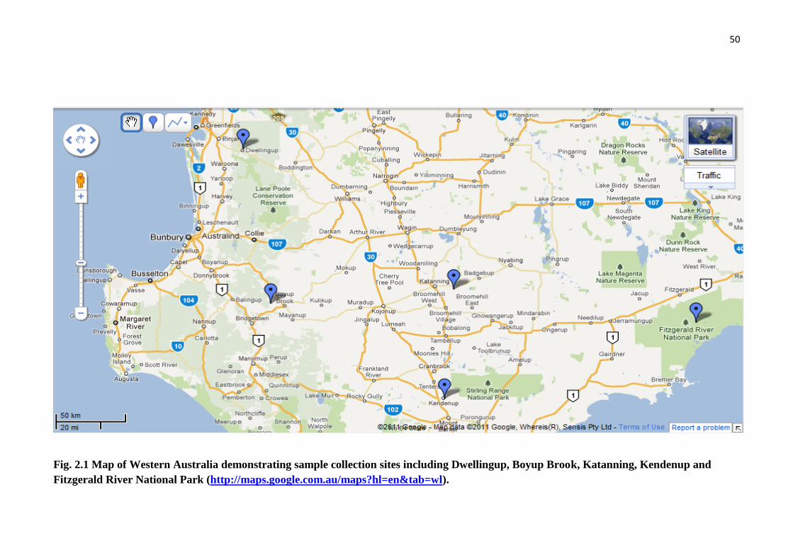
50
Fig. 2.1 Map of Western Australia demonstrating sample collection sites including Dwellingup, Boyup Brook, Katanning, Kendenup and
Fitzgerald River National Park (http://maps.google.com.au/maps?hl=en&tab=wl).
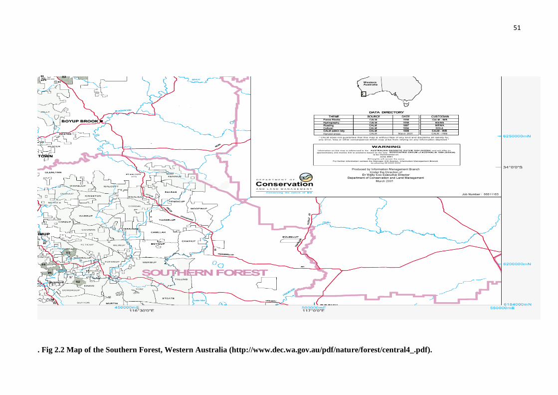
51
. Fig 2.2 Map of the Southern Forest, Western Australia (http://www.dec.wa.gov.au/pdf/nature/forest/central4_.pdf).

52
Fig. 2.3 Map of Dorre and Bernier Islands
(http://www.environment.gov.au/biodiversity/threatened/publications/recovery/djo
ongari/index.html).
53
2.2 DNA extraction
After morphological identification, ectoparasites (ticks and fleas) were chopped
into small sections using a disposable scalpel blade for each one to avoid cross
contamination. New disposable glass slides or Petri dishes were used as a chopping
board for each extraction to prevent cross contamination. One individual tick was used
per DNA extraction and three to four fleas were pooled per DNA extraction. DNA was
extracted from blood and tissue samples using a DNeasy Blood and Tissue Kit (Qiagen,
Maryland, USA) according to the manufacturer‟s instructions, with the following
modification: the volume of elution buffer used to dissolve DNA from spin columns
was decreased to 50-100 µl (lower than the recommended volume of 200 µl) to
concentrate the amount of DNA present, particularly from blood samples. One negative
control using sterile water was included in each batch of DNA extractions. DNA
extracts were stored at −20°C until further processing.
2.3 PCR detection of Bartonella species
Bartonella DNA in tick and flea samples was detected using nested-PCRs of the
ITS region and the gltA gene. External primers for the gltA gene and the ITS region
were designed for the present study from GenBank B. henselae DNA sequences
(GenBank, gltA; L38987, ITS; L35101). A nested-PCR of the ITS region was
performed using new external primers designed for this study; WITS-F (5‟-
ACCTCCTTTCTAAGGATGAT-3‟) and WITS-R (5‟-AAAGACCAGCTTCTCG-
AGAT-3‟) and internal primers Bh311-332F (5‟-CTCTTTCTTCAGATGATGATCC-
3‟) and Bh473-452R (5‟-AACCAACTGAGCTACAAGCCCT-3‟) as previously
described (Jensen et al., 2000).
54
The PCR reactions for the external primers were performed using 1 µL of DNA
in a 25 µL reaction containing 1 x PCR buffer, 2.5 mM MgCl2, 0.2 mM dNTPs, 1 µM
of each primer and 0.02 U/µL TAQ-Ti (hot start) Taq DNA polymerase (Fisher Biotech
Australia, Wembley, W.A., Australia). The cycling conditions consisted of a pre-PCR
step of 96 ºC for 2 minutes, followed by 40 cycles of 94 ºC for 50 seconds, 48 ºC for 60
seconds and an extension of 72 ºC for 90 seconds, with a final extension of 72 ºC for 10
minutes. PCR reactions for the internal primers were the same as the first round PCR
with the exception that the MgCl2 concentration was 2 mM. Cycling conditions
consisted of a pre-PCR step of 96 ºC for 2 minutes, followed by 45 cycles of 94 ºC for
45 seconds, 60 ºC for 30 seconds and an extension of 72 ºC for 45 seconds, with a final
extension of 72 ºC for 5 minutes.
Nested-PCR for the gltA gene was also performed using newly designed external
primers, gltA-F2 (5‟-GCTTCGTGTGAATCGAAAATCA-3‟) and gltA-R2 (5‟-GCGG-
TTAAGCTTCCAATCATA-3‟) and internal primers BhCS.781F (5‟-GGGGACCAGC-
TCATGGTGG-3‟) and BhCS.1137R (5‟-AATGCAAAAAGAACAGTAA-ACA-3‟) as
previously described (Norman et al., 1995). Nested-PCR for the gltA gene produced a
379 bp amplicon. The PCR reaction conditions and cycles for gltA external primers
were the same as for the external ITS PCR primers. PCR reactions for the internal
primers were performed using 1 µL of DNA in a 25 µL reaction containing 1 x PCR
buffer, 2 mM MgCl2, 0.2 mM dNTPs, 1 µM of each primer and 0.02 U/µL TAQ-Ti (hot
start) Taq DNA polymerase (Fisher Biotec, Australia, Wembley, W.A., Australia).
Cycling conditions consisted of a pre-PCR step of 96 ºC for 2 minutes, followed by 45
cycles of 94 ºC for 45 seconds, 55 ºC for 30 seconds and an extension of 72 ºC for 45
seconds, with a final extension of 72 ºC for 5 minutes. All new primers designed in this
study were created using Primer3 primer design (http://biotools.Umassmed.edu /bioapp-

55
s/primer3_www.cgi), Whitehead Institute for Biomedical Research). Primers were
synthesised by GeneWorks (Hindmarsh, South Australia). A list of primer sequences
used in this study is described in Table 2.3.
2.4 PCR for phylogenetic analysis at additional loci
Following successful amplification of Bartonella DNA from ectoparasite
samples, 3 other loci were amplified using single-step PCRs targeting the 16S rRNA,
ftsZ and rpoB genes. New primers for the 16S rRNA, W16S-F (5‟-AGTAACGCGTG-
GGAATCTAC-3‟) and W16S-R (5‟-CACAGCACCTTCGGGTAAAACCAA-3‟) were
designed from a B. henselae DNA sequence (GenBank, M73229). The 16S rRNA
primers produced an approximately 1.4 kb PCR product (the amplicon size varies
depending on the species of Bartonella). Degenerate primers used for the ftsZ gene,
FtsZ-F (5‟-ATTAATCTGCAYCGGCCAGA-3‟) and FtsZ-R (5‟-ACBGAHACACGA-
ATAACACC 3‟) and the rpoB gene, RpoB-F (5‟-CGCATTGGYTTRCTTCGTATG-
3‟) and RpoB-R (5‟-GTRGAYTGATTRGAACGYTG-3‟) were modified from previous
studies (Zeaiter et al., 2002; Renesto et al., 2001). These degenerate primers produced a
925-bp PCR product at the ftsZ locus and an 863-bp PCR product at the rpoB locus. A
new internal reverse primer, Inner ITS-R (5‟-GCGGTTAAGCTTCCAATCATA-3‟)
targeting the ITS region was also designed for amplification and sequencing of a large
fragment of the ITS region. The size of the large ITS fragment ranged from 0.9 kb to 1.6
kb in different Bartonella species. PCR reactions for the 16S rRNA primers were
performed using 1 µL of DNA in a 25 µL reaction containing 1 x PCR buffer, 2 mM
MgCl2, 0.2 mM dNTPs, 1 µM of each primer and 0.02 U/µL TAQ-Ti (hot start) Taq
DNA polymerase (Fisher Biotec Australia, Wembley, W.A., Australia). Cycling
conditions consisted of a pre-PCR step of 96 ºC for 2 minutes, followed by 45 cycles of

56
94 ºC for 50 seconds, 55 ºC for 50 seconds and an extension of 72 ºC for 90 seconds,
with a final extension of 72 ºC for 10 minutes. PCR reactions for the ftsZ primers were
performed using 1 µL of DNA in a 25 µL reaction containing 1 x PCR buffer, 2 mM
MgCl2, 0.2 mM dNTPs, 1 µM of each primer and 0.02 U/µL TAQ-Ti (hot start) Taq
DNA polymerase (Fisher Biotec Australia, Wembley, W.A., Australia). Cycling
conditions consisted of a pre-PCR step of 96 ºC for 2 minutes, followed by 45 cycles of
94 ºC for 50 seconds, 52 ºC for 30 seconds and an extension of 72 ºC for 45 seconds,
with a final extension of 72 ºC for 7 minutes. PCR reactions for the rpoB primers were
performed using 1 µL of DNA in a 25 µL reaction containing 1 x PCR buffer, 2.5 mM
MgCl2, 0.2 mM dNTPs, 1 µM of each primer and 0.02 U/µL TAQ-Ti (hot start) Taq
DNA polymerase (Fisher Biotec Australia, Wembley, W.A., Australia). Cycling
conditions consisted of a pre-PCR step of 96 ºC for 2 minutes, followed by 45 cycles of
94 ºC for 50 seconds, 55 ºC for 50 seconds and an extension of 72 ºC for 60 seconds,
with a final extension of 72 ºC for 7 minutes.
2.5 Agarose gel electrophoreses and PCR product purification
PCR products were electrophoresed on 1% w/v of agarose gel (Ultrapure
Agarose gel, Invitrogen®) in tris acetate-EDTA (TAE) buffer (40nmM Tris-HCL, 20
mM acetate, 2 mM EDTA, pH 7.9) stained with SYBR® Safe DNA Gel Stain
(Invitrogen®). Gels were run at 100 volts for approximately 45 minutes and PCR
products were visualized using a Dark Reader transilluminator (Clare Chemical
Research®, USA). PCR products from all genes were purified from agarose gel slices
using an UltraCleanTM
15 DNA Purification Kit (MO BIO Laboratories Inc. West
Carlsbad, California, USA). The DNA concentrations of all purified PCR products were
measured using a NanoDrop®
ND-1000 spectrophotometer (Thermo Scientific®, USA).
57
2.6 DNA sequencing and phylogenetic analysis
Sequencing was performed for all loci using an ABI PrismTM
Terminator Cycle
Sequencing kit (Applied Biosystems, Foster City, California, USA) on an Applied
Biosystems 3730 DNA Analyzer, following the manufacturer‟s instructions.
Sequencing reactions were performed using approximately 2-5 µL of DNA template
(depending on the DNA concentration of purified PCR products and the fragment
length of PCR products) in a 10 µL reaction containing 1 µL of Big Dye version 3.1
terminator mix (Applied Biosystems, Foster City, California, USA), 3.2 pM of primer
and 1.5 x buffer for sequencing reaction. The reaction mix was made up to 10 µL using
molecular grade water. Nucleotide sequences generated for all 5 loci were analysed
using Chromas lite version 2.0 (http://www.technelysium.com.au) and aligned with
reference sequences of Bartonella spp. from GenBank using Clustal W
(http://www.clustalw.genome.jp). Phylogenetic relationships of all 5 loci were
constructed using Distance and Maximum-parsimony methods using Mega version 4
(Mega4: Molecular Evolutionary Genetics Analysis software, Arizona State University,
Tempe, Arizona, U.S.A.) (Tamura et al., 2007). Genbank accession numbers of
Bartonella species used for the concatenated phylogenetic analysis are shown in Table
2.3 and GenBank accession numbers of Bartonella species discovered in the present
study are shown in Table 2.4.

58
Table 2.3 Oligonucleotide primers used for nested-PCR and single step PCR
amplifications of the 16S rRNA, gltA, ftsZ, rpoB loci and the ribosomal ITS region.
Primer Locus Nucleotide sequence Reference
gltA-F2†
gltA GCTTCGTGTGAATCGAAAATCA This study
gltA-R2†
GCGGTTAAGCTTCCAATCATA
BhCS.781F (gltA)* gltA GGGGACCAGCTCATGGTGG (Norman et al., 1995)
BhCS.1137R(gltA)* AATGCAAAAAGAACAGTAAACA
WITS-F† ITS ACCTCCTTTCTAAGGATGAT This study
WITS-R† AAAGACCAGCTTCTCGAGAT
Bh311-332F (ITS) *
Bh473-452R (ITS) *
ITS CTCTTTCTTCAGATGATGATCC
AACCAACTGAGCTACAAGCCCT
(Jensen et al., 2000)
Inner ITS-R (ITS)* GCGGTTAAGCTTCCAATCATA This study
W16S-F 16S rRNA AGTAACGCGTGGGAATCTAC This study
W16S-R CACAGCACCTTCGGGTAAAACCAA
RpoB-F rpoB CGCATTGGYTTRCTTCGTATG (Renesto et al., 2001)
RpoB-R GTRGAYTGATTRGAACGYTG
FtsZ-F ftsZ ATTAATCTGCAYCGGCCAGA (Zeaiter et al., 2002)
FtsZ-R ACBGAHACACGAATAACACC
*Inner primers, †Outer primers.

59
Table 2.4 Genbank accession numbers of Bartonella species used for the
concatenated phylogenetic analysis
Bartonella species 16S rRNA ftsZ gltA ITS rpoB
Bartonella tamiae EF672729 EF605281 DQ395177 EF605183 EF672730
Candidatus Bartonella rudakovii EF682086 EF682092 EF682090 EF682087 EF682088
Bartonella rochalimae DQ683196 DQ676486 DQ683195 DQ683199 DQ676489
Bartonella rattimassiliensis AY515120 AY515133 AY515125 AY515121 AY515131
Bartonella rattaustraliani EU111753 EU111771 EU111797 EU111760 EU111786
Bartonella queenslandensis EU111758 EU111780 EU111801 EU111765 EU111790
Bartonella phoceensis AY515119 AY515135 GU056197 AY515123 AY515132
Bartonella coopersplainsensis EU111759 EU11781 EU111803 EU111770 EU111792
Bartonella chomelii NR025736 AB290193 AY254308 AB498010 AB290189
Bartonella capreoli NR025120 AB290192 AF293392 AB498009 AB290188
Bartonella australis DQ538394 DQ538399 DQ538395 DQ538396 DQ538397
Bartonella alsatica AJ002139 AF467763 AF204273 AF312506 AF165987
Bartonella bacilliformis Z11683 AF007266 U28076 L26364 AF165988
Bartonella birtlesii AF204274 AF467762 AF204272 AY116640 AF165989
Bartonella bovis AF199502 AF467761 AF293394 AY116638 AF166581
Bartonella clarridgeiae U64691 AF141018 U84386 AF167989 AF165990
Bartonella doshiae Z31351 AF467754 AF207827 AJ269786 AF165991
Bartonella elizabethae L01260 AF467760 U28072 L35103 AF165992
Bartonella grahamii Z31349 AF467753 Z70016 AJ269785 AF165993
Bartonella henselae M73229 AF061746 L38987 L35101 AF171070
Bartonella koehlerae AF076237 AF467755 AF176091 AF312490 AY166580
Bartonella quintana M11927 AF061747 Z70014 L35100 AF165994
Bartonella schoenbuchensis AJ278187 AF467765 AJ278183 AY116639 AY167409
Bartonella taylorii Z31350 AF467756 AF191502 AJ269784 AF165995
Bartonella tribocorum AJ003070 AF467759 AJ005494 AF312505 AF165996
Bartonella vinsonii subsp.arupensis AF214558 AF467758 AF214557 AF312504 AY166582
Bartonella vinsonii subsp. berkhoffii U26258 AF467764 U28075 AF167988 AF165989
Bartonella vinsonii subsp.vinsonii M73230 AF467757 Z70015 L35102 AF165997

60
Table 2.5 GenBank accession numbers of Bartonella species detected from possible
arthropod vectors collected from animals in Western Australia in the present study
Host animals Possible
arthropod
vectors
Bartonella spp. GenBank accession numbers
16S rRNA ITS ftsZ rpoB gltA
Mardo
(Antechinus
flavipes)
Acanthopsylla
jordani
flea
Candidatus
B. antechini
GU168960 GU168959 GU168958 GU168961 GU168962
Woylie
(Bettongia
penicillata)
Pygiopsylla hilli
flea
Candidatus
B. woyliei
HQ444148 HQ444149 HQ444150 HQ444151 HQ444152
Ixodes
australiensis
tick
Bartonella sp. - HQ444153 - - HQ44
4154
Western
barred
bandicoot
(Perameles
bougainville)
Pygiopsylla
tunneyi
flea
Candidatus
B. bandicootii
HQ444161 HQ444162 HQ444163 HQ444164 HQ444165
Rodent
(Rattus
fuscipes)
Stephanocircus
pectinipes
flea
B.
coopersplainsensis
- HQ444157 HQ444158 HQ444159 HQ444160
Ixodes sp.
tick
B.
rattaustraliani
- HQ444155 - - HQ444156
Red fox
(Vulpes
vulpes)
Ctenocephalides
felis
flea
B. henselae HM990954 HM990957 HM990958 HM990956 HM990955
B. clarridgeiae HM990959 HM990962 HM990963 HM990961 HM990960

61
Chapter 3
Candidatus Bartonella antechini: a novel Bartonella species detected in
fleas and ticks from the yellow-footed antechinus (Antechinus flavipes),
an Australian marsupial
(This chapter forms the basis of the article by Kaewmongkol et al. (2011)
Candidatus Bartonella antechini: A novel Bartonella species detected in fleas and
ticks from the yellow-footed antechinus (Antechinus flavipes), an Australian
marsupial. Veterinary Microbiology. 149: 517-521.)
3. 1 Introduction
Recently, four novel species of Bartonella were described from animals in
Australia. Bartonella australis was isolated from the blood of grey kangaroos
(Macropus giganteus) from central coastal Queensland (Fournier et al., 2007).
Bartonella rattaustraliani, B. queenslandensis and B. coopersplainsensis were
identified from rodent blood from Queensland (Gundi et al., 2009). Bartonella-like
organisms have also been detected in ticks (Ixodes tasmani) collected from koalas
(Phascolarctos cinereus) (Vilcins et al., 2009). However, very little is known about
Bartonella spp. in other marsupials in Australia.
The mardo (Antechinus flavipes) is a small carnivorous marsupial belonging to
the genus Antechinus, order Dasyuromorphia (Dasyuridae). These native Australian
marsupials are found in a variety of habitats across Queensland, New South Wales,

62
Victoria, South Australia and the southwest of Western Australia. All species in this
genus have the unusual physiological feature of male semelparity (total male mortality)
after a single breeding season (Naylor et al., 2008). This event, immediately following
mating, is thought to be due to excessive production of testosterone and corticosteroids,
resulting in gastrointestinal haemorrhage and failure of the individual‟s immune system
(McAllean et al., 1998; Naylor et al., 2008). This chapter reports the discovery of a
novel species of Bartonella that was detected in both fleas (Acanthopsylla jordani) and
ticks (Ixodes antechini) removed from mardos from the southwest of Western Australia.
3. 2. Materials and Methods
3.2.1 Sample collection and vectors identification
Thirty six mardos were trapped in dry sclerophyll forests surrounding the town
of Dwellingup in the southwest of Western Australia in 2003 to 2004 (Fig. 2.1). Twenty
seven ticks and 39 fleas were collected from the animals‟ skin and hair coats using a
flea comb or tweezers. The ectoparasites were stored in 70% ethanol until required for
DNA extraction. Tick morphology was characterized by light microscopy using the
standard key for Australian ticks (Roberts, 1970). Fleas were also identified using light
microscopy according to a standard key for Australian fleas (Dunnet and Mardon,
1974).

63
3.2.2 DNA extraction
Ticks and fleas were chopped into small sections using a disposable scalpel
blade and DNA was extracted using a DNeasy Blood and Tissue Kit (Qiagen,
Maryland, USA).
3.2.3 PCR screening and genotyping analysis
The methods performed for PCR assays and phylogenetic analysis at additional
loci have been described previously in Chapter 2.
3.3 Results
The genetic similarity of the concatenated sequences compared with sequences
from other Bartonella spp., shown in Fig. 3.1 and 3.2, revealed that the Bartonella spp.
from the mardo was 94.5% to 98.8% similar to other Bartonella spp. at the 16S rRNA
locus, 80.3% to 87.8% similar at the gltA locus, 84.6% to 89.2% similar at the ftsZ locus
and 82.5% to 87.9% similar at the rpoB locus (Table 3.1). The proposed cut-off values
for differentiation of a new Bartonella species are genetic identities of less than 96.0%
and 95.4% for gltA and rpoB respectively (La Scola et al., 2003). On this basis, the
Bartonella isolate described here from mardo ticks and fleas should be considered a
new species and we propose the name Candidatus Bartonella antechini n. sp. The
concatenated phylogenetic analysis in this study showed the close relationship between
this new species and B. australis isolated from grey kangaroos (Macropus giganteus) in
Queensland (Fig. 3.1 and 3.2). A separate clustering of Bartonella species harboured by
marsupials or marsupial vectors is also evident. Bartonella DNA was also detected from

64
the tick samples using nested-PCR targeting the gltA and ITS loci, but we were unable
to amplify PCR products from the same tick samples using a single step PCR for the
16S rRNA, ftsZ, and rpoB genes. Amplification of Bartonella DNA from all five loci
was successful for the flea extracts. The gltA fragment and ITS sequences generated
from flea samples were identical to the corresponding Bartonella DNA sequences from
ticks. Partial sequences for the five loci corresponding to this new species were
submitted to GenBank under the accession numbers GU168958; ftsZ, GU168959; 16S-
23S rRNA intergenic spacer (ITS), GU168960; 16S rRNA, GU168961; rpoB,
GU168962; gltA.

65
Table 3.1 Percentage genetic similarity of the concatenated sequences from
Candidatus Bartonella antechini n. sp. compared with other confirmed Bartonella
spp.
Percent genetic similarity to other Bartonella sp.
16S rRNA ftsZ rpoB gltA
Candidatus B. antechini
94.5-98.8 84.6-89.2 82.5-87.9 80.3-87.8

66
Fig. 3.1 Neighbor-Joining concatenated phylogenetic tree of 16S rRNA, gltA, ftsZ,
rpoB, and the ITS region of Australian marsupial isolates and validated species
and subspecies of Bartonella. Percentage bootstrap support (>50%) from 1000
pseudoreplicates is indicated at the left of the supported node.

67
Fig. 3.2 Maximum-Parsimony concatenated phylogenetic tree of 16S rRNA, gltA,
ftsZ, rpoB, and the ITS region of Australian marsupial isolates (B. australis and
Candidatus B. antechini) and validated species and subspecies of Bartonella.
Percentage bootstrap support (>40%) from 1000 pseudoreplicates is indicated at
the left of the supported node.

68
3.4 Discussion
The novel Bartonella species described here is named Candidatus Bartonella
antechini after the genus of marsupial host (Antechinus) from which the ectoparasites
were collected. Genetic characterization of this new species detected from fleas
(Acanthopsylla jordani) and ticks (Ixodes antechini), based on multi-locus sequence
analysis of the 16S rRNA, gltA, ftsZ, rpoB loci and the ITS region, have confirmed its
genetic distinctness. The results of the present study have confirmed that two different
types of possible vectors, ticks and fleas, from one mammalian host in a geographical
area, are able to harbour the same species of Bartonella. While fleas and ticks have both
been identified as potential vectors of Bartonella henselae, the reports were made from
different studies involving different animal species (Chomel et al., 1996; Cotté et al.,
2008).
It is not known why the amplification of 16S rRNA, ftsZ and rpoB genes in the
tick-positive samples was not successful, but this may be due to the different PCR
sensitivities between single step and nested-PCR. Differences in Bartonella
multiplication rates inside ticks and fleas have been observed in experimental
Bartonella infections (Cotté et al., 2008; Chomel et al., 2009). Single step PCR may
lack the sensitivity required to detect Bartonella spp. in some stages of infection in tick
vectors.
Little is known about Bartonella spp. occurring in marsupials and their
arthropod vectors in Australia. The identification and characterization of Bartonella spp.
in both marsupials and marsupial vector populations is important for understanding the
epidemiology of Bartonella infections in Australia. Multilocus sequence analysis of the

69
Bartonella species will help to inform the debate over the evolution, ecology, host
interactions and virulence of these bacteria in native Australian fauna.

70
Chapter 4
Diversity of Bartonella species detected in arthropod vectors from
animals in Western Australia
(This chapter forms the basis of the article by Kaewmongkol et al. (2011) Diversity
of Bartonella species detected in arthropod vectors from animals in
Australia. Comparative Immunology, Microbiology & Infectious Diseases,
doi:10.1016/j.cimid.2011.07.002)
4.1 Introduction
In the last decade, Bartonella spp. have been isolated from a wide variety of
free-roaming and captive mammals and their ectoparasites worldwide, including wild
canids (Henn et al., 2009; Gabriel et al., 2009; Marquez et al., 2009), wild rodents
(Inoue et al., 2009; Gundi et al., 2009; Morick et al, 2010; Kosoy et al., 2010), wild
cervids (Dehio et al., 2004; Skarphédinsson et al., 2005; Matsumoto et al., 2008) and
stray dogs (Inoue et al., 2009; Bai et al., 2010).
Bartonella henselae and B. clarridgeiae are known to occur in cats, cat fleas and
humans in Australia (Iredell et al., 2003; Barrs et al., 2010). Bartonella henselae
sequence type 1 (ST 1) or strain Houston-1 has been proposed as a major strain causing
human bartonellosis and is distributed widely in the cat population in Australia (Iredell
et al., 2003; Arvand et al., 2007). Additionally, five novel Bartonella species have been
reported from mammalian hosts in Australia. Three novel Bartonella species, including

71
B. rattaustraliani, B. queenslandensis and B. coopersplainsensis, were isolated from the
blood of native Australian rodents (Gundi et al., 2009). Bartonella australis was first
isolated from the blood of eastern grey kangaroos (Macropus giganteus) (Fournier et
al., 2007) and was the first report of Bartonella species isolated from an Australian
marsupial. Candidatus Bartonella antechini n. sp. was detected in ticks and fleas from
an Australian marsupial, Antechinus flavipes (mardo or yellow-footed antechinus)
(Kaewmongkol et al., 2011a). However, to date, knowledge of the genetic diversity
among Bartonella species in marsupial animals and their arthropod vectors in Australia
is still largely unknown.
The woylie (or brush-tailed bettong) (Bettongia penicillata) once inhabited
southwest Western Australia, most of South Australia, the northwest of Victoria and the
central area of New South Wales. In Western Australia, the species has undergone a
rapid population decline over the last 10 years, the reasons for which are currently
unclear. The “woylie decline” is currently being investigated by the Department of
Environment and Conservation, Western Australia (DEC, 2008). The western barred
bandicoot (Perameles bougainville) is a small nocturnal omnivorous marsupial species
that was originally found on the mainland of Australia but due to predation by red foxes
(Vulpes vulpes) and feral cats (Felis catus), competition with the European rabbit
(Oryctolagus cuniculus), altered fire regimes and loss of habitat (Richards, 2003),
natural populations are now only found on Bernier and Dorre Islands in Western
Australia. The last report of the western barred bandicoot found on the Australian
mainland was in 1931 (Kitchener and Vicker, 1981). These two species of Australian
marsupials are classified as threatened species and are vulnerable to extinction.

72
The aim of this chapter was to identify the genetic diversity of Bartonella
species from ectoparasites, ticks and fleas collected from native rodents and marsupials
in Western Australia.
4.2 Materials and Methods
4.2.1 Sample collection and ectoparasite identification
Twenty-eight fleas including Pygiopsylla tunneyi (25), Pulex irritans (1),
Xenopsylla vexabilis vexabilis (2) were collected from 55 anaesthetized bandicoots
living on Bernier and Dorre Islands from 2005 to 2007 (Table 2.1, 2.2) (Fig. 2.3) during
an investigation into BPCV1 (bandicoot papillomatosis carcinomatosis virus type 1)
infections. Woylies and rodents were trapped in various locations in Western Australia,
including Keninup, Balban, Karakamia and Fitzgerald River National Park in 2003 to
2004 as part of mammal monitoring studies by DEC (Fig. 2.1, 2.2). Fifty three ticks
including Ixodes spp. (45), Amblyomma spp. (8) and 21 fleas including Pygiopsylla hilli
(18), Echidnophaga myrcobii (1), Spectinocircus dasyure (2) were collected from 62
woylies. One tick (Ixodes spp.) and 1 flea (Spectinocircus pectinipes) were collected
from 2 rodents (Rattus fuscipes). Tick morphology was characterized by light
microscopy using the standard key for Australian ticks (Robert, 1970). Fleas were also
identified using light microscopy according to a standard key for Australian fleas
(Dunnet and Mardon, 1974).

73
4.2.2 DNA extraction
Ticks and fleas were chopped into small sections using a disposable scalpel
blade and the DNA was extracted using a DNeasy Blood and Tissue Kit (Qiagen,
Maryland, USA) according to the manufacturer‟s protocol, as described in Chapter 2.
4.2.3 PCR detection and phylogenetic analysis at additional loci
The methods performed for PCR detections and phylogenetic analysis at
additional loci have been described previously in Chapter 2 and 3.
4.3 Results
Bartonella species were detected in 7 of the 104 DNA extractions from various
ectoparasite samples described in Table 1 using nested PCR assays of the ITS region
and gltA gene. Four of 7 positive samples were from fleas and ticks collected from
woylies. Two of 7 positive samples were from fleas and ticks collected from bush rats
(Rattus fuscipes). Another positive sample was from fleas collected from western barred
bandicoots. Amplification of Bartonella DNA from all tick extracts was successful
using nested-PCR targeting the gltA gene and ITS region, but the single step PCR for
the 16S rRNA, ftsZ, and rpoB genes was unsuccessful. Amplification of Bartonella
DNA from all five loci was successful for the flea extracts. Two of 4 positive samples
from ectoparasites collected from woylies were from fleas. DNA sequencing and
phylogenetic analysis of the 16S rRNA, gltA, ftsZ and rpoB genes and the ITS region in
these two Bartonella PCR positive samples revealed that the Bartonella species in fleas
from woylies constituted a new species which shared < 96% genetic similarity at the

74
gltA locus and <95.4% at the rpoB locus with other confirmed Bartonella species. We
propose the name Candidatus Bartonella woyliei n. sp. for this new species. The
remaining 2 positive samples were from ticks collected from woylies. DNA from one
tick sample was identical to the corresponding Bartonella DNA sequences from flea
extracts from woylies at the gltA and ITS loci, but DNA amplified from the second tick
sample was genetically distinct and shared less than 96.0% genetic similarity at the gltA
locus to other confirmed Bartonella species (Fig. 4.1). A genetic similarity of less than
96.0% at the gltA locus is considered a valid criterion for delimiting species in
Bartonella (La Scola et al., 2003). Therefore the novel Bartonella spp. detected from a
second tick from woylies should be considered as a potential new species, tentatively
named Bartonella species strain WC2. DNA sequencing and phylogenetic analysis of
all 5 loci revealed that the positive flea sample from a western barred bandicoot was
also a new species and shared < 96% genetic similarity at the gltA locus and < 95.4% at
the rpoB locus to other confirmed Bartonella species. We propose the name Candidatus
Bartonella bandicootii n. sp.
The concatenated phylogenetic analysis in the present study identified a
marsupial cluster of Bartonella species in Australia and revealed the close relationship
among new Bartonella species isolated or detected from marsupial hosts and their
ectoparasites, including B. australis isolated from eastern grey kangaroos (Macropus
giganteus) (Fournier et al., 2007), Candidatus Bartonella woyliei detected from ticks
and fleas from woylies (this study), Candidatus Bartonella bandicootii detected from
fleas from bandicoots (this study) and Candidatus Bartonella antechini detected from
ticks and fleas from Antechinus flavipes (Kaewmongkol et al., 2011a) (Fig. 4.1 and 4.2).
Bartonella coopersplainsensis and Bartonella rattaustraliani were detected in flea and

75
tick extracts respectively from rodents. The accession numbers of all DNA sequences
submitted in GenBank are shown in Table 2.4.
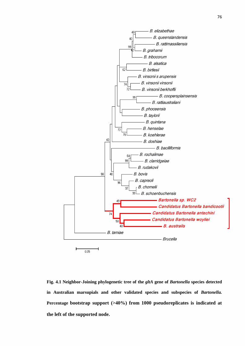
76
Fig. 4.1 Neighbor-Joining phylogenetic tree of the gltA gene of Bartonella species detected
in Australian marsupials and other validated species and subspecies of Bartonella.
Percentage bootstrap support (>40%) from 1000 pseudoreplicates is indicated at
the left of the supported node.
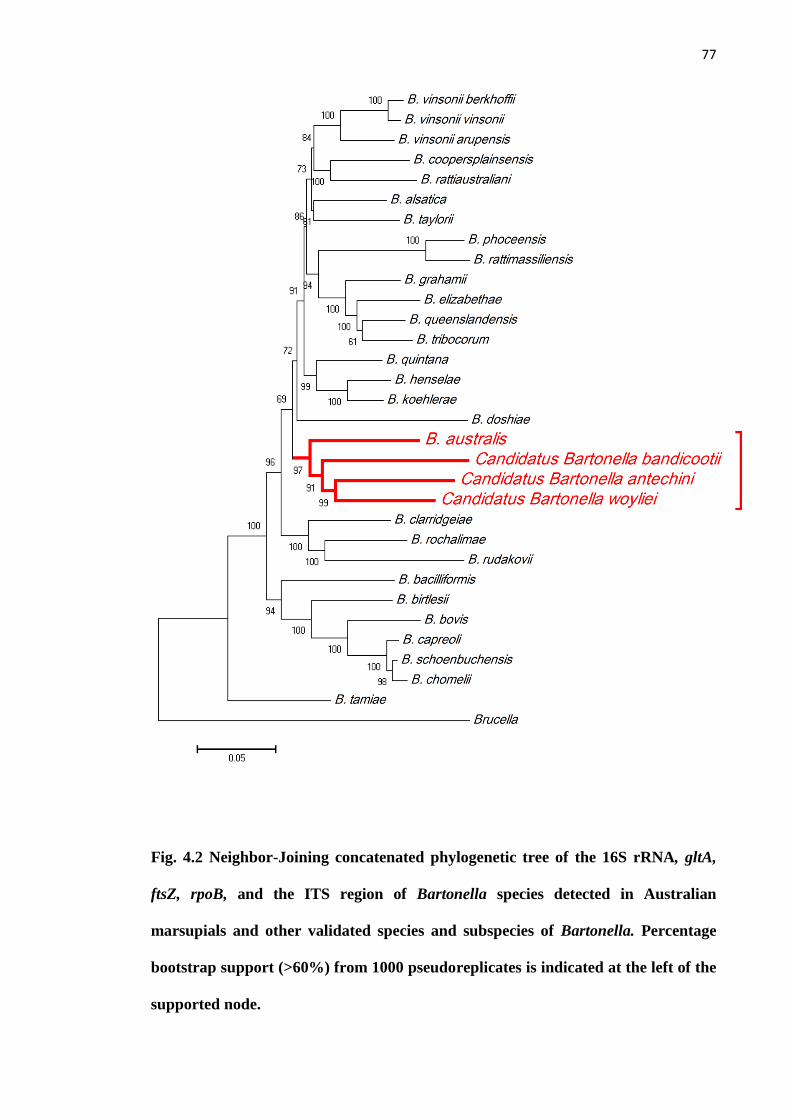
77
Fig. 4.2 Neighbor-Joining concatenated phylogenetic tree of the 16S rRNA, gltA,
ftsZ, rpoB, and the ITS region of Bartonella species detected in Australian
marsupials and other validated species and subspecies of Bartonella. Percentage
bootstrap support (>60%) from 1000 pseudoreplicates is indicated at the left of the
supported node.
78
4. Discussion
The present study identified two novel Bartonella species, which were named
after the marsupial hosts from which the ectoparasites were collected. Candidatus
Bartonella woyliei is named after its woylie host and Candidatus Bartonella bandicootii
is named after its western barred bandicoot host. According to a report of the ad hoc
committee for the re-evaluation of species definition in bacteriology, the Candidatus
concept was suggested for well characterized but as-yet uncultured organisms
(Stackebrandt et al., 2002). For example, this nomenclature was adopted for Candidatus
B. thailandensis n. sp. detected in the blood of rodents in Thailand (Saisongkoh et al.,
2009) and for Candidatus B. mayotimonensis n. sp. detected from heart valve
specimens in a human patient (Lin et al., 2010).
The first Bartonella species detected in a marsupial host was B. australis from
eastern grey kangaroos and the researchers proposed that B. australis was closely
related to Bartonella species in rodents (Fournier et al., 2007). However, with additional
detections in Australia, it has become clear that Bartonella species from marsupial hosts
and their ectoparasites form a separate marsupial cluster (Kaewmongkol et al., 2011a).
Concatenated phylogenetic analysis of 5 loci revealed that Candidatus B. antechini,
detected from ticks and fleas from a small carnivorous marsupial named the mardo
(Antechinus flavipes), was closely related to B. australis. These two species were
detected from marsupials and their ectoparasites and were less closely related to
Bartonella species from rodent ectoparasites (Kaewmongkol et al., 2011a). In this study
the clustering of marsupial-derived Bartonella species confirmed the existence of a
marsupial cluster of Bartonella species in Australia which has evolved separately from
Bartonella species in other mammals.

79
Single step PCR assays of the 16S rRNA, rpoB and ftsZ loci were unable to
amplify Bartonella DNA in tick samples. The inability of single step PCR to detect
Bartonella DNA in ticks in the present study was also reported in ticks from marsupials
in a previous study and is discussed in chapter 3 (Kaewmongkol et al., 2011a).
The present study is the first report of Bartonella species from possible
arthropod vectors from rodents in Australia and has also confirmed that two types of
possible vectors (ticks and fleas) could be infected with the same species of Bartonella.
All species of ectoparasites infected with Bartonella species in the marsupial cluster
have been found only in Australia (Dunnet and Mardon, 1972; Kolonin, 2009). The
marsupial-derived fleas (P. hilli and P. tunneyi) have previously been reported in
woylies, common ringtail possums (Pseudocheirus peregrinus) and southern brown
bandicoots (Isoodon obesulus) (ABRS, 2009). The tick (I. australiensis) has previously
been reported in burrowing bettongs (Bettongia lesueur), woylies, long-nosed potoroos
(Potorous tridactylus), quokkas (Setonix brachyurus) and also in dogs, cattle and
humans in Australia (Kolonin, 2009). The extent of host adaptation in these Bartonella
species is still unknown. However, the compatibility between these Bartonella species
and several arthropod vectors may be one of the keys to explain the host range of these
Bartonella species in animals in Australia. Variations in host adaptation to bacteria in
the genus Bartonella could result in different pathogenicities in different mammal hosts
(Dehio, 2008). Wildlife in Australia could be significant reservoirs for these Bartonella
species, which may contribute to diseases in native marsupials, companion animals and
humans.

80
Chapter 5
Zoonotic Bartonella species in Fleas and Blood from Red Foxes in
Australia
(This chapter forms the basis of the article by Kaewmongkol et al. (2011) Zoonotic
Bartonella species in Fleas and Blood from Red Foxes in Australia. Vector
Borne and Zoonotic Diseases, doi: 10.1089/vbz.2011.0646.)
5.1 Introduction
The zoonotic Bartonella species, Bartonella henselae and B. clarridgeiae, which
are the cause of cat scratch disease and endocarditis in humans, have been reported from
cats, cat fleas and humans in Australia. (Iredell et al., 2003; Barrs et al., 2010).
However, to date, there has been no report of B. henselae or B. clarridgeiae in
Australian wild animals and their ectoparasites.
The European red fox (Vulpes vulpes) occupies a wide variety of habitats across
the continents of Europe, Asia and North America. In the Southern Hemisphere, the red
fox occurs only in Australia (Rolls, 1984), where it was imported during the 19th
century by English colonists for the purpose of hunting (Rolls, 1984). However, this
activity was not sufficient to keep red fox numbers in check and they have since
contributed to major population declines in a number of native Australian fauna species
(Saunders et al, 2007). They are now considered a serious invasive pest species and
programs to eradicate the animals are conducted throughout Australia (Saunders et al,
81
2007). Previous research has discussed the distribution of some of their diseases and
parasites (Glen et al., 2005).
Bartonella species have been reported in wild canids in North America and
Europe, including coyotes (Canis latrans), gray foxes (Urocyon cinereoargenteus) and
red foxes (Vulpes vulpes) (Henn et al., 2009; Gabriel et al., 2009; Marquez et al., 2009)
and the possibility that these canid species may act as Bartonella reservoirs has been
discussed (Henn et al., 2009; Gabriel et al., 2009; Marquez et al., 2009). So far, only
Bartonella rochalimae has been isolated or detected from red foxes (Henn et al., 2009).
Bartonella spp. have also been detected in various flea species collected from the wild
canids (Marquez et al., 2009) and fleas have been proposed to be major vectors of
Bartonella spp. among wild canids (Henn et al., 2009; Gabriel et al., 2009).
In this chapter, the first detection of zoonotic Bartonella species is reported,
including B. henselae and B. clarridgeiae, from both fleas (Ctenocephalides felis) and
blood collected from European red foxes from southwest Western Australia.
5.2 Materials and methods
5.2.1 Sample collection and DNA extraction
Flea and blood samples were collected from red foxes in March 2010, in the
areas surrounding the towns of Katanning (20th
– 21st March) and Boyup Brook (27
th –
28th
March) in southwest Western Australia (Fig. 2.1). In excess of 500 red foxes were
shot by volunteers and farmers on or around these dates as part of the „Red Card for the
Red Fox’ 2010 culling program coordinated by the Department of Agriculture and

82
Food, Western Australia. Carcasses were brought to a central location for recording
within 48 hours (Fig 5.1). At this point flea and blood samples were collected from 164
red foxes. Storage of red fox carcasses together in vehicles during culling implied that
fleas were able to move between carcasses and as such it was not possible to identify
individual fleas originating from specific red fox carcasses. A total of 151 fleas
(Ctenocephalides felis) were randomly collected from 164 red foxes using a flea comb
or tweezers. Fleas were stored in 70% ethanol until processed for DNA extraction.
Blood samples were collected into EDTA tubes from the peritoneal cavity of 14 red
foxes and were stored at -20°C until required. Of these, ten were collected from red
foxes in Katanning and four from Boyup Brook. Species of fleas were identified as
Ctenocephalides felis by light microscopy using the standard key for Australian fleas
(Dunnet and Mardon, 1974).
Thirty four pooled DNA samples were extracted from 151 fleas, with three to
five fleas pooled in each DNA extraction. Twenty-five pooled DNA samples were
extracted from 117 fleas collected from Katanning and nine pooled DNA samples were
extracted from 34 fleas collected from Boyup Brook (Table 5.1). DNA was extracted
from fleas and blood samples using a DNeasy Blood and Tissue Kit (Qiagen, Maryland,
USA) according to the manufacturer‟s instructions.

83
Fig. 5.1 Red fox carcasses shot by volunteers and farmers as part of the ‘Red Card
for the Red Fox’ 2010 culling program coordinated by the Department of
Agriculture and Food, Western Australia.

84
Table 5.1 Number of fleas, pooled flea DNA, blood samples and Bartonella spp. in two locations in Western Australia.
Katanning Boyup Brook
Sample Number of
samples
DNA
samples
PCR
positive
B.
henselae
B.
clarridgeiae
Number of
samples
DNA
samples
PCR
positive
B.
henselae
B.
clarridgeiae
C. felis 117 25 24 4 20 34 9 0 - -
Blood 10 10 1 0 1 4 4 0 - -
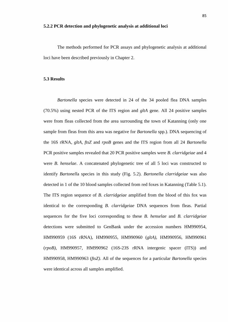
85
5.2.2 PCR detection and phylogenetic analysis at additional loci
The methods performed for PCR assays and phylogenetic analysis at additional
loci have been described previously in Chapter 2.
5.3 Results
Bartonella species were detected in 24 of the 34 pooled flea DNA samples
(70.5%) using nested PCR of the ITS region and gltA gene. All 24 positive samples
were from fleas collected from the area surrounding the town of Katanning (only one
sample from fleas from this area was negative for Bartonella spp.). DNA sequencing of
the 16S rRNA, gltA, ftsZ and rpoB genes and the ITS region from all 24 Bartonella
PCR positive samples revealed that 20 PCR positive samples were B. clarridgeiae and 4
were B. henselae. A concatenated phylogenetic tree of all 5 loci was constructed to
identify Bartonella species in this study (Fig. 5.2). Bartonella clarridgeiae was also
detected in 1 of the 10 blood samples collected from red foxes in Katanning (Table 5.1).
The ITS region sequence of B. clarridgeiae amplified from the blood of this fox was
identical to the corresponding B. clarridgeiae DNA sequences from fleas. Partial
sequences for the five loci corresponding to these B. henselae and B. clarridgeiae
detections were submitted to GenBank under the accession numbers HM990954,
HM990959 (16S rRNA), HM990955, HM990960 (gltA), HM990956, HM990961
(rpoB), HM990957, HM990962 (16S-23S rRNA intergenic spacer (ITS)) and
HM990958, HM990963 (ftsZ). All of the sequences for a particular Bartonella species
were identical across all samples amplified.

86
Phylogenetic analysis of B. henselae was conducted using an 862-bp fragment
of the ITS region. Distance analysis of the ITS region showed the close relationship
between B. henselae detected in fleas from red foxes and B. henselae strain Houston-1
(0.3% genetic distance) (Fig. 5.3). An 893-bp fragment of the ITS region of B.
clarridgeiae from the present study was also compared with other isolates of B.
clarridgeiae using distance analysis and the resultant tree revealed that the B.
clarridgeiae detected in red foxes was genetically distinct from previously published
sequences of B. clarridgeiae (0.1-2.8% genetic distance) (Fig. 5.4).
87
Fig. 5.2 Neighbor-Joining concatenated phylogenetic tree of 16S rRNA, gltA, ftsZ,
rpoB and the ITS region of Bartonella henselae and Bartonella clarridgeiae isolates
from red foxes. Percentage bootstrap support (>45%) from 10000 pseudoreplicates
is indicated at the left of the supported node.

88
Fig. 5.3 Neighbor-Joining phylogenetic tree of the ITS region of Bartonella
henselae isolates. Percentage bootstrap support (>45%) from 10000
pseudoreplicates is indicated at the left of the supported node.
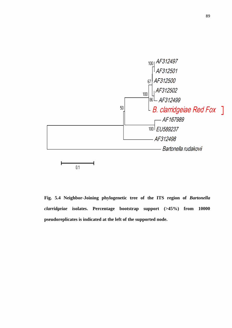
89
Fig. 5.4 Neighbor-Joining phylogenetic tree of the ITS region of Bartonella
clarridgeiae isolates. Percentage bootstrap support (>45%) from 10000
pseudoreplicates is indicated at the left of the supported node.

90
5.4 Discussion
This is the first report of B. henselae and B. clarridgeiae, two zoonotic species
of Bartonella, from red foxes and their fleas in Australia, and this is the first time that B.
clarridgeiae has been identified in a red fox. Until now, only B. rochalimae has been
isolated or detected from red foxes from France (Henn et al., 2009). In the present
study, concatenated phylogenetic analysis of all 5 loci confirmed the species status of B.
clarridgeiae detected from fleas from red foxes, which exhibited 9.5% genetic distance
for B. rochalimae (Fig. 5.2). Single step PCRs for the 16S rRNA, ftsZ, and rpoB loci
were unable to amplify B. clarridgeiae DNA in the blood of a red fox. However, B.
clarridgeiae detected in the blood of a red fox was identical to the corresponding B.
clarridgeiae sequences from flea extracts at the ITS region. Mixed sequences between
B. henselae and B. clarridgeiae were not detected in this study.
Bartonella clarridgeiae was detected in only one blood sample from a red fox.
The true prevalence of Bartonella in the fox host may be higher than this result
suggests, as a pre-enrichment procedure followed by PCR detection has been shown to
greatly improve the sensitivity of detecting Bartonella DNA in dog blood samples
(Duncan et al., 2007). However, the red fox may not be the only natural reservoir for B.
henselae and B. clarridgeiae in this region. Investigation of other wildlife reservoirs
living in the same area, including native marsupials, feral cats and rabbits, as well as pet
dogs, should be performed to further elucidate the ecology of the organisms.
The distance between the towns of Katanning and Boyup Brook in the southwest
of Western Australia is approximately 120 kilometers. However, all positive samples
were detected only in fleas collected from red foxes in the area of Katanning. It is not

91
known why Bartonella species were not detected in flea samples from Boyup Brook
and further work needs to be performed to elucidate the ecology of Bartonella across
the region.
Multi-locus sequence typing has previously been conducted to differentiate B.
henselae strains detected in cats and humans in Australia (Iredell et al., 2003).
Bartonella henselae sequence type 1 (ST 1), also known as strain Houston-1, has been
identified as the principal strain causing human bartonellosis and it is distributed widely
in the cat populations in Australia and North America (Iredell et al., 2003; Arvand et al.,
2007). Distance analysis of the ITS region revealed that the B. henselae strains detected
in the present study are closely related to B. henselae strain Houston-1 (ST 1). The
distribution of this strain in other mammalian hosts and their fleas should be defined,
particularly in domestic cats in southwest Western Australia.
Bartonella clarridgeiae detected in the present study seems to be distinct from
other isolates using distance analysis of a large fragment of the ITS region. Although a
prevalence study of B. clarridgeiae in cats and cat fleas from Eastern Australia has been
reported previously (Barrs et al., 2010), there is little information on the ITS sequences
of B. clarridgeiae in Australia in GenBank.
In conclusion, the identification and characterization of Bartonella species in red
foxes in the SW of Western Australia suggests that foxes may act as reservoirs of
infection for other animals, both wild and domesticated, and humans in this region.
92
Chapter 6
Investigation of Bartonella species in pet animals and their
ectoparasites in Australia
6.1 Introduction
Bartonella henselae and B. clarridgeiae were recently discovered in red foxes in
Western Australia (Kaewmongkol et al., 2011c). Bartonella species have also been
detected in ectoparasites collected from marsupials in Western Australia. Candidatus B.
antechini n. sp., Candidatus B. woyliei n. sp. and Candidatus B. bandicootii were
detected in ticks and fleas from Antechinus flavipes (mardo or yellow-footed
antechinus), Bettongia penicillata (woylies or brush-tailed bettong) and Perameles
bougainville (western barred bandicoots), respectively (Kaewmongkol et al., 2011a;
2011b). In contrast with rodents, marsupials, cats and humans, there has been less
research to describe Bartonella species infection in dogs and their ectoparasites in
Australia. Dogs are reported to be one of most important hosts for Bartonella infection
in other countries and the close contact between humans and their pets makes it
important to study Bartonella in dogs to determine if dogs may serve as a source of
human bartonellosis. The aim of this chapter was to search for evidence of Bartonella
species in pet dogs and their ectoparasites in Australia and to compare genotypes with
other species to determine ecological relationships.
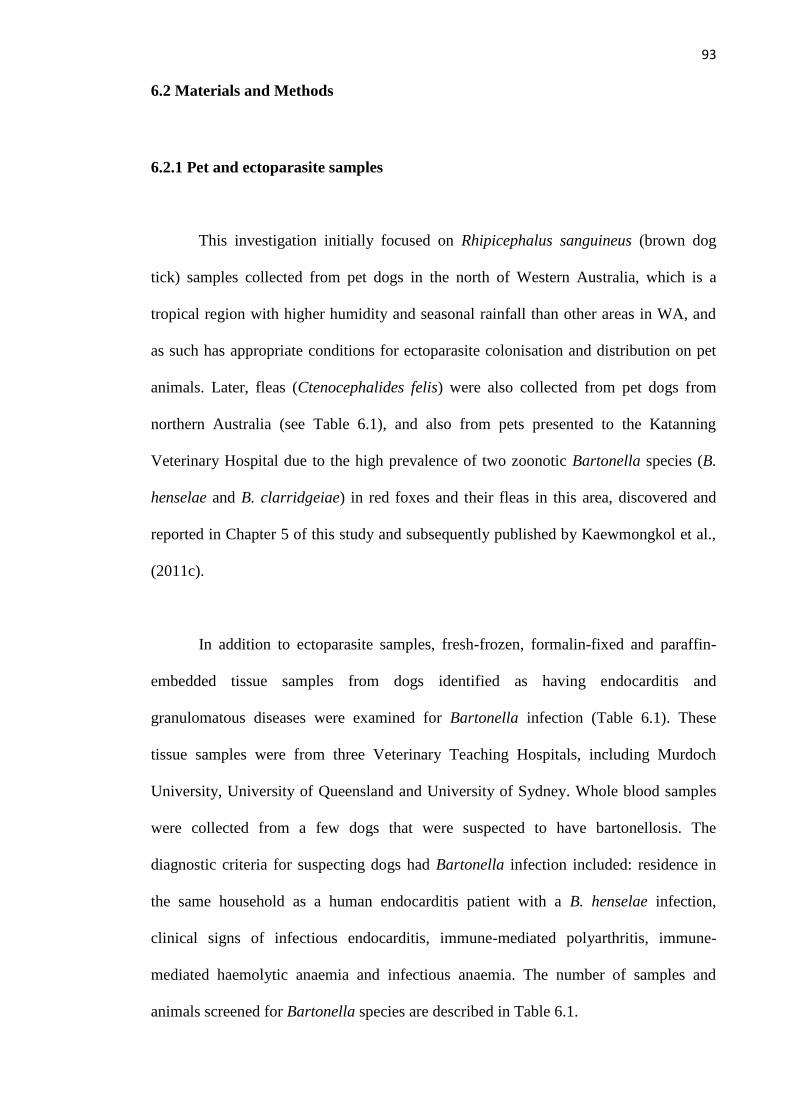
93
6.2 Materials and Methods
6.2.1 Pet and ectoparasite samples
This investigation initially focused on Rhipicephalus sanguineus (brown dog
tick) samples collected from pet dogs in the north of Western Australia, which is a
tropical region with higher humidity and seasonal rainfall than other areas in WA, and
as such has appropriate conditions for ectoparasite colonisation and distribution on pet
animals. Later, fleas (Ctenocephalides felis) were also collected from pet dogs from
northern Australia (see Table 6.1), and also from pets presented to the Katanning
Veterinary Hospital due to the high prevalence of two zoonotic Bartonella species (B.
henselae and B. clarridgeiae) in red foxes and their fleas in this area, discovered and
reported in Chapter 5 of this study and subsequently published by Kaewmongkol et al.,
(2011c).
In addition to ectoparasite samples, fresh-frozen, formalin-fixed and paraffin-
embedded tissue samples from dogs identified as having endocarditis and
granulomatous diseases were examined for Bartonella infection (Table 6.1). These
tissue samples were from three Veterinary Teaching Hospitals, including Murdoch
University, University of Queensland and University of Sydney. Whole blood samples
were collected from a few dogs that were suspected to have bartonellosis. The
diagnostic criteria for suspecting dogs had Bartonella infection included: residence in
the same household as a human endocarditis patient with a B. henselae infection,
clinical signs of infectious endocarditis, immune-mediated polyarthritis, immune-
mediated haemolytic anaemia and infectious anaemia. The number of samples and
animals screened for Bartonella species are described in Table 6.1.
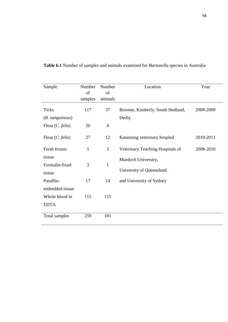
94
Table 6.1 Number of samples and animals examined for Bartonella species in Australia
Sample Number
of
samples
Number
of
animals
Location Year
Ticks
(R. sanguineus)
117 37 Broome, Kimberly, South Hedland,
Derby
2008-2009
Fleas (C. felis) 20 4
Fleas (C. felis) 27 12 Katanning veterinary hospital 2010-2011
Fresh frozen
tissue
5 3 Veterinary Teaching Hospitals of
Murdoch University,
University of Queensland
and University of Sydney
2008-2010
Formalin-fixed
tissue
3 1
Paraffin-
embedded tissue
17 14
Whole blood in
EDTA
115 115
Total samples 259 181

95
6.2.2 DNA extraction
Ticks and fleas were chopped into small sections as previously described in
Chapter 1. Five fleas were pooled in one DNA extract. Formalin-fixed tissues were
washed with PBS solution and paraffin-embedded tissues were lysed with Xylene
followed by the DNA extraction. DNA was extracted using a DNeasy Blood and Tissue
Kit (Qiagen, Maryland, USA). Fresh samples were collected from tissues at the time of
necropsy examination (Fig. 6.1).
6.2.3 PCR detection and phylogenetic analysis at additional loci
The methods for PCR detection and phylogenetic analysis at additional loci have
been described previously in Chapter 2.

96
Fig. 6.1 Aortic valve endocarditis in a dog suspected to have Bartonella infection.

97
6.3 Results
Bartonella DNA was not detected in any of the tissue samples tested. Similarly,
Bartonella DNA was not detected in tick and flea samples collected from the north of
Western Australia and all blood samples from dogs suspected of having bartonellosis
were negative by PCR. Bartonella henselae and B. clarridgeiae DNA were detected
from fleas collected from 3 pet cats in Katanning by nested-PCR of the ITS region.
Further confirmation was performed by amplification and sequencing of a large
fragment of the ITS region in all positive samples (3 of 15 flea pools). DNA sequencing
of the ITS region revealed that one positive sample was identified as B. henselae and
two samples were B. clarridgeiae. Phylogenetic analysis of the B. clarridgeiae-positive
sample from these cat fleas was conducted using an 893-bp fragment of the ITS region
and showed a close relationship (0.1% genetic distance) with the B. clarridgeiae strain
detected in red foxes and their fleas from the same geographical region of Western
Australia (Katanning) which was reported in the previous chapter (Kaewmongkol et al.,
2011c) (Fig. 6.2).
Additionally, an 862-bp fragment of the ITS region of B. henselae detected from
these fleas was also compared with other isolates of B. henselae using distance analysis
and the resultant tree (Fig. 6.2) revealed that the B. henselae detected in pet cats‟ fleas
from Katanning was genetically distinct from a previously published B. henselae
sequence from the same location (Kaewmongkol et al., 2011c). The phylogenetic tree
showed the close relationships between B. henselae in pet cats from Katanning and B.
henselae strains CAL-1 and FIZZ, which were isolated from human Cat Scratch Disease
(CSD) patients from France (GenBank accession number AF369527and AF369526,
respectively) (0.3-0.7% genetic distance) (Fig. 6.3).
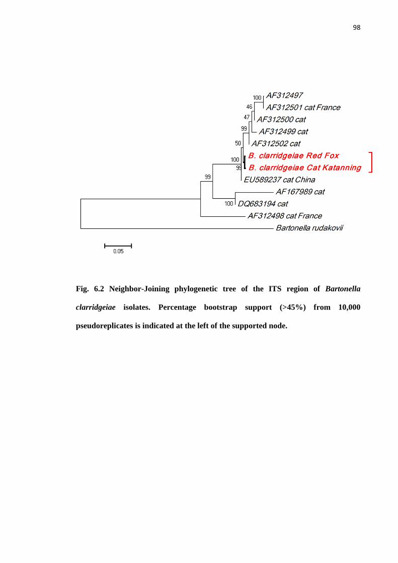
98
Fig. 6.2 Neighbor-Joining phylogenetic tree of the ITS region of Bartonella
clarridgeiae isolates. Percentage bootstrap support (>45%) from 10,000
pseudoreplicates is indicated at the left of the supported node.
99
Fig. 6.3 Neighbor-Joining phylogenetic tree of the ITS region of Bartonella
henselae isolates. Percentage bootstrap support (>40%) from 10,000
pseudoreplicates is indicated at the left of the supported node.

100
6.4 Discussion
The detection of zoonotic Bartonella species, B. henselae and B. clarridgeiae, in
red foxes and their fleas in Katanning, Western Australia (reported in Chapter 5 and
Kaewmongkol et al., 2011c) indicates that red foxes in this area could be an important
reservoir of Bartonella infection for other animals and also for humans in the same
region. Additional information presented in this chapter, the identification of B.
henselae from fleas collected from pet cats in the same area, confirms the presence of
zoonotic Bartonella species in both wild and domestic animals and is consistent with
their transmission via C. felis fleas in this area. Bartonella henselae and B. clarridgeiae
have been previously reported only from cats and C. felis fleas collected from cats in
Australia (Iredell et al., 2003; Barrs et al., 2010). A genetic association between
Bartonella species in wildlife and pet animals was demonstrated in the study reported
here. Therefore, it is possible that zoonotic Bartonella species from pets have been
transmitted to or from wild animals in the same habitats via the flea vector, C. felis.
Pet cats are recognized as an important reservoir of B. henselae and B.
clarridgeiae and have been identified as a major source for Bartonella infection in
humans (Chomel et al., 2006; 2009). Furthermore, the identification and
characterization of zoonotic Bartonella species in cat fleas collected from red foxes and
pet cats in the same environment in Western Australia suggests that a local cycle of
Bartonella species exists between wild and pet animals. This is surprising as close
contact between these two animal species is unlikely and may indicate a peri-urban red
fox ecology in this small rural town. Flea control in pets is still the main strategy to
reduce the risk of Bartonella transmission to wildlife, pet animals and pet owners.

101
Attempts to detect Bartonella DNA in a variety of other clinically indicated
samples or ticks however were not successful. As discussed in Chapter 1, detection of
Bartonella in such samples by other researchers has met with similar difficulty. A
previous study reported a very low prevalence of Bartonella infection in blood collected
from stray dogs (1/278) in Bangkok using nested-PCR of the ITS region (Inoue et al.,
2008). However, another study used a novel technique with combined pre-enrichment
culture followed by PCR for Bartonella detection and reported more than 30%
prevalence for Bartonella infection in blood collected from stray dogs from the same
region (Bai et al., 2010). In the present study, nested-PCR detection alone may lack the
sensitivity required to detect Bartonella species in blood and tissue samples from
infected dogs or from engorged ticks.
In addition, non-specific amplification of other bacteria was identified in the
present study using nested-PCR of the ITS region. These DNA amplicons were
identified as belonging to the genus Mesorhizobium as reported in another study
(Brietchwerdt et al., 2005). Here, Ochrobactrum anthropi was identified in a
granulomatous lung lesion (fresh frozen tissue) and a granulomatous lesion from the
jejunum (paraffin-embedded tissue) using the same set of ITS primers. Cross
contamination by this bacterium via medical instruments and human specimens has
been previously reported (Ezzedine et al., 1994; Labarca et al., 2007). This bacterium is
commonly isolated from soil, plants and water sources (Kettaneh et al., 2003) and is an
opportunistic pathogen in immunocompromised patients (Ozdemir et al., 2006; Song et
al., 2007). The possibility that this bacterium could actually be an unrecognized cause
of disease in animals requires further investigation.
102
The unsuccessful amplification of Bartonella DNA using single step PCR in
Ixodes spp. ticks was demonstrated in Chapters 3 and 4. The limitations, including the
low sensitivity of single step PCRs and PCR inhibitors in ticks have been considered as
possible reasons for the inability of single step PCR to detect Bartonella DNA in tick
samples. Therefore, the failure to amplify Bartonella DNA in R. sanguineus could
explain all negative results for Bartonella investigation in these ticks. In the last 15
years, a diverse range of Bartonella species DNA has been reported in a variety of tick
species from various geographic locations (Agelakis et al., 2010). However, only one
report described the detection of Bartonella DNA in R. sanguineus and it could be that
this tick species is a poor reservoir or vector of Bartonella spp. (Wikswo et al., 2007).

103
Chapter 7
Genetic characterization of flea-derived Bartonella species from native
animals in Australia suggests host-vector-parasite co-evolution
(This chapter forms the basis of the article by Kaewmongkol et al. (2011) Genetic
characterization of flea-derived Bartonella species from native animals in Australia
suggests host-parasite co-evolution. Infection, Genetics and Evolution, doi:
10.1016/j.meegid.2011.07.021 )
7.1 Introduction
Fleas (order Siphonaptera, meaning wingless-tube) are blood-sucking insects
that mostly infect mammals and some avian species. They are hosts for a variety of
pathogens including Yersinia pestis (the causative agent of bubonic plague), Rickettsia
species (e.g. flea-borne spotted fever) and Bartonella species (bartonellosis, including
cat scratch disease), all of which are considered emerging or re-emerging diseases of
humans worldwide (Azad et al., 1997; Bitam et al., 2010).
The co-evolution of Australian fauna and their fleas has been discussed and
elucidated in the context of zoogeography (Traub et al., 1972; Dunnet and Mardon,
1974; Lewis et al., 1993; Whiting et al., 2008) and the taxonomic classification of fleas
associated with Australian marsupials has provided evidence for the origin and
distribution of metatherian hosts prior to the separation of Gondwana (Traub et al.,
1972; Dunnet and Mardon, 1974; Whiting et al., 2008). Morphological identification of

104
fleas is still the major tool for flea taxonomy, which is used for the study of
phylogenetic relationships. However, this approach is limited by the requirement for
skilled entomologists to elucidate flea morphology (Whiting et al., 2008; Bitam et al.,
2010).
A recent study used multilocus sequence analysis of four genes (18S rRNA, 28S
rRNA, cytochrome oxidase II and elongation 1-alpha) to examine phylogenetic
relationships in the order Siphonaptera (Whiting et al., 2008). Since then a re-evaluation
of flea taxonomy using molecular tools has begun to explore the relationship between
fleas as vectors of infectious agents and their mammalian hosts (Bitam et al., 2010).
Distribution of arthropod-borne pathogens is influenced by the host range of arthropod
vectors (Chomel et al., 2009). The presence of co-evolution between marsupial hosts
and their fleas in the same habitats has been reviewed (Traub et al., 1972; Dunnet and
Mardon, 1974; Lewis et al., 1993; Whiting et al., 2008), but to the authors‟ knowledge
co-evolution of hosts, vectors and Bartonella species has not been demonstrated.
Recently, new genotypes and novel species of Bartonella were detected in Australian
marsupials and their fleas (Fournier et al., 2007; Kaewmongkol et al., 2011a; 2011b). It
has been suggested that the compatibility between Bartonella species and fleas may be
a contributing factor to the host specificity of these Bartonella species in Australian
marsupials (Kaewmongkol et al., 2011a; 2011b). We examine this concept further in
the present study by evaluating the genetic relationships among flea species harbouring
a variety of Bartonella species and their marsupial hosts in Australia.

105
7.2 Materials and methods
7.2.1 Flea samples and morphological identification
Ten species of fleas were collected from a variety of locations in Western
Australia (Table 7.1). Species of fleas were characterized by light microscopy using the
standard key for Australian fleas (Dunnet and Mardon, 1974).
7.2.2 DNA extraction, PCR and phylogenetic analysis
DNA was extracted from fleas using a DNeasy Blood and Tissue Kit (Qiagen,
Maryland, USA) according to the manufacturer‟s protocol. DNA samples extracted
from these fleas were screened for Bartonella species using PCR as described in
previous studies (Kaewmongkol et al., 2011a; 2011b; 2011c).
For amplification of flea DNA, PCR primers for the 18S rRNA gene were
designed based on Ctenocephalides canis DNA sequences (GenBank accession no.
AF423914) and consisted of 18S-F (5‟-GATCGTACCCACATTACTTG-3‟) and 18S-R
(5‟-AAAGAGCTCTCAATCTGTCA-3‟). PCR reactions for the 18S rRNA gene were
performed using 1 µL of DNA in a 25 µL reaction containing 1 x PCR buffer, 2 mM
MgCl2, 0.2 mM dNTPs, 1 µM of each primer and 0.02 U/µL TAQ-Ti (hot start) Taq
DNA polymerase (Fisher Biotech Australia, Wembley, W.A., Australia). The cycling
conditions consisted of a pre-PCR step of 96ºC for 2 minutes, followed by 45 cycles of
94ºC for 50 seconds, 55ºC for 60 seconds and an extension of 72ºC for 90 seconds, with
a final extension of 72ºC for 10 minutes. PCR primers for the cytochrome c oxidase
subunit I (the COI gene), LCO1490: (5'-GGTCAACAAATCATAAAGATATTGG-3')
106
and HC02198: (5'-TAAACTTCAGGGTGACCAAAAAATCA-3') were used as per a
previous study (Folmer et al., 1994). The PCR reactions for the COI gene were
performed using 1 µL of DNA in a 25 µL reaction containing 1 x PCR buffer, 2 mM
MgCl2, 0.2 mM dNTPs, 1 µM of each primer and 0.02 U/µL TAQ-Ti (hot start) Taq
DNA polymerase (Fisher Biotech Australia, Wembley, W.A., Australia). The cycling
conditions consisted of a pre-PCR step of 96 ºC for 2 minutes, followed by 40 cycles of
94ºC for 30 seconds, 50ºC for 30 seconds and an extension of 72ºC for 60 seconds, with
a final extension of 72ºC for 7 minutes. PCR products from all genes were purified from
agarose gel slices using an UltraCleanTM
15 DNA Purification Kit (MO BIO
Laboratories Inc. West Carlsbad, California, USA). Sequencing was performed using an
ABI PrismTM
Terminator Cycle Sequencing kit (Applied Biosystems, Foster City,
California, USA) on an Applied Biosystems 3730 DNA Analyzer, following the
manufacturer‟s instructions. Nucleotide sequences generated for 2 loci were analysed
using Chromas lite version 2.0 (http://www.technelysium.com.au) and aligned with
reference sequences from GenBank using Clustal W (http://www.clustalw.genome.jp).
Phylogenetic analysis was conducted on concatenated 18S rRNA and COI sequences.
Distance and maximum-parsimony were conducted using MEGA version 4.1
(MEGA4.1: Molecular Evolutionary Genetics Analysis software, Arizona State
University, Tempe, Arizona, USA), based on evolutionary distances calculated with the
Kimura‟s distance and grouped using Neighbour-Joining. Bootstrap analyses were
conducted using 10,000 replicates to assess the reliability of inferred tree topologies.
DNA sequences generated in the present study were submitted to GenBank (see Table
7.2 for accession numbers).
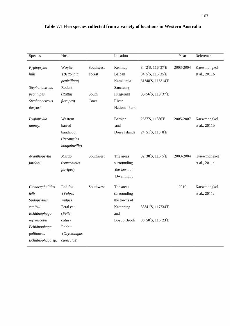
107
Table 7.1 Flea species collected from a variety of locations in Western Australia
Species Host Location Year Reference
Pygiopsylla
hilli
Stephanocircus
pectinipes
Stephanocircus
dasyuri
Woylie
(Bettongia
penicillata)
Rodent
(Rattus
fuscipes)
Southwest
Forest
South
Coast
Keninup
Balban
Karakamia
Sanctuary
Fitzgerald
River
National Park
34°2/S, 116°37
/E
34°5/S, 116°35
/E
31°48/S, 116°14
/E
33°56/S, 119°37
/E
2003-2004 Kaewmongkol
et al., 2011b
Pygiopsylla
tunneyi
Western
barred
bandicoot
(Perameles
bougainville)
Bernier
and
Dorre Islands
25°7/S, 113°6
/E
24°51/S, 113°8
/E
2005-2007 Kaewmongkol
et al., 2011b
Acanthopsylla
jordani
Mardo
(Antechinus
flavipes)
Southwest The areas
surrounding
the town of
Dwellingup
32°38/S, 116°5
/E 2003-2004 Kaewmongkol
et al., 2011a
Ctenocephalides
felis
Spilopsyllus
cuniculi
Echidnophaga
myrmecobii
Echidnophaga
gallinacea
Echidnophaga sp.
Red fox
(Vulpes
vulpes)
Feral cat
(Felis
catus)
Rabbit
(Oryctolagus
cuniculus)
Southwest The areas
surrounding
the towns of
Katanning
and
Boyup Brook
33°41/S, 117°34
/E
33°50/S, 116°23
/E
2010 Kaewmongkol
et al., 2011c

108
7.3 Results
The phylogenetic relationships among the 10 species of fleas, based on
concatenated sequences from 2 loci are shown in Fig. 7.1. Genetic clustering of these
fleas correlated with the Family classification based on morphological identification
(Dunnet and Mardon, 1974). The 10 species of fleas were categorized into 3 Families:
Pulicidae (Ctenocephalides felis, Spilopsyllus cuniculi, Echidnophaga myrmecobii,
Echidnophaga gallinacea, and an unidentified Echidnophaga spp.), Stephanocircidae
(Stephanocircus pectinipes and Stephanocircus dasyuri) and Pygiopsyllidae
(Pygiopsylla hilli, Pygiopsylla tunneyi and Acanthopsylla jordani).
The corresponding mammalian hosts from which these fleas were collected were
included in the concatenated tree (Fig. 7.1). These results demonstrate the relationship
between fleas in the Family Pulicidae and a wide host range of introduced (i.e. non-
native) mammalian species; red foxes, feral cats and rabbits (Fig. 7.1). In contrast,
association between mammalian hosts and fleas in the Families Stephanocircidae and
Pygiopsyllidae, are restricted to Australian rodents (bush rats) and marsupials
respectively (Fig. 7.1).
The Bartonella species detected in these fleas were also included in the
concatenated tree (Fig. 7.1) which indicates that two zoonotic Bartonella species (B.
henselae and B. clarridgeiae) are distributed among flea species belonging to the
Family Pulicidae, known to share a wide variety of mammalian hosts. However,
restriction of host range was evident in Bartonella species detected in marsupials, as
both the fleas and the Bartonella species were genetically distinct (Fig. 7.1 and 7.2)
from those infecting non-Gondwana origin hosts. This study also demonstrates the

109
relationship between Bartonella species (B. rattaustraliani and B. coopersplainsensis)
and fleas in the Family Stephanocircidae from a native Australian rodent, Rattus
fuscipes (Fig. 7.1). The genetic position of B. rattaustraliani and B. coopersplainsensis
is also separated from other Bartonella species from rodents in other geographical areas
(Fig. 7.2).

110
Table 7.2 GenBank accession numbers of flea species from animals in Australia
Family Species Host Bartonella species GenBank accession numbers
18S rRNA Cytochrome
oxidase I
Pygiopsyllidae Pygiopsylla hilli
Woylie
(Bettongia
penicillata)
Candidatus
B. woyliei
JN008926 JN008915
Pygiopsylla tunneyi
Western barred
bandicoot
(Perameles
bougainville)
Candidatus
B. bandicootii
JN008933 JN008924
Acanthopsylla
jordani
Mardo
(Antechinus
flavipes)
Candidatus
B. antechini
JN008925 JN008916
Stephanocircidae Stephanocircus
pectinipes
Rodent
(Rattus fuscipes)
B. coopersplainsensis
B. rattaustraliani
JN008932 JN008923
Stephanocircus
dasyuri
JN008930 JN008920
Pulicidae Ctenocephalides
felis
Red fox
(Vulpes vulpes)
Feral cat
(Felis catus)
Rabbit
(Oryctolagus
cuniculus)
B. henselae
B. clarridgeiae
JN008927 JN008917
Spilopsyllus
cuniculi
JN008928 JN008918
Echidnophaga
myrmecobii
JN008929 JN008919
Echidnophaga
gallinacea
JN008931 JN008921
Echidnophaga sp. JN008934 JN008922

111
Fig. 7.1 Neighbor-Joining concatenated phylogenetic tree of the 18S rRNA, and COI genes of flea species and their associated Bartonella
species from marsupials and other mammals in Australia. Percentage bootstrap support (>40%) from 10,000 pseudoreplicates is indicated at
the left of the supported node. The tree is rooted using Calliphora vomitoria (fly) as the outgroup.

112
Fig. 7.2 Neighbor-Joining concatenated phylogenetic tree of 16S rRNA, gltA, ftsZ, rpoB,
and the ITS region of Bartonella species showing the separate clustering of Bartonella
spp. from Australian marsupials and B. rattaustraliani and B. coopersplainsensis from
native Australian rodents. Percentage bootstrap support (>60%) from 1000
pseudoreplicates is indicated at the left of the supported node (Kaewmongkol et al.,
2011b).

113
7.4 Discussion
Co-evolution between Bartonella species and flea vectors has been proposed
previously for flea species infesting rodents and felids (Chomel et al., 1996; 2009; Bown et
al., 2004) and the demonstration of host specificity between B. washoensis and squirrels has
also been reported (Inoue et al., 2010). Moreover, a geographical relationship was noted for
B. grahamii and various rodent hosts in Japan, China (represent Asian group), Canada, UK,
Russia and USA (represent American/European group) (Inoue et al., 2009). In the present
study, the close association between Australian marsupials, their fleas and Bartonella species
suggests adaptation by Bartonella species to a specific ecological niche, which is comprised
of specific mammalian hosts and specific flea vectors. The Bartonella species in the
marsupial cluster, reported in previous studies (Kaewmongkol et al., 2011a; 2011b), appear to
have evolved separately in marsupials and their fleas within Australia.
The introduction of mammalian pest species has been postulated to interfere with
native host-bacteria interactions (Telfer et al., 2005; 2007; Chomel et al., 2009) and the
impact of the red fox on Australia‟s native animals has been discussed in the context of the
spread of diseases and parasites (Glen et al., 2005). Our finding of B. henselae and B.
clarridgeiae in flea species of the Family Pulicidae only may reflect the limited number of
samples examined, but is consistent with these bacteria being co-introduced with pest species
in post-colonial Australia. Alternatively, these two zoonotic Bartonella species could be
distributed across a wide variety of native species via a broad range of Pulicidae fleas.
Therefore, genetic segregation of Bartonella species in marsupials could provide an
opportunity to study Bartonella genetics in the context of host and vector specificity.
114
Three Bartonella species, B. queenslandensis, B. rattaustraliani and B.
coopersplainsensis, were isolated from native rodents including Rattus fuscipes, Rattus
leucopus, Rattus sordidus (Rattus conatus), Rattus tunneyi, Uromys caudimaculatus and
Unidentified Melomys spp. in Queensland, eastern Australia (Gundi et al., 2009). The
evolutionary lineage of the genus Uromys and Melomys in Australia is still largely unknown.
However, the evolutionary relationships within the genus Rattus have been defined (Robins
et al., 2007; 2008; 2010). All of these Rattus species were grouped into the Australo-Papuan
clade, which is one of two major groups of the genus Rattus. Another major group is the
Asian clade, which includes Rattus rattus, Rattus norvegicus and Rattus exulans (Robins et
al., 2007; 2008; 2010). Rattus fuscipes, a rodent host for Stephanocircidae fleas in this study,
is the oldest lineage of the Australo-Papuan clade in Australia, and the divergence of the
genus Rattus into two major groups (Asian and Australo-Papuan clades) occurred
approximately 2.7 million years ago (Robins et al., 2010). The concatenated phylogenetic
multigene analysis revealed that B. queenslandensis was closely related to Bartonella species
from other rodent isolates (Gundi et al., 2009) (Fig 7.2). In contrast to B. queenslandensis, B.
rattaustraliani and B. coopersplainsensis were less closely related to other Bartonella species
isolated from rodents (Gundi et al., 2009) (Fig. 7.2). In the present study, genetic clustering
of fleas in the Family Stephanocircidae correlated with the morphological classifications
from previous studies (Traub et al., 1972; Dunnet and Mardon, 1974; Lewis et al., 1993) and
also correlated well with genetic classification of fleas in the study by Whiting et al., (2008).
Fleas in the Family Stephanocircidae are well discriminated from other flea families using
both morphological and genetic classifications (Traub et al., 1972; Dunnet and Mardon,
1974; Lewis et al., 1993; Whiting et al., 2008). In addition, these fleas are unique and most
likely originated in Australia prior to the separation of the continents (Whiting et al., 2008).
In the present study, the compatibility between B. rattaustraliani and B. coopersplainsensis

115
and Australian fleas from the Family Stephanocircidae was evident (Fig. 7.1) and this
relationship could explain the genetic positioning of B. rattaustraliani and B.
coopersplainsensis, which are less related to other Bartonella species isolated from rodents
outside Australia. It can be hypothesized that fleas in the Family Stephanocircidae may play
an important role in Australia in the evolution of Bartonella species in Australian rodents.
Therefore, B. queenslandensis may have evolved with non-native rodents, which were
introduced by European colonists in the 19th
century.
The study of Australian flea morphology and phylogeny has provided evidence for the
origin and distribution of marsupial ancestors (Traub et al., 1972; Dunnet and Mardon, 1974;
Whiting et al., 2008). Investigation of the fossil record reveals that ancestral marsupials
dispersed from Australia (east Gondwana) to South America (west Gondwana) approximately
165-180 million years ago (Beck et al., 2008). Dispersal directions of marsupial ancestors
have also been associated with the dispersal of fleas in the Family Pygiopsyllidae into New
Guinea and the Family Stephanocircidae into South America (Dunnet and Mardon, 1974;
Whiting et al., 2008). The investigation of Bartonella species in these two Families of fleas in
other geographical areas would be useful for the study of the origin of metatherians and their
radiation across the continents. Furthermore, deeper study of the Bartonella genome may also
provide insights into the evolution of these enigmatic bacteria and their mammalian and
vector hosts.

116
Chapter 8
General Discussion
8.1 Introduction
The development of advanced molecular tools is one of the most significant factors in
improving the accurate delineation of Bartonella species (Boulouis et al., 2005). In the last 15
years, a number of Bartonella species have been reported based on PCR detection and
characterization of multiple genes (Chomel et al., 2004; Chomel et al 2006; Boulouis et al.,
2005). Bartonella species have been detected in several human cases (Cheung et al., 2010;
Kossiva et al., 2010; Gan et al., 2011), dogs (Mellor et al., 2006; Saunders et al., 2006;
Cockwill et al., 2007) and also in unusual arthropods using PCR testing (Dehio et al., 2004;
Halos et al., 2004; Valerio et al., 2005; Reeves et al., 2006). The limitations of conventional
techniques for Bartonella identification, discussed in previous chapters, include low
sensitivity using conventional culture methods (Fournier et al., 2002) and the lack of
commercial tests for species-specific serological testing (Boulouis et al., 2005). Provided that
the test is conducted properly and with appropriate controls, PCR is more sensitive and more
specific than conventional methods and can be applied to various types of samples. This
study developed original, practical PCR techniques for detection and genetic characterization
of Bartonella species in animals and their ectoparasites in Australia.

117
The study of Bartonella adaptation has been increasingly focused on arthropod
vectors, mammalian hosts and environments in which the hosts, vectors and bacteria have co-
evolved (Chomel et al., 2009). Bartonella adaptation is required for the evasion of the host
immune responses and to maintain the life cycle between mammalian hosts and arthropod
vectors (Chomel et al., 2009). Previous studies have identified Bartonella arthropod vector
specificity among Bartonella species and fleas infesting rodents and felids (Chomel et al.,
1996; 2009; Bown et al., 2004) and host specificity of Bartonella species was demonstrated
in B. washoensis and squirrels (Inoue et al., 2010). Additionally, a geographical relationship
was noted for B. grahamii and various rodent hosts in Japan, China (representing the Asian
group), Canada, UK, Russia and USA (representing the American/European group) (Inoue et
al., 2009). In the present study, a diverse range of Bartonella species was discovered in
ectoparasites from various mammalian hosts and the relationships among these Bartonella
species, mammalian hosts and ectoparasites were elucidated in the unique Australian
ecosystem.
8.2 Genotypic status of Bartonella species in animals and their ectoparasites in Australia
The first Bartonella species detected in a marsupial host was B. australis from eastern
grey kangaroos (Fournier et al., 2007) and it was proposed that B. australis was closely
related to Bartonella species in Australian placental rodents. However, evidence from this
thesis indicates that Bartonella species from marsupial hosts and their vectors form a separate
marsupial cluster. In Chapter 3, we reported the identification of a novel Bartonella species,
Candidatus B. antechini detected from fleas (Acanthopsylla jordani) and ticks (Ixodes
antechini) collected from a small carnivorous marsupial, Antechinus flavipes (Mardos or
Yellow-footed antechinus) in the southwest of Western Australia. DNA sequencing of the

118
16S rRNA, gltA, ftsZ and rpoB genes and the ribosomal ITS region revealed it to be a distinct
Bartonella species, closely related to Bartonella australis from kangaroos. These two species
detected from marsupials and their ectoparasites were not closely related to Bartonella
species from rodents.
In Chapter 4, Bartonella species were described in various ectoparasites from other
mammals. A variety of Bartonella species were detected in two species of ticks and three
species of fleas collected from marsupial hosts; woylies (Bettongia penicillata) and western
barred bandicoots (Perameles bougainville) and from a rodent host; Rattus fuscipes in
Western Australia. Bartonella species were detected using nested-PCR of the gltA gene and
the ITS, and species were characterized using DNA sequencing of the 16S rRNA, gltA, rpoB,
ftsZ genes and the ITS region. Bartonella rattaustraliani and B. coopersplainsensis had been
previously detected in Ixodes spp. ticks and fleas (Stephanocircus pectinipes) collected from
rodents. In this thesis, two novel Bartonella species were discovered from marsupials;
Candidatus Bartonella woyliei n. sp. was detected in both fleas (Pygiopsylla hilli) and ticks
(Ixodes australiensis) collected from woylies and Candidatus Bartonella bandicootii n. sp.
was detected in fleas (Pygiopsylla tunneyi) collected from western barred bandicoots on a
remote offshore island. Concatenated phylogenetic analysis of all 5 loci clarified the
marsupial cluster of Bartonella species in Australia and confirmed the species status of these
two Bartonella species in ticks and fleas from woylies and western barred bandicoots, both of
which are classified as threatened marsupial species and are vulnerable to extinction.
In Chapter 5, investigation of Bartonella species hosts in Western Australia was
expanded to include the European red fox, an introduced pest animal species, and its fleas (C.
felis). Zoonotic Bartonella species; Bartonella henselae and B. clarridgeiae were detected in

119
fleas from red foxes and B. clarridgeiae was detected in the blood of one red fox.
Phylogenetic analysis of the ITS region revealed that the B. henselae isolates detected in the
present study were related to B. henselae strain Houston-1, the major pathogenic strain in
humans in Australia and worldwide, and confirmed the genetic distinctness of B. clarridgeiae
from red foxes. Furthermore, this is the first time that B. clarridgeiae has been identified in a
red fox. Until now, B. rochalimae has been the only Bartonella spp. isolated or detected from
red foxes, from France (Henn et al., 2009). The identification and characterization of
Bartonella species in red foxes in the Southwest of Western Australia suggests that red foxes
may act as reservoirs of infection for other animals and humans in this region.
The association between zoonotic Bartonella species in wildlife and pet animals was
described in Chapter 6. Distance analysis of the ITS region showed the close relationship
between a B. clarridgeiae strain detected in fleas from pet cats and B. clarridgeiae detected
in red foxes and their fleas from the same location (reported in chapter 5). Phylogenetic
analysis using a large fragment of the ITS region revealed the close relationship between B.
henselae in pet cats from Katanning and B. henselae strains CAL-1 and FIZZ which were
isolated from human CSD patients from France (GenBank accession number AF369527and
AF369526, respectively). These findings in fleas collected from pet cats confirmed the
distribution of zoonotic Bartonella species in both wild and domestic animals via C. felis
fleas in this area and suggested that a local cycle exists between wild and pet animals. Cat
fleas may be a major vector capable of transmitting these Bartonella species between wild
and domestic animals and also humans in this region. It is curious that Bartonella spp. were
not identified in dogs, and further investigation of dogs in this area is warranted.
120
8.3 Co-evolution of Bartonella species, Australian fleas and Australian fauna
Fleas are important arthropod vectors for a variety of diseases in veterinary and
human medicine, and bacteria belonging to the genus Bartonella are among the organisms
most commonly transmitted by these ectoparasites. Recently, a number of Bartonella species
have been detected in fleas in Australia, as described in previous chapters. In chapter 7, the
genetic diversity of marsupial fleas was investigated; ten species of flea were collected from
7 different mammalian hosts in Western Australia, including woylies, western barred
bandicoots, mardos, bush rats, red foxes, feral cats, and wild rabbits. PCR and sequence
analysis of the cytochrome oxidase subunit I (COI) and the 18S rRNA genes from these fleas
was performed. Concatenated phylogenetic analysis of the COI and 18S rRNA genes
revealed a close genetic relationship between marsupial fleas, with P. hilli from woylies, P.
tunneyi from western barred bandicoots and A. jordani from mardos, forming a separate
cluster from other mammalian fleas collected in the same geographical area. The clustering of
Bartonella species with their marsupial flea hosts suggests co-evolution of marsupial hosts,
marsupial fleas and Bartonella species in Australia. The present study demonstrated a clear
compatibility between B. rattaustraliani and B. coopersplainsensis, Australian fleas from the
Family Stephanocircidae and Australian rodents, Rattus fuscipes. This relationship explained
the genetic positioning of B. rattaustraliani and B. coopersplainsensis, which are less related
to other Bartonella species isolated from rodents outside Australia.
In conclusion, the close association between Australian fauna, Australian fleas and
Bartonella species suggests adaptation by Bartonella species to a specific ecological niche,
which is comprised of specific mammalian hosts and specific flea vectors in specific

121
environments (Fig. 8.1). The Bartonella species in the marsupial cluster discovered in this
thesis appear to have evolved separately in marsupials and their fleas within Australia.

122
Hosts
Parasites
Vectors
Parasite community in hosts
Immune System
Parasite community in vectors
Adaptations
Adaptations
Immune System
Environment
•Climate change
•Pesticides
Native host-vector-parasite interactions
Non-native interferences; introduced hosts, vectors, parasites
Fig. 8.1 Native host-vector-parasite interactions and non-native interferences

123
8.4 Future direction of Bartonella research in Australia
In the present study, new external primers for nested-PCRs targeting the gltA gene
and the ITS region were developed from single step PCRs used in previous studies (Norman
et al., 1995, Jensen et al., 2000) to increase the sensitivity and specificity of the PCR. The
primary PCR conditions and PCR cycling conditions for both loci were optimized for use
with high concentrations of MgCl2 (2.5 mM) and low annealing temperatures (48-50°C) to
increase the chances of amplification of novel species of Bartonella. However, the
disadvantage of this approach, particularly at the ITS region, was to reduce the specificity of
detection, resulting in non-specific amplification of other organisms at the ITS region
including Mesorhizobium and Ochrobactrum species (O. anthropi and O. intermedium),
which are also members of the Alphaproteobacteria group. All positive results using these
nested-PCRs however, were confirmed by sequencing, and therefore all non-specific (i.e.
non-Bartonella) amplifications were correctly identified.
To increase the efficiency of Bartonella detection, a novel technique which combines
culture and PCR has been developed in recent years (Maggi et al., 2005; Duncan et al., 2007),
which comprises two steps including pre-enrichment of Bartonella species in liquid media
(BAPGM; Bartonella Alpha Proteobacteria Growth Medium) for 7 days, followed by PCR
detection. This combined technique may be the solution to improve both sensitivity and
specificity of PCR detection and could increase the possibility of identifying further novel
species of Bartonella. Development of this technique locally is required for further studies of
Bartonella species in Australia.

124
Degenerate primers used for amplification of additional loci (the rpoB and ftsZ genes)
in the present study were modified from primers reported in previous studies (Renesto et al.,
2001; Zeaiter et al., 2002). Because variation at the binding sites of primers to these loci were
observed in many species of Bartonella in GenBank, these degenerate primers were created
to ensure more consistent amplification of a range of Bartonella species.
A nested-PCR for detection of a large fragment of the ITS region was successfully
developed in the present study and was designed for multiple applications in Bartonella
studies. The first application was as a detection tool, because nested-PCR is more sensitive
and specific than single step PCRs developed in previous studies (Jensen et al., 2000). The
ITS region of Bartonella species contains highly variable sequences in both inter- and intra-
species strains. The nested-PCR of the ITS region developed in the present study is suitable
for both inter- and intra-species detection and characterization of Bartonella species. The
large fragment of the ITS region amplified by this nested-PCR covered a 335-bp fragment of
the ITS region (S1 fragment) used for the Multi Spacer Typing (MST) technique for strain
identification of B. henselae that was reported in a previous study (Li et al., 2006).
Additional sixth and seventh loci may be needed to confirm the species status of
Bartonella in the future and to identify strains within the same species. Other loci, such as the
groEL and ribC genes, were reported as the most appropriate loci for genetic characterization
of Bartonella species (La Scola et al., 2003). Multi locus sequence analysis of 7 loci,
including the 16S rRNA, gltA, groEL, rpoB, ribC, ftsZ genes and the ITS region, was recently
performed for strain clustering (intra-species identification) of B. washoensis isolates (Inoue
et al., 2010).

125
Projects to determine the complete genome sequences of 4 Bartonella species,
including B. henselae, B. quintana, B. bacilliformis and B. tribocorum, were launched in
2004-2007 (Alsmark et al., 2004; Saenz et al., 2007). Since then, post-genomic approaches
have begun to explore the genetic information of bacteria in the genus Bartonella. The
Bartonella genomes consist of circular chromosomes with the size ranging from 1.44 Mb
(encoding 1,283 genes) to 2.62 Mb (encoding 2,136 genes) (Alsmark et al., 2004; Saenz et
al., 2007). Comparative genome analysis among these 4 complete genome sequences
revealed the set of orthologous genes shared by B. henselae, B. quintana, B. bacilliformis and
B. tribocorum, and this set of conserved genes among 4 species of Bartonella was referred to
as the core genome (conserved 959 coding genes of a total 1,283-2,136 coding genes) (Saenz
et al., 2007; Chomel et al., 2009). The host-integrated metabolisms of Bartonella species are
mostly programmed by genes encoded in the core genome and these metabolisms are
required to support the intra-erythrocytic infection life cycle of the genus (Saenz et al., 2007).
The coding genes excluded from the core genome were organized into genomic islands and
were also called the accessory genes. These accessory genes may play an important role in
Bartonella-host cell interactions and may carry the functions for the Bartonella adaptations to
their hosts (mammalian and arthropod hosts) (Saenz et al., 2007). Therefore, the genome
sequencing of unique genotypes of Bartonella species in Australian fauna that were
demonstrated in this study could provide an opportunity to study Bartonella genetics in more
depth in the context of host and vector specificity.

126
In summary, the principle findings presented in this thesis are as follows:
Multilocus sequence analysis of the 16S rRNA, gltA, ftsZ and rpoB genes and the
ribosomal ITS region was applied to identify and confirm the status of all Bartonella
species identified in this study.
Three novel Bartonella species were detected and identified in flea and tick vectors
from marsupials in Western Australia.
Candidatus Bartonella antechini was detected in ticks and fleas from mardos
(Antechinus flavipes).
Candidatus Bartonella woyliei was detected in fleas and ticks from brush-tailed
bettongs or woylies (Bettongia penicillata).
Candidatus Bartonella bandicootii was detected in fleas from western barred
bandicoots (Perameles bougainville).
These novel Bartonella species were further analysed to confirm the existence of a
marsupial cluster of Bartonella species in Australia.
Zoonotic Bartonella species, B. henselae and B. clarridgeiae, were detected in red
foxes and their fleas (Ctenocephalides felis). This detection indicated that red foxes
could be an important reservoir of Bartonella infections for other animals and humans
in this geographical locality.
The genetic association of these zoonotic Bartonella species detected in red foxes and
pet animals has demonstrated and confirmed the distribution of zoonotic Bartonella
species in fleas from both wild and domestic animals in this region and the existence
of a wild-domestic animal cycle of fleas and Bartonella spp.
Sequence analysis of the COI and 18S rRNA genes of flea vectors harbouring a
diversity of Bartonella species were performed in this study.

127
The genetic clustering of Bartonella species and flea vectors with their Australian
fauna hosts suggests co-evolution of hosts, fleas and Bartonella species in Australia.

128
References
Al-Majali, A. M., and K. M. Al-Qudah. 2004. Seroprevalence of Bartonella henselae and
Bartonella quintana infections in children from Central and Northern Jordan. Saudi
medical journal 25: 1664-1669.
Alsmark, C. M., A. C. Frank, E. O. Karlberg, B. A. Legault, D. H. Ardell, B. Canback, A. S.
Eriksson, A. K. Naslund, S. A. Handley, M. Huvet, B. La Scola, M. Holmberg, and S.
G. Andersson. 2004. The louse-borne human pathogen Bartonella quintana is a
genomic derivative of the zoonotic agent Bartonella henselae. Proceedings of the
National Academy of Sciences of the United States of America 101: 9716-9721.
Al-Soud, W.A., Jonsson, L.J., Radstrom, P. 2000. Identification and characterization of
immunoglobulin G in blood as a major inhibitor of diagnostic PCR. Journal of clinical
microbiology 38: 345-350.
Amerein, M.P., De Briel, D., Jaulhac, B., Meyer, P., Monteil, H., Piemont, Y. 1996.
Diagnostic value of the indirect immunofluorescence assay in cat scratch disease with
Bartonella henselae and Afipia felis antigens. Clinical and diagnostic laboratory
immunology 3: 200-204.
Angelakis, E., Billeter, S.A., Breitschwerdt, E.B., Chomel, B.B., Raoult, D. 2010. Potential
for tick-borne bartonelloses. Emerging infectious diseases 16: 385-391.
Arvand, M., E. J. Feil, M. Giladi, H. J. Boulouis, and J. Viezens. 2007. Multi-locus sequence
typing of Bartonella henselae isolates from three continents reveals hypervirulent and
feline-associated clones. PloS one 2: e1346.
Austen, J. M., R. Jefferies, J. A. Friend, U. Ryan, P. Adams, and S. A. Reid. 2009.
Morphological and molecular characterization of Trypanosoma copemani n. sp.
(Trypanosomatidae) isolated from Gilbert's potoroo (Potorous gilbertii) and quokka
(Setonix brachyurus). Parasitology 136: 783-792.
129
Avidor, B., M. Graidy, G. Efrat, C. Leibowitz, G. Shapira, A. Schattner, O. Zimhony, and M.
Giladi. 2004. Bartonella koehlerae, a new cat-associated agent of culture-negative
human endocarditis. Journal of clinical microbiology 42: 3462-3468.
Azad, A. F., S. Radulovic, J. A. Higgins, B. H. Noden, and J. M. Troyer. 1997. Flea-borne
rickettsioses: ecologic considerations. Emerging infectious diseases 3: 319-327.
Bacher, A., Fischer, M., Kis, K., Kugelbrey, K., Mortl, S., Scheuring, J., Weinkauf, S.,
Eberhardt, S., Schmidt-Base, K., Huber, R., Ritsert, K., Cushman, M., Ladenstein, R.
1996. Biosynthesis of riboflavin: structure and mechanism of lumazine synthase.
Biochemical Society transactions 24: 89-94.
Bai, Y., M. Kosoy, A. Martin, C. Ray, K. Sheff, L. Chalcraft, and S. K. Collinge. 2008.
Characterization of Bartonella strains isolated from black-tailed prairie dogs
(Cynomys ludovicianus). Vector borne and zoonotic diseases (Larchmont, N.Y) 8: 1-
5.
Bai, Y., M. Y. Kosoy, S. Boonmar, P. Sawatwong, S. Sangmaneedet, and L. F. Peruski. 2010.
Enrichment culture and molecular identification of diverse Bartonella species in stray
dogs. Veterinary microbiology 146: 314-319.
Barka, N.E., Hadfield, T., Patnaik, M., Schwartzman, W.A., Peter, J.B. 1993. EIA for
detection of Rochalimaea henselae-reactive IgG, IgM, and IgA antibodies in patients
with suspected cat-scratch disease. The Journal of infectious diseases 167: 1503-1504.
Barrs, V. R., J. A. Beatty, B. J. Wilson, N. Evans, R. Gowan, R. M. Baral, A. E. Lingard, G.
Perkovic, J. R. Hawley, and M. R. Lappin. 2010. Prevalence of Bartonella species,
Rickettsia felis, Haemoplasmas and the Ehrlichia group in the blood of cats and fleas
in eastern Australia. Australian veterinary journal 88: 160-165.
Barton, A. 1909. Descripcion de elementos endoglobulares en los enfermos de Fiebre de
Verruga. Cron. Med. Lima 26: 7.

130
Battisti, J. M., and M. F. Minnick. 2008. Laboratory maintenance of Bartonella quintana.
Current protocols in microbiology Chapter 3: Unit 3C 1 1-3C 1 13.
Battisti, J. M., K. N. Sappington, L. S. Smitherman, N. L. Parrow, and M. F. Minnick. 2006.
Environmental signals generate a differential and coordinated expression of the heme
receptor gene family of Bartonella quintana. Infection and immunity 74: 3251-3261.
Battisti, J. M., L. S. Smitherman, K. N. Sappington, N. L. Parrow, R. Raghavan, and M. F.
Minnick. 2007. Transcriptional regulation of the heme binding protein gene family of
Bartonella quintana is accomplished by a novel promoter element and iron response
regulator. Infection and immunity 75: 4373-4385.
Beck, R. M., H. Godthelp, V. Weisbecker, M. Archer, and S. J. Hand. 2008. Australia's oldest
marsupial fossils and their biogeographical implications. PloS one 3: e1858.
Benson, M.J., Gawronski, J.D., Eveleigh, D.E., Benson, D.R. 2004. Intracellular symbionts
and other bacteria associated with deer ticks (Ixodes scapularis) from Nantucket and
Wellfleet, Cape Cod, Massachusetts. Applied and Environmental Microbiology 70:
616-620.
Bereswill, S., S. Hinkelmann, M. Kist, and A. Sander. 1999. Molecular analysis of riboflavin
synthesis genes in Bartonella henselae and use of the ribC gene for differentiation of
Bartonella species by PCR. Journal of clinical microbiology 37: 3159-3166.
Bergmans, A. M., M. F. Peeters, J. F. Schellekens, M. C. Vos, L. J. Sabbe, J. M. Ossewaarde,
H. Verbakel, H. J. Hooft, and L. M. Schouls. 1997. Pitfalls and fallacies of cat scratch
disease serology: evaluation of Bartonella henselae-based indirect fluorescence assay
and enzyme-linked immunoassay. Journal of clinical microbiology 35: 1931-1937.

131
Bermond, D., R. Heller, F. Barrat, G. Delacour, C. Dehio, A. Alliot, H. Monteil, B. Chomel,
H. J. Boulouis, and Y. Piemont. 2000. Bartonella birtlesii sp. nov., isolated from
small mammals (Apodemus spp.). International journal of systematic and evolutionary
microbiology 50 Pt 6: 1973-1979.
Billeter, S. A., M. G. Levy, B. B. Chomel, and E. B. Breitschwerdt. 2008. Vector
transmission of Bartonella species with emphasis on the potential for tick
transmission. Medical and veterinary entomology 22: 1-15.
Billeter, S. A., M. K. Miller, E. B. Breitschwerdt, and M. G. Levy. 2008. Detection of two
Bartonella tamiae-like sequences in Amblyomma americanum (Acari: Ixodidae)
using 16S-23S intergenic spacer region-specific primers. Journal of medical
entomology 45: 176-179.
Birtles, R. J. 1995. Differentiation of Bartonella species using restriction endonuclease
analysis of PCR-amplified 16S rRNA genes. FEMS microbiology letters 129: 261-
265.
Birtles, R. J., S. M. Hazel, M. Bennett, K. Bown, D. Raoult, and M. Begon. 2001.
Longitudinal monitoring of the dynamics of infections due to Bartonella species in
UK woodland rodents. Epidemiology and infection 126: 323-329.
Birtles, R. J., and D. Raoult. 1996. Comparison of partial citrate synthase gene (gltA)
sequences for phylogenetic analysis of Bartonella species. International journal of
systematic bacteriology 46: 891-897.
Bitam, I., K. Dittmar, P. Parola, M. F. Whiting, and D. Raoult. 2010. Fleas and flea-borne
diseases. International Journal of Infectious Diseases 14: e667-676.

132
Boonjakuakul, J. K., H. L. Gerns, Y. T. Chen, L. D. Hicks, M. F. Minnick, S. E. Dixon, S. C.
Hall, and J. E. Koehler. 2007. Proteomic and immunoblot analyses of Bartonella
quintana total membrane proteins identify antigens recognized by sera from infected
patients. Infection and immunity 75: 2548-2561.
Boulouis, H. J., C. C. Chang, J. B. Henn, R. W. Kasten, and B. B. Chomel. 2005. Factors
associated with the rapid emergence of zoonotic Bartonella infections. Veterinary
research 36: 383-410.
Bowers, T.J., Sweger, D., Jue, D., Anderson, B. 1998. Isolation, sequencing and expression
of the gene encoding a major protein from the backteriophage associated with
Bartonella henselae. Gene 206: 49-52.
Bown, K. J., M. Bennet, and M. Begon. 2004. Flea-borne Bartonella grahamii and
Bartonella taylorii in bank voles. Emerging infectious diseases 10: 684-687.
Breitschwerdt, E. B., C. E. Atkins, T. T. Brown, D. L. Kordick, and P. S. Snyder. 1999.
Bartonella vinsonii subsp. berkhoffii and related members of the alpha subdivision of
the Proteobacteria in dogs with cardiac arrhythmias, endocarditis, or myocarditis.
Journal of clinical microbiology 37: 3618-3626.
Breitschwerdt, E.B., Blann, K.R., Stebbins, M.E., Munana, K.R., Davidson, M.G., Jackson,
H.A., Willard, M.D. 2004. Clinicopathological abnormalities and treatment response
in 24 dogs seroreactive to Bartonella vinsonii (berkhoffii) antigens. Journal of the
American Animal Hospital Association 40: 92-101.
Breitschwerdt, E. B., B. C. Hegarty, R. Maggi, E. Hawkins, and P. Dyer. 2005. Bartonella
species as a potential cause of epistaxis in dogs. Journal of clinical microbiology 43:
2529-2533.

133
Breitschwerdt, E. B., and D. L. Kordick. 2000. Bartonella infection in animals: carriership,
reservoir potential, pathogenicity, and zoonotic potential for human infection. Clinical
Microbiology Reviews 13: 428-438.
Breitschwerdt, E. B., D. L. Kordick, D. E. Malarkey, B. Keene, T. L. Hadfield, and K.
Wilson. 1995. Endocarditis in a dog due to infection with a novel Bartonella
subspecies. Journal of clinical microbiology 33: 154-160.
Breitschwerdt, E. B., and R. G. Maggi. 2009. A confusing case of canine vector-borne
disease: clinical signs and progression in a dog co-infected with Ehrlichia canis and
Bartonella vinsonii ssp. berkhoffii. Parasites & vectors 2 Suppl 1: S3.
Breitschwerdt, E. B., R. G. Maggi, W. L. Nicholson, N. A. Cherry, and C. W. Woods. 2008.
Bartonella sp. bacteremia in patients with neurological and neurocognitive
dysfunction. Journal of clinical microbiology 46: 2856-2861.
Breitschwerdt, E. B., R. G. Maggi, B. Sigmon, and W. L. Nicholson. 2007. Isolation of
Bartonella quintana from a woman and a cat following putative bite transmission.
Journal of clinical microbiology 45:270-272.
Brenner, D. J., S. P. O'Connor, H. H. Winkler, and A. G. Steigerwalt. 1993. Proposals to
unify the genera Bartonella and Rochalimaea, with descriptions of Bartonella
quintana comb. nov., Bartonella vinsonii comb. nov., Bartonella henselae comb.
nov., and Bartonella elizabethae comb. nov., and to remove the family Bartonellaceae
from the order Rickettsiales. International journal of systematic bacteriology 43: 777-
786.
Brouqui, P., and D. Raoult. 2006. Arthropod-borne diseases in homeless. Annuals of the New
York Academy of Sciences 1078: 223-235.
134
Cadenas, M. B., J. Bradley, R. G. Maggi, M. Takara, B. C. Hegarty, and E. B. Breitschwerdt.
2008. Molecular characterization of Bartonella vinsonii subsp. berkhoffii genotype III.
Journal of clinical microbiology 46:1858-1860.
Chang, C. C., B. B. Chomel, R. W. Kasten, R. M. Heller, H. Ueno, K. Yamamoto, V. C.
Bleich, B. M. Pierce, B. J. Gonzales, P. K. Swift, W. M. Boyce, S. S. Jang, H. J.
Boulouis, Y. Piemont, G. M. Rossolini, M. L. Riccio, G. Cornaglia, L. Pagani, C.
Lagatolla, L. Selan, and R. Fontana. 2000. Bartonella spp. isolated from wild and
domestic ruminants in North America. Emerging infectious diseases 6: 306-311.
Chenoweth, M. R., C. E. Greene, D. C. Krause, and F. C. Gherardini. 2004. Predominant
outer membrane antigens of Bartonella henselae. Infection and immunity 72: 3097-
3105.
Chenoweth, M. R., G. A. Somerville, D. C. Krause, K. L. O'Reilly, and F. C. Gherardini.
2004. Growth characteristics of Bartonella henselae in a novel liquid medium:
primary isolation, growth-phase-dependent phage induction, and metabolic studies.
Applied and environmental microbiology 70: 656-663.
Cherry, N. A., P. P. Diniz, R. G. Maggi, J. B. Hummel, E. M. Hardie, E. N. Behrend, E.
Rozanski, T. C. Defrancesco, M. B. Cadenas, and E. B. Breitschwerdt. 2009. Isolation
or molecular detection of Bartonella henselae and Bartonella vinsonii subsp.
berkhoffii from dogs with idiopathic cavitary effusions. Journal of veterinary internal
medicine / American College of Veterinary Internal Medicine 23: 186-189.
Cheung, V.W., Moxham, J.P. 2010. Cat scratch disease presenting as acute mastoiditis.
Laryngoscope 120 Suppl 4: S222.
Chomel, B. B. 1996. Cat-scratch disease and bacillary angiomatosis. Revue scientifique et
technique (International Office of Epizootics) 15: 1061-1073.

135
Chomel, B.B., Abbott, R.C., Kasten, R.W., Floyd-Hawkins, K.A., Kass, P.H., Glaser, C.A.,
Pedersen, N.C., Koehler, J.E. 1995. Bartonella henselae prevalence in domestic cats
in California: risk factors and association between bacteremia and antibody titers.
Journal of clinical microbiology 33: 2445-2450.
Chomel, B. B., H. J. Boulouis, and E. B. Breitschwerdt. 2004. Cat scratch disease and other
zoonotic Bartonella infections. Journal of the American Veterinary Medical
Association 224: 1270-1279.
Chomel, B. B., H. J. Boulouis, E. B. Breitschwerdt, R. W. Kasten, M. Vayssier-Taussat, R. J.
Birtles, J. E. Koehler, and C. Dehio. 2009. Ecological fitness and strategies of
adaptation of Bartonella species to their hosts and vectors. Veterinary research 40: 29.
Chomel, B. B., E. T. Carlos, R. W. Kasten, K. Yamamoto, C. C. Chang, R. S. Carlos, M. V.
Abenes, and C. M. Pajares. 1999. Bartonella henselae and Bartonella clarridgeiae
infection in domestic cats from The Philippines. The American journal of tropical
medicine and hygiene 60: 593-597.
Chomel, B.B., Kasten, R.W. 2010. Bartonellosis, an increasingly recognized zoonosis.
Journal of Applied Microbiology 109: 743-750.
Chomel, B. B., R. W. Kasten, K. Floyd-Hawkins, B. Chi, K. Yamamoto, J. Roberts-Wilson,
A. N. Gurfield, R. C. Abbott, N. C. Pedersen, and J. E. Koehler. 1996. Experimental
transmission of Bartonella henselae by the cat flea. Journal of clinical microbiology
34: 1952-1956.
Chomel, B. B., R. W. Kasten, J. B. Henn, and S. Molia. 2006. Bartonella infection in
domestic cats and wild felids. Annals of the New York Academy of Sciences 1078:
410-415.
136
Chomel, B. B., Y. Kikuchi, J. S. Martenson, M. E. Roelke-Parker, C. C. Chang, R. W.
Kasten, J. E. Foley, J. Laudre, K. Murphy, P. K. Swift, V. L. Kramer, and J. O'Brien
S. 2004. Seroprevalence of Bartonella infection in American free-ranging and captive
pumas (Felis concolor) and bobcats (Lynx rufus). Veterinary research 35: 233-241.
Chomel, B. B., K. A. Mac Donald, R. W. Kasten, C. C. Chang, A. C. Wey, J. E. Foley, W. P.
Thomas, and M. D. Kittleson. 2001. Aortic valve endocarditis in a dog due to
Bartonella clarridgeiae. Journal of clinical microbiology 39: 3548-3554.
Chomel, B. B., A. C. Wey, and R. W. Kasten. 2003. Isolation of Bartonella washoensis from
a dog with mitral valve endocarditis. Journal of clinical microbiology 41: 5327-5332.
Chung, C. Y., R. W. Kasten, S. M. Paff, B. A. Van Horn, M. Vayssier-Taussat, H. J.
Boulouis, and B. B. Chomel. 2004. Bartonella spp. DNA associated with biting flies
from California. Emerging infectious diseases 10: 1311-1313.
Cockwill, K. R., S. M. Taylor, H. M. Philibert, E. B. Breitschwerdt, and R. G. Maggi. 2007.
Bartonella vinsonii subsp. berkhoffii endocarditis in a dog from Saskatchewan. The
Canadian veterinary journal 48: 839-844.
Cotté, V., S. Bonnet, D. Le Rhun, E. Le Naour, A. Chauvin, H. J. Boulouis, B. Lecuelle, T.
Lilin, and M. Vayssier-Taussat. 2008. Transmission of Bartonella henselae by Ixodes
ricinus. Emerging infectious diseases 14: 1074-1080.
Dalton, M.J., Robinson, L.E., Cooper, J., Regnery, R.L., Olson, J.G., Childs, J.E. 1995. Use
of Bartonella antigens for serologic diagnosis of cat-scratch disease at a national
referral center. Archives of internal medicine 155: 1670-1676.
Daly, J. S., M. G. Worthington, D. J. Brenner, C. W. Moss, D. G. Hollis, R. S. Weyant, A. G.
Steigerwalt, R. E. Weaver, M. I. Daneshvar, and S. P. O'Connor. 1993. Rochalimaea
elizabethae sp. nov. isolated from a patient with endocarditis. Journal of clinical
microbiology 31: 872-881.
137
Dauga, C., I. Miras, and P. A. Grimont. 1996. Identification of Bartonella henselae and B.
quintana 16S rDNA sequences by branch-, genus- and species-specific amplification.
Journal of medical microbiology 45: 192-199.
Dehio, C. 2008. Infection-associated type IV secretion systems of Bartonella and their
diverse roles in host cell interaction. Cellular microbiology 10: 1591-1598.
Dehio, C., M. Meyer, J. Berger, H. Schwarz, and C. Lanz. 1997. Interaction of Bartonella
henselae with endothelial cells results in bacterial aggregation on the cell surface and
the subsequent engulfment and internalisation of the bacterial aggregate by a unique
structure, the invasome. J Cell Sci 110 ( Pt 18): 2141-2154.
Dehio, C., U. Sauder, and R. Hiestand. 2004. Isolation of Bartonella schoenbuchensis from
Lipoptena cervi, a blood-sucking arthropod causing deer ked dermatitis. Journal of
clinical microbiology 42: 5320-5323.
Delphine Bermond, H.-J. B., Re!my Heller, Guy Van Laere,, B. B. C. Henri Monteil, Anna
Sander, Christoph Dehio, and a. Y. Pie!mont. 2002. Bartonella bovis Bermond et al.
sp. nov. and Bartonella capreoli sp. nov., isolated from European ruminants.
International journal of systematic and evolutionary microbiology 52: 383–390.
Demers, D. M., J. W. Bass, J. M. Vincent, D. A. Person, D. K. Noyes, C. M. Staege, C. P.
Samlaska, N. H. Lockwood, R. L. Regnery, and B. E. Anderson. 1995. Cat-scratch
disease in Hawaii: etiology and seroepidemiology. The Journal of pediatrics 127: 23-
26.
Dillon, B., J. Valenzuela, R. Don, D. Blanckenberg, D. I. Wigney, R. Malik, A. J. Morris, J.
M. Robson, and J. Iredell. 2002. Limited diversity among human isolates of
Bartonella henselae. Journal of clinical microbiology 40: 4691-4699.
138
Duncan, A. W., R. G. Maggi, and E. B. Breitschwerdt. 2007. A combined approach for the
enhanced detection and isolation of Bartonella species in dog blood samples: pre-
enrichment liquid culture followed by PCR and subculture onto agar plates. Journal of
microbiological methods 69: 273-281.
Dunnet, G. M., Mardon, D. K. 1974. A Monograph of Australian Fleas (Siphonaptera).
Australian Journal of Zoology Supplementary Series 22: 1-273.
Dupon, M., A. M. Savin De Larclause, P. Brouqui, M. Drancourt, D. Raoult, A. De Mascarel,
and J. Y. Lacut. 1996. Evaluation of serological response to Bartonella henselae,
Bartonella quintana and Afipia felis antigens in 64 patients with suspected cat-scratch
disease. Scandinavian journal of infectious diseases 28: 361-366.
Durden, L. A., B. A. Ellis, C. W. Banks, J. D. Crowe, and J. H. Oliver, Jr. 2004. Ectoparasites
of gray squirrels in two different habitats and screening of selected ectoparasites for
Bartonella. The Journal of parasitology 90: 485-489.
Ehrenborg, C., L. Wesslen, A. Jakobson, G. Friman, and M. Holmberg. 2000. Sequence
variation in the ftsZ gene of Bartonella henselae isolates and clinical samples. Journal
of clinical microbiology 38: 682-687.
Ellis, B. A., R. L. Regnery, L. Beati, F. Bacellar, M. Rood, G. G. Glass, E. Marston, T. G.
Ksiazek, D. Jones, and J. E. Childs. 1999. Rats of the genus Rattus are reservoir hosts
for pathogenic Bartonella species: an Old World origin for a New World disease? The
Journal of infectious diseases 180: 220-224.
Eremeeva, M. E., H. L. Gerns, S. L. Lydy, J. S. Goo, E. T. Ryan, S. S. Mathew, M. J. Ferraro,
J. M. Holden, W. L. Nicholson, G. A. Dasch, and J. E. Koehler. 2007. Bacteremia,
fever, and splenomegaly caused by a newly recognized Bartonella species. The New
England journal of medicine 356: 2381-2387.

139
Eskow, E., R. V. Rao, and E. Mordechai. 2001. Concurrent infection of the central nervous
system by Borrelia burgdorferi and Bartonella henselae: evidence for a novel tick-
borne disease complex. Archives of neurology 58: 1357-1363.
Ezzedine, H., Mourad, M., Van Ossel, C., Logghe, C., Squifflet, J.P., Renault, F., Wauters,
G., Gigi, J., Wilmotte, L., Haxhe, J.J. 1994. An outbreak of Ochrobactrum anthropi
bacteraemia in five organ transplant patients. Journal of Hospital Infection 27: 35-42.
Finkelstein, J.L., Brown, T.P., O'Reilly, K.L., Wedincamp, J., Jr., Foil, L.D. 2002. Studies on
the growth of Bartonella henselae in the cat flea (Siphonaptera: Pulicidae). Journal of
medical entomology 39: 915-919.
Foil, L., E. Andress, R. L. Freeland, A. F. Roy, R. Rutledge, P. C. Triche, and K. L. O'Reilly.
1998. Experimental infection of domestic cats with Bartonella henselae by
inoculation of Ctenocephalides felis (Siphonaptera: Pulicidae) feces. Journal of
medical entomology 35: 625-628.
Folmer, O., M. Black, W. Hoeh, R. Lutz, and R. Vrijenhoek. 1994. DNA primers for
amplification of mitochondrial cytochrome c oxidase subunit I from diverse metazoan
invertebrates. Molecular marine biology and biotechnology 3: 294-299.
Fournier, P. E., M. F. Minnick, H. Lepidi, E. Salvo, and D. Raoult. 2001. Experimental
model of human body louse infection using green fluorescent protein-expressing
Bartonella quintana. Infection and immunity 69: 1876-1879.
Fournier, P. E., J. Robson, Z. Zeaiter, R. McDougall, S. Byrne, and D. Raoult. 2002.
Improved culture from lymph nodes of patients with cat scratch disease and genotypic
characterization of Bartonella henselae isolates in Australia. Journal of clinical
microbiology 40: 3620-3624.

140
Fournier, P.E., Taylor, C., Rolain, J.M., Barrassi, L., Smith, G., Raoult, D. 2007. Bartonella
australis sp. nov. from kangaroos, Australia. Emerging infectious diseases 13: 1961-
1962.
Fox, G.E., Wisotzkey, J.D., Jurtshuk, P., Jr. 1992. How close is close: 16S rRNA sequence
identity may not be sufficient to guarantee species identity. International journal of
systematic bacteriology 42: 166-170.
Gabriel, M. W., J. Henn, J. E. Foley, R. N. Brown, R. W. Kasten, P. Foley, and B. B.
Chomel. 2009. Zoonotic Bartonella species in fleas collected on gray foxes (Urocyon
cinereoargenteus). Vector borne and zoonotic diseases (Larchmont, N.Y) 9: 597-602.
Gan, J.J., Mandell, A.M., Otis, J.A., Holmuhamedova, M., Perloff, M.D. 2011. Suspecting
optic neuritis, diagnosing Bartonella cat scratch disease. Archives of neurology 68:
122-126.
Gillespie, T. N., R. J. Washabau, M. H. Goldschmidt, J. M. Cullen, A. R. Rogala, and E. B.
Breitschwerdt. 2003. Detection of Bartonella henselae and Bartonella clarridgeiae
DNA in hepatic specimens from two dogs with hepatic disease. Journal of the
American Veterinary Medical Association 222: 47-51, 35.
Glen, A. S., and C. R. Dickman. 2005. Complex interactions among mammalian carnivores
in Australia, and their implications for wildlife management. Biological reviews of
the Cambridge Philosophical Society 80: 387-401.
Gundi, V. A., C. Taylor, D. Raoult, and B. La Scola. 2009. Bartonella rattaustraliani sp.
nov., Bartonella queenslandensis sp. nov. and Bartonella coopersplainsensis sp. nov.,
from Australian rats. International journal of systematic and evolutionary
microbiology 59: 2956-2961.
141
Guptill, L. 2003. Bartonellosis. The Veterinary clinics of North America 33: 809-825.
Haake, D.A., Summers, T.A., McCoy, A.M., Schwartzman, W. 1997. Heat shock response
and groEL sequence of Bartonella henselae and Bartonella quintana. Microbiology
143 (Pt 8): 2807-2815.
Hadfield, T. L., R. Warren, M. Kass, E. Brun, and C. Levy. 1993. Endocarditis caused by
Rochalimaea henselae. Human pathology 24: 1140-1141
Halos, L., T. Jamal, R. Maillard, F. Beugnet, A. Le Menach, H. J. Boulouis, and M. Vayssier-
Taussat. 2005. Evidence of Bartonella sp. in questing adult and nymphal Ixodes
ricinus ticks from France and co-infection with Borrelia burgdorferi sensu lato and
Babesia sp. Veterinary research 36: 79-87.
Halos, L., T. Jamal, R. Maillard, B. Girard, J. Guillot, B. Chomel, M. Vayssier-Taussat, and
H. J. Boulouis. 2004. Role of Hippoboscidae flies as potential vectors of Bartonella
spp. infecting wild and domestic ruminants. Applied and environmental microbiology
70: 6302-6305.
Heller, R., M. Kubina, P. Mariet, P. Riegel, G. Delacour, C. Dehio, F. Lamarque, R. Kasten,
H. J. Boulouis, H. Monteil, B. Chomel, and Y. Piemont. 1999. Bartonella alsatica sp.
nov., a new Bartonella species isolated from the blood of wild rabbits. International
journal of systematic bacteriology 49: 283-288.
Heller, R., P. Riegel, Y. Hansmann, G. Delacour, D. Bermond, C. Dehio, F. Lamarque, H.
Monteil, B. Chomel, and Y. Piemont. 1998. Bartonella tribocorum sp. nov., a new
Bartonella species isolated from the blood of wild rats. International journal of
systematic bacteriology 48 Pt 4: 1333-1339.
Hemachudha, T. 2005. Rabies and dog population control in Thailand: success or failure? J
Med Assoc Thai 88: 120-123.

142
Henn, J. B., B. B. Chomel, H. J. Boulouis, R. W. Kasten, W. J. Murray, G. K. Bar-Gal, R.
King, J. F. Courreau, and G. Baneth. 2009. Bartonella rochalimae in raccoons,
coyotes, and red foxes. Emerging infectious diseases 15: 1984-1987.
Henn, J. B., M. W. Gabriel, R. W. Kasten, R. N. Brown, J. E. Koehler, K. A. MacDonald, M.
D. Kittleson, W. P. Thomas, and B. B. Chomel. 2009. Infective endocarditis in a dog
and the phylogenetic relationship of the associated "Bartonella rochalimae" strain
with isolates from dogs, gray foxes, and a human. Journal of clinical microbiology 47:
787-790.
Henn, J. B., M. W. Gabriel, R. W. Kasten, R. N. Brown, J. H. Theis, J. E. Foley, and B. B.
Chomel. 2007. Gray foxes (Urocyon cinereoargenteus) as a potential reservoir of a
Bartonella clarridgeiae-like bacterium and domestic dogs as part of a sentinel system
for surveillance of zoonotic arthropod-borne pathogens in northern California. Journal
of clinical microbiology 45: 2411-2418.
Henn, J. B., C. H. Liu, R. W. Kasten, B. A. VanHorn, L. A. Beckett, P. H. Kass, and B. B.
Chomel. 2005. Seroprevalence of antibodies against Bartonella species and
evaluation of risk factors and clinical signs associated with seropositivity in dogs.
American journal of veterinary research 66: 688-694.
Hertig, M. 1942. Phlebotomus and Carrion's disease. American Journal of Tropical Medicine
and Hygiene 22: 1-80.
Higgins, J. A., S. Radulovic, D. C. Jaworski, and A. F. Azad. 1996. Acquisition of the cat
scratch disease agent Bartonella henselae by cat fleas (Siphonaptera:Pulicidae).
Journal of medical entomology 33: 490-495.
Hoffmann, M., C. E. Hawkins, and P. D. Walsh. 2008. Action needed to prevent extinctions
caused by disease. Nature 454:159.

143
Houpikian, P., and D. Raoult. 2001. 16S/23S rRNA intergenic spacer regions for
phylogenetic analysis, identification, and subtyping of Bartonella species. Journal of
clinical microbiology 39: 2768-2778.
Houpikian, P., and D. Raoult. 2003. Western immunoblotting for Bartonella endocarditis.
Clinical and diagnostic laboratory immunology 10: 95-102.
Huarcaya, E., C. Maguina, R. Torres, J. Rupay, and L. Fuentes. 2004. Bartonellosis (Carrion's
Disease) in the pediatric population of Peru: an overview and update. Brazil Journal
of Infectious Diseases 8: 331-339.
Inoue, K., H. Kabeya, K. Hagiya, M. Y. Kosoy, Y. Une, Y. Yoshikawa, and S. Maruyama.
2010. Multi-locus sequence analysis reveals host specific association between
Bartonella washoensis and squirrels. Veterinary microbiology 148: 60-65.
Inoue, K., H. Kabeya, M. Y. Kosoy, Y. Bai, G. Smirnov, D. McColl, H. Artsob, and S.
Maruyama. 2009. Evolutional and geographical relationships of Bartonella grahamii
isolates from wild rodents by multi-locus sequencing analysis. Microbial ecology 57:
534-541.
Inoue, K., H. Kabeya, H. Shiratori, K. Ueda, M. Y. Kosoy, B. B. Chomel, H. J. Boulouis, and
S. Maruyama. 2009. Bartonella japonica sp. nov. and Bartonella silvatica sp. nov.,
isolated from Apodemus mice. International journal of systematic and evolutionary
microbiology 60: 759-763.
Inoue, K., S. Maruyama, H. Kabeya, K. Kawanami, K. Yanai, S. Jitchum, and S.
Jittaparapong. 2009. Prevalence of Bartonella infection in cats and dogs in a
metropolitan area, Thailand. Epidemiology and infection: 137(11): 1568-1573.
Iredell, J., D. Blanckenberg, M. Arvand, S. Grauling, E. J. Feil, and R. J. Birtles. 2003.
Characterization of the natural population of Bartonella henselae by multilocus
sequence typing. Journal of clinical microbiology 41: 5071-5079.

144
Ishida, C., H. Tsuneoka, H. Iino, K. Murakami, H. Inokuma, T. Ohnishi, and M. Tsukahara.
2001. [Bartonella henselae infection in domestic cat and dog fleas]. Kansenshogaku
zasshi 75: 133-136.
Jacomo, V., P. J. Kelly, and D. Raoult. 2002. Natural history of Bartonella infections (an
exception to Koch's postulate). Clinical and diagnostic laboratory immunology 9: 8-
18.
Jeanclaude, D., Godmer, P., Leveiller, D., Pouedras, P., Fournier, P.E., Raoult, D., Rolain,
J.M. 2009. Bartonella alsatica endocarditis in a French patient in close contact with
rabbits. Clinical Microbiology and Infection 15 Suppl 2: 110-111.
Jensen, W. A., M. Z. Fall, J. Rooney, D. L. Kordick, and E. B. Breitschwerdt. 2000. Rapid
identification and differentiation of Bartonella species using a single-step PCR assay.
Journal of clinical microbiology 38: 1717-1722.
Jeyaprakash, A., M. A. Hoy, and M. H. Allsopp. 2003. Bacterial diversity in worker adults of
Apis mellifera capensis and Apis mellifera scutellata (Insecta: Hymenoptera) assessed
using 16S rRNA sequences. Journal of invertebrate pathology 84: 96-103.
Joblet, C., V. Roux, M. Drancourt, J. Gouvernet, and D. Raoult. 1995. Identification of
Bartonella (Rochalimaea) species among fastidious gram-negative bacteria on the
basis of the partial sequence of the citrate-synthase gene. Journal of clinical
microbiology 33: 1879-1883.
Jones, R. T., K. F. McCormick, and A. P. Martin. 2008. Bacterial communities of Bartonella-
positive fleas: diversity and community assembly patterns. Applied and
environmental microbiology 74: 1667-1670.
K. Hercı´k, O. M., J. Janecˇek and P. Branny. 2002. In situ detection of Bartonella henselae
cells. Molecular and Cellular Probes 16: 49–56.
145
Kabeya, H., Colborn, J.M., Bai, Y., Lerdthusnee, K., Richardson, J.H., Maruyama, S., Kosoy,
M.Y. 2010. Detection of Bartonella tamiae DNA in ectoparasites from rodents in
Thailand and their sequence similarity with bacterial cultures from Thai patients.
Vector borne and zoonotic diseases (Larchmont, N.Y) 10: 429-434.
Kaewmongkol, G., S. Kaewmongkol, H. Owen, P. A. Fleming, P. J. Adams, U. Ryan, P. J.
Irwin, and S. G. Fenwick. 2011a. Candidatus Bartonella antechini: a novel Bartonella
species detected in fleas and ticks from the yellow-footed antechinus (Antechinus
flavipes), an Australian marsupial. Veterinary microbiology 149: 517-521.
Kaewmongkol, G., Kaewmongkol, S., Owen, H., Fleming, P. A., Adams, P. J., Ryan, U.,
Irwin, P. J., Fenwick, S. G., Burmej, H., Bennett, M. D., and Wayne, A. F. 2011b.
Diversity of Bartonella species detected in arthropod vectors from animals in
Australia. Comparative Immunology, Microbiology & Infectious Diseases. 34(5):
411-417.
Kaewmongkol, G., Kaewmongkol, S., Fleming, P. A., Adams, P. J., Ryan, U., Irwin, P. J.,
and Fenwick, S. G. 2011c. Zoonotic Bartonella species in Fleas and Blood from Red
Foxes in Australia. Vector-Borne and Zoonotic Disease. 11(12): 1549-1553.
Kaewmongkol, G., Kaewmongkol, S., McInnes, L., Burmej, H., Bennett, M. D., Adams, P.
J., Ryan, U., Irwin, P. J., Fenwick, S. G. 2011d. Genetic characterization of flea-
derived Bartonella species from native animals in Australia suggestss host-parasite
co-evolution. Infection, Genetics and Evolution. 11(8): 1868-1872.
Kelly, P., J. M. Rolain, R. Maggi, S. Sontakke, B. Keene, S. Hunter, H. Lepidi, K. T.
Breitschwerdt, and E. B. Breitschwerdt. 2006. Bartonella quintana endocarditis in
dogs. Emerging infectious diseases 12: 1869-1872.
146
Kelly, T. M., I. Padmalayam, and B. R. Baumstark. 1998. Use of the cell division protein
FtsZ as a means of differentiating among Bartonella species. Clinical and diagnostic
laboratory immunology 5: 766-772.
Kettaneh, A., Weill, F.X., Poilane, I., Fain, O., Thomas, M., Herrmann, J.L., Hocqueloux, L.
2003. Septic shock caused by Ochrobactrum anthropi in an otherwise healthy host.
Journal of clinical microbiology 41: 1339-1341.
Kim, C. M., J. Y. Kim, Y. H. Yi, M. J. Lee, M. R. Cho, D. H. Shah, T. A. Klein, H. C. Kim,
J. W. Song, S. T. Chong, M. L. O'Guinn, J. S. Lee, I. Y. Lee, J. H. Park, and J. S.
Chae. 2005. Detection of Bartonella species from ticks, mites and small mammals in
Korea. Journal of veterinary science 6: 327-334.
Kitchener DJ, Vicker E. Catalogue of modern mammals in the Western Australian Museum,
Western Australian Museum, Perth, Western Australia, 1981. p. 1895-1981.
Koehler, J. E. 1994. Bacillary angiomatosis: investigation of the unusual interactions between
Rochalimaea bacilli and endothelial cells. The Journal of laboratory and clinical
medicine 124: 475-477.
Koehler, J.E., Quinn, F.D., Berger, T.G., LeBoit, P.E., Tappero, J.W. 1992. Isolation of
Rochalimaea species from cutaneous and osseous lesions of bacillary angiomatosis.
The New England journal of medicine 327: 1625-1631.
Kolonin GV. FAUNA OF IXODID TICKS OF THE WORLD, (Acari, Ixodidae),
Moscow, 2009. Viewed 17 September 2010. http://www.kolonin.org.
Koehler, J. E., M. A. Sanchez, C. S. Garrido, M. J. Whitfeld, F. M. Chen, T. G. Berger, M. C.
Rodriguez-Barradas, P. E. Le Boit, and J. W. Tappero. 1997. Molecular epidemiology
of Bartonella infections in patients with bacillary angiomatosis-peliosis. The New
England journal of medicine 337: 1876-1883.

147
Kordick, D. L., and E. B. Breitschwerdt. 1998. Persistent infection of pets within a household
with three Bartonella species. Emerging infectious diseases 4: 325-328.
Kordick, D. L., B. Swaminathan, C. E. Greene, K. H. Wilson, A. M. Whitney, S. O'Connor,
D. G. Hollis, G. M. Matar, A. G. Steigerwalt, G. B. Malcolm, P. S. Hayes, T. L.
Hadfield, E. B. Breitschwerdt, and D. J. Brenner. 1996. Bartonella vinsonii subsp.
berkhoffii subsp. nov., isolated from dogs; Bartonella vinsonii subsp. vinsonii; and
emended description of Bartonella vinsonii. International journal of systematic
bacteriology 46: 704-709.
Kosoy, M., Y. Bai, K. Sheff, C. Morway, H. Baggett, S. A. Maloney, S. Boonmar, S.
Bhengsri, S. F. Dowell, A. Sitdhirasdr, K. Lerdthusnee, J. Richardson, and L. F.
Peruski. 2010. Identification of Bartonella infections in febrile human patients from
Thailand and their potential animal reservoirs. The American journal of tropical
medicine and hygiene 82: 1140-1145.
Kosoy, M., C. Morway, K. W. Sheff, Y. Bai, J. Colborn, L. Chalcraft, S. F. Dowell, L. F.
Peruski, S. A. Maloney, H. Baggett, S. Sutthirattana, A. Sidhirat, S. Maruyama, H.
Kabeya, B. B. Chomel, R. Kasten, V. Popov, J. Robinson, A. Kruglov, and L. R.
Petersen. 2008. Bartonella tamiae sp. nov., a newly recognized pathogen isolated
from three human patients from Thailand. Journal of clinical microbiology 46: 772-
775.
Kosoy, M., Murray, M., Gilmore, R.D., Jr., Bai, Y., Gage, K.L.. 2003. Bartonella strains
from ground squirrels are identical to Bartonella washoensis isolated from a human
patient. Journal of clinical microbiology 41: 645-650.

148
Kosoy, M. Y., R. L. Regnery, T. Tzianabos, E. L. Marston, D. C. Jones, D. Green, G. O.
Maupin, J. G. Olson, and J. E. Childs. 1997. Distribution, diversity, and host
specificity of Bartonella in rodents from the Southeastern United States. The
American journal of tropical medicine and hygiene 57: 578-588.
Kossiva, L., Vakaki, M., Pasparakis, D., Kallergi, K., Karavanaki, K., Garoufi, A. 2010. Hip
monoarthritis in a child with cat-scratch disease: Rare manifestation. Pediatrics
International 52: 680-681.
Kruszewska, D., and S. Tylewska-Wierzbanowska. 1996. Unknown species of Rckettsiae
isolated from Ixodes ricinus tick in Walcz. Roczniki Akademii Medycznej w
Bialymstoku (1995) 41: 129-135.
La Scola, B., and D. Raoult. 1996. Serological cross-reactions between Bartonella quintana,
Bartonella henselae, and Coxiella burnetii. Journal of clinical microbiology 34: 2270-
2274.
La Scola, B., and D. Raoult. 1999. Culture of Bartonella quintana and Bartonella henselae
from human samples: a 5-year experience (1993 to 1998). Journal of clinical
microbiology 37: 1899-1905.
La Scola, B., Z. Zeaiter, A. Khamis, and D. Raoult. 2003. Gene-sequence-based criteria for
species definition in bacteriology: the Bartonella paradigm. Trends in microbiology
11: 318-321.
La, V. D., B. Clavel, S. Lepetz, G. Aboudharam, D. Raoult, and M. Drancourt. 2004.
Molecular detection of Bartonella henselae DNA in the dental pulp of 800-year-old
French cats. Clinical infectious disease 39: 1391-1394.

149
Labarca, J.A., Garcia, P., Balcells, M.E., de la Cerda, G., Salinas, A.M., Roman, J.C.,
Guzman, A.M., Perez, C.M. 2007. Pseudo-outbreak of Ochrobactrum anthropi
bacteremia related to cross-contamination from erythrocyte sedimentation tubes.
Infection Control and Hospital Epidemiology 28: 763-765.
Lawson, P.A., Clarridge, 1. E. & Collins, M. D. 1996. Description of Bartonella clarridgeiae
sp.nov. isolated from the cat of a patient with Bartonella henselae-septicemia.
Medical Microbiology Letters 5: 64-73.
Lax, A. J., and W. Thomas. 2002. How bacteria could cause cancer: one step at a time.
Trends in microbiology 10: 293-299.
Lewis, R. E. 1993. Notes on the geographical distribution and host preferences in the order
Siphonaptera. Part 8. New taxa described between 1984 and 1990, with a current
classification of the order. Journal of medical entomology 30: 239-256.
Li, W., B. B. Chomel, S. Maruyama, L. Guptil, A. Sander, D. Raoult, and P. E. Fournier.
2006. Multispacer typing to study the genotypic distribution of Bartonella henselae
populations. Journal of clinical microbiology 44: 2499-2506.
Lin, E. Y., C. Tsigrelis, L. M. Baddour, H. Lepidi, J. M. Rolain, R. Patel, and D. Raoult.
2010. Candidatus Bartonella mayotimonensis and endocarditis. Emerging infectious
diseases 16: 500-503.
Lindh, J.M., Terenius, O., Faye, I. 2005. 16S rRNA gene-based identification of midgut
bacteria from field-caught Anopheles gambiae sensu lato and A. funestus mosquitoes
reveals new species related to known insect symbionts. Applied and environmental
microbiology 71: 7217-7223.

150
Lucey, D., Dolan, M.J., Moss, C.W., Garcia, M., Hollis, D.G., Wegner, S., Morgan, G.,
Almeida, R., Leong, D., Greisen, K.S., et al. 1992. Relapsing illness due to
Rochalimaea henselae in immunocompetent hosts: implication for therapy and new
epidemiological associations. Clinical Infectious Diseases 14: 683-688.
MacDonald, K. A., B. B. Chomel, M. D. Kittleson, R. W. Kasten, W. P. Thomas, and P.
Pesavento. 2004. A prospective study of canine infective endocarditis in northern
California (1999-2001): emergence of Bartonella as a prevalent etiologic agent.
Journal of veterinary internal medicine / American College of Veterinary Internal
Medicine 18: 56-64.
Maggi, R.G., Chomel, B., Hegarty, B.C., Henn, J., Breitschwerdt, E.B. 2006. A Bartonella
vinsonii berkhoffii typing scheme based upon 16S-23S ITS and Pap31 sequences from
dog, coyote, gray fox, and human isolates. Molecular and Cellular Probes 20: 128-
134.
Maggi, R. G., A. W. Duncan, and E. B. Breitschwerdt. 2005. Novel chemically modified
liquid medium that will support the growth of seven Bartonella species. Journal of
clinical microbiology 43: 2651-2655.
Margolis, B., I. Kuzu, M. Herrmann, M. D. Raible, E. Hsi, and S. Alkan. 2003. Rapid
polymerase chain reaction-based confirmation of cat scratch disease and Bartonella
henselae infection. Archives of pathology & laboratory medicine 127: 706-710.
Marquez, F. J., J. Millan, J. J. Rodriguez-Liebana, I. Garcia-Egea, and M. A. Muniain. 2009.
Detection and identification of Bartonella sp. in fleas from carnivorous mammals in
Andalusia, Spain. Medical and veterinary entomology 23: 393-398.
Marston, E. L., J. W. Sumner, and R. L. Regnery. 1999. Evaluation of intraspecies genetic
variation within the 60 kDa heat-shock protein gene (groEL) of Bartonella species.
International journal of systematic bacteriology 49 Pt 3: 1015-1023.
151
Maruyama, S., T. Sakai, Y. Morita, S. Tanaka, H. Kabeya, S. Boonmar, A. Poapolathep, T.
Chalarmchaikit, C. C. Chang, R. W. Kasten, B. B. Chomel, and Y. Katsube. 2001.
Prevalence of Bartonella species and 16S rRNA gene types of Bartonella henselae
from domestic cats in Thailand. The American journal of tropical medicine and
hygiene 65: 783-787.
Masui, S., Sasaki, T. & Ishikawa, H., 1997, groE-homologous operon of Wolbachia, an
intracellular symbiont of arthropods: a new approach for their phylogeny. Zoological
Science 14: 701-706.
Matar, G. M., B. Swaminathan, S. B. Hunter, L. N. Slater, and D. F. Welch. 1993.
Polymerase chain reaction-based restriction fragment length polymorphism analysis
of a fragment of the ribosomal operon from Rochalimaea species for subtyping.
Journal of clinical microbiology 31: 1730-1734.
Matsumoto, K., Z. L. Berrada, E. Klinger, H. K. Goethert, and S. R. Telford, 3rd. 2008.
Molecular detection of Bartonella schoenbuchensis from ectoparasites of deer in
Massachusetts. Vector borne and zoonotic diseases (Larchmont, N.Y) 8: 549-554.
Maurin, M., F. Eb, J. Etienne, and D. Raoult. 1997. Serological cross-reactions between
Bartonella and Chlamydia species: implications for diagnosis. Journal of clinical
microbiology 35: 2283-2287.
Maurin, M., A. Stein, and D. Raoult. 1994. [A new bacterium: Rochalimaea]. La Revue du
praticien 44: 865-869.
McAllan, B. M., J. R. Roberts, and T. O'Shea. 1998. The effects of cortisol and testosterone
on renal function in male Antechinus stuartii (Marsupialia). Journal of comparative
physiology 168: 248-256.

152
McDonald, I. R., A. K. Lee, K. A. Than, and R. W. Martin. 1986. Failure of glucocorticoid
feedback in males of a population of small marsupials (Antechinus swainsonii) during
the period of mating. The Journal of endocrinology 108: 63-68.
Mellor, P.J., Fetz, K., Maggi, R.G., Haugland, S., Dunning, M., Villiers, E.J., Mellanby, R.J.,
Williams, D., Breitschwerdt, E., Herrtage, M.E. 2006. Alpha1-proteinase inhibitor
deficiency and Bartonella infection in association with panniculitis, polyarthritis, and
meningitis in a dog. Journal of Veterinary and internal medicine 20: 1023-1028.
Minnick, M. F., K. N. Sappington, L. S. Smitherman, S. G. Andersson, O. Karlberg, and J. A.
Carroll. 2003. Five-member gene family of Bartonella quintana. Infection and
immunity 71: 814-821.
Minnick, M. F., L. S. Smitherman, and D. S. Samuels. 2003. Mitogenic effect of Bartonella
bacilliformis on human vascular endothelial cells and involvement of GroEL.
Infection and immunity 71: 6933-6942.
Minnick, M. F., J. C. Strange, and K. F. Williams. 1994. Characterization of the 16S-23S
rRNA intergenic spacer of Bartonella bacilliformis. Gene 143: 149-150.
Minnick, M. F., Z. R. Wilson, L. S. Smitherman, and D. S. Samuels. 2003. gyrA mutations in
ciprofloxacin-resistant Bartonella bacilliformis strains obtained in vitro.
Antimicrobial agents and chemotherapy 47: 383-386.
Mollet, C., Drancourt, M., Raoult, D. 1997. rpoB sequence analysis as a novel basis for
bacterial identification. Molecular microbiology 26: 1005-1011.
Monteil, M., B. Durand, R. Bouchouicha, E. Petit, B. Chomel, M. Arvand, H. J. Boulouis,
and N. Haddad. 2007. Development of discriminatory multiple-locus variable number
tandem repeat analysis for Bartonella henselae. Microbiology (Reading, England)
153: 1141-1148.

153
Morick, D., B. R. Krasnov, I. S. Khokhlova, G. I. Shenbrot, M. Y. Kosoy, and S. Harrus.
2010. Bartonella genotypes in fleas (Insecta: Siphonaptera) collected from rodents in
the Negev Desert, Israel. Applied and environmental microbiology. 76: 6864-6869.
Muller, S., Boulouis, H.J., Viallard, J., Beugnet, F. 2004. Epidemiological survey of canine
bartonellosis to Bartonella vinsonii subs. berkofhoffii and canine monocytic
ehrlichiosis in dogs on the Island of reunion. Revue de Medecine Veterinaire 155:
377-380.
Naylor, R., S. J. Richardson, and B. M. McAllan. 2008. Boom and bust: a review of the
physiology of the marsupial genus Antechinus. Journal of comparative physiology
178: 545-562.
Noguchi, H., and T. S. Battistini. 1926. Etiology of Oroya Fever : I. Cultivation of Bartonella
bacilliformis. The Journal of experimental medicine 43: 851-864.
Norman, A. F., R. Regnery, P. Jameson, C. Greene, and D. C. Krause. 1995. Differentiation
of Bartonella-like isolates at the species level by PCR-restriction fragment length
polymorphism in the citrate synthase gene. Journal of clinical microbiology 33: 1797-
1803.
Ohkuma, M. 2003. Termite symbiotic systems: efficient bio-recycling of lignocellulose.
Applied microbiology and biotechnology 61: 1-9.
Olsen, J.E., Larsen, J.L. 1993. Ribotypes and plasmid contents of Vibrio anguillarum strains
in relation to serovar. Applied and environmental microbiology 59: 3863-3870.
Ozdemir, D., Soypacaci, Z., Sahin, I., Bicik, Z., Sencan, I. 2006. Ochrobactrum anthropi
endocarditis and septic shock in a patient with no prosthetic valve or rheumatic heart
disease: case report and review of the literature. Japanese journal of infectious
diseases 59: 264-265.

154
Padmalayam, I., B. Anderson, M. Kron, T. Kelly, and B. Baumstark. 1997. The 75-kilodalton
antigen of Bartonella bacilliformis is a structural homolog of the cell division protein
FtsZ. Journal of bacteriology 179: 4545-4552.
Pappalardo, B. L., T. Brown, D. Gebhardt, S. Sontakke, and E. B. Breitschwerdt. 2000.
Cyclic CD8+ lymphopenia in dogs experimentally infected with Bartonella vinsonii
subsp. berkhoffii. Veterinary immunology and immunopathology 75: 43-57.
Pappalardo, B. L., T. Brown, J. L. Gookin, C. L. Morrill, and E. B. Breitschwerdt. 2000.
Granulomatous disease associated with Bartonella infection in 2 dogs. Journal of
veterinary internal medicine / American College of Veterinary Internal Medicine 14:
37-42.
Parola, P., S. Shpynov, M. Montoya, M. Lopez, P. Houpikian, Z. Zeaiter, H. Guerra, and D.
Raoult. 2002. First molecular evidence of new Bartonella spp. in fleas and a tick from
Peru. The American journal of tropical medicine and hygiene 67: 135-136.
Patnaik, M., and J. B. Peter. 1995. Cat-scratch disease, Bartonella henselae, and the
usefulness of routine serological testing for Afipia felis. Clinical infectious diseases
21: 1064.
Pesavento, P. A., B. B. Chomel, R. W. Kasten, K. A. McDonald, and F. C. Mohr. 2005.
Pathology of Bartonella endocarditis in six dogs. Veterinary pathology 42: 370-373.
Podsiadly, E., T. Chmielewski, E. Sochon, and S. Tylewska-Wierzbanowska. 2007.
Bartonella henselae in Ixodes ricinus ticks removed from dogs. Vector borne and
zoonotic diseases (Larchmont, N.Y) 7: 189-192.
Raoult, D., Birtles, R.J., Montoya, M., Perez, E., Tissot-Dupont, H., Roux, V., Guerra, H.
1999. Survey of three bacterial louse-associated diseases among rural Andean
communities in Peru: prevalence of epidemic typhus, trench fever, and relapsing
fever. Clinical Infectious Diseases 29: 434-436.
155
Raoult, D., P. E. Fournier, M. Drancourt, T. J. Marrie, J. Etienne, J. Cosserat, P. Cacoub, Y.
Poinsignon, P. Leclercq, and A. M. Sefton. 1996. Diagnosis of 22 new cases of
Bartonella endocarditis. Annals of internal medicine 125: 646-652.
Raoult, D., Roblot, F., Rolain, J.M., Besnier, J.M., Loulergue, J., Bastides, F., Choutet, P.
2006. First isolation of Bartonella alsatica from a valve of a patient with endocarditis.
Journal of clinical microbiology 44: 278-279.
Rathbone, P., Graves, S., Miller, D., Odorico, D., Jones, S., Hellyar, A., Sinickas, V., Grigg,
A. 1996. Bartonella (Rochalimaea) quintana causing fever and bacteremia in an
immunocompromised patient with non-Hodgkin's lymphoma. Pathology 28: 80-83.
Reed, D.L., Hafner, M.S. 2002. Phylogenetic analysis of bacterial communities associated
with ectoparasitic chewing lice of pocket gophers: a culture-independent approach.
Microbial Ecology 44: 78-93.
Reeves, W. K., A. D. Loftis, J. A. Gore, and G. A. Dasch. 2005. Molecular evidence for
novel Bartonella species in Trichobius major (Diptera: Streblidae) and Cimex
adjunctus (Hemiptera: Cimicidae) from two southeastern bat caves, U.S.A. Journal of
vector ecology 30: 339-341.
Reeves, W. K., M. P. Nelder, K. D. Cobb, and G. A. Dasch. 2006. Bartonella spp. in deer
keds, Lipoptena mazamae (Diptera: Hippoboscidae), from Georgia and South
Carolina, USA. Journal of wildlife diseases 42: 391-396.
Reeves, W. K., M. P. Nelder, and J. A. Korecki. 2005. Bartonella and Rickettsia in fleas and
lice from mammals in South Carolina, U.S.A. J Vector Ecol 30: 310-315.
Reeves, W. K., T. E. Rogers, L. A. Durden, and G. A. Dasch. 2007. Association of
Bartonella with the fleas (Siphonaptera) of rodents and bats using molecular
techniques. Journal of vector ecology 32: 118-122.

156
Regnery, R., Martin, M., Olson, J. 1992. Naturally occurring "Rochalimaea henselae"
infection in domestic cat. Lancet 340: 557-558.
Regnery, R. L., J. E. Childs, and J. E. Koehler. 1995. Infections associated with Bartonella
species in persons infected with human immunodeficiency virus. Clinical infectious
diseases 21 Suppl 1: S94-98.
Relman, D.A., Loutit, J.S., Schmidt, T.M., Falkow, S., Tompkins, L.S. 1990. The agent of
bacillary angiomatosis. An approach to the identification of uncultured pathogens.
The New England journal of medicine 323: 1573-1580.
Renesto, P., J. Gouvernet, M. Drancourt, V. Roux, and D. Raoult. 2001. Use of rpoB gene
analysis for detection and identification of Bartonella species. Journal of clinical
microbiology 39: 430-437.
Richards JD. Report on threatened Shark Bay marsupials, western barred bandicoot
Perameles bougainville bougainville, burrowing bettong Bettongia lesueur lesueur,
banded hare-wallaby Lagostrophus fasciatus fasciatus and rufous hare-wallabies
Lagorchestes hirsutus bernieri and Lagorchestes hirsutus dorreae. Report prepared
for Department of Environment and Heritage, Canberra, ACT, Australia, 2003.
Roberts, F. H. S. 1970. Australian Ticks. Melbourn: CSIRO Publishing.
Robins, J. H., Hingston, M.,Matisoo-Smith, E., Ross, H. A. 2007. Identifying Rattus species
using mitochondrial DNA. Molecular Ecology Notes 7: 717-729
Robins, J. H., P. A. McLenachan, M. J. Phillips, L. Craig, H. A. Ross, and E. Matisoo-Smith.
2008. Dating of divergences within the Rattus genus phylogeny using whole
mitochondrial genomes. Molecular phylogenetics and evolution 49: 460-466.
Robins, J. H., P. A. McLenachan, M. J. Phillips, B. J. McComish, E. Matisoo-Smith, and H.
A. Ross. 2010. Evolutionary relationships and divergence times among the native rats
of Australia. BMC evolutionary biology 10: 375.

157
Rodriguez-Barradas, M. C., R. J. Hamill, E. D. Houston, P. R. Georghiou, J. E. Clarridge, R.
L. Regnery, and J. E. Koehler. 1995. Genomic fingerprinting of Bartonella species by
repetitive element PCR for distinguishing species and isolates. Journal of clinical
microbiology 33: 1089-1093.
Rolain, J. M., O. Bourry, B. Davoust, and D. Raoult. 2005. Bartonella quintana and
Rickettsia felis in Gabon. Emerging infectious diseases 11: 1742-1744.
Rolain, J. M., P. E. Fournier, D. Raoult, and J. J. Bonerandi. 2003. First isolation and
detection by immunofluorescence assay of Bartonella koehlerae in erythrocytes from
a French cat. Journal of clinical microbiology 41: 4001-4002.
Rolls EC. They all ran wild: the animals and plants that plague Australia. Angus and
Robertson. Sydney 1984.
Roux, V., and D. Raoult. 1995. The 16S-23S rRNA intergenic spacer region of Bartonella
(Rochalimaea) species is longer than usually described in other bacteria. Gene 156:
107-111.
Roux, V., and D. Raoult. 1995. Inter- and intraspecies identification of Bartonella
(Rochalimaea) species. Journal of clinical microbiology 33: 1573-1579.
Rydkina, E. B., V. Roux, E. M. Gagua, A. B. Predtechenski, I. V. Tarasevich, and D. Raoult.
1999. Bartonella quintana in body lice collected from homeless persons in Russia.
Emerging infectious diseases 5: 176-178.
Saenz, H.L., Engel, P., Stoeckli, M.C., Lanz, C., Raddatz, G., Vayssier-Taussat, M., Birtles,
R., Schuster, S.C., Dehio, C. 2007. Genomic analysis of Bartonella identifies type IV
secretion systems as host adaptability factors. Nature Genetics 39: 1469-1476.
Saisongkorh, W., W. Wootta, P. Sawanpanyalert, D. Raoult, and J. M. Rolain. 2009.
"Candidatus Bartonella thailandensis": a new genotype of Bartonella identified from
rodents. Veterinary microbiology 139: 197-201.

158
Sander, A., Posselt, M., Oberle, K., Bredt, W. 1998. Seroprevalence of antibodies to
Bartonella henselae in patients with cat scratch disease and in healthy controls:
evaluation and comparison of two commercial serological tests. Clinical and
diagnostic laboratory immunology 5: 486-490.
Sanogo, Y. O., Z. Zeaiter, G. Caruso, F. Merola, S. Shpynov, P. Brouqui, and D. Raoult.
2003. Bartonella henselae in Ixodes ricinus ticks (Acari: Ixodida) removed from
humans, Belluno province, Italy. Emerging infectious diseases 9: 329-332.
Sasaki, T., M. Kobayashi, and N. Agui. 2002. Detection of Bartonella quintana from body
lice (Anoplura: Pediculidae) infesting homeless people in Tokyo by molecular
technique. Journal of medical entomology 39: 427-429.
Saunders, G. K., and W. E. Monroe. 2006. Systemic granulomatous disease and
sialometaplasia in a dog with Bartonella infection. Veterinary pathology 43: 391-392.
Schwartzman, W.A., Nesbit, C.A., Baron, E.J. 1993. Development and evaluation of a blood-
free medium for determining growth curves and optimizing growth of Rochalimaea
henselae. Journal of clinical microbiology 31: 1882-1885.
Segal, G., Ron, E.Z. 1995. The groESL operon of Agrobacterium tumefaciens: evidence for
heat shock-dependent mRNA cleavage. Journal of bacteriology 177: 750-757.
Skarphedinsson, S., P. M. Jensen, and K. Kristiansen. 2005. Survey of tick-borne infections
in Denmark. Emerging infectious diseases 11: 1055-1061.
Smith, A., P. Clark, S. Averis, A. J. Lymbery, A. F. Wayne, K. D. Morris, and R. C.
Thompson. 2008. Trypanosomes in a declining species of threatened Australian
marsupial, the brush-tailed bettong Bettongia penicillata (Marsupialia: Potoroidae).
Parasitology 135: 1329-1335.

159
Solano-Gallego, L., J. Bradley, B. Hegarty, B. Sigmon, and E. Breitschwerdt. 2004.
Bartonella henselae IgG antibodies are prevalent in dogs from southeastern USA.
Veterinary research 35: 585-595.
Song, S., Ahn, J.K., Lee, G.H., Park, Y.G. 2007. An epidemic of chronic pseudophakic
endophthalmitis due to Ochrobactrum anthropi: clinical findings and managements of
nine consecutive cases. Ocular Immunology and Inflammation 15: 429-434.
Sontakke, S., Cadenas, M.B., Maggi, R.G., Diniz, P.P., Breitschwerdt, E.B. 2009. Use of
broad range16S rDNA PCR in clinical microbiology. Journal of microbiological
methods 76: 217-225.
Spach, D. H., K. P. Callis, D. S. Paauw, Y. B. Houze, F. D. Schoenknecht, D. F. Welch, H.
Rosen, and D. J. Brenner. 1993. Endocarditis caused by Rochalimaea quintana in a
patient infected with human immunodeficiency virus. Journal of clinical microbiology
31: 692-694.
Stackebrandt, E., Goebel, B.M. 1994. A place for DNA–DNA reassociation and 16S rRNA
sequence analysis in the present species definition in bacteriology. International
Journal of Systematic Bacteriology 44: 846–849.
Stackebrandt, E., W. Frederiksen, G. M. Garrity, P. A. Grimont, P. Kampfer, M. C. Maiden,
X. Nesme, R. Rossello-Mora, J. Swings, H. G. Truper, L. Vauterin, A. C. Ward, and
W. B. Whitman. 2002. Report of the ad hoc committee for the re-evaluation of the
species definition in bacteriology. International journal of systematic and evolutionary
microbiology 52: 1043-1047.
Strong, R.P. 1918. Trench fever: Report of Commission, Medical Research Committee,
American Red Cross. Oxford University Press. Oxford, 40–60.

160
Suksawat, J., Y. Xuejie, S. I. Hancock, B. C. Hegarty, P. Nilkumhang, and E. B.
Breitschwerdt. 2001. Serologic and molecular evidence of coinfection with multiple
vector-borne pathogens in dogs from Thailand. Journal of veterinary internal
medicine / American College of Veterinary Internal Medicine 15: 453-462.
Sumner, J.W., Nicholson, W.L., Massung, R.F. 1997. PCR amplification and comparison of
nucleotide sequences from the groEL heat shock operon of Ehrlichia species. Journal
of Clinical Microbiology 35: 2087-2092.
Swift, H.F. 1920. Trench fever. Archives of Internal Medicine 26: 76-98.
Sykes, J. E., M. D. Kittleson, P. A. Pesavento, B. A. Byrne, K. A. MacDonald, and B. B.
Chomel. 2006. Evaluation of the relationship between causative organisms and
clinical characteristics of infective endocarditis in dogs: 71 cases (1992-2005).
Journal of the American Veterinary Medical Association 228: 1723-1734.
Tamura, K., J. Dudley, M. Nei, and S. Kumar. 2007. MEGA4: Molecular Evolutionary
Genetics Analysis (MEGA) software version 4.0. Molecular biology and evolution
24: 1596-1599.
Tang, Y. W. 2009. Duplex PCR assay simultaneously detecting and differentiating Bartonella
quintana, B. henselae, and Coxiella burnetii in surgical heart valve specimens. Journal
of clinical microbiology 47: 2647-2650.
Telfer, S., M. Begon, M. Bennett, K. J. Bown, S. Burthe, X. Lambin, G. Telford, and R.
Birtles. 2007. Contrasting dynamics of Bartonella spp. in cyclic field vole
populations: the impact of vector and host dynamics. Parasitology 134: 413-425.
Telfer, S., K. J. Bown, R. Sekules, M. Begon, T. Hayden, and R. Birtles. 2005. Disruption of
a host-parasite system following the introduction of an exotic host species.
Parasitology 130: 661-668.

161
Telfer, S., X. Lambin, R. Birtles, P. Beldomenico, S. Burthe, S. Paterson, and M. Begon.
2010. Species interactions in a parasite community drive infection risk in a wildlife
population. Science (New York, N.Y) 330: 243-246.
Tou, S.P., Adin, D.B., Castleman, W.L. 2005. Mitral valve endocarditis after dental
prophylaxis in a dog. Journal of veterinary internal medicine / American College of
Veterinary Internal Medicine 19: 268-270.
Traub, R. 1972. Notes on zoogeography, convergent evolution and taxonomy of fleas
(Siphonaptera), based on collections from Gunong Benom and elsewhere in South-
East Asia. III. Zoogeography. Bull. Br. Mus. (Nat. Hist.) Zool., Lond. 23: 291-450.
Valerio, C.R., Murray, P., Arlian, L.G., Slater, J.E. 2005. Bacterial 16S ribosomal DNA in
house dust mite cultures. The Journal of allergy and clinical immunology 116: 1296-
1300.
Vayssier-Taussat, M., D. Le Rhun, S. Bonnet, and V. Cotte. 2009. Insights in Bartonella host
specificity. Annals of the New York Academy of Sciences 1166: 127-132.
Vilcins, I. M., M. Kosoy, J. M. Old, and E. M. Deane. 2009. Bartonella-Like DNA Detected
in Ixodes tasmani Ticks (Acari: Ixodida) Infesting Koalas (Phascolarctos cinereus) in
Victoria, Australia. Vector borne and zoonotic diseases (Larchmont, N.Y). 9(5): 499-
503.
Vilcins, I. M., J. M. Old, and E. M. Deane. 2008. Detection of a spotted fever group
Rickettsia in the tick Ixodes tasmani collected from koalas in Port Macquarie,
Australia. Journal of medical entomology 45: 745-750.
Welch, D. F., K. C. Carroll, E. K. Hofmeister, D. H. Persing, D. A. Robison, A. G.
Steigerwalt, and D. J. Brenner. 1999. Isolation of a new subspecies, Bartonella
vinsonii subsp. arupensis, from a cattle rancher: identity with isolates found in
conjunction with Borrelia burgdorferi and Babesia microti among naturally infected

162
mice. Journal of clinical microbiology 37: 2598-2601.
Welch, D. F., D. A. Pickett, L. N. Slater, A. G. Steigerwalt, and D. J. Brenner. 1992.
Rochalimaea henselae sp. nov., a cause of septicemia, bacillary angiomatosis, and
parenchymal bacillary peliosis. Journal of clinical microbiology 30: 275-280.
Wikswo, M.E., Hu, R., Metzger, M.E., Eremeeva, M.E. 2007. Detection of Rickettsia
rickettsii and Bartonella henselae in Rhipicephalus sanguineus ticks from California.
Journal of medical entomology 44: 158-162.
Woolley, M. W., D. L. Gordon, and B. L. Wetherall. 2007. Analysis of the first Australian
strains of Bartonella quintana reveals unique genotypes. Journal of clinical
microbiology 45: 2040-2043.
Woolley, P. 1966. Reproduction in Antechinus spp. and other dasyurid marsupials. Symposia
of the Zoological Society of London 15: 281–294.
Worthen, P.L., Gode, C.J., Graf, J. 2006. Culture-independent characterization of the
digestive-tract microbiota of the medicinal leech reveals a tripartite symbiosis.
Applied and environmental microbiology 72: 4775-4781.
Zangwill, K.M., Hamilton, D.H., Perkins, B.A., Regnery, R.L., Plikaytis, B.D., Hadler, J.L.,
Cartter, M.L., Wenger, J.D. 1993. Cat scratch disease in Connecticut. Epidemiology,
risk factors, and evaluation of a new diagnostic test. The New England journal of
medicine 329: 8-13.
Zeaiter, Z., Z. Liang, and D. Raoult. 2002. Genetic classification and differentiation of
Bartonella species based on comparison of partial ftsZ gene sequences. Journal of
clinical microbiology 40: 3641-3647.