current treatments for traumatic synovitis, … treatments for traumatic synovitis, capsulitis, and...
TRANSCRIPT

Current Treatments for Traumatic Synovitis,Capsulitis, and Osteoarthritis
C. Wayne McIlwraith, BVSc, PhD, FRCVS, Diplomate ACVS;David D. Frisbie, DVM, PhD, Diplomate ACVS; andChristopher E. Kawcak, DVM, PhD, Diplomate ACVS
Authors’ address: Orthopaedic Research Center, College of Veterinary Medicine and BiomedicalSciences, Colorado State University, Fort Collins, Colorado 80523. © 2001 AAEP.
Pre-Treatment Considerations
In its broadest sense, the term traumatic arthritisincludes a diverse collection of pathologic and clinicalstates that develop after single or repetitive episodes oftrauma and may include one or all of the following: 1)synovitis (inflammation of the synovial membrane), 2)capsulitis (inflammation of the fibrous joint capsule),3) sprain (injury of specific ligaments associated withthe joint), 4) intra-articular fractures, and 5) meniscaltears (femorotibial joints).
Any of the above situations can potentiallyprogress to osteoarthritis. To facilitate discussionof pathogenesis, diagnosis and treatment, it is con-venient to divide articular trauma into three enti-ties:
Type 1. Traumatic synovitis and capsulitis with-out disturbance of articular cartilage or dis-ruption of major supporting structures. Thisincludes acute synovitis and most sprains.
Type 2. Disruptive trauma with damage to thearticular cartilage or complete rupture of majorsupporting structures. This includes severesprains (A), meniscal tears (B), and intra-artic-ular fractures (C).
Type 3. Posttraumatic osteoarthritis. This in-cludes cases of disruptive trauma in which ma-jor residual damage is present. Patients mayhave deformity, limited motion, or instability ofjoints.
It must be recognized that there is considerableoverlap in that cases of osteochondral fragmentationin the carpus or fetlock typically present as synovitis/capsulitis. The pathobiology associated with injuryto each of the tissues of the joint have been detailedpreviously in these proceedings. There is obviousoverlap between the entities of articular trauma andthis needs to be recognized. However, each entitywill be discussed separately as the specific treatmentsfor each condition are most conveniently dealt with inthis fashion. It should also be recognized that failureof a good response to treatment of traumatic synovitisand capsulitis commonly implies other damage withinthe joint. If one takes a problem-based approach andtreats specifically for each problem present in the joint,the best results will be attained.
Radiographs are commonly made to eliminate thepossibility of osteochondral damage. This is notalways routine in sports medicine practices if the
180 2001 / Vol. 47 / AAEP PROCEEDINGS
IN DEPTH: CURRENT CONCEPTS IN EQUINE OSTEOARTHRITIS
NOTES
Reprinted in the IVIS website with the permission of AAEP Close window to return to IVIS
Proceedings of the Annual Convention of the AAEP 2001
clinician feels comfortable that the problem is pri-marily localized in soft tissues. On the other hand,if there is failure to respond to therapy or the syno-vial fluid tap at the time of treatment revealschanges suggestive of more structural damage (andobviously if the lameness is of sufficient severity),radiographs needs to be taken. Synovial fluid anal-ysis (even gross inspection) is always useful. Anevaluation of color and viscosity can be done whileaspirating fluid or injecting the joint. With severelameness associated with synovial effusion, synovialfluid analysis should always be performed to ruleout infective arthritis. As implied above, diagnos-tic arthroscopy may be the only way to truly definethe internal state of the joint and the degree ofdisease.
Principles of Treatment
There are a number of treatments for acute synovi-tis with or without accompanying capsulitis. Theaim of these treatments is to return the joint tonormal as quickly as possible. In addition to bring-ing relief to the patient and allowing it to return tonormal work, suppression of synovitis and capsulitisis important to prevent the products of inflammationfrom compromising the articular cartilage and lead-ing to osteoarthritis. These processes have beenpreviously discussed in the first article. In additionto the potential deleterious effects of synovitis onarticular cartilage, it is important to provide painrelief and minimize the potential microinstabilityassociated with excessive synovial effusion. It hasalso been shown experimentally in the rabbit thatjoint inflammation weakens intra-articular liga-ments in addition to affecting the cartilage.
In all traumatic entities in the joint, the goal—inaddition to returning the joint to normal as quicklyas possible—is to prevent the occurrence or decreasethe severity of osteoarthritis. This in-depth semi-nar addresses medical treatments but it is impor-tant to note that timely removal of osteochondralchip fragments, timely and appropriate reductionand fixation of larger intra-articular fractures, accu-rate diagnosis of ligamentous and meniscal injurieswith arthroscopy, and the appropriate treatment forosteochondritis dissecans entities are all criticaltreatments to preventing osteoarthritis. The re-mainder of this article details options available fortreatment of the traumatic joint.
Rest and ImmobilizationThe usefulness of rest in cases of acute inflammationand capsular injury is obvious. The realities of rac-ing or other athletic activities often prevent theproper application of this modality, which wouldallow a complete recovery in many cases. Bandagesupport may also assist healing of an acutely dam-aged joint. It has also been shown that a pressurebandage stimulates mechanoreceptors and this inturn can decrease pain sensation. Immobilizationis important when there is any destabilizing injury
but is not ideal if the problem is limited to synovitis/capsulitis. However, prolonged immobilization maylead to muscle atrophy and adhesion formationwithin the joint as well as articular cartilage atro-phy. Casting is only appropriate in cases of desta-bilizing injury. Passive flexion of limbs may helpretain mobility and some hand-walking is recom-mended in most instances. We use passive flexionof the fetlock routinely after surgery to maintaincapsular range of motion and minimize adhesionsand fibrosis and, if it is considered appropriate (ifthe patient is willing), for primary capsular injury aswell. Hand walking should always be continuedeven if training is stopped. If there is no destabi-lizing injury, hand walking will maintain motion onthe joint capsule as well as prevent atrophic changein the articular cartilage.
Physical Therapy
Hydrotherapy may be useful immediately after atraumatic joint injury. Although the use of cold orhot water seems to be regularly debated, it is rea-sonable to assume that cold hydrotherapy is indi-cated in the acute stage of joint injury to retard theinflammatory processes of exudation and diapedesisand reduce edema.1 The application of ice is ex-tremely beneficial as a primary treatment for mostacute joint injuries. After 48 hours, hot hydrother-apy may be indicated to relieve pain and reducetension in inflamed tissues. The vasodilatory effectcan aid in both fluid resorption as well as providingphagocytic cells.2
Swimming has also been used in the convalescentperiod with joint injury to maintain the horse’s con-dition while relieving joint trauma. It is the closesttreatment that can approximate nonweightbearingmotion as practiced in human sports medicine. Itis also possible that the massaging effect of thewater on the limbs may help prevent fibrosis of thejoint capsule. However, it should be recognizedthat swimming does not maintain joint tone, and aquick return of the horse to fast work is potentiallydangerous.
There has been considerable use made in recentyears of modalities such as electromagnetic therapy,electrostimulation, and low level laser for variousmusculoskeletal conditions including traumaticjoint disease. There have been no controlled stud-ies documenting their value but anecdotally symp-tomatic relief is considered to be achieved with thesevarious modalities by people using them.
Any process that causes chronic fibrosis in thecapsular tissues is contraindicated because it de-creases joint motion and decreases the shock absorb-ing capabilities of the joint capsule. Diathermyand ultrasound have been used to produce deep heatin the tissues and to enhance vascularity andhealing.1 Repeated applications of ultrasound willcause bone resorption (osteoporosis). However,these techniques have not attained a prominentplace in the treatment of joint conditions. Lini-
AAEP PROCEEDINGS / Vol. 47 / 2001 181
IN DEPTH: CURRENT CONCEPTS IN EQUINE OSTEOARTHRITIS
Reprinted in the IVIS website with the permission of AAEP Close window to return to IVIS
Proceedings of the Annual Convention of the AAEP 2001
ments are also commonly used.2 The massagingeffect of their application is probably as useful astheir ability to produce heat.
Dimethyl Sulfoxide (DMSO [Domoso])This polar chemical solvent has been used in thehorse alone or mixed with corticosteroids to reducesoft tissue swelling and inflammation resulting fromacute trauma.3 Its main value in this regard isconsidered to be the reduction of edema.4 Morerecently, DMSO has been shown to possess super-oxide dismutase activity, whereby it can inactivatesuperoxide radicals. The drug has also been shownto enhance penetration of various agents throughthe skin and a 3-fold increase in the penetration ofpercutaneous steroid when mixed with DMSO hasbeen reported.4 It has also been noted that whencortisone has been dissolved in DMSO the dilution ofcortisone necessary to stabilize lysosomes was re-duced from 1/10 to 1/1000 times. There has alsobeen work to show an increased blood flow throughexperimental flaps and the presence of vascular di-lation with DMSO application. This may also helpwith the resolution of soft tissue inflammation.The drug is bacteriostatic and produces collagendissolution which may help in restoring pliability tofibrosed tissue.4
Such properties provide some rationale for its use injoint inflammation, and it has been demonstrated thatthe development of adjuvant polyarthritis in the rat issignificantly inhibited by the local use of DMSO. Thedrug has a definite antiarthritic effect that seems in-dependent of its ability to promote the absorption ofcorticosteroids.5 It was also shown that the local an-tiarthritic effect of hydrocortisone was increased 10-fold when DMSO was used as a carrier.5 Dimethylsulfoxide has been used in the treatment against sy-novitis in horses.6 It is important that the medicalgrade DMSO (liquid or gel) be used. Gloves should beworn during its application.7
Joint LavageThis technique was initially proposed to remove carti-laginous debris that caused synovitis.8 Production ofsynovitis with articular cartilage fragments and pu-
rified chondroitin sulfate has been demonstratedexperimentally.9 In addition, synovitis of inflamedsynovial membrane acts as a source of deleteriousmediators (discussed in pathobiology) and this con-stitutes an additional reason for the use of jointlavage.
Lavage may be done under general anesthesia orstanding. It is obviously possible to do more ex-tensive lavage under general anesthesia. Clippingand aseptic preparation of the joint is performed afterwhich two 12- to 14-gauge needles are inserted in thejoint. The use of a fluid pump saves time for la-vage. The clinical results from joint lavage are par-ticularly gratifying in a patient with severe lamenessassociated with acute synovitis. Following the com-pletion of lavage, therapeutic agents such as hyaluro-nan may then be administered. Joint lavage is usedmost commonly in association with arthroscopic sur-gery and is considered to be a significant part of thebenefit achieved from surgery. It can, however, alsobe done standing and although the volume put in thejoint is less than under general anesthesia, it stillseems quite effective and there is no known data onminimum volume or flow requirements.
References1. Hickman J. Veterinary orthopaedics. Philadelphia: J. B.
Lippincott Co., 1964.2. Adams OR. Lameness in horses, 3rd ed. Philadelphia: Lea
& Febiger, 1974.3. Koller LD. Clinical application of DMSO by veterinarians in
Oregon and Washington. VM/SAC 1976;71:591.4. Wood DC, Wood J. Pharmacologic and biochemical consider-
ations of dimethyl sulfoxide. Ann NY Acad Sci 1975;243:7.5. Gorog P, Kovacs JB. Antiarthritic and antithrombotic effects
of topically applied dimethyl sulfoxide. Ann NY Acad Sci1975;243:91.
6. Tiegland MB, Metcalf JW, Levesque F. Observations on thetherapeutics of DMSO, in Proceedings. 11th Annu Conv AmAssoc Equine Practnr 1965;371–375.
7. Rubin LE. Toxicity of dimethyl sulfoxide, alone and in com-bination. Ann NY Acad Sci 1975;243:98.
8. Norrie RD. The treatment of joint disease by saline lavage, inProceedings. 21st Annu Conv Am Assoc Equine Practnr,1975;91–94.
9. Chrisman OD, Fessel JM, Southwick WO. Experimental pro-duction of synovitis and marginal articular exostoses in theknee joints of dogs. Yale J Biol Med 1965;37:409–412.
Nonsteroidal Anti-Inflammatory Drugs
Nonsteroidal anti-inflammatory drugs (NSAIDs) aresubstances other than steroids that suppress one ormore components of the inflammatory response.1
Such a broad definition would include both phenyl-butazone-type drugs and the new intra-articularpreparations such as HA, PSGAG, and pentosansulfate. The term NSAID tends to be used morerestrictively to describe anti-inflammatory agents
that inhibit some component of the enzyme systemthat converts arachidonic acid into prostaglandinsand thromboxanes.2 There are various nonsteroidalanti-inflammatory drugs available for and used inthe treatment of joint disease in the horse. Themost common one is phenylbutazone.
As defined above, all NSAIDs inhibit cyclooxygen-ase activity to some degree.3 With phenylbuta-
182 2001 / Vol. 47 / AAEP PROCEEDINGS
IN DEPTH: CURRENT CONCEPTS IN EQUINE OSTEOARTHRITIS
Reprinted in the IVIS website with the permission of AAEP Close window to return to IVIS
Proceedings of the Annual Convention of the AAEP 2001
zone, this effect is marked and one of the mostimportant aspects of its therapeutic potential.However, other agents such as carprofen are rela-tively weak cyclooxygenase inhibitors leading to theconclusion that in addition to these agents’ effects onprostaglandins, other mechanisms may contributeto their overall anti-inflammatory activity. Drugssuch as ketoprofen inhibit 5-lipoxygenase in vitroin addition to cyclooxygenase but this property hasnot been demonstrated in vivo.4 Meclofenamatealso inhibits human polymorphonuclear neutrophil(PMN) chemotaxis as well as degranulation andgeneration of superoxide free radicals. However,the significance of such findings to clinical anti-in-flammatory therapy remains unclear. Although it isclear that in low doses aspirin and most of the newerNSAIDs inhibit the biosynthesis of prostaglandins(PGs) from arachidonic acid (and PGs have beenshown to mediate fever, hyperalgesia, vasodilation[edema], and several interleukin-1 dependent re-sponses), at higher doses these drugs inhibit non–PG-dependent processes such as the activity of avariety of enzymes, proteoglycan synthesis by chon-drocytes, transmembrane ion fluxes and chemoat-tractant binding.5 The potential deleterious effectof suppressing prostaglandin E2 (PGE2) levels andinducing increased IL-1 secretion has recently beenpointed out. It is well recognized that PGE2 exertsa negative feedback on IL-1 release, an action whichshould lead to chondroprotection, so it is possiblethat NSAID-induced reduction of PGE2 synthesiscould adversely affect cartilage matrix by inhibitionof this negative feedback pathway.6
Differential Activities of NSAIDs
A recent advance that should greatly increase ourunderstanding of variations in the activity of differ-ent NSAIDs, as well as offering enhanced thera-peutic usefulness, is the discovery of constitutive(COX-1) and inducible (COX-2) forms of cyclooxy-genase.7–9 It is suggested that COX-1 is responsi-ble for the production of PGs involved in normalphysiologic functions, whereas COX-2, the produc-tion of which is produced by bacterial lipopolysac-charide and cytokines, would appear to have a rolein inflammation. Many NSAIDs such as aspirinand indomethacin have been shown to be more po-tent inhibitors of COX-1 than COX-2, suggestingthat they are likely to affect physiologic processesmore than inflammatory ones.1 It may be possibleto reduce the toxicity of NSAIDs by choosing indi-vidual agents that are specifically active againstthe inflammation-associated isoenzyme COX-2 andleave the “physiologic” isoenzyme COX-1 unaffectedto perform its homeostatic role.
There are clinical impressions that some NSAIDsare most useful in the treatment of orthopedic prob-lems, where others are better in the treatment ofcolic. This new data suggests a potential for tar-geting individual agents on inflammatory prob-lems in specific areas.10 It has been suggested that
drugs such as aspirin and indomethacin, which aremore effective inhibitors of COX-1 than COX-2 aremore likely to produce mucosal damage and ulcer-ation in the gastrointestinal tract than such agentsas naproxen, carprofen, and meloxicam, which arerelatively more effective against COX-2.11,12
Other Activities of NSAIDs
NSAIDs are well known for their toxic effects and, inparticular, gastric ulceration. However, providedthe drugs are used at clinical dose rates, such prob-lems seem relatively uncommon in the horse.1,2
Although it is recognized that several factors maypredispose toward phenylbutazone toxicity in thehorse—for instance, breed and age—high dosageis considered to be particularly important.1 Thegreatest range of side effects have been reported inhumans and include edema, renal papillary necro-sis, tubular nephritis, hepatotoxicity, blood dyscra-sias (bone marrow depression and aplastic anemia),and skin rashes. Marked individual human varia-tion is seen between NSAIDs in terms of reportedefficacy and toxicity and differential toxicities arenow being recognized in the horse.
Another action of NSAIDs that should be consid-ered in relation to joint disease is their effect onproteoglycan synthesis. It is now known that anumber of NSAIDs affect cartilage anabolism in ad-dition to modulating the inflammatory cascade.Most work has been done with sodium salicylate andaspirin in which it has been demonstrated there isinhibition of proteoglycan synthesis.13,14 Salicy-late produces much more profound suppression ofproteoglycan synthesis in osteoarthritic cartilagecompared with nondiseased cartilage. Differencesbetween drugs and the question of levels in thesynovial fluid need resolution. Currently NSAIDssuch as tiaprofenic acid, piroxicam, and sulindac donot appear to affect articular cartilage anomalismand phenylbutazone does not seem to have any ef-fect on equine cartilage.a It has also been sug-gested that some NSAIDs such as benoxaprofenstimulate proteoglycan synthesis2 and it has beenrecently shown that carprofen increases proteogly-can synthesis by equine chondrocytes and explantsin vitro.b The other issue that always needs con-sideration is the beneficial anti-inflammatory effectof a NSAID versus potential deleterious effects inthe cartilage. In a recently published study usingcultured explants of equine carpal articular carti-lage, it was shown that at all concentrations, theanticatabolic effects of both phenylbutazone andDepo-Medrol influenced the proteoglycan content ofthe explants far more than did the antianabolic orcytotoxic drug effect.15 The normal proteoglycanloss in culture was reduced by the presence of eitherphenylbutazone or Depo-Medrol and the effect wassignificant at clinically relevant concentrations ofphenylbutazone (2–20 mg/ml) but not Depo-Medrol(20–200 mg/ml). Depo-Medrol caused a dose-de-pendent suppression of proteoglycan synthesis at
AAEP PROCEEDINGS / Vol. 47 / 2001 183
IN DEPTH: CURRENT CONCEPTS IN EQUINE OSTEOARTHRITIS
Reprinted in the IVIS website with the permission of AAEP Close window to return to IVIS
Proceedings of the Annual Convention of the AAEP 2001
all concentrations and chondrocyte viability wasaffected only at the 2000 mg/ml dose. Phenylbuta-zone affected proteoglycan synthesis and cell via-bility only at 2000 mg/ml concentration.15
Classification of NSAIDs
NSAIDs can be divided into two groups based ontheir chemical structure. Most NSAIDs are car-bolic acids but a few, most noticeably phenylbuta-zone, are enolic acids. To the clinician this divisionis probably academic. However, as knowledge ofthe cyclooxygenase isoenzyme system increases, ithas been proposed that it will prove possible to iden-tify the features of individual agents in each groupthat affect their relative affinities for COX-1, COX-2,and any other form of COX. For instance the aceticacid derivative sulindac has been shown to be 100times more active against COX-1 than COX-2whereas the related compound, diclofenac, isslightly more active against COX-2.2 Carprofen isequipotent against COX-1 and COX-2 and naproxenis relatively more potent in its effect on COX-2. This knowledge should contribute to the devel-opment of the “ideal” anti-inflammatory acetic acidor propionic acid derivative.
PhenylbutazonePhenylbutazone is the most widely used NSAID inthe horse. Although the plasma elimination half-life (T1/2) is the traditional way of assessing theduration of action of various drugs, including anti-biotics and anti-inflammatory agents, many NSAIDsdemonstrate different kinetics at the tissue level.It has been shown with phenylbutazone that al-though the T1/2 is 4 to 8 h, the inflammatory exudateT1/2 is 24 h.16 Similar results have been reported inthe horse for flunixin, meloxicam, and carprofen.1
It is felt that the drug is relatively nontoxicat repeated doses of 2.2 mg/kg twice a day orless.17 Because of the extended duration of actionof phenylbutazone in inflammatory exudate, singledaily dosing (4.4 mg/kg) seems to be sufficient inmost cases. Although it is called an NSAID, likemost cyclooxygenase inhibitors phenylbutazone in-hibits prostaglandin synthesis at much lower dosesthan those required to suppress edema and leuko-cyte accumulation in inflammatory foci. It is there-fore most likely to be useful where PGE2 plays amajor role. Increased PGE2 production has beendemonstrated a number of times in joint disease inthe horse. PGE2 also has the ability to amplifythe pain-inducing properties of histamine andbradykinin.18 It is considered that phenylbutazoneexerts its analgesic effect in part by inhibiting theproduction of PGE2 peripherally at sites of inflam-mation. In addition, recent evidences highlighted aprostaglandin-dependent component of pain medi-ated at the spinal level and NSAIDs also exert someaction at this level.2 Because the major signs ofinflammation such as swelling result from the accu-mulation of leukocytes and proteinaceous fluid at
the site of injury and infection, it is reasonable tounderstand that inhibition of a single mediator suchas PGE2 is hardly going to have an effect on leuko-cyte infiltration. Clinical experience suggests phe-nylbutazone can provide anti-inflammatory benefitssuch as reduction in edema associated with surgicalwounds. However, such an effect, which has beenseen at clinical doses of other NSAIDs like carprofenand ketoprofen, has been difficult to demonstrateexperimentally for phenylbutazone. PGE2 alsoplays a major role in pain production. Phenylbuta-zone definitely has a pronounced analgesic effectand although scientific demonstration of moremarked anti-inflammatory effects have not beenmade, it is felt to work well clinically. It is com-monly used as a first line of treatment with minorjoint injuries.
The usual accepted dose for phenylbutazone is 4.4mg/kg/d (administered once or twice daily). In se-vere musculoskeletal problems such as laminitis,the double of this dose has been used but this shouldonly be for brief periods and augmentation withopioid-type analgesics would probably be preferred.High doses of phenylbutazone (15 or 30 mg/kg/d)cause anorexia and depression with death occurringin 4–7 d. A dose of 8.8 mg/kg is toxic if repeated ona daily basis. A leukopenia as well as a markedreduction in total serum protein level has been re-ported after 14 days at this dose rate.19 Postmortemexaminations performed on horses, ponies, and foalsdying of phenylbutazone toxicity have revealed gas-trointestinal ulceration, renal papillary necrosis,and vascular thrombosis. Sites of ulcers includedthe glandular portion of the stomach and the smalland large intestine. In addition, in animals givenphenylbutazone orally, there were prominent ulcer-ations of the oral cavity. This appears to be a localeffect as it was seen in two horses given phenyl-butazone orally but not in two animals given thesame dose intravenously. Renal papillary necrosisseems to be dose-dependent with mild lesions occur-ring in a horse given 8 mg/kg/d and more extensivenecrosis involving the tubular epithelial cells inhorses receiving 15 or 30 mg/d. It should also beremembered from a clinical point of view that thetoxic effects of NSAIDs are additive since many ofthese result from their common ability to inhibitcyclooxygenase. Therefore, it is recommended thatwhen the clinically recommended dose of an NSAIDfails to give adequate analgesia, another type ofanalgesic such as an opiate or an alpha-2 agonistshould be used. Phenylbutazone should be usedwith care in old and debilitated animals where it isless efficiently metabolized and eliminated.1 It hasbeen argued that phenylbutazone is relatively dis-appointing as an anti-inflammatory agent in termsof reduction of edema and leukocyte infiltration intoinflammatory foci.20 It may be that newer drugswill provide more effect at this level.
184 2001 / Vol. 47 / AAEP PROCEEDINGS
IN DEPTH: CURRENT CONCEPTS IN EQUINE OSTEOARTHRITIS
Reprinted in the IVIS website with the permission of AAEP Close window to return to IVIS
Proceedings of the Annual Convention of the AAEP 2001
Aspirin (Acetylsalicylic Acid)Acetylsalicylate differs from other NSAIDs in itsability to acetylate and thereby irreversibly inhibitcyclooxygenase. This has a profound effect onplatelet function. Aspirin is commonly adminis-tered at around 25 to 35 mg/kg PO. Aspirin hashad limited clinical use in the horse.1 However, itsunique effect on platelets at low dose rates wouldsuggest a rationale for its use in conditions such asnavicular syndrome, chronic laminitis, and throm-boembolic colic in which vasoactive agents are advo-cated and have been found of some value. Dailydosing or even administration of aspirin every twodays should reduce clotting and thrombus formationin horses.
Meclofenamic Acid (Arquel)Meclofenamic acid is used in the oral granule format a dose of 2.2 mg/kg/d. Compared to otherNSAIDs it appears it has an onset of action that isslow, requiring 36 to 48 h for full effect.21 Clinicalexperience suggests it is particularly useful in thetreatment of chronic musculoskeletal problems.22
In clinical trials with 304 horses, it was found toimprove 78% of the horses with navicular syndrome,76% of those with laminitis and 61% of those withosteoarthritis.21 In a double-blind study compar-ing seven-day treatments of phenylbutazone (4.4mg/kg) and meclofenamic acid (2.2 mg/kg), meclofe-namic acid produced a favorable clinical responsein 60% of the animals suffering from navicularsyndrome or osteoarthritis, whereas phenylbuta-zone only produced improvement in 36% of suchpatients.1 However, the drug has not achieved rou-tine use because of the differential costs.
Excessive doses of meclofenamic acid producesigns of toxicity similar to those of phenylbutazone(the dose 13–18 mg/kg). Signs include anorexia,depression, weight loss, edema, diarrhea, oral ulcer-ation, and reduced hematocrit.1
Flunixin (Banamine, Finadyne)Flunixin is used clinically in the horse at a dose of1.1 mg/kg. Whether it is given orally or parenter-ally, the onset of action occurs after about 2 h andpersists for as long as 30 h.2 The maximal effect isobtained between 2 and 16 h. Because the drug hasa short plasma half life, it is assumed that there isaccumulation of the drug at inflammatory foci (themeasured range is 1.6 to 2.5 h for T1/2). Flunixinis rapidly absorbed after oral intramuscular admin-istration with plasma levels occurring within30 h. Like all NSAIDs, except salicylate, it is morethan 90% protein-bound. In experimental systemsfor studying inflammation, a single intravenousdose of 1.1 mg/kg of flunixin suppressed PGE2 pro-duction in inflammatory exudates for 12–24 h.This dose also inhibited the ex vivo production ofthromboxane ETXB2 by equine platelets. Flunixinhas been used most frequently for the treatment ofcolic but it is useful in the treatment of lameness in
horses. However, for economic reasons phenylbu-tazone is preferred when the latter drug is effica-cious.
No adverse clinical or biochemical signs have beenrecorded in horses given three times the recom-mended dose of flunixin for 10 days or five times therecommended dose for 5 days. However, cases oftoxicity have been reported in ponies and foals.
Naproxen (Equiproxen, Naprosyn)
Naproxen is given orally at a dose of 10 mg/kg. It isused much less than phenylbutazone for musculo-skeletal conditions. It was originally marketed forprimarily muscular conditions but experience in hu-mans shows it to be very valuable in joint conditions.Naproxen has relatively close anti-inflammatoryand analgesic doses and it would therefore be ex-pected to have a greater anti-inflammatory effectthan drugs such as phenylbutazone and aspirin.Work has demonstrated that naproxen does indeedseem to be more effective than phenylbutazone in anexperimental model of equine myositis. In clinicalcases of azoturia, naproxen produced a favorableresponse in 90% of horses with an average time forremission of five days.2 Naproxen also seems tohave a wide safety margin with no sign of toxicity inhorses given the drug at three times the recom-mended dose for 42 days.
Carprofen (Zenecarp, Rimadyl)
Carprofen is a relatively recent addition to theNSAID armamentarium. It has a longer T1/2 thanthe other NSAIDs. It is used in the horse at a doseof 0.7 mg/kg administered intravenously and can begiven once daily. Carprofen has been tested in thehorse for its anti-inflammatory and analgesic activ-ity. A single dose of 0.7 mg/kg reduced the concen-tration of PGE2 and inflammatory exudate for up toeight hours and the ex vivo generation of TXB2 inblood for up to 15 hours.23 It was, however, notedthat the reduction in eicosanoid production bycarprofen was modest compared with the reductionsproduced by therapeutic doses of phenylbutazoneand flunixin.24 However, carprofen demonstratedat 12, 24, 36, and 48 h an anti-inflammatory effectby way of reduced volume on a swollen area ex-perimentally created in the necks of ponies.23
Subsequent studies have demonstrated greater in-hibition of PGE2 and inflammatory exudate bycarprofen at 4 mg/kg administered intravenouslyand this dose also causes moderate but significantinhibition of LTB4 which indicates inhibition of 5-lipoxygenase by the high dose rate. Carprofen ismore tolerated at the dose of 0.7 mg/kg given by theoral or intravenous route. However, intramuscularadministration resulted in an increase in CPK lev-els, suggesting muscle damage.25 In a randomizedcontrolled study in osteoarthritis in dogs, carprofenwas shown to be of significant benefit.26
AAEP PROCEEDINGS / Vol. 47 / 2001 185
IN DEPTH: CURRENT CONCEPTS IN EQUINE OSTEOARTHRITIS
Reprinted in the IVIS website with the permission of AAEP Close window to return to IVIS
Proceedings of the Annual Convention of the AAEP 2001
Ketoprofen (Ketofen)
Ketoprofen (Ketofen) was originally marketed as adual inhibitor of cyclooxygenase and 5-lipoxygenase.27
Such activity would broaden the anti-inflammatorypotential of the compound, in theory making it su-perior to other NSAIDs. However, such claims arebased on early in vitro data4 and have been chal-lenged with results from experimental models inrats. Ketoprofen had no effect on LTB-4 con-centration in such models at doses that producedvirtually 100% inhibition of PGE2 and TXB-2production.28 In two studies involving ketoprofenon exudate eicosanoid concentrations in the horse,the drug significantly reduced PGE2 but not LTB-4levels. The dose rate used was 2.2 mg/kg once ortwice daily (two doses). In another study, synovitiswas induced in the mid carpal joint of 12 horses bythe injection of carrageenan. Although intrave-nous administration of ketoprofen significantly re-duced PGE2 concentrations in synovial fluid at 6 and9 h after administration, the LTB-4 levels were un-affected. Joint effusion was reduced at 3 h andlameness was reduced at 3 and 6 h after ketoprofentreatment.29 At clinical doses of 2.2 mg/kg/d, thedrug should not be considered as superior to otherNSAIDs based on claims about its ability to inhibit5-lipoxygenase.
In a recent study with experimentally inducedsynovitis in horses (sterile carrageenan) the anal-gesic and anti-inflammatory effects of ketoprofen(2.2 and 3.6 mg/kg) and phenylbutazone (4.4 mg/kg)were compared. All NSAID-treated horses hadPGE2 compared with saline treated horses. Theeffect lasted longer with phenylbutazone treatedhorses than ketoprofen treated horses.30 Therewere no treatment effects on leukotriene B4 (whichwould supposedly happen if ketoprofen was indeedinhibiting the lipoxygenase pathway). Only phe-nylbutazone treatment reduced lameness, jointtemperature, and synovial fluid volume. The con-clusion was that phenylbutazone was more effectivethan ketoprofen in reducing lameness, joint temper-ature, synovial fluid volume, and synovial fluid PGE2.The results do not support lipoxygenase inhibitionby either NSAID.
References and Footnotes1. Lees P, Higgins AJ. Clinical pharmacology in therapeutic
uses of non-steroidal anti-inflammatory drugs in the horse.Equine Vet J 1985;17:83–96.
2. May SA, Lees P. Nonsteroidal anti-inflammatory drugs. InMcIlwraith CW, Trotter GW, eds. Joint disease in the horse.Philadelphia: WB Saunders, 1996;223–237.
3. Vane JR. Inhibition of prostaglandin synthesis as a mecha-nism of action for aspirin-like drugs. Nature 1971;231:232–235.
4. Dawson W, Boot JR, Harvey J, Walker JR. The pharmacol-ogy of benoxaprofen with particular reference to effects onlipoxygenase product formation. Eur J Rheumatol Inflamm1982;5:61–68.
5. Abramson SB, Weissmann G. The mechanisms of action ofnonsteroidal anti-inflammatory drugs. Arth Rheum 1989;32:1–9.
6. Landoni MF, Foote R, Frean S, Lees P. Effects of flunixin,tolfenamic acid, R(2) and S(1) ketoprofen on the responseof equine synoviocytes to lipopolysaccharide stimulation.Equine Vet J 1996;28:468–475.
7. Lee SH, Soyoola E, Chanmugam P, et al. Selective expres-sion of mitogen-inducible cyclooxygenase in macrophagesstimulated with lipopolysaccharide. J Biol Chem 1992;267:2934–2938.
8. Mead EEA, Smith WL, Dewitt DL. Differential inhibition ofprostaglandin endoperoxide synthase (cyclo-oxygenase)isozymes by aspirin and other non-steroidal anti-inflamma-tory drugs. J Biol Chem 1993;268:6610–6614.
9. Xie W, Robertson DL, Simmons DL. Mitogen-inducibleprostaglandin G/H synthase: A new target for non-steroidalanti-inflammatory drugs. Drug Dev Res 1992;25:249–265.
10. Insel PA. Analgesic-antipyretics and anti-inflammatorydrugs: Drugs employed in the treatment of rheumatoid ar-thritis in gout. In Gilman AG, Rall TW, Nies AS, Taylor P,eds. The pharmacological basis of therapeutics, ed 8. NewYork: Pergamon Press, 1990;638–681.
11. Huskisson EC, Woolf DL, Balme HW, et al. Four new anti-inflammatory drugs: Responses and variations. Br Med J1976;1:1048–1049.
12. Vane JR, Botting RM. New insights into the mode of actionof anti-inflammatory drugs. Inflamm Res 1995;44:1–10.
13. Brandt KD. Nonsteroidal anti-inflammatory drugs and ar-ticular cartilage. J Rheumatol 1987;14(Suppl):132–133.
14. Slowmans-Kovacs SD, Albrecht ME, Brandt KD. Effects ofsalicylate on chondrocytes from osteoarthritic and contralat-eral knees of dogs with unilateral anterior cruciate ligamenttransection. Arth Rheum 1989;32:486–490.
15. Jolly WT, Whittem T, Jolly AC, Firth EC. The dose-relatedeffects of phenylbutazone and a methylprednisolone acetateformulation (Depo-MedrolR) on cultured explants of equinecarpal articular cartilage. J Vet Pharmacol Ther 1995;18:429–437.
16. Lees P, Taylor KBO, Higgins AJ, Sharma SC. Phenylbuta-zone and oxyphenbutazone distribution into tissue fluids inthe horse. J Vet Pharmacol Ther 1986;9:204–212.
17. Collins LG, Tyler DE. Phenylbutazone toxicosis in thehorse: A clinical study. J Am Vet Med Assoc 1984;184:699–703.
18. Higgins AJ, Lees P. Phenylbutazone inhibition of prosta-glandin E2 production in equine acute inflammatory exudate.Vet Rec 1983;113:622–623.
19. MacKay RJ, French TW, Nguyen HT, Mayhew IG. Effectsof large doses of phenylbutazone administration in horse.Am J Vet Res 1983;44:774–780.
20. Higgs GA, Eakins KE, Mugridge KG, et al. The effects ofnon-steroid anti-inflammatory drugs on leukocyte migrationin carrageenan-induced inflammation. Eur J Pharmacol1980;66:81–86.
21. Cotter GH, Riley WF, Beck CC, Coppock RW. Arquel (Cl-1583). A new nonsteroidal anti-inflammatory drug forhorses, in Proceedings. Am Assoc Equine Practnr 1973;19:81–90.
22. Snow DH, Baxter P, Whiting B. The pharmacokinetics ofmeclofenamic acid in the horse. J Vet Pharmacol Ther 1981;4:147–156.
23. Lees P, McKellar Q, May SA, Ludwig B. Pharmacodynam-ics and pharmacokinetics of carprofen in the horse. EquineVet J 1994;26:203–208.
24. Lees P, Ewins CP, Taylor JBO, Sedgwick AD. Serumthromboxane in the horse and its inhibition by aspirin, phe-nylbutazone and flunixin. Brit Vet J 1987;143:462–476.
25. McKellar QA, Bogan JA, Fellenberg RL, et al. Pharmacoki-netic, biochemical and tolerance studies on carprofen in thehorse. Equine Vet J 1991;23:280–284.
26. Vasseur PB, Johnson AL, Budsberg SC, et al. Randomized,controlled trial of the efficacy of carprofen, a nonsteroidalanti-inflammatory drug, in the treatment of osteoarthritis indogs. J Am Vet Med Assoc 1995;807–811.
27. Betley M, Sutherland SF, Gregoricka MJ, Pollett RA. Theanalgesic effect of ketoprofen for use in treating equine colic
186 2001 / Vol. 47 / AAEP PROCEEDINGS
IN DEPTH: CURRENT CONCEPTS IN EQUINE OSTEOARTHRITIS
Reprinted in the IVIS website with the permission of AAEP Close window to return to IVIS
Proceedings of the Annual Convention of the AAEP 2001
as compared to flunixin meglumine. Equine Pract 1991;13:11–16.
28. Salman JA, Tilling LC, Monscada S. Benoxaprofen does notinhibit formation of leukotriene B4 in a model of acute in-flammation. Biochem Pharmacol 1984;33:28–2930.
29. Owens JG, Kamerling SG, Keowen ML. Anti-inflammatoryeffects and pharmacokinetics of ketoprofen in a model ofequine synovitis, in Proceedings. 6th Int Cong Eur AssocVet Pharmacol Toxicol 1994;170–171.
30. Owens JG, Kamerling SG, Stanton SR, et al. Effects ofpretreatment with ketoprofen and phenylbutazone on experi-mentally induced synovitis in dogs. Am J Vet Res 1996;57:866–872.
aCambridge H, Lees P, unpublished data.bAbrahams L, Frean S, Lees P, unpublished data.
Intra-Articular Corticosteroids
Although it has been implied by some that intra-articular corticosteroids have been replaced by HAand PSGAG, many clinicians have returned to orpersisted in the use of corticosteroids.1 The un-toward effects of intra-articular corticosteroids inhorses have been repeatedly cited in the veterinaryliterature and more recently in the lay press. Thefirst report of intra-articular corticosteroid use wasin 1955.2 More recently, various investigators haveattempted to critically evaluate the effects of cor-ticosteroids in equine joints3–10 and these resultsare helping identify a more definitive role for theseagents in the management of joint disease.
Historical Perspectives
Wheat first reported the use of hydrocortisone totreat clinical muscular conditions in 94 horses andcattle.2 This report was followed by a series of in-vestigations by Van Pelt and coworkers evaluating anumber of corticosteroid preparations as treatmentsfor a variety of clinical conditions.6,11–16 A few clin-ical trials have been reported since.17–21 Mostlyfavorable results have been reported but all studieswere poorly controlled.
The first study indicating corticosteroids as harm-ful in the horse was written by O’Connor in1968.22 The report was based on some studies inthe human literature. The statement, “An endlessdestructive cycle is set into motion, which if contin-ued will produce a steroid arthropathy which canrender the horse useless” was referenced and thereference was an abstract written by an anonymousauthor.23 Six other human-based references (fourtextbook chapters and two journal papers) werequoted in this paper and one of them alluded tocorticosteroids producing Charcot-like arthropathy.Charcot’s arthropathy is a neurogenic disease thatresults in the loss of sensation, loss of proprioceptivecontrol, instability, and arthritis (most often seen asa sequel to syphilis). There has never been anyscientific demonstration of a comparable responseassociated with corticosteroid use in horses.
A noted veterinary pharmacologist made somerather alarming statements in discussing corticoste-roids in his textbook.24 Examples include “A pa-tient on corticosteroids can walk all the way to theautopsy room” and “A horse can wear a joint surfaceright down to the bone running on a glucocorticoid-injected joint.” Photographs of a normal fetlockfrom an immature horse and a severely degenerativefetlock (that had been injected with corticosteroids)were also included. However, no substantiation wasmade for corticosteroids causing such gross damage.Instances of degenerative joint disease (DJD) causedby corticosteroids were persistently presented with-out proof of such pathogenesis.24
More recently, the beneficial versus deleteriouseffects of corticosteroids have been revisited in hu-mans25–29 and in horses.1 Recent studies havelooked at the morphologic and biochemical changesin equine articular cartilage under the influence ofcorticosteroids with or without the added effect ofexercise3,4,8,10,30 as well as articular cartilage matrixmetabolism and synovial membrane hyaluronanproduction under the influence of corticosteroids.9,30–32
Much new information has been gained and it isclear many previous generalizations are wrong andthat there are many differences with regard to thetype and dose of corticosteroid used, as well as thereaction of individual tissues.
Effect of Corticosteroids
These effects have been reviewed recently.33
Corticosteroid effects are exerted through an inter-action with steroid-specific receptors in the cellularcytoplasm of steroid-responsive tissues.28,34,35 Thecorticosteroid binds to the receptor, resulting in achange in the allosteric nature of the receptor-ste-roid complex. This then allows the complex revers-ibly bind to specific sites on the nuclear chromatinof glucocorticoid-responsive genes. Different cor-ticosteroids interact differently with these re-ceptors.34 Due to this interaction, transcription ofthese genes is modulated and messenger RNA is
AAEP PROCEEDINGS / Vol. 47 / 2001 187
IN DEPTH: CURRENT CONCEPTS IN EQUINE OSTEOARTHRITIS
Reprinted in the IVIS website with the permission of AAEP Close window to return to IVIS
Proceedings of the Annual Convention of the AAEP 2001
produced that encodes for other proteins that pro-duce the hormonal effect.28,34
Corticosteroids are potent anti-inflammatoryagents and inhibit inflammatory processes at virtu-ally all levels. Traditionally, the primary anti-in-flammatory effect of corticosteroids has been relatedto stabilization of lysosomal membranes with aconcomitant decrease in the release of lysosomalenzymes. However, the anti-inflammatory ef-fects are considerably more complex than this.36
Glucocorticoid receptors have been demonstrated inneutrophils, lymphocytes, and eosinophils and it ispossible that all glucocorticoid anti-inflammatoryeffects are exerted through receptor-mediatedmechanisms.36 A major effect of corticosteroids istheir inhibition of movement of inflammatory cells(including neutrophils and monocyte-macrophages)into a site of inflammation.28,34–36 Corticosteroidsalso affect neutrophil function but to a lesser extentthan movement and the effect on neutrophil func-tion seems to be dose-dependent.28,34–36 It hasbeen suggested that at physiologic (lower) doses ofcorticosteroids, the effects on neutrophilic phagocy-tosis and lysosomal membrane stabilization, inhibi-tion of lysosomal enzyme release, and inhibition ofneutrophilic chemotaxis may be less significant inhow corticosteroids elicit their effect than previouslybelieved.36 There is also some evidence that theinhibitory effects of corticosteroids on prostaglandinproduction by leukocytes is more profound on mono-cytes-macrophages than it is on neutrophils.37,38 Apoor correlation has been reported between neutro-phil numbers and PGE2 concentrations in synovialfluid after corticosteroid treatment of chronic in-flammatory joint disease in man, suggesting eitheralternative sources of prostaglandin (macrophage)or differential effects of corticosteroids on cellularfunction.39
Corticosteroids affect the humoral aspects of in-flammation, predominantly by inhibition of prosta-glandin production.36,39–41 There is much evidenceto support inhibition of the generation of pro-inflam-matory metabolites (prostaglandins) from arachidonicacid as the primary mechanism of anti-inflammatoryaction of corticosteroids (Fig. 7–22).39–41 This ac-tion is considered to be largely due to the inhibitionof phospholipase A2 by the steroid-inducible groupof proteins called lipocortins.40–44 NSAIDs exerttheir effect at an adjacent location along the path-way of eicosanoid production with corticosteroidsbeing effective at inhibiting both the cyclooxygen-ase and lipoxygenase pathways and NSAIDs mainlyacting by inhibiting the cyclooxygenase pathway,thereby limiting production of prostacyclin andthromboxane. It has been recently suggested thatin addition to inhibition of the pro-inflammatoryprostaglandin pathways, corticosteroids may haveother anti-inflammatory effects at different levelsand the finding that some prostaglandins have alsobeen shown to exhibit anti-inflammatory effects
complicates the complete definition of the anti-inflammatory mode of action of corticosteroids.
The eicosanoids are important in both the induc-tion and maintenance of inflammation and, onceproduced, they can interact with various cytokines.This further complicates accurate identification ofmodes of action. Many of the effects of IL-1 areassociated with stimulation of prostaglandin produc-tion and inflammation and tumor necrosis factor(TNF) is known to induce production of PGE2 inmacrophages.44 PGE2 has been shown to suppressTNF activity and phospholipase A2 synthesis is en-hanced by IL-1, TNF, and lipopolysaccharide.
Data on specific activity of proteinases under theinfluence of corticosteroids varies. In one study inhumans, messenger RNA expression for collagenaseand tissue inhibitor of metalloproteinase (TIMP)as well as histologic inflammation scores were de-creased after triamcinolone administration in ar-thritic joints.45 However, corticosteroids did notsuppress stromelysin activity by activated equinesynovial cells under in vitro conditions.46 In an-other study in humans, the presence of hydro-cortisone caused decreased PGE2 concentrations,increased TIMP concentrations, and decreased col-lagenase concentrations in normal osteoarthriticand rheumatoid synovial membrane.47
Low doses of corticosteroids have also been asso-ciated with inhibition of plasminogen activator ac-tivity in human synovial fibroblasts.48 Althoughhyaluronate synthesis has been observed to bedecreased by corticosteroids in cultures of humanskin fibroblasts as well as canine synovial mem-brane,49,50 synovial fluid concentrations of HA wereincreased after intra-articular corticosteroid injec-tion in horses.32
There were a number of early reports describingdeleterious effects of corticosteroids on normal artic-ular cartilage.49,51–55 Cortisone acetate adminis-tration in mice resulted in a decrease in chondrocytesize and impaired organelle development in asso-ciation with single as well as repeated systemiccorticosteroid injections.54 In another study ofintra-articular corticosteroids (once weekly for 2 to12 weeks) in rabbits, progressive loss of endoplasmicreticulum, mitochondria, and Golgi apparatus wasnoted.55 The same author also noted progressiveloss of proteoglycans as well as an overall decreasein protein, collagen and proteoglycan synthesis andgross evidence of cartilage thinning fibrillation andfissuring. It is to be noted that in many of thesestudies, corticosteroids were administered daily fortime periods of up to 12 weeks or very high dosageswere used.
Equine Studies
Methylprednisolone Acetate (Depo-Medrol)
A number of studies have evaluated the effect ofmethylprednisolone acetate (MPA) injected into nor-mal equine joints. The first was done by Marcoux
188 2001 / Vol. 47 / AAEP PROCEEDINGS
IN DEPTH: CURRENT CONCEPTS IN EQUINE OSTEOARTHRITIS
Reprinted in the IVIS website with the permission of AAEP Close window to return to IVIS
Proceedings of the Annual Convention of the AAEP 2001
in 1977.56 Methylprednisolone (80 mg) was in-jected into equine carpal joints and compared theresponse to repeated injections of blood (simulatinghemarthrosis). Marcoux injected 80 mg of MPA perjoint in all four carpal joints of 6 horses for a total offive injections. The four joints of each horse wereinjected with either 80 mg of MPA, MPA plus blood,blood alone, or the vehicle associated with cortico-steroid; two joints of each horse therefore receivedMPA. The author concluded that repeated injec-tions did not have any direct toxic effects on thearticular cartilage and that the injection of thevehicle did not alter the articular structures.However, evaluation methods were not well descri-bed. Levels of MPA were still elevated in the joint47 days after a single injection. White color depos-its were also noted in the synovial membrane of allMPA-injected joints.
In a second study, 8 mature horses with no priorsigns of joint disease or history of intra-articulartherapy were treated with eight weekly intra-artic-ular injections of MPA.3 Treatments were given ata dose of 120 mg/joint into the antebrachiocarpal(radiocarpal) and middle carpal (intercarpal) jointswith the left joints used as untreated controls.There were no gross differences but chondrocyte ne-crosis and hypocellularity were observed and therates of proteoglycan and collagen synthesis werereduced in MPA-injected joints. After eight weeklyinjections, the proteoglycan content of the articularcartilage was reduced to 56.52% of the control valuesand the proteoglycan content decreased further at 4-and 8-week recovery periods to 40.77% and 35.17%of the control values respectively. The rate of pro-teoglycan synthesis as measured by 35SO4 uptakewas reduced to 17.04% of the control values after thelast injection. Four and eight weeks later, the rateof synthesis increased to 55.31% and 71.28% of thecontrol values respectively, indicating a positive re-sponse. The authors asked whether cartilage thathad lost 50% proteoglycan would be vulnerable tobreakdown with exercise. The doses used in thisstudy are high and with both joints injected, theybecome particularly high.
In a third study, we injected 100 mg MPA threetimes at two weekly intervals into the middle carpaljoints of four normal horses and tissues were col-lected two weeks after the last injection.10 Horsesremained clinically normal during the study andsignificant radiographic changes were not observed.However, safranin 0 matrix staining intensity anduronic acid content were significantly lower in thetreated joints. Articular cartilage fibrillation wasnot evident in any joints. The latter two studies didshow, however, that some regressive changes oc-curred to normal equine articular cartilage when6a-methyl-prednisolone acetate was used.
MPA has also been evaluated using equine osteo-chondral fracture models.4–6,57 Synovitis is a com-mon feature of all of these. Meagher created largeosteochondral fractures off the distal aspect of the
radial carpal bone bilaterally in 5 horses using ar-throtomy; one other horse was used as a nonoper-ated control. Fragments were 1-cm wide and 2.5cm in length, which is more like a slab fracture.Three weeks later these horses were galloped for 4.5to 5 miles by chasing them around a pasture in apickup truck. One middle carpal joint receivedmethylprednisolone acetate (120 mg), the other onewas an untreated control. The first injection wasgiven three weeks after surgery and injections wererepeated every two weeks for four injections.Horses were galloped from the 22nd day until the78th day. Changes occurred including cartilageerosion and periarticular proliferation in the non-treated joints (probably related to instability or ar-throtomy) with change being more severe in thejoints injected with MPA. This study was consid-ered to confirm previous statements that adequaterest is required after injection of intra-articular cor-ticosteroids.
Most recently we have re-evaluated the effects ofintra-articularly administered MPA in exercisedhorses with our arthroscopic carpal osteochondralfragmentation model. Eighteen horses were ran-domly assigned to each of three groups (6 horses ineach group). An osteochondral chip fragment wascreated in one randomly chosen middle carpal jointof each horse. Both middle carpal joints in the pla-cebo control group (CNT) horses were injected intra-articularly (IA) with a polyionic fluid. The MPAcontrol group horses (MPA CNT) were injected with100 mg MPA IA in the middle carpal joint withoutan osteochondral fragment and the opposite middlecarpal joint was injected with a similar volume ofpolyionic fluid. The MPA treated group horses(MPA TX) were treated with 100 mg MPA IA in thejoint that contained the osteochondral fragment andthe opposite middle carpal joint was injected witha single volume of polyionic fluid. All horses weretreated IA on days 14 and 28 after surgery andexercised on a high speed treadmill for six weeksstarting on day 15 after surgery.
The results of this experiment were that there wasno significant clinical improvement in the degreeof lameness associated with MPA administra-tion. The lack of a significant reduction of jointpain (manifested by lameness) was in contrast to ourprevious work with triamcinolone acetonide (TA)and also with anecdotal reports of decreased lame-ness associated with the clinical use of MPA.Joints that contained an osteochondral fragmentand were treated with MPA had lower PGE2 concen-trations in the synovial fluid and lower scores forintimal hyperplasia and vascularity (there was noeffect on cellular infiltration) in synovial membranecompared to placebo-treated joints. However otherparameters observed at post mortem and evaluatedin the articular cartilage (histologic and histochem-ical evaluation of articular cartilage glycosaminogly-can (GAG) content and rate of GAG synthesis)suggested possible deleterious effects of intra-artic-
AAEP PROCEEDINGS / Vol. 47 / 2001 189
IN DEPTH: CURRENT CONCEPTS IN EQUINE OSTEOARTHRITIS
Reprinted in the IVIS website with the permission of AAEP Close window to return to IVIS
Proceedings of the Annual Convention of the AAEP 2001
ular MPA administration when compared to the con-trols.
It was noted that there was lower synovial fluidvolume in 10 of 12 MPA-treated joints independentof fragmentation. These findings are compatiblewith anecdotal clinical reports of “red, dry joints” inassociation with MPA administration and are incontrast to results with intra-articular administra-tion of TA where similar color or volume changeswere not seen. There was also a higher color scoreattributable to MPA plus fragmentation. Synovialfluid protein concentration was higher in frag-mented joints but at day 72 after surgery all jointsfrom horses receiving MPA treatment had signifi-cantly more total protein in the synovial fluid com-pared to nonfragmented joints in the control grouphorses. Fragmented joints from MPA control grouphorses had higher total protein concentrations in thesynovial fluid as compared to fragmented joints orcontrol group horses, suggesting that MPA admin-istration in a nonfragmented joint increased proteinconcentration compared to saline administrationand this also is in contrast to the decreased proteinlevels in synovial fluid after TA administration.Significantly higher GAG concentration was seen inthe synovial fluid from joints injected directly withMPA and this was felt to be a direct result of MPAadministration. GAG synthesis in the articularcartilage was decreased under the influence of MPAso that it was felt that increased fluid GAG levelswere most likely resulting from increased degrada-tion of GAG in the articular cartilage. IncreasedHA levels were also associated with MPA adminis-tration which has also been reported after adminis-tration of other corticosteroids.58 In addition, alljoints receiving MPA treatment had significantlyinferior modified Mankin scores on histologic evalu-ation, illustrating the deleterious effect with in-tra-articular administration of MPA on articularcartilage. This is again in contrast to the lack ofdeleterious effects with betamethasone4 and the im-provement in Mankin scores associated with TAadministration.5 Although a significant loss of sa-franin 0 fast green staining is observed in nonfrag-mented joints treated with MPA compared to thecontralateral joints, no differences between jointswere observed in the MPA treated or control groupsand there was poor correlation between safranin 0staining and biochemical analysis of the GAG con-tent of the articular cartilage. Recent studies ques-tion the accuracy of Safranin O–Fast Green (SOFG)staining in assessing the GAG content in the artic-ular cartilage58 and we feel the results of SOFGstaining need to be interpreted with caution. Bio-chemical analysis of the total articular cartilageGAG content did not reveal a detrimental effect withMPA treatment. However, cartilage from joints op-posite those receiving MPA had significantly higherGAG content compared to both contralateral jointsand joints from the control group horses. This sug-gests that there may be a beneficial remote effect on
GAG content of cartilage associated with MPA ad-ministration. Similar remote effects were seenwith TA. Although total articular cartilage GAGcontent was not adversely affected by MPA admin-istration, GAG synthesis on day 72 after surgerywas lower in MPA treated joints as compared tojoints from control horses, suggesting a direct nega-tive effect on articular cartilage GAG synthesis as-sociated with MPA treatment. This is in contrastto previous data following TA administration wherethere were no negative effects on the rate of GAGsynthesis but is consistent with previous in vitroand in vivo studies.
In conclusion, there was no significant clinicalimprovement in lameness associated with MPA al-though there was a decrease in PGE2 levels in thesynovial fluid and lower synovial membrane vascu-larity and intimal hyperplasia scores. On the otherhand, there were deleterious effects on articular car-tilage with direct administration of MPA with pos-sible deleterious effects associated with MPA in thecontralateral joint. These findings are in contrastto the positive effects seen when TA was assessedusing the same model but are consistent with pre-vious studies where MPA has been administeredintra-articularly in normal and abnormal joints.Our studies further confirm the potential detrimen-tal effects of MPA in articular cartilage in horses.More recently the effect of intra-articular MPA onthe biomechanical properties of articular cartilagehas been evaluated.59 Eight two-year-old horseshad MPA or 2.5 ml of pH-adjusted polyethylene gly-col, sodium chloride, and Myrastyl-gamma-pico-linium chloride. They were injected at 14-dayintervals for a total of four treatments per horse (100mg MPA each time). Horses underwent a standardtreadmill exercise protocol until euthanasia at day70. There were significant differences demon-strated between intrinsic material properties andthickness of the cartilage between MPA and treatedjoints. Diluent-treated cartilage had 97% increasein compressive stiffness modulus, was 121% morepermeable and had an 88% increase in shear mod-ulus compared to MPA-treated articular cartilage.Diluent-treated cartilage was also 24% thicker thanMPA-treated cartilage. These findings indicatedthat repetitive intra-articular administration ofMPA to exercising horses alters the mechanical in-tegrity of articular cartilage, which could lead toearly cartilage degeneration.
In another study, the effect of MPA was tested injoints that also had lipopolysaccharide-induced sy-novitis. Intra-articular MPA alone was associatedwith decreased proteoglycan synthesis and in-creased protein and collagen synthesis in the artic-ular cartilage. Total protein synthesis by synovialmembrane was also increased by MPA alone. Incontrast, no differences in protein or proteoglycansynthesis were observed in explants from the jointswith synovitis with or without intra-articularMPA. The results suggested that the effect of in-
190 2001 / Vol. 47 / AAEP PROCEEDINGS
IN DEPTH: CURRENT CONCEPTS IN EQUINE OSTEOARTHRITIS
Reprinted in the IVIS website with the permission of AAEP Close window to return to IVIS
Proceedings of the Annual Convention of the AAEP 2001
tra-articular MPA on joint metabolism was differentbetween inflamed and normal joints. The authorsalso suggested caution in interpretation of in vitroculture results when investigating the effect of intra-articular corticosteroids on chondrocyte function.60
Caron et al showed that MPA inhibited the stimu-lation of MMP-13 expression by rhIL-1b.
We have recently done an in vitro study withcartilage explants to try to determine a minimallyeffective dose for MPA. A traditional dose in acarpal joint, for instance, has been 100 mg. Ourhypothesis was that we could perhaps inject consid-erably less MPA and still have the same effect.Clinical reports from equine veterinarians confirmthat they indeed do get clinical responses with lowerdoses. However, in our in vitro study using humanrecombinant IL-1 with equine cartilage explants, weneeded a dose equivalent to 100 mg to achieve effec-tive suppression of IL-1 mediated degradation in thecartilage. We plan to do this work again with ournewly acquired equine interleukin-1.
In the meantime, we need to be cautious with theuse of MPA. We should try to use as low a dose aspossible and be well aware of the deleterious sideeffects. We prefer using betamethasone or triam-cinolone acetonide (described in the following sec-tions).
Betamethasone
Using an osteochondral fragment exercise modelthat we developed, we evaluated betamethasoneesters. Osteochondral fragments were createdarthroscopically on the distal aspect of both antebra-chiocarpal bones in 12 horses to evaluate the effectsof intra-articular betamethasone with and withoutexercise.4,6 One middle carpal joint of each horsewas injected with 2.5 ml betamethasone at 14 daysafter surgery and the procedure was repeated at 35days. The opposite joint was injected with 2.5 mlsaline as a control. Six of the horses were main-tained in box stalls throughout the study as nonex-ercised controls and six were exercised five days perweek on a high speed treadmill with a regimen of 2min trot, 2 min gallop, 2 min trot. Three weeksafter the second injection, horses were clinically ex-amined for lameness and synovial effusion, radio-graphs were taken and the horses euthanized.Mild lameness was seen in all horses in the exer-cised group at the end of the study. Four of thesewere lame in the saline-injected limb, one in thecorticosteroid treated limb and one had bilaterallameness. Of the five nonexercised horses evalu-ated for lameness (one horse was removed from thestudy), two were lame in the saline-injected joint,two in the steroid-treated limb and one wassound. No differences were noted on radiographsor on palpation of the steroid-treated limbs vs con-trol limbs in either exercise group. Firm reattach-ment of the osteochondral fragment was seen in allbut three joints. Gross articular cartilage damagesubjectively seemed worse in the exercised horses
but was not different between steroid and saline-treated joints in the same horse. The results ofhistologic examination did not show any consistentdetrimental effects of betamethasone with or with-out exercise. Histochemical staining showed a de-crease in glycosaminoglycans in the steroid-treatedlimbs of rested horses, although the decrease wasnot significant at p , 0.05. The exercised horseshad similar levels of glycosaminoglycans in treatedvs control joints. Chemical assays showed no sig-nificant difference in water content or uronic acidconcentration (a measure of GAG content) of thetreated vs control joints. The use of betamethasonein this carpal chip model did not show any consistentdetrimental effects in either rested or exercisedhorses. This was our first evaluation of an in-tra-articular corticosteroid using our arthroscopicfragmentation-exercise model. At that stage ourlaboratory was not doing good biochemical analysisof GAG content or GAG synthetic rate. In subse-quent experiments, we have modified our fragmen-tation (to increase the degree of synovitis anddecrease the tendency for healing of the fragment)and also are evaluating the articular cartilage withmore sophisticated means.
This study also addressed the question of whetherexercise after corticosteroid injection causes signifi-cant deleterious effects on articular cartilage, atleast in the short term. It showed that exercise didnot harm articular cartilage exposed to betametha-sone. It also implies that there may be consider-able differences in metabolic responses of articularcartilage to the various corticosteroids used rou-tinely and also pointed out that no evaluation oftherapeutic dose (in the way of dose titration stud-ies) has ever been done with any of the intra-artic-ular corticosteroids used in the horse.1
Triamcinolone Acetonide
Our work with triamcinolone acetonide (TA) sug-gests that it may indeed be chondroprotective in thehorse.6 In this study 18 horses were trained on ahigh speed treadmill and then had an osteochondralfragment created at the distal aspect of the radialcarpal bone of one randomly chosen midcarpal joint.Six horses were treated with intra-articular injec-tion of polyionic fluid in both middle carpal joints(CNT), six horses were treated with 12 mg TA intra-articularly in the middle carpal joint without anosteochondral fragment (the opposite midcarpaljoint was treated IA with a similar volume of poly-ionic fluid) (TA CNT), six horses were treated with12 mg TA in the joint that contained the osteochon-dral fragment (the opposite middle carpal joint wastreated IA with a similar volume of polyionic fluid)(TA TX). Triamcinolone and placebo treatmentswere repeated at days 13 and day 27 after surgeryand treadmill exercise proceeded five days per week,beginning on days 14 and ending on day 72. Clin-ical exams were performed at the beginning and endof the study. Synovial fluid samples were obtained
AAEP PROCEEDINGS / Vol. 47 / 2001 191
IN DEPTH: CURRENT CONCEPTS IN EQUINE OSTEOARTHRITIS
Reprinted in the IVIS website with the permission of AAEP Close window to return to IVIS
Proceedings of the Annual Convention of the AAEP 2001
from the joints on days 0, 14, 21, 28, 35, and 72, andanalyzed for total protein, nucleated cell counts,hyaluronan levels (HA), glycosaminoglycan (GAG)and prostaglandin E2 (PGE2). Euthanasia wasdone at day 72 and synovial membrane sampleswere taken and assessed histologically. Articularcartilage samples were aseptically collected to deter-mine GAG content of the cartilage as well as GAGsynthetic rate. A split plot with repeated measuresdesign was used as the statistical model and a mul-tivariant analysis of variance was performed to de-termine statistical significance of both main andinteraction effects of independent variables.
Horses that were treated intra-articularly withTA in a joint containing a fragment (TA TX) wereless lame than horses in the CNT and TA CNTgroups. Horses treated with TA in either joint hadlower protein and higher HA and GAG concentra-tions in synovial fluid. Synovial membrane fromCNT and TA CNT groups had less inflammatory cellinfiltration, intimal hyperplasia and subintimal fi-brosis. Analysis of articular cartilage morphologicparameters evaluated using a standardized scoringsystem were significantly better from TA CNT andTA TX groups, irrespective of which joint receivedTA. There was less staining with SOFG in the TACNT group compared with the TA TX group and theCNT group, although the GAG synthetic rate waselevated in the TA CNT group as compared with theother two groups.
In conclusion, the results from this study sup-ported favorable effects of TA on degree of clinicallydetectable lameness and on synovial fluid, synovialmembrane and articular cartilage morphological pa-rameters, both with direct intra-articular adminis-tration and remote site administration as comparedto placebo treatments. The beneficial effects wererecorded in both synovial membrane morphologicbiochemical articular cartilage parameters. In-creased HA concentrations were observed in TAtreated joints which also suggests a favorable corti-costeroid effect on synoviocyte metabolism. Thisresearch supports a chondroprotective effect of cor-ticosteroids in a controlled model of osteoarthritisand is in marked contrast to the detrimental effectsof corticosteroids seen in in vivo osteochondral frag-ment models where methylprednisolone was used.
In the same study, the effect of TA on dynamicsof bone remodeling and fragility was assessed.7
Third carpal bones from joints with fragmentsshowed significantly more vascularity, single-la-beled surface, and total-labeled surface of mineral-izing surface in subchondral and subjacenttrabecular bone. Trends were also seen towardhigh vascular canal volume and osteochondral junc-tion remodeling sites in third carpal bones fromfragmented joints. No significant differences wereseen in microdamage density or size between frag-mented and nonfragmented joints. No significantinfluence of TA treatment was seen on any param-eter measured. The results from this study show
that osteochondral fragmentation induces signifi-cant changes in remodeling of opposing bone andthat administration of corticosteroids into jointswith fragmentation does not significantly alter boneremodeling or fragility. This information is partic-ularly useful in view of the extrapolation fromhuman clinical work that has suggested that intra-articular corticosteroids in horses may cause osteo-porosis in the adjacent bone.
In summary, the critical evaluation of intra-artic-ular TA administration in this study resulted in nosubstantial detrimental effects and some chondro-protective effects on joint tissues.
Clinical Impressions
There have also been some clinical reports question-ing the extent of the deleterious effects of corticoste-roids. McKay and Milne looked at Thoroughbredsthat received intra-articular corticosteroids on theracetrack.19 Conclusive evidence of corticosteroidarthropathy in racehorses was not seen where therewas no prior radiographic evidence of osseouschanges in the joint. In another review of caserecords by Owen in which the intra-articular injec-tion of a corticosteroid had been considered to resultin arthropathy, all cases had evidence of prior osse-ous changes in the joint, including three cases ofcarpal chip fractures, two of osselets, one of a prox-imal first phalanx chip fracture and one of a frac-tured tuber scapulae in the shoulder.62 Thisauthor pointed out in his paper how the term Char-cot’s arthropathy in man was sometimes used incor-rectly to describe corticosteroid-induced arthropathy.Unfortunately, the lay public has been told thatcorticosteroids purely inhibit pain and therefore per-mit horses to continue to run and to degenerate theirjoints. It would seem that the beneficial effects ofcorticosteroids go far beyond being painkillers.Some of the beneficial effects in clinical practicehave been outlined by Genovese.63
References1. McIlwraith CW. The usefulness and side effects of in-
tra-articular corticosteroids—What do we know? inProceedings. 38th Annu Conv Am Assoc Equine Practnr1992;21–30.
2. Wheat JD. The use of hydrocortisone in the treatment ofjoint and tendon disorders in large animals. J Am Vet MedAssoc 1955;127:64–67.
3. Chunekamrai S, Krook L, Lust G, et al. Changes in articu-lar cartilage after intra-articular injections of methylpred-nisolone acetate in horses. Am J Vet Res 1989;50:1733–1741.
4. Foland JW, McIlwraith CW, Trotter GW, et al. Effect ofbetamethasone and exercise on equine carpal joints withosteochondral fragments. Vet Surg 1994;23:369–376.
5. Frisbie DD, Kawcak CE, Baxter GM, Trotter GW, Powers BE,Lassen ED, McIlwraith CW. The effects of 6-alpha-methyl-prednisolone acetate on an in vivo equine osteochondral frag-ment exercise model. Am J Vet Res 1998;59:1619–1628.
6. Frisbie D, Kawcak CS, McIlwraith CW, Trotter GW, PowersBE. Unpublished data, 1995.
7. Kawcak CE, Norrdin RW, Frisbie DD, Trotter GW, McIl-wraith CW. Effects of osteochondral fragmentation andintra-articular triamcinolone acetonide treatment on sub-
192 2001 / Vol. 47 / AAEP PROCEEDINGS
IN DEPTH: CURRENT CONCEPTS IN EQUINE OSTEOARTHRITIS
Reprinted in the IVIS website with the permission of AAEP Close window to return to IVIS
Proceedings of the Annual Convention of the AAEP 2001
chondral bone in the equine carpus. Equine Vet J 1998;30:66–71.
8. Shoemaker RS, Bertone AL, Martin GS, et al. Effects ofintra-articular administration of methylprednisolone acetateon normal articular cartilage and on healing of experimen-tally induced osteochondral defects in horses. Am J Vet Res1992;53:1446–1453.
9. Todhunter RJ, Fubini SL, Lust G. In vitro dose-responsestudy on effect of methylprednisolone acetate (Depo-Medrol)on proteoglycan metabolism in equine articular cartilage.Vet Surg 1993;22:402.
10. Trotter GW, McIlwraith CW, Yovich JV, et al. Effects ofintra-articular administration of methylprednisolone acetateon normal equine articular cartilage. Am J Vet Res 1991;52:83–87.
11. Van Pelt RW. Clinical and synovial fluid response to intra-articular synovial injection of 6a-methylprednisolone acetateinto horses and cattle. J Am Vet Med Assoc 1963;143:738–748.
12. Van Pelt RW. Intra-articular injection of 6a-methyl, 17a-hydroxyprogesterone acetate in tarsal hydrarthrosis (bogspavin) in the horse. J Am Vet Med Assoc 1967;151:1159–1171.
13. Van Pelt RW, Riley WF. Tarsal hydrarthrosis in the horse:Response to intra-articular injection of synthetic steroids.Can Vet J 1969;10:130–135.
14. Van Pelt RW, Tillotson PJ, Gertsen KE. Intra-articular in-jection of betamethasone in arthritis in horses. J Am VetMed Assoc 1970;156:1589–1599.
15. Van Pelt RW, Tillotson PJ, Gertsen KE, et al. Effects ofintra-articular injection of flumethasone suspension in jointdiseases of horses. J Am Vet Med Assoc 1971;159:739–753.
16. Van Pelt RW, Tillotson PJ, Gertsen KE, et al. Effects ofintra-articular flumethasone suspension on synovial effusionenzyme activity of arthritic horses. J Am Vet Med Assoc1972;160:186–190.
17. Houdeshell JW. Field trials of a new long acting corticoste-roid in the treatment of equine arthropathies. VM/SAC1969;64:782–784.
18. Houdeshell JW. The effect of a corticosteroid combinationon blood and synovial fluid in horses. VM/SAC 1970;65:963–966.
19. McKay AG, Milne FJ. Observations of the intra-articularuse of corticosteroids in the racing Thoroughbred. J Am VetMed Assoc 1976;168:1039–1041.
20. Swanstrom OG, Dawson HA. Intra-articular betasone andDepo-Medrol: A comparative study, in Proceedings. AnnuConv Am Assoc Equine Practnr 1974;20:249–254.
21. Vernimb GD, Van Hoose LM, Hennessey PW. Onset andduration of corticosteroid effect after injection of betasone fortreating equine arthropathies. VM/SAC 1977;72:241–244.
22. O’Conner JT. The untoward effects of the corticosteroids inequine practice. J Am Vet Med Assoc 1968;153:1614–1617.
23. Anonymous. Abstracts of medical literature. JAMA 1958;173:2302.
24. Tobin T. Steroidal anti-inflammatory agents: The cortico-steroids and ACTH. In Tobin T, ed. Drugs and the perfor-mance horse. Springfield, IL: 1981;132–148.
25. Gray RG, Gottlieb NL. Intra-articular corticosteroids: Anupdated assessment. Clin Orthop Rel Res 1983;177:235–263.
26. Gray RG, Tenenbaum J, Gottlieb NL. Local corticosteroidinjection treatment in rheumatic disorders. Sem ArthRheum 1981;10:231–254.
27. Munck A, Guyre PM. Anti-inflammatory steroid action:Basic and clinical aspects. San Diego: Academic Press,
1989;30–47.28. Sternberg EM, Wilder RL. Corticosteroids. In: McCarty
DJ, Koopman WJ, eds. Arthritis and allied conditions, 12thed. Philadelphia: Lea & Febiger, 1993;665.
29. Weiss MM. Corticosteroids in rheumatoid arthritis. SemArth Rheum 1989;19:9–21.
30. Bertone AL, Carter BG, Weisbrode SE, et al. Influence ofsteroid suppression on more and less weightbearing osteo-
chondral defects in equine tarsocrural joints. Proc Vet Or-thop Soc 1993;20:8.
31. Roneus B, Lindblad A, Lindholm A, et al. Effects of intra-articular corticosteroid and sodium hyaluronate injections onsynovial fluid production and synovial fluid content of sodiumhyaluronate and proteoglycans in normal equinejoints. Zentralbl Veterinarmed A 1993;40:10–16.
32. Tulamo R-M. Comparison of high-performance liquid chro-matography with a radiometric assay for determination ofthe effect of intra-articular administration of corticosteroidand saline solution on synovial hyaluronate concentration inhorses. Am J Vet Res 1991;52:1940–1944.
33. Trotter GW. Intra-articular corticosteroids. In: McIl-wraith CW, Trotter GW, eds. Joint Disease in theHorse. WB Saunders, 1996;237–256.
34. LaPointe MC, Baxter JD. Molecular biology of glucocorti-coid hormone action. In: Schleimer RP, Claman HN,Oronsky AL, eds. Anti-inflammatory steroid action: Basicand clinical aspects. San Diego: Academic Press, 1989;3.
35. Nelson A, Conn D. Glucocorticoids in rheumatic disease.Mayo Clin Proc 1980;55:758–769.
36. Axelrod L. Glucocorticoids. In: Harris ED, Kelley WN,Ruddy S, Sledge CB, eds. Textbook of rheumatology, 4thed. Philadelphia: WB Saunders, 1993;779.
37. Lees P, Higgins AJ. Influence of betamethasone on the com-position of inflammatory exudate in the horse: A prelimi-nary report. Equine Vet J 1984;16:539–541.
38. Lees P, Higgins AJ, Sedgwick AD, et al. Actions of beta-methasone in models of acute nonimmune inflammation.Brit Vet J 1987;143:143–158.
39. Bombardieri S, Cattani P, Ciabattoni G, et al. The synovialprostaglandin system in chronic inflammatory arthritis:Differential effects of steroidal and nonsteroidal anti-inflam-matory drugs. Brit J Pharmacol 1981;73:893–901.
40. Russo-Marie F, Duval D. Prostaglandin synthetase inhibi-tors: New clinical applications. New York: Alan R. Liss,Inc, 1980;13–29.
41. Tsurufuji S, Ohuchi K. In vivo models of inflammation: Areview with special reference to the mechanisms of action ofglucocorticoids. In: Schleimer RP, Claman HN, OronskyAL, eds. Anti-inflammatory steroid action: Basic and clin-ical aspects. San Diego: Academic Press Inc, 1989;259.
42. Clements PJ, Paulus HE. Nonsteroidal anti-inflammatorydrugs (NSAIDs). In: Kelley WN, Harris ED, Ruddy S,Sledge CB, eds. Textbook of rheumatology, 4th ed. Phila-delphia: WB Saunders Co, 1993;700.
43. DiRosa M. Role in inflammation of glucocorticoid-inducedphospholipase inhibitory proteins. Prog Biochem Pharma-col 1985;20:55–62.
44. Zurier RB. Prostaglandins, leukotrienes, and related com-pounds. In: Kelley WN, Harris Ed, Ruddy S, Sledge CB,eds. Textbook of rheumatology, 4th ed. Philadelphia: WBSaunders Co, 1993;201.
45. Firestein GS, Paine MM, Littmann BH. Gene expression(collagenase, tissue inhibitor of metalloproteinases, comple-ment, and HLA-DR) in rheumatoid arthritis and osteoarthri-tis synovium. Arth Rheum 1991;34:1094–1105.
46. May SA, Hooke RE, Lees P. The effect of drugs used in thetreatment of osteoarthrosis on stromelysin (proteoglycanase)of equine synovial cell origin. Equine Vet J 1988;S6:28–32.
47. McGuire MB, Murphy G, Reynolds JJ, et al. Production ofcollagenase and inhibitor hydrocortisone and indomethacin.Clin Sci 1981;61:703–710.
48. Hamilton JA, Bootes A, Phillips PE, et al. Human synovialfibroblast plasminogen activator. Modulation of enzyme ac-tivity by anti-inflammatory steroids. Arth Rheum 1981;24:1296–1303.
49. Mankin HJ, Conger KA. The effect of cortisol on articularcartilage of rabbits. I. Effect of a single dose of cortisol onglycine-C14 incorporation. Lab Invest 1966;15:794–800.
50. Smith TJ. Glucocorticoid regulation of glycosaminoglycansynthesis in cultured human skin fibroblasts: Evidence fora receptor-mediated mechanism involving effects on specificde novo protein synthesis. Metabolism 1988;37:179–184.
AAEP PROCEEDINGS / Vol. 47 / 2001 193
IN DEPTH: CURRENT CONCEPTS IN EQUINE OSTEOARTHRITIS
Reprinted in the IVIS website with the permission of AAEP Close window to return to IVIS
Proceedings of the Annual Convention of the AAEP 2001
51. Higuchi M, Masuda T, Susuda K, et al. Ultrastructure ofthe articular cartilage after systemic administration of hy-drocortisone in the rabbit: an electron microscope study.Clin Orthop Rel Res 1980;152:296–302.
52. Mankin HJ, Conger KA. The acute effects of intra-articularhydrocortisone on articular cartilage in rabbits. J Bone JtSurg 1966;48A:1383–1388.
53. Shaw NE, Lacey E. The influence of corticosteroids on nor-mal and papain-treated articular cartilage in the rab-bit. J Bone Jt Surg 1973;55B:197–205.
54. Silberberg M, Silberberg R, Hasler M. Fine structure ofarticular cartilage in mice receiving cortisone acetate. ArchPathol 1966;82:569–582.
55. Silberman M, Lewinson D, Toister Z. Early cartilage re-sponse to systemic glucocorticoid administration: An ultra-structural study. Metab Bone Dis Rel Res 1980;2:267–279.
56. Marcoux M. The effects of methylprednisolone and blood onequine articular structures, in Proceedings. Annu Conv AmAssoc Equine Practnr 1977;23:333–341.
57. Meagher DM. The effects of intra-articular corticosteroidsand continued training on carpal chip fractures of horses, in
Proceedings. Annu Conv Am Assoc Equine Practnr 1970;16:405–412.
58. Frisbie DD, Kawcak CE, Trotter GW, et al. Effects of tri-amcinolone acetonide on an in vivo osteochondral fragmentexercise model. Equine Vet J 1997;29:349–359.
59. Murray RC, deBowes RM, Gaughan et al. The effects ofintra-articular methylprednisolone and exercise on the me-chanical properties of articular cartilage in the horse. Os-teoarth Cart 1998;6:106–114.
60. Todhunter RJ, Fubini SL, Veriner-Singer M, et al. Acutesynovitis and intra-articular methylprednisolone acetate inponies. Osteoarth Cart 1998;6:94–105.
61. Caron JP, Tardiff G, Martel-Pelletier J, et al. Modulation ofmatrix metalloproteinase 13 (collagenase 3) gene expressionin equine chondrocytes by interleukin 1 and corticosteroids.Am J Vet Res 1996;57:1631–1634.
62. Owen R, Ap R. Intra-articular corticosteroid therapy inhorses. J Am Vet Med Assoc 1980;177:710–713.
63. Genovese RL. The use of corticosteroids in racetrack prac-tice, in Proceedings. Symp Eff Use Corticosteroids Vet Prac1983;56–65.
Sodium Hyaluronate (Hyaluronan)
Hyaluronic acid (hyaluronan) is a linear polydisac-charide and polyionic nonsulfated glycosaminogly-can. The disaccharide units are linked by 1–4glycosidic bonds to form a long unbranched chainconsisting of 10,000 to 12,000 disaccharide units1
forming particles of widely varying size. Underphysiologic conditions, hyaluronic acid is anionicand associated with monovalent cations. It hasbeen suggested that when the cation of a polysac-charide is undetermined, the compound is properlyreferred to as hyaluronan (HA).1 Estimates of mo-lecular weight vary and depend on the source of thecompound, the method of isolation and the methodused in determination of molecular weight. Stud-ies in humans and animals have determined themolecular weight of synovial fluid hyaluronate tobe 2–6 million daltons.2 Concentration of synovialfluid HA varies between species and between jointsof an individual with the smaller joints generallyexhibiting a higher concentration.3,4 The variousmethods employed in the determination of equinesynovial fluid HA concentration have resulted in arange of normal values. The values for normalequine synovial fluid have fallen into the range of0.33 to 1.5 mg/ml, depending on the investigator andtechnique employed. The wide range of values re-ported indicates a comparison of absolute valuesbetween studies is impossible.
Synthesis and Function of Endogenous Hyaluronate
Hyaluronan is an integral component of both syno-vial fluid and articular cartilage in normal synovialjoints. Synovial fluid HA is synthesized by the sy-noviocyte of the synovial membrane. Hyaluronan
that is incorporated in the extracellular matrix ofarticular cartilage is synthesized locally by the chon-drocyte. Hyaluronan is removed from the joint viathe lymphatic system. Once in the peripheral cir-culation, HA is rapidly taken up primarily by theliver and degraded by endothelial cells of the hepaticsinusoids.1,5 It has been shown recently that inaddition to the liver, the articular tissues are capa-ble of local degradation of HA; however, no degra-dation is apparent within the joint cavity.6
Hyaluronan confers the property of viscoelasticityto synovial fluid and this viscoelasticity is propor-tional to and dependent upon the concentration anddegree of polymerization of HA in the fluid.7,8 Ithas been demonstrated that HA is responsible forboundary lubrication of the synovial membrane and,more recently, is a significant factor in the lubrica-tion of articular cartilage.9,10 Hyaluronan may alsoinfluence the composition of synovial fluid throughsteric hindrance of active plasma components andleukocytes from the joint cavity.11 Solutions con-taining HA have the ability to exclude solutes andparticles from the solution in proportion to the sizeof the particle, concentration, and molecular weightof the hyaluronate in solution.11,12 Solutions con-taining HA may also modulate the chemotactic re-sponse within the extracellular fluid of connectivetissues through reduction of cell migration13 andreduced rates of perfusion and flow of solutes.14
A molecule of HA is the nucleus of the proteogly-can aggregates (aggrecan) in the extracellular ma-trix of articular cartilage. It is believed that thecompressive stiffness in articular cartilage is depen-dent on the integrity of the matrical proteoglycans.15
194 2001 / Vol. 47 / AAEP PROCEEDINGS
IN DEPTH: CURRENT CONCEPTS IN EQUINE OSTEOARTHRITIS
Reprinted in the IVIS website with the permission of AAEP Close window to return to IVIS
Proceedings of the Annual Convention of the AAEP 2001
Possible Mechanism of Action of Exogenous SodiumHyaluronate
Beneficial effects after intra-articular administra-tion of HA have been reported in a number of equinestudies16–25 as well as other animals. The mecha-nism through which beneficial effects have beenachieved remains controversial. The therapeuticeffect(s) of exogenously administered HA may resultfrom the supplementation of the actions of depletedor depolymerized endogenous HA or, alternatively,result from other properties that have been ascribedto HA based on experimental work, including mod-ulation of increased synthesis of endogenous HA.The mechanisms through which HA has been hy-pothesized to benefit diseased joints has been variedand highly speculative.
Alterations in synovial fluid HA concentration andmolecular weight in various pathologic states havebeen described but the results are somewhat con-flicting. Generally, there is a reduction in synovialHA concentration and molecular weight with equinejoint disease. The concentration has been reportedas lower in horses with traumatic arthritis.26 Onthe other hand, in another study synovial fluid fromequine joints with acute traumatic synovitis was notsignificantly different in HA concentration thanfrom normal joints.27 In a third study, there wasno significant difference in the concentration of sy-novial fluid HA between normal equine joints andthose with acute or chronic arthritis; however, jointswith septic arthritis and those with radiographicevidence of osteoarthritis had reduced concen-trations compared to controls.28 The molecularweight of synovial fluid HA was not significantlydifferent when fluid from nonclinical equine jointswere compared with those of acute or chronic arthri-tis.
The mechanisms through which HA has been hy-pothesized to benefit diseased joints has been variedand highly speculative. It has not been determinedwhat concentration or degree of polymerization ofHA is necessary for effective intra-articular soft tis-sue lubrication. In one study utilizing a synovialmembrane assay to evaluate the ability of varioussolutions containing HA to lubricate soft tissues,synovial fluid from human rheumatoid arthritis pa-tients had similar lubricating properties to normalbovine synovial fluid.10 The half-life of exogenousintra-articular HA injected into normal equinejoints has been estimated to be 96 h.29 The half-lifeof exogenously administered HA is reduced in dis-eased joints. In a sheep experimental model thehalf-life was reduced from 20.8 h in normal joints to11.5 h in arthritic joints.30 It has, however, beenshown that although most exogenously adminis-tered hyaluronate is rapidly cleared from the joint,a proportion remains associated with synovialtissues.31 It has been suggested that some of theexogenous HA and its breakdown products localizein the intercellular space surrounding the synovio-
cytes, influencing the metabolic activity of thesecells.32 The mechanism by which exogenous HAproduces clinical benefit beyond its presence in thejoint is of great interest.
Other effects of exogenous HA have been iden-tified experimentally. Anti-inflammatory effectshave been demonstrated in a number of in vitrostudies and include an inhibition of chemotaxis ofgranulocytes, macrophages, and migration of lym-phocytes, as well as reduction of phagocytosis bygranulocytes and macrophages.32–41 It has beensuggested that the anti-inflammatory effect of HA isthe result of reduced interaction of enzymes, anti-gens, or cytokines with target cells through sterichindrance.5,13,14,40 Recent evidence suggests thatreduced chemotaxis and phagocytosis of activatedneutrophils are mediated through the interac-tion of HA with the CD44 cell receptors ofneutrophils.42 The HA-inhibited neutrophil-medi-ated degradation in a concentration and molecularweight-dependent fashion and it has been showneffective in reducing the production of prostaglandinE2 by interleukin-1-stimulated rabbit chondro-cytes.42,43 In a controlled clinical trial in humanarthritic patients, hyaluronate treatment reducedsynovial fluid levels of PGE2 and elevated levels ofcyclic AMP.44 These studies suggest that the anti-inflammatory properties of HA may be attributablein part to its ability to reduce production of solubleinflammatory mediators and to augment signaltransduction pathways. The proliferation of rabbitsynovial cells in culture was inhibited by the addi-tion of HA to the culture medium. It was foundthat this effect was markedly dependent on the mo-lecular weight and concentration of HA. At themolecular weight and concentration of HA presentin normal synovial fluid, proliferation was inhibited.At lower molecular weights or concentrations asfound in rheumatoid synovial fluid, HA was signifi-cantly less inhibitory. It is therefore felt thatchanges in synovial fluid HA associated with ar-thropathies may contribute to a favorable environ-ment for rheumatoid pannus expansion.45
It has also been recently demonstrated that acommercial preparation of 800-kDa HAa was testedin an in vitro cartilage chondrolytic system. It wasfound that the HA was effective in blocking theability of a fibronectin fragment to cause cartilagedegradation and release of half of the total cartilagePG from cartilage and this was associated with adecreased concentration of fibronectin fragment onthe superficial cartilage surface and decreased pen-etration into the cultured cartilage tissue. It wasconcluded that the blocking activity appeared to beassociated with the ability of HA to block penetra-tion of the fibronectin fragment rather than directeffects on cartilage tissue.46 In a study evaluatingthe value of hyaluronic acid in a canine stifle immo-bilization model, it was found that the immunolocal-ization of TNFa was absent or greatly reduced inarticular cartilage of the injected stifle along with
AAEP PROCEEDINGS / Vol. 47 / 2001 195
IN DEPTH: CURRENT CONCEPTS IN EQUINE OSTEOARTHRITIS
Reprinted in the IVIS website with the permission of AAEP Close window to return to IVIS
Proceedings of the Annual Convention of the AAEP 2001
increased retention of proteoglycan histochemicalstaining. Immunoreactivity of TNF receptors weresimilar to that of TNFa. In this study the patternof distribution of stromelysin in regions where pro-teoglycans were degraded supported the role ofstromelysin in the destruction of proteoglycans inatrophic articular cartilage.47
Effect of Molecular WeightIt has been repeatedly stated that the injection ofHA into a pathologic joint results in increased syn-thesis of high molecular weight endogenous hyaluro-nate by the synoviocytes.18,21,23,48 Many of theseauthors reached such conclusion based on a hypoth-esis by Asheim and Lindblad in 197649 and opinionsexpressed by Balazs.50 Direct effect on HA synthe-sis was not clearly demonstrated, however. In alater in vitro study it was demonstrated that hyal-uronate and molecular weight greater than 5 3 105
daltons stimulated the synthesis of hyaluronatein a concentration-dependent manner. However,HA preparations of molecular weight less than 5 3105 daltons had little or no effect except at highconcentrations where HA synthesis was depressed.51
In a more recent study on the influence of exogenousHA on the synthesis of HA and collagenase byequine synoviocytes (monolayer culture), it wasfound that exogenous HA influenced neither the rateof synthesis nor the hydrodynamic size of the newlyproduced HA by control or principal cell cultures.The authors concluded that the principal mecha-nism of action of HA did not appear to be stimulationof synthesis of HA, of augmented molecular weight,or marked inhibition of collagenase synthesis.52
Exposure of synoviocytes from normal and diseasedjoints to a number of commercial HA preparationsfailed to significantly influence endogenous HA bio-synthetic activity and in higher concentrations sig-nificantly stimulated collagenase synthesis. Thisstudy provided some objective evidence that HA inthe extracellular environment may modulate thesynthesis of HA via synoviocytes. Whether these invitro effects occur in vivo have not been clearly dem-onstrated. It is also possible that normalizationof synovial fluid HA concentration and molecularweight may occur secondarily as a result of otherbenefits derived from the exogenous sodium hyal-uronate therapy rather than through direct pharma-cologic effects.
Direct Effects on Cartilage Healing
It is doubtful that exogenous HA has any directeffect on articular cartilage. It is well recognizedthat there is decreased proteoglycan aggregation inarticular cartilage with osteoarthritis (and proteo-glycan aggregation is mediated by a link protein toan HA backbone); various investigators have dem-onstrated in vitro that addition of HA to a mediumof disaggregated proteoglycan subunits results inaggregation.53 In view of these findings, some au-thors have theorized that one of the benefits of
intra-articular HA lies in its ability to increase pro-teoglycan aggregation in articular cartilage. How-ever, there are no convincing data to supportproteoglycan-aggregating effects of exogenous HA inhyaline cartilage in vivo. If one considers the mo-lecular size of pharmaceutical HA, it would seemunlikely that exogenous HA would gain access to thecell membrane of the chondrocyte. Exogenouslyadministered HA has been shown to interact withproteoglycans and the chondrocyte cell surface viathe HA binding domain of the proteoglycan mole-cule.
There have been no demonstrated direct effects onintact articular cartilage. However, in vitro studieshave demonstrated that high concentrations of HAsuppress IL-1a and TNFa induced release of 35SO4proteoglycans from chondrocytes in culture.54,55
The influence of intra-articularly injected high mo-lecular weight HA on the healing of superficial anddeep lesions of the articular cartilage in an experi-mental animal model has been investigated.56 TheHA injections appeared to have no effect, either pos-itive or negative, on the healing of intracartilagi-nous and osteochondral joint lesions. However, thepositive effects of high molecular weight HA on ex-perimentally induced cartilage degeneration havebeen recognized. In one study using a partial me-niscectomy of OA in the rabbit knee, high molecularweight HA injected intra-articularly twice a weekstarting immediately after surgery inhibited carti-lage degeneration in both the femoral condyle andtibial plateau. High molecular weight HA offeredbetter protection than a lower molecular weightproduct and therefore showed that at least in therabbit model, intra-articular high molecular weightHA was more effective than lower molecular weightHA in inhibiting cartilage degeneration and earlyosteoarthritis.57
Clinical Use of Hyaluronan
The first report of the clinical use of HA for intra-articular treatment of equine joint disease waspublished in 1970,23 in which cases of traumaticdegenerative equine arthritis were treated withmethylprednisolone acetate versus HA/methyl-prednisolone acetate combination in 20 racing Thor-oughbreds and Standardbreds. The investigatorsconcluded the combination of HA and methylpred-nisolone acetate resulted in a better and more last-ing improvement than the corticosteroid alone. In1976, Asheim and Lindblad provided the first reportof treatment of equine traumatic arthritis with in-tra-articular HA alone in 54 joints of 45 racehorsespreviously treated unsuccessfully by conventionalmeans. Through a one-year observation period,38 of 45 horses were free of lameness and 32 re-turned to the racetrack after treatment.49 Sincethese early reports, numerous clinical and experi-mental studies have been conducted to evaluate theefficacy of HA in the treatment of equine joint dis-ease.17–19,21,24,25,29–31,35,36,58,59 The clinical reports
196 2001 / Vol. 47 / AAEP PROCEEDINGS
IN DEPTH: CURRENT CONCEPTS IN EQUINE OSTEOARTHRITIS
Reprinted in the IVIS website with the permission of AAEP Close window to return to IVIS
Proceedings of the Annual Convention of the AAEP 2001
have generally supported the use of HA but in manyof them the evaluations are subjective and the defi-nitions of criteria for successful treatment are ab-sent. The duration of post-treatment observationperiods are varied and some studies were of shortduration. Most studies include response to intra-articular anesthesia as a criterion for case selection,which helps in localizing the problem but provideslittle information about the specific diagnosis. Anumber of studies state or imply that the conditiontreated was osteoarthritis but the criteria for osteo-arthritis were not demonstrated. It would seemthat many of the cases were synovitis or capsulitisrather than osteoarthritis.
Attempts have been made to assess the clinicalresponse to sodium hyaluronate therapy in thehorse more objectively.6,58 In one model using bi-lateral osteochondral fractures created by arthrot-omy, the authors concluded that HA had a protectiveeffect on the articular cartilage and resulted in re-duced lameness. However, the conclusion that HAtreatment was responsible for the return to normalweightbearing is suspect since both treated and non-treated limbs returned to presurgical weightbearingvalues. The effectiveness of intra-articular Hylan,a derivative of HA modified by cross-linking, wasevaluated in a double blind study that employed theuse of gait analysis.59 In this study, treatmentwith Hylan did not significantly alter any of thosevariables compared to baseline or control values andthe conclusion was, at least in this model of acutesynovitis (amphotericin), that there was no benefi-cial effect.
A chondroprotective effect for HA was reportedbased on a study involving experimentally inducedarthritis in dogs.60,61 However, treatment withHA has been reported to result in exacerbationof histologic, biochemical, and gross morphologicchanges associated with osteoarthritis experiment-ally induced in sheep by medial meniscectomy.62,63
Treatment with HA improved weightbearing andresulted in a lower gross pathologic score of osteoar-thritis but resulted in a higher score for osteophytosisand higher histologic score for osteoarthritis as wellas reduced proteoglycan content. One of the argu-ments espoused by proponents of the use of HA hasbeen its actions of physiologic therapeutic modalityin the treatment of joint disease allowing rapid re-turn to athletic function without the risk of delete-rious effects that have been associated with someother treatments. This notion of safety has beenchallenged and the sheep meniscectomy demon-strates that rapid return to function may not be anappropriate goal in every case.
Controversy exists concerning the relationship be-tween molecular weight of exogenous HA and theclinical efficacy of treatment in equine joint disease.Certain advantages have been claimed in promo-tional material for products of higher molecularweight.64 Although many of the in vitro effects of
HA have been shown to be enhanced with highermolecular weight hyaluronate (including inhibitionof fibroblast proliferation, inhibition of phagocytosis,enhanced synthesis of hyaluronate by cultured sy-noviocytes, and inhibition of PGE2 production byIL-1 stimulated chondrocytes),2,13,34,37,39,51 the cor-relation between molecular weight and clinical ef-fect are less clear. In a comparative study of fivesodium hyaluronate products in the treatment oftraumatic arthritis in horses, horses treated withhyaluronate of molecular weight greater than 2 3106 daltons exhibited significantly longer duration ofsoundness than those treated with hyaluronate lessthan 2 3 106 daltons.65 In another blinded study,the clinical effect of sodium hyaluronate with a mo-lecular weight of 0.13 3 106 vs 2.88 3 106 daltonswere compared in the treatment of 69 racing Thor-oughbreds with carpitis. There were no clinicallysignificant differences in the response to the twodrugs, questioning whether the molecular weightof administered HA had any effect on therapeuticresponse.66 The post-treatment observation periodin this study was two weeks, therefore the durationof effect was not evaluated.
Recently, a randomized double-blind and placebo-controlled clinical study was carried out in Stan-dardbred trotters. Seventy-seven trotters withmoderate to severe lameness were grouped accord-ing to number of affected joints and within eachgroup were randomized for treatment with polysul-fated glycosaminoglycan (PSGAG), sodium hyaluro-nate (HA), or placebo for three weeks. The horseswere inspected weekly with final examination two tofour weeks after the end of treatment. The meanand initial lameness score was significantly reducedduring treatment and at the last examination in allthree groups (p , 0.01).67 Additionally, the preva-lence of sound horses increased significantly fromone to three weeks of treatment and to the lastexamination in all three groups (p , 0.03). Com-parison of the two treatment groups with regard tothe development of the lameness curve and timeuntil soundness indicated a small nonsignificant dif-ference in favor of HA. No significant differencewas detected between the two treatment groups andthe prevalences or cumulative incidence of sound-ness. The study detected a superior effect of thetwo drugs compared with a placebo for reduction oflameness score during the treatment period (p 50.03) and the total study period (p , 0.01), time untilsoundness (p 5 0.04), and the prevalence of soundhorses at the last examination (p , 0.01). All threetreatments affected traumatic arthritis in horsesbut HA and PSGAG gave better results than pla-cebo.
Viscosupplementation
It has been known for many years that synovial fluidfrom osteoarthritic joints is lower in elasticity andviscosity than synovial fluid from normal joints and
AAEP PROCEEDINGS / Vol. 47 / 2001 197
IN DEPTH: CURRENT CONCEPTS IN EQUINE OSTEOARTHRITIS
Reprinted in the IVIS website with the permission of AAEP Close window to return to IVIS
Proceedings of the Annual Convention of the AAEP 2001
that the decrease in rheologic properties of synovialfluid results from reduction in molecular size andconcentration of HA in the synovial fluid.2,68 Thisphenomenon led Balazs to introduce viscosupple-mentation therapy, which is the injection of HA orits derivative in an attempt to return the elasticityand viscosity of the synovial fluid to normal orhigher levels.69 Viscosupplementation with HAhas been used as a specific therapeutic technique inosteoarthritis (OA), especially in Italy and Japan.However, 6 to 10 injections are often required toachieve efficiency and suggested reasons for thishave included that the elastoviscous properties ofcurrent HA preparations are inadequate to restoresufficiently the elasticity and viscosity of the syno-vial fluid in the arthritic knee, or that the injectedHA has eliminated too quickly from the joint to beeffective.68 Because of this limitation in visco-supplementation with conventional HA prepara-tions, hylans (chemically cross-linked hyaluronans)were developed to improve the efficacy of visco-supplementation therapy of OA. Cross-linked HAimproves its utility for viscosupplementation in sev-eral ways: 1) the rheologic properties are in-creased, 2) it has a longer retention time in thesynovial space, and 3) because of the cross-links, itbecomes more resistant to free radical production.One particular combination of hylan, G-F20 (Syn-viscR)b has been developed specifically as a device forviscosupplementation therapy in OA of the knee.In a Canadian multicenter trial of human osteoar-thritis, it was shown that patients treated with Syn-visc had an equal response to nonsteroidal anti-inflammatory drugs (without the consequent sideeffects).68 The results of four clinical trials in Ger-many have also validated the efficacy and safety ofSynvisc.70 In another more recent study in Can-ada, 1537 injections were performed in 336 patientsinvolving 458 knees. The overall response and thechange of activity level were judged better or muchbetter for 77% and 76% of the treated knees after thefirst course of treatment (three weekly injections)and 87% and 84% after a second course. The meantime elapsing between the first and second course(8.2 6 0.5 mo) is an evaluation of the duration ofbenefits. Local adverse events were observed in 28patients (32 knees) with an overall rate of 2.7%adverse events per injection. The adverse eventswere characterized by pain and/or transient swell-ing in the injected joint, mostly mild or moderate inintensity. The conclusion was that Synvisc pro-vided good clinical benefits and an acceptable safetyprofile in current clinical practice. The occurrenceof adverse events after an intra-articular injection isinfrequent and unpredictable. Hylan G-F20 is across-linked HA preparation of average molecularweight over 6 million. It is to be noted that purifiedhuman umbilical hyaluronate and a commercialpreparation of HAd intended for intra-articular vis-cosupplementation did not demonstrate the same
degree of boundary lubricating ability as bovine sy-novial fluid or purified lubricin. The data did showthat HA possesses some boundary lubricating abil-ity in excess of that produced by physiologic salinealone but could not replicate the boundary lubrica-tion provided by synovial mucin. This study alsosupported earlier observations of an interaction be-tween lubricin and hyaluronate, however.71
As mentioned previously, a double blind studywith amphotericin-induced synovitis has been donein the middle carpal joint of horses. The responseto treatment with Hylan was compared with that inuntreated horses using 3-D motion analysis, syno-vial fluid analysis, and synovial histologic exam.Treatment with hylan did not significantly alter anyof these variables compared with baseline controlvalues. When one considers where Hylan has beenused in humans compared to an acute synovitismodel in the horse, it may be that viscosupplemen-tation is more appropriate for osteoarthritis.
Intravenous Hyaluronan
A formulation of HA for intravenous administrationhas been developed for use in horses and has beenlicensed for several years now. It is given as a 40mg (4 ml) intravenous injection and goes under thetrade name of Legend in the United States andHyonate everywhere else.e Anecdotal informationfrom personal communication with veterinariansand personal experience suggests efficacyf; howeverdata from a controlled study has only recentlybecome available. A controlled investigation ofintravenously administered HA was done usingan osteochondral fragmentation model of equinearthritis.72 Osteochondral fragments were createdunilaterally on the distal aspect of the radial carpalbone of 12 horses and the horses were subjected to acontrolled program of exercise using a high speedtreadmill. Six horses were treated with 40 mg so-dium hyaluronate intravenously on day 13, 20, and27 after osteochondral fragmentation and 6 controlhorses were similarly treated with physiologic sa-line. Horses treated with HA intravenously hadlower lameness scores (were less lame), significantlybetter synovial membrane histologic scores (cellularinfiltration and vascularity), and significantly lowerconcentrations of total protein and PGE2 within sy-novial fluid 72 days after surgery compared withplacebo-treated horses. Treatment with IV admin-istered HA had no significant effects on glycosami-noglycan synthetic rate or morphologic scores inarticular cartilage (no deleterious effects occurredwith HA treatment) and synovial fluid HA levelswere not changed.
The mechanism by which intravenously adminis-tered hyaluronate achieves therapeutic levels intra-articularly is uncertain. Assuming the plasmaclearance of HA in the horse is similar to that iden-tified in rabbits,8 it must be assumed that the ben-eficial effects seen in the experimental study are
198 2001 / Vol. 47 / AAEP PROCEEDINGS
IN DEPTH: CURRENT CONCEPTS IN EQUINE OSTEOARTHRITIS
Reprinted in the IVIS website with the permission of AAEP Close window to return to IVIS
Proceedings of the Annual Convention of the AAEP 2001
associated with localization of HA (or part of themolecule) at the synovial membrane level. It iswell recognized that synovial membrane of the horseis highly vascularized and it is perhaps possible anintravenous administration provides the synovio-cyte with more exposure to exogenous HA thanintra-articular administration. Hyaluronan recep-tors are not confined to connective tissue cells.There are three main groups of HA cell receptors iden-tified to date: CD 44, RHAMM, and ICAM-1. Somehave yet to be classified and the first and third of thesewere already known as cell adhesion molecules withother recognized ligands before their HA binding wasdiscovered.73 CD44 is a multipurpose receptor. It iswidely distributed in the body and recent studies inour laboratory have shown expression of this receptoron equine synoviocytes, neutrophils, and lymphocytes.Although there is low expression of CD44 receptors onchondrocytes in normal cartilage, we have shown in-creased expression in osteoarthritic equine chondro-cytes.
Intravenous HA has achieved widespread useclinically in the U.S. It has been used as a directtherapeutic agent as well as on a prophylactic ba-sis. A prospective blinded study was done in 1996to evaluate the effects of regular injections of in-travenous HA at two weekly intervals.74 Seventyhorses were treated from May 1 to December 1 and70 horses received placebo (racing Quarter Horses).Positive trends were noted but the hypothesis thatprophylactic use of HA would cut down the amountof other medication was not proven.
References and Footnotes1. Balazs EA, Laurent TC. Nomenclature of hyaluronic acid.
Biochem J 1986;235:903.2. Balazs EA, Watson D, Duff IF, Roseman S. Hyaluronic acid
in synovial fluid. I. Molecular parameters of hyaluronicacid in normal and arthritic human synovial fluid. ArthRheum 1967;10:357–376.
3. Auer JA, Fackelman GF, Gingerich DA, Fetter AW. Effectof hyaluronic acid in naturally occurring and experimentalosteoarthritis. Am J Vet Res 1980;41:568–574.
4. Persson L. On the synovial in horses: A clinical and exper-imental study. Acta Vet Scand 1971;35:29–43.
5. Eriksson S, Fraser JRE, Laurent TC, et al. Endothelial cellsare a site of uptake and degradation of hyaluronic acid in theliver. Exp Cell Res 1983;144:223–228.
6. Laurent UB, Fraser JR, Laurent-Engstrom A, et al. Catabo-lism of hyaluronan in the knee joint of the rabbit. Matrix1992;12:130–136.
7. Gibbs DA, Merrill EW, Smith KA. Rheology of hyaluronicacid. Biopolymers 1968;6:777–791.
8. Hamerman D, Rojkind M, Sandson J. Protein bound to hya-luronate: Chemical and immunological studies. Fed AmSoc Exp Biol 1966;25:1040–1045.
9. Radin EL, Paul IL. A consolidated concept of joint lubrica-tion. J Bone Jt Surg 1972;54A:607–616.
10. Swann DA, Radin EL, Nazimiec M, et al. Role of hyaluronicacid in joint lubrication. Ann Rheum Dis 1974;33:318–326.
11. Ogston AG, Phelps CF. The partition of solutes betweenbuffer solutions and solutions containing hyaluronic acid.Biochem J 1960;78:827–833.
12. Comper WD, Laurent TC. Physiological function of connec-tive tissue polysaccharides. Physiol Rev 1978;58:255–315.
13. Forrester JV, Wilkinson PC. Inhibition of leukocyte locomo-tion by hyaluronic acid. J Cell Sci 1981;48:315–330.
14. Ogston AG, Sherman TF. Effects of hyaluronic acid upondiffusion of solutes and flow of solvent. J Physiol 1961;156:67–74.
15. Freeman MAR, Kempson GE. Load carriage, adult articu-lar cartilage. In: Freeman MAR, ed. Adult articular car-tilage, 1st ed. New York: Grune & Stratton, Inc., 1972;228.
16. Cannon JH. Clinical evaluation of intra-articular sodiumhyaluronate in the Thoroughbred horse. Equine Vet Sci1985;5:147–148.
17. Galley RH. The use of hyaluronic acid in the racehorse, inProceedings. Annu Conv Am Assoc Equine Practnr 1986;32:657–661.
18. Irwin DHG. Sodium hyaluronate in equine traumatic ar-thritis. J South African Vet Assoc 1980;50:231–233.
19. Phillips MW. Intra-articular sodium hyaluronate in thehorse: A clinical trial, in Proceedings. Annu Conv Am As-soc Equine Practnr 1980;26:389–394.
20. Roneus B, Lindblad A, Lindholm A, Jones B. Effects ofintra-articular corticosteroid and sodium hyaluronate injec-tions on synovial fluid production and synovial fluid contentof sodium hyaluronate and proteoglycans in normal equinejoints. Zentralbl Veterinar Med A 1993;40:10–16.
21. Rose RJ. Intra-articular use of sodium hyaluronate for thetreatment of osteoarthrosis in the horse. NZ Vet J 1979;27:528.
22. Ruth DT, Swites BJ. Comparison of the effectiveness ofintra-articular hyaluronic acid and conventional therapy forthe treatment of naturally occurring arthritic conditions inhorses. Equine Pract 1985;7:25–29.
23. Rydell NV, Butler J, Balazs EA. Hyaluronic acid in synovialfluid. VI. Effect of intra-articular injection of hyaluronicacid on the clinical symptoms of arthritis in track horses.Acta Vet Scand 1970;11:139–155.
24. Swanstrom OG. Hyaluronate (hyaluronic acid) and its use,in Proceedings. Annu Conv Am Assoc Equine Practnr 1978;24:345–348.
25. Vernon GT. Clinical successes and failures using a newhyaluronic acid-Synacid, in Proceedings. Annu Conv AmAssoc Equine Practnr 1983;29:397–402.
26. Hilbert BJ, Rowley G, Antonas KN. Hyaluronic acid concen-tration in synovial fluid from normal and arthritic joints ofhorses. Aust Vet J 1984;61:22–24.
27. Saari H, Konttinen YT, Tulamo RM, et al. Concentrationand degree of polymerization of hyaluronate in equine syno-vial fluid. Am J Vet Res 1989;50:2060–2063.
28. Tulamo RM, Heiskanen T, Salonen M. Concentration andmolecular weight distribution of hyaluronate in synovial fluidfrom clinically normal horses and horses with diseased jointsAm J Vet Res 1994;55:710–715.
29. Hilbert BJ, Rowley G, Antonas KN, et al. Changes in thesynovia after the intra-articular injection of sodium hyal-uronate into normal horse joints and after arthrotomy andexperimental cartilage damage. Aust Vet J 1985;62:182–184.
30. Fraser JR, Kimpton WG, Pierscionek BK, Cahill RNP. Thekinetics in normal and acutely inflamed synovial joints: Ob-servations with experimental arthritis in sheep. Sem ArthRheum 1993;22:9–17.
31. Laurent UBG, Fraser JRE, Engstrom-Laurent A, et al. Ca-tabolism of hyaluronan in the knee joint of the rabbit. Ma-trix 1992;12:130–136.
32. Ghosh P. Osteoarthritis and hyaluronan—palliative or dis-ease-modifying treatment? Sem Arth Rheum 1993;22:1–3.
33. Balazs EA, Darzynkiewiez Z. The effect of hyaluronic acidon fibroblasts, mononuclear phagocytes and lymphocytes.In: Kulonen E, Pikkarainen JPKK, eds. Biology of fibro-blasts. London: Academic Press, 1973;237.
34. Brandt KD. Modification of chemotaxis by synovial fluidhyaluronate [abst]. Arth Rheum 1970;13:308.
AAEP PROCEEDINGS / Vol. 47 / 2001 199
IN DEPTH: CURRENT CONCEPTS IN EQUINE OSTEOARTHRITIS
Reprinted in the IVIS website with the permission of AAEP Close window to return to IVIS
Proceedings of the Annual Convention of the AAEP 2001
35. Brandt KD. The effect of synovial fluid hyaluronate on theingestion of monosodium urate crystals by leukocytes. ClinChem Acta 1974;55:307–315.
36. Forrester JB, Balazs EA. Inhibition of phagocytosis by highmolecular weight hyaluronate. Immunol 1980;40:435–446.
37. Grecomoro G, Martorana U, DiMarco C. Intra-articulartreatment with sodium hyaluronate in gonarthrosis: A con-trolled clinical trial versus placebo. Pharmatherapeutica1987;5:137–141.
38. Hakansson L, Hallgren R, Benge P. Effect of hyaluronicacid on phagocytosis of opsonized latex particles. ScandJ Immunol 1980;11:649–653.
39. Partsch G, Schwarzer C, Neumuller J, et al. Modulation ofthe migration and chemotaxis of PMN cells for hyaluronicacid. J Rheumatol 1989;48:123–128.
40. Pisko EJ, Turner RA, Soderstrom LP, et al. Inhibition ofneutrophil phagocytosis and enzyme release by hyaluronicacid. Clin Exp Rheumatol 1983;1:41–44.
41. Treadway WJ, Sederstrom LP, Turner RA, et al. The role ofhyaluronic acid flux on modulation of neutrophil function.Arth Rheum 1981;24(Suppl):94.
42. Tamoto K, Tada M, Shimada S, et al. Effects of highmolecular weight hyaluronates on the functions of guineapig polymorphonuclear leukocytes. Sem Arth Rheum 1993;22:4–8.
43. Akatsuka M, Yamamoto Y, Tobetto K, et al. In vitro effectsof hyaluronan on prostaglandin E2 induction by interleukin-1in rabbit articular chondrocytes. Agents Actions 1993;38:122–125.
44. Punzi L, Schiavon F, Cavasin F, et al. The influence ofintra-articular hyaluronic acid on PGE2 and cAMP of syno-vial fluid. Clin Exp Rheumatol 1989;7:247–250.
45. Goldberg RL, Toole BP. Hyaluronate inhibition of cell pro-liferation. Arth Rheum 1987;30:769–778.
46. Homandberg GA, Hui F, Wen C, et al. Hyaluronic acidsuppresses fibronectin fragment mediated cartilage chon-drolysis: I. In vitro. Osteoarthritis and Cartilage 1997;5:309–319.
47. Kincaid SA, Kurkendall JA, Beard AR, KammermanJR. Influence of hyaluronate on regulators and productsof articular chondrocyte metabolism. In: Ewart K, ed. Hy-aluronic acid workshop. Kansas City: Bayer, 1995;56–63.
48. Tobetto K, Nakai K, Akatsuka M, et al. Inhibitory effects ofhyaluronan on neutrophil-mediated cartilage degradation.Connect Tiss Res 1994;29:181–190.
49. Asheim A, Lindblad G. Intra-articular treatment of arthri-tis in racehorses with sodium hyaluronate. Acta Vet Scand1976;17:379–394.
50. Burkhardt D, Ghosh P. Laboratory evaluation of anti-ar-thritic drugs as potential chondroprotective agents. SeminArth Rheum 1987;17:3–34.
51. Smith MM, Ghosh P. The synthesis of hyaluronic acid byhuman synovial fibroblasts is influenced by the extracellularenvironment. Rheumatol Int 1987;7:113–122.
52. Lynch TM, Caron JP, Arnoczky SP, et al. Influence of exog-enous hyaluronan on synthesis of hyaluronan and collage-nase by equine synoviocytes. Am J Vet Res 1998;59:888–892.
53. Brandt KD, Palmoski MJ, Perricone E. Aggregation of car-tilage proteoglycans. II. Evidence for the presence of ahyaluronate-binding region on proteoglycans from osteoar-thritic cartilage. Arthritis Rheum 1976;19:1308.
54. Larsen NE, Lombard KM, Parent E, Balazs EA. Effect ofhylan on cartilage and chondrocyte cultures. J Orthop Res1992;10:23–32.
55. Shimazu A, Jikko A, Iwamato M, et al. Effects of hyaluronicacid on the release of proteoglycans from the cell matrix inrabbit chondrocyte cultures in the presence and absence ofcytokines. Arth Rheum 1993;36:247–253.
56. Wygren A, Falk J, Wik O. The healing of cartilage injuriesunder the influence of joint immobilization and repeated hy-aluronic acid injections. An experimental study. Acta Or-thop Scand 1978;49:121–133.
57. Kikuchi T, Yamada H, Shimmei M. Effect of high molecularweight hyaluronan on cartilage degeneration in a rabbitmodel of osteoarthritis. Osteo Cartilage 1996;4:99–110.
58. Gingerich DA, Auer JA, Fackelman GE. Force plate studieson the effect of exogenous hyaluronic acid on joint function inequine arthritis. J Vet Pharmacol Ther 1979;2:291–298.
59. Peloso JG, Stick JA, Caron JP, et al. Effects of Hylan onamphotericin-induced carpal lameness in equids. Am J VetRes 1993;54:1527–1534.
60. Abatangelo G, Botti P, Delbue M, et al. Intra-articular so-dium hyaluronate injections in the Pond-Nuki experimentalmodel of osteoarthritis in dogs. I. Biochemical re-sults. Clin Orthop 1989;241:278–285.
61. Schiavinato A, Lini E, Guidolin D, et al. Intra-articularsodium hyaluronate injections in the Pond-Nuki experimen-tal model of osteoarthritis in dogs. II. Morphological find-ings. Clin Orthop Rel Res 1989;241:286–299.
62. Ghosh P, Read R, Armstrong S, et al. The effects of intra-articular administration of hyaluronan in a model of early os-teoarthritis in sheep. I. Gait analysis and radiological andmorphological studies. Sem Arth Rheum 1993;22:18–30.
63. Ghosh P, Read R, Numata Y, et al. The effects of intra-articular administration of hyaluronan in a model of earlyosteoarthritis in sheep. II. Cartilage composition and pro-teoglycan metabolism. Sem Arth Rheum 1993;22:31–42.
64. McIlwraith CW. Intra-articular medication for traumaticjoint problems: Do we understand the choices? CompendContin Educ Pract Vet 1989;11:1287–1311.
65. Phillips MW. Clinical trial comparison of intra-articular so-dium hyaluronate products in the horse. Equine Vet Sci1989;9:39–40.
66. Aviad AD, Arthur RM, Brencick VA, et al. Sinacid vs Hy-lartin-V in equine joint disease. Equine Vet Sci 1988;8:112–116.
67. Gaustad G, Larsen S. Comparison of polysulfated glycos-aminoglycan and sodium hyaluronate with placebo in treat-ment of traumatic arthritis in horses. Equine Vet J 1995;27:356–362.
68. Adams ME, Atkinson MH, Lussier AJ, et al. The role ofviscosupplementation with Hylan G-F20 (SynviscR) in thetreatment of osteoarthritis of the knee: A Canadian multi-center trial comparing Hylan G-F20 alone, Hylan G-F20 withnonsteroidal anti-inflammatory drugs (NSAIDs) and NSAIDsalone. Osteo Cartilage 1995;3:213–226.
69. Belazs EA, Denlinger JL. Viscosupplementation: A newconcept in the treatment of osteoarthritis. J Rheumatol1993;20(S39):3–9.
70. Adams ME. An analysis of clinical studies of the use ofcross-linked hyaluronan, hylan, in the treatment of osteoar-thritis. J Rheumatol 1993;20(S39):16–18.
71. Jay GD, Haberstroh K, Cha C-J. Comparison of the bound-ary-lubricating ability of bovine synovial fluid, Lubricen andHealon. J Biomed Mater Res 1998;40:414–418.
72. Kawcak CE, Frisbie DD, McIlwraith CW, et al. Effects ofintravenous administration of sodium hyaluronate on carpaljoints in exercising horses after arthroscopic surgery andosteochondral fragmentation. Am J Vet Res 1997;58:1132–1140.
73. Fraser JRE, Laurent TC. Hyaluronan. In: Comper W,ed. Extracellular matrix, vol 2: Molecular components andinteractions. Amsterdam: Harwood Academic Publishers,1996;141–199.
74. McIlwraith CW, Goodman NL, Frisbie DD. Prospectivestudy in the prophylactic value of intravenous hyaluronanin two year old racing Quarter Horses, in Proceedings.44th Annu Conv Am Assoc Equine Practnr 1998;269 –271.
aArtz Seikagaku, Inc., Japan.bSynvisc, Biomatrix, Inc., 65 Railroad Ave., Ridgefield, NJ
07657.cHylartinR V, Pharmacia, Piscataway, NJ 08854.dLegend, Bayer, Inc., Shawnee Mission, KS.eMcIlwraith, CW, unpublished data.
200 2001 / Vol. 47 / AAEP PROCEEDINGS
IN DEPTH: CURRENT CONCEPTS IN EQUINE OSTEOARTHRITIS
Reprinted in the IVIS website with the permission of AAEP Close window to return to IVIS
Proceedings of the Annual Convention of the AAEP 2001
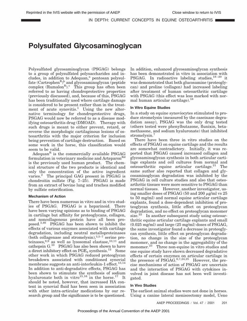
Polysulfated Glycosaminoglycan
Polysulfated glycosaminoglycan (PSGAG) belongsto a group of polysulfated polysaccharides and in-cludes, in addition to Adequan,a pentosan polysul-fate (CartrophenR)b and glycosaminoglycan (peptidecomplex (RumalonR).c This group has often beenreferred to as having chondroprotective properties(previously discussed), and, because of this, PSGAGhas been traditionally used where cartilage damageis considered to be present rather than in the treat-ment of acute synovitis.1 Using the new alter-native terminology for chondroprotective drugs,PSGAG would now be referred to as a disease mod-ifying osteoarthritis drug (DMOAD). Therapy withsuch drugs is meant to either prevent, retard, orreverse the morphologic cartilaginous lesions of os-teoarthritis with the major criterion for inclusionbeing prevention of cartilage destruction. Based onsome work in the horse, this classification wouldseem to be valid.
AdequanR is the commercially available PSGAGformulation in veterinary medicine and ArteparonRd
is the previously used human product. The chem-ical structure of the two products is identical andonly the concentration of the active ingredientvaries.2 The principal GAG present in PSGAG ischondroitin sulfate (Fig. 7–25). PSGAG is madefrom an extract of bovine lung and trachea modifiedby sulfate esterification.
Mechanism of Action
There have been numerous in vitro and in vivo stud-ies of PSGAG. PSGAG is a heparinoid. Therehave been varying opinions as to binding of PSGAGin cartilage but affinity for proteoglycans, collagen,and noncollagenous protein have all been pro-posed.3,46 PSGAG has been shown to inhibit theeffects of various enzymes associated with cartilagedegradation, including neutral metalloproteinases(both collagenase and stromelysin),3,5–7 serine pro-teinases,8,9 as well as lysosomal elastase,10,11 andcathepsin G.10 PSGAG has also been shown to havea direct inhibitory effect on PGE2 synthesis.12 Someother work in which PSGAG reduced proteoglycanbreakdown associated with conditioned synovialmembrane suggests an anti-interleukin-1 effect.13,14
In addition to anti-degradative effects, PSGAG hasbeen shown to stimulate the synthesis of sodiumhyaluronate both in vitro15,16 in the horse.17 Itshould be noted, however, that increased HA con-tent in synovial fluid has been seen in associationwith other intra-articular medications by our re-search group and the significance is to be questioned.
In addition, enhanced glycosaminoglycan synthesishas been demonstrated in vitro in association withPSGAG. In radioactive labeling studies,18–20 itwas demonstrated that both glucosamine (proteogly-can) and proline (collagen) had increased labelingafter treatment of human osteoarthritic cartilagewith PSGAG (this effect was less marked with nor-mal human articular cartilage).18
In Vitro Equine Studies
In a study on equine synoviocytes stimulated to pro-duce stromelysin (measured by the caseinase degra-dation assay), PSGAG was the only drug tested(others tested were phenylbutazone, flunixin, beta-methasone, and sodium hyaluronate) that inhibitedstromelysin.7
There have been three in vitro studies on theeffects of PSGAG on equine cartilage and the resultsare somewhat contradictory. Initially, it was re-ported that PSGAG caused increased collagen andglycosaminoglycan synthesis in both articular carti-lage explants and cell cultures from normal andosteoarthritic equine articular cartilage.21 Thesame author also reported that collagen and gly-cosaminoglycan degradation was inhibited by thePSGAG in cell culture studies and also that osteo-arthritic tissues were more sensitive to PSGAG thannormal tissues. However, another investigator, us-ing smaller doses of PSGAG (50 and 200 mg/ml vs 25to 50 mg/ml) and normal equine articular cartilageexplants, found a dose-dependent inhibition of pro-teoglycan synthesis, little effect on proteoglycandegradation, and no effect on proteoglycan monomersize.22 In another subsequent study using osteoar-thritic equine articular cartilage explants and small(0.025 mg/ml) and large (25 mg/ml) doses of PSGAG,the same investigator found a decrease in proteogly-can synthesis, little effect on proteoglycan degrada-tion, no change in the size of the proteoglycanmonomer, and no change in the aggregability of themonomer.23 Three non-equine in vitro studies andone equine study have shown decreased degradativeeffects of certain enzymes on articular cartilage inthe presence of PSGAG.6,7,24,25 However, the pre-cise mechanisms of action of PSGAG are uncertainand the interaction of PSGAG with cytokines in-volved in joint disease has not been well investi-gated.
In Vivo Studies
The earliest animal studies were not done in horses.Using a canine lateral meniscectomy model, Ueno
AAEP PROCEEDINGS / Vol. 47 / 2001 201
IN DEPTH: CURRENT CONCEPTS IN EQUINE OSTEOARTHRITIS
Reprinted in the IVIS website with the permission of AAEP Close window to return to IVIS
Proceedings of the Annual Convention of the AAEP 2001
demonstrated dramatic morphologic differences be-tween articular cartilage from control and PSGAG-treated joints. PSGAG was given intramuscularlyat 25 mg/kg for 13 treatments.26 Later work with acanine meniscectomy model also showed a protec-tive effect when PSGAG was administered sub-cutaneously.19 These authors suggested that thePSGAG likely acts by inhibiting matrix degradingenzymes. Favorable effects (lower active neutralmetalloproteinase activity, increased chondrocytecounts, and maintenance proteoglycan content)have also been reported using intra-articularly ad-ministered PSGAG in a meniscectomy model of os-teoarthritis in rabbits.6,25 PSGAG has also beentested on the canine anterior cruciate ligament tran-section model and is reported to have both a prophy-lactic and therapeutic effect.24,27 PSGAG has alsobeen tested using the rat air pouch model of inflam-mation and improved proteoglycan content extract-ability and aggregation as well as reduction ofleukocyte infiltration into the pouch were noted.28
It was felt that reduced leukocyte infiltration wouldreduce cartilage exposure to leukocyte derived pro-teinases and other mediators of cartilage dam-age. PSGAG was also tested in clinical cases ofosteoarthritis in boars (intramuscularly at 5.2mg/kg for 6 treatments and saline was put intocontrol joints). The degree of lameness was signif-icantly decreased and there was also increased hy-aluronic acid in the synovial fluid.29 The drug hasalso been used in the treatment of canine hip dys-plasia.30
The first equine investigation involved 250-mg in-jections of PSGAG intra-articularly twice weekly for3 weeks and then once weekly for the next 3 weeksin clinical equine cases with joint swelling andlameness.31 A significant improvement in synovialfluid protein concentration and synovial fluid viscos-ity was reported, as well as an overall impressionof decreased clinical signs (lameness, swellingand effusion, and increased flexion). Intra-articu-lar PSGAG was then tested using a Freund’s adju-vant-induced model in the carpus of 30 horses.This study concluded that the clinical signs of ar-thritis were reduced in treated animals.24 The lat-ter investigators, in a clinical trial in 109 horses,also felt that PSGAG improved clinical signs morefrequently than horses not treated.
PSGAG was then tested on chemically-induced, aswell as physically-induced lesions in the horse in ourlaboratory.32,33 Treatment with intra-articular in-jections of 250 mg PSGAG once weekly for 5 weeksin carpal joints injected with sodium monoiodoac-etate revealed less articular cartilage fibrillationand erosion, less chondrocyte death, and markedlyimproved safranin 0 staining. PSGAG, however,did not have any effect on physically induced lesions(partial and full thickness). Our conclusions fromthis study were that PSGAG could markedly de-crease the development of osteoarthritis but wasof no benefit in healing cartilage lesions already
present at the initiation of treatment. A secondstudy using intramuscular PSGAG (500 mg q 4 d for7 treatments) showed relatively insignificant effectswith treatment. The effects were limited to slightlyimproved safranin 0 staining in sodium monoiodoac-etate joints when PSGAG was used.
More recent studies have evaluated the effects ofPSGAG with or without exercise on the repair ofarticular cartilage defects as well as the develop-ment of osteoarthritis in the carpus of ponies.34,35
The authors concluded that PSGAG was beneficialin ameliorating the clinical, radiographic, and scin-tigraphic signs of joint disease. In another study,the effects of both hyaluronan and PSGAG wereevaluated in the repair of equine articular cartilagedefects in ponies.36 Neither drug showed any ben-eficial effects. However, the project was termi-nated 11 weeks after defect induction.
Potential Complications of Intra-Articular Use
Intra-articular infection after intra-articular injec-tion is always a potential risk. However, researchhas demonstrated that PSGAG may have greaterpotential in this regard. Potentiation of a subinfec-tive dose of Staphylococcus aureus in the middlecarpal joint of horses has been demonstrated in ourlaboratory.37,38 Using a subinfective dose, infec-tion occurred in 8 out of 8 PSGAG injected joints,whereas it only occurred in 3 horses that receivedintra-articular sodium hyaluronate and 4 that re-ceived intra-articular methylprednisolone acetate.This infection could be prevented by 125 mg amika-cin but was not abolished by prior filtration of Ad-equan. In another study, PSGAG was shown toinhibit equine complement activity.39 The classicaland ultimate complement pathways have bacteri-cidal activity and both were shown to be inhibited byPSGAG in vitro. This inhibition could be a factor inthe ability of bacteria to induce septic arthritis.
Because PSGAG is classified as a heparinoid,some effect on the hemostatic mechanisms is ex-pected.40,41 Local hematomas were sometimes de-scribed as transient complications in early clinicaltrials in humans and heparin-associated thrombocy-topenia is known to occur with heparin use inpeople.41 However, the drug has been used exten-sively in humans in earlier reports with a low com-plication rate. However, it has been suggested thatthe risk of hemarthrosis in humans is high.40 Itis therefore interesting that hemarthrosis has notbeen seen in the horse, despite extensive use. Inaddition, one of the author’s primary uses of Ad-equan is following arthroscopic surgery where thereis considerable articular cartilage loss and subchon-dral bone exposure. These joints typically havepersistent effusion, which could be classified as hem-arthrosis and the use of intra-articular PSGAGseems to treat this condition quite effectively.
202 2001 / Vol. 47 / AAEP PROCEEDINGS
IN DEPTH: CURRENT CONCEPTS IN EQUINE OSTEOARTHRITIS
Reprinted in the IVIS website with the permission of AAEP Close window to return to IVIS
Proceedings of the Annual Convention of the AAEP 2001
Intramuscular Use of PSGAG
Most Adequan is used intramuscularly. As dis-cussed previously, the positive effects using themonoiodoacetate (MIA) model that had been seenwith intra-articular PSGAG could not be emulatedby the intramuscular route. However, defects inthis MIA model have since been recognized. Thereis minimal objective data supporting effectivenessfor intramuscular Adequan but the drug is widelyused and anecdotal reports support its value. Theissue of absorption after intramuscular injectionwas addressed by Burba et al.17 In this study, PS-GAG was labeled with tritium and scintillation doneon synovial fluid as well as joint tissues. It was feltthat levels of drug consistent with that seen in othernon-equine studies were obtained and it was con-cluded that therapy every 4 days was effective inmaintaining anti-inflammatory levels in the joint.
Clinical Use of PSGAG
We primarily use Adequan following surgery whenthere is significant loss of articular cartilage (gradeIII or grade IV damage). Typically these horseswill have persistent bloody synovial effusion. Theuse of intra-articular Adequan has marked benefi-cial effects in these instances. We like to give oneinjection intra-articularly (using 0.5 ml amikacinconcurrently) and then follow up with intramuscu-lar therapy at weekly intervals and a dose of 500mg. The drug is also used to considerable extent ona prophylactic basis. Caron et al42 did a survey of1522 equine practitioner members of AAEP seekinginformation on Adequan use. Of practitioners re-sponding, 90.5% of practitioners reported use ofPSGAG. Use of PSGAG was significantly morecommon by practitioners involved predominantlywith racehorses or show horses. Standardbredracehorse practitioners had a significantly higherlevel of intra-articular use of Adequan. Overall,PSGAG was reported to be perceived as moderatelyeffective for all four categories of joint disease:idiopathic synovitis, acute synovitis (with lame-ness), subacute OA (mild radiographic changes), andchronic OA (moderate to severe radiographicchanges). Use of PSGAG was considered more ef-fective than HA for the treatment of subacute OAand less effective for idiopathic joint effusion andacute synovitis.
Pentosan Polysulfate
The use of this drug in the treatment of equinejoint disease has been recently extensively re-viewed.43 Although pentosan polysulfate (PPS) asthe sodium salt has been used in Europe for over 30years as an antithrombotic–antilipidemic agent, itspotential as a disease-modifying antiarthritic agenthas only been realized in recent years. Also, a newcalcium derivative of PPS (CAPPS) has recentlybeen developed that is more effectively absorbedafter oral administration than sodium pentosan
polysulfate (NaPPS) and this offers hope for wideruse of this drug. PPS could also be considered as adisease-modifying OA drug. It has been pointedout by Little and Ghosh that PPS, unlike NSAIDs,does not possess analgesic activity.43 Therefore, inorder to provide symptomatic relief and efficacy, adrug such as PPS must be capable of correcting thepathobiologic imbalances that are present withinthe OA joint, and these authors feel that PPS fulfillsthese requirements.
PPS is not derived from animal or bacterialsources but, rather, the “backbone” of PPS is iso-lated from beechwood hemicellulose, which consistsof repeating units of (1-4)-linked beta-D-xylano-pyranoses. An anabolic effect on chondrocytes hasbeen demonstrated in a focal model of OA induced byunilateral meniscectomy in sheep.44 Studies onchondrocytes in agarose culture showed that PPSstimulated proteoglycan synthesis.45 Also, in anexperimental model of joint disease in rabbits, oraladministration of CAPPS (10 mg/kg q 7 d) main-tained the normal articular cartilage ratio of aggre-can to dermatan sulfate (interpreted by the authorsas chondrocyte phenotype).46 PPS also stimulatesHA synthesis by cultured synoviocytes obtained byboth rheumatoid and osteoarthritic joints.47 Thesein vitro effects of PPS in HA synthesis were con-firmed in a rat air pouch model of inflammation andin this model increased synthesis of HA was notstimulated by PSGAG.28
A number of in vitro and in vivo studies haveshown that PPS will inhibit various processes thatinduce degeneration of the articular cartilage ma-trix. For example, PPS has been shown to inhibitMMP3.48 There is a suggestion that PPS may mod-ulate receptor-mediated binding of cytokines.43
In an ovine model of OA (medial meniscectomy),weekly intra-articular injections of PPS for fourweeks improved joint function and reduced meanradiographic scores and Mankin histologic scores ofarticular cartilage damage in the femoral condyle.49
There are no published studies describing the ap-plication of PPS for equine joint disease but the drughas been used in Australia. Anecdotally it is con-sidered that treatment improves the clinical param-eters of synovial effusion and lameness in mosthorses when used in clinical cases. Marked allevi-ation of lameness after racing was the most notedchange with this drug.43 It is also felt that becauseof the vascular effects of the drug that it could aid ordecrease the rate of subchondral bone necrosis andsclerosis.
An interesting study involved the simultaneousadministration of sodium pentosan polysulfate andIGF-1 early in the pathogenesis of iatrogenic canineOA. The combination of drugs significantly re-duced the severity of lesions, whereas IGF-1 alonehad little effect.50 The presence of PPS appeared todecrease the amount of total and active metallopro-teinases in the cartilage. The authors suggestedthat the PPS reduce the enzymatic breakdown of
AAEP PROCEEDINGS / Vol. 47 / 2001 203
IN DEPTH: CURRENT CONCEPTS IN EQUINE OSTEOARTHRITIS
Reprinted in the IVIS website with the permission of AAEP Close window to return to IVIS
Proceedings of the Annual Convention of the AAEP 2001
IGF-1, binding protein or receptor, thus allowingIGF-1 to exert its influence.
Oral Glycosaminoglycans
Oral glycosaminoglycan products available forhorses include a purified chondroitin sulfate productfrom bovine trachea (Flex-FreeR)e and a complex ofglycosaminoglycans and other nutrients from thesea mussel Perna canaliculus (Syno-FlexR).f Morerecently, a combination of glucosamine hydrochlo-ride, chondroitin sulfate, manganese, and vitamin Chas been marketed as a “nutraceutical” (CosequinR).g
There have been some positive anecdotal reports forsuch supplements.51–55 Cosequin has been evalu-ated using the Freund’s adjuvant model of in-flammatory joint disease in horses.56 The oralsupplement was used at the recommended dose be-ginning 10 days prior to arthritis induction andcontinuing for a further 26 days. No benefit wasdemonstrated based on clinical (lameness, stridelength, carpal circumference, carpal flexion) and sy-novial fluid (protein) parameters. However, it isquestionable whether the Freund’s adjuvant modelsimulates any clinical equine joint entity and onemust question the value of studies using this model.
The oral administration of glucosamine sulfatehas been associated with decreased pain and im-proved range of motion compared to placebo in acontrolled clinical trial in humans.55 In anothercontrolled study, glucosamine sulfate was as effec-tive as ibuprofen at relieving symptoms of osteo-arthritis in people.54 In vitro studies usingglucosamine sulfate have demonstrated increasedglycosaminoglycan and proteoglycan synthesis andin vivo studies have demonstrated anti-inflamma-tory activity through inhibition of lysosomal enzymeactivity and free radical production.
There is conflicting evidence regarding the enteralabsorption of orally administered glycosaminogly-cans.51–53 The initial focus with oral glycosamino-glycans was on chondroitin sulfate and there hasbeen some supportive evidence presented for absorp-tion of active molecules.30,52 In interpretation ofsuch studies, however, one has to be careful whetherradiolabeled macromolecular sulfate, chondroitinsulfate, or labeled but inactive monomer or otherdegradation products are being absorbed. It is notvalid to extrapolate between antienzymic data (in-volving intact chondroitin sulfate molecules) and thedetection of tritium label in tissues. Some earlierstudies that failed to show absorption have beensince criticized for the lack of specificity of the meth-ods used. However, favorable absorption of chon-droitin sulfate and dermatan sulfate from thegastrointestinal tract with reduced N-acetylglu-cosaminodase and granulocyte elastase activity,as well as increased HA concentration in treatedpatients, has been reported.52 For instance, inanother study when chondroitin sulfate was admin-istered to healthy human volunteers and serum con-centrations of GAGs using the dimethyl methylene
blue assay done, it was reported that neither intactnor depolymerized chondroitin sulfate was effec-tively absorbed.51 This study was criticized for lowdosage combined with a low sensitivity with theDMMB assay used.53
As mentioned in the previous section, absorptionof calcium pentosan polysulfate after oral adminis-tration in rats has been reported as effective andwhat has been reported to be of sufficient levelto maintain cartilage proteoglycan concentrationand biosynthesis.46 There was also some evi-dence for oral bioavailability of glucosamine sul-fate and tropism for articular cartilage after oraladministration.54 The pharmacokinetics, organdistribution, metabolism, and excretion of glu-cosamine were studied in the dog using uniformlylabeled (14-C)-glucosamine (sulfate) intravenouslyor orally in single doses. In humans, unlabeledglucosamine sulfate was given intravenously andorally and glucosamine was measured in plasmaand urine with a glucosamine-specific ion-exchangechromatographic method. The result showed thatthe bioavailability, pharmacokinetics, and excretionpattern of glucosamine was consistent with thosefound in the dog with radiolabeled glucosamine andwith those reported in a previous study in the rat.57
In the dog study, radiolabeled glucosamine wasgiven intravenously and orally and the distributionof radioactivity orally was the same as intrave-nously.
In one human study, cartilage biopsies were takenbefore and after 4 weeks of glucosamine sulfate oralsupplementation in a few treated subjects as well asassessing the overall results clinically. There werereductions in joint pain, analgesic use and the im-provement of joint function with glucosamine sul-fate administration. Electron microscopy showeda typical picture of established osteoarthritis. Inthose given glucosamine sulfate, it was consideredthat they “showed a picture more similar to healthycartilage.”58 Exogenous glucosamine increasesmatrix production and seems likely to alter the nat-ural history of OA. Glucosamine also has a mildanti-inflammatory activity that is probably via afree radical scavenging effect.59 Numerous in vitrostudies have demonstrated that glucosamine stimu-lates the synthesis of proteoglycan and collagen bychondrocytes.47
An additional mechanism by which chondroitinsulfate may benefit joint tissues is by the preven-tion of fibrin thrombi in synovial or subchondralmicrovasculature.60 The anti-synovitis effect maybe significant. It is interesting that in a study withextract of Perna canaliculus in humans, at the endof six months 19 of the 28 rheumatoid patients(67.9%) and 15 of the 38 osteoarthritic patients(39.5%) felt they had benefitted from the treat-ment.61 However, other studies have questionedboth the GAG content and the therapeutic value ofthis extract.
204 2001 / Vol. 47 / AAEP PROCEEDINGS
IN DEPTH: CURRENT CONCEPTS IN EQUINE OSTEOARTHRITIS
Reprinted in the IVIS website with the permission of AAEP Close window to return to IVIS
Proceedings of the Annual Convention of the AAEP 2001
There is better evidence for glucosamine absorp-tion than with chondroitin sulfate. Glucosamine isan amino monosaccharide that is a basic constituentof the disaccharide units of glycosaminoglycansof articular cartilage.45 Glucosamine is the hex-osamine present in keratan sulfate and the pre-cursor of D-galactosamine (the hexosamine inchondroitin sulfate).2 Exogenous glucosamine hasbeen suggested as the preferred substrate for GAGsynthesis because a higher energy expenditure isrequired with endogenous glucose.45 There is goodevidence for effective absorption of glucosamine sul-fate (up to 87%) after oral administration in humans.In vitro studies have also documented enhancedchondrocyte synthesis of GAGs and collagen by glu-cosamine. It has been pointed out that althoughresearch mainly documents the effects of glu-cosamine as the sulfate salt, the veterinary productcontains glucosamine hydrochloride.
New oral glycosaminoglycan/glucosamine prod-ucts continue to be developed. There is quite anexplosive market in both humans and horses despitevery little scientific validation. A control study isneeded to address the effectiveness of these oralglycosaminoglycan-type products and also to evalu-ate their efficacy relative to other systemically ad-ministered products, such as Adequan.
A clinical trial was conducted in 25 horses over a6-week period. In this study, Cosequin was associ-ated with decreased lameness, improved lameness,and improved lameness scores, but there were nocontrols.62
References and Footnotes1. McIlwraith CW. Intra-articular medication for traumatic
joint problems: Do we understand the choices? CompendContin Educ Pract Vet 1989;11:1287–1311.
2. Trotter GW. Polysulfated glycosaminoglycan (Ad-equanR). In: McIlwraith CW, Trotter GW, eds. Joint dis-ease in the horse. Philadelphia: WB Saunders, 1996;270–280.
3. Altman RD, Dean DD, Muniz O, et al. Therapeutic treat-ment of osteoarthritis with glycosaminoglycan polysulfuricacid ester. Arth Rheum 1989;32:1300–1307.
4. Andrews JL, Sutherland J, Ghosh P. Distribution and bind-ing of glycosaminoglycan polysulfate to intervertebral disc,knee joint cartilage and meniscus. Arnzeim-Forsch/DrugRes 1985;35:144–148.
5. Altman RD, Dean DD, Muniz O, et al. Prophylactic treat-ment of canine osteoarthritis with glycosaminoglycan poly-sulfuric acid ester [abstr]. Arth Rheum 1989;32:759–766.
6. Howell DS, Carreno M, Pelletier J-P, et al. Articular carti-lage breakdown in a lapine model of osteoarthritis. ClinOrthop Rel Res 1986;213:69–76.
7. May SA, Hooke RE, Lees P. The effect of drugs used in thetreatment of osteoarthrosis on stromelysin (proteoglycanase)of equine synovial cell origin. Equine Vet J 1988;S6:28–32.
8. Kruze D, Fehr K, Menninger H, et al. Effect of antirheu-matic drugs on neutral protease from human leukocyte gran-ules. J Rheumatol 1976;35:337–346.
9. Theiler R, Thosh P, Brooks P. Clinical, biochemical andimaging methods of assessing osteoarthritis and clinical tri-als with agents claiming “chondromodulating” activity. Os-teoarth Cart 1994;2:1–23.
10. Baici A, Fehr K. Inhibition of human lysosomal elastase byArteparon. In: Dettmer N, Greiling H, eds. IXth Euro-
pean Congress of Rheumatology, Basel: Eular Publishers,1982;19.
11. Baici A, Salgam P, Fehr K, et al. Inhibition of human elas-tase from polymorphonuclear leukocytes by a glycosamino-glycan polysulfate (Arteparon). Biochem Pharmacol 1980;29:1723–1727.
12. Dietmar EGG. Effects of glycosaminoglycan-polysulfateand two nonsteroidal anti-inflammatory drugs on prostaglan-din E2 synthesis in Chinese hamster ovary cell cultures.Pharm Res Comm 1983;15:709–717.
13. Ghosh P, Smith M, Wells C. Second-line agents in osteoar-thritis. In: Dixon JS, Furst DE, eds. Second-line agentsin the treatment of rheumatic diseases. New York: MarcelDekker, Inc, 1992;363.
14. Jones IL, Sandstrom T. Enhanced breakdown of bovine ar-ticular cartilage proteoglycans by conditioned synovial me-dium in vitro. Arnzeim-Forsch/Drug Res 1985;35:141–144.
15. Nishikawa H, Mori I, Umemoto J. Glycosaminoglycan poly-sulfate-induced stimulation of hyaluronic acid synthesis inrabbit knee synovial membrane: Involvement of bindingprotein and calcium ion. Arch Biochem Biophys 1988;266:201–209.
16. Smith MM, Ghosh P. The effects of some polysulfated poly-saccharides on hyaluronate synthesis by human synovialfibroblasts. Recent advances in connective tissue re-search. Agents/Actions 1987;518:55–62.
17. Burba DJ, Collier M. In vivo kinetic study on uptake anddistribution of intramuscular tritium-labeled polysulfatedglycosaminoglycan in equine synovial fluid and articular car-tilage, in Proceedings. Annu Conv Am Assoc Equine Practnr1991;37:241–242.
18. Adam M, Krabcova M, Musilova J, et al. Contribution to themode of action of glycosaminoglycan-polysulfate (GAGPS)upon human osteoarthritic cartilage. Arnzeim-Forsch/DrugRes 1980;30:1730–1732.
19. Hannan N, Ghosh P, Bellenger C, et al. Systemic adminis-tration of glycosaminoglycan polysulphate (Arteparon) pro-vides partial protection of articular cartilage from damageproduced by meniscectomy in the canine. J Orthop Res1987;5:47–59.
20. Vacha J, Pesakova V, Krajickova J, et al. Effect of glycos-aminoglycan polysulfate on the metabolism of cartilage ribo-nucleic acid. Arnzeim-Forsch/Drug Res 1984;34:607–609.
21. Glade MJ. Polysulfated glycosaminoglycan accelerates netsynthesis of collagen and glycosaminoglycans by arthriticequine cartilage tissues and chondrocytes. Am J Vet Res1990;51:779–785.
22. Caron JP, Eberhart SW, Nachreiner R. Influence of polysul-fated glycosaminoglycan on equine articular cartilage in ex-plant culture. Am J Vet Res 1991;52:1622–1625.
23. Caron JP, Toppin DS, Block JA. Effect of polysulfated gly-cosaminoglycan on osteoarthritic equine articular cartilage inexplant culture. Am J Vet Res 1993;54:1116–1121.
24. Altman RD, Dean DD, Muniz O, et al. Prophylactic treat-ment of canine osteoarthritis with glycosaminoglycan poly-sulfuric acid ester [abstr]. Arth Rheum 1989;32:759–766.
25. Howell DS, Muniz O, Carreno MR. Effect of glycosamino-glycan polysulfate ester on proteoglycan-degrading enzymeactivity in an animal model of osteoarthritis. Adv InflamRes 1986;11:197–206.
26. Ueno R. Results of the treatment of experimental arthrosisof the knee joint with a mucopolysaccharide polysulfuric acidester. Z Orthop 1973;111:886–892.
27. Altman RD, Kapila P, Dean DD, et al. Future therapeutictrends in osteoarthritis. Scand J Rheumatol 1989;S77:37–42.
28. Francis DJ, Forrest MJ, Brooks PM, et al. Retardation ofarticular cartilage degradation by glycosaminoglycan poly-sulfate, pentosan polysulfate, and DH-40J in the rat airpouch model. Arth Rheum 1989;32:608–616.
29. Brennan JJ, Aherne F, Nakano T. Effects of glycosamino-glycan polysulfate treatment on soundness, hyaluronic acidcontent of synovial fluid and proteoglycan aggregate in artic-ular cartilage of lame boars. Can J Vet Res 1987;51:394–398.
AAEP PROCEEDINGS / Vol. 47 / 2001 205
IN DEPTH: CURRENT CONCEPTS IN EQUINE OSTEOARTHRITIS
Reprinted in the IVIS website with the permission of AAEP Close window to return to IVIS
Proceedings of the Annual Convention of the AAEP 2001
30. DeHaan JJ, Goring R, Beale BS. Evaluation of polysulfatedglycosaminoglycan for the treatment of hip dysplasia in dogs.Vet Surg 1994;23:177–181.
31. Tew WP. Demonstration by synovial fluid analysis of theefficacy in horses of an investigational drug (L-1016). JEquine Vet Sci 1982;March/April:42–50.
32. Trotter GW, Yovich J, McIlwraith CW, et al. Effects of in-tramuscular polysulfated glycosaminoglycan on chemical andphysical defects in equine articular cartilage. Can J Vet Res1989;43:224–230.
33. Yovich J, Trotter GW, McIlwraith CW, et al. Effects of poly-sulfated glycosaminoglycan on chemical and physical defectsin equine articular cartilage. Am J Vet Res 1987;48:1407–1414.
34. Todhunter RJ, Minor RR, Wootton J, et al. Effects of exer-cise and polysulfated glycosaminoglycan on repair of articu-lar cartilage defects in the equine carpus. J Orthop Res1993;11:782–795.
35. Todhunter RJ, Freeman KP, Yeager AE. Effects of exerciseand polysulfated glycosaminoglycan on the development ofosteoarthritis in equine carpal joints with osteochondral de-fects. Vet Surg 1993;22:330–342.
36. Barr ARS, Duance VC, Wotton SF, et al. Influence of intra-articular sodium hyaluronate and polysulphated glycosami-noglycans on the biochemical composition of equine articularsurface repair tissue. Equine Vet J 1994;26:40–42.
37. Gustafson SB, McIlwraith CW, Jones RL. Comparison ofthe effect of polysulfated glycosaminoglycan, corticosteroids,and sodium hyaluronate in the potentiation of a subinfectivedose of Staphylococcus aureus in the midcarpal joint ofhorses. Am J Vet Res 1989;50:2014–2017.
38. Gustafson SB, McIlwraith CW, Jones RL. Further investi-gations into the potentiation of infection by intra-articularinjection of polysulfated glycosaminoglycan and the effect offiltration and intra-articular injection of amikacin. Am JVet Res 1989;50:2018–2022.
39. Rashmir-Raven AM, Coyne CP, Fenwick BW, et al. Inhibi-tion of equine complement activity by polysulfated glycosami-noglycans. Am J Vet Res 1992;53:87–90.
40. Bauer F, Schulz P, Reber G, et al. Anticoagulant propertiesof three mucopolysaccharides used in rheumatology.Thromb Haemostas 1983;50:652–655.
41. Greinacher A, Michels I, Schafer M, et al. Heparin-associ-ated thrombocytopenia in a patient treated with polysulfatedchondroitin sulphate: Evidence of immunological cross-re-activity between heparin and polysulphated glycosaminogly-can. Br J Hematol 1992;81:252–254.
42. Caron JP, Kaneene JB, Miller R. Results of a survey ofequine practitioners on the use and perceived efficacy ofpolysulfated glycosaminoglycan. Am J Vet Res 1996;209:1564–1568.
43. Little C, Ghosh P. Potential use of pentosan polysulfate forthe treatment of equine joint disease. In: McIlwraith CW,Trotter GW, eds. Joint disease in the horse. Philadelphia:WB Saunders, 1996;281–292.
44. Little C, Ghosh P, Bellinger C. Meniscectomy increases ag-grecan, decoran (PGS2) and biglycan (PG-S1) metabolism incartilage. Proc Orthop Res Soc 1993;18:707.
45. Reichelt L, Forster K, Fischer M, et al. Efficacy and safety ofintramuscular glucosamine sulfate in osteoarthritis of theknee. Arzneimittelforschung 1994;44:75–80.
46. Smith MM, Ghosh P, Numata Y, et al. The effects of orallyadministered calcium pentosan polysulfate on inflammation
and cartilage degradation produced in rabbit joints by intra-articular injection of a hyaluronate-polylysine complex.Arth Rheum 1993;37:125–136.
47. Jimenez SA. The effect of glucosamine on human chondro-cyte gene expression. Presented at the 9th EULAR Sympo-sium, Madrid, Spain, 1996;8–10.
48. Nethery A, Giles I, Jenkins K, Jackson C, et al. The chon-droprotective drugs, Arteparon and sodium pentosan polysul-fate, increase collagenase activity and inhibit stromelysinactivity in vitro. Biochem Pharmacol 1992;44:1549–1553.
49. Ghosh P, Armstrong S, Read R, et al. Animal models ofearly osteoarthritis: Their use for the evaluation of poten-tial chondroprotective agents. In: VandenBerg WB, vander Kraan PM, van Lent PLEM, eds. Joint destruction inarthritis and osteoarthritis. Austin, TX: Birkhauser, 1993;195.
50. Rogachefsky RA, Dean DD, Howell DS, Altman RD. Treat-ment of canine osteoarthritis with insulin-like growth factor(IGF-1) and sodium pentosan polysulfate. Osteoarth Cart1993;1:105–114.
51. Baici A, Horler D, Moser B, et al. Analysis of glycosamino-glycans in human serum after oral administration of chon-droitin sulfate. Rheumatol Int 1992;12:81–88.
52. Conte A, DeBernardi M, Palmieri L, et al. Metabolic fateof exogenous chondroitin sulfate in man. Arnzeim-Forsch/Drug Res 1991;41:768–772.
53. Lualdi P. Bioavailability of oral chondroitin sulfate[ltr]. Rheumatol Int 1993;13:39–40.
54. Muller-Fassbender H, Bach G, Haase W, et al. Glucosaminesulfate compared to ibuprofen in osteoarthritis of theknee. Osteoarth Cart 1994;2:61–69.
55. Noack W, Fischer M, Forster K, et al. Glucosamine sulfatein osteoarthritis of the knee. Osteoarth Cart 1994;2:51–59.
56. White GW, Jones EW, Hamm J, et al. The efficacy of orallyadministered sulfated glycosaminoglycan in chemically-in-duced equine synovitis and degenerative joint disease. JEquine Vet Sci 1994;14:350–353.
57. Setnikar I, Giacchetti C, Zanolo G. Pharmacokinetics ofglucosamine in the dog and in man. Arzneim-Forsch/DrugRes 1986;36:729–735.
58. Pujalte JM, Llavore E, Ylescupidez FR. Double-blind clini-cal evaluation of oral glucosamine sulfate in the basic treat-ment of osteoarthrosis. Curr Med Res Opin 1980;7:110–114.
59. Raiss R, Einfluss von D. Glucosaminsulfat auf experimen-tell geschaedigten gelenkknorpel. Fortschr Med 1985;103:658–668.
60. Bucci L. Chondroprotective agents: glucosamine and chon-droitin sulfates. Townsend Let Doctor (suppl.) 1994;52–54.
61. Gibson RG, Gibson SLM, Conway V, Chappell D. Pernacanaliculus in the treatment of arthritis. Practitioner 1980;224:955–960.
62. Hanson R. Oral glycosaminoglycans in treatment of degen-erative joint disease in horses. Equine Pract 1996;18:18–22.
aAdequanR, Luitpold Pharmaceuticals, Inc., Shirley, NY.bCartrophenR, not available in US.cRumalonR, not available in US.dArteparonR, Luitpold Pharmaceuticals, no longer manufac-
tured.eFlex-FreeR, Vita-Flex Nutrition Co., Staten Island, NY.fSyno-FlexR, Vetri-Science Laboratories, Essex Junction, VT.gCosequinR, Nutramax Laboratories, Edgewood, MD.
206 2001 / Vol. 47 / AAEP PROCEEDINGS
IN DEPTH: CURRENT CONCEPTS IN EQUINE OSTEOARTHRITIS
Reprinted in the IVIS website with the permission of AAEP Close window to return to IVIS
Proceedings of the Annual Convention of the AAEP 2001