lymphoid neogenesis in rheumatoid synovitis · lymphoid neogenesis in rheumatoid synovitis1 ......
TRANSCRIPT

of September 13, 2018.This information is current as
SynovitisLymphoid Neogenesis in Rheumatoid
Goronzy and Cornelia M. WeyandKurtin, Robert H. Cofield, William M. O'Fallon, Jörg J. Seisuke Takemura, Andrea Braun, Cynthia Crowson, Paul J.
http://www.jimmunol.org/content/167/2/1072doi: 10.4049/jimmunol.167.2.1072
2001; 167:1072-1080; ;J Immunol
Referenceshttp://www.jimmunol.org/content/167/2/1072.full#ref-list-1
, 16 of which you can access for free at: cites 41 articlesThis article
average*
4 weeks from acceptance to publicationFast Publication! •
Every submission reviewed by practicing scientistsNo Triage! •
from submission to initial decisionRapid Reviews! 30 days* •
Submit online. ?The JIWhy
Subscriptionhttp://jimmunol.org/subscription
is online at: The Journal of ImmunologyInformation about subscribing to
Permissionshttp://www.aai.org/About/Publications/JI/copyright.htmlSubmit copyright permission requests at:
Email Alertshttp://jimmunol.org/alertsReceive free email-alerts when new articles cite this article. Sign up at:
Print ISSN: 0022-1767 Online ISSN: 1550-6606. Immunologists All rights reserved.Copyright © 2001 by The American Association of1451 Rockville Pike, Suite 650, Rockville, MD 20852The American Association of Immunologists, Inc.,
is published twice each month byThe Journal of Immunology
by guest on September 13, 2018
http://ww
w.jim
munol.org/
Dow
nloaded from
by guest on September 13, 2018
http://ww
w.jim
munol.org/
Dow
nloaded from

Lymphoid Neogenesis in Rheumatoid Synovitis1
Seisuke Takemura,2* Andrea Braun, 2* Cynthia Crowson,‡ Paul J. Kurtin, † Robert H. Cofield,§
William M. O’Fallon, ‡ Jorg J. Goronzy,* and Cornelia M. Weyand3*
In rheumatoid arthritis (RA), tissue-infiltrating lymphocytes can be arranged in sophisticated organizations that resemble mi-crostructures usually formed in secondary lymphoid organs. Molecular pathways and host risk factors involved in this process oflymphoid neogenesis remain to be defined. In a series of 64 synovial tissue biopsies, lymphoid follicles with germinal centers (GCs)were found in 23.4% of the patients. Follicular dendritic cells (FDCs) were exclusively present in tissues with GCs, suggesting thatthe recruitment or in situ maturation of FDCs is a critical factor for GC formation in the synovial membrane. Primary follicleswere absent, emphasizing the role of Ag recognition in the generation of inflammation-associated lymphoid organogenesis. Mul-tivariate logistic regression analysis of tissue cytokines and chemokines identified two parameters, in situ transcription of lym-photoxin (LT)- b and of B lymphocyte chemoattractant (BLC; BLC/CXCL13), that were predictors for FDC recruitment andsynovial GC formation. LT- b and BLC/CXCL13 were found to be independent variables that could, in part, compensate for eachother to facilitate GC formation. Prediction models incorporating in situ transcription of LT- b and BLC/CXCL13 had highnegative yet moderate positive predictive values, suggesting that LT-b and BLC/CXCL13 are necessary but not sufficient. LT-bprotein was detected on a subset of mantle zone and GC B cells, but also on T cells in follicular structures. BLC/CXCL13 wasproduced by FDCs in follicular centers, but was predominantly found in endothelial cells and synovial fibroblasts, suggestingheterotypic signaling between cells of the synovial membrane and infiltrating lymphocytes in regulating extranodal lymphoidneogenesis. The Journal of Immunology,2001, 167: 1072–1080.
T he preferred targets of inflammatory attack in rheumatoidarthritis (RA)4 are the synovial membrane, cartilage, andbone of diarthrodial joints. T cells, B cells, macrophages,
and dendritic cells (DCs) accumulate in the synovial layer, induc-ing hyperplasia and tissue invasiveness of the synoviocytes. A dis-tinguishing feature of the rheumatoid lesion is the high degree ofcellular organization acquired by the tissue-infiltrating lympho-cytes. Rheumatoid synovitis is associated with the formation ofcomplex lymphoid microstructures, to the extent that the rheuma-toid process induces the formation of T cell-B cell follicles withgerminal center (GC) reactions in the synovium (1–3). These mi-crostructures share many features with secondary lymphoid tissue,and the formation of GCs at an extranodal site can, therefore, beconsidered as an example of lymphoid neogenesis. The structuralrequirements permitting the generation of tertiary lymphoid tissuehave not been examined. Also, it is not known which molecularpathways are used or which factors determine the type of lymphoidorganization in individual patients.
A close relationship between inflammation and lymphoid orga-nogenesis has been suggested by the finding that proinflammatorycytokines, such as members of the TNF superfamily, have a crit-ical role in both processes (4, 5). Initially, it was found that micedeficient in lymphotoxin (LT) had no lymph nodes or Peyer’spatches and failed to form GCs in the spleen (6, 7). Despite theabsence of GCs, LT-a2/2 mice still produced high-affinity IgG1responses, provided the mice were immunized with high doses ofAg (8). Gene targeting has been successfully used to implicateother molecules in the process of lymphoid organogenesis, includ-ing LT-b, type I TNFR, and the LT-bR (9–11). Mice with defectsin these genes display different structural abnormalities and func-tional impairment of secondary lymphoid organs. In essence, sig-nals transmitted by LT-a1b2 appear to be pivotal in the ontogenyof secondary lymphoid organs (12, 13).
More recently, studies of lymphoid organogenesis have focusedon the contribution of chemokines that provide the cues to guidecell movement inside lymphoid organs (14, 15). Much progresshas been made in understanding how the two major populations oflymphocytes are directed either to B cell or T cell zones and howchemokines control the movements of such cells during the devel-opment of Ag-specific immune responses. Mice homozygous forthe spontaneous mutation, paucity of lymph node T cells (plt), arecharacterized by major abnormalities in T cell trafficking intolymph nodes and disturbances in the organization of T cell zones(16, 17). These mice lack expression of secondary lymphoid che-moattractant (SLC/CCL21) (18), which binds to CCR7. Defects inthe movement of lymphocytes and DCs through the T cell areas ofspleen, lymph nodes, and Peyer’s patches are shared byplt andCCR7-deficient mice, establishing a critical role of this receptor-ligand pair in compartmental homing of T cells (19). Homeostatictrafficking of B cells into lymphoid tissue and B cell follicles ap-pears to be critically controlled by B lymphocyte chemoattractant(BLC/CXCL13) (20, 21). The receptor for BLC/CXCL13,
Departments of *Medicine and Immunology,†Laboratory Medicine and Pathology,‡Health Services Research, and§Orthopedic Surgery, Mayo Clinic, Rochester, MN55905
Received for publication January 24, 2001. Accepted for publication May 9, 2001.
The costs of publication of this article were defrayed in part by the payment of pagecharges. This article must therefore be hereby markedadvertisementin accordancewith 18 U.S.C. Section 1734 solely to indicate this fact.1 This work was supported by grants from the National Institutes of Health (R01AI44142, R01 AR42527, and R01 AR41974) and the Mayo Foundation.2 S.T. and A.B. contributed equally to this study.3 Address correspondence and reprint requests to Dr. Cornelia M. Weyand, MayoClinic, Guggenheim 401, 200 First Street SW, Rochester, MN 55905. E-mail address:[email protected] Abbreviations used in this paper: RA, rheumatoid arthritis; GC, germinal center;BLC, B lymphocyte chemoattractant; CD21L, CD21 long isoform; DC-CK1, den-dritic cell-derived C-C chemokine 1; FDC, follicular DC; LT, lymphotoxin; MCP,macrophage chemoattractant protein;plt, paucity of lymph node T cells; SLC, sec-ondary lymphoid chemoattractant.
Copyright © 2001 by The American Association of Immunologists 0022-1767/01/$02.00
by guest on September 13, 2018
http://ww
w.jim
munol.org/
Dow
nloaded from

CXCR5, is expressed on recirculating B cells; in in vitro chemo-taxis assays, BLC/CXCL13 attracts B cells. In mice with a targetedinactivation of CXCR5, the normal development of Peyer’spatches, inguinal lymph nodes, splenic follicles, and peripherallymphocytes is disrupted, making the BLC/CXCL13-CXCR5 re-ceptor-ligand pair critical in lymphoid tissue organization (15).
We studied a large cohort of patients with RA who presentedwith different phenotypes of lymphoid microarchitectures in thesynovial lesions and examined whether cytokines and chemokinesimplicated in the genesis of secondary lymphoid organs are in-volved in the process of lymphoid neogenesis in RA. Analysis ofthe cellular elements in the synovium demonstrated that T cells, Bcells, macrophages, and DCs were universally present in rheuma-toid synovitis irrespective of the topographical organization of theinfiltrate. In contrast, follicular DCs (FDCs) were limited to a sub-set of patients. Their presence perfectly correlated with the forma-tion of secondary follicles and GCs. Multivariate regression anal-ysis identified LT-b and BLC/CXCL13 as independent criticalvariables in distinguishing patients with and without synovial GCs.These data suggest that seeding with FDCs or their precursors isthe critical step in follicle formation in the synovium and occurs insome, but not all, patients. LT-a1b2 and BLC/CXCL13 may re-cruit or retain this highly specialized cell to extranodal tissue sites,thus determining the ultimate organization and function of tissue-invading lymphocytes in rheumatoid synovitis.
Materials and MethodsStudy population
Synovial tissue was obtained from 64 patients with active RA who fulfilledthe American College of Rheumatology 1987 revised criteria for RA andwho underwent joint surgery. All patients provided informed consent. Thestudy was approved by the Mayo Clinic Internal Review Board.
Histopathological evaluations
Hematoxylin sections of the synovial tissue samples were analyzed for theorganizational structure of the inflammatory infiltrate with particular at-tention to the topographical arrangement of T cells, B cells, and macro-phages. All analysis was performed by one hemopathologist (P.J.K.) who
was unaware of any clinical or laboratory findings. Tissue specimens weregrouped according to the following criteria: 1) T cell-B cell aggregateswith GCs, 2) T cell-B cell aggregates without GCs, or 3) diffuse infiltrationof T cells and B cells and the absence of lymphoid organization. GCswithin lymphoid aggregates were identified by standard histological crite-ria (22). These included well-circumscribed clusters of centrocytes andcentroblasts with variable numbers of tingible body macrophages and mi-totic figures within aggregates of small lymphocytes. In most cases, cellswith the morphological features of FDCs were also associated with thecentrocytes and centroblasts.
RT-PCR and cytokine semiquantification
Total RNA was extracted from synovial tissue specimens using a commer-cially available reagent (TRIzol; Invitrogen Life Technologies, Grand Is-land, NY). cDNA from synovial tissue specimens was analyzed forb-ac-tin-specific sequences by semiquantitative PCR-ELISA and then adjustedto contain equal numbers. Adjusted cDNA was amplified by PCR for 30cycles under nonsaturating conditions with cytokine-specific primers (Ta-ble I) in parallel with a standard containing a known number of cytokinesequences (23, 24). Each PCR amplification cycle consisted of denatur-ation at 94°C for 30 s, annealing at either 55°C (b-actin, BLC/CXCL13,LT-b, MCP-1/CCL2, and DC-derived C-C chemokine (DC-CK1/CCL18)),58°C (LT-a, and LT-bR), or 60°C (SLC/CCL21) for 1 min, and polymer-ization at 72°C for 1 min with 10-min denaturation at 94°C at the start ofthe reaction and a final 10-min extension at 72°C. Amplified products werelabeled with digoxygenin-11-dUTP (Roche Molecular Biochemicals, Indi-anapolis, IN) and then semiquantified in a liquid hybridization assay withbiotinylated internal probes (Table I) using a commercially available PCR-ELISA kit (Roche Molecular Biochemicals). Labeled PCR products werehybridized for 2.5 h with 200 ng/ml probe at 42°C forb-actin, at 50°C forDC-CK1/CCL18 and macrophage chemoattractant protein 1 (MCP-1)/CCL2, and at 55°C for LT-a, LT-b, LT-bR, BLC/CXCL13, and SLC/CCL21. Hybrids were immobilized on streptavidin-coated microtiter platesand, after washing, were detected with a peroxidase-labeled anti-digoxigenin Ab. Plates were developed by a color reaction using 2,29-azino-bis-(3-ethylbenzthiazoline-6-sulfonic acid) (diammonium salt) sub-strate and quantified using a kinetic microplate reader (Molecular Devices,Sunnyvale, CA). The number of cytokine-specific sequences was deter-mined by interpolation with a standard curve and was expressed as thenumber of cytokine sequences per 13 106 b-actin sequences. A ratio ofone cytokine-specific sequence per 13 106 b-actin sequences was arbi-trarily defined as 1 U.
cDNA was amplified using a specific primer set for the CR-2/CD21 longisoform (CD21L) selectively expressed by FDCs (Table I) (25). Amplified
Table I. Nucleotide sequences of PCR primers and biotinylated probes
Gene Accession No. Oligonucleotide Sequence
b-actin NM_001101 59Primer,ATGGCCACGGCTGCTTCCAGC39 Primer,CATGGTGGTGCCGCCAGACAGProbe,TTCCTTCCTGGGCATGGAGT
LT-a E01275 59 Primer,GCTGCTCACCTCATTGGAGA39 Primer,GGTGGATAGCTGGTCTCCCTProbe,CCAGTGGCATCTACTTCGTCTAC
LT-b L11015 59 Primer,ATCAGGGAGGACTGGTAACGGA39 Primer,GAGGTAATAGAGGCCGTCCTGCProbe,GAGGAGCCAGAAACAGATCTCAG
LT-bR NM_002342 59Primer,GGTGCCTCCATATGCGTCGG39 Primer,GGGGACGCAGTGGTTGTTACProbe,TGCAGGGACCAGGAAAAGGAATAC
BLC/CXCL13 AF044197 59Primer,TCTCTGCTTCTCATGCTGCTGG39 Primer,AGCTTGAGGGTCCACACACACAProbe,TCCCTAGACGCTTCATTGATCG
DC-CK1/CCL18 Y13710 59 Primer,GGTGTCATCCTCCTAACCAAG39 Primer,GGAAAGGGGAAAGGATGATAProbe,CTTTTAAGAGTCCCATCTGCTATG
MCP-1/CCL2 M24545 59 Primer,CAAGGGCTCGCTCAGCC39 Primer,GCAATTTCCCCAAGTCTCTGProbe,GAAGACTTGAACACTCACTCCAC
SLC/CCL21 AB002409 59Primer,CCCCAGGACCCAAGGCAGTGAT39 Primer,TGTGACCGCTCAGTCCTCTTGCProbe,CTCCATCCCAGCTATCCTGTTC
CD21L J03565 59 Primer,GTGGATTTACTTTGAAGGGCA39 Primer,GGCATGTTTCTTCACACCG
1073The Journal of Immunology
by guest on September 13, 2018
http://ww
w.jim
munol.org/
Dow
nloaded from
products were analyzed by 2% agarose gel electrophoresis. PCR conditionswere as described for amplification of products for PCR-ELISA. The an-nealing temperature for the primer set was 55°C.
Antibodies
The following Abs were used for immunohistochemistry; mouse anti-human CD4 mAb (1:100; Dako, Carpinteria, CA), mouse anti-humanCD20 mAb (1:100; Dako), goat anti-human BLC/BCA-1/SCYB13 poly-clonal Ab (1:200; R&D Systems, Minneapolis, MN), mouse anti-humanLT-b mAb, B9.C9 (AC10) and B27.B2 (26, 27) (from J. L. Browning,Biogen, Cambridge, MA), mouse anti-human TNF-b (LT-a) mAb (R&DSystems), and mouse anti-human CXCR5 (BLR-1) mAb (R&D Systems).Secondary Ab used were peroxidase-labeled goat anti-mouse IgG (1:300;Kirkegaard & Perry Laboratories, Gaithersburg, MD) and biotinylated rab-bit anti-goat Ig (1:300; Dako).
Immunohistochemistry
Frozen synovial tissues embedded in OCT (Sakura Finetek USA, Torrance,CA) were cut into 5-mm sections, mounted on slides (SuperFrost/Plus;Fisher Scientific, Pittsburgh, PA), and stored at270°C. Before staining,the slides were fixed in acetone for 10 min, air dried, and fixed in 1%paraformaldehyde/EDTA (pH 7.2) for 3 min. Endogenous peroxidase wasblocked with 0.3% H2O2 in 0.1% sodium azide. Nonspecific binding wasblocked for 15 min with 5% normal goat serum (Invitrogen Life Technol-ogies) or porcine serum (Sigma, St. Louis, MO), dependent on the speciesof secondary Ab.
For double-staining with LT-a1b2 and CD20 Abs, the EnVision1 sys-tem (Dako) was used. Sections were fixed as above, blocked with 5%swine serum for 15 min, and incubated with mouse anti-human LT-a1b2mAb (1:200) for 30 min at room temperature. Thoroughly washed sectionswere treated for 30 min with the EnVision1 reaction system and developedwith 3,39-diaminobenzidine tetrahydrochloride. The 3,39-diaminobenzi-dine tetrahydrochloride-stained slides were washed in tap water. Nonspe-cific binding was blocked for 15 min with 5% goat serum, and sectionswere stained with mouse anti-human CD20 mAb (1:100; Dako) for 60 minat room temperature. After incubation with biotinylated rabbit anti-mouseIg Ab (1:300; Dako), the slides were incubated with the VectaStain avidin-
biotin complex-alkaline phosphatase kit (Vector Laboratories, Burlingame,CA) for 30 min and then developed with a Vector Red substrate kit (VectorLaboratories) for 3 min. Slides were counterstained with hematoxylin for5 s and permanently mounted in Cytoseal-60 (Stephens Scientific, River-dale, NJ). Negative controls were stained with secondary Ab without theprimary Ab.
Statistical analysis
Synovial tissue types were compared by nonparametric testing using Sig-maStat software (SPPS, Chicago, IL) for the relative level of cytokinetranscripts as determined by RT-PCR. Continuous variables (relative cy-tokine transcript concentrations) were analyzed by recursive partitioning todefine optimal cutoffs. Logistic models were then used to identify variablesthat correlated with an increased likelihood of GC formation. Analysis wasdone using SAS statistical software (SAS Institute, Cary, NC).
ResultsSynovial lymphoid microstructures
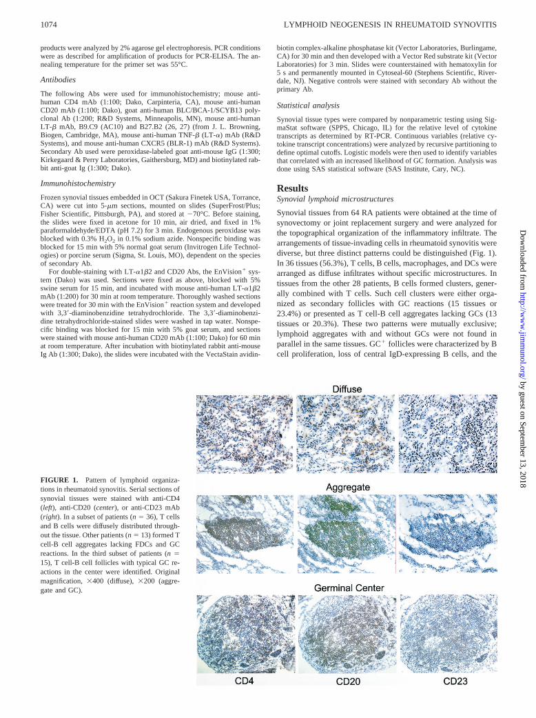
Synovial tissues from 64 RA patients were obtained at the time ofsynovectomy or joint replacement surgery and were analyzed forthe topographical organization of the inflammatory infiltrate. Thearrangements of tissue-invading cells in rheumatoid synovitis werediverse, but three distinct patterns could be distinguished (Fig. 1).In 36 tissues (56.3%), T cells, B cells, macrophages, and DCs werearranged as diffuse infiltrates without specific microstructures. Intissues from the other 28 patients, B cells formed clusters, gener-ally combined with T cells. Such cell clusters were either orga-nized as secondary follicles with GC reactions (15 tissues or23.4%) or presented as T cell-B cell aggregates lacking GCs (13tissues or 20.3%). These two patterns were mutually exclusive;lymphoid aggregates with and without GCs were not found inparallel in the same tissues. GC1 follicles were characterized by Bcell proliferation, loss of central IgD-expressing B cells, and the
FIGURE 1. Pattern of lymphoid organiza-tions in rheumatoid synovitis. Serial sections ofsynovial tissues were stained with anti-CD4(left), anti-CD20 (center), or anti-CD23 mAb(right). In a subset of patients (n5 36), T cellsand B cells were diffusely distributed through-out the tissue. Other patients (n5 13) formed Tcell-B cell aggregates lacking FDCs and GCreactions. In the third subset of patients (n515), T cell-B cell follicles with typical GC re-actions in the center were identified. Originalmagnification,3400 (diffuse), 3200 (aggre-gate and GC).
1074 LYMPHOID NEOGENESIS IN RHEUMATOID SYNOVITIS
by guest on September 13, 2018
http://ww
w.jim
munol.org/
Dow
nloaded from

presence of FDC networks. B cells in T cell-B cell aggregateswithout GCs did not actively proliferate and FDC networks werenot detectable (3). Both types of tissues with clustering of B cellsand T cells contained plasma cells that tended to accumulate underthe synovial lining layer. CD831 DCs were present in all variantsof rheumatoid synovitis.
FDCs are restricted to a subset of RA tissues
FDCs are an essential cellular component of B cell follicles insecondary lymphoid tissues. To assess whether all synovial tissuesamples contained these cells, immunohistochemical stains forCD23 were performed (Fig. 1). To avoid sampling artifacts, PCRfor a constitutive marker of FDCs, CD21L (25), was performed inparallel (Fig. 2). Both approaches yielded the same result. FDCswere found in 15 of 64 tissues (23.4%) analyzed. In all biopsieswith a positive signal for CD21L mRNA, follicles with GC reac-tions were present. In all tissues lacking GC1 follicles, CD21Lwas undetectable. In particular, tissues with T cell-B cell aggre-gates without GCs lacked FDCs, indicating that these structuresare different from primary follicles. This finding demonstrated thatthe lymphoid neogenesis in the rheumatoid synovium fundamen-tally differed from secondary lymphoid organs. The exclusive find-ing of GCs but no primary follicles suggested that the lymphoidneogenesis in the synovium is strictly dependent on an Ag recog-nition event, in distinction to normal lymph nodes. The selective
presence of FDCs also indicated that these cells are not a regularcomponent of the synovial tissue.
Correlation of tissue cytokine pattern and lymphoidmicrostructures
To identify cytokines and chemokines contributing to the forma-tion of synovial GCs, LT-a and LT-b transcripts were semiquan-tified after adjustment for the number ofb-actin transcripts. Asshown in Fig. 3, tissues with GC1 follicles contained significantlymore LT-a and LT-b mRNA than the two other tissue types. Bothcytokines were expressed at distinctly low levels in tissues withaggregates or diffuse synovitis. In biopsies with GC1 synovitis, themedian for LT-a was 182 U; LT-b-specific sequences werepresent with a median of 441 U. In synovial tissue samples with Tcell-B cell aggregates, the median values for LT-a and LT-b were42 and 35 U, respectively. Tissues with diffuse synovitis wereessentially negative for LT-a-specific sequences and had lowLT-b mRNA.
LT-a1b2 binds to the LT-bR. Functional activity of LT-b,therefore, depends on the availability of LT-bR1 cells. Expressionof LT-bR in the synovial microenvironment was analyzed by PCR.All tissue extracts contained mRNA for LT-bR. There was a trendfor GC tissues to have higher numbers of LT-bR transcripts, butthis was not statistically significant.
Correlation of tissue chemokine profiles and lymphoidmicrostructures
BLC/CXCL13 and SLC/CCL21 are critical players in the forma-tion of secondary lymphoid tissues (28–30) and are expressed inchronic inflammatory lesions. BLC/CXCL13 mRNA was detectedin synovial tissue extracts at varying levels depending on the lym-phoid microstructure encountered in the biopsy (Fig. 4). The high-est quantities of BLC/CXCL13 mRNA were detected in GC-pos-itive synovium, 15- to 30-fold higher than in tissues with T cell-Bcell aggregates (p 5 0.001) or diffuse lymphocytic infiltrates (p ,0.001). There was a trend for BLC/CXCL13 to be higher in ag-gregate-positive tissues than in diffuse tissues (median of 159 vs 89
FIGURE 2. Expression of CD21L, a specific marker for FDCs, is lim-ited to synovial tissues with GC1 follicles. cDNA was generated from allsynovial biopsies and amplified with primers specific for CD21L and forb-actin. Four representative examples for each histological pattern of RAsynovitis are shown. CD21L-specific sequences were exclusively ex-pressed in follicular tissues with GC reactions.
FIGURE 3. In situ transcription of LT-a and LT-b correlates with the pattern of lymphoid microstructure formed in RA synovitis. Tissue samples werestratified according to the pattern of synovitis. LT-a, LT-b, and LT-bR transcripts in tissue extracts were semiquantified by RT-PCR and liquid oligo-nucleotide hybridization. Results of the measurements are given as arbitrary units after normalization forb-actin. Box plots show medians, 25th and 75thpercentiles as boxes, and 10th and 90th percentiles as whiskers. Levels of LT-a and LT-b transcripts were significantly higher in tissues with GC1 follicles(GC) compared with tissues with T cell-B cell aggregates lacking GCs (Agg) or tissues with diffuse synovitis (Diff). There was no statistically significantdifference in LT-bR expression. GC-negative aggregates and diffuse synovitis did not differ for any of the three markers.
1075The Journal of Immunology
by guest on September 13, 2018
http://ww
w.jim
munol.org/
Dow
nloaded from
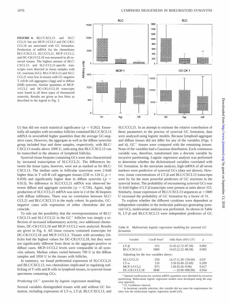
U) that did not reach statistical significance (p 5 0.262). Essen-tially all samples with secondary follicles contained BLC/CXCL13mRNA in severalfold higher quantities than the average GC-neg-ative cases. However, the aggregate as well as the diffuse synovitisgroup included four and three samples, respectively, with BLC/CXCL13 results above 1000 U, indicating that BLC/CXCL13 canbe transcribed in the absence of lymphoid follicles.
Synovial tissue biopsies containing GCs were also characterizedby increased transcription of SLC/CCL21. The differences be-tween the tissue types, however, were not as marked as for BLC/CXCL13. The median units in follicular synovium were 2-foldhigher than in T cell-B cell aggregate tissues (258 vs 124 U,p 50.059) and significantly higher than in diffuse synovitis (p 50.013). No difference in SLC/CCL21 mRNA was observed be-tween diffuse and aggregate synovitis (p 5 0.726). Again, highproduction of SLC/CCL21 mRNA was seen in 2 of the 36 biopsieswith diffuse infiltrates. There was no correlation between SLC/CCL21 and BLC/CXCL13 in the study cohort. In particular, GC-negative cases with expression of either chemokine did notcoincide.
To rule out the possibility that the overrepresentation of BLC/CXCL13 and SLC/CCL21 in the GC1 follicles was simply a re-flection of increased inflammatory activity, two additional chemo-kines, DC-CK1/CCL18 and MCP-1/CCL2 were analyzed. Resultsare given in Fig. 4. All tissue extracts contained transcripts forDC-CK1/CCL18 and MCP-1/CCL2. Tissues with secondary fol-licles had the highest values for DC-CK1/CCL18, but they werenot significantly different from those in the aggregate-positive ordiffuse cases. MCP-1/CCL2 levels were comparable in all syno-vitis subsets. Median values varied between 700 U in the diffusesamples and 1050 U in the tissues with follicles.
In summary, we found preferential expression of SLC/CCL21and BLC/CXCL13, two chemokines implicated in regulating traf-ficking of T cells and B cells in lymphoid tissues, in synovial tissuespecimens containing GCs.
Predicting GC1 synovitis by logistic regression modeling
Several variables distinguished tissues with and without GC for-mation, including expression of LT-a, LT-b, BLC/CXCL13, and
SLC/CCL21. In an attempt to estimate the relative contribution ofthese parameters to the process of synovial GC formation, datawere analyzed using logistic models. Because lymphoid aggregateand diffuse tissues did not differ for any of the variables (Figs. 3and 4), GC1 tissues were compared with the remaining tissues.None of the variables had a Gaussian distribution. Each continuousvariable was, therefore, transformed into a discrete variable byrecursive partitioning. Logistic regression analysis was performedto determine whether the dichotomized variables correlated withGC formation. In the univariate analysis, high mRNA of all sevenmarkers were predictive of synovial GCs (data not shown). How-ever, tissue concentrations of LT-b and BLC/CXCL13 transcriptswere by far the most powerful predictors of GC reactions in thesynovial lesion. The probability of encountering synovial GCs was31-fold higher if LT-b transcripts were present at units above 315.Similarly, tissue expression of BLC/CXCL13 sequences at.1800U increased the probability of GC formation by a factor of 31.
To explore whether the different cytokines were dependent orindependent variables in the molecular pathways generating syno-vial GCs, multivariate analysis was performed. As shown in TableII, LT- b and BLC/CXCL13 were independent predictors of GC
FIGURE 4. BLC/CXCL13 and SLC/CCL21 but not MCP-1/CCL2 and DC-CK1/CCL18 are associated with GC formation.Production of mRNA for the chemokinesBLC/CXCL13, SLC/CCL21, MCP-1/CCL2,and DC-CK1/CCL18 was measured in all sy-novial tissues. The highest amount of BLC/CXCL13- and SLC/CCL21-specific tran-scripts were detected in tissue samples withGC reactions (GC). BLC/CXCL13 and SLC/CCL21 were low in tissues with GC-negativeT cell-B cell aggregates (Agg) and in diffuse(Diff) synovitis. Similar quantities of MCP-1/CCL2 and DC-CK1/CCL18 transcriptswere found in all three types of rheumatoidsynovitis. Results are given as box blots asdescribed in the legend to Fig. 3.
Table II. Multivariate logistic regression modeling for synovial GCformation
Variable Cutoff Pointa Odds Ratio (95% CIb) p
LT-b 315 15.45 (2.72–87.59) 0.002BLC/CXCL13 1800 14.03 (2.22–88.58) 0.005
Adjusting for the two variables above:
SLC/CCL21 550 14.17 (1.30–159.60) 0.03c
LT-a 110 3.56 (0.49–25.69) 0.209MCP-1/CCL2 875 1.60 (0.26–9.96) 0.617DC-CK1/CCL18 3840 — (0.00–999.00) 0.954
a Optimal cutoff points for cytokine mRNA quantities were identified by recursivepartitioning. Multivariate logistic regression models were developed using the step-wise approach.
b CI, Confidence interval.c In bootstrap variable selection, this variable did not meet the requirements for
entry into the multivariate logistic regression model (43).
1076 LYMPHOID NEOGENESIS IN RHEUMATOID SYNOVITIS
by guest on September 13, 2018
http://ww
w.jim
munol.org/
Dow
nloaded from

formation. Significance for SLC/CCL21 was lost after bootstrap-ping was performed for validation of the variables in the model(43). None of the other parameters continued to be significant aftercorrection for LT-b and BLC/CXCL13 measurements. The modelthat best predicted GC formation was based on the following twoassumptions, reflecting the independent contribution of BLC/CXCL13 and LT-b: 1) patients with low BLC/CXCL13 (,1800U) and low LT-b (,315 U, defined by the recursive partitioning)have no GCs, and 2) patients with high BLC/CXCL13 and/or LT-bhave GCs. The model had a sensitivity of 86.7% and a specificityof 87.8% to correctly predict GC formation (Table III). The pos-itive predictive value was 68.4%, i.e., only 11 of the 17 tissuespredicted were true GC formers. Six of the 17 tissues fulfilled thecytokine criteria but did not form GCs, indicating that the merepresence of high LT-b and/or BLC/CXCL13 transcripts was notsufficient to guarantee follicle formation and that other variables,e.g.,cytokines not included in our analysis, may play a role. Thenegative predictive value of the model was excellent (95.6%, Ta-ble III), suggesting that GC formation does not occur if both ofthese mediators are below a certain threshold. The alternativemodel, requiring high concentrations of either cytokine, lackedsensitivity.
The cellular origin of LT-b and BLC/CXCL13 in rheumatoidsynovitis
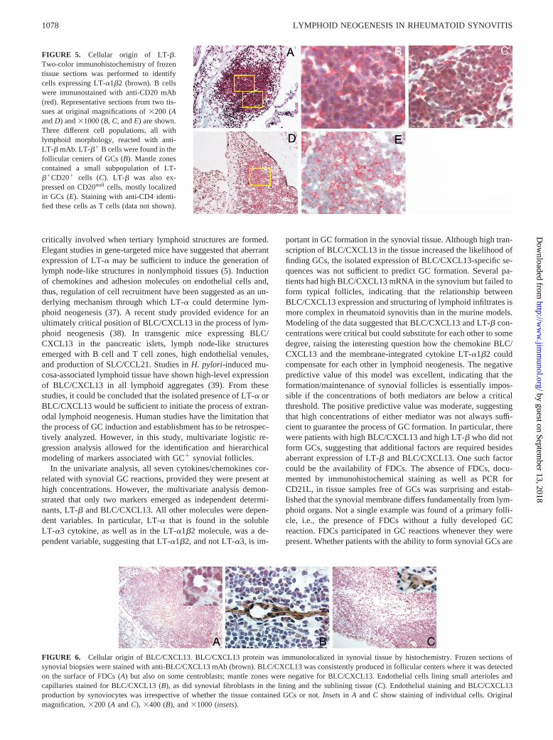
The strong predictive value of either LT-b or BLC/CXCL13 inidentifying tissues with GC reactions raised the question of whichcell types supplied these two factors in the synovial microenvi-ronment. Immunohistochemical staining was used to detect mem-brane-bound LT-b in rheumatoid synovitis. Staining of peripheralblood B cells served as positive controls; 80–85% of all circulat-ing CD201 B cells expressed LT-b. In secondary lymphoid tis-sues, such as tonsils, weak staining could be localized to few cellsin secondary follicles. More prominent staining results were ob-tained in synovial tissue sections. Sections from biopsies with dif-fuse synovitis or T cell-B cell aggregates were negative. Synovialtissue B cell follicles with GC reactions yielded a positive signalfor LT-b (Fig. 5). A subset of the CD201 B cells in the follicularcenters stained with anti-LT-b Ab. A subpopulation of B cells inthe mantle zone was also positive for surface LT-b protein. Nomorphological or topographical characteristics were found distin-guishing LT-b2 and LT-b1 B cells. LT-b staining was not re-stricted to CD201 cells. Follicular structures also included lym-phoid non-B cells staining positive. These cells expressed CD4,identifying them as CD41 T cells. The majority of T and B cellsin the tissue, however, did not stain for LT-b. LT-b positivity ofCD201 and CD20null cells was associated with follicles and wasnot encountered in interfollicular regions.
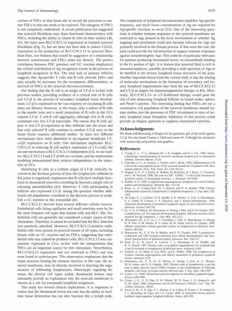
Production of BLC/CXCL13 in the spleen and lymph nodes hasbeen attributed to stromal cells, most likely FDCs. Immunohisto-
chemical analysis of synovial sections revealed intense stainingwith anti-BLC/CXCL13 Ab within the GCs. The staining patternwas compatible with FDCs expressing BLC/CXCL13 (Fig. 6).Frequently, the cytoplasm of follicular center B cells also stainedwith the Ab, whereas mantle zone cells were consistently negative.BLC/CXCL13 production in the synovial tissue was not restrictedto follicular centers. In patients with or without GC1 follicles,BLC/CXCL13 protein was detected on endothelial cells of capil-laries and small arterioles. Yet another cell population contributedto BLC/CXCL13 production in the synovial lesions; intense stain-ing was consistently found on synovial lining cells. In addition,isolated synoviocytes, dispersed throughout the tissue, expressedBLC/CXCL13 protein.
DiscussionLymphoid follicles with GC reactions are a characteristic of sy-novial lesions in RA, yet these sophisticated microstructures aregenerated in only a subset of patients. The current study incorpo-rated a large series of synovial tissue biopsies to define molecularcomponents of the process of lymphoid neogenesis in extranodalsites and to identify parameters predicting GC formation in indi-vidual patients. Distinct clustering of T cells and B cells was foundin 44% of all synovial tissues, approximately one-half of them hadGC reactions. Our study documents that the emergence of GCs inthe synovium is not dependent on a single variable but requires theconcerted action of several independent cellular responses, in par-ticular, the production of LT-b and BLC/CXCL13. Interestingly,LT-b and BLC/CXCL13 were independently regulated and could,in part, substitute for each other. LT-a1b2 originated from lym-phoid cells, mainly B cells, whereas BLC/CXCL13 was predom-inantly supplied by ancillary cells of the synovial membrane. Theability of synovial tissue cells to participate in BLC/CXCL13 pro-duction is obviously a factor guiding lymphoid neogenesis to thistissue site. Recruitment of FDCs or their precursors was identifiedas the ultimate determinant in regulating lymphoid organization inthe joint.
Emergence of GC1 follicles in extranodal sites is considered asa critical step in the generation of autoimmune process (31). It isnot unique for RA and has also been observed in other chronicinflammatory syndromes such as hepatitis C infection, Sjogren’ssyndrome,Heliobacter pylori-associated gastric mucosa-associ-ated lymphoid tissue, and Hashimoto’s thyroiditis (32–35), butusually in only a subset of cases. It is not known why only somepatients can generate these highly structured lymphoid organiza-tions. Evidence for a critical role of host-response factors in de-termining whether or not GCs are formed in the synovium comesfrom prospective monitoring of multiple successive tissue lesionsin RA patients. We have found that the pattern of lymphoid ar-rangement in the joint lesions is stable over time in individualpatients and that samples harvested from multiple different jointswill contain the same type of synovitis (S. Takemura, P. J. Kurtin,J. J. Guronzy, and C. M. Weyand, manuscript in preparation). Anadditional possible explanation is that Ags driving rheumatoid sy-novitis are diverse and that the particular topography of the lym-phoid infiltrates is a reflection of the type of Ag encountered in thesynovial membrane. Precedence for this model comes from theobservation that lymphadenitis is characterized by preferential ac-tivation of submicroenvironments in lymph nodes, depending onthe Ag that elicits the immune response. Forexample, EBV-inducedlymphadenitis is known to lead to profound activation of paracorticalT cell zones, whereas bacterial infections with streptococci can beexpected to produce follicular lymphoid hyperplasia (36).
Results of the current study indicate that the molecules impli-cated in the development of secondary lymphoid organs are also
Table III. Prediction of GC formation assuming independentcontribution of tissue LT-b and tissue BLC/CXCL13
GC Present
GC Predicteda
TotalNo Yes
No 43 6 49Yes 2 13 15Total 45 19 64
a In this model, GCs were predicted to form if either BLC/CXCL13 or LT-bexceeded the cutoff defined by recursive partitioning. (BLC/CXCL13. 1800 U;LT-b . 315 U). Positive predictive value of the model, 68.4%; negative predictivevalue of the model, 95.6%.
1077The Journal of Immunology
by guest on September 13, 2018
http://ww
w.jim
munol.org/
Dow
nloaded from
critically involved when tertiary lymphoid structures are formed.Elegant studies in gene-targeted mice have suggested that aberrantexpression of LT-a may be sufficient to induce the generation oflymph node-like structures in nonlymphoid tissues (5). Inductionof chemokines and adhesion molecules on endothelial cells and,thus, regulation of cell recruitment have been suggested as an un-derlying mechanism through which LT-a could determine lym-phoid neogenesis (37). A recent study provided evidence for anultimately critical position of BLC/CXCL13 in the process of lym-phoid neogenesis (38). In transgenic mice expressing BLC/CXCL13 in the pancreatic islets, lymph node-like structuresemerged with B cell and T cell zones, high endothelial venules,and production of SLC/CCL21. Studies inH. pylori-induced mu-cosa-associated lymphoid tissue have shown high-level expressionof BLC/CXCL13 in all lymphoid aggregates (39). From thesestudies, it could be concluded that the isolated presence of LT-a orBLC/CXCL13 would be sufficient to initiate the process of extran-odal lymphoid neogenesis. Human studies have the limitation thatthe process of GC induction and establishment has to be retrospec-tively analyzed. However, in this study, multivariate logistic re-gression analysis allowed for the identification and hierarchicalmodeling of markers associated with GC1 synovial follicles.
In the univariate analysis, all seven cytokines/chemokines cor-related with synovial GC reactions, provided they were present athigh concentrations. However, the multivariate analysis demon-strated that only two markers emerged as independent determi-nants, LT-b and BLC/CXCL13. All other molecules were depen-dent variables. In particular, LT-a that is found in the solubleLT-a3 cytokine, as well as in the LT-a1b2 molecule, was a de-pendent variable, suggesting that LT-a1b2, and not LT-a3, is im-
portant in GC formation in the synovial tissue. Although high tran-scription of BLC/CXCL13 in the tissue increased the likelihood offinding GCs, the isolated expression of BLC/CXCL13-specific se-quences was not sufficient to predict GC formation. Several pa-tients had high BLC/CXCL13 mRNA in the synovium but failed toform typical follicles, indicating that the relationship betweenBLC/CXCL13 expression and structuring of lymphoid infiltrates ismore complex in rheumatoid synovitis than in the murine models.Modeling of the data suggested that BLC/CXCL13 and LT-b con-centrations were critical but could substitute for each other to somedegree, raising the interesting question how the chemokine BLC/CXCL13 and the membrane-integrated cytokine LT-a1b2 couldcompensate for each other in lymphoid neogenesis. The negativepredictive value of this model was excellent, indicating that theformation/maintenance of synovial follicles is essentially impos-sible if the concentrations of both mediators are below a criticalthreshold. The positive predictive value was moderate, suggestingthat high concentrations of either mediator was not always suffi-cient to guarantee the process of GC formation. In particular, therewere patients with high BLC/CXCL13 and high LT-b who did notform GCs, suggesting that additional factors are required besidesaberrant expression of LT-b and BLC/CXCL13. One such factorcould be the availability of FDCs. The absence of FDCs, docu-mented by immunohistochemical staining as well as PCR forCD21L, in tissue samples free of GCs was surprising and estab-lished that the synovial membrane differs fundamentally from lym-phoid organs. Not a single example was found of a primary folli-cle, i.e., the presence of FDCs without a fully developed GCreaction. FDCs participated in GC reactions whenever they werepresent. Whether patients with the ability to form synovial GCs are
FIGURE 6. Cellular origin of BLC/CXCL13. BLC/CXCL13 protein was immunolocalized in synovial tissue by histochemistry. Frozen sections ofsynovial biopsies were stained with anti-BLC/CXCL13 mAb (brown). BLC/CXCL13 was consistently produced in follicular centers where it was detectedon the surface of FDCs (A) but also on some centroblasts; mantle zones were negative for BLC/CXCL13. Endothelial cells lining small arterioles andcapillaries stained for BLC/CXCL13 (B), as did synovial fibroblasts in the lining and the sublining tissue (C). Endothelial staining and BLC/CXCL13production by synoviocytes was irrespective of whether the tissue contained GCs or not.Insetsin A and C show staining of individual cells. Originalmagnification,3200 (AandC), 3400 (B), and31000 (insets).
FIGURE 5. Cellular origin of LT-b.Two-color immunohistochemistry of frozentissue sections was performed to identifycells expressing LT-a1b2 (brown). B cellswere immunostained with anti-CD20 mAb(red). Representative sections from two tis-sues at original magnifications of3200 (AandD) and31000 (B,C, andE) are shown.Three different cell populations, all withlymphoid morphology, reacted with anti-LT-b mAb. LT-b1 B cells were found in thefollicular centers of GCs (B). Mantle zonescontained a small subpopulation of LT-b1CD201 cells (C). LT-b was also ex-pressed on CD20null cells, mostly localizedin GCs (E). Staining with anti-CD4 identi-fied these cells as T cells (data not shown).
1078 LYMPHOID NEOGENESIS IN RHEUMATOID SYNOVITIS
by guest on September 13, 2018
http://ww
w.jim
munol.org/
Dow
nloaded from
carriers of FDCs at that tissue site or recruit the precursor or ma-ture FDCs to that site needs to be explored. The ontogeny of FDCsis not completely understood. A recent publication has suggestedthat synovial fibroblasts may share functional characteristics withFDCs, including the ability to cluster B cells on their surface (40,41). We have seen BLC/CXCL13 expression in isolated synovialfibroblasts (Fig. 5), but we have not been able to induce CD21Lexpression or the production of BLC/CXCL13 in synovial fibro-blast lines, two features that would be suggestive of a relationshipbetween synoviocytes and FDCs (data not shown). The perfectcorrelation between FDC presence and GC reaction emphasizesthe critical contribution of Ag recognition events in the process oflymphoid neogenesis in RA. The total lack of primary folliclessuggests that Ag-specific T cells and B cells precede FDCs andmay actually be necessary for the recruitment, differentiation, orsurvival of FDCs in the synovial microenvironment.
Our finding that the B cell is an origin of LT-b is in-line withprevious studies, providing evidence of a critical role of B cellsand LT-b in the process of secondary lymphoid tissue develop-ment. LT-b is expressed on the vast majority of circulating B cells(data not shown). However, in the tissue, only a subset of B cellsin the mantle zone and a small proportion of B cells in the GCsexpress LT-b. T cell-B cell aggregates, although rich in B cells,contained very few LT-b transcripts. The reason that B cells ap-pear to lose LT-b expression as they infiltrate into the tissue andthat only selected B cells continue to produce LT-b once in thetissue lesion requires additional studies. At least two differentmechanisms have been identified to up-regulate membrane LT-a1b2 expression on B cells. One mechanism implicates BLC/CXCL13 in inducing B cell surface expression of LT-a1b2; thesecond mechanism is BLC/CXCL13 independent (15). In our stud-ies, BLC/CXCL13 and LT-b did not correlate, and the multivariatemodeling demonstrated their relative independence in the induc-tion of GCs.
Identifying B cells as a cellular origin of LT-b, critically in-volved in the decision process of how the lymphocytic infiltrate inRA joints is organized, emphasizes that B cells have multiple func-tions in rheumatoid synovitis extending far beyond a simple role ofreleasing autoantibodies (42). However, T cells participating infollicles also expressed LT-b, raising the question whether addi-tional cell populations contribute to the decision process to estab-lish a GC reaction in this extranodal site.
BLC/CXCL13 derived from several different cellular sources.Endothelial cells lining capillaries and small arterioles were by farthe most frequent cell types that stained with anti-BLC Abs. En-dothelial cells are generally not considered a major source of thischemokine. Therefore, it cannot be excluded that the BLC detectedwas passively adsorbed. However, BLC/CXCL13-positive endo-thelial cells were present in synovial tissues of all types, includingtissues with no GC reaction and no FDCs, suggesting that endo-thelial cells may indeed be producer cells. BLC/CXCL13 was con-sistently expressed in GCs, in-line with the interpretation thatFDCs are an important source for this chemokine. Nevertheless,BLC/CXCL13 expression was not restricted to FDCs and waseven found in synoviocytes. This observation emphasizes that thetissue structure hosting the immune reaction, in this case, the sy-novial membrane, may be directly involved in directing the orga-nization of infiltrating lymphocytes. Heterotypic signaling be-tween the diverse cell types within rheumatoid lesions mayultimately provide an explanation why the synovial membrane ischosen as a site for extranodal lymphoid neogenesis.
This study has several clinical implications. It is important torealize that the rheumatoid lesion not only has the ability to facil-itate tissue destruction but can also function like a lymph node.
The complexity of lymphoid microstructures amplifies Ag-specificresponses, and much lower concentrations of Ag are required forAg-specific reactions to occur (31). One of the interesting ques-tions is whether immune responses in the synovial membrane arerestricted to Ags present in the local environment or whether Agtrapping and enrichment could also become relevant for Ags notprimarily involved in the disease process. If that were the case, thejoint could provide the infrastructure to support immune responsesagainst nonarthritogenic Ags. This could be of particular relevancefor patients producing rheumatoid factor, an autoantibody bindingto the Fc portion of IgG. It is known that synovial fluid is rich inimmunocomplexes, possibly providing a wide spectrum of Ags tobe handled in the tertiary lymphoid tissue structures of the joint.Another important lesson from the current study is that the sharingof molecular mechanisms in the formation of secondary and ter-tiary lymphoid organizations may limit the use of BLC/CXCL13and LT-b as targets for immunosuppressive therapy in RA. Obvi-ously, BLC/CXCL13- or LT-b-directed therapy would pose therisk of also destroying lymphoid follicles in lymph nodes, spleen,and Peyer’s patches. The interesting finding that FDCs are not aconstitutive cell population of the synovial membrane should ini-tiate studies into the processes of FDC recruitment to sites of ter-tiary lymphoid tissue formation. Inhibition of that process couldprovide an elegant approach to suppress rheumatoid synovitis.
AcknowledgmentsWe thank Jeff Browning at Biogen for his generous gift of the mAb againstLT-b. We also thank Tammy J. Dahl and James W. Fulbright for assistancewith manuscript preparation and graphics.
References1. Young, C. L., T. C. Adamson III, J. H. Vaughan, and R. I. Fox. 1984. Immu-
nohistologic characterization of synovial membrane lymphocytes in rheumatoidarthritis.Arthritis Rheum. 27:32.
2. Schroder, A. E., A. Greiner, C. Seyfert, and C. Berek. 1996. Differentiation of Bcells in the nonlymphoid tissue of the synovial membrane of patients with rheu-matoid arthritis.Proc. Natl. Acad. Sci. USA 93:221.
3. Wagner, U. G., P. J. Kurtin, A. Wahner, M. Brackertz, D. J. Berry, J. J. Goronzy,and C. M. Weyand. 1998. The role of CD81 CD40L1 T cells in the formation ofgerminal centers in rheumatoid synovitis.J. Immunol. 161:6390.
4. Ruddle, N. H. 1999. Lymphoid neo-organogenesis: lymphotoxin’s role in inflam-mation and development.Immunol. Res. 19:119.
5. Kratz, A., A. Campos-Neto, M. S. Hanson, and N. H. Ruddle. 1996. Chronicinflammation caused by lymphotoxin is lymphoid neogenesis.J. Exp. Med. 183:1461.
6. De Togni, P., J. Goellner, N. H. Ruddle, P. R. Streeter, A. Fick, S. Mariathasan,S. C. Smith, R. Carlson, L. P. Shornick, and J. Strauss-Schoenberger. 1994.Abnormal development of peripheral lymphoid organs in mice deficient in lym-photoxin.Science 264:703.
7. Fu, Y. X., H. Molina, M. Matsumoto, G. Huang, J. Min, and D. D. Chaplin. 1997.Lymphotoxin-a (LTa) supports development of splenic follicular structure that isrequired for IgG responses.J. Exp. Med. 185:2111.
8. Matsumoto, M., S. F. Lo, C. J. Carruthers, J. Min, S. Mariathasan, G. Huang,D. R. Plas, S. M. Martin, R. S. Geha, M. H. Nahm, and D. D. Chaplin. 1996.Affinity maturation without germinal centres in lymphotoxin-a-deficient mice.Nature 382:462.
9. Matsumoto, M., Y. X. Fu, H. Molina, and D. D. Chaplin. 1997. Lymphotoxin-a-deficient and TNF receptor-I-deficient mice define developmental and func-tional characteristics of germinal centers.Immunol. Rev. 156:137.
10. Koni, P. A., R. Sacca, P. Lawton, J. L. Browning, N. H. Ruddle, andR. A. Flavell. 1997. Distinct roles in lymphoid organogenesis for lymphotoxinsa andb revealed in lymphotoxinb-deficient mice.Immunity 6:491.
11. Futterer, A., K. Mink, A. Luz, M.H., and K. Pfeffer. 1998. The lymphotoxinbreceptor controls organogenesis and affinity maturation in peripheral lymphoidtissues.Immunity 9:59.
12. Matsumoto, M., Y. X. Fu, H. Molina, G. Huang, J. Kim, D. A. Thomas,M. H. Nahm, and D. D. Chaplin. 1997. Distinct roles of lymphotoxina and thetype I tumor necrosis factor (TNF) receptor in the establishment of folliculardendritic cells from non-bone marrow-derived cells.J. Exp. Med. 186:1997.
13. Cyster, J. G. 1999. Chemokines and cell migration in secondary lymphoid organs.Science 286:2098.
14. Cyster, J. G., V. N. Ngo, E. H. Ekland, M. D. Gunn, J. D. Sedgwick, andK. M. Ansel. 1999. Chemokines and B-cell homing to follicles.Curr. Top. Mi-crobiol. Immunol. 246:87.
15. Ansel, K. M., V. N. Ngo, P. L. Hyman, S. A. Luther, R. Forster, J. D. Sedgwick,J. L. Browning, M. Lipp, and J. G. Cyster. 2000. A chemokine-driven positivefeedback loop organizes lymphoid follicles.Nature 406:309.
1079The Journal of Immunology
by guest on September 13, 2018
http://ww
w.jim
munol.org/
Dow
nloaded from
16. Gunn, M. D., S. Kyuwa, C. Tam, T. Kakiuchi, A. Matsuzawa, L. T. Williams, andH. Nakano. 1999. Mice lacking expression of secondary lymphoid organ che-mokine have defects in lymphocyte homing and dendritic cell localization.J. Exp. Med. 189:451.
17. Nakano, H., S. Mori, H. Yonekawa, H. Nariuchi, A. Matsuzawa, and T. Kakiuchi.1998. A novel mutant gene involved in T-lymphocyte-specific homing into pe-ripheral lymphoid organs on mouse chromosome 4.Blood 91:2886.
18. Vassileva, G., H. Soto, A. Zlotnik, H. Nakano, T. Kakiuchi, J. A. Hedrick, andS. A. Lira. 1999. The reduced expression of 6Ckine in theplt mouse results fromthe deletion of one of two 6Ckine genes.J. Exp. Med. 190:1183.
19. Forster, R., A. Schubel, D. Breitfeld, E. Kremmer, I. Renner-Muller, E. Wolf, andM. Lipp. 1999. CCR7 coordinates the primary immune response by establishingfunctional microenvironments in secondary lymphoid organs.Cell 99:23.
20. Forster, R., T. Emrich, E. Kremmer, and M. Lipp. 1994. Expression of the G-protein-coupled receptor BLR1 defines mature, recirculating B cells and a subsetof T-helper memory cells.Blood 84:830.
21. Forster, R., A. E. Mattis, E. Kremmer, E. Wolf, G. Brem, and M. Lipp. 1996. Aputative chemokine receptor, BLR1, directs B cell migration to defined lymphoidorgans and specific anatomic compartments of the spleen.Cell 87:1037.
22. van der Valk, P., and C. Meijer. 1987. The histology of reactive lymph nodes.Am. J. Surg. Pathol. 11:866.
23. Brack, A., H. L. Rittner, B. R. Younge, C. Kaltschmidt, C. M. Weyand, andJ. J. Goronzy. 1997. Glucocorticoid-mediated repression of cytokine gene tran-scription in human arteritis-SCID chimeras.J. Clin. Invest.99:2842.
24. Klimiuk, P. A., J. J. Goronzy, J. Bjornsson, R. D. Beckenbaugh, andC. M. Weyand. 1997. Tissue cytokine patterns distinguish variants of rheumatoidsynovitis.Am. J. Pathol. 151:1311.
25. Liu, Y. J., J. Xu, O. de Bouteiller, C. L. Parham, G. Grouard, O. Djossou,B. de Saint-Vis, S. Lebecque, J. Banchereau, and K. W. Moore. 1997. Folliculardendritic cells specifically express the long CR2/CD21 isoform.J. Exp. Med.185:165.
26. Browning, J. L., I. Dougas, A. Ngam-ek, P. R. Bourdon, B. N. Ehrenfels,K. Miatkowski, M. Zafari, A. M. Yampaglia, P. Lawton, W. Meier, et al.. 1995.Characterization of surface lymphotoxin forms: use of specific monoclonal an-tibodies and soluble receptors.J. Immunol. 154:33.
27. Cannella, B., I. D. Sizing, C. D. Benjamin, J. L. Browning, and C. S. Raine. 1997.Antibodies to lymphotoxina (LT a) and LTb recognize different glial cell typesin the central nervous system.J. Neuroimmunol. 78:172.
28. Hjelmstrom, P., J. Fjell, T. Nakagawa, R. Sacca, C. A. Cuff, and N. H. Ruddle.2000. Lymphoid tissue homing chemokines are expressed in chronic inflamma-tion. Am. J. Pathol. 156:1133.
29. Cyster, J. G. 2000. Leukocyte migration: scent of the T zone.Curr. Biol. 10:R30.30. Ngo, V. N., H. Korner, M. D. Gunn, K. N. Schmidt, D. S. Riminton,
M. D. Cooper, J. L. Browning, J. D. Sedgwick, and J. G. Cyster. 1999. Lym-
photoxina/b and tumor necrosis factor are required for stromal cell expressionof homing chemokines in B and T cell areas of the spleen.J. Exp. Med. 189:403.
31. Fehr, T., C. Lopez-Macias, B. Odermatt, R. M. Torres, D. B. Schubart, T. L.O’Keefe, P. Matthias, H. Hengartner, and R. M. Zinkernagel. 2000. Correlationof anti-viral B cell responses and splenic morphology with expression of B cell-specific molecules.Int. Immunol. 12:1275.
32. Banerjee, S. K., A. P. Weston, M. N. Zoubine, D. R. Campbell, and R. Cherian.2000. Expression of cdc2. and cyclin B1 inHelicobacter pylori-associated gastricMALT and MALT lymphoma: relationship to cell death, proliferation, and trans-formation.Am. J. Pathol. 156:217.
33. Freni, M. A., D. Artuso, G. Gerken, C. Spanti, T. Marafioti, N. Alessi,A. Spadaro, A. Ajello, and O. Ferrau. 1995. Focal lymphocytic aggregates inchronic hepatitis C: occurrence, immunohistochemical characterization, and re-lation to markers of autoimmunity.Hepatology 22:389.
34. Lennert, K., and U. Schmid. 1983. Prelymphoma, early lymphoma, and manifestlymphoma in immunosialadenitis (Sjogren’s syndrome): a model of lym-phomagenesis.Haematol. Bluttransfus 28:418.
35. Imal, Y., and M. Yamakawa. 1996. Morphology, function and pathology of fol-licular dendritic cells.Pathol. Int. 46:807.
36. Schnitzer, B. 1995. Reactive lymphoid hyperplasia. InSurgical Pathology of theLymph Nodes and Related Organs, 2nd Ed. E. S. Jaffe, ed. Saunders, Philadel-phia, p. 98.
37. Cuff, C. A., J. Schwartz, C. M. Bergman, K. S. Russell, J. R. Bender, andN. H. Ruddle. 1998. Lymphotoxina3 induces chemokines and adhesion mole-cules: insight into the role of LTa in inflammation and lymphoid organ devel-opment.J. Immunol. 161:6853.
38. Luther, S. A., T. Lopez, W. Bai, D. Hanahan, and J. G. Cyster. 2000. BLCexpression in pancreatic islets causes B cell recruitment and lymphotoxin-depen-dent lymphoid neogenesis.Immunity 12:471.
39. Mazzucchelli, L., A. Blaser, A. Kappeler, P. Scharli, J. Laissue, M. Baggiolini,and M. Uguccioni. 1999. BCA-1 is highly expressed inHelicobacter pylori-induced mucosa-associated lymphoid tissue and gastric lymphoma.J. Clin. In-vest. 104:49.
40. Lindhout, E., M. van Eijk, M. van Pel, J. Lindeman, H. J. Dinant, andC. de Groot. 1999. Fibroblast-like synoviocytes from rheumatoid arthritis patientshave intrinsic properties of follicular dendritic cells.J. Immunol. 162:5949.
41. Bofill, M., A. N. Akbar, and P. L. Amlot. 2000. Follicular dendritic cells share amembrane-bound protein with fibroblasts.J. Pathol. 191:217.
42. Weyand, C. M., J. J. Goronzy, S. Takemura, and P. J. Kurtin. 2000. T cell-B cellinteractions in rheumatoid arthritis.Arthritis Res. 2:457.
43. Sauerbrei, W., and M. Schumacher. 1992. A bootstrap resampling procedure formodel building: application to the Cox regression model.Stat. Med. 11:2093.
1080 LYMPHOID NEOGENESIS IN RHEUMATOID SYNOVITIS
by guest on September 13, 2018
http://ww
w.jim
munol.org/
Dow
nloaded from