congenital ichthyosis in pediatric age group - a...
TRANSCRIPT

CONGENITAL ICHTHYOSIS IN PEDIATRIC AGE
GROUP - A CLINICAL STUDY
Dissertation Submitted to
THE TAMIL NADU DR. M.G.R. MEDICAL UNIVERSITY
in partial fulfillment of the regulations
for the award of the degree of
M.D. (Dermatology, Venereology and Leprology)
BRANCH - XII A
MADRAS MEDICAL COLLEGE
THE TAMIL NADU DR. M.G.R. MEDICAL
UNIVERSITY, CHENNAI, INDIA.
MARCH 2008

CERTIFICATE
Certified that this dissertation entitled “Congenital
ichthyosis in pediatric age group – a clinical study” is a bonafide work
done by Dr.P.Sivayadevi postgraduate student of the department of
Dermatology, Venereology and Leprosy, Madras Medical College,
Chennai- 600003, during the academic year 2005-2008. This work has
not previously formed the basis for the award of any degree.
Prof. Dr. B. Parveen M.D., D.D., Professor and Head of the Department, Department of Dermatology, and Leprology, Madras Medical College, Chennai – 600003.
PROF. DR.T.P. KALA NITI M.D,
DEAN,
Madras Medical College,
Chennai – 600003.

SPECIAL ACKNOWLEDGEMENT
My sincere thanks to
PROF.DR.T.P. KALA NITI M.D, DEAN,
Madras Medical College
for allowing me to do this dissertation and utilize the institutional facilities.

ACKNOWLEDGEMENT
I am gratefully indebted to Prof. Dr. B. Parveen M.D., D.D.,
Professor and Head, Department of Dermatology and Leprology for
her invaluable guidance, motivation and help through out the study. I
would like to express my sincere and heartfelt gratitude to Prof. Dr.
V.S.Dorairaj, M.D., D.V., Director, Institute of Venereology.
I am very grateful to Dr.S.Jayakumar M.D.,D.D., Additional
Professor, Department of Dermatology for his invaluable guidance
and help. I sincerely thank Dr.C.Janaki,M.D.,D.D., Reader of
Dermatology (Mycology) for her priceless support.
I express my earnest gratefulness to Dr.P.Prabavathy M.D., D.D.,
professor and Head of the Department of Occupational Dermatology
and Contact Dermatitis for her constant motivation and guidance. I
thank Dr.V.Somasundaram M.D., D.D., Additional Professor,
Department of Occupational Dermatology and contact Dermatitis for
his benevolent help and support.
I incline to thank Dr. V. Anandan, M.D.,(Derm), D.C.H.,
D.N.B.,(Paed), Dr.R.Priyavathani, M.D., D.D., D.N.B., Dr.

G.k.Tharini M.D., and Dr.N.Hema M.D.,(DVL) Dr.S.Anupama
Roshan, D.D.V.L., Assistant Professors, Department of Dermatology
for their kind support and encouragement.
I thank Dr.A.Hameedullah M.D.,D.D., Dr.S.Kumaravelu
M.D.,D.D., Dr.J.Manjula M.D.,D.N.B.,(Derm) and Dr.Aftab
Jameela Wahab M.D.,D.D., Assistant Professors, Department of
Occupational Dermatology and Contact Dermatitis for their support
and help.
My sincere thanks to Dr.N.Kumar, M.D., D.V., D.M.R.D.,
Additional professor, Dr.V.Thirunavukkarasu M.D., D.V., Registrar,
Dr.K.Venkateshwaran M.D., D.V., Dr.S.Thilagavathy M.D., D.V.,
Dr.P.Mohan M.D., D.V., Dr.S.Arunkumar M.D., D.V., and
Dr.S.Kalaivani M.D., D.V., Dr.P.Prabakar,M.D.,(DVL), Assistant
Professors, Institute of Venereology, for their help and suggestions.
I express my sincere gratitude to Dr.R.Arunadevi M.D.,
D.D., Lecturer /Registrar, Department of Dermatology for her
support.

I wish to thank Dr.N.Gomathy, M.D.,D.D., former Professor,
Department of Dermatology and Dr.N.S.Usman, M.D., D.V.,Ph.D.,
former Director, Institute of Venereology, Dr.K.Rathinavelu M.D.,
D.D., former Professor of Leprosy for their constant support and
motivation.
I am also thankful to Dr.S.Mohan M.D., D.V., former registrar,
Dr.D.Ramachandra Reddy M.D., D.V., Dr.P.Elangovan M.D.,
D.V., and Dr.V.Sampath M.D.,D.D., for their continuing guidance
and support.
I duly acknowledge the paramedical staff and my colleagues
for their help and favour.
Last but not least I am profoundly grateful to all patients for their
cooperation and participation in the study.

CONTENTS
Sl.No Title Page No.
1 INTRODUCTION 1
2 REVIEW OF LITERATURE 2
3 AIMS OF THE STUDY
4 MATERIALS AND METHODS
5 OBSERVATIONS AND RESULTS
6 DISCUSSION
7 CONCLUSION
BIBLIOGRAPHY
PROFORMA
MASTER CHART
INTRODUCTION
Ichthyoses comprise of a heterogeneous group of disorders,
due to defect in keratinization or cornification with abnormal
differentiation and desquamation of epidermis. It is clinically
characterized by dry rough skin with scaling over much or the entire
body surface.
The terminology and nosology of congenital ichthyosis has
continuously evolved and has lead to a confusing medley of different
terms and classification systems. Recent advances in the molecular
genetics has provided tools to categorize ichthyosis, on the basis of their
underlying genetic defects.
A number of well defined types of ichthyoses have characteristic
features and can be reliably diagnosed. But a specific diagnosis can be
challenging in certain patients and families due to great clinical
heterogeneity.
In general, determination of whether an ichthyosis is inherited or
acquired, presented at birth or later in life, and whether it is limited to
the skin or part of multisystem disorder, helps in diagnosis. Quality and
distribution of scale, presence or absence of erythroderma, blistering,

associated abnormalities of skin adnexae are other useful clinical
features. A thorough family history is essential for recognizing the
inheritance pattern. Establishing the correct clinical diagnosis in a
patient with ichthyosis is a prerequisite for making prognostic
predictions, therapeutic decisions and offering genetic counselling.
REVIEW OF LITERATURE
CONGENITAL ICHTHYOSIS
SYNONYMS
Fish skin disease, Alligator skin disease, Sauriasis, Congenital
hyperkeratosis.
HISTORY
The term ichthyosis derives from the Greek root
‘ichthys’ for fish.
First historic reference to ichthyosis appears in an
Indian text in 250Bc – ‘Ekakushtha, skin disease
like scales of a fish’1.
Hystrix type ichthyosis – ‘porcupine men’ in the
lambert family of Suffolk was reported by Machin
(1732)
Harlequin fetus – one of the first genodermatoses
recorded by Oliver Hart (1750)1.
‘Ichthyosis nacree’ described by Alibert (1806) is
the most likely first well documented report of
ichthyosis vulgaris.
Willan (1808) – classified ichthyosis as a
‘squamous disease’.

Brocq (1902) - distinguished bullous and
nonbullous2 ichthyosiform erythroderma.
Cockayne (1933) – first to use genetic classification
of ichthyosis.
Refsum’s disease – described by Norwegian
neurologist Refsum (1946)4.
Sjögren – Larsson syndrome- described by Swedish
psychiatrists (1957).
Wells and kerr (1965) - recognized X- linked
ichthyosis3.
CLASSIFICATION
Early classification was based on scale description
– ichthyosis larvata/ tarda/ mitis/ inversa.
Wells and Kerr classified ichthyosis according to inheritance pattern
- AD/ AR/ XLR.
Vanscott, Frost, Weinstern5 –classified based on rates of epidermal
turn over.
Retention ichthyoses
Ichthyosis vulgaris
Recessive X-linked ichthyosis
Lamellar ichthyosis
Hyperproliferative ichthyoses
Non-bullous ichthyosiform erythroderma

Bullous ichthyosiform erythroderma
Refsum’s disease
Sjögren-Larsson syndrome
CONGENITAL ICHTHYOSES
MAJOR FORMS:
Ichthyosis vulgaris
X-linked recessive ichthyosis
Non- bullous ichthyosiform erythroderma
Lamellar ichthyosis
Harlequin ichthyosis
Bullous ichthyosiform erythroderma
Ichthyosis bullosa of Siemens
Ichthyosis hystrix
ICHTHYOSIFORM SYNDROMES:
Netherton’s syndrome
Sjögren- Larsson syndrome
Neutral lipid storage disease
Refsum’s disease
Kallman’s syndrome
Multiple sulphatase deficiency syndrome
X-linked dominant ichthyosis
IBIDS (trichothiodystrophy)

KID syndrome
CHILD syndrome
Ichthyosis follicularis with alopecia and photophobia
Rud’s syndrome
Congenital ichthyosis variants
Isolated genetic syndromes with ichthyosis
PATHOGENESIS:
The primary function of the stratum corneum is to provide
a barrier to water loss, without which terrestrial life is not possible.
Defective barrier function leads to increased transepidermal water loss, a
characteristic feature of ichthyosis.
The stratum corneum is a double compartment system
analogous to a brick wall, in which the corneocytes (“bricks”) provide
the structural building blocks, around which a lipid enriched
extracellular matrix (the “mortar”) is deposited, to provide the
permeability barrier to systemic water loss6. These
lipids,predominantly,the neutral lipids,cholesterol sulphate, free fatty
acids,and the polar lipids,ceramides,form repeating units of electron-
lucent and electron-dense membranes,termed lamellar unit structures,
when visualized with ruthenium tetroxide.Two or three lamellar unit
structures fill the intercellular domains of stratum corneum. Lipids are

delivered to this site through secretion of lamellar bodies at the stratum
granulosum-stratum corneum interface. Lamellar bodies contain
glycosylceramides, phospholipids and cholesterol sulphate, as well as
certain hydrolytic enzymes9. Upon secretion, some of these enzymes
process glycosylceramides and phospholipids to ceramides and free fatty
acids respectively. Conversion of cholesterol sulphate to cholesterol by
cholesterol sulphatase present on the cell membrane surface, leads to
breakdown of the intercellular lipid lamellae, and resultant
desquamation.
In addition to lipids,the mortar of inner stratum corneum
contains corneodesmosomes that span adjacent corneocytes7. Proteolysis
of corneodesmosomes is required for normal desquamation8. In normal
skin, desmosome density and cohesion lessens in transit from lower to
upper stratum corneum.
Disordered keratinization also results from alterations in
structural proteins like cornified cell envelope and enclosed aggregated
keratin filaments,which are the major components of the stratum
corneum. Envelope precursors such as involucrin, loricrin, small
proline- rich proteins and envoplakin, are synthesized late in
stratification and then cross- linked by the action of transglutaminase
enzymes, which are synthesized in the granular layer.The corneocyte

protein envelope is linked covanently to an outer ceramide layer, the
lipid envelope, which also contains a variety of membrane associated
glycoproteins such as the integrins. Keratin intermediate filaments are
the major stress bearing cytoskeletal proteins. They are aggregated by
interaction with filaggrin (filament aggregating protein), a basic
histidine rich protein, stored as profilaggrin in keratohyaline granules.
Normal desquamation is an invisible process by which a
single corneocyte or small clump of corneocytes detaches from its
neighbours and shed. In the retention hyperkeratoses, normal epidermal
homeostasis is maintained, but desquamation is retarded and
corneocytes are shed in large clumps. In the hyperproliferative
ichthyoses, epidermal homeostasis is disturbed, the process of
desquamation is abnormal as well. Chronic barrier damage induces
keratinocyte DNA synthesis and results in epidermal hyperplasia10, with
knock-on effects on the activity of cytokines, growth factors, calcium
gradients, adhesion molecules and lytic enzymes.
ICHTHYOSIS VULGARIS
Synonyms
Ichthyosis simplex
Autosomal dominant ichthyosis
Most common of the inherited ichthyoses, with a reported
incidence of 1 in 25012. It is an autosomal dominant disorder with
variable phenotypic expression and penetrance, so severity can vary
between generations and affected siblings.
Pathogenesis
Absence or decrease of filaggrin and its precursor, profilaggrin is
seen in the epidermis from patients with ichthyosis vulgaris in
biomedical studies13 . Expression of mRNA is reduced14.There may be
selectively impaired post transcriptional control of profilaggrin synthesis
or the profilaggrin gene may be influenced by other mutated genes15.
Scale formation is thought to result from loss of water retaining
aminoacids derived from filaggrin catabolism16. The hyperkeratosis is
regarded as a retention keratosis resulting from increased adhesiveness
of stratum corneum. Labelling with tritiated thymidine shows a normal
rate of epidermal proliferation18.
Pathology
In the epidermis, there is moderate degree of hyperkeratosis with
a thin or absent granular layer. Hyperkeratosis often extends in to the
hair follicles, resulting in large keratotic follicular plugs.
Clinical features
Scaling is obvious from two months of age or may be further
delayed. Clinical symptoms and severity depend on season and climate,
improving during the summer with increasing humidity, and worsening

in a dry, cold environment. It usually improves with advancing age12 .
Pruritus is not a problem but will be present if associated with atopy.
Rarely hypohidrosis with heat intolerance may be present. Scaling is
most pronounced on the extensor surfaces of the arms and lower legs.
The groin and flexural areas are spared because of increased humidity in
those regions.Coexistant atopic dermatitis may obscure this feature.
Face is usually spared, if involved localizes to forehead and cheeks,
perioral region. Scaling on the trunk is less pronounced and diaper area
is spared.
Scales are white or grey, small, flaky or branny and semi
adherent with turned up edges giving a “pasted- on” appearance. Palms
and soles show accentuated skin markings – hyperlinearity, due to mild
hyperkeratosis. If severe, furrows or painful fissures may occur on the
heels.
Associations17
Keratosis pilaris- involves posterior arms, thighs and
buttocks.
Atopic triad of asthma, hayfever and atopic dermatitis in
as many as 25 – 50% patients.
Ocular manifestations and testicular cancer have been
reported.

X LINKED RECESSIVE ICHTHYOSIS
An X linked recessive disorder, which almost exclusively affects
the male patients.
Estimated incidence ranges between 1 in 2000 to 1 in 9500 male
births19.
Pathogenesis
It is due to the inheritance of mutated steroid sulfatase gene (90%
deletion) on chromosome XP 22.32 from the carrier mothers20. Steroid
sulfatase is a membrane bound microsomal enzyme responsible for
hydrolyzing sulphate groups from cholesterol sulphate and sulfated
steroid hormones. Continued hydrolysis of cholesterol sulphate during
corneocyte transit from inner to outer stratum corneum is a critical step
normally leading to desquamation. Due to decrease in enzyme activity,
cholesterol sulphate accumulates in the scale, constituting 30% of
stratum corneum lipids21(normal 3%) .Concomitant placental steroid
sulfatase deficiency causes inadequate deconjugation of DHEAS
necessary for estrogen synthesis which in turn leads to failure to initiate
labour22.Altered sex hormone profile may in part explain the abnormal
testicular development in some patients.
Pathology
Epidermis shows slightly thickened with orthokeratotic
hyperkeratosis23.
Granular layer is normal or slightly thickened.
Kinetic studies show normal rates of cell turn over.
Clinical features
Age of onset –In 75% patients, scaling is evident within first week of
life22 as pronounced peeling or desquamation.In 6% manifestations
occur after 1 year24.Scaling tends to increase throughout childhood
spreading up from lower legs to the trunk. and stabilizes in the
teens.Scaling diminishes in summer months. But does not subside with
age.
Site
Scaling is more prominent on the extensor surface extremities
with significant flexural surface involvement. Posterior and lateral part
of neck is almost invariably involved- ‘dirty neck disease’. Upper and
lateral abdominal wall and preauricular facial skin are commonly
affected. Palm /soles are spared.
Scales
Medium to large, polygonal, dull, light to dark brown scales with
tight adherence to skin. Light grey scaling may be seen in face, scalp,
axillae, flexor aspect of limbs.
Systemic manifestations
Comma shaped asymptomatic corneal opacities in slit lamp
examination is seen in 50 to 90% of adult patients and 25% of carrier
females,due to stromal deposits in posterior surface of Descemet’s
membrane25.
Cryptorchidism is seen in 25% patients26.
Abnormalities of sperm count or motility and testicular cancer27
independent of
cryptorchidism.
Inguinal hernia
Unilateral renal agenesis
Rare – epilepsy, acute lymphoblastic leukaemia, paraplegia /
parapareis due to prolonged labour.
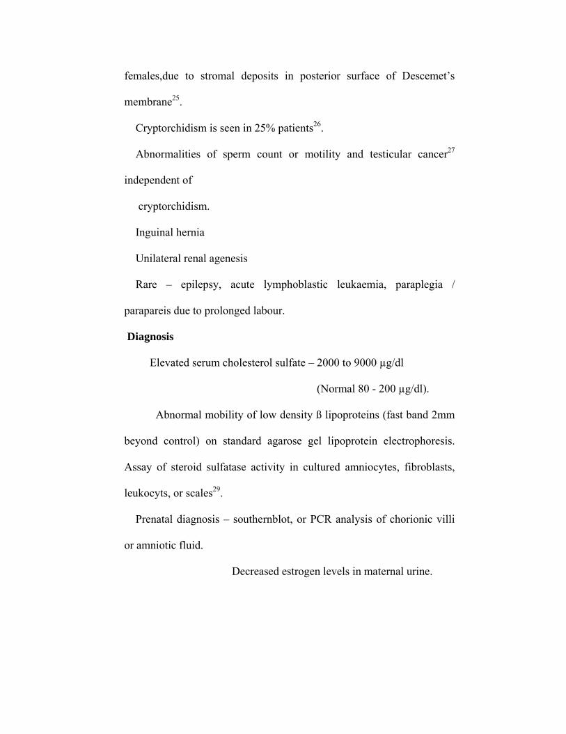
Diagnosis
Elevated serum cholesterol sulfate – 2000 to 9000 µg/dl
(Normal 80 - 200 µg/dl).
Abnormal mobility of low density ß lipoproteins (fast band 2mm
beyond control) on standard agarose gel lipoprotein electrophoresis.
Assay of steroid sulfatase activity in cultured amniocytes, fibroblasts,
leukocyts, or scales29.
Prenatal diagnosis – southernblot, or PCR analysis of chorionic villi
or amniotic fluid.
Decreased estrogen levels in maternal urine.

COLLODION BABY
It is a descriptive term for the baby who is born encased in a
transulcent, parchment like membranous covering resembling a dried
film of collodion.
It is the usual presentation of congenital recessive ichthyoses30
like non-bullous ichthyosiform erythroderma and lamellar ichthyosis34.
Others include trichothiodystrophy, Sjögren Larsson syndrome, and
Conradi Hünermann disease.
Clinical features
Babies are born prematurely. At birth, the neonate is covered
with a taught, shiny and transparent membrane formed by the thickened
stratum corneum that resembles a plastic wrap or clingfilm . It’s tautness
leads to ectropion, eclabion, hypoplasia of nasal and aural cartilage.
Normal skin markings are obliterated.
The membrane dessicates and cracks around flexures during the
first days of life and completely shed within first few weeks of life and
often reveals an erythrodermic ichthyosis.
Variants – self healing type – lamellar exfoliation of newborn or (10-
20%) spontaneously healing collodion
baby31.
localized type
Complications
Impaired thermal regulation and hypothermia

Increased transcutaneous water loss and hypernatremic
dehydration leading to renal failure and neurologic
equelae.
Sepsis as fissures provide a portal of entry for
microorganisms.
Pneumonitis due to aspiration of squamous material shed in to
the amniotic fluid.
Hypoxia due to inelasticity of collodion membrane leading to
restricted lung movement.
Vascular obstruction and distal oedema due to constricting
band.
Malnutrition due to impaired sucking.
Pathology
Compact hyperkeratosis with a thick eosinophilic PAS positive
stratum corneum.
Biopsy should be deferred until transition into the underlying disease
phenotype occur.
HARLEQUIN ICHTHYOSIS
Synonym:
Ichthyosis congenita gravior
The term is derived from the variegated textile pattern used to
clothe medieval jesters. Very rare disease, inherited in a autosomal
recessive manner35 but sporadic cases were reported due to new
dominant mutation36.
Pathogenesis
Massive accumulation of scales are caused by a reduced
activity of keratinocyte serine / theronine protein phosphatase, which
leads to a block in profilaggrin processing to filaggrin37.
Expressions of calpain 1 which plays a important role in
epidermal differentiation as a regulator of signal transduction and
cytoplasmic protease activity is reduced in the epidermis.
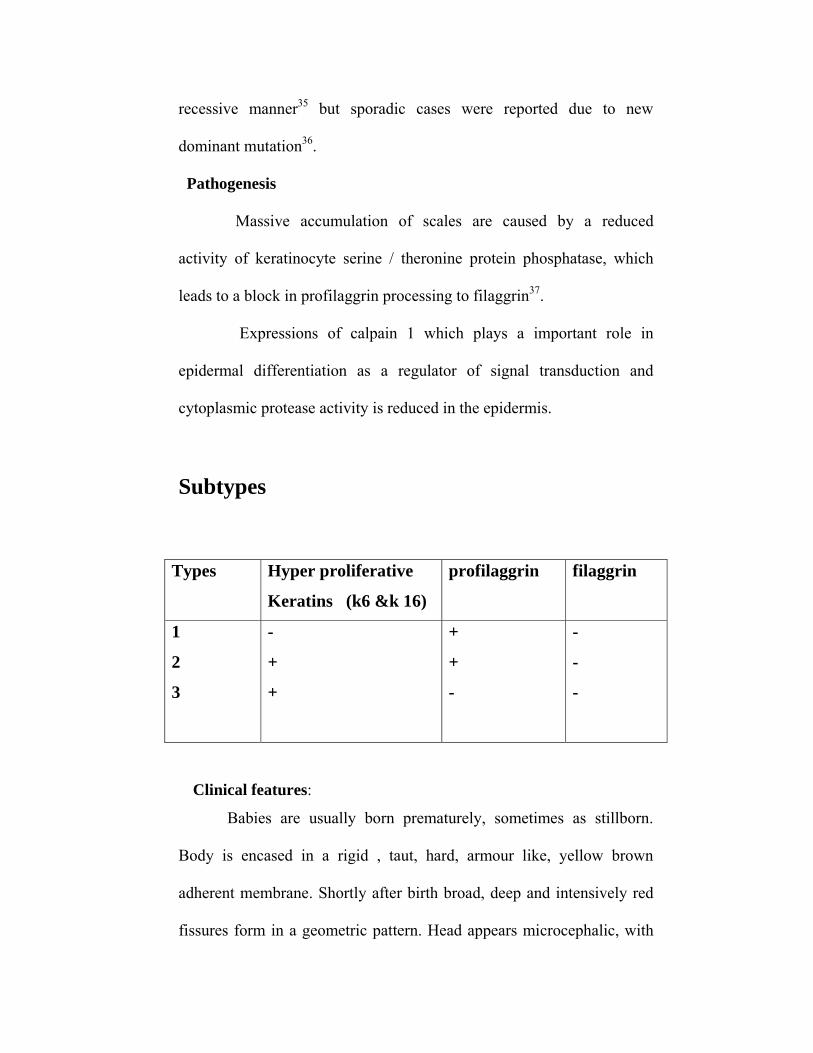
Subtypes
Clinical features:
Babies are usually born prematurely, sometimes as stillborn.
Body is encased in a rigid , taut, hard, armour like, yellow brown
adherent membrane. Shortly after birth broad, deep and intensively red
fissures form in a geometric pattern. Head appears microcephalic, with
Types Hyper proliferative
Keratins (k6 &k 16)
profilaggrin filaggrin
1
2
3
-
+
+
+
+
-
-
-
-

severe ectropion, conjunctival oedema, eclabion, rudimentary ear and
nose, giving a grotesque appearance. Hands and feet are edematous and
swollen,covered by mitten like casing, with well developed digits.
Pathology
Extraordinary thickened and compact orthokeratotic stratum
corneum. Hair follicles show marked, concentric accumulation of
keratotic material around hair shafts.
Electron microscopy show abnormal or missing lamellar bodies
in the granular layer, absent extracellular lipid lamellae and presence of
lipid inclusion or remnant organelles in the stratum corneum39.
Prenatal diagnosis is possible by fetal skin biopsy, which
demonstrates premature cornification at 20weeks of gestational age40.
Outcome
Most patients do not survive more than few days, due to
temperature instability and sepsis. In survivors, severe generalized
ichthyosiform erythroderma eventuates41. Oldest child reported alive to
date being 9 years42.
LAMELLAR ICHTHYOSIS
Classical lamellar ichthyosis is inherited as an autosomal
recessive disorder. But an autosomal dominant pattern of inheritance has
been described43.
Reported incidence is 1 per 1,00,000 live births.

Pathogenesis
It is due to deleterious mutation of the transglutaminase gene on
chromosome 14q11, leading to transglutaminase-1 deficiency44, which
catalyzes the calcium dependent cross linking of proteins of cornified
envelope, through γ glutamyl – lysine isopeptide bonds45.
Pathology
Lamellar ichthyosis is a retention type ichthyosis.There is
massive orthokeratotic hyperkeratosis with acanthosis and mild
papillomatosis. Granular layer is normal or increased.
Electron microscopy shows elongated cholesterol clefts and
translucent lipid droplets in stratum corneum with a thin or absent
cornified cell envelope46.
Clinical features
At birth the disease presents as collodion baby, followed by
scaling within the first month of life. Erythroderma is usually less
intense.
Scales are large, dark brown or grey, plate like, firmly adherent
and form a mosaic or bark like pattern. Traction and compression by taut
skin leads to scarring alopecia. Hair shafts are encased by thickened
stratum corneum. Severe ectropion leads to exposure keratitis. Deep
fissures in flexures cause limitation of joint movement, flexion
contracture and sclerodactyly.

Other features are palmoplantar keratoderma, secondary nail
dystrophy, severe heat intolerance due to epidermal constriction of
sweat ducts and recurrent ear infection due to accumulation of scale in
the external ear canal.
In mild lamellar ichthyosis, typical scales occur only in lower legs
and upper arm. Fine white branny scales occur on the flexures and
neck47.
Autosomal dominant Lamellar ichthyosis
There is no collodion membrane at birth. These patients have non
erythrodermic, lamellar type generalized scaling from birth,
palmoplantar hyperkeratoses and lichenification of dorsa of hands and
feet.
NON BULLOUS ICHTHYOSIFORM ERYTHRODERMA
Rare and usually severe autosomal recessive, inflammatory
ichthyosis, although autosomal dominant transmission was reported. It is
more common than lamellar ichthyosis, affecting 1 in 3,00,000
population.
Pathogenesis
Few patients carry recessive mutation in the transglutaminase1
gene, leading to abnormal formation of cornified cell envelope48.
Ultrastructural and biochemical abnormalities such as an
increased number of lamellar bodies, accumulation of lipid droplets,
abnormal activity of lamellar body enzymes in the stratum corneum, are
suggestive of abnormalities in the lamellar body secretory system49.
In some patients, inactivating mutations were identified in
lipoxygenase-3 (ALOXE3) and 12- lipoxygenase (ALOX12B), which
encode non heme, iron containing dioxygenases50 which catalyzes the
oxygenation of free and esterified polyunsaturated fatty acids,
phospholipids and triglycerides, and are crucial for formation of
epidermal lipid barrier.
Pathology
In the epidermis, there is variable mild parakeratosis and
acanthosis with normal or prominent granular layer. Prominent blood
vessels and mild upper dermal lymphocytic infiltrate are seen in the
dermis.
Kinetic studies show increased epidermal turnover rate51.
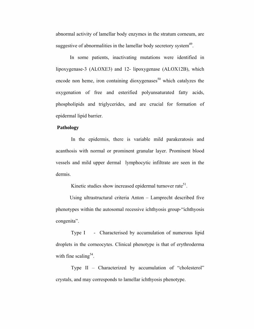
Using ultrastructural criteria Anton – Lamprecht described five
phenotypes within the autosomal recessive ichthyosis group-“ichthyosis
congenita”.
Type I - Characterised by accumulation of numerous lipid
droplets in the corneocytes. Clinical phenotype is that of erythroderma
with fine scaling54.
Type II – Characterized by accumulation of “cholesterol”
crystals, and may corresponds to lamellar ichthyosis phenotype.

Type III – Characterized by reticulate scale pattern with
erythroderma. Ultrastructurarly lamellated membrane structures are
seen55.
Type IV & V – Individual case reports.
Clinical features
Clinical spectrum is more variable with respect to severity.
Collodion baby presentation is seen at birth in 90% cases and
generalized scaly erythroderma is apparent after that. Scale is white or
grey, thin, superficial and semi adherent and appear feathery in face,
arm, trunk and lamellar on lower legs. Scaling may be cyclical with
shedding over 2-4 weeks. Summer deterioration is often present.
Other features
Ectropion which may leads to exposure keratitis52.
Palmoplantar hyperkeratosis(70%) with painful fissures.
Tinea amiantacea with patchy cicatrical alopecia53.
Nail dystrophy, subungual hyperkeratosis (50%), onychomycosis
Hypohidrosis and heat intolerance.
Wide spread fungal infection reported.
Mild growth retardation.

Difference between non bullous ichthyosiform erythroderma
and Lamellar Ichthyosis
Non bullous ichthyosiform erythroderma Clinical Fine white scale Erythroderma (variable) Epidermal turnover times Accelerated mitotic rates Increased epidermal labeling indices ultrastructural Abnormal and increased number of lamellar bodies Numerous lipid droplets within cornified cells Extensive bilayer stacks within intercellular spaces of stratum corneum. Histopathology Stratum corneum four times thicker More parakeratosis Prominent mucin or glycosaminoglycans in stratum corneum cell membranes Biochemical Butyrase/glucosidase ratio 90 to 100
Lamellar ichthyosis Clinical Thick plate like scale Erythroderma- little to absent Epidermal turnover times Normal proliferation kinetics Ultrastructural Normal lamellar bodies Not seen Not seen Histopathology Stratum corneum massively thickened (nine times) No parakeratosis Normal granular layer No PAS+ membranes Biochemical Butyrase/ glucosidase ratio < 5

BULLOUS ICHTHYOSIFORM ERYTHRODERMA OF BROCQ
Synonyms
Epidermolytic hyperkeratosis –The term was coined by Frost &
Vanscott Bullous ichthyosis.
Rare autosomal dominant disorder with complete penetrance, which is
associated with blistering in early phases. Sporadic occurrence is seen
in 50% cases56.
Estimated prevalence is 1 in 2,00,000.
Pathogenesis
K1 (Type II –basic) and k10 (Type I- acidic) are the keratins
specific to the differentiated epidermis. BIE is caused by heterozygous
mutation in the genes coding keratin 1 and 10, localized on
chromosomes 12q11-q13 and 17q12-q21, respectively57. Pathogenic
mutations, leading to nonconservative aminoacid substituitions, cluster
at the boundaries of the α helical rod domains – ‘hot spot arginine’58.
Mutation perturb keratin alignment, oligomerization and filament
assembly, thus weakening the cytoskeleton, compromising mechanical
strength and cellular integrity of the epidermis. The association of
desmosomes and tonofilaments is disturbed, so that many desmosomes
are attached to only one keratinocyte instead of connecting two
neighbouring keratinocyts. Real acantholysis occur leading to blister
formation.
Keratin 1 mutations are usually associated with severe
palmoplantar keratoderma, where as KRT10 mutations spare the palms
and soles, because the gene is not expressed here.
Keratin filament disruption may impair lamellar body secretion
resulting in an impaired barrier and secondary epidermal hyperplasia59.
Epidermal hyperplasia results in expression of the alternate
“hyperproliferative” keratin pair (k6 & k10) which could ameliorate the
expression of k1 and k10 and the blistering phenotype, after the neonatal
period .
Pathology
Nikolsky (1897) first recognized the characteristic histopathology
of bullous ichthyosiform erythroderma known as epidermolytic
hyperkeratosis or granular degeneration61.
Epidermis shows marked hyperkeratosis and acanthosis. In the
prominent and degenerate granular layer and in upper stratum spinosum,
there are variously sized clear spaces around the nuclei. Peripheral to the
clear spaces, cells show indistinct boundaries formed by irregular
shaped keratohyaline granules. Intraepidermal bullae form through
separation of edematous cells from one another62.
Dermis shows moderately severe chronic inflammation.
Labelling with tritiated thymidine show increased proliferative activity
in the epidermis.

Clinical features
At birth, mild generalized erythroderma is present. Flaccid
blisters and superficial erosions at sites of minor trauma are apparent
within first few hours of life. Erosions heal rapidly without scarring.
During childhood, localized blistering at sites of trauma
continues and erythroderma fades. Hyperkeratosis become obvious and
is prominent around anterior neck, flexures, abdominal wall and scalp.
Yellow brown waxy, ridged or corrugated scale sometimes forming
spiny outgrowth builds up in the skin creases. Cobble stone like
keratoses occur over dorsa of hands and feet.. Verrucous plaques are
easily dislodged, leaving tender erosions. Skin colonization by
staphylococcus, brevibacterium and fungi produces embarrassing body
odour and repeated skin infection. Scarring alopecia may occur.
Palm and soles show hyperkeratosis in 60% and it may leads to
recurrent painful, fissures and contracture. Clinical subtyping of bullous
ichthyosiform erythroderma is based on the presence or absence of
severe palmar / plantar hyperkeratosis.

Clinical subtypes of epidermolytic
hyperkeratosis
Charact-
eristic
NPS-1 NPS-2 NPS-3 PS-1 PS-2 PS-3
Palm/
sole
Keratosis
_ _ _ + + +
Palm
/sole
surface
Normal Normal Hyper
linear,
Minimal
scale
Smooth
Smooth
Cerebr
-iform
Digital
Contr-
actures
_ _ _ _ _ _
Scale Hystrix Brown Fine,
white
Mild White
Scale,
peel
Tan
Distri-
bution
Genera-
lized
Genera-
lized
Genera-
lized
Localize
d
Genera-
lized
Genera
-lized
Erythro-
derma
_ _ + _ + _
Blistering + + + Locali-
zed
+ Neona
-tal
Variants
Cyclic ichthyosis with epidermolytic hyperkeratosis
Annular epidermalytic ichthyosis63
Naevoid BIE – unilateral or bilateral streaks of hyperkeratosis that
follow the lines of Blaschko64 showing histological
features of EHK. It is caused by somatic mutation in
KRT1 or KRT10 ,arising postzygotically during
early embryogenesis.
ICHTHYOSIS BULLOSA OF SIEMENS
Synonyms
Ichthyosis exfoliativa
Rare autosomal dominantly inherited ichthyosis.
Pathogenesis
It is due to heterozygous mutation in the gene for keratin 2e
(KRT2e)65, which is expressed only in the upper most spinous and
granular layer of the epidermis.
Clinical features
At birth, the skin may appear normal or show mild blistering.
Trauma induced small blisters on the extremities occur during infancy
but usually subside during early childhood, while hyperkeratosis
develops. Predilection sites are the skin overlying the joints, flexures
and the dorsa of the hands and feet, and there is always sparing of the
palms and soles present. The skin may appear ridged, shiny or
lichenified. A characteristic feature is superficially denuded areas with
collarette-like borders described as ‘moulting’ or ‘mauserung’, which
develop due to superficial blistering and shedding of the stratum
corneum.
Histopathology
Features of epidermolytic hyperkeratosis are confined to the
prominent granular layer and upper spinous layer. Sites of mauserung
reveal intracorneal blistering with orthohyperkerotosis above and below
the split.
ICHTHYOSIS HYSTRIX
It is a descriptive term for a clinically and genetically
heterogenous group of skin disorders, characterized by spiny
hyperkeratotic scale similar to that of BIE. They differ from BIE in that
blistering is not a feature, erythroderma is usually mild or absent, and
limited or naevoid forms are more common.
Clinical features
At birth there may be a generalized or naevoid scaly erythema.
Hystrix (porcupine spine) scaling, often a muddy brown or grey color,
accumulates during childhood and affects extensor aspects of the limbs
and truncal areas. Palmoplantar keratoderma either diffuse or striate,
affects most people which may lead to functional impairment.
Variants
Icthyosis hytrix curth-macklin – due to mutation affecting V2
domain of keratin 1
Electron microscopy show characteristic continuous
perinuclear tonofilament shell in the upper spinous and granular layer,
resulting in three distinct cytoplasmic compartments. Double nuclei
occur in 10% of spinous and granular keratinocytes.
IH of Rheydt – now known as HID syndrome, characterized by
hystrix like ichthyosis and deafness.
COMEL NETHERTON’S SYNDROME
A rare autosomal recessive disorder that is characterized by the
concurrence of ichthyosis, characteristic hair shaft abnormality and
atopy66. 18% of all congenital erythrodermas were attributable to
Netherton’s syndrome69.
Comel – described the clinical features of ichthyosis linearis
Circumflexa67.
Netherton – discovered the hair shaft abnormality68.
Wilkinson – delineated the triad of clinical features.
Pathogenesis
It is due to mutation in the SPINK5 gene (serine protease inhibitor
Kazal type 5) which encodes LEKT1 (lympho epithelial Kazal type
related inhibitor) 70 which may be important in the down regulation of
inflammatory pathways. Loss of LEKT1 leads to premature and
uncontrolled proteolytic activity of serine proteases which could result
in abnormalities of structure of lamellar lipid membrane71.
In the hair, failure to convert sulphydryl groups to disulphide
bonds leads to weak coherence of cortical cells. Such focal softening of
hair shaft may allow invagination of distal shaft in to the dilated
proximal cup.
Pathology
Varies with the type and phase of the lesion . .
Erythroderma –shows hyperkratosis, parakeratosis, exocytosis,
spongiosis intraepidermal microabscesses and
subcorneal split .
Double edged scale –show parakeratosis with reduced or absent
granular layer. Focal accumulation of PAS
positive, diastase- resistant, homogeneous
material representing exuded serum is seen
within the parakeratotic stratum corneum72.
Older ILC lesions –show psoriasiform epidermal hyperplasia,
Papillomatosis and a mixed perivascular

inflammatory infiltrate.
Microscopy of Hair – taken from scalp / eyebrow73 show trichorrhexis
Invaginata (bamboo hair) in 20 – 50% cases. It consists
of a bulbous distal hair end invaginating a concave
dilated proximal hair terminal giving a ball and socket
appearance . Nodular thickening of the distal end of
broken hair shafts (‘golf tee’)74, pilitorti, trichorrhexis
nodosa and helical hair may also be seen.
Clinical features
At birth generalized erythroderma is present and scaling
quickly develops. In approximately 20% patients, complications like
hypernatremic dehydration75, temperature instability
bronchopneumonia, sepsis, which may be fatal, occur. Failure to thrive
and diarrhoea may occur resulting in short stature.
Erythema tends to improve but may recur. During child hood,
50% develop ichthyosis linearis circumflexa (ILC). It is an
erythematous, scaly, annular or polycyclic, flat patch with an incomplete
advancing double edge of peeling scale. It is episodic,often migrating in
a cephalocaudal pattern. Fluctuating erythroderma triggered by
intercurrent illness may occur and in between attacks, skin may look
normal. Pruritus and flexural lichenification may occur. Impaired
sweating can lead to hyperpyrexia. Bacterial, fungal infection and viral
warts are common.

Atopy – Atopic diathesis is seen in 50% of patients and manifests as
atopic dermatitis or asthma. Eosinophilia and allergic reactions to
various foods are common. Serum IgE levels are markedly elevated
ranging from 100 to 10,000 Iu/ml76.
Hair – scalp, eyebrow, eyelash, body hair remain sparse, lustreless and
brittle. Hair is unruly, short and spiky. Broken hair shafts at follicular
orifice produces a peppered appearance.
Others – Intermittent amino aciduria77, deficient IQ, seizures and
impaired CMI.
Chronic erythroderma, with disabling flexural edema and
papillomatosis may occur in the axilla, groin, vulva, which is
premalignant.
SJÖGREN LARSSON SYNDROME
It is a rare autosomal recessive neurocutaneous disorder,
comprising of congenital ichthyosis, spastic diplegia and mental
retardation.
Pathogenesis
It is caused by deficiency of the microsomal enzyme fatty
aldehyde dehydrogenase(FALDH), due to inactivating mutations in the
FALDH gene, located on chromosome 17 p11.218. This enzymes
catalyzes the oxidation of long chain aliphatic aldehydes into fatty acids,
a pathway that is important for the synthesis of epidermal lipids as well

as the catabolism of phospholipids and sphingolipids in the brain79. The
symptoms of Sjogren-Larsson syndrome are thought to stem from
membrane alterations due to accumulation of fatty alcohol or fatty
aldehyde- modified lipids and proteins. Retarded myelination and a
variable degree of dysmyelination probably results from the
accumulation of free lipids in the periventricular white matter.
Pathology
Epidermis shows orthohyperkeratosis, acanthosis, and
papillomatosis.
Granular layer may be normal or increased.
Electron microscopy may show lamellar membrane
inclusions and cleft in corneocytes.
Clinical features
Skin – At birth skin is dry and mildly erythrodermic .Collodion baby
presentation is rare80. Scaling develop within three months, tends to
follow a cyclical pattern of accumulation and shedding, predominantly
affecting limbs and face. Lamellar type ichthyosis occur in lowerlimbs.
A velvety orange or brown lichenification topped with verrucous
hyperkeratosis is seen in and around flexures, neck and periumbilical
folds and is helpful in diagnosis. Perisistent pruritus may occur leading
to visible excoriations. 70% patients develop palmoplantar keratoderma.
Neural

Symptoms are evident during first year of life. It is stable and
nonprogressive. Features include spastic di or tetraplegia, mental
retardation81, speech defects, seizures and learning disability.
Ocular
Glistening dots on fovea and para fovea82 of the retina, which
do not interfere with vision, may develop during childhood and fade
with advancing age. It is seen in 80% patients and occurs due to the
deposition of lipofuscin pigment granules in the specialized retinal
epithelium83.
REFSUM’S DISEASE
Synonyms
Heredopathia atactica polyneuritiformis
Phytanic acid storage disease
A rare autosomal recessive neurocutaneous disorder.
Pathogenesis
This disorder is caused by inactivating mutations in both copies of
the peroxisomal phytanoyl-CoA hydroxylase(PAHX) gene on the short
arm of chromosomes 1085. Deficiency of the peroxisomal phytanoyl-
CoA hydroxylase causes impaired degradation of phytanic acid, a 20-
carbon, saturated, branched- chain fatty acid, exclusively derived from
exogeneous sources like plant chlorophyll, leading to an excessive
accumulation of phytanic acid in tissues and body fluids.

Normal serum level of phytanic acid is 1mg/ 100ml but in
Refsum’s disease, it accounts for 5 – 30% of serum lipids and level rises
to 60mg / 100ml. Phytanic acid interferes with membrane structure and
function84.
Pathology
Besides nonspecific orthokeratotic hyperkeratosis, lipid containing
vacuoles are seen in the basal keratinocytes .
Clinical features
Scales resemble ichthyosis vulgaris, which develop during
childhood or adolescence. In late untreated cases, lamellar scaling
develops. Hyperkeratosis of palms and soles may be seen.
Neural features
Start in childhood and progressive, but have undulating
course with periods of acute exacerbation and remission. Features
include failing night vision and concentric visual field constriction due
to progressive retinitis pigmentosa, cataract, deafness, tinnitus,anosmia,
progressive weakness, foot drop, cerebellar ataxia, mixed sensorimotor
polyneuropathy with hypertrophied peripheral nerves and an elevated
CSF protein.
Others
Renal tubular dysfunction, skeletal hyperostosis,cardiomyopathy
leading to sudden death.
TAY’S SYNDROME.
Synonyms
IBIDS syndrome- ichthyosis, brittle hair, intellectual
impairment, decreased fertility and short
stature.
Trichothiodystrophy E,F.
Heterogeneous group of rare autosomal recessively inherited
disorders, sharing sulfur deficient, short, brittle hair as a possible marker
for neuro ectodermal disease.
Pathogenesis
There is reduced stability or altered function of the transcription/
repair complex TF11H, due to mutation in the genes, that encode
helicase subunits ERCC2/ XPD and ERCC3/ XPB86. This leads to
defective DNA repair leading to photosensitivity. The growth defects,
brittle hair and nails, and neurological defects may be caused by
impairment of transcription function of XPD and XPB gene products,
leading to reduced RNA synthesis87.
Reduced level (up to 50%) of hair sulphur and sulphur containing
aminoacids, cysteine, methionine and proline are the hall mark of
trichothiodystrophy. Based on cellular response to uvlight, three groups
were identified.
1. Normal cell survival and excision repair after UV irradiation.
2. Repair synthesis was only 50 percent of normal and RNA
synthesis was severely reduced but cell survival was normal.
3. A severe excision repair defect that appears to be identical to
xeroderma pigmentosum complementation group D203.
Pathology
Skin –In the epidermis there is hyperkeratosis, focal parakeratosis,
acanthosis and a normal or reduced granular layer.
Hair
Light microscopy – hair shafts show transverse fractures (trichoschisis)
. A nodal appearance similar to trichorrhexis nodosa or 180° twists as in
pilitorti may be observed.
Polarizing light microscopy – gold standard for detecting the
characteristic light and dark bands, the so-called ‘tiger tail’ or ‘zigzag’
pattern88.
Transmission electron microscopy –may show an abnormal alignment
of microfibers as well as an absence of the exocuticle and A-layer.
Clinical features
Skin
At birth, the disease presents with a collodion membrane or
erythroderma in a premature neonate. Scales vary from fine, translucent
to dark and large. Erythroderma improves after infancy. Other features
are increased susceptibility to infection, follicular keratosis,
erythema,eczema, pruritus, hypohidrosis, cheilitis, pyoderma,

folliculitis, palmar pustules, telangiectasia, poikiloderma. An elfin like
and progeric face is seen due to fat atrophy with prominent ears and chin
recession89 . Photosensitivity occurs in 50% patients but there is no
report of cutaneous malignancies88.
Nails may show thinning, longitudinal ridging, yellow
discolouration, onychogryphosis.
Hair
Hair may be broken, brittle, sparse, sometimes absent in scalp,
eyebrow, eyelashes, axillary, pubis. Few nasal vibrissae and otic hairs
are present. Marked susceptibility to weathering is present.
CNS
Mental retardation, seizures, tremor, ataxia, neurosensory
deafness, dysarthria, spasticity, hemi-and tetraparesis, cerebellar
deficiency, pyramidal signs, intention tremor, impaired motor
coordination, peripheral neuropathy, nystagmus. Sociable behaviour is
frequently seen88.
Dental
Enamel hypoplsia and caries, maxillary hypoplasia, high-arched
palate, bifid uvula, and leukoplakia.
Ophthalmologic
Antimongoloid slant, ectropion, conjunctivitis, hypertelorism,
enophthalmus, exophthalmus, astigmatism, chorioretinal atrophy, retinal

pigmentation, pale optic disc, photophobia and diminished red-green
discrimination.
Genital
Decreased fertility, cryptorchidism, hypoplasia of both male and
female genitals, and hypospadiasis.
Orthopaedic
Genu valgum, coxa valga, valgus deformity of the great toe,
cubital and tibial valgus deformity, pectus excavatum, scoliosis, thoracic
kyphosis and lumbosacral lordosis.
Related syndromes
BIDS – Brittle hair, intellectual impairment, decreased fertility and
short stature.
PIBIDS - Association with photosensitivity seen.
SIBIDS90 – osteosclerosis, ichthyosis,brittle hair, intellectual
impairment,decreased fertility and short stature.
X LINKED DOMINANT ICHTHYOSIS
Synonyms
Conradi-Hünermann- Happle syndrome
X linked dominant chondrodyplasia punctata type Π
Very rare, X linked dominant disorder,which be lethal to male
fetuses but may be compatible with life in xxy karyotype. It is one of the
variants of chondrodysplasia punctata.

Pathogenesis
It occurs due to primary defect in cholesterol biosynthesis. A
deficiency of dihydroxyacetone phosphate acyl transferase enzyme
which is responsible for synthesis of plasmalagen phospholipids and
glycolipids is seen. Distinct mutation in the emopamil binding protein
(EBP) on the short arm of the X chromosome, leads to accumulation of
8 dehydrochholesterol and 8(9)- cholesterol91.
Pathology
In epidermis, orthohyperkeratosis with focal parakeratosis, reduced
granular layer, prominent perifollicular atrophy in old cases are seen.
Electron microscopy may show needle like inclusions in the
granular layer, persistant desmosomal structures in the stratum corneum,
degenerate mitochondria and vacuolated lamellar bodies.
Clinical features
Typically females are affected. Babies are born with either
collodion membrane or generalized ichthyosiform erythroderma which
resolves within first year. Generalized linear and swirling patterns of
erythroderma and scaling are established along Blaschko lines. In adults,
hyperkeratosis is replaced by follicular atrophoderma and icepick scars,
linear pigmentary changes and scarring alopecia all in a Blaschkoid
distribution. Palmoplantar hyperkeratosis and nail dystrophy may
occur92. Recurrent infection especially in the flexures is common.
Skeletal anamolies
Skeletal anomalies are usually asymmetric. They include
frontal bossing, flat nasal bridge, facial asymmetry, shortening of limbs,
scoliosis, hexadactyly. Widespread premature calcifications manifesting
as stippled epiphyses involving the trachea and vertebrae may be seen
on radiographs during infancy.
Others
Unilateral cataracts (60%), congenital heart defects, sensorineural
deafness, central nervous system malformations and renal anomalies.
Intellect is not impaired and life expectancy is normal. Spontaneous
improvement with age may occur, which reflect progressive elimination
of mutant cells by adjacent normal cells, which have a growth
advantage.
Related syndromes
(1) Rhizomelic chondrodysplasia punctata – autosomal recessively
inherited,characterized by combination of punctate epiphyseal
calcification, rhizomelia (proximal limb shortening), deficient
red blood cell plasmalogens, and accumulation of phytanic acid.
Ichthyosis occur in 25% of patients which is similar to Conradi
Hümermann syndrome.
(2) X linked chondrodysplasia punctata with steroid surfatase
deficiency.
CHANARIN – DORFMAN SYNDROME
Synonym – Neutral lipid storage disease.
Autosomal recessively inherited, rare inborn error of lipid
metabolism, characterized by ichthyosis, myopathy and vacuolated
leukocytes93.
Aetiology
Increased fibroblast triglyceride synthesis, with a complete failure
of endogenous triglyceride breakdown has been demonstrated.
Exogenous triglyceride metabolism and serum lipid levels are normal94.
Pathology
Light microscopy –In peripheral smear there are numerous lipid
containing vacuoles in the circulating granulocytes and monocytes but
not in lymphocytes called ‘Jordans anomaly’96 is seen. Lipid staining of
skin shows closely packed lipid droplets of varying sizes in epithelium
of eccrine sweat glands and ducts, basal cells, fibroblast and
endothellial cells. Biopsy of muscle, liver, and bone marrow also reveal
lipid droplets.
Ultastructure – Epidermal lamellar bodies and intercellular lipid
lamellae are disturbed by globular electron lucent inclusions96.
Clinical features
Babies are either collodion or erythrodermic at birth,and scaling
resemble that of non bullous ichthyosiform erythroderma. Troublesome
pruritus hypohidrosis,and flexural lichenification may occur.

Others
Subclinical to marked proximal myopathy with elevated muscle
enzymes, hepatomegaly with abnormal liver enzymes, cataract of
nuclear type (50%), nerve deafness, ataxia, short stature, mental
retardation.
KERATITIS ICHTHYOSIS DEAFNESS SYNDROME
Synonym
Erythro keratodermia progressive of Burns.
Skinner (1981) – coined the acronym ‘KID’97
Majority of the cases are sporadic and there is overlap
with ectodermal dysplasias seen..
Pathogenesis
Caused by missense mutations in the connexin gene GJB2 encoding
the gap junction protein 2698.
Pathology
Orthohyperkeratosis, acanthosis, papillomatosis and vacuolization
of the cells in the granular layer are seen.Corneal epithelium may be
dyskeratotic, atrophic or absent in the center, and Bowman,s membrane
may be absent. In the inner ear, the organ of corti is immature or
atrophic.
Ultrastructural study may show changes similar to ichthyosis
hystrix.
Clinical features
Transient erythroderma is present at birth. During infancy, fixed,
symmetrically distributed, well demarcated, hyperkeratotic plaques
with an erythematous base and a leathery rough verrucous surface
develops in face usually cheeks, forehead, ear, chin, nose, small
joints.Thick perioral rugae and leonine facies may occur. Prominent
follicular hyperkeratosis with scarring alopecia of scalp99, loss of
eyebrows, eye lashes and body hair occur. A reticulated PPK
resembling grained leather is characteristic. Nails may be dystrophic
and show leukonychia. Absent teeth, delayed eruption and cavities may
occur. Acneform eruptions, deep abscesses and discharging sinuses,
chronic cutaneous granulomatous candidial and fungal infection may
develop. Squamous cell carcinomas may develop even in childhood100.
Others
Sensorineural deafness during infancy, progressive corneal
vascularization (96%) leading to blindness by adolescence may be seen.
Variants
HID syndrome – Allelic variant of KID, characterized by hystrix like
ichthyosis and deafness.
CHILD SYNDROME
The acronym CHILD describes a very rare disorder comprising
congenital hemidysplasa, ichthyosiform erythroderma, and limb
defects, which is found almost exclusively in females104.

Pathogenesis
It is an X linked dominant disorder. Inactivating mutations in
NSDHL(NADH steroid dehydrogenase like protein) which encodes the
enzyme 3ß – hydroxysteroid dehydrogenase has been demonstrated105
Pathology
Marked ortho and parakeratosis with acanthotic and papillomatous
epidermis with neutrophilic collection in stratum corneum overlying a
mild superficial perivascular infiltrate is seen. Multiple layers of
granular cells surround the sweat ducts, while the granular zone in the
surrounding epidermis may be absent, normal or thickened.
Clinical features
Child presents at birth or neonatal period with a unilateral
erythema with hyperkeratosis, crusted plaques covering most of one
side of body, commonly on right side, with a sharp demarcation in
midline, sparing the face with affinity for the skin folds
(ptychotropism). Linear bands of ichthyotic skin may occur in the other
side due to somatic mutation106. Linear alopecia of the affected side and
claw like nail dystrophy are common. The lesions often improves with
time.
Skeletal system
Anamolies include hypoplasia of digits, ribs, ipsilateral limb
hypoplasia, spina bifida scoliosis. Stippled epiphyses analogous to
Conradi Hünermann syndrome has been noted.

Others - Renal defects107, CNS, and CVS malformations.
Ichthyosis follicularis with alopecia and photophobia
It is characterized by non inflammatory follicular keratoses,
persistent non cicatrical scalp and body alopecia and severe
photophobia from birth.Generalised xerosis and lamellar scaling are
common. Lesions must be distinguished from keratosis follicularis
spinulosa decalvans and keratosis pilaris rubra atrophicans facei.
CHIME syndrome
Coloboma, heart disease, ichthyosis, mental retardation
and ear defects.
Kallmann’s syndrome – caused by large deletion of the short arm of
the X chromosome proximal to and involving the STS
gene.Characterized by XLRI with hypogonadotrophic hypogonadism,
anosmia, nystagmus, synkinesis101. Other features are developmental
delay, spastic paraplegia, osteoporosis.
Rud’s syndrome – X linked ichthyosis, obesity, hypogonadism, mental
retardation, epilepsy and endocrinopathies102.
ICE syndrome – AD, characterized by ichthyosis vulgaris like eruption,
‘full’ cheeks, sparse lateral eyebrows, craniofacial and skeletal
abnormalities103.
Multiple sulfatase deficiency
Rare autosomal recessive disorder characterized by a deficiency
of several sulfatides, glycosaminoglycans, spingolipids, steroid sulfates

in tissues and body fluids108. Clinical features include neurologic
deterioration, skeletal abnormalities, facial dysmorphism and ichthyosis
resembling that of X linked steroid sulfatase deficiency.

TREATMENT
Regular, paraffin based emollient application is of benefit to
all ichthyosis patients. Urea containing emollients improve epidermal
hydration and lyse keratin. Emollients are of limited value in bullous
ichthysiform erythroderma as the scale is often waxy and macerated.
Keratolytic agents such as 1-5% salicylic acid can be used to
encourage shedding of scale. Alpha-hydroxy acids, such as lactate,
glycolic, malic, mandelic, citric, pyruvic, gluconic and tartaric acid
enhance corneocyte shedding by virtue of their effect in breaking
intercellular bonds108.
Topical calcipotriol has been efficacious with lamellar
ichthyosis109.
Topical tacrolimus 0.1% produce significant improvement in
Netherton’s syndrome
Systemic retinoids are effective in most patients with severe
congenital ichthyosis except in Netherton’s syndrome, where
deterioration with systemic retinoid theropy is noted. The starting dose
of acitretin is 0.5-0.75mg/kg/day and the maintenance dose is titrated
down to the lowest effective level between 0.1 and 0.5mg/kg/day.
AIMS OF THE STUDY
To study the incidence of Congenital ichthyosis in pediatric age
group in Institute of child health, Government General Hospital,
Chennai during the period of two years between september 05
to september 07.
To study the incidence of various types of congenital ichthyosis
and ichthyosiform syndromes.
To study the age of onset of various types of congenital
ichthyosis.
To study the sex incidence of various types of congenital
ichthyosis.
To study the presenting symptoms and clinical patterns.

To study the associated skin disorders with particular reference
to atopy, keratosis pilaris, and fungal infection.
To study the histopathological pattern.
To study the prevalence of ichthyosis in family members.
MATERIALS AND METHODS
Materials for this study were gathered from the pediatric out patient
wing of dermatology, Institute of child health, Government general
hospital, Chennai during the period between September 05 to
September 07. A total of 64 cases of ichthyosis were enrolled in the
study on the basis of clinical pattern. The total number of new patients
who attended the department during the period was enrolled for
comparative studies.
Procedure
History
In eliciting the history, a set pattern of questionnaire was
followed (as given in proforma). Enquiries were made with regard to
symptoms, age of onset, duration, history of collodion baby, blisters,
seasonal variation, repeated skin infection and atopy. History regarding
involvement of other systems like central nervous system, skeletal
system was taken.
Clinical examination

A careful and detailed dermatological and systemic examination
with necessary investigations was conducted methodically. Patients up
to fourteen years of age, were examined for distribution and nature of
scales, presence of erythroderma, and blisters along with a note of
associated disorders if any. Referral to other specialties like neurology
and opthalmology were done to confirm or to rule out associated
features of some syndromes as and when suspected.
Exclusion criteria
Acquired ichthyosis – Malnutrition
Congenital hypothyroidism
Acquired immune deficiency syndrome
Laboratory investigations
Routine hematological investigation was done in all cases.
Microscopic examination of hair was done. Skin biopsy was done and
specimens were studied with H & E stain.
All the datas were compiled and the inference was drawn .

OBSERVATION AND RESULTS
Incidence Out of 10,200 patients attended dermatology out patient
department, Institute of child health, Government General Hospital
during the period between September 2005 and September 2007, total
number of patients with congenital ichthyosis was 64.
Incidence of congenital ichthyosis was 0.63% or 6 per 1000.
TABLE – 1
Total number of patients attended as skin out patients
at Institute of child health, Government General
Hospital (sep 05 to sep 07)
10,200
Total number of patients with congenital ichthyosis 64
Incidence of congenital ichthyosis 0.63%
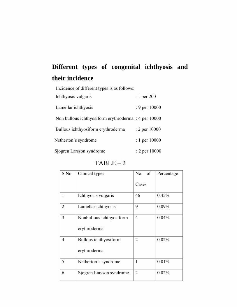
Different types of congenital ichthyosis and
their incidence Incidence of different types is as follows:
Ichthyosis vulgaris : 1 per 200
Lamellar ichthyosis : 9 per 10000
Non bullous ichthyosiform erythroderma : 4 per 10000
Bullous ichthyosiform erythroderma : 2 per 10000
Netherton’s syndrome : 1 per 10000
Sjogren Larsson syndrome : 2 per 10000
TABLE – 2 S.No Clinical types No of
Cases
Percentage
1 Ichthyosis vulgaris 46 0.45%
2 Lamellar ichthyosis 9 0.09%
3 Nonbullous ichthyosiform
erythroderma
4 0.04%
4 Bullous ichthyosiform
erythroderma
2 0.02%
5 Netherton’s syndrome 1 0.01%
6 Sjogren Larsson syndrome 2 0.02%

Relative incidence of different types of
congenital ichthyosis
Out of 64 patients with congenital ichthyosis, ichthyosis vulgaris
accounts for 72% followed by lamellar ichthyosis 14%. Non bullous
ichthyosiform erythroderma constitutes 6% followed by bullous
ichthyosiform erythroderma and Sjogren Larsson syndrome each
constitutes 3%. Netherton’s syndrome constitutes 1.5% of cases.
Table – 3
S.No Clinical types No of Cases Percentage
1 Ichthyosis vulgaris 46 71.8%
2 Lamellar ichthyosis 9 14.1%
3 Non bullous ichthyosiform
erythroderma
4 6.3%
4 Bullous ichthyosiform
erythroderma
2 3.1%
5 Netherton’s syndrome 1 1.5%
6 Sjogren Larsson syndrome 2 3.1%

Sex incidence of different types of ichthyosis
Incidence of ichthyosis vulgaris was almost equal in both sexes.
Incidence of lamellar ichthyosis was more in females. Equal sex
distribution was seen in non bullous ichthyosiform erythroderma.
Bullous ichtyosiform erythroderma, Sjogren Larsson syndrome and
Netherton’s syndrome were seen only in males.
TABLE – 4
S.No Clinical Type M F Total
1 Ichthyosis vulgaris 25 21 46
2 Lamellar ichthyosis 2 7 9
3 NBIE 2 2 4
4 BIE 2 _ 2
5 Sjogren Larsson
Syndrome
2 _ 2
6 Netherton’s syndrome 1 _ 1
Total 34 30 64

Age of onset of different types of ichthyosis
All except two cases of ichthyosis vulgaris had age of onset from
three to six months. Lamellar ichthyosis, non bullous ichthyosiform
erythroderma, bullous ichthyosiform erythroderma, and other
ichthyosiform syndromes had age of onsets since birth.
TABLE – 5
Months Types
of
Ichthyosis
Birth 3
months
6
months
1
year
Ichthyosis vulgaris - 22 22 2
Lamellar ichtyosis 9 - - -
NBIE 4 - - -
BIE 1 - - -
Sjogren Larsson
syndrome
2 - - -
Nethertron’s
syndrome
1 - - -
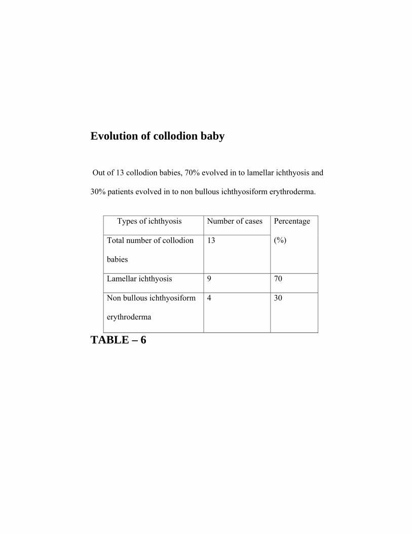
Evolution of collodion baby
Out of 13 collodion babies, 70% evolved in to lamellar ichthyosis and
30% patients evolved in to non bullous ichthyosiform erythroderma.
TABLE – 6
Types of ichthyosis Number of cases
Total number of collodion
babies
13
Percentage
(%)
Lamellar ichthyosis 9 70
Non bullous ichthyosiform
erythroderma
4 30
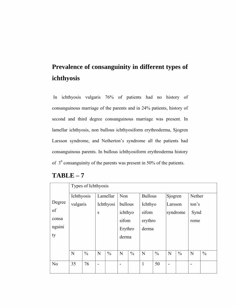
Prevalence of consanguinity in different types of
ichthyosis
In ichthyosis vulgaris 76% of patients had no history of
consanguinous marriage of the parents and in 24% patients, history of
second and third degree consanguinous marriage was present. In
lamellar ichthyosis, non bullous ichthyosiform erythroderma, Sjogren
Larsson syndrome, and Netherton’s syndrome all the patients had
consanguinous parents. In bullous ichthyosiform erythroderma history
of 30 consanguinity of the parents was present in 50% of the patients.
TABLE – 7 Types of Ichthyosis
Ichthyosis
vulgaris
Lamellar
Ichthyosi
s
Non
bullous
ichthyo
sifom
Erythro
derma
Bullous
Ichthyo
sifom
erythro
derma
Sjogren
Larsson
syndrome
Nether
ton’s
Synd
rome
Degree
of
consa
nguini
ty
N % N % N % N % N % N %
No 35 76 - - 1 50 -
-
20 2 4 4 45 2 50 - 1 50 1 100
30 9 20 5 55 2 50 1 50 1 50 -
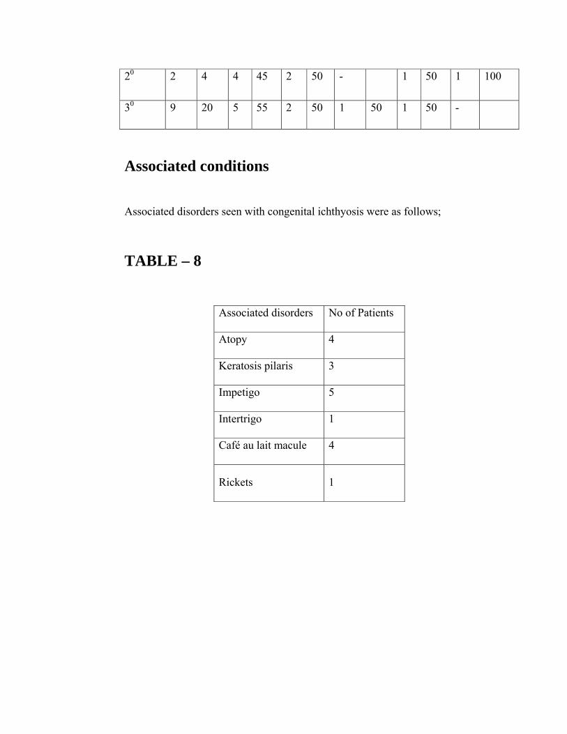
Associated conditions
Associated disorders seen with congenital ichthyosis were as follows;
TABLE – 8
Associated disorders No of Patients
Atopy 4
Keratosis pilaris 3
Impetigo 5
Intertrigo 1
Café au lait macule 4
Rickets 1
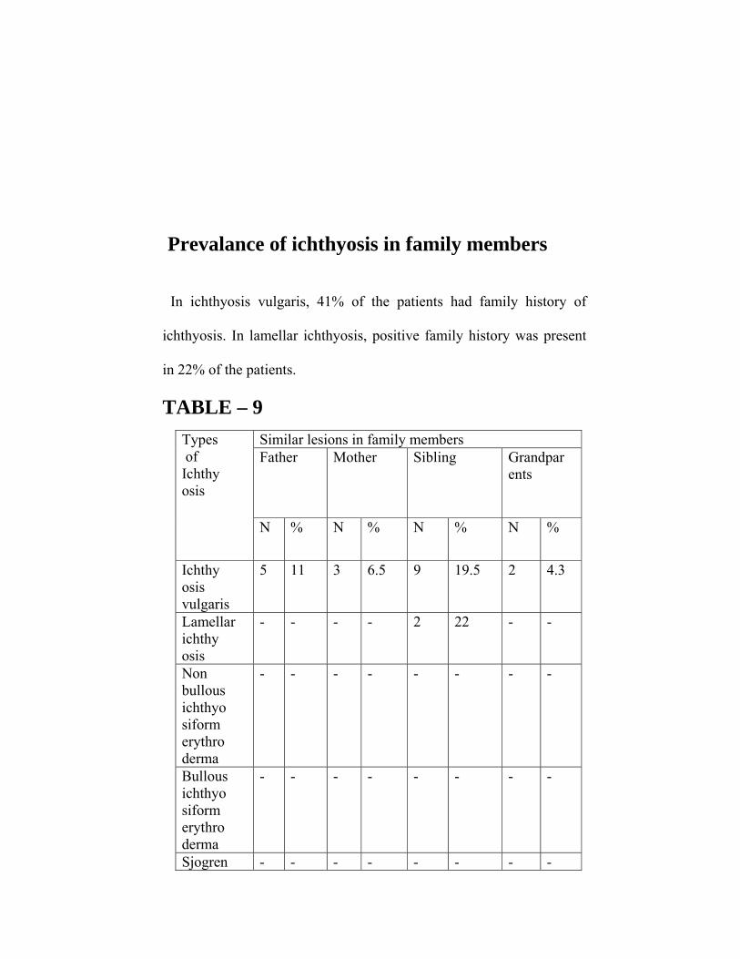
Prevalance of ichthyosis in family members
In ichthyosis vulgaris, 41% of the patients had family history of
ichthyosis. In lamellar ichthyosis, positive family history was present
in 22% of the patients.
TABLE – 9 Similar lesions in family members Father Mother Sibling Grandpar
ents
Types of Ichthy osis
N % N % N % N %
Ichthy osis vulgaris
5 11 3 6.5 9 19.5 2 4.3
Lamellar ichthy osis
- - - - 2 22 - -
Non bullous ichthyo siform erythro derma
- - - - - - - -
Bullous ichthyo siform erythro derma
- - - - - - - -
Sjogren - - - - - - - -

Larsson synd rome Nether ton’s synd rome
- - - - - - - -
Lab Investigations
Percentage of patients with eosinophilia in ichthyosis vulgaris was
11%
Table 10
S.No Type of ichthyosis No of patients
with eosinophilia
Percentage
1 Ichthyosis vulgaris 5 11
2 Netherton’s syndrome 1 100
Serum IgE level
Serum IgE level was elevated (2000 IU/ml) in Netherton’s
syndrome patient.

DISCUSSION In this study of 64 patients, the incidence of congenital
ichthyosis was found to be 6 per 1000 (Table 1).
Incidence of various clinical types of congenital ichthyosis
were as follows. (Table 2)
Ichthyosis vulgaris : 1 per 200
Lamellar ichthyosis : 9 per 10000
Non bullous ichthyosiform erythroderma : 4 per 10000
Bullous ichthyosiform erythroderma : 2 per 10000
Netherton’s syndrome : 1 per 10000
Sjögren Larsson syndrome. : 2 per 10000
A study by Wells and Kerr CB showed, that the incidence of
ichthyosis vulgaris may be as common as 1 in 250. In this study, the
incidence of ichthyosis vulgaris was 1in 200 which complies with the
above study.
Out of 64 cases, the relative incidence of different types of
congenital ichthyosis were as follows. (Table 3)

Ichthyosis vulgaris : 71.8%
Lamellar ichthyosis : 14%
Non bullous ichthyosiform erythroderma : 6%
Bullous ichthyosiform erythroderma : 3%
Netherton’s syndrome : 1.5%
Sjögren Larsson syndrome : 3%
Sex incidence of ichthyosis vulgaris and non bullous
ichthyosiform erythroderma were equal in this study (Table 4). But
females had increased incidence than males (1:3) in lamellar
ichthyosis,In Netherton’s syndrome and Sjögren Larsson syndrome
only males were affected in this study.
Age of onset of ichthyosis vulgaris was around 3-6 months in
96% patients (Table 5). In lamellar ichthyosis , non bullous
ichthyosiform erythroderma, bullous ichthyosiform erythroderma , the
age of onset of the disease was from birth. This complies with that of
the description about the age of onset of diseases given by Traupe et al,
in the guide to clinical diagnosis of ichthyosis.
In Vangysel study of follow up of 17 cases of collodion baby,
60-80% of the infants developed non bullous ichthyosiform
erythroderma and lamellar ichthyosis. In this study of 13 cases of
collodion babies, 70% of the patients developed lamellar ichthyosis
and 30% of the patients developed non bullous ichthyosiform
erythroderma . Thus the ratio of non bullous ichthyosiform
erythroderma Vs bullous ichtyosiform erythroderma was 1:2. 3
(Table 6).
The most common presenting complaints of the patients were
dryness, roughness and disfigurement. Apart from that, itching was
present in patients with associated atopy. Winter exacerbation of the
disease was present in 46% of ichthyosis vulgaris patients.
A study by Kuokanen et al showed an association of atopy in
37-50% of patients. In this study 6.5% patients had associated atopy
and other associations seen were kerotosis pilaris, café au lait macules,
pyoderma, intertrigo and dermatophytosis. (Table 8)
There were several reports of individual cases with congenital
ichthyosis occurring in association with extra cutaneous defects. In this
study, a patient with ichthyosis vulgaris had associated rickets.
In ichthyosis vulgaris, 76% of patients had no history of
consanguinous marriage of the parents and in 24% of patients, history
of 20 and 30 consanguinous marriage was present. (Table 7). In lamellar

ichthyosis, 20 consanguinous marriage was present in 44% of patients
and 30 consanguinous marriage was present in 55% of patients. In non
bullous ichthyosiform erythroderma, 50% patients had 20
consangiunous parents and 50% had 30 consanguinous parents. This
complies with that of autosomal recessive inheritance.
In bullous ichthyosiform erythroderma, 50% had no history of
consanguinity and 30 consanguinity was present in 50% patients. In
Netherton’s syndrome, 20 consanguinous marriage was present in the
parents and it complies with that of autosomal recessive inheritance. In
Sjögren Larsson syndrome, 20 consanguinous marriage was present in
50% of patients and 30 consanguinous marriage was present in 50%
patients and it complies with that of autosomal recessive inheritance.
In ichthyosis vulgaris, family history of ichthyosis was present
in41% of patients (Table 9). In lamellar ichthyosis, positive family
history was present in one family where two siblings were affected. As
it is a autosomal dominantly inherited disorder, the risks of having a
further affected child is 25% which is seen in this case.
In bullous ichthyosiform erythroderma, no family history of
ichthyosis was present in the patient. Because it is a autosomal

dominantly inherited disorder it can be presumed that the patient
suffered a new keratin gene mutation.
Histopathological examination of biopsy from the patients with
ichthyosis vulgaris showed features of hyperkeratosis with decreased
granular layer. In bullous ichthyosiform erythroderma intra epidermal
cleavage with vacuolated cells in the granular layer were seen. Hair
shaft examination in Netherton’s syndrome showed trichorrhexis
invaginata of hair shaft in scalp hair sample.
Laboratory examination showed increased eosinophil count in
11% of ichthyosis vulgaris patients (Table 10). Serum IgE level was
elevated (2000 IU/ ml) in patient with Netherton’s syndrome.
Conclusion
The incidence of congenital ichthyosis in Institute of
child health, Government General Hospital during the
period between September 2005 and September 2007 was
6 per 1000.
The incidence of various clinical types of congenital
ichthyosis were as follows
Ichthyosis vulgaris : 1 per 200
Lamellar ichthyosis : 9 per 10000
Non bullous ichthyosiform erythroderma : 4 per 10000
Bullous ichthyosiform erythroderma : 2 per 10000
Netherton’s syndrome : 1 per 10000
Sjögren Larsson syndrome. : 2 per 10000
The relative incidence of various clinical types of
congenital ichthyosis were as follows
Ichthyosis vulgaris : 71.8%
Lamellar ichthyosis : 14%

Non bullous ichthyosiform erythroderma : 6%
Bullous ichthyosiform erythroderma : 3%
Netherton’s syndrome : 1.5%
Sjögren Larsson syndrome : 3%
Sex distribution was equal in ichthyosis vulgaris patients.
In lamellar ichthyosis, the male to female sex ratio was
1:3
The age of onset of ichthyosis vulgaris was around 3 to 6
months in 96% of patients. In other clinical types, the age
of onset was from birth.
Follow up of collodion baby showed that 70% of patients
developed lamellar ichthyosis and 30% of patient
developed non bullous ichthyosiform erythroderma, the
ratio being 1:2.3.
The main complaints of the patients were dryness,
roughness and disfigurement. Winter exacerbation of the
disease was present in 46% of ichthyosis vulgaris
patients.

6.5% of the patients with ichthyosis vulgaris had
associated atopy.
Other associated condition seen were keratosis pilaris,
café au lait macules, pyoderma and intertrigo.
Rickets was seen in association with ichthyosis vulgaris
in one patient.
Ichthyosis was prevalent in family members of 41% of
the patients with ichthyosis vulgaris and in 22% of the
patients with lamellar ichthyosis.
Laboratory investigations showed eosinophilia in 11% of
patients with ichthyosis vulgaris. Serum IgE was
elevated in patient with Netherton’s syndrome.

BIBLIOGRAPHY
1. Menon IA, Hoberman HF. Dermatological writings of ancient
India Med Hist 1969; 13:387-92.
2. Brocq L.erythrodermie congenitale ichthyosiforme avec
hyperepidermotrophie. Ann dermatol syphiligr 1902; 4: 1-31.
3. Wells RS, Kerr CB. Ichthyosis Br Med j.1966; 2:1504-6.
4. refsums. Heredopathia atactica polyneuritiformis . Acta
psychiatr scand 1946; 38: 1-303.
5. frost P: ichthyosiform dermatoses J invest dermatol 60: 541,
1973.
6. Elias PM, Menon GK: structural and lipid biochemical
correlales of the epidermal permeability barrier. Adv lipid Res
24:1 1991.
7. chapman SJ, walsh A: desmosomes, corneo desmosomes and
desquamation. Arch dermatol Res 262: 304, 1989.
8. Feingold KR: the regulation and role of epidermal lipid
synthesis Adv lipid Res 24:57, 1991.
9. lampe MA, Williams ML, Elias PM. Human epidermal lipids.
Characterization and modulations during differentiation j lipid
Res 24- 131, 1991.

10. Proksch E, Holleran WM, Menon GK etal. Barrier function
regulates epidermal lipid and DNA synthesis. Br J Dermatol
1993;128 : 473 – 82.
11. Williams ML : the ichthyoses – pathogenesis and prenatal
diagnosis: a review of recent advances peadiatr dermatol 1:1,
1983.
12. wells RS, Kerr CB, clinical features of autosomal dominant and
sexlinked ichthyosis in an English population BMS 1966 : 1 :
947 – 50.
13. Sybert VP, Dale BA, Holbrook KA ichthyosis vulgaris :
identification of a defect in the synthesis of filaggrin correlated
with an absence of keratohyaline granules. J invest Dermatol
1985; 84: 191- 4.
14. nirunsuksiri W, presland RB, Brumbaugh SG etal. Decreased
profilaggrin expression in ichthyosis vulgaris is a result of
selectively impaired post transcriptional control. J Biol chem.
1995; 270: 871 – 6.
15. fleckman P, Bumbaughs. Absence of granular layer and
keratohyaline define a morphologically distinct subset of
individuals with ichthyosis vulgaris exp dermatol 2002; 11: 327
– 36.
16. Scott IR, Harding CR, Battett JG. Histidine rich protein of
keratohyaline granules: source of the free aminoacids, urocanic

acid pyrrolidone carboxylic acids in trtum corneum. Biochem
Biophysics Acta 1982; 719:110 – 7.
17. Mevorah B etal : Autosomal dominant ichthyosis and X-linked
ichthyosis. Comparison of their clinical and histological
phenotype ActaDerm venereo 71: 431, 1991.
18. Frost P, vanscott EJ. Ichthyosiform dermatoses. Arch Dermatol
1966; 94: 113.
19. LykkesfeldtG etal: placental steroid sulfatase deficiency:
Biochemical diagnosis and clinical review Obstet Gynecol 64:
49 1984.
20. Bonifas JM, Morley BJ Oakey RE, etal. Cloning of a c DNA for
steroid sulfatase : frequent occurrence of gene deletions in
patients with recessive X linked ichthyosis. Proc Natl Acad sci
USA. 1987; 84:92 48-51.
21. Judge MR, MankuMS, Stewart C, Harper JL scale lipid analysis
in inherited ichthyoses. Br J Dermatol 1990; 123: 820.
22. Paige DG, Emilion GG, Bouloux PMG, Harper JF, A clinical
and genetic study of Xlinked recessive ichthyosis and
contiguous gene defects. BR J Dermatol 1994; 131: 622 – 9.
23. Felinstein A, Ackerman AB, Ziprkows Ki L. Histology of
autosomal dominant ichthyosis vulgaris and Xlinked ichthyosis.
Arch Dermatol Forsch 1974; 248:361.
24. Okano M, KitcinoY, Yoshikawa Ketal. Xlinked ichthyosis and
ichthyosis vulgaris: comparison of their clinical feature based
on biochemical analysis: Br J Dermatol 1988; 119 : 777 – 83.
25. Castagloia C, Fabbrocini G, illianoGMetal. Occularfindings in
Xlinked ichthyosis: a survey opthalmogica 1991; 202: 152- 5.
26. Taylor NF, shackleton CHL: Gas chromatographic steroid
analysis for diagnosis of placental sulfatase deficiency: a study
of nine patients . J clin endocrinol Metab 49: 78, 1979.
27. Hoyer H, Lykkesfeldt AE, Skakkebaek NE : steroid surfatase
deficiency associated with testis cancer Lancet 2: 1456, 1983.
28. Epstein EH,Krauss RM, X linked ichthyosis increased blood
cholesterol sulfate and electro phoretic mobility lowdensity
lipoprotein. Science 1981; 214: 659 – 60.
29. Balden HP, Hooker P, Kubilos J, Tarascio A: Analysis of
Keratinizing tissue for sulfatase deficiency J invest Dermatol
17: 262, 1980.
30. Lareque m, Gharbi R, Daniel J etal: Le be’be’ collodion
e’volution a’ propos de 29cas. Ann Dermatol venereal 103; 31,
1976.
31. Frenk E, detechtermann F: self healing collodion baby: evidence
for autosomal recessive inheritance. Prediatr Dermatol 19: 95,
1992.

32. Garty BB, MetzkerA, NitzanM:Hypernatraemis in congenital
lamellar ichthyosis. J pediatr 95:814, 1979.
33. perlman M, Bar-ziv: congenital ichthyosis and neonatal
pulmonary disease . paediatric 53: 573,1974.
34. VonElysel D, Lijnen R, Moekti petal collodion baby: afollowup
study of 17 cases. J Eur Acad dermatol venereal 2002;16 472 –
5.
35. Unamuno Petal: Harlequin Fetus in four siblings Br. J Dermatol
116: 569, 1987.
36. Lawlor F. Peiris S. Harlequin Fetus successfully healed with
etretinate. Br J Dermatol 1985; 112, 585.
37. KamE, Nirunsuksiri W, Hager B, etal. Prtein phosphatase
activity in human keratinocytes cultured from normal epidermis
and epidermis from patients with harlequin ichthyosis. Br J
Dermatol . 1997; 137: 874 – 82.
38. Michel M, Fleckman P,Smith LT, Dale BA. The calcium
activated neutral protease calpain 1 is present in normal Fetal
\skin and is decreased in neonatal harlequin ichthyosis Br J
Dermatol 1999, 141 : 1017 – 26.
39. Dale BA, Holbrook KA, Fleckman P, etal. Heterogeneity in HL,
an inborn error of epidermal keratinization: variable
morphology and structural protein expression and a defect in
lamellar granules. J invest Dermatol. 1990; 94:6 6 – 18.

40. Blanchet – Bardon C, Domezy: prenatal dignosis of a harlequin
Fetus,semin Dermatal 1984; 3: 225 – 8.
41. Buxman MM, Goudkin PE, Fahrenbach WH, Diamond RL:
Harlequin ichthyosis with epidermal lipid abnormality. Arch
Dermatol 115: 189, 1979.
42. Lawler F progress of a Hairelequin Fetus to non bullous
ichthyosiform erythroderma. Paediatrics 1989; 82: 870 – 3.
43. Traube H, Kolde G, Happle R, autosomal dominant lamellar
ichthyosis ; a new skin disorder clin Genet 1984; 26: 457 – 61.
44. Huber M, Rettler I, Bernasconi K, etal. Mutations of
keratinocyte transgllutaminase in lamellar ichthyosis. Science
1995, 267: 525 – 8.
45. Hohl D, Huber M, Frenk E. Analysis of the cornified cell
envelope in lamellar ichthyosis. Arch Dermatol 1993; 129: 618
– 24.
46. Karnerva L, Lauharanta J, Niemi KM, Lassus A. New
observation on the fine structure of lamellar ichthyosis and the
effect of treatment with etretinate Amj Dermatopathol. 1983; 5:
555 – 68.
47. Niemi KM, Kanerva L, Kuokanen K. Recessive ichthyosis
congenital, type 2. Arch DermaldRes 1991; 283: 211 – 8.
48. Shevchenkoyo. Ampton JG, Toro JR, etal. Splice site mutatuion
in TGM-1 in congenital recessive ichthyosis in American

families: molecular, genetic , genealogic and clinical studies.
Hum Genet . 2000; 106: 492-9.
49. Ghadially R, Williams ML, Housy,llas PM, membrane
structural abnormalities in the stratum corneum of the
autosomal recessive ichthyosis. J invest Dermatol. 1992; 99:
755 – 63.
50. Jobard F, Lefevere C, Karaduman A, etal. Lipoxy genase 3 and
12 are mutated in non bullous congenital ichthyosiform
erythroderma linked to chromosome 17P 13-1. Hummol Gehet,
2002; 11: 107-13.
51. Hazell M, Marks R. clinical histologic and ccell kinetic
discriminants between lamellar ichthyosis and non bullous
ichthyosiform erythroderma. Arch Dermatol 1985; 121:489-93.
52. Murduch ME, Judge MR, Dowd P. Non bullous ichthyosiform
erythroderma: response to etretinate therapy Br J Dermatol1991
; 125: 62-4.
53. Traupe H, Happle R: Alopecia ichthyotica.a characteristic
eature of congenital ichthyosis: Dermatological 167:225, 1983.
54. Neimi KM, Kanerva L, Wahlgren CF,Ignatius J:clinical light
and electron microscopic feature of recessive ichthyosis
congenital typeШ. Arch Dermatol Res 284: 259, 1992.

55. Arnold ML, Anton Lamprecht I, Melz-Roth Fuss B, Hartschuch
W. ichthyosis congenital, type 3.Arch Dermatol Res 1988; 280
– 78.
56. Simpson JR: congenital ichthyisiform erythroderma . trans st.
john Hospital Derm soc 50: 93, 1964.
57. Jrvine AD, Mclean WH, Human keratin disease: the increasing
spectrum of disease and subtley of the phonotype – genotype
correlation Br. J Dermatol 1999; 140: 815 – 28
58. Chipev CC, Yang JM, DIGIOvann JJ etas . preferential sites in
keratin 10 mutated in epidermolytic hyperkeratosis.Am J
Human Genet 1994; 54: 179- 90.
59. Williams ML, Elias PM, from basket weave to barrier: unifying
concepts for the pathogenesis of the disorders of cornification.
Argh Dermatol. 129 ; 626, 1993.
60. Bales J. clinical heterogeneity in epidermolytic hyperkeratosis
arch Dermatol . 1994; 130: 1026 – 35.
61. Ishibashi Y. Klingmuller G. Eythrodemia ichthyosiformis
congenital bullosa Brocq. Arch Klin Exp Dermatol 1968;
232:205.
62. Mccurdy J, Beare JM. Congenital bullous ichthyosiform
erythroderma.Br J Dermatol 1976;79:294.
63. Sahn EE, Weimer CE, Garen PD. Annular Epidermolytic
ichthyosis: a unique phenotype.J Am Acad Dermatol 1992;
27:348- 55.
64. Paller AS, Syder AJ, Chan YM etal. Genetic and clinical
mosaicism in a type of epidermal naevus. NEng J med. 1994;
331: 140 – 15.
65. Smith LT, underwood RA, Mclean WH1. ontogeny and regional
variability of keratin2 e in developing human fetalskin: a unique
special and temporal pattern of keratin expression in
development. Br J Dermatol 1999; 140 : 582-9
66. Altman J. stroud J: Netheston’s syndrome and ichthyosis
linearis circum flexa. Arch Dermatol 100: 550, 1969.
67. comel M. ichthyosis linearis circumflexa. Dermatoligical 1949;
98: 133 – 6.
68. Nethetrons EW. A unique case of trichorrhexis nodosa:
‘bamboo hair’. Arch Dermatol 1958; 78: 483 – 7.
69. PruszKowski A, Bodemerc, Fraitag S, etal. Neonatal and
infantile erythrodermas: a retrospective study of 51 patients
Arch Dermatol . 2000; 136: 875 – 80.
70. Chavanas S etal . mutation in SPINK5 encoding a serine
proteaseinhibitor, cause Netherton syndrome Nat Genet 25:
141,2000.
71. Fartasch M, Wiiliams ML, Elias PM. Allerted lamellar body
secretion and stratum corneum membrane structure in Netherton
syndrome: differentiation from other infantile erythrodermas
and pathogenic implications. Arch Dermatol. 1999; 135: 823 –
32.
72. Thoine EG, Zelickson AS, Mohaz JH, etal. Netherton’s
syndrome an electron microscopic study Arch dermatol Res
1975; 253: 177.
73. Powell J. Dawber RP, Ferguson DJ, Griffiths WA. Netherton’s
syndrome increased likelihood of diagnosis by examining
eyebrowhairs Br J Dermatol 1999; 141: 544 – 6.
74. De Berker DAR, paige DG, Ferguson DJP, Dawber RPR. Golf
tee hairs in Netherton disease paediatr Dermatol 1995; 12:7- 11.
75. Jones SK, Thomas LM, surbrug SK, Weston WL. Neonatal
hypernatraemia in two siblings with Netherton’s syndrome Br J
Dermatol 1986; 114: 741-3
76. Greens S, Muller S: Nethertons syndrome . Report of a case and
review of the literature. J Am Acad Dermatol 13: 329,1985.
77. Krafchik B.: Nethetron’s syndrome pediatr Dermatol 9: 157,
1992.
78. De laurenzi V, Roger GR, Hamrock Dj , etal sjögren – larsson
syndrome is caused by mutations in the fatty aldehyde
dehydrogenase gene. NatGenet 1996; 12: 52-7.

79. Rizzo WB, Craft DA. sjögren – larsson syndrome.deficient
activity of the fatty aldehyde dehydrogenase complement of
fatty alcohol: NADT oxide reductase in cultured fibroblasts . J
clin invest 1991; 88: 1643 – 8.
80. Taube B, Billeadud C, Labreze C etal. sjögren – larsson
syndrome early diagnosis dietary management and biochemical
studies in two cases. Dermatology 1999, 198: 340 -5.
81. Jagell S, Hejjbel J: sjögren – larsson syndrome : physical and
neurological features Helv paediatr Act a 37: 519, 1982.
82. Polland W, SansgrenO specific changes in the fund us typical
for the sjögren – larsson syndrome: an opthamological study.
Acta opthamol scand 1980; 58: 321 -30.
83. Nilsson SEG, Jagell S. Lipofusun and metanin content of the
retinal pigment epithelium in a case of sjögren – larsson
syndrome Br J opthamol 1987: 7: 224-6.
84. Davies MG, Marks R, Dykes PJ, Reynolds D: Epidermal
Abnormalities in refsum,s disease is caused by mutations in the
phytanoyl WA hydroxylase gene. Nat Genet 1997; 17:190-3.
85. mihalik SJ, Morrell JC, Kim D, etal Refsum disease is caused
by mutation in the phytanoyl WA hydroxylase gene Nat Genet
1997; 17:190-3.

86. Takayama K, Salazar EP, Broughton BC etal. Defects in the
repour and transcription gene ERCc2 (XPD) in trichothio
dystrophy. Am J Hum Genet 1996; 58: 263- 70.
87. Vermeulen W, Scott RJ, Rodgers S etal clinical heterogeneity
within XP associated with mutations in the DNA repair and
transcription gene ERCC3. Am J Hum Genet 1994; 54: 191 -
200.
88. Itin PH, Sarasin A, Pitelkow MR: Trichothiodystrophy: update
on the sulfurdeficient brittle hair syndromes. J Am Acad
Dermatol 20011 ; 44: 891-920; quiz 921 – 824.
89. Happle R, Traube H, Grobe H, Bonsmann G. The Tay
Syndrome. Euj Pediatric 141 : 147, 1984.
90. Champman S The Tricho thiodystrophy Syndrome of Pollih
Peadiatric Radiol 18: 154, 1988.
91. Derry JM, Golmally E, Means GD etal. Murations in a delta 8-
delta 7 steroi isomerase in the tallered mouse and Xlinked
dominant Chondrodysplasia punctata. Nat Genet 1999; 22: 286-
90.
92. Happle R, Xlinked dominant chondrodysplasia punctata.
Review of literature and report of a case Hum Genet 1979; 53:
65- 73.

93. Srebrinik A, Tur E, Perluk C etal. Dorfman- Chanarin
syndrome: a case report and a review J Am Acad Dermatol 17:
801, 1987.
94. Judge MR, Hilaire N, Salvagre R etal. Neutral lipid storage
disease: case report and lipid studies Br J Dermatol 1994; 130:
507-10.
95. Rozens zajn L, Klazman A, Yatte D, Edrati P. Jordan’s
anamolyin white blood cells. Blood 1966; 28 : 258-65.
96. Elias PM, Williams ML, Neutral lipid storage disease: defective
lamellar body contents and intracellular dispersion. Arch
Dermatol 1985; 121: 1000-8.
97. Skinner BA, Greist MC, Norins AL. The keratitis, Ichtyosis and
deafness (KID) syndrome. Arch Dermatol. 1981; 117: 285-9.
98. Richard G, Rouan F, Willough by CE, etal. Missense mutations
in the GjB2 encoding connexin-26 cause the ectodermal
dysplasia Keratitis. Ichtyosis- Deafness syndrome. Am j Hum
Genet 2002; 70: 1341-8.
99. Freden ij, estery NB; selected geno dermatoses in infants
children. Clin Dermatol 3:14, 1985.
100. Baden Ep, Alper Jc. Ichthyosiform dermatosis, Keratitis and
deafness Arch Dermatol 1977; 113: 1701-4.
101. Kallmann Fj. Schonfeid WA, Berresa SE. The genetic aspects
of primary enuchoidism Amj Ment Defic 1944: 48: 203-36.
102. Traubo H, ed. Ichthyosis and hypogonadism : Reflections on
the so called Rud,s Syndrome .in: the ichthyosis Berlin spinger-
verlog, 1989: 91-7.
103. Sidransky E, Feinstein A, Goodman RM: Ichthyosis cheek
eyebrow syndrome. A new autosomal dominant disorder , clin
Genet 31: 137, 1987.
104. Happle R, Koch H, Lenz W. the Child syndrome: congenital
hemi dysplasia, ichthyosiform erythroderma and Limb defects,
Eur j peadiatr 1980; 134: 27-33.
105. Koinig A, Bornholdt etal. Mutations in the NSHDL gene,
encoding a 3 beta. Hydroxyl steroid dehydrogenase; cause
CHILD syndrome. Am J Med Genet 2000; 90: 339-46.
106. Hashi moto K, Toppers, sharata H, Edwards M. CHILD
syndrome : analysis of abnormal keratinisation and ultra
structure. Peadiatr Dermatol 1995; 12:116-29
107. Herbert AA, Holbrook KA, Hall jc. The CHILD syndrome
histologic and ultrastructural studies Arch Dermatol 1987, 123:
503-9.
108. YU RJ. Hyperkeratinisation, corneocyte cohesion and alpha
hydroxy acids. J Am Acad Dermatol 1984; 11.867-79.
109. Lucker GPH, Vande kerkhof PCM, sleijlen PM. Effect of
topical calcipotriol on congenital ichthyosis Br j Dermatol 1994;
131: 546- 50.

PROFORMA DATE : INFORMANT:
CASE NO : ADDRESS :
NAME :
AGE :
SEX :
COMPLAINTS:
HISTORY OF PRESENT ILLNESS
Scaling – age of onset/ distribution
Course of lesion- tends to improve/ persistent/ remission
Seasonal variation- summer / winter exacerbation/
cyclical shedding or peeling
H/o blisters
H/o itching
H/o diminished sweating and heat intolerance
H/o photosensitivity/ photophobia
H/o repeated skin infections
H/o seizures
History s/o mental retardation
H/o difficulty in speech.
H/o difficulty in hearing
H/o disturbance in vision
H/o disturbance in perception of smell

H/o difficulty in walking
H/o any abnormal movements
H/o restriction of limb movements
H/o difficulty in climbing upstairs
H/o diarrhea/ malabsorption
History s/o atopy/ asthma
BIRTH HISTORY
ANTENATAL – Maternal illness / medication/ investigation
NATAL – Prematurity / Prolonged labour
NEONATAL – collodion baby / erythrodermic / blisters
DEVELOPMENTAL HISTORY
MILE STONES – social smile/ head control/ sitting with and without
Support / standing with and without support/
Walking / speech
FAMILY HISTORY
Pedigree chart
Consanguinous marriage of parents
Similar lesions in parents/ siblings
GENERAL EXAMINATION
Nourishment

Anemia
Jaundice
Lymphadenopathy
Short stature Height for age
Microcephaly Weight for age
Cataract Head circumference
Gait
Bony deformity
SYSTEM EXAMINATION
Central nervous system – higher function/ speech/ hearing /
Nystagmus / hypertonicity / thickened
peripheral nerves
Respiratory system
Cardio vascular system
Abdomen - hepatosplenomegaly
DERMATOLOGICAL EXAMINATION
SCALES
Small / large
White / dark
Loose / adherent
Lamellar / warty
Site – scalp / face / neck / trunk / upper & lower limb / cubital &
popliteal fossa.
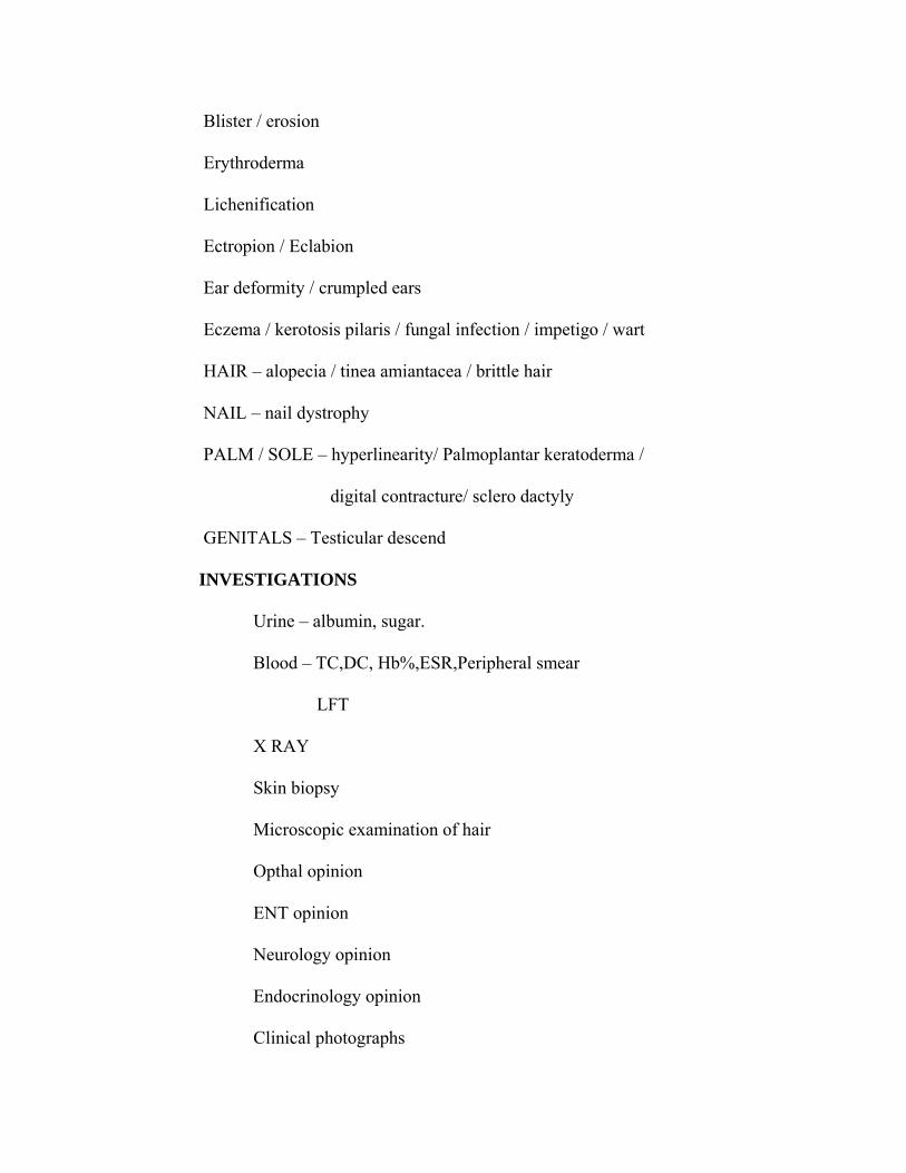
Blister / erosion
Erythroderma
Lichenification
Ectropion / Eclabion
Ear deformity / crumpled ears
Eczema / kerotosis pilaris / fungal infection / impetigo / wart
HAIR – alopecia / tinea amiantacea / brittle hair
NAIL – nail dystrophy
PALM / SOLE – hyperlinearity/ Palmoplantar keratoderma /
digital contracture/ sclero dactyly
GENITALS – Testicular descend
INVESTIGATIONS
Urine – albumin, sugar.
Blood – TC,DC, Hb%,ESR,Peripheral smear
LFT
X RAY
Skin biopsy
Microscopic examination of hair
Opthal opinion
ENT opinion
Neurology opinion
Endocrinology opinion
Clinical photographs

KEY TO MASTER CHART
F - Female
M - Male
ICH VULG - Ichthyosis vulgaris
LI - Lamellar ichthyosis
BIE - Bullous ichthyosiform erythroderma
NBIE - Non bullous ichthyosiform erythroderma
SJO LAR - Sjogren Larsson syndrome
Nether synd - Netherton’s syndrome
Mon - Month
Y - Year
B - Birth
SIB - Sibling
MO - Mother
Gra Fa - Grand Father
Med - medium
SE - Summer exacerbation
WE - Winter exacerbation
HL - Hyper linearity
PPK - Palmoplantar keratoderma
AD - Atopic dermatitis
KP - Keratosis pilaris
CALM - café au lait macule
TI - Trichorrhexis invaginata
Eos - eosinophils