congenital recessive ichthyosis
TRANSCRIPT
ACTAUNIVERSITATIS
UPSALIENSISUPPSALA
2019
Digital Comprehensive Summaries of Uppsala Dissertationsfrom the Faculty of Medicine 1541
Congenital Recessive Ichthyosis
Studies of Gene Expressions Related to KeratinocyteDifferentiation and Skin Barrier Repair
HANQIAN ZHANG
ISSN 1651-6206ISBN 978-91-513-0578-3urn:nbn:se:uu:diva-377557
Dissertation presented at Uppsala University to be publicly examined in Fåhraeussalen,Rudbeck Laboratory, C5, entréplan, Uppsala, Thursday, 11 April 2019 at 13:15 for thedegree of Doctor of Philosophy (Faculty of Medicine). The examination will be conductedin English. Faculty examiner: Associate Professor Mikael Ivarsson (School of HealthSciences, Örebro University).
AbstractZhang, H. 2019. Congenital Recessive Ichthyosis. Studies of Gene Expressions Related toKeratinocyte Differentiation and Skin Barrier Repair. Digital Comprehensive Summaries ofUppsala Dissertations from the Faculty of Medicine 1541. 58 pp. Uppsala: Acta UniversitatisUpsaliensis. ISBN 978-91-513-0578-3.
Autosomal recessive congenital ichthyosis (ARCI) is a rare monogenetic disorder characterizedby a defective skin barrier, hyperkeratosis, and dry, scaly skin. It affects keratinocytedifferentiation and is caused by mutations in any of at least 12 genes believed to control theformation of ω-O-acylceramide and the corneocyte lipid envelope (CLE): ABCA12, ALOXE3,ALOX12B, CERS3, CYP4F22, ELOVL4, LIPN, NIPAL4, PNPLA1, SDR9C7, SLC27A4, andTGM1.
Studies of keratinocyte differentiation and gene expression in ARCI may help us betterunderstand the pathobiology of skin barrier formation. One way to verify that ARCI-relatedgene products are operating in a chain of events essential for lipid barrier formation is touse immunofluorescence and in situ proximity ligation assays to demonstrate the proteins’colocalizations in the epidermis. In paper I, a new method for the objective quantitative imageanalysis of protein expression and colocalization in different epidermal layers of skin sectionswas developed using free, open-source software, CellProfiler. Using this method and microarrayanalyses of skin biopsies from ARCI patients with TGM1 mutations (n = 5) compared withthose of healthy controls (n = 4), many ARCI-related genes were found to be upregulated inpatient epidermis (paper II). Because many other genes involved in keratinocyte differentiationand immune/inflammatory response, including PPARD, were also induced in the patients’microarrays, the effects of a ligand-dependent transcription factor, PPARδ, encoded by PPARD,were studied on ARCI-related gene expression in cultured keratinocytes, usually showing thepronounced upregulation by PPARδ agonists (paper III). Furthermore, using previous arraydata obtained from cultured differentiated keratinocytes and from skin biopsies of patientswith TGM1 mutations, nine novel candidate markers of differentiation were identified, and theupregulation was verified by qPCR of mRNA from cultured keratinocytes and skin biopsies.These transcripts were also induced by PPARδ agonists in cultured proliferating keratinocytes(paper IV).
To conclude, the upregulation of other ARCI-related genes in patients with TGM1 mutationsmight reflect a feedback mechanism in ω-O-acylceramide biosynthesis, which, however,is unable to restore the patients’ skin barrier. In theory, substitution therapy with ω-O-acylceramide and recombinant TGm-1 may be required. Because PPARδ activation appearsinvolved in upregulating ARCI-related genes and nine novel differentiation marker genes, allpotentially important for barrier repair, this approach could become a treatment option forseveral types of ichthyosis and wound healing.
Keywords: genodermatoses, oligoarray, trancriptomics, transglutaminase-1, cornifiedenvelope, peroxisome proliferator-activated receptor δ, all-trans retinoic acids.
Hanqian Zhang, Department of Medical Sciences, Dermatology and Venereology, Akademiskasjukhuset, Uppsala University, SE-75185 Uppsala, Sweden.
© Hanqian Zhang 2019
ISSN 1651-6206ISBN 978-91-513-0578-3urn:nbn:se:uu:diva-377557 (http://urn.kb.se/resolve?urn=urn:nbn:se:uu:diva-377557)

Dedicated to my beloved family The fear of the LORD is the beginning
of wisdom, and the knowledge of the HolyOne is insight.
Proverbs 9:10

List of Papers
This thesis is based on the following papers, which are referred to in the text by their Roman numerals:
I Hanqian Zhang, Maja Ericsson, Marie Virtanen, Simone
Weström, Carolina Wählby, Anders Vahlquist, Hans Törmä. Quantitative image analysis of protein expression and colocalisation in skin sections. Experimental Dermatology. 2018;27(2):196–199.
II Hanqian Zhang, Maja Ericsson, Simone Weström, Anders Vahlquist, Marie Virtanen, Hans Törmä. Patients with congenital ichthyosis and TGM1 mutations overexpress other ARCI genes in the skin: part of a barrier repair response? Experimental Dermatology. 2018. (In press)
III Hanqian Zhang, Simone Weström, Anders Vahlquist, Hans Törmä. PPARδ agonists upregulate transcripts in the ω-O-acylceramide pathway essential for the formation of corneocyte lipid envelopes in epidermis and pathogenic in ichthyosis. (Manuscript)
IV Hanqian Zhang*, Simone Weström*, Anders Vahlquist, Marie Virtanen, Hans Törmä. Identification of novel gene products involved in epidermal keratinocyte differentiation and pathologic skin barrier repair. (Manuscript)
* These authors contributed equally.
Reprints were made with permission from the respective publishers.
Supplementary publication not included
A. Hotz, C. Fagerberg, A. Vahlquist, A. Bygum, H. Törmä, M.-A. Rauschendorf, H. Zhang, L. Heinz, E. Bourrat, I. Hausser, V. Vestergaard, A. Dragomir, A.D. Zimmer, J. Fischer. Identification of mutations in SDR9C7 in six families with autosomal recessive congenital ichthyosis. British Journal of Dermatology. 2018;178(3):e207–e209.
Contents
Introduction ................................................................................................... 11 Skin .......................................................................................................... 11 Epidermal layers ....................................................................................... 12 Acylceramide biosynthesis ....................................................................... 13 The formation of the corneocyte lipid envelope (CLE) ........................... 14 Regulation of keratinocyte differentiation ............................................... 15 Autosomal recessive congenital ichthyosis (ARCI) ................................. 17 Immunofluorescence and in situ proximity ligation assay ....................... 18
Aim of the Study ........................................................................................... 19 Specific aims: ........................................................................................... 19
Materials and Methods .................................................................................. 20 Human skin samples ................................................................................. 20 Immunofluorescence (IF) and the in situ proximity ligation assay (isPLA) ..................................................................................................... 20 Quantitative analysis of IF and isPLA signals ......................................... 21 RNA isolation and microarray expression analysis .................................. 21 Microarray data analysis .......................................................................... 21 Cell culture ............................................................................................... 22 Human epidermal equivalents (HEEs) ..................................................... 23 Analysis of mRNA expression using quantitative PCR ........................... 23 Statistical analysis .................................................................................... 23
Results ........................................................................................................... 24 Paper I ...................................................................................................... 24 Paper II ..................................................................................................... 26 Paper III .................................................................................................... 30 Paper IV ................................................................................................... 33
Discussions & Conclusions ........................................................................... 35 Paper I ...................................................................................................... 35 Paper II ..................................................................................................... 36 Paper III .................................................................................................... 37 Paper IV ................................................................................................... 39
Future Perspectives ....................................................................................... 42

Summary in Swedish .................................................................................... 43
Summary in Mandarin .................................................................................. 45
Acknowledgements ....................................................................................... 46
References ..................................................................................................... 51
Abbreviations
12R-LOX 12R-lipoxygenase encoded by ALOX12B
ABCA12 ATP-binding cassette, sub-family A (ABC1), member 12
acylCer ω-O-acylceramide
AKR1B10 Aldo-keto reductase family 1 member B10
ANGPTL4 Angiopoietin-like 4
AP-1 Activator protein 1
ARCI Autosomal recessive congenital ichthyosis
atRA All-trans retinoic acid
BLNK B cell linker
cDNA Complementary deoxyribonucleic acid
CE Cornified envelope
CerS3 Ceramide synthase 3
CLE Corneocyte lipid envelope
Ct Cycle threshold
CYP4F22 cytochrome P450 family 4 subfamily F member 22
DAG Diacylglycerol
DEG Differently expressed gene
DEJ Dermal-epidermal junction
ELOVL4 ELOVL fatty acid elongase 4
eLOX3 Epidermal lipoxygenase-3 encoded by ALOXE3
ENDOU Endoribonuclease, poly(U) specific
ER Endoplasmic reticulum
FA Fatty acid
FATP4 Fatty acid transporter protein 4 encoded by SLC27A4
GBA β-glucocerebrosidase
GCNT4 Glucosaminyl (N-Acetyl) transferase 4
GEO Gene expression omnibus
GlcCer Glycosylated acylceramide
GLTP Glycolipid transfer protein
HEE Human epidermal equivalent
IF Immunofluorescence
IP3 Inositol trisphosphate
isPLA in situ proximity ligation assay
KRT Keratin
LA Linoleic acid
LB Lamellar body
LC Long chain
LCB Long-chain base, sphingosine
LIPN Lipase N
mRNA Messenger ribonucleic acid
NIPAL4 NIPA-like 4, ichthyin
PKC Protein kinase C
PLC-1 Phospholipase C-1
PNPLA1 Patatin like phospholipase domain containing 1
PPAR Peroxisome proliferator-activated receptor
qPCR Quantitative polymerase chain reaction
RAR Retinoic acid receptor
RHCG Rh family C glycoprotein
ROI Region-of-interest
RXR Retinoid X receptor
sc Stratum corneum
SDR9C7 Short-chain dehydrogenase/reductase family 9C member 7
sg Stratum granulosum
SLC15A1 Solute carrier family 15 member 1
SULT2B1 Sulfotransferase Family 2B Member 1
TEWL Trans-epidermal water loss
TGm-1 Transglutaminase-1 encoded by TGM1
TMEM86A Transmembrane protein 86A
ULC Ultra long chain
ULCFA Ultra-long chain fatty acid
VLC Very long chain
VSNL1 Visinin-like protein 1
ZBTB7C Zinc finger and BTB domain containing 7C
ω-OH-Cer ω-hydroxyl ceramide
11
Introduction
Skin The human skin is the largest organ and the outer covering of the human body. It is the first line of defence and the barrier protecting inner organs from various insults, such as external mechanical damage, pathogen invasion, chemical damage, and ultraviolet (UV) radiation. The skin is also important for regulating body temperature and fluid balance to maintain homeostasis. Moreover, it is also essential for vitamin D synthesis and for storing water and fat, and is a vital organ for sensing heat, cold, touch, pressure, and pain. It can also excrete waste materials by sweating. More importantly, the skin plays crucial roles in both innate and adaptive immunity.
Generally, the skin is composed of three distinct layers, the subcutis, dermis, and epidermis. The lowermost layer, the subcutis, consists of loose connective tissue and adipocytes, and is mainly used for fat storage. The subcutis also works as an insulator and shock absorber to protect internal organs.
Above the subcutis and beneath the epidermis, the dermis is mainly composed of dense connective tissue and fibroblasts. The fibroblasts are involved in synthesizing structural components in the dermis, such as collagen, elastic fibres, and extrafibrillar matrix, and in secreting signal factors to support dermal and epidermal growth. Collagen provides the skin with tensile strength and elastic fibres with elasticity, cushioning the body from strain and stress. Also, sweat glands, hair follicles, nerves, and lymphatic and blood vessels are located in the dermis. The dermis is tightly connected to the epidermis by the dermal–epidermal junction (DEJ), a basement membrane.
The epidermis is the uppermost layer of the skin. It is a stratified squamous non-vascularized epithelium mainly consisting of keratinocytes, which account for 95% of the epidermis (1). In addition to keratinocytes, the epidermis also contains melanocytes, Langerhans cells, and Merkel cells. Melanocytes can produce melanin, which is a pigment that determines skin colour. Melanin also can absorb UV light to prevent DNA damage due to UV exposure (2). Langerhans cells are dendritic cells residing in the skin to capture and process antigens, which is essential for initiating adaptive
12
immunity. Merkel cells are oval-shaped and work as mechanoreceptors, which are essential for light-touch sensation (3).
Epidermal layers Morphologically, the epidermis is divided into several layers, i.e., the stratum basale, stratum spinosum, stratum granulosum, and stratum corneum, representing various stages of keratinocyte differentiation (Figure 1).
Stratum basale The stratum basale, or basal layer, is the deepest layer of the epidermis; it is attached to the DEJ by hemidesmosomes, and is made up of small, cuboidal, proliferating keratinocytes with stem cell properties. Melanocytes and Merkel cells also appear in this layer. In the basal proliferating keratinocytes, keratin K5 and K14 are the major cytoskeletal structural proteins.
Stratum spinosum As the basal keratinocytes differentiate and gradually move upwards, the cells become enlarged to form the spinous layer, or stratum spinosum, and start to lose their proliferating ability. Spinous keratinocytes are interconnected via desmosomes and start to produce lamellar bodies (LBs), enriched in polar lipids, including glucosylceramides, phospholipids, free sterols, and hydrolytic enzymes (4). Langerhans cells are mainly located in this layer. Keratin K1 and K10 are expressed to replace K5 and K14, since the keratinocyte differentiation programme has started. K1 and K10 are also the structural protein markers of suprabasal differentiated keratinocytes, and dominant negative mutations of K1 or K10 cause epidermolytic ichthyosis (5).
Stratum granulosum The keratinocytes continue to migrate upwards and become the granular cell layer containing keratohyalin granules, which are protein structures that appear darkly stained when subject to hematoxylin and eosin (HE) staining and are filled with the histidine-rich protein profilaggrin. Profilaggrin is cleaved by calcium-dependent proteases into filaggrin monomers when the granular cells transit to corneocytes (6), and harmful mutations in filaggrin result in atopic dermatitis or ichthyosis vulgaris (5). Also, lipids in the LBs are released into the extracellular space by exocytosis to generate an intercellular lipid barrier. At the same time, the granular cells start to lose their nuclei and cellular organelles to become dead corneocytes in the stratum corneum (6).
13
Stratum corneum The stratum corneum is the uppermost layer of the skin, consisting of non-viable flattened corneocytes at the final stage of keratinocyte differentiation. The dead cells are surrounded with a rigid protein shell, also known as the cornified envelope (CE), which is composed of different proteins crosslinked by transglutaminases (7). The polar lipids released from the LBs are enzymatically converted into non-polar lipids (4), which are stacked between the corneocytes to form intercellular lipid lamellae (Figure 1). The main components of the lipid lamellae are ceramides with a sphingolipid backbone (Cer), cholesterol, and free fatty acids (4, 8, 9). A unique epidermal ceramide, ω-O-acylceramide (acylCer), can be covalently crosslinked to the CE to form the corneocyte lipid envelope (CLE) by transglutaminase-1 (TGm-1), which is encoded by the gene TGM1 (5, 7, 10), or by a yet unknown enzyme (11). The CLE is the essential structure of the skin barrier function and ichthyosis pathogenesis (12). Most of the barrier functions of the skin, such as the permeability barrier function preventing trans-epidermal water loss (TEWL) and the antimicrobial function, rely on the correct organization of the CE, CLE, and lipid lamellae (11-13). The corneocytes are tightly connected with one another by corneodesmosomes, which are modified desmosome structures that can be degraded through proteolysis in the upper stratum corneum to allow desquamation (7), i.e., the shedding of corneocytes from the skin surface. During desquamation, the low pH in the stratum corneum is important for maintaining the activity of proteases for corneodesmosome degradation (14). The total transit time for a keratinocyte passing from the stratum basale to shedding from the stratum corneum is 52–75 days (1).
Acylceramide biosynthesis The key component of lipid lamellae in the stratum corneum is acylCer made from an ultra-long chain (C28–C36) fatty acid (ULCFA), a long-chain base (LCB, sphingosine), and a ω-esterified linoleic acid (8). In acylCer biosynthesis, ELOVL fatty acid elongase 4 (ELOVL4) is required for ULCFA synthesis (15, 16), and cytochrome P450 family 4 subfamily F member 22 (CYP4F22) is responsible for the hydroxylation of ω-carbon in the ULCFA, which is later used for the esterification of ω-hydroxyl ceramide (ω-OH-Cer) with linoleic acid (8). Three other proteins also appear to be essential for acylCer production. Fatty acid transporter protein 4 (FATP4) encoded by SLC27A4 is a highly putative acyl-CoA synthetase that activates ULCFA (12, 17, 18), which serves as a substrate for ceramide synthase 3 (CerS3), catalysing an amide bond between LCB and ULCFA to produce ω-OH-Cer (8, 12, 19). Finally, patatin-like phospholipase domain-containing
14
protein 1 (PNPLA1) is required for the esterification of ω-OH-Cer with linoleic acid at the ω-carbon to generate acylCer (20-22). The biosynthesis of acylCer is proposed to occur in the endoplasmic reticulum (ER) membrane of upper spinous keratinocytes and granular cells (8).
The formation of the corneocyte lipid envelope (CLE) After acylCer is synthesized in the ER, it is moved to the Golgi apparatus and glycosylated by ceramide glucosyltransferase encoded by UGCG, whose deficiency results in an ichthyosis phenotype in mice (12, 23, 24). Then, the glycosylated acylCer (GlcCer) is transported with the assistance of a vital lipid transporter, ATP-binding cassette (ABC) sub-family A member 12 (ABCA12), to the LBs and later released into extracellular space at the interface between the stratum granulosum and corneum by the fusion of the LBs with the granular cell membrane (12, 25, 26). Following that, the linoleate moiety of GlcCer is oxygenated by 12R-lipoxygenase (12R-LOX) and epidermal lipoxygenase-3 (eLOX3) encoded by the ALOX12B and ALOXE3 genes, respectively (12, 27-31). The oxidized linoleate moiety is then cleaved from GlcCer by hydrolysis to form a free ω-hydroxyl group on the residual GlcCer (12, 31, 32). The free ω-hydroxyl group of GlcCer is used for esterification by TGm-1 with involucrin, a protein component of the CE (5, 10, 12), or by an unknown enzyme (11). The GlcCer can be deglycosylated and hydrolyzed to form ceramide by β-glucocerebrosidase encoded by GBA, a deficiency of which causes Gaucher disease, sometimes associated with ichthyosis (12, 33, 34). Accordingly, the deglycosylation process is also crucial for the correct formation of the CLE and intercellular lipid lamellae and for skin barrier function (12, 33). Defects in any of the above-mentioned enzymes of acylCer synthesis, the lipid transporter, and related enzymes in CLE formation may cause ichthyosis.

15
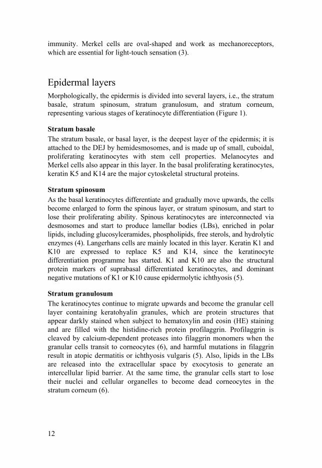
Figure 1. Schematic showing the epidermis, ω-O-acylceramide (acylCer) biosynthesis, and the formation of the corneocyte lipid envelope (CLE), which is the crucial structure for skin barrier function and ichthyosis pathogenesis (12). The ω-hydroxyl ceramide (ω-OH-Cer) in the intercellular lipid lamellae of the stratum corneum is either present as a linoleic acyl ester or is protein bound (35). The autosomal recessive congenital ichthyosis (ARCI)-related genes are underlined. FA = fatty acid; LC = long chain; VLC = very long chain; ULC = ultra-long chain. Adapted with permission from Ohno et al. (8, 22).
Regulation of keratinocyte differentiation Keratinocyte differentiation, a crucial process for skin barrier formation, is well-tuned by many factors. The calcium concentration gradient and a number of transcription factors and microRNAs reportedly regulate this crucial process (36-42).
Calcium is the central regulator of keratinocyte differentiation (41). The calcium concentration gradually rises from the stratum basale, peaks in the stratum granulosum, and declines in the stratum corneum (42). When keratinocytes are switched to a high extracellular calcium concentration, calcium-dependent desmosomes, adherens junctions, and tight junctions are formed to bind cells together, and the calcium receptor is activated to stimulate phospholipase C-1 (PLC-1) via the E-cadherin–catenin complex to generate two second messengers, inositol trisphosphate (IP3) and
16
diacylglycerol (DAG), essential for keratinocyte differentiation (40). IP3 stimulates the release of calcium from intracellular stores, the ER, and the Golgi apparatus, and DAG activates protein kinase C (PKC) (40). PLC-1 is also required to activate store-operated channels for calcium influx to replenish the intracellular calcium store (40). The sustained rise in intracellular calcium and PKC-regulated transcription factor activator protein 1 (AP-1) regulates the gene transcription required for keratinocyte differentiation, i.e., IVL encoding involucrin and TGM1 (40, 43, 44).
Peroxisome proliferator-activated receptors (PPARs) are ligand-activated nuclear receptors comprising three subtypes, i.e., PPAR, PPARδ, and PPAR, all of which work as transcription factors stimulating keratinocyte differentiation and improving the skin permeability barrier function (37-39). PPAR activators inhibit keratinocyte proliferation and induce apoptosis. In contrast, PPAR activation slightly increases cell proliferation, whereas PPARδ has no anti-proliferative or pro-apoptotic effects to facilitate cell survival (37-39). PPAR and PPARδ are also important for epidermal lipid metabolism (39, 45).
PPARδ stimulates epidermal keratinocyte differentiation by an indirect mechanism via an angiopoietin-like 4 (ANGPTL4)-mediated signalling pathway (46). PPAR heterodimerizes with other nuclear hormone receptor, the retinoid X receptors (RXRs), to directly induce the target gene expression of ANGPTL4 (47, 48). ANGPTL4 deficiency/knock-down diminishes the pro-differentiating effects of PPAR agonist GW501516 and the expression of PKC and active phosphorylated components of AP-1, which can be restored by recombinant ANGPTL4 (46). This indicates that calcium and PPAR have a convergent common signalling pathway of PKC and AP-1 to regulate a number of genes important for keratinocyte differentiation. Although TGM1, ABCA12, CERS3, and ELOVL4 have already been shown to be induced by PPAR agonists (38, 49, 50), it is not known whether other genes involved in the formation of acylCer and the CLE in keratinocytes are similarly inducible.
However, unlike calcium, the pivotal factor in regulating keratinocyte differentiation, PPAR preferably plays an essential role via ANGPTL4 in skin barrier repair and wound healing processes, including inflammation, cell migration, proliferation, and extracellular matrix remodelling (51-55). In addition, phorbol esters also can mimic the action of DAG and activate PKC to induce late differentiation markers, i.e., filaggrin, involucrin, and loricrin, and induce ANGPTL4 expression via AP-1 in airway smooth muscle cells, but not in keratinocytes (40, 46, 56).
By contrast, all-trans retinoic acid (atRA), the agonist of retinoic acid receptors (RARs) that are also nuclear hormone receptors, inhibits keratinocyte differentiation by various mechanisms (57, 58). Like PPAR, RARs form heterodimers with the RXRs as the transcription factor to regulate the transcription of target genes (57). atRA suppresses the protein
17
markers of cornification, the genes responsible for the synthesis of epidermal lipids, cholesterol, long-chain fatty acids, and sphingolipids, and the genes implicated in acylCer synthesis and CLE formation (57, 58). Interestingly, in this context, retinoids, i.e., vitamin A derivatives, are commonly used in treating ichthyosis (58-60).
Autosomal recessive congenital ichthyosis (ARCI) Ichthyosis is a collective name for a wide variety of primarily genetically determined disorders of cornification. Autosomal recessive congenital ichthyosis (ARCI) is a heterogeneous group of rare monogenic skin disorders with a prevalence of 1:100,000 (5). ARCI is characterized by a defective epidermal barrier measured as an increase in TEWL inducing a compensatory hyperkeratosis. Hyperkeratosis is presented as a dry, thick epidermis and grey or brownish scales covering the whole body. ARCI patients are often born as collodion babies, a condition in which the infant is born with tight, thick, shiny skin sheets (collodion membranes) covering the entire body surface. Although the condition may sometimes be self-improving (61, 62), it usually continues as a chronic ichthyosis. Clinically, ARCI is divided into four major subtypes: harlequin ichthyosis (HI), lamellar ichthyosis (LI), congenital ichthyosiform erythroderma (CIE), and pleomorphic ichthyosis (PI) (63).
Etiologically, it has so far been reported that ARCI can be caused by mutations in one of the following genes, i.e., TGM1, ALOXE3, ALOX12B, ABCA12, CYP4F22, CERS3, NIPAL4, PNPLA1, SDR9C7, SLC27A4, LIPN, and SULT2B1 (5, 64-66). Moreover, mutations in one of some other genes, i.e., SPINK5, ALDH3A2, PHYH, SUMF1, GJB2, NSDHL, EBP, MBTPS2, ST14, ABHD5, ELOVL4, CLDN1, GBA, STS, FLG, KRT1, KRT10, and KRT2, also can cause an ichthyosis phenotype (5, 34, 67-71). The mutations in ARCI usually fall into several categories: missense mutation (i.e., one base pair change in a DNA coding sequence results in one amino acid substitution in a protein), non-sense mutation (i.e., peptide synthesis is prematurely stopped, resulting in a truncated protein), and splice site mutation (resulting in a changed reading frame). Many of the above genes are involved in the common pathway of acylCer biosynthesis and CLE formation, as mentioned above (12). This could also explain why such divergent single-gene mutations can give rise to similar and overlapping phenotypes. However, some of the above-mentioned ichthyosis-related genes are involved in keratinocyte cholesterol metabolism and the formation of structural proteins, indicating several other essential pathological mechanisms also involved in ichthyosis pathogenesis.
Among the ARCI-related genes, TGM1 is the most commonly affected one (5, 63, 72). Its gene product, TGm-1, plays a vital role in the formation

18
of the CE and CLE by crosslinking proteins and covalently binding acylCer to the CE, and is thereby essential for skin barrier function. However, the effects of TGM1 mutations on the other ARCI genes remain elusive.
Immunofluorescence and in situ proximity ligation assay Immunofluorescence (IF) and in situ proximity ligation assay (isPLA) are methods that have been widely used for studies of protein expression and colocalization, respectively. IF staining can reveal the location and relative expression level of a target protein in tissue sections. The isPLA detects the colocalization (i.e., putative interaction) of two target proteins at a maximal distance of 30 nm in situ with high specificity (73). A positive isPLA colocalization signal, seen as a bright fluorescent spot under the microscope, is the result of a series of reactions, including target binding by primary and secondary antibodies, DNA ligation, and rolling circle amplification (73).
It is conceivable that a better understanding of the disturbed acylCer biosynthesis underlying ARCI could advance the development of new treatment strategies. One way to verify that all the described enzymes and proteins are involved in the same metabolic pathway is to study their localization and colocalization using IF and/or isPLA.
Both IF and isPLA have already been applied in the semi-quantitative analysis of protein expression and colocalization in skin sections (18, 74). However, an efficient method for quantitative image analysis of IF and isPLA is lacking.
19
Aim of the Study
The aim is to better understand the pathological mechanisms of ARCI in the hope of finding new therapeutic approaches.
Specific aims: I To develop a new, efficient method for the objective quantitative
image analysis of protein expression and colocalization in different epidermal layers of skin sections.
II To investigate the impacts of TGM1 mutations on the global transcriptomic expression profile, focusing on the expression of ARCI-related genes in patient skin.
III To investigate whether PPAR regulates the expression of ARCI-related genes involved in acylCer synthesis and CLE formation in keratinocytes.
IV To identify new markers of keratinocyte differentiation that might also be involved in skin barrier repair.
20
Materials and Methods
Human skin samples All studies have been approved by the Regional Ethical Review Board (EPN) in Uppsala and were conducted according to the Declaration of Helsinki principles. Informed and written consent was obtained from the ARCI patients and healthy controls. After local anaesthesia, 3-mm punch biopsies were obtained from lesioned skin on the trunk of the patients and controls for microarray analysis and IF, as previously described (75).
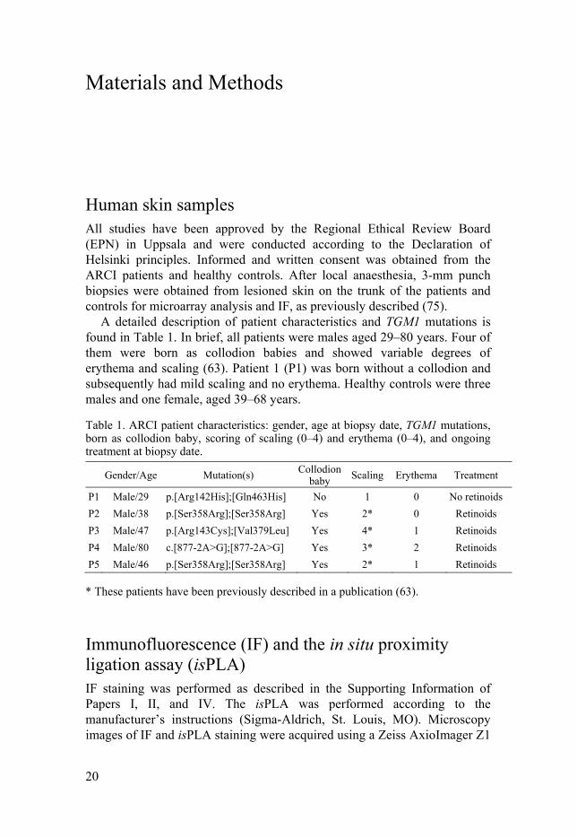
A detailed description of patient characteristics and TGM1 mutations is found in Table 1. In brief, all patients were males aged 29–80 years. Four of them were born as collodion babies and showed variable degrees of erythema and scaling (63). Patient 1 (P1) was born without a collodion and subsequently had mild scaling and no erythema. Healthy controls were three males and one female, aged 39–68 years.
Table 1. ARCI patient characteristics: gender, age at biopsy date, TGM1 mutations, born as collodion baby, scoring of scaling (0–4) and erythema (0–4), and ongoing treatment at biopsy date.
Gender/Age Mutation(s) Collodion baby Scaling Erythema Treatment
P1 Male/29 p.[Arg142His];[Gln463His] No 1 0 No retinoids
P2 Male/38 p.[Ser358Arg];[Ser358Arg] Yes 2* 0 Retinoids
P3 Male/47 p.[Arg143Cys];[Val379Leu] Yes 4* 1 Retinoids
P4 Male/80 c.[877-2A>G];[877-2A>G] Yes 3* 2 Retinoids
P5 Male/46 p.[Ser358Arg];[Ser358Arg] Yes 2* 1 Retinoids
* These patients have been previously described in a publication (63).
Immunofluorescence (IF) and the in situ proximity ligation assay (isPLA) IF staining was performed as described in the Supporting Information of Papers I, II, and IV. The isPLA was performed according to the manufacturer’s instructions (Sigma-Aldrich, St. Louis, MO). Microscopy images of IF and isPLA staining were acquired using a Zeiss AxioImager Z1
21
microscope (Carl Zeiss, Stockholm, Sweden) at 40× magnification using the ZEN2012 software. Images for IF were acquired in a single focal plane where the sharpest, strongest IF staining was located, whereas isPLA images were acquired as z-stacks to enable detection of vertical isPLA signals present at different depths in the skin sections.
Quantitative analysis of IF and isPLA signals Detailed instructions on how to use the CellProfiler pipelines can be found in the Supporting Information of Paper I. In brief, the analysis of epidermal protein expression and colocalization was performed using three pipelines: I) Projection: isPLA signals in different focal planes of a z-stack image were projected into a single .tif image. This step was bypassed for IF images, which were acquired in a single focal plane and directly exported as lossless .tif files. II) Region-of-interest (ROI): Using the output of step 1, a ROI was manually outlined by drawing a line along the uppermost nucleated epidermal layer, and including the epidermis and dermis. III) Measurement: The epidermal outline from pipeline 2 was used to generate a segmentation of the image from pipeline 1. The segmentation included two layers above the outline (i.e., the stratum corneum) and up to 18 layers below. The fluorescence intensity in red, green, and blue image channels and the isPLA signal count were automatically measured for each layer.
RNA isolation and microarray expression analysis Total RNA was isolated using TRI Reagent (Ambion – Thermo Fisher Scientific, Foster City, CA) from homogenized skin biopsies or cultured cells and RNA quality was evaluated using the Agilent 2100 Bioanalyzer system (Agilent Technologies Inc, Palo Alto, CA). The GeneChip Clariom D Human Array (Thermo Fisher Scientific, Waltham, MA) was used for the microarray analysis of gene expression. RNA from each sample was used to generate amplified and biotinylated sense-strand cDNA from the entire expressed genome according to the GeneChip WT PLUS Reagent Kit User Manual. GeneChip Clariom D Human Array was hybridized. Washing and staining were performed using the Fluidics Station 450 (Thermo Fisher Scientific) and finally scanned using the GeneChip Scanner 3000 7G.
Microarray data analysis The raw data were normalized in the free software Expression Console from Affymetrix (http://www.affymetrix.com) using the robust multi-array
22
average (RMA) method (76, 77). The RMA algorithm fits a robust linear model at the probe level to minimize the effect of probe-specific affinity differences. The gene expression data were analysed in the freely available statistical computing language R (http://www.rproject.org) using packages available from the Bioconductor project (www.bioconductor.org). To search for genes that are differentially expressed between groups, an empirical Bayes moderated t-test was then performed (78) using the ‘limma’ package (79). To address the problem of multiple testing, the p-values were adjusted using the method of Benjamini and Hochberg (80).
Heat maps of gene expression were generated using the online tool Morpheus (https://software.broadinstitute.org/morpheus). The genes, of which the expression altered more than 1.5 log2fold-change (FC) compared to controls (adjusted p-value < 0.05), were used in functional annotation clustering analysis using the Database for Annotation, Visualization and Integrated Discovery (DAVID) (76, 81). Gene ontology clusters with Bonferroni adjusted p-value < 0.01 were retained for evaluation. The complete data are submitted to Gene Expression Omnibus repository (GSE107462).
Cell culture Normal human epidermal keratinocytes from adults were cultured in EpiLife serum-free medium containing human keratinocyte growth supplement and gentamicin/amphotericin B (cells, medium, and reagents were all from Life Technologies, Stockholm, Sweden) and were used at passage 2. The cells were incubated at 37°C in 5% CO2. The medium was replaced 24 h after the culture was initiated and subsequently every second day. Cells were detached using trypsin/EDTA and Defined Trypsin Inhibitor (Life Technologies). N/TERT-1 keratinocytes were maintained in EpiLife medium (kindly provided by Dr. J. Rheinwald) (82).
Proliferating keratinocytes were cultured until 75% confluency, and then exposed to various drugs. For differentiated keratinocytes, cells were cultured as above until reaching 100% confluency; the cells were then maintained in EpiLife medium supplemented with 1.5 mM CaCl2 for 4 days. Proliferating and differentiated keratinocytes were exposed to the PPAR agonists WY14643/pirinixic acid (PPAR), GW501516 and GW0742 (PPAR), and ciglitizone (PPAR) and to GSK0660, an antagonist selective for PPAR, all dissolved in DMSO (the agonists, antagonist, and solvent were all from Sigma-Aldrich) for up to 48 h. The cells were then harvested in TRI Reagent; the total RNA was isolated and subsequently used for qPCR analysis.
23
Human epidermal equivalents (HEEs) Primary human epidermal keratinocytes of neonatal origin were cultured in EpiLife medium with supplements. After trypsinization, 300,000 cells were seeded per insert (12-mm diameter, 0.4-µm pore size; Merck Millipore, Burlington, MA) in EpiLife medium and incubated as submerged culture for 48 h. The medium was then changed to CnT-Prime 3D Barrier Medium (CELLnTec, Bern, Switzerland) supplemented with 20% DMEM (CnT/DMEM; Sigma-Aldrich) and incubated for another 24 h in submerged culture. To raise the inserts to the air–liquid interface, the medium inside was removed and the inserts were transferred to 6-cm dishes containing 3.2 mL of CnT/DMEM (three or four inserts per dish). After 12 days at the air–liquid interface, the HEEs were treated with vehicle DMSO, 1 µM all-trans retinoic acid (atRA) (Sigma-Aldrich), or 5 µM GW501516 for 24, 48, or 72 h. During these three days, the medium or treatment was changed every day. The HEEs were then harvested in TRI Reagent, and the total RNA was isolated and subsequently used for qPCR analysis.
Analysis of mRNA expression using quantitative PCR First-strand cDNA was synthesized from total RNA by combining oligo(d)T, random hexamers, buffer, and MMLV reverse transcriptase (Invitrogen, Carlsbad, CA) as previously described (83, 84). Quantitative PCR was performed using cDNA as the template and TaqMan Gene Expression Assays (Thermo Fisher Scientific) in an ABI 7500 Fast Real-Time PCR system (Applied Biosystems, Foster City, CA). Expression levels were measured in triplicate. The relative mRNA expression was determined by the 2(–ΔΔCt) method using RPL19 as the reference gene.
Statistical analysis The protein expression detected by IF staining was analysed using the multiple t-test with Holm–Sidak correction. The normalized relative mRNA expression detected by qPCR analysis was analysed using multiple t-tests, one-way ANOVAs, and two-way ANOVAs with different corrections for post hoc multiple comparisons. All data were analysed using GraphPad Prism software (GraphPad Software, La Jolla, CA).
24
Results
Paper I In this paper, we developed a new, efficient method for the objective quantitative image analysis of protein expression (IF) and colocalization (isPLA) in different epidermal layers of skin sections using the CellProfiler software. The method consists of three steps/pipelines:
I Projection: For IF, this step is skipped, because the images can
be acquired in single focal plane and directly exported as lossless .tif files. For isPLA, since the signals are located in different focal planes, a z-stack image can be acquired and projected into a single .tif image.
II Region-of-interest (ROI): Using the output image from step 1, a ROI was outlined manually by drawing a continuous line just above the uppermost nucleated epidermal layer, circling the epidermis and dermis, excluding small parts of the image corners, and looping around to rejoin the starting point.
III Measurement: The outlined ROI binary image from step 2 was used to segment the image from step 1. The segmentation comprises two layers above the outline (i.e., the stratum corneum) and up to 18 layers below (i.e., the stratum granulosum, spinosum, and basale, and the dermis). The fluorescence intensities of the image colour channels (i.e., red, green, and blue) and the isPLA signal counts were automatically measured for each layer.
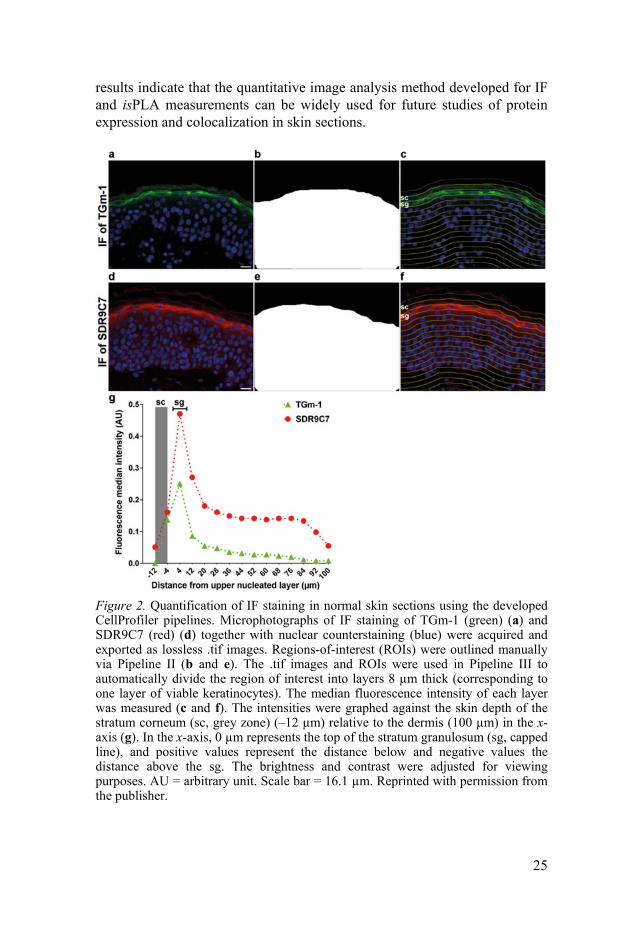
The CellProfiler software and the developed pipelines used in this study can be downloaded at www.cellprofiler.org. We then used the developed method to analyse the IF images of TGm-1 and SDR9C7 (Figure 2), two ARCI-related proteins, as well as isPLA images of TGm-1/SDR9C7 (Figure 3). The median fluorescence intensities of TGm-1 and SDR9C7 reached their maximum expression in the upper stratum granulosum (Figure 2g). The isPLA signal density in each layer indicated that TGm-1 and SDR9C7 mainly colocalized in the stratum granulosum, whereas the technical and biological negative controls displayed almost no isPLA signal, indicating that these two proteins colocalize and might be functionally connected. The
25
results indicate that the quantitative image analysis method developed for IF and isPLA measurements can be widely used for future studies of protein expression and colocalization in skin sections.
Figure 2. Quantification of IF staining in normal skin sections using the developed CellProfiler pipelines. Microphotographs of IF staining of TGm-1 (green) (a) and SDR9C7 (red) (d) together with nuclear counterstaining (blue) were acquired and exported as lossless .tif images. Regions-of-interest (ROIs) were outlined manually via Pipeline II (b and e). The .tif images and ROIs were used in Pipeline III to automatically divide the region of interest into layers 8 µm thick (corresponding to one layer of viable keratinocytes). The median fluorescence intensity of each layer was measured (c and f). The intensities were graphed against the skin depth of the stratum corneum (sc, grey zone) (–12 µm) relative to the dermis (100 µm) in the x-axis (g). In the x-axis, 0 µm represents the top of the stratum granulosum (sg, capped line), and positive values represent the distance below and negative values the distance above the sg. The brightness and contrast were adjusted for viewing purposes. AU = arbitrary unit. Scale bar = 16.1 µm. Reprinted with permission from the publisher.

26
Figure 3. Quantification of isPLA signals of TGm-1/SDR9C7 colocalization using the developed CellProfiler pipelines. (a) Projected lossless .tif image of isPLA of TGm-1/SDR9C7 (red) together with nuclear counterstaining (blue) was generated using Pipeline I. (b) The region-of-interest (ROI) was manually outlined using Pipeline II. (c) The projected image and ROI were used in Pipeline III to automatically divide the region of interest into layers 8 µm thick (corresponding to one layer of viable keratinocytes). The isPLA signals outlined in white indicate that they were detected and counted by Pipeline III. (d) The numbers of isPLA signals 1000 µm–2 for each layer of isPLA colocalization of TGm-1/SDR9C7, TGm-1/filaggrin (biological control), and technical control graphed against skin depth. sc = stratum corneum (grey zone); sg = stratum granulosum. Scale bar = 16.1 µm. Reprinted with permission from the publisher.
Paper II In this paper, a microarray study was performed to investigate the impacts of TGM1 mutations on the global transcriptome expression profile focusing on ichthyosis-related genes.
We examined the mRNA expression profile in skin punch biopsies from five male patients (P1–5, aged 29–80 years) with ARCI of different degrees of severity and with various types of inactivated TGM1 mutations (see Table 1) and from four healthy volunteers (aged 39–68 years; C1, C2, C4 male and
27
C3 female). Within the global transcripts, there were a total of 22,559 gene transcripts with UniGene ID, of which 599 represented differentially expressed genes (DEGs). The healthy controls and ARCI patients were well separated by two principal components generated from the transcriptomic data. Of all DEGs, 205 were upregulated >1.5 log2fold-change (FC) and 67 were downregulated < –1.5 FC (denoting a 2.8-fold change after back transformation), functionally clustering into different gene ontology (GO) terms, for example, keratinocyte differentiation (GO:0030216) and immune response (GO:0006955).
Many of the upregulated DEGs were assigned to keratinocyte differentiation and sporadically involved in CE formation, for example, eight small proline-rich proteins (SPRRs, 3.18–9.31 FC), involucrin (IVL, 1.98 FC), and two late cornified envelope proteins (LCE3D and LCE3E, 4.51–4.91 FC). Additionally, PPARD was increased by 2.25 FC. This gene encodes PPAR, a ligand-activated nuclear receptor serving as a transcription factor essential for keratinocyte differentiation, lipid metabolism, and skin barrier repair (39, 55, 85). Among the DEGs annotated to immune response, several antimicrobial peptide (AMP) genes (S100A7, S100A8, S100A9, DEFB4A, and DEFB4B, 4.16–11.29 FC), several cytokine genes (CCL20, 11.64 FC and IL36G, 5.13 FC), and a lipid transporter CD36 (4.12 FC) were highly induced.
Interestingly, some DEGs were identified as ichthyosis-related and lipid-synthesis genes. For example, the transcription of seven out of 12 ARCI-related genes, i.e., ABCA12, ALOX12B, CERS3, CYP4F22, LIPN, SDR9C7, and SLC27A4 (0.98–2.05 FC), was significantly upregulated, as was the expression of three genes related to other types of ichthyosis, i.e., ELOVL4, GBA, and GJB2 (1.66–5.69 FC). In contrast, CLDN1 displayed downregulated transcription (–1.38 FC), and ALOXE3, NIPAL4, PNPLA1, and TGM1 displayed non-significant differences compared with healthy controls (Figure 4).

28
Figure 4. Heatmap of gene expression profiles of ichthyosis-related genes in ARCI patients with TGM1 mutations (P1–5; n = 5) compared with controls (C1–4; n = 4) as determined by microarray analysis. Genes with significantly altered expressions (adjusted P < 0.05) between patients and controls are shown in the blue box. Reprinted with permission from the publisher.
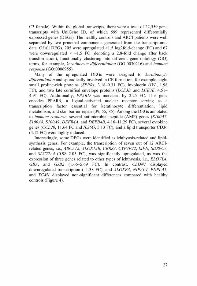
The microarray data, which indicated the altered expression of many ichthyosis-related genes, prompted verification of the transcripts using qPCR. In complete agreement with the microarray results, the transcripts of ABCA12, ALOX12B, CERS3, CYP4F22, ELOVL4, LIPN, SDR9C7, and SLC27A4 were all upregulated in patients versus controls, but not the transcripts of ALOXE3, NIPAL4, and PNPLA1 (Figure 5).
29
Figure 5. Relative mRNA expression of ichthyosis-related genes in ARCI patients with TGM1 mutations (n = 5) and healthy controls (n = 4) was reanalysed using qPCR. Expression was normalized to the reference gene RLP19. Values are presented as boxes and whiskers, with the latter showing min/max (expression/RLP19). *P < 0.05; n.s. = not significant.
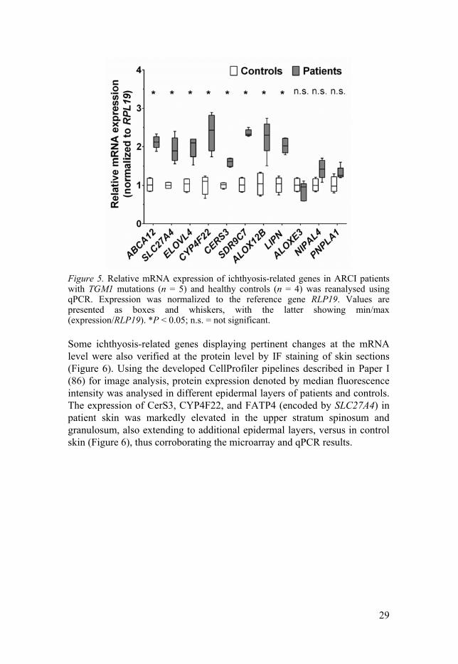
Some ichthyosis-related genes displaying pertinent changes at the mRNA level were also verified at the protein level by IF staining of skin sections (Figure 6). Using the developed CellProfiler pipelines described in Paper I (86) for image analysis, protein expression denoted by median fluorescence intensity was analysed in different epidermal layers of patients and controls. The expression of CerS3, CYP4F22, and FATP4 (encoded by SLC27A4) in patient skin was markedly elevated in the upper stratum spinosum and granulosum, also extending to additional epidermal layers, versus in control skin (Figure 6), thus corroborating the microarray and qPCR results.
30
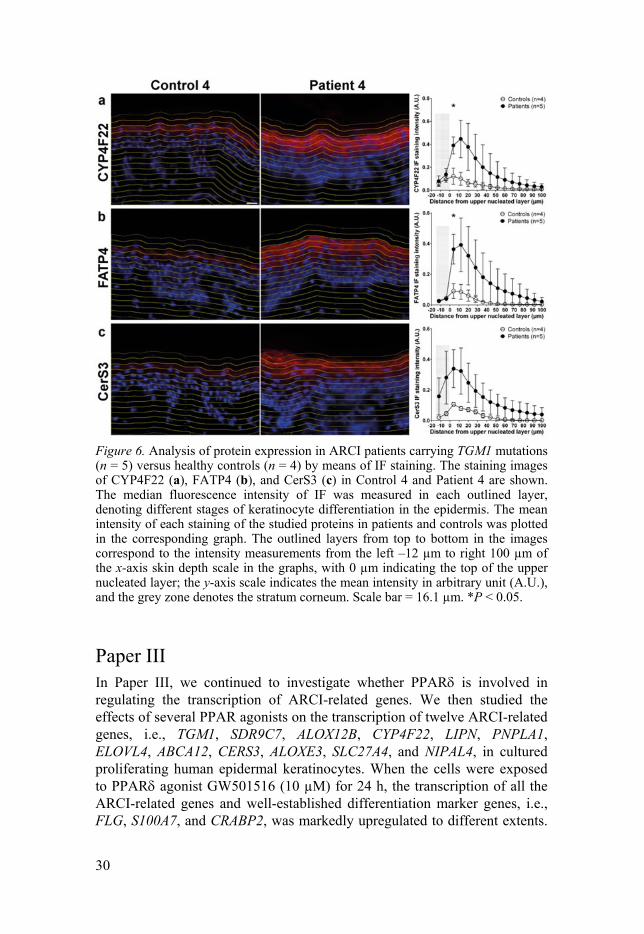
Figure 6. Analysis of protein expression in ARCI patients carrying TGM1 mutations (n = 5) versus healthy controls (n = 4) by means of IF staining. The staining images of CYP4F22 (a), FATP4 (b), and CerS3 (c) in Control 4 and Patient 4 are shown. The median fluorescence intensity of IF was measured in each outlined layer, denoting different stages of keratinocyte differentiation in the epidermis. The mean intensity of each staining of the studied proteins in patients and controls was plotted in the corresponding graph. The outlined layers from top to bottom in the images correspond to the intensity measurements from the left –12 µm to right 100 µm of the x-axis skin depth scale in the graphs, with 0 µm indicating the top of the upper nucleated layer; the y-axis scale indicates the mean intensity in arbitrary unit (A.U.), and the grey zone denotes the stratum corneum. Scale bar = 16.1 µm. *P < 0.05.
Paper III In Paper III, we continued to investigate whether PPAR is involved in regulating the transcription of ARCI-related genes. We then studied the effects of several PPAR agonists on the transcription of twelve ARCI-related genes, i.e., TGM1, SDR9C7, ALOX12B, CYP4F22, LIPN, PNPLA1, ELOVL4, ABCA12, CERS3, ALOXE3, SLC27A4, and NIPAL4, in cultured proliferating human epidermal keratinocytes. When the cells were exposed to PPAR agonist GW501516 (10 µM) for 24 h, the transcription of all the ARCI-related genes and well-established differentiation marker genes, i.e., FLG, S100A7, and CRABP2, was markedly upregulated to different extents.
31
In contrast, no clear effects of PPAR agonist WY14643 (20 µM) were observed, whereas the PPAR agonist ciglitizone (5 µM) exhibited only weak inducing effects on the transcription of TGM1 and NIPAL4.
Furthermore, we investigated whether or not the inducing effects of PPAR agonist GW501516 on the ARCI-related gene transcription are time and/or dose dependent. The peak mRNA expression was observed at 24 h rather than at 6 and 48 h for selected genes, i.e., TGM1, SDR9C7, ELOVL4, ABCA12, CERS3, and NIPAL4. Our further experiments also found that the effects of GW501516 are dose dependent: compared with 5 µM GW501516, 10 µM elicited >5-fold higher inductions for TGM1, SDR9C7, ALOX12B, CYP4F22, LIPN, and PNPLA1. However, almost no effect was observed for 1 µM GW501516.
After observing the markedly inducing effect of GW501516 on ARCI-related gene transcription in proliferating keratinocytes, we tried to confirm a similar effect of GW501516 on differentiated keratinocytes. Proliferating and differentiated keratinocytes were identically exposed to GW501516 (10 µM) for 24 h. GW501516 induced all the ARCI-related genes in both proliferating and differentiated keratinocytes, but the inducing effects were more pronounced in pre-confluent proliferating cells than post-confluent differentiated cells. This was because of a low basal transcription level of all ARCI-related genes in proliferating cells, genes that can be markedly induced by keratinocyte differentiation to a higher level in differentiated cells.
To further explore whether or not the induction effects of GW501516 on ARCI-related genes were ligand dependent, we then exposed proliferating keratinocytes to PPAR antagonist GSK0660 (10 µM) for 2 h before adding PPAR agonist GW501516 (5 µM). After this combined exposure, the transcription of SLC27A4 and NIPAL4 remained at the baseline/vehicle level (Figure 7k and l), indicating that GSK0660 completely blocked the inducing effect of GW501516 and that the induction of both genes resulted from the ligand activation of PPAR. Interestingly, the transcription of six ARCI genes, i.e., TGM1, SDR9C7, ALOX12B, CYP4F22, LIPN, and PNPLA1, was even reduced below that of the vehicle due to the combined exposure of PPAR agonist and antagonist (Figure 7a–f), and they are also the same genes most highly upregulated by GW501516 alone. Together with the fact that GSK0660 alone reduced the basal transcription of several ARCI-related genes in both proliferating and differentiated keratinocytes, this suggests that GSK0660 might even block the endogenous ligand activation of PPAR. However, the induced transcription of ELOVL4, ABCA12, CERS3, and ALOXE3 by GW501516 was unaffected by pre-exposure to GSK0660 (Figure 7g–j), indicating that these genes were not upregulated through the ligand activation of PPAR.

32
Figure 7. Effects of PPAR antagonist GSK0660 (GSK) on PPAR agonist GW501516 (GW)-induced transcription of ARCI-related genes important for the epidermal acylCer synthesis pathway in proliferating keratinocytes (a–l). The cells were pre-exposed to 10 µM GSK for 2 h before adding 5 µM GW for 24 h. The induced transcription of some genes by GW was inhibited by GSK (a–f, k, and l), whereas the induction of other genes was not inhibited (g–j). All expression values are normalized to vehicle cells (V) and to the reference gene RPL19. ago = agonist, anta = antagonist. Values represent mean ±SD (n = 3). *P-value < 0.05, ** < 0.01, *** < 0.001, **** < 0.0001; ns = not significant.
To confirm that the induced transcription of ARCI-related genes by PPAR agonist GW501516 in cultured keratinocytes was reproducible in an in vivo-like model, HEEs were exposed to GW501516 (5 µM) for 24, 48, and 72 h. The transcription of all ARCI-related genes except LIPN was markedly upregulated at one or several time points, suggesting that the inducing effects of PPAR agonist GW501516 in 2D monolayer culture can also be reproduced in a 3D organotypic model.
33
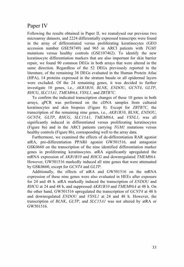
Paper IV Following the results obtained in Paper II, we reanalysed our previous two microarray datasets, and 2224 differentially expressed transcripts were found in the array of differentiated versus proliferating keratinocytes (GEO accession number GSE58749) and 965 in ARCI patients with TGM1 mutations versus healthy controls (GSE107462). To identify the new keratinocyte differentiation markers that are also important for skin barrier repair, we found 90 common DEGs in both arrays that were altered in the same direction. Regardless of the 52 DEGs previously reported in the literature, of the remaining 38 DEGs evaluated in the Human Protein Atlas (HPA), 14 proteins expressed in the stratum basale or all epidermal layers were excluded. Of the 24 remaining genes, it was decided to further investigate 10 genes, i.e., AKR1B10, BLNK, ENDOU, GCNT4, GLTP, RHCG, SLC15A1, TMEM86A, VSNL1, and ZBTB7C.
To confirm the indicated transcription changes of these 10 genes in both arrays, qPCR was performed on the cDNA samples from cultured keratinocytes and skin biopsies (Figure 8). Except for ZBTB7C, the transcription of the remaining nine genes, i.e., AKR1B10, BLNK, ENDOU, GCNT4, GLTP, RHCG, SLC15A1, TMEM86A, and VSNL1, was all significantly induced in differentiated versus proliferating keratinocytes (Figure 8a) and in the ARCI patients carrying TGM1 mutations versus healthy controls (Figure 8b), corresponding well to the array data.
Furthermore, we examined the effects of de-differentiation RAR agonist atRA, pro-differentiation PPAR agonist GW501516, and antagonist GSK0660 on the transcription of the nine identified differentiation marker genes in proliferating keratinocytes. atRA significantly upregulated the mRNA expression of AKR1B10 and RHCG and downregulated TMEM86A. However, GW501516 markedly induced all nine genes that were attenuated by GSK0660, except for GCNT4 and GLTP.
Additionally, the effects of atRA and GW501516 on the mRNA expression of these nine genes were also evaluated in HEEs after exposure for 24 and 48 h. atRA markedly induced the transcription of ENDOU and RHCG at 24 and 48 h, and suppressed AKR1B10 and TMEM86A at 48 h. On the other hand, GW501516 upregulated the transcription of GCNT4 at 48 h and downregulated ENDOU and VSNL1 at 24 and 48 h. However, the transcription of BLNK, GLTP, and SLC15A1 was not altered by atRA or GW501516.
34
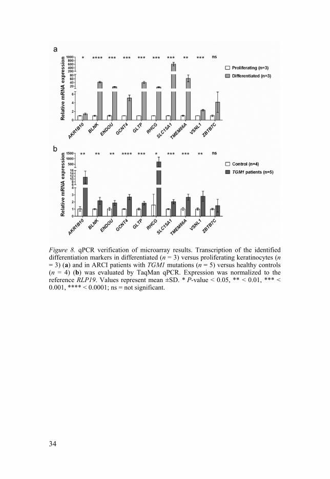
Figure 8. qPCR verification of microarray results. Transcription of the identified differentiation markers in differentiated (n = 3) versus proliferating keratinocytes (n = 3) (a) and in ARCI patients with TGM1 mutations (n = 5) versus healthy controls (n = 4) (b) was evaluated by TaqMan qPCR. Expression was normalized to the reference RLP19. Values represent mean ±SD. * P-value < 0.05, ** < 0.01, *** < 0.001, **** < 0.0001; ns = not significant.
35
Discussions & Conclusions
Paper I The image analysis of IF and isPLA stainings indicates that the new method clearly captures the unique pattern of protein expression and colocalization, because the developed CellProfiler pipelines for the image analysis of immunostained skin sections have four unique advantages, as discussed below. The new method provides:
1. quantification of staining fluorescence intensity; 2. number counts of isPLA colocalization signals; 3. layered measurements; and 4. simultaneous analysis of large numbers of images.
By automating the image analysis procedure, the same standards could be applied in processing all images, avoiding subjective bias in evaluating the staining results. Additionally, the developed method can simultaneously measure the fluorescence intensity of IF staining and the number of isPLA signals, although these results were presented separately in this paper. More importantly, the novel layered measurements of IF staining intensity and isPLA signals precisely determine in which layers of the epidermis the proteins of interest are expressed and colocalized, enabling comparisons of staining signals between layers within and between images, and even between skin sections immunostained at one time. By reporting the median rather than mean fluorescence intensity, the image analysis method furthermore limits the bias of staining artefacts. Moreover, the CellProfiler can simultaneously analyse a large number of images more efficiently than can the currently available method of time-consuming manual image analysis performed individually. Applying the developed method in the analysis of IF images of TGm-1 and SDR9C7 and of isPLA images of TGm-1/SDR9C7 clearly revealed the expression and colocalization patterns of those two ARCI-related proteins in the epidermis, suggesting that they might be functionally connected and act in the same pathway.
To conclude, the developed quantitative image analysis method for IF and isPLA stainings can be widely applied in future studies of protein expression and colocalization in skin sections.
36
Paper II This study investigated global transcriptomic changes in the epidermis of ARCI patients with TGM1 mutations versus healthy controls. The most pertinent findings are summarized below:
1. the increased expression of genes involved in the biosynthesis of acylCer and the CLE is important for skin barrier function and attributable to the aetiology of what might be termed lipodysgenic ichthyoses;
2. the upregulation of various AMPs and cytokine genes is implicated in the antimicrobial response and in the regulation of the immune response; and
3. a number of genes affecting keratinocyte proliferation and differentiation are altered.
All of these changes are probably involved in re-establishing a functional skin barrier in the skin of ARCI patients with a disrupted skin barrier.
Epidermal lipid synthesis is essential for a proper barrier function. In the microarray data, the reduced mRNA transcription of two genes, i.e., CERS6 and SLC27A2, involved in the synthesis of fatty acids and ceramides with shorter carbon chain lengths indicates that the production of ULCFAs, which are acylCer precursors, is prioritized in ARCI patients with defective skin barrier. Corroborating this observation, the seven ARCI-related genes implicated in the biosynthesis of acylCer and the CLE were upregulated 2–4 fold. Most of these altered expressions were verified by qPCR analysis of mRNA and IF stainings of proteins in patient and control skin samples (Figures 5 and 6).
Moreover, the incorporation of proteins into the CE mediated by TGm-1 is crucial for skin barrier formation. The increased transcription of CE precursor genes, such as SPRRs, IVL, and LCEs, in TGm-1-deficient epidermis might be a feedback response to compensate for a defective skin barrier.
Elevated AMP expression has previously been reported in ARCI patients carrying TGM1 mutations and in TGm-1-deficient mice (87). Here, our study also revealed several highly upregulated transcripts encoding proteins with antimicrobial activity, i.e., S100A7, S100A8, S100A9, PI3, DEFB4A, and DEFB4B, which might protect TGM1-mutated patients from skin infections despite their barrier abnormalities.
Speculatively, both innate and adaptive immune responses seem to be stimulated in ARCI patient skin, since the transcription of the innate immunity suppressor gene IL37 (88) was reduced in the microarray results, and another important innate and adaptive immunity regulator gene IL36G,
37
encoding IL-36, a dendritic cell activator (89), was upregulated in both microarray and qPCR analyses.
Regarding the other changes in the gene expression profiles of ARCI patients, it is interesting to draw some analogies with psoriasis. A previous study has identified five genes, i.e., PPARD, GATA3, TIMP3, WNT5A, and TPPG1, essential for psoriasis development (90). In our study, altered expression of PPARD, GATA3, and WNT5A was also found. Interestingly, PPAR encoded by PPARD as a nuclear ligand-activated transcription factor plays an important role in keratinocyte differentiation, epidermal lipid metabolism, and skin barrier repair (39, 55, 85). Moreover, PPAR agonists reportedly induce the expression of the ARCI-related genes ABCA12, CERS3, and ELOVL4 (49, 50), prompting further investigation of the impacts of PPAR activation on the expression of other ARCI-related genes involved in acylCer synthesis and CLE formation and crucial for skin barrier function, since PPAR plays an essential role in skin barrier repair and wound healing via the ANGPTL4 signalling pathway (51-55).
The observed upregulation of numerous genes in the skin of ARCI patients with TGM1 mutations might represent a compensatory mechanism for the severe epidermal barrier dysfunction elicited by TGm-1 deficiency. The inducing factors implicated in such a hypothesized compensatory mechanism remain unclear, but elevated water flux in the cornified layer (91), abnormal signalling by skin lipids, and a disruptive [Ca2+] gradient might all be involved. In fact, the last possibility is supported by the marked upregulation of GJB2 observed in ARCI patients, because in the GJB2-deficient mouse model of keratitis-ichthyosis-deafness (KID) syndrome, altered epidermal calcium distribution leads to abnormal lipid processing (92), suggesting that GJB2 is a crucial factor in barrier function. Further investigations are needed to clarify whether the altered expression of many ARCI-related genes in patients carrying TGM1 mutations is unique to this specific type of ichthyosis or represents part of a general barrier repair mechanism.
In conclusion, the marked induction of ARCI-related genes and genes important for CE formation and immune or inflammatory regulation might reflect a compensatory repair mechanism for the defective epidermal barrier function in the skin of ARCI patients with disruptive TGM1 mutations.
Paper III Following Paper II, we wished to see whether PPAR is involved in regulating the expression of ARCI-related genes and thus is an essential player in acylCer synthesis and CLE formation. As PPARs reportedly play essential roles in orchestrating epidermal keratinocyte differentiation (37-39) and PPAR agonists upregulate the gene expression of TGM1, ABCA12,
38
CERS3, and ELOVL4 (38, 49, 50), we further studied the effects of agonists for PPAR, PPAR, and PPAR on ARCI-related gene transcription in proliferating keratinocytes. The results indicate that the ARCI genes were specifically upregulated by PPAR agonist GW501516 in cultured keratinocytes and HEEs, but not by PPAR agonist WY14643, though the PPAR agonist ciglitizone had only minor inducing effects on some of the studied genes. This result adds to the finding in Paper II that a 4.8-fold increased mRNA expression of PPARD, but unaltered PPARA and PPARG transcripts, was observed together with 2–4-fold upregulated expressions of eight ARCI-related genes in ARCI patients with TGM1 mutations (59), implying that increased PPARD expression might contribute to the compensatory upregulation of ARCI genes attempting to restore a functional skin barrier in the patients. This is also corroborated by previous findings that PPAR knockout mice display delayed permeability barrier repair after acute injury and the impaired generation and secretion of lamellar bodies (55), in comparison with PPAR and PPAR knockouts (37, 38). Conversely, ligand activation of PPAR accelerates foetal barrier development in rats (85). Taken together, these data indicate that PPAR is an essential regulator of skin barrier repair, including acylCer synthesis and CLE formation.
To further investigate the mechanism of the inducing effect of PPAR agonist GW501516, we pre-exposed proliferating keratinocytes to PPAR antagonist GSK0660 prior to adding GW501516. GSK0660 attenuated the induced transcription of eight ARCI-related genes, i.e., TGM1, SDR9C7, ALOX12B, CYP4F22, LIPN, PNPLA1, SLC27A4, and NIPAL4, suggesting that the transcription of at least these genes is induced by PPAR ligand activation. However, the upregulation of ELOVL4, ABCA12, CERS3, and ALOXE3 by GW501516 was not markedly attenuated by GSK0660, indicating that the induction of these four genes might not result from the ligand-dependent activation of PPAR, implying that apart from ligand activation, other molecular mechanisms of PPAR regulate gene expression and biological functions (93, 94), and that the off-target effects of GW501516 could be involved.
Additionally, it seems that the induction of many ARCI-related genes was attenuated by sole exposure to GSK0660 in proliferating and differentiated keratinocytes, implying that GSK0660 could even inhibit the action of endogenous PPAR ligands or ligands available in culture medium. Interestingly, previous studies have reported that unsaturated fatty acids, such as the essential linoleic acid (LA), are endogenous PPAR ligands (95-97). As mentioned above, LA is a moiety of acylCer, which is generated from the esterification of LA with ω-OH-ceramide by PNPLA1 (20-22). Afterwards, the linoleate moiety is oxygenated and released from acylCer by 12R-LOX and eLOX3 encoded by ALOX12B and ALOXE3, respectively (27-31). The product of this reaction is incorporated onto the CE to form the
39
CLE (12). It can be speculated that the released LA could reflux and lead to continuous acylCer synthesis through PPAR ligand activation. Moreover, a recent report indicated that both endogenous and exogenous ceramides can induce the expression of PPAR, but not PPAR and PPAR, and also upregulate ABCA12 transcription in a PPAR-mediated pathway, as the siRNA knockdown of PPARD attenuates the ceramide-induced ABCA12 transcription (98). These findings indicate that PPAR is a ceramide-sensitive transcription factor (24) and an important mediator of ABCA12 transcription, corroborating our finding that PPAR agonist activation mediates the induction of ARCI-related genes essential for acylCer synthesis, CLE formation, and skin barrier function.
To conclude, the marked upregulation of all the ARCI-related genes involved in acylCer synthesis and CLE formation by PPAR agonist GW501516 suggests that the ligand activation of PPAR might be used in restoring a functional skin barrier by activating the acylCer pathway, for example, in several types of ichthyosis and wound healing, except of course when the deleterious mutations are the cause of ARCI.
Paper IV Keratinocyte differentiation is crucial for generating a functional skin barrier. However, many factors involved in this process, particularly those essential for skin barrier function and repair, are still largely unknown. We took advantage of our previous two microarray studies of differentiated versus proliferating keratinocytes and ARCI patients carrying TGM1 mutations versus healthy controls to identify genes potentially associated with normal keratinocyte differentiation and skin barrier repair in ARCI epidermis with deficient barrier function.
Well-known keratinocyte differentiation markers, i.e., KRT1, KRT10, and FLG, were excluded in this study, because of their similar expression levels in ARCI patients and controls. To identify potential novel differentiation markers also involved in skin barrier repair, only the DEGs altered in the same direction in data from both arrays were filtered for further evaluation. Apart from genes previously reported to be associated with keratinocyte differentiation, six of which were ARCI-related genes, we finally identified 10 potential target genes that were upregulated in both arrays and not previously related to epidermal keratinocyte differentiation or barrier repair. Except for ZBTB7C, the induced transcription of nine genes, i.e., AKR1B10, RHCG, GLTP, VSNL1, SLC15A1, BLNK, ENDOU, GCNT4, and TMEM86A, was verified using qPCR in cultured keratinocytes and skin biopsies. As far as we know, the functions of these nine genes in skin have not been clarified. Regarding the regulation of these nine genes, we further studied two well-known transcription factors that regulate keratinocyte differentiation, i.e., de-
40
differentiation RAR and pro-differentiation PPAR. The transcription of these genes was altered by RAR agonist atRA and PPAR agonist GW501516 in both cultured keratinocytes and HEEs.
Regarding the potentially novel markers, the transcription of AKR1B10 and RHCG was induced by atRA. Aldo-keto reductase family 1 member B10 (AKR1B10) is a reductase implicated in retinoid metabolism; it back-converts retinal to retinol and thus diminishes the formation of atRA (99-101). Since atRA has de-differentiating effects, AKR1B10 upregulation might decrease the atRA level to allow unopposed keratinocyte differentiation to occur. The Rh family C glycoprotein (RHCG), on the other hand, is an ammonia transporter (102) that plays an essential role in ammonium handling and pH homeostasis in the kidney (103). Speculatively, induced RHCG expression during keratinocyte differentiation might help maintain a correct pH gradient even in the epidermis. The low pH in the cornified layer is crucial for epidermal lipid metabolism, keratinocyte differentiation, skin barrier function, and desquamation (14).
Glycolipid transfer protein (GLTP) and VSNL1-encoded visinin-like protein 1 (VILIP-1) are reportedly present in lamellar bodies (104). GLTP specifically accelerates the intermembrane transport of glycosphingolipids (105) and might participate in the intracellular translocation of glucosylceramide from the Golgi apparatus to the cell membrane (106-108). Moreover, non-glycosylated precursor ceramide induces the transcription of GLTP (109). Therefore, we speculate that GLTP might be an important player in translocating glucosylated acylCer in lamellar bodies. VILIP-1, on the other hand, is a neuronal calcium sensor protein and induces cAMP in neural cells (110). VILIP-1 overexpression in mouse pancreatic islet β-cells elevates cAMP and enhances exocytosis and insulin secretion in a cAMP-associated manner (110). Speculatively, VILIP-1 might facilitate the exocytosis of epidermal lipids, including glucosylated acylCer encapsulated in lamellar bodies for skin barrier formation.
Solute carrier family 15 member 1 (SLC15A1) is known as peptide transporter 1 (PEPT1), a H+-coupled oligopeptide transporter (111). It is important for the intestinal uptake of orally administered collagen hydrolysate dipeptides, i.e., prolyl-hydroxyproline and hydroxyprolyl-glycine (112), which improve skin barrier dysfunction by lowering TEWL and elevating the water content in the cornified layer (113). Interestingly, SLC15A2-encoded peptide transporter 2 (PEPT2) shares the same function as PEPT1 and is expressed in keratinocyte and implicated in skin oligopeptide uptake (114). Our qPCR results indicated that SLC15A1 was the highest induced gene of all nine identified keratinocyte differentiation marker genes, suggesting that it might play a key role in ameliorating deficient skin barrier function by the uptake of beneficial oligopeptides, for example, the above-mentioned collagen dipeptides and probably a tripeptide
41
glycyl-L-histidyl-L-lysine reported to accelerate wound healing and skin repair (115).
To the best of our knowledge, the functions of BLNK, ENDOU, GCNT4, and TMEM86A in epidermis have not yet been studied.
BLNK-encoded B-cell linker protein is a downstream adaptor protein of the B-cell receptor and is essential for normal B-cell development (116). A recent report indicated that BLNK is important for IL-10 production in regulatory B cells, and BLNK deficiency enhances contact hypersensitivity and experimental autoimmune encephalomyelitis in mice because of defective IL-10 production in regulatory B cells (117). Therefore, we speculate that BLNK might participate in regulating inflammatory/immune response during epidermal development and skin barrier repair.
ENDOU encodes a poly(U)-specific endoribonuclease, also known as placental protein 11 (PP11) (118). B cells with ENDOU overexpression undergo activation-induced cell death (AICD); the disruption of ENDOU prevents AICD, and ENDOU-deficient mice produce auto-antibodies, indicating that ENDOU is a key regulator of an RNA-dependent pathway controlling peripheral B-cell fate and self-antigen responsiveness (119). We therefore speculate that ENDOU might be involved in directing transcriptional changes in keratinocyte terminal differentiation and in guiding cell fate into a nonviable corneocyte.
Glucosaminyl (N-acetyl) transferase 4, core 2 (GCNT4) is implicated in the synthesis of core structures of mucin biosynthesis (120), and might facilitate the biosynthesis of glycoproteins and/or glycolipids in epidermis during epidermal differentiation.
Transmembrane protein 86A (TMEM86A) is also known as lysoplasmalogenase-like protein TMEM86A and has no known function. An important paralog TMEM86B encodes lysoplasmalogenase, which hydrolyses the vinyl ether bond of lysoplasmalogen and may determine the respective levels of plasmalogens and lysoplasmalogens in cells and modulate cell membrane properties (121). Therefore, TMEM86A might participate in regulating keratinocyte membrane properties when the cells are undergoing terminal differentiation.
In conclusion, we identified nine novel marker genes potentially implicated in keratinocyte differentiation and skin barrier repair. These results extend our knowledge of the marker genes of human epidermal keratinocyte differentiation and skin barrier repair. These identified genes could be used as biomarkers of keratinocyte differentiation and skin barrier repair in future dermatological studies.
42
Future Perspectives
Following on this thesis work, some interesting and necessary investigations need to be conducted in the near future:
1. It would be interesting to know more about whether the induction
of several ARCI-related genes in ARCI patients with TGM1 mutations is only associated with this genotype of ichthyosis or is part of a general skin barrier repair mechanism.
2. Because this thesis supports a concept in which CLE is essential
for skin barrier function and is at the core of ARCI aetiology, two key factors, acylCer and TGm-1, could be new therapeutic agents for ARCI patients.
3. Since PPAR preferably plays an essential role in skin barrier
repair and wound healing via the ANGPTL4 signalling pathway and upregulates the ARCI-related genes and novel differentiation marker genes related to barrier repair, further studies are needed to investigate whether the agonists of PPAR or recombinant ANGPTL4 can be used as therapeutic agents to improve skin barrier repair and wound healing.
4. Regarding the discovery of nine new keratinocyte differentiation
markers, their functions in the epidermis should be clarified, if possible, certain proposed interesting roles need to be investigated, for example, whether atRA works through RHCG to modulate the epidermal pH gradient to accelerate desquamation.
43
Summary in Swedish
Medfödd autosomalt recessiv iktyos (ARCI) är en sällsynt monogenetisk hudsjukdom kännetecknad av ett tjockt hornlager och en torr fjällning till följd av en abnorm celldifferentiering (utmognaden) av nybildade keratinocyter i överhuden (epidermis). Sjukdomen orsakas av mutationer i någon av >12 gener som anses behövas för bildningen av en hudspecifik lipid (ω-O-acylceramid) och dess koppling (via enzymet TGM-1) till protein i hornlagret. Defekter i denna hudbarriär leder till ökat utflöde av vatten som automatisk initierar olika försvarsmekanismer, t ex en ökad bildning av hornceller (hyperkeratos).
Studier av keratinocytdifferentiering och genuttryck i huden vid ARCI kan bidra till bättre förståelse av hudbarriärens patobiologi och ge nya behandlingsidéer. Ett sätt att verifiera att de ARCI-relaterade genprodukterna är verksamma i samma kedja av reaktioner nödvändiga för bildning av lipidbarriären är att använda immunfluorescensteknik och in situ proximity ligation assay (PLA) för att påvisa proteinernas samlokalisation i epidermis. En ny metod utvecklades för kvantitativ analys av mikroskopiska snitt med avseende på specifik proteinförekomst i olika skikt av epidermis genom utnyttjande av open source-programvaran CellProfiler (artikel I). Med denna metod och mikroarray-analyser av extraherat mRNA jämfördes hudbiopsier från ARCI-patienter med friska kontroller. Många av de ARCI-relaterade generna (förutom TGM1 som var sjukdomsorsaken) var överaktiva hos patienterna tydande på en s k feed-back reglering. Även andra gener involverade i överhudens inflammationssvar och celldifferentiering var inducerade, inklusive PPARD som är överordnad i detta sammanhang (artikel II). Genom ligand-aktivering av motsvarande transkriptionsfaktor (PPAR) i odlade keratinocyter påvisades även ett ökat mRNA-uttryck av flera ARCI-relaterade gener (artikel III). Utifrån arraydata erhållna från differentierade versus prolifererande keratinocytodlingar och hudbiopsier från patienter med TGM1-mutationer kunde 9 nya differentieringsmarkörer identifieras, alla verifierade med kvantitativ qPCR analys av mRNA. Dessa gener kunde också aktiveras av PPAR-agonister i odlade keratinocyter (artikel IV).
Sammanfattningsvis styrker studieresultaten de ARCI-relaterade genernas samordnade betydelse för bildningen av en effektiv hudbarriär. Men även om syntesen av ω-O-acylceramid förefaller ökad hos ARCI patienter med TGM1-mutationer är det inte tillräckligt för att förbättra hudbarriären; andra

44
reparationsmekanismer aktiveras också vilket leder till kronisk hudinflammation, ändrad celldifferentiering och fjällning. Eftersom aktivering av PPAR förefaller involverad i uppreglering av ARCI-relaterade gener och 9 nya differentieringsmarkörer - alla potentiellt viktiga för barriärfunktionen - kan detta tillvägagångssätt tänkas bli ett behandlingsalternativ vid vissa typer av iktyos och sårläkningsproblem.
45
Summary in Mandarin
常染色体隐性先天性鱼鳞病中与角质形成细胞的分化以及表皮屏障修复相关的基因表达研究
常染色体隐性先天性鱼鳞病(ARCI)是一类罕见单基因遗传皮肤
病,以表皮屏障缺陷,过度角质化,表皮干燥和鱼鳞状皮屑为特征。该病影响表皮角质形成细胞的分化,并由控制 ω-O-酰基神经酰胺和角质细胞脂质包膜(CLE)形成的若干基因中任何一个基因突变所引发。目前已知的这些基因至少有 12 个之多,分别为 ABCA12, ALOXE3, ALOX12B, CERS3, CYP4F22, ELOVL4, LIPN, NIPAL4, PNPLA1, SDR9C7, SLC27A4 和 TGM1。
角质形成细胞分化及 ARCI 中基因表达的研究有助于理解表皮屏障形成的病理生物学机制。要验证这些 ARCI 相关基因编码蛋白是否都作用于 CLE 形成的通路,一种方法是用免疫荧光(IF)和原位邻位链接技术(isPLA)来研究这些 ARCI 相关蛋白在表皮上的共定位。论文 I 描述了,我们用免费开源图像分析软件 CellProfiler 开发了一种可以高效同时自动定量分层检测 IF 和 isPLA 信号的图像分析新方法。这个新方法与微阵列基因芯片技术一起揭示出,相比于健康对照表皮(n=4),许多其他ARCI 相关蛋白在 TGM1 编码转谷氨酰胺酶-1 突变的 ARCI 病人(n=5)表皮中表达上调。此外,与角质形成细胞分化和免疫炎症反应相关的一些其他基因也在这些 ARCI 病人表皮中上调表达(论文 II)。这其中就包括对角质形成细胞分化起到促进作用的 PPARD 基因,其编码的过氧化物酶体增殖物激活受体-(PPAR)在其激动剂的作用下,在培养的表皮细胞中,上调了 ARCI 相关基因的表达(论文 III)。进而,我们用之前从分化的角质形成细胞中和 ARCI 病人表皮中获得的两组基因芯片转录组数据鉴定了 9 个新的表皮细胞分化标记基因,这些基因在这两组数据中的上调表达均得到了 qPCR 的验证,并且 PPAR激动剂也在培养的增殖的角质形成细胞中上调这些基因的表达(论文 IV)。
综上所述,在 TGM1 突变的 ARCI 病人表皮中,ARCI 相关基因表达的上调反映了 ω-O-酰基神经酰胺和角质细胞脂质包膜(CLE)形成通路的代偿反馈调节机制,然而这并不足以帮助 ARCI 病人重构表皮屏障。理论上,重组的转谷氨酰胺酶-1 和 ω-O-酰基神经酰胺的替代疗法是治疗这类 ARCI 病人所需要的。又因为 PPAR激动剂能够上调 ARCI 相关基因和新鉴定的表皮细胞分化标记基因的表达,而这些基因又对表皮屏障修复非常重要,所以 PPAR有望成为一个其他鱼鳞病,表皮屏障修复和伤口愈合的治疗靶标。
46
Acknowledgements
The thesis work was conducted in the Dermatology Group at the Department of Medical Sciences, Uppsala University, and funded by the Swedish Research Council, Uppsala University Hospital, and HudFonden.
With the compilation of my thesis, my memorable PhD journey is coming to an end. This thesis would not be complete without my thanking all those mentioned below who contributed immensely to this work, guided me, helped me, supported me, and cheered me up along this challenging but wonderful journey.
Hans Törmä, my main supervisor and Head of the Dermatology Research Group – I am really grateful for your recruiting me as a PhD student in this wonderful group, for introducing me to the field of dermatology. It is my great pleasure to work with you in this wonderful group. My deepest gratitude for your wonderful, patient supervision and endless help. I really appreciate the deep and rewarding discussions with you about research and life – I have really learned a lot from you. Thank you very much for always being encouraging, supportive, and understanding over the past several years.
Anders Vahlquist, my co-supervisor and Emeritus Professor of Dermatology and Venereology – I am deeply grateful for your very insightful and helpful comments and suggestions and for your valuable feedback on my project, manuscripts, and thesis to lift me and my work to a higher intellectual level. I have been fascinated with your knowledgeable discussions of so many different subjects, from which I have learned a lot.
Marie Virtanen, my co-supervisor and Dermatology Specialist physician – I really appreciate your great help in my study and with the collection of precious skin biopsies for my research.
Simone Weström, my excellent colleague and Research Engineer in our lab – I am sincerely grateful to you for kindly guiding me through the lab methods and procedures when needed, which was a great help in my work. Without your generous help and support, my PhD studies would not have gone so smoothly.
I also would like to express my considerable gratitude to all the patients and healthy volunteers who kindly donated their skin for my research and made this thesis possible.
Eva Hagforsen, Senior Lecturer and Associate Professor of Dermatology – thank you very much for taking the time to hold group meetings with me,
47
for your valuable feedback on my posters, and for the very useful presentations and discussions in our journal club.
Maja Ericsson, my former classmate in the UGSBR programme and excellent Research Engineer in our lab – I would like to express my sincere gratitude for your immense contribution to the work on Papers I and II. I really appreciated your very careful, critical, and valuable suggestions on how to improve our manuscripts and posters. I miss the time when we talked about different kinds of food and had delicious Chinese dinners with Hans, Simone, and Karolina.
Carolina Wählby, professor in Quantitative Microscopy at Uppsala University – Thank you very much for introducing me into the fascinating world of image analysis. I am really grateful for your very rewarding lectures and discussions on CellProfiler and great help and support to make Paper I possible.
Karolina Tandre, my nice office mate and a brilliant Research Engineer in the lab – I really appreciate all the pleasant talks and the sharing about various aspects of life. Thanks very much for all your great help and care for me, and for organizing a julbord for our group.
Mohammad Alimohammadi, my excellent colleague and a Senior Lecturer and clinical Dermatologist – I am really grateful for your excellent presentations in our lunch meetings, from which I learned what a good presentation should be like. Also, thank you very much for your inspiration and encouragement, and for your help in my future career development.
Per Kappelin, clinical Dermatologist – Thanks very much for your very professional and helpful suggestions on my mole removal and for the useful discussions of clinical, research, and lab work. I am also very happy to work with you on our cooperated project.
Hao Li, my very good friend and a ‘big brother’ fellow PhD in the Dermatology Group – Thanks for sharing your valuable insights into PhD studies and for your great help with my family.
Hanna Norsted, my good office mate as a postdoc in the Dermatology Group – I appreciate the precious experience of working with you, from which I learned how to work with colleagues in a better way.
Håkan Hedstrand and Torborg Hoppe, clinical Dermatologists and Lecturers – Thanks very much for generously giving your time to hold group meetings with me and for the productive discussions of my research.
Thank all the doctors and nurses in Dermatology clinic at the University Hospital who helped me and my work. In particular, Ola Rollman, very nice to meet you and thanks for your kind advice for my ichthyosis symptoms.
To all the previous project workers, Marianne, Sara, Marika, Albin, Emelie, Diyar, and Cornelia – Thanks to all of you for your great help with the lab work. I greatly enjoyed the time spent with all of you. Especial thanks go to Sara for helping me learn Swedish.

48
Thanks to Mikael Ivarsson, Gunnar Westin, and Mårten Fryksnäs, for your valuable remarks and rewarding discussions during my half-time review.
Elham and Samuel, my very good office mates – I have been very happy working with you in the office. Thank you, Elham, for your great help in my studies and for the happy lunchtime conversations. Samuel, your passion and enthusiasm for work and science inspired me a lot, and are appreciated very much.
Thank all my pleasant and excellent colleagues from neighbouring research groups for your great help in my study, sharing lab equipment and reagents, rewarding discussions, and happy talks. In particular,
Su-Chen, thanks very much for your great help with my family, happy lunch talks, and helpful advice for my child;
Azita, I really appreciate your great recommendation of choosing the preschool for my daughter Elsa and encouragement for my Swedish learning;
Masoud, I am very grateful for your great help with my western-blot and interesting talks about research and life;
Andrei and Pascal, I would like to express my gratitude for your valuable suggestions for the statistical analysis of my data;
Niklas and Johanna, thanks very much for rewarding discussions and talks about research;
Maria, I really appreciate your valuable advices for selecting the preschool for my daughter Elsa, happy lunch talks, and great laughter;
Prasad, Cherno, Assel and Ketan, thanks very much for your help and sharing equipment and reagents with me, and all your highly energetic hard work inspired me a lot.
Xesus, I really appreciate your well-pronounced Mandarin greetings every day and inspiring encouragements.
Anas, My deep thanks for your great help with my study and generous sharing of inert gas for protecting my sensitive reagents and of tasteful Chinese candies.
Thank Desirée, Ana, Dariush, Gretha, Ulf, Malin, Abir, Isabella, Britt, Birgitta, Lars, Gunnel, Maija-Leena, Cane, Alina, Dag, Karin, Juliana, Christian, Sarah, Elin, Brittmarie and Navya for all your friendly accompany, warm care, and nice talks.
Susanne Kerje, my first project tutor in my master study at Uppsala University. Thank you very much for introducing me into research. It is my pleasure to work with you and learn how to work in the lab. I really appreciate your endless encouragement and helpful guidance and advice. Thank you very much for giving me warm comfort when I am in difficulty. Also thank you Professor Leif Andersson for giving me the opportunity to work in your great lab.
Camilla Russell, my second project tutor of master study. Thank you very much for bringing me to the fascinating methodology area of proximity
49
ligation assays (PLA) that I have been working with in my PhD study. Also thank you Professor Ulf Landegren very much for allowing me to work in your dynamic lab, patiently introducing me different well-designed molecular tools and giving me valuable comments on my mini-review of the PLA technology. Your knowledgeable talks inspired me a lot.
Annika Ahlford, my master project tutor. It is really my great pleasure and luck to work with you so that I learned from you many useful skills that are good preparations for my PhD study. My deepest thanks for your warm care, endless help, valuable advices for me. Also thank you Professor Mats Nilsson for giving me the opportunity to work in your creative lab and your passion for science and innovation inspired me a lot.
Thank all my Chinese friends in Rudbeck Laboratory, Su-Chen Li, Di Yu, Chuan Jin, Jing Ma, Yuan Xie, Hua Huang, Lei Zhang, An-Qi Xiong, Ren Sun, Jitong Sun, Liqun He, Ying Sun, Yan Zhang, and Lei Liu, for all your nice lunch-time talks and inspiriting discussions about research and life.
Thanks to Professor De-Li Zhang, my supervisor at Northwest A&F University in China, for your understanding, encouragement, and support of my study abroad.
Thank Karin Axell from Proper English AB for careful language check with this thesis summary.
My warmest thanks to all my dear brothers and sisters in the family of Uppsala Fellowship of Nordic Chinese Christian Church, Ping Ren, Dan Wu, Mei-Shan Li, Zui Tao, Bing Cai, King Yin Pong, Lei Huang, Jie-Fang Zhu, An-Shu Liu, Niko Laxström, Enen Guo, Qin Peng, Pin Wang, Steve Choy, Bing Liu, Xiang-Yu Luan, Hiu Tung Lo, Kai-Lin Hou, Wing Kwan Wong, Ke-Rong Wang, Qin-Jie Zhang, Peng Gao, and Hao Huang, for all your everlasting love, care, and help with my family both physically and spiritually, for rewarding discussions of Biblical teachings in our weekly Bible Study Meetings, and for very nice dinners, memorable trips, and happy games during our Youth Group Meetings. Besides, my deep gratitude to Pastor Xi-Da Chen for your endless encouragement and spiritual support helping me overcome my weakness and strengthen my faith. Also, thanks to all my dear christian friends from different nordic countries for memorable meetings in annual Easter and Summer Camps and short missionary trips to Uppsala.
My deep thanks to my relatives, Rui Xu, Wei Xu, Hai-Bo Li, Jian Xu, Tie-Nan Xu, and Shu-Ping Tian in China for your great help and care with my family, especially when my mum had an operation last year and I was unable to accompany with my parents. 特别感谢大舅一家在我不能回国照顾做手术的母亲时,给予我们家及时的关心和照顾。特别感谢表哥徐睿在家对我父母一直以来的照顾,特别感激表姐徐巍能安排每天给我住院的母亲送饭送去关爱,还有感谢表姐夫李海波、表哥徐健、大舅徐铁男和大舅妈田淑萍到医院看望我母亲。

50
My deepest gratitude to my dear parents, Jing-Li Xu and Shi-De Zhang, for your unconditional love, care, and help. Without your endless understanding, encouragement, and support, I cannot accomplish my dream of studying abroad and even finishing my PhD study in Sweden. I am really lucky to be your son and have great parents like you, giving me a warm family filled with love to grow up, and providing me the best conidtions and education as you can. You are the BEST. 感谢爸妈从小到大给我无条件的爱和帮助,无微不至的关心和照顾,让我能在一个充满爱的家庭长大,也感谢爸妈你们能尽其所能给我提供 好的条件、环境和教育让我能够长大成才,也正是你们无条件的鼓励和支持才能帮助我实现出国留学的梦想,并完成在瑞典的硕士和博士的学业,我很幸运能成为你们的儿子,因为你们是 棒的!
To my dear wife Xue Zhao, my beloved lover, best friend and truly better half. It is really my great luck to meet you, know you and get married with you. Thank you for accepting all of me, the good and even not good. The time that we spent together for the past was the most romantic part in my life, I want this romance to be continued forever. Thank you for making our family full of love, sweetness, and fun. Without your endless care, encouragement, and support, I would not achieve this goal. Also, special thanks to our dear daughter Elsa, the proof of our love. Elsa, you are the great sweetness and joy in my life. I love you all!
Being in Sweden is one of the best chapters of my life, I would like to thank all my friends, colleagues, and family members for making my stay an amazing experience. Thank you all for your help, warm care and affection. My appologies if I missed someone.
Last but not least, special thanks from my deep heart to my Lord Jesus Christ for your everlasting love, comforts, and help. Thank you for accepting my weakness and forgiving my sin. Your love, word, and help have been changing me all the time to be a better person, so that I can enjoy the abundant life full of blessings you prepare for me. Your word is a lamp for my feet and a light on my path, and make me walk in the light together with you until eternity.
51
References
1. Rook A, Burns T. Rook's textbook of dermatology. 7th ed. Malden, Mass.: Blackwell Science; 2004.
2. Agar N, Young AR. Melanogenesis: a photoprotective response to DNA damage? Mutat Res. 2005;571(1-2):121-32.
3. Maricich SM, Wellnitz SA, Nelson AM, Lesniak DR, Gerling GJ, Lumpkin EA, et al. Merkel cells are essential for light-touch responses. Science. 2009;324(5934):1580-2.
4. Proksch E, Brandner JM, Jensen JM. The skin: an indispensable barrier. Exp Dermatol. 2008;17(12):1063-72.
5. Vahlquist A, Fischer J, Törmä H. Inherited Nonsyndromic Ichthyoses: An Update on Pathophysiology, Diagnosis and Treatment. Am J Clin Dermatol. 2018;19(1):51-66.
6. Ovaere P, Lippens S, Vandenabeele P, Declercq W. The emerging roles of serine protease cascades in the epidermis. Trends Biochem Sci. 2009;34(9):453-63.
7. Candi E, Schmidt R, Melino G. The cornified envelope: a model of cell death in the skin. Nat Rev Mol Cell Biol. 2005;6(4):328-40.
8. Ohno Y, Nakamichi S, Ohkuni A, Kamiyama N, Naoe A, Tsujimura H, et al. Essential role of the cytochrome P450 CYP4F22 in the production of acylceramide, the key lipid for skin permeability barrier formation. Proc Natl Acad Sci U S A. 2015;112(25):7707-12.
9. Downing DT, Stewart ME, Wertz PW, Colton SW, Abraham W, Strauss JS. Skin lipids: an update. J Invest Dermatol. 1987;88(3 Suppl):2s-6s.
10. Nemes Z, Marekov LN, Fesus L, Steinert PM. A novel function for transglutaminase 1: attachment of long-chain omega-hydroxyceramides to involucrin by ester bond formation. Proc Natl Acad Sci U S A. 1999;96(15):8402-7.
11. Elias PM, Schmuth M, Uchida Y, Rice RH, Behne M, Crumrine D, et al. Basis for the permeability barrier abnormality in lamellar ichthyosis. Exp Dermatol. 2002;11(3):248-56.
12. Akiyama M. Corneocyte lipid envelope (CLE), the key structure for skin barrier function and ichthyosis pathogenesis. J Dermatol Sci. 2017;88(1):3-9.
13. Elias PM. The skin barrier as an innate immune element. Semin Immunopathol. 2007;29(1):3-14.
14. Proksch E. pH in nature, humans and skin. J Dermatol. 2018;45(9):1044-52. 15. Agbaga MP, Brush RS, Mandal MN, Henry K, Elliott MH, Anderson RE. Role
of Stargardt-3 macular dystrophy protein (ELOVL4) in the biosynthesis of very long chain fatty acids. Proc Natl Acad Sci U S A. 2008;105(35):12843-8.
16. Kihara A. Synthesis and degradation pathways, functions, and pathology of ceramides and epidermal acylceramides. Prog Lipid Res. 2016;63:50-69.
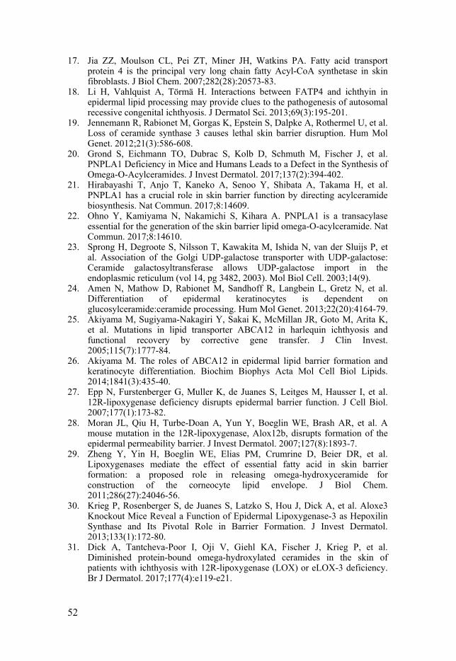
52
17. Jia ZZ, Moulson CL, Pei ZT, Miner JH, Watkins PA. Fatty acid transport protein 4 is the principal very long chain fatty Acyl-CoA synthetase in skin fibroblasts. J Biol Chem. 2007;282(28):20573-83.
18. Li H, Vahlquist A, Törmä H. Interactions between FATP4 and ichthyin in epidermal lipid processing may provide clues to the pathogenesis of autosomal recessive congenital ichthyosis. J Dermatol Sci. 2013;69(3):195-201.
19. Jennemann R, Rabionet M, Gorgas K, Epstein S, Dalpke A, Rothermel U, et al. Loss of ceramide synthase 3 causes lethal skin barrier disruption. Hum Mol Genet. 2012;21(3):586-608.
20. Grond S, Eichmann TO, Dubrac S, Kolb D, Schmuth M, Fischer J, et al. PNPLA1 Deficiency in Mice and Humans Leads to a Defect in the Synthesis of Omega-O-Acylceramides. J Invest Dermatol. 2017;137(2):394-402.
21. Hirabayashi T, Anjo T, Kaneko A, Senoo Y, Shibata A, Takama H, et al. PNPLA1 has a crucial role in skin barrier function by directing acylceramide biosynthesis. Nat Commun. 2017;8:14609.
22. Ohno Y, Kamiyama N, Nakamichi S, Kihara A. PNPLA1 is a transacylase essential for the generation of the skin barrier lipid omega-O-acylceramide. Nat Commun. 2017;8:14610.
23. Sprong H, Degroote S, Nilsson T, Kawakita M, Ishida N, van der Sluijs P, et al. Association of the Golgi UDP-galactose transporter with UDP-galactose: Ceramide galactosyltransferase allows UDP-galactose import in the endoplasmic reticulum (vol 14, pg 3482, 2003). Mol Biol Cell. 2003;14(9).
24. Amen N, Mathow D, Rabionet M, Sandhoff R, Langbein L, Gretz N, et al. Differentiation of epidermal keratinocytes is dependent on glucosylceramide:ceramide processing. Hum Mol Genet. 2013;22(20):4164-79.
25. Akiyama M, Sugiyama-Nakagiri Y, Sakai K, McMillan JR, Goto M, Arita K, et al. Mutations in lipid transporter ABCA12 in harlequin ichthyosis and functional recovery by corrective gene transfer. J Clin Invest. 2005;115(7):1777-84.
26. Akiyama M. The roles of ABCA12 in epidermal lipid barrier formation and keratinocyte differentiation. Biochim Biophys Acta Mol Cell Biol Lipids. 2014;1841(3):435-40.
27. Epp N, Furstenberger G, Muller K, de Juanes S, Leitges M, Hausser I, et al. 12R-lipoxygenase deficiency disrupts epidermal barrier function. J Cell Biol. 2007;177(1):173-82.
28. Moran JL, Qiu H, Turbe-Doan A, Yun Y, Boeglin WE, Brash AR, et al. A mouse mutation in the 12R-lipoxygenase, Alox12b, disrupts formation of the epidermal permeability barrier. J Invest Dermatol. 2007;127(8):1893-7.
29. Zheng Y, Yin H, Boeglin WE, Elias PM, Crumrine D, Beier DR, et al. Lipoxygenases mediate the effect of essential fatty acid in skin barrier formation: a proposed role in releasing omega-hydroxyceramide for construction of the corneocyte lipid envelope. J Biol Chem. 2011;286(27):24046-56.
30. Krieg P, Rosenberger S, de Juanes S, Latzko S, Hou J, Dick A, et al. Aloxe3 Knockout Mice Reveal a Function of Epidermal Lipoxygenase-3 as Hepoxilin Synthase and Its Pivotal Role in Barrier Formation. J Invest Dermatol. 2013;133(1):172-80.
31. Dick A, Tantcheva-Poor I, Oji V, Giehl KA, Fischer J, Krieg P, et al. Diminished protein-bound omega-hydroxylated ceramides in the skin of patients with ichthyosis with 12R-lipoxygenase (LOX) or eLOX-3 deficiency. Br J Dermatol. 2017;177(4):e119-e21.
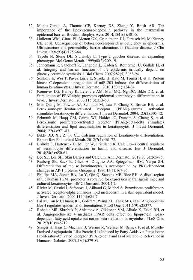
53
32. Munoz-Garcia A, Thomas CP, Keeney DS, Zheng Y, Brash AR. The importance of the lipoxygenase-hepoxilin pathway in the mammalian epidermal barrier. Biochim Biophys Acta. 2014;1841(3):401-8.
33. Holleran WM, Ginns EI, Menon GK, Grundmann JU, Fartasch M, McKinney CE, et al. Consequences of beta-glucocerebrosidase deficiency in epidermis. Ultrastructure and permeability barrier alterations in Gaucher disease. J Clin Invest. 1994;93(4):1756-64.
34. Tayebi N, Stone DL, Sidransky E. Type 2 gaucher disease: an expanding phenotype. Mol Genet Metab. 1999;68(2):209-19.
35. Jennemann R, Sandhoff R, Langbein L, Kaden S, Rothermel U, Gallala H, et al. Integrity and barrier function of the epidermis critically depend on glucosylceramide synthesis. J Biol Chem. 2007;282(5):3083-94.
36. Sonkoly E, Wei T, Pavez Lorie E, Suzuki H, Kato M, Torma H, et al. Protein kinase C-dependent upregulation of miR-203 induces the differentiation of human keratinocytes. J Invest Dermatol. 2010;130(1):124-34.
37. Komuves LG, Hanley K, Lefebvre AM, Man MQ, Ng DC, Bikle DD, et al. Stimulation of PPARalpha promotes epidermal keratinocyte differentiation in vivo. J Invest Dermatol. 2000;115(3):353-60.
38. Mao-Qiang M, Fowler AJ, Schmuth M, Lau P, Chang S, Brown BE, et al. Peroxisome-proliferator-activated receptor (PPAR)-gamma activation stimulates keratinocyte differentiation. J Invest Dermatol. 2004;123(2):305-12.
39. Schmuth M, Haqq CM, Cairns WJ, Holder JC, Dorsam S, Chang S, et al. Peroxisome proliferator-activated receptor (PPAR)-beta/delta stimulates differentiation and lipid accumulation in keratinocytes. J Invest Dermatol. 2004;122(4):971-83.
40. Bikle DD, Xie Z, Tu CL. Calcium regulation of keratinocyte differentiation. Expert Rev Endocrinol Metab. 2012;7(4):461-72.
41. Elsholz F, Harteneck C, Muller W, Friedland K. Calcium--a central regulator of keratinocyte differentiation in health and disease. Eur J Dermatol. 2014;24(6):650-61.
42. Lee SE, Lee SH. Skin Barrier and Calcium. Ann Dermatol. 2018;30(3):265-75. 43. Rutberg SE, Saez E, Glick A, Dlugosz AA, Spiegelman BM, Yuspa SH.
Differentiation of mouse keratinocytes is accompanied by PKC-dependent changes in AP-1 proteins. Oncogene. 1996;13(1):167-76.
44. Phillips MA, Jessen BA, Lu Y, Qin Q, Stevens ME, Rice RH. A distal region of the human TGM1 promoter is required for expression in transgenic mice and cultured keratinocytes. BMC Dermatol. 2004;4:2.
45. Rivier M, Castiel I, Safonova I, Ailhaud G, Michel S. Peroxisome proliferator-activated receptor-alpha enhances lipid metabolism in a skin equivalent model. J Invest Dermatol. 2000;114(4):681-7.
46. Pal M, Tan MJ, Huang RL, Goh YY, Wang XL, Tang MB, et al. Angiopoietin-like 4 regulates epidermal differentiation. PLoS One. 2011;6(9):e25377.
47. Robciuc MR, Skrobuk P, Anisimov A, Olkkonen VM, Alitalo K, Eckel RH, et al. Angiopoietin-like 4 mediates PPAR delta effect on lipoprotein lipase-dependent fatty acid uptake but not on beta-oxidation in myotubes. PLoS One. 2012;7(10):e46212.
48. Staiger H, Haas C, Machann J, Werner R, Weisser M, Schick F, et al. Muscle-Derived Angiopoietin-Like Protein 4 Is Induced by Fatty Acids via Peroxisome Proliferator-Activated Receptor (PPAR)-delta and Is of Metabolic Relevance in Humans. Diabetes. 2009;58(3):579-89.
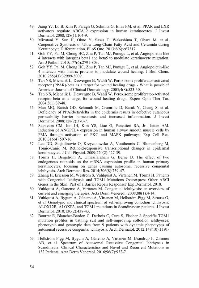
54
49. Jiang YJ, Lu B, Kim P, Paragh G, Schmitz G, Elias PM, et al. PPAR and LXR activators regulate ABCA12 expression in human keratinocytes. J Invest Dermatol. 2008;128(1):104-9.
50. Mizutani Y, Sun H, Ohno Y, Sassa T, Wakashima T, Obara M, et al. Cooperative Synthesis of Ultra Long-Chain Fatty Acid and Ceramide during Keratinocyte Differentiation. PLoS One. 2013;8(6):e67317.
51. Goh YY, Pal M, Chong HC, Zhu P, Tan MJ, Punugu L, et al. Angiopoietin-like 4 interacts with integrins beta1 and beta5 to modulate keratinocyte migration. Am J Pathol. 2010;177(6):2791-803.
52. Goh YY, Pal M, Chong HC, Zhu P, Tan MJ, Punugu L, et al. Angiopoietin-like 4 interacts with matrix proteins to modulate wound healing. J Biol Chem. 2010;285(43):32999-3009.
53. Tan NS, Michalik L, Desvergne B, Wahli W. Peroxisome proliferator-activated receptor (PPAR)-beta as a target for wound healing drugs - What is possible? American Journal of Clinical Dermatology. 2003;4(8):523-30.
54. Tan NS, Michalik L, Desvergne B, Wahli W. Peroxisome proliferator-activated receptor-beta as a target for wound healing drugs. Expert Opin Ther Tar. 2004;8(1):39-48.
55. Man MQ, Barish GD, Schmuth M, Crumrine D, Barak Y, Chang S, et al. Deficiency of PPARbeta/delta in the epidermis results in defective cutaneous permeability barrier homeostasis and increased inflammation. J Invest Dermatol. 2008;128(2):370-7.
56. Stapleton CM, Joo JH, Kim YS, Liao G, Panettieri RA, Jr., Jetten AM. Induction of ANGPTL4 expression in human airway smooth muscle cells by PMA through activation of PKC and MAPK pathways. Exp Cell Res. 2010;316(4):507-16.
57. Lee DD, Stojadinovic O, Krzyzanowska A, Vouthounis C, Blumenberg M, Tomic-Canic M. Retinoid-responsive transcriptional changes in epidermal keratinocytes. J Cell Physiol. 2009;220(2):427-39.
58. Törmä H, Bergström A, Ghiasifarahani G, Berne B. The effect of two endogenous retinoids on the mRNA expression profile in human primary keratinocytes, focusing on genes causing autosomal recessive congenital ichthyosis. Arch Dermatol Res. 2014;306(8):739-47.
59. Zhang H, Ericsson M, Weström S, Vahlquist A, Virtanen M, Törmä H. Patients with Congenital Ichthyosis and TGM1 Mutations Overexpress Other ARCI Genes in the Skin: Part of a Barrier Repair Response? Exp Dermatol. 2018.
60. Vahlquist A, Ganemo A, Virtanen M. Congenital ichthyosis: an overview of current and emerging therapies. Acta Derm Venereol. 2008;88(1):4-14.
61. Vahlquist A, Bygum A, Gånemo A, Virtanen M, Hellström-Pigg M, Strauss G, et al. Genotypic and clinical spectrum of self-improving collodion ichthyosis: ALOX12B, ALOXE3, and TGM1 mutations in Scandinavian patients. J Invest Dermatol. 2010;130(2):438-43.
62. Bourrat E, Blanchet-Bardon C, Derbois C, Cure S, Fischer J. Specific TGM1 mutation profiles in bathing suit and self-improving collodion ichthyoses: phenotypic and genotypic data from 9 patients with dynamic phenotypes of autosomal recessive congenital ichthyosis. Arch Dermatol. 2012;148(10):1191-5.
63. Hellström Pigg M, Bygum A, Gånemo A, Virtanen M, Brandrup F, Zimmer AD, et al. Spectrum of Autosomal Recessive Congenital Ichthyosis in Scandinavia: Clinical Characteristics and Novel and Recurrent Mutations in 132 Patients. Acta Derm Venereol. 2016;96(7):932-7.
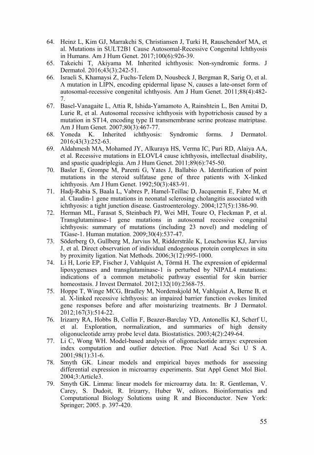
55
64. Heinz L, Kim GJ, Marrakchi S, Christiansen J, Turki H, Rauschendorf MA, et al. Mutations in SULT2B1 Cause Autosomal-Recessive Congenital Ichthyosis in Humans. Am J Hum Genet. 2017;100(6):926-39.
65. Takeichi T, Akiyama M. Inherited ichthyosis: Non-syndromic forms. J Dermatol. 2016;43(3):242-51.
66. Israeli S, Khamaysi Z, Fuchs-Telem D, Nousbeck J, Bergman R, Sarig O, et al. A mutation in LIPN, encoding epidermal lipase N, causes a late-onset form of autosomal-recessive congenital ichthyosis. Am J Hum Genet. 2011;88(4):482-7.
67. Basel-Vanagaite L, Attia R, Ishida-Yamamoto A, Rainshtein L, Ben Amitai D, Lurie R, et al. Autosomal recessive ichthyosis with hypotrichosis caused by a mutation in ST14, encoding type II transmembrane serine protease matriptase. Am J Hum Genet. 2007;80(3):467-77.
68. Yoneda K. Inherited ichthyosis: Syndromic forms. J Dermatol. 2016;43(3):252-63.
69. Aldahmesh MA, Mohamed JY, Alkuraya HS, Verma IC, Puri RD, Alaiya AA, et al. Recessive mutations in ELOVL4 cause ichthyosis, intellectual disability, and spastic quadriplegia. Am J Hum Genet. 2011;89(6):745-50.
70. Basler E, Grompe M, Parenti G, Yates J, Ballabio A. Identification of point mutations in the steroid sulfatase gene of three patients with X-linked ichthyosis. Am J Hum Genet. 1992;50(3):483-91.
71. Hadj-Rabia S, Baala L, Vabres P, Hamel-Teillac D, Jacquemin E, Fabre M, et al. Claudin-1 gene mutations in neonatal sclerosing cholangitis associated with ichthyosis: a tight junction disease. Gastroenterology. 2004;127(5):1386-90.
72. Herman ML, Farasat S, Steinbach PJ, Wei MH, Toure O, Fleckman P, et al. Transglutaminase-1 gene mutations in autosomal recessive congenital ichthyosis: summary of mutations (including 23 novel) and modeling of TGase-1. Human mutation. 2009;30(4):537-47.
73. Söderberg O, Gullberg M, Jarvius M, Ridderstråle K, Leuchowius KJ, Jarvius J, et al. Direct observation of individual endogenous protein complexes in situ by proximity ligation. Nat Methods. 2006;3(12):995-1000.
74. Li H, Lorie EP, Fischer J, Vahlquist A, Törmä H. The expression of epidermal lipoxygenases and transglutaminase-1 is perturbed by NIPAL4 mutations: indications of a common metabolic pathway essential for skin barrier homeostasis. J Invest Dermatol. 2012;132(10):2368-75.
75. Hoppe T, Winge MCG, Bradley M, Nordenskjold M, Vahlquist A, Berne B, et al. X-linked recessive ichthyosis: an impaired barrier function evokes limited gene responses before and after moisturizing treatments. Br J Dermatol. 2012;167(3):514-22.
76. Irizarry RA, Hobbs B, Collin F, Beazer-Barclay YD, Antonellis KJ, Scherf U, et al. Exploration, normalization, and summaries of high density oligonucleotide array probe level data. Biostatistics. 2003;4(2):249-64.
77. Li C, Wong WH. Model-based analysis of oligonucleotide arrays: expression index computation and outlier detection. Proc Natl Acad Sci U S A. 2001;98(1):31-6.
78. Smyth GK. Linear models and empirical bayes methods for assessing differential expression in microarray experiments. Stat Appl Genet Mol Biol. 2004;3:Article3.
79. Smyth GK. Limma: linear models for microarray data. In: R. Gentleman, V. Carey, S. Dudoit, R. Irizarry, Huber W, editors. Bioinformatics and Computational Biology Solutions using R and Bioconductor. New York: Springer; 2005. p. 397-420.
56
80. Benjamini Y, Hochberg Y. Controlling the false discovery rate: a practical and powerful approach to multiple testing. J R Statist Soc B. 1995;57(1):289-300.
81. Huang DW, Sherman BT, Lempicki RA. Systematic and integrative analysis of large gene lists using DAVID bioinformatics resources. Nat Protoc. 2009;4(1):44-57.
82. Dickson MA, Hahn WC, Ino Y, Ronfard V, Wu JY, Weinberg RA, et al. Human keratinocytes that express hTERT and also bypass a p16(INK4a)-enforced mechanism that limits life span become immortal yet retain normal growth and differentiation characteristics. Mol Cell Biol. 2000;20(4):1436-47.
83. Buraczewska I, Berne B, Lindberg M, Loden M, Törmä H. Moisturizers change the mRNA expression of enzymes synthesizing skin barrier lipids. Arch Dermatol Res. 2009;301:587-94.
84. Buraczewska I, Berne B, Lindberg M, Lodén M, Törmä H. Long-term treatment with moisturizers affects the mRNA levels of genes involved in keratinocyte differentiation and desquamation Arch Dermatol Res. 2009;301:175-81.
85. Jiang YJ, Barish G, Lu B, Evans RM, Crumrine D, Schmuth M, et al. PPARdelta activation promotes stratum corneum formation and epidermal permeability barrier development during late gestation. J Invest Dermatol. 2010;130(2):511-9.
86. Zhang H, Ericsson M, Virtanen M, Weström S, Wählby C, Vahlquist A, et al. Quantitative image analysis of protein expression and colocalisation in skin sections. Exp Dermatol. 2018;27(2):196-9.
87. Haneda T, Imai Y, Uchiyama R, Jitsukawa O, Yamanishi K. Activation of Molecular Signatures for Antimicrobial and Innate Defense Responses in Skin with Transglutaminase 1 Deficiency. PLoS One. 2016;11(7):e0159673.
88. Nold MF, Nold-Petry CA, Zepp JA, Palmer BE, Bufler P, Dinarello CA. IL-37 is a fundamental inhibitor of innate immunity. Nat Immunol. 2010;11(11):1014-U64.
89. Gabay C, Towne JE. Regulation and function of interleukin-36 cytokines in homeostasis and pathological conditions. J Leukoc Biol. 2015;97(4):645-52.
90. Dou J, Zhang L, Xie X, Ye L, Yang C, Wen L, et al. Integrative analyses reveal biological pathways and key genes in psoriasis. Br J Dermatol. 2017;177(5):1349-57.
91. Grubauer G, Elias PM, Feingold KR. Transepidermal water loss: the signal for recovery of barrier structure and function. J Lipid Res. 1989;30(3):323-33.
92. Bosen F, Celli A, Crumrine D, vom Dorp K, Ebel P, Jastrow H, et al. Altered epidermal lipid processing and calcium distribution in the KID syndrome mouse model Cx26S17F. FEBS letters. 2015;589(15):1904-10.
93. Peters JM, Hollingshead HE, Gonzalez FJ. Role of peroxisome-proliferator-activated receptor beta/delta (PPAR beta/delta) in gastrointestinal tract function and disease. Clin Sci. 2008;115(3-4):107-27.
94. Beyaz S, Yilmaz OH. Molecular Pathways: Dietary Regulation of Stemness and Tumor Initiation by the PPAR-delta Pathway. Clin Cancer Res. 2016;22(23):5636-41.
95. Desvergne B, Wahli W. Peroxisome proliferator-activated receptors: Nuclear control of metabolism. Endocr Rev. 1999;20(5):649-88.
96. Grygiel-Gorniak B. Peroxisome proliferator-activated receptors and their ligands: nutritional and clinical implications - a review. Nutr J. 2014;13.
97. Noy N. Non-classical Transcriptional Activity of Retinoic Acid. Subcell Biochem. 2016;81:179-99.
57
98. Jiang YJ, Uchida Y, Lu B, Kim P, Mao C, Akiyama M, et al. Ceramide stimulates ABCA12 expression via peroxisome proliferator-activated receptor in human keratinocytes. J Biol Chem. 2009;284(28):18942-52.
99. Gallego O, Belyaeva OV, Porte S, Ruiz FX, Stetsenko AV, Shabrova EV, et al. Comparative functional analysis of human medium-chain dehydrogenases, short-chain dehydrogenases/reductases and aldo-keto reductases with retinoids. Biochem J. 2006;399(1):101-9.
100. Ruiz FX, Porte S, Pares X, Farres J. Biological role of aldo-keto reductases in retinoic Acid biosynthesis and signaling. Front Pharmacol. 2012;3:58.
101. Jumper N, Hodgkinson T, Arscott G, Har-Shai Y, Paus R, Bayat A. The Aldo-Keto Reductase AKR1B10 Is Up-Regulated in Keloid Epidermis, Implicating Retinoic Acid Pathway Dysregulation in the Pathogenesis of Keloid Disease. J Invest Dermatol. 2016;136(7):1500-12.
102. Gruswitz F, Chaudhary S, Ho JD, Schlessinger A, Pezeshki B, Ho CM, et al. Function of human Rh based on structure of RhCG at 2.1 Å. P Natl Acad Sci USA. 2010;107(21):9638-43.
103. Biver S, Belge H, Bourgeois S, Van Vooren P, Nowik M, Scohy S, et al. A role for Rhesus factor Rhcg in renal ammonium excretion and male fertility. Nature. 2008;456(7220):339-43.
104. Raymond AA, Gonzalez de Peredo A, Stella A, Ishida-Yamamoto A, Bouyssie D, Serre G, et al. Lamellar bodies of human epidermis: proteomics characterization by high throughput mass spectrometry and possible involvement of CLIP-170 in their trafficking/secretion. Mol Cell Proteomics. 2008;7(11):2151-75.
105. Malinina L, Malakhova ML, Teplov A, Brown RE, Patel DJ. Structural basis for glycosphingolipid transfer specificity. Nature. 2004;430(7003):1048-53.
106. D'Angelo G, Polishchuk E, Di Tullio G, Santoro M, Di Campli A, Godi A, et al. Glycosphingolipid synthesis requires FAPP2 transfer of glucosylceramide. Nature. 2007;449(7158):62-U43.
107. Mattjus P. Glycolipid transfer proteins and membrane interaction. Bba-Biomembranes. 2009;1788(1):267-72.
108. Halter D, Neumann S, van Dijk SM, Wolthoorn J, de Maziere AM, Vieira OV, et al. Pre- and post-Golgi translocation of glucosylceramide in glycosphingolipid synthesis. J Cell Biol. 2007;179(1):101-15.
109. Zou XQ, Gao YG, Ruvolo VR, Gardner TL, Ruvolo PP, Brown RE. Human Glycolipid Transfer Protein Gene (GLTP) Expression Is Regulated by Sp1 and Sp3 INVOLVEMENT OF THE BIOACTIVE SPHINGOLIPID CERAMIDE. J Biol Chem. 2011;286(2):1301-11.
110. Dai FHF, Zhang Y, Kang YH, Wang QH, Gaisano HY, Braunewell KH, et al. The neuronal Ca2+ sensor protein visinin-like protein-1 is expressed in pancreatic islets and regulates insulin secretion. J Biol Chem. 2006;281(31):21942-53.
111. Terada T, Inui K. Recent advances in structural biology of peptide transporters. Curr Top Membr. 2012;70:257-74.
112. Aito-Inoue M, Lackeyram D, Fan MZ, Sato K, Mine Y. Transport of a tripeptide, Gly-Pro-Hyp, across the porcine intestinal brush-border membrane. J Pept Sci. 2007;13(7):468-74.
113. Shimizu J, Asami N, Kataoka A, Sugihara F, Inoue N, Kimira Y, et al. Oral collagen-derived dipeptides, prolyl-hydroxyproline and hydroxyprolyl-glycine, ameliorate skin barrier dysfunction and alter gene expression profiles in the skin. Biochem Biophys Res Commun. 2015;456(2):626-30.

58
114. Kudo M, Katayoshi T, Kobayashi-Nakamura K, Akagawa M, Tsuji-Naito K. H+/peptide transporter (PEPT2) is expressed in human epidermal keratinocytes and is involved in skin oligopeptide transport. Biochem Bioph Res Co. 2016;475(4):335-41.
115. Pickart L, Vasquez-Soltero JM, Margolina A. GHK Peptide as a Natural Modulator of Multiple Cellular Pathways in Skin Regeneration. Biomed Res Int. 2015.
116. Minegishi Y, Rohrer J, Coustan-Smith E, Lederman HM, Pappu R, Campana D, et al. An essential role for BLNK in human B cell development. Science. 1999;286(5446):1954-7.
117. Jin G, Hamaguchi Y, Matsushita T, Hasegawa M, Le Huu D, Ishiura N, et al. B-cell linker protein expression contributes to controlling allergic and autoimmune diseases by mediating IL-10 production in regulatory B cells. J Allergy Clin Immunol. 2013;131(6):1674-82.
118. Laneve P, Gioia U, Ragno R, Altieri F, Di Franco C, Santini T, et al. The Tumor Marker Human Placental Protein 11 Is an Endoribonuclease. J Biol Chem. 2008;283(50):34712-9.
119. Poe JC, Kountikov EI, Lykken JM, Natarajan A, Marchuk DA, Tedder TF. EndoU is a novel regulator of AICD during peripheral B cell selection. J Exp Med. 2014;211(1):57-69.
120. Rinaldi M, Dreesen L, Hoorens PR, Li RW, Claerebout E, Goddeeris B, et al. Infection with the gastrointestinal nematode Ostertagia ostertagi in cattle affects mucus biosynthesis in the abomasum. Vet Res. 2011;42:61.
121. Wu LC, Pfeiffer DR, Calhoon EA, Madiai F, Marcucci G, Liu SJ, et al. Purification, Identification, and Cloning of Lysoplasmalogenase, the Enzyme That Catalyzes Hydrolysis of the Vinyl Ether Bond of Lysoplasmalogen. J Biol Chem. 2011;286(28):24916-30.

Acta Universitatis UpsaliensisDigital Comprehensive Summaries of Uppsala Dissertationsfrom the Faculty of Medicine 1541
Editor: The Dean of the Faculty of Medicine
A doctoral dissertation from the Faculty of Medicine, UppsalaUniversity, is usually a summary of a number of papers. A fewcopies of the complete dissertation are kept at major Swedishresearch libraries, while the summary alone is distributedinternationally through the series Digital ComprehensiveSummaries of Uppsala Dissertations from the Faculty ofMedicine. (Prior to January, 2005, the series was publishedunder the title “Comprehensive Summaries of UppsalaDissertations from the Faculty of Medicine”.)
Distribution: publications.uu.seurn:nbn:se:uu:diva-377557
ACTAUNIVERSITATIS
UPSALIENSISUPPSALA
2019