complications of coronary intervention - intech - open science
TRANSCRIPT

3
Complications of Coronary Intervention
Seung-Jin Lee Soonchunhyang University Cheonan Hospital
South Korea
1. Introduction
Drug-eluting stents (DES) substantially reduce restenosis compared with bare metal stents and represent a significant advance in percutaneous coronary interventions (PCIs). Accordingly, DES have been rapidly adopted into practice and are currently used in the vast majority of PCI procedures. As PCIs for more complicated lesions increase, various complications, such as stent thrombosis, fracture, dissection or perforation, are also increase. For example, PCIs for patients who have chronic total occlusion increase and these patients tend to have more risk factors like diabetes mellitus, hypertension, dyslipidemia, and previous myocardial infarction and also have multi-vessel diseases and have decreased left ventricular ejection fraction. If major procedure-related complications were developed in these high risk patients, it may leads to fatal results. So it is important to understand possible complications of PCIs and eliminate potential risk factors before procedures.
1.1 Stent fracture
Drug-eluting stents (DES) have proven very effective in reducing restenosis by suppressing neointimal hyperplasia. However, potentially serious complications such as in-stent restenosis and thrombus still occur. Stent fracture has been identified as a possible contributor to these adverse outcomes. A number of risk factors for the development of stent fracture have been described, although a detailed analysis of the angiographic factors predisposing to stent fracture is lacking.
1.2 Incidence and definition
Stent fracture is defined as the cases where the linear or curvilinear connections of stent struts are interrupted and areas of the stented segment are uncovered by stent struts visible on coronary angiography. The incidence of stent fracture is reported in 0.8-7.7% of cases.1-8 However, because of limited sensitivity of angiography to detect fracture, its true incidence is still unknown. In a recent report analyzed from autopsy findings, stent fracture was observed in 29% of total patients.9 So, the real incidence of stent fracture is assumed to be a little higher than what has been clinically reported. Stent fracture from patients treated with Cypher stents is more frequently observed than in cases of Taxus stents. The incidence of stent fracture of Cypher stent was 1.3% in the SIRIUS trial,9 compared to 0.58% incidence with the Taxus stent is in the Taxus IV/V/VI trials. Stent fracture has previously been recognized in noncoronary vessels, especially in the superficial femoral and popliteal arteries
www.intechopen.com
Coronary Interventions
48
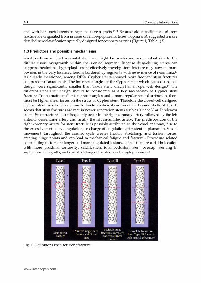
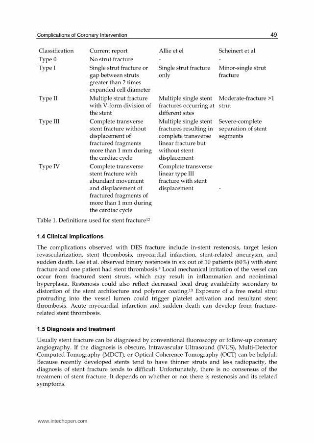
and with bare-metal stents in saphenous vein grafts.10,11 Because old classifications of stent fracture are originated from in cases of femoropopliteal arteries, Popma et al. suggested a more detailed new classification specially designed for coronary arteries (Figure 1, Table 1).12
1.3 Predictors and possible mechanisms
Stent fractures in the bare-metal stent era might be overlooked and masked due to the diffuse tissue overgrowth within the stented segment. Because drug-eluting stents can suppress neointimal hyperplasia more effectively thereby stent fracture may now be more obvious in the very localized lesions bordered by segments with no evidence of neointima.13 As already mentioned, among DESs, Cypher stents showed more frequent stent fractures compared to Taxus stents. The inter-strut angles of the Cypher stent which has a closed-cell design, were significantly smaller than Taxus stent which has an open-cell design.14 The different stent strut design should be considered as a key mechanism of Cypher stent fracture. To maintain smaller inter-strut angles and a more regular strut distribution, there must be higher shear forces on the struts of Cypher stent. Therefore the closed-cell designed Cypher stent may be more prone to fracture when shear forces are beyond its flexibility. It seems that stent fractures are rare in newer generation stents such as Xience V or Eendeavor stents. Stent fractures most frequently occur in the right coronary artery followed by the left anterior descending artery and finally the left circumflex artery. The predisposition of the right coronary artery for stent fracture is possibly attributed to the vessel anatomy, due to the excessive tortuosity, angulation, or change of angulation after stent implantation. Vessel movement throughout the cardiac cycle creates flexion, stretching, and torsion forces, creating hinge points and can lead to mechanical fatigue and fracture.2 Procedure related contributing factors are longer and more angulated lesions, lesions that are ostial in location with more proximal tortuosity, calcification, total occlusion, stent overlap, stenting in saphenous vein grafts, and overstretching of the stents with high pressure.12
Fig. 1. Definitions used for stent fracture
www.intechopen.com
Complications of Coronary Intervention
49
Classification Current report Allie et el Scheinert et al
Type 0 No strut fracture - -
Type I Single strut fracture or gap between struts greater than 2 times expanded cell diameter
Single strut fracture only
Minor-single strut fracture
Type II Multiple strut fracture with V-form division of the stent
Multiple single stent fractures occurring at different sites
Moderate-fracture >1 strut
Type III Complete transverse stent fracture without displacement of fractured fragments more than 1 mm during the cardiac cycle
Multiple single stent fractures resulting in complete transverse linear fracture but without stent displacement
Severe-complete separation of stent segments
Type IV Complete transverse stent fracture with abundant movement and displacement of fractured fragments of more than 1 mm during the cardiac cycle
Complete transverse linear type III fracture with stent displacement
-
Table 1. Definitions used for stent fracture12
1.4 Clinical implications
The complications observed with DES fracture include in-stent restenosis, target lesion revascularization, stent thrombosis, myocardial infarction, stent-related aneurysm, and sudden death. Lee et al. observed binary restenosis in six out of 10 patients (60%) with stent fracture and one patient had stent thrombosis.5 Local mechanical irritation of the vessel can occur from fractured stent struts, which may result in inflammation and neointimal hyperplasia. Restenosis could also reflect decreased local drug availability secondary to distortion of the stent architecture and polymer coating.13 Exposure of a free metal strut protruding into the vessel lumen could trigger platelet activation and resultant stent thrombosis. Acute myocardial infarction and sudden death can develop from fracture-related stent thrombosis.
1.5 Diagnosis and treatment
Usually stent fracture can be diagnosed by conventional fluoroscopy or follow-up coronary angiography. If the diagnosis is obscure, Intravascular Ultrasound (IVUS), Multi-Detector Computed Tomography (MDCT), or Optical Coherence Tomography (OCT) can be helpful. Because recently developed stents tend to have thinner struts and less radiopacity, the diagnosis of stent fracture tends to difficult. Unfortunately, there is no consensus of the treatment of stent fracture. It depends on whether or not there is restenosis and its related symptoms.
www.intechopen.com

Coronary Interventions
50
2. No-reflow phenomenon
The phenomenon of no-reflow is defined as inadequate myocardial perfusion through a
given segment of the coronary circulation without angiographic evidence of mechanical
vessel obstruction.15 The underlying cause of no-reflow is microvascular obstruction, which
may be produced by various mechanisms. The concept of no-reflow was first described in
experimental models in 196616 and then in the clinical setting of reperfusion after myocardial
infarction in 1985.17 No-reflow has been documented in ≥30% of patients after
thrombolysis18 or mechanical intervention for acute myocardial infarction. The prevalence is
variable, ranging from 5% up to 50%, according to the methods used to assess the
phenomenon and to the population under study.
A series of consistent data has clearly shown that no-reflow has a strong negative impact on outcome, negating the potential benefit of primary percutaneous coronary intervention (PCI).19,20 Indeed, patients with no-reflow exhibit a higher prevalence of: 1) early post-infarction complications (arrhythmias, pericardial effusion, cardiac tamponade, early congestive heart failure); 2) left adverse ventricular remodeling; 3) late repeat hospital stays for heart failure; and 4) mortality.
Therefore, it is important to prevent and effectively treat the no-reflow phenomenon during PCI to achieve an optimal outcome.
2.1 Historical overview
The term no-reflow was first used by Majno and colleagues21 in the setting of vertebral ischemia in 1967. This phenomenon was initially described by Krug et al.16 during induced myocardial infarction in the canine model in 1966 and again by Kloner et al.15 in 1974 in which it occurred for 90 min after temporary epicardial coronary artery occlusion followed by reperfusion. Electron microscopic examination showed severe myocardial capillary damage with loss of pinocytonic vesicles in the endothelial cells, endothelial blisters or blebs and endothelial gaps with neutrophil infiltration. Intraluminal capillary plugging by neutrophils and/or microthrombi with myocardial cell swelling was also noted. Galiuto et al.22 with sequential measurements of myocardial perfusion by myocardial contrast echocardiography, have recently shown that in humans no-refow detected 24 h after successful PCI spontaneously improves over time in approximately 50% of patients. Thus, no-reflow can be categorized as sustained or reversible. Sustained no-reflow is probably the result of anatomical irreversible changes of the coronary microcirculation, whereas reversible no-reflow is the result of functional and thus, reversible, changes of the microcirculation.
In humans, no-reflow is caused by the variable combination of 4 pathogenetic components: 1) distal atherothrombotic embolization; 2) ischemic injury; 3) reperfusion injury; and 4) susceptibility of the coronary microcirculation to injury.
1. Distal embolization
Emboli of different sizes can originate from epicardial coronary thrombus and from fissured atherosclerotic plaques, in particular during primary PCI.23 Large emboli (>200μm diameter) can obstruct pre-arterioles, causing infarctlets. Experimental observations have shown that myocardial blood flow decreases irreversibly when microspheres obstruct more than 50% of coronary capillaries.24
www.intechopen.com

Complications of Coronary Intervention
51
2. Ischemia-related injury
Changes in endothelial cells, visible after prolonged ischemia, are represented by endothelial protrusions and membrane-bound bodies, which often fill the capillaries up to luminal obliteration. Furthermore, large endothelial gaps with extravascular erythrocytes are common.25
3. Reperfusion-related injury
A massive infiltration of the coronary microcirculation by neutrophils and platelets occurs at the time of reperfusion.25,26 Reintroduction of neutrophils in post-ischemic myocardium results in their activation, with subsequent adhesion to the endothelial surface and migration in the surrounding tissue. Activated neutrophils, in turn, release oxygen free radicals, proteolytic enzymes, and pro-inflammatory mediators that can directly cause tissue and endothelial damage. Neutrophils also form aggregates with platelets that plug capillaries, thus mechanically blocking flow.27,28 Finally, vasoconstrictors released by damaged endothelial cells, neutrophils, and platelets contribute to sustained vasoconstriction of the coronary microcirculation.29 Tumor necrosis factor-alpha expression is induced by reperfusion, and can impair endothelium-dependent coronary flow reserve.30 Interleukin-1β also has recently been associated with ischemia-reperfusion injury, because interleukin-1β knockout animals exhibit marked reduction of ischemic induced inflammation.31 Selectin expression on cell surfaces is also important for mechanical plugging of the microcirculation.32 Finally, the balance between nitric oxide and superoxide is tipped in favor of superoxide within minutes of reperfusion of ischemic tissues, due to increased production of xanthine oxidase by neutrophils, endothelial cells, and cardiac myocytes, which leads to an exacerbation of the inflammatory state.33
Reperfusion might also cause irreversible injury to myocytes.34 During ischemia there is an increase of the intracellular sodium (Na+) content due to accumulation of hydrogen (H+), which is exchanged by the Na+/H+ exchanger. The subsequent exchange of doubly charged positive calcium ion (Ca++) with Na+ by the sarcolemmal Na+/Ca++ exchanger produces a calcium overload that triggers uncontrolled hypercontraction and stimulates opening of the mitochondrial permeability transition pore (m0PTP), which further enhances calcium overload. Furthermore, Na+ extrusion trough Na+/potassium (K+) adenosine triphosphate (ATP)-ase is impaired and together with Ca++ accumulation leads to myocyte cell swelling, which contributes to subsequent rupture of the cell membrane when the extracellular osmolality is rapidly normalized by reperfusion. Of note, cyclosporine, which blocks the m-PTP, has been recently shown to reduce infarct size by 20% when administered intravenously in patients undergoing primary PCI.35 Finally, ischemic pre-conditioning might also reduce infarct size by blockade of m-PTP.36
Natriuretic peptides might modulate ischemia-reperfusion injury. Atrial natriuretic peptide might suppress the rennin-angiotensin-aldosterone system and endothelin (ET)-1 that increase infarct size, microvascular obstruction, and cardiac remodeling.37
4. Individual predisposition of coronary microcirculation to injury
In humans, no-reflow is occasionally observed during elective procedures,38 whereas it can be absent after primary PCI in patients with acute myocardial infarction. In particular, diabetes and hypercholesterolemia has been associated with impaired microvascular
www.intechopen.com

Coronary Interventions
52
reperfusion by enhancing endothelial oxidative stress.39,40 Pre-conditioning by using nicorandil seems to have a beneficial effect on microvascular function.41
2.2 Diagnosis
1. Coronary angiography
Reduced coronary flow after primary PCI (TIMI flow 0 to 2) is associated with worse outcome than normal (TIMI 3) flow, even when no significant epicardial obstruction remains.42 More sensitive markers of tissue perfusion have now been identified and provide prognostic information beyond that of TIMI flow grade. The TIMI frame count assesses the number of angiographic frames required for the contrast medium to reach standardized distal landmarks of the coronary tree, and the myocardial blush grade (MBG) is a quantitative assessment of myocardial contrast density. The MBG is scored on a scale of 0 to 3, with higher scores indicating better perfusion. An MBG 0 to 1, suggestive of no-reflow, is observed in as high as 50% of patients with TIMI flow grade 3.43 Taken together, angiographic no-reflow can be defined as a TIMI flow grade <3 or 3 with an MBG 0 to 1.
2. Electrocardiography
Rapid ST-segment resolution defined as a reduction of ≥50% in the ST-segment elevation index is highly specific (91%) for myocardial reperfusion (or the absence of no-reflow on myocardial contrast echocardiography) although less sensitive (77%).44
3. Myocardial contrast echocardiography (MCE)
Lack of intramyocardial contrast opacification is due to microvascular obstruction; thus, it represents the extent of no-reflow.45 In the AMICI study, the extent of no-reflow at MCE was demonstrated to be the best predictor of adverse left ventricular remodeling after acute myocardial infarction, being superior to ST-segment resolution and to MBG among patients exhibiting TIMI flow grade 3.20
4. Cardiac magnetic resonance imaging
No-reflow can be diagnosed as: 1) lack of gadolinium enhancement during first pass; and 2) lack of gadolinium enhancement within a necrotic region, identified by late gadolinium hyperenhancement.46
2.3 Prevention and treatment (Figure 2)
1. Distal embolization
No specific technique is currently recommended in guidelines to prevent distal embolization during primary PCI. Direct stent implantation, by avoiding balloon-induced thrombus fragmentation and by entrapping the atherothrombus under the stent struts, has been suggested as a possible technique to reduce distal embolization in a specific subset of patients i.e. those with good distal visualization of the infarct-related artery after guidewire passage.47
A more promising technique is the use of thrombectomy and distal filter devices. Although distal filter devices did not improve early or late prognosis compared with standard primary PCI, thrombectomy performed with a simple manual aspiration catheter revealed improved myocardial reperfusion and significantly reduced no-reflow.48 A recent large trial
www.intechopen.com

Complications of Coronary Intervention
53
by Svilaas et al.49 confirmed the improvement of reperfusion associated with manual thrombus-aspiration as compared with standard primary PCI showing a strikingly lower mortality at 12-month follow-up.50 So, it is suggested that manual thrombus aspiration should be used in the setting of primary PCI, particularly in patients with a high thrombus burden.51
2. Ischemia-related injury
Strategies aimed at reducing pain onset-to-balloon time might reduce no-reflow by decreasing total ischemic time. Drugs known to reduce myocardial oxygen consumption and consequently the severity of ischemia and improve myocardial perfusion include carvedilol, fosinopril, and valsartan.52,53
3. Reperfusion-related injury
Intracoronary nitroglycerin is usually suggested as the first-line agent, mainly to reverse epicardial vessel spasm, even if the blood pressure is reduced. Theoretically, nitroglycerin should have little impact on arteriolar tone and hence on no-reflow since physiologically it produces little effect in the microvasculature.
Patients at high risk of no-reflow can be treated with drugs such as glycoprotein IIb/IIIa antagonists, adenosine, nicorandil, and nitroprusside aimed at counteracting endothelial, platelet, and neutrophil activation.
Fig. 2. Therapies of no-reflow targeted to main pathogenetic mechanisms
www.intechopen.com

Coronary Interventions
54
Among glycoprotein IIb/IIIa antagonists, abciximab has been found to improve myocardial perfusion when started during primary PCI and infused for 12 h thereafter. Interestingly, intracoronary abciximab has been proven to be superior to intravenous abciximab in patients treated by primary PCI.54 Adenosine is an endogenous purine nucleoside that decreases arteriolar resistance and activates intracellular cardioprotective signaling pathways. Its mechanism of action may involve opening ATP-sensitive potassium channels (KATP), inhibition of neutrophil migration, prevention of superoxide generation, or blockade of coronary endothelin release. Nitroprusside is a nitric oxide donor that does not depend on intracellular metabolism to derive nitric oxide, with potent vasodilator properties. Nicorandil is a hybrid of a KATP opener and nitrate and may prevent reperfusion injury by blocking the mitochondrial permeability transition pore. Verapamil is a calcium-channel blocker that has several beneficial effects in the setting of no-reflow in addition to attenuation of microvascular spasm. Varapamil may also inhibit platelet aggregation and thrombus formation in the microvasculature and may have a direct effect on calcium flux across the sarcolemmal membrane or within intracellular compartment that could protect reversibly injuried myocytes.
3. Coronary artery perforation and cardiac tamponade
Coronary artery perforation complicating percutaneous coronary intervention (PCI) occurs in 0.1-3.0% of cases. Perforation with balloon angioplasty or stenting is rare, occurring in 0.1% of cases. However, when ablation devices, such as rotablator, directional coronary atherectomy, transluminal extraction catheter, and excimer laser, are used, the frequency is substantially higher than with balloon angioplasty or stents, occurring up to 3.0%.55 Recently, the use of ablation devices in PCI has tended to decline. Instead, procedures for more complex lesions including calcification, severe angulation, and chronic total occlusion have increased. To treat these complex lesions, stiffer and hydrophilic wires are necessary and high pressure for balloon dilatation is needed. Increasing numbers of procedures using glycoprotein IIb/IIIa inhibitors is also contributing to the fact that coronary artery perforation still occurs.
3.1 Classification
The Ellis classification depends on angiographic findings is most widely used (table 2).55
Type I Extraluminal crater without extravasation
Type II Pericardial or myocardial blush without contrast jet extravasation
Type III Extravasation through frank (>1 mm) perforation
Cavity Perforation into an anatomic cavity chamber, spilling coronary sinus, etc
Table 2. Perforation Classification
3.2 Risk factors
Many factors are involved in coronary perforation during PCI. Related risk factors can be divided into patient-related, vessel-related, and procedure-related factors.56 Patients-related factors are old age, hypertension, PCI for unstable angina or non-ST segment elevation
www.intechopen.com

Complications of Coronary Intervention
55
myocardial infarction.57 Vessel-related factors are ACC/AHA type C lesions, calcified lesions, and chronic total occlusion lesions.3 Procedure-related factors include use of stiff hydrophilic wires, device oversizing, use of atheroablative devices. One observational study reports that 87% of perforation due to guidewires is attributed to hydrophilic wires.58 In case of rotational atherectomy use, if lesions are eccentric, lesion length are >10cm, or lesions are very tortuous, the risk of perforation is high.
3.3 Symptoms and signs
If coronary perforation is develop, usually patients feel severe chest pain. In addition, nausea, dizziness, and vomiting can occur. The heart rate can rise suddenly, blood pressure can drop and if cardiac tamponade happens, an increase of central venous pressure with neck vein engorgement develops. Sustained ST-segment elevation or depression can be observed even though coronary balloons are deflated.
3.4 Diagnosis
The diagnosis of coronary artery perforation is not difficult by coronary angiography. If cardiac tamponade is suspected, then echocardiography is very useful. Sometimes perforation can occur 12-48 h later after PCI, if vital signs become unstable and serum hemoglobin and hematocrit levels decrease, cardiac tamponade must be suspected and urgent echocardiography should be performed.
3.5 Clinical outcomes and prognosis
Complications related to coronary artery perforation are diverse and depend on the degree of perforation. It has been reported that in cases of perforation, myocardial infarction can occurs in 13-34%, emergency coronary artery bypass graft in 11-39%, cardiac tamponade in 12-31%, and mortality in 7.6-19%.59-61 The degree of perforation is a important marker to predict the late prognosis. Ellis classification type I perforation has a clinically good prognosis in 60% and it is rare that type II perforation has a poor prognosis. However, type III perforation reveals high major adverse events rate.55,62
3.6 Treatment
Generally, guidewire-related perforation does not cause grave results except with concomitant use of glycoprotein IIb/IIIa inhibitors. However, perforation due to balloon, atherectomy devices, or laser can produce hemopericardium or hemodynamic collapse.
1. Prolonged balloon inflation
The most important thing to stop bleeding is prolonged balloon inflation at the perforation site at least for 10-15 min at 2-6 atm. If bleeding does not stop, a perfusion balloon catheter can be used for 15-45 min inflated at low pressure. This prolonged balloon inflation and timely pericardiocentesis can avoid surgical treatment in patients with Ellis type I perforation.
2. Stent
In some cases, a polytetrafluoroethylene(PTFE)-covered stent is effective. Rarely, one or more conventional stent (bare metal or drug-eluting stent) implantation can be considered.
www.intechopen.com

Coronary Interventions
56
3. Pericardiocentesis
If perforation is suspected, echocardiography should be performed and if hemopericardium
is confirmed, pericardiocentesis should be performed immediately. An indwelling pericardial
catheter should be maintained for 6-24 h and echocardiography should repeated every 6-12 h.
4. Management of anticoagulation
When perforation occurs, anticoagulation should be maintained to prevent thrombus
formation. However, if perforation happens after use of atherectomy or laser, it is
recommended that protamine sulfate should be administrated intravenously in order to
partially reverse the effect of heparin. If contrast leakage is sustained despite prolonged
balloon inflation, repeated balloon inflation should be performed; meanwhile the dosage of
protamine sulfate should increase under activated clotting time monitoring.
Glycoprotein IIb/IIIa inhibitors must be stopped. The effect of abciximab can be reversed
after platelet transfusion of 6-10 units, but there are not any known antidotes for eptifibatide
or tirofiban.
Fig. 3. Management algorithm of coronary artery perforation
www.intechopen.com

Complications of Coronary Intervention
57
5. Embolization
In case of small vessel size or distal location, limited involved myocardium, chronic total occlusion, or situation where surgery is unavailable, embolization using coils or gelfoam can successfully occlude the perforation.
6. Surgical treatment
If the perforation is severe, with hemodynamic instability or perforation is sustained despite nonsurgical management, emergency surgery is necessary.
4. Coronary dissection and acute closure
4.1 Angiographic definition
Dissection is defined as disruption of an arterial wall resulting in splitting and separation of the intimal (or subintimal) layers.
Feature Definition Abrupt Obstruction of contrast flow (TIMI 0 or 1) in a dilated segment with closure previously documented anterograde flow Ectasia A lesion diameter greater than the reference diameter in one or more areas Luminal irregularities
Arterial contour that has a saw-toothed pattern consisting of opacification but not fulfilling the criteria for dissection or intracoronary thrombus
Intimal flap A discrete filling defect in apparent continuity with the arterial wall Thrombus Discrete, mobile angiographic filling defect with or without contrast staining Dissection A Small radiolucent area within the lumen of the vessel B Linear, nonpersisting extravasation of contrast material C Extraluminal, persisting extravasation of contrast material D Spiral-shaped filling defect E Persistent lumen defect with delayed anterograde flow F Filling defect accompanied by total coronary occlusion Dissection, length(mm)
Measure end to end for type B through F dissections
Dissection, staining
Persistence of contrast within the dissection after washout of contrast material from the remaining portion of the vessel
Table 3. Standardized criteria for postprocedural lesion morphology63,64
4.2 Pathogenesis and incidence of coronary dissection
Intima-media cracks and medial dissection can be developed by balloon injury and if dissection involves the adventitia layer, narrowing of the lumen can occur.65 In NHLBI classification, coronary dissection occurs in 32-41% of total balloon procedures. If the lumen narrows >50% or the length of dissection is >10mm, the risk of abrupt vessel closure increases.
In the modern PCI era, where coronary dissection can be promptly resolved by stent implantation, clinically significant dissection is reported only 1.7%. Residual dissection increases the risk of post procedure MI, emergency CABG, and stent thrombosis and mortality also increases threefold.66
www.intechopen.com

Coronary Interventions
58
4.3 Pathogenesis and incidence of abrupt vessel closure
Coronary balloon dilatation leads to endothelial denudation, intimal fissuring, and medial penetration and extensive damage causes obstructive dissection or intramural hematoma. When subintimal structures are exposed to the blood, then activation of platelets and thrombin formation occur. Obstructive thrombus can be formed with or without medial dissection.
An autopsy finding of patients who experienced abrupt closure within 30 days after balloon angioplasty revealed that over 50% have intimal/medial dissection flaps with or without thrombi. Cases presenting with pure thrombi without dissection was very rare.
It has been reported that the incidence of abrupt closure due to balloon angioplasty or atherectomy is 2-13.5%. About two thirds of cases of abrupt closure arise inside the catheterization laboratory and the majority occurs within the first 6 h after angioplasty. After routine stent implantation had replaced balloon angioplasty, the incidence of abrupt closure was dramatically decreased. Proper deployment technique and supportive drugs such as dual antiplatelet therapy and heparin also contribute toward the lower incidence.
4.4 Clinical manifestations of abrupt closure
Before the stent era, the incidence of abrupt closure-related mortality, myocardial infarction, and emergency CABG were 5%, 45%, and 55% respectively. However, since bailout stenting had been introduced, the incidence of emergency CABG owing to abrupt closure was reported to be 0.8%. Long term prognosis of abrupt closure in related to the increase of restenosis, 2-year mortality, myocardial infarction, and CABG. Well known predictors of abrupt closure are in Table 4.
Unstable angina
Diabetes mellitus
Female gender
Advanced age
Intraluminal thrombus
ACC/AHA score
Lesion length ≥2 luminal diameters or >10 mm
Extensive proximal tortuosity
Bend point ≥45 degrees
Branch point
Other stenoses ≥50% in same vessel
Multivessel disease
Ostial right coronary artery
Degenerated saphenous vein grafts
“Inoperable” surgical status
Collaterals originating from target vessel
Preangioplasty stenosis 90-99%
Table 4. Predictor of abrupt closure
www.intechopen.com

Complications of Coronary Intervention
59
4.5 Managements
Fig. 4. Management algorithm of abrupt closure
1. Initial management
When acute chest pain is recognized, nitroglycerin 100-200mcg should be injected into the
coronary artery. If activated clotting time is less than 250 sec, additional heparin should be
injected. Before the stent era, a size-matched balloon was inflated for at least 5 min and
sometimes perfusion balloon was used. Currently, bailout stenting is considered as the most
effective tool for abrupt or threatened coronary closure. Additional balloon inflation can
recover blood flow to normal. Because the effect of rescue abciximab has been reported in
many clinical trials and since it reduces subacute thrombus formation, administration of
glycoprotein IIb/IIIa inhibitors is recommended.
2. Coronary artery bypass graft
In cases of acute closure due to left main coronary artery injury or coronary artery
perforation, installation of intra-aortic balloon counterpulsation is necessary and emergency
coronary artery bypass grafting (CABG) should be considered. A long dissection which does
not resolve with stenting also needs CABG.
5. Stent thrombosis
5.1 Definition
Definitions of stent thrombosis (ST) range from “angiographically proven to “clinically
suspected” ST with the inclusion of myocardial infarction involving the target vessel to
unexplained death (within 30 days). Although the first definition has a well-defined
mechanism limited to selected patients undergoing angiography at the time of ST, there is
concern for underestimation of the true incidence of ST. On the other hand, the other
www.intechopen.com

Coronary Interventions
60
broader definitions include events potentially related to disease progression, life threatening
arrhythmias, myocardial infarction of non-culprit lesions, and non-cardiac sudden death
and therefore overestimate the true incidence. Accounting for these limitations, an academic
research consortium (ARC) proposed a new standardised definition of ST (Table 5).67 It is
based on 2 principles: level of certainty that ST is underlying mechanism of the adverse
event and the time of the adverse event relative to the index procedure.
ST can be classified based on the time of adverse event (Figure 5). Early ST refers to the first
30 days after stent implantation and is further stratified into acute (<24 hours) and subacute
(24 hours to 30 days). Late ST is time between 1 month and 1 year. Very late ST means
beyond 1 year. The rationale of this classification is to account for different
pathophysiological mechanisms that may be at work at various times.
Definite ST
Definite stent thrombosis is diagnosed when either angiographic or pathological confirmation is present
Angiographic confirmation of ST*
The presence of a thrombus originating in the stent or in the segment 5 mm proximal to the stented region and at least one of the following criteria within a 48-h time window:
Acute onset of ischemic symptoms at rest (typical chest pain >20 min)
New ischemic ECG changes suggestive of acute ischemia
Typical rise and fall in cardiac biomarkers
Pathological confirmation of stent thrombosis
Evidence of recent thrombus within the stent determined at autopsy
Probable ST
Clinical definition of probable ST is diagnosed after intracoronary stenting in the following cases
Any unexplained death within the first 30 d
Regardless of the time after the index procedure, any MI that is related to documented acute ischemia in the territory of the implanted stent without angiographic confirmation of ST and in the absence of any other obvious cause
Possible ST
Clinical definition of possible ST is diagnosed with any unexplained death from 30 d after intracoronary stenting until the end of trial follow-up
*The incidental angiographic documentation of stent occlusion in the absence of clinical signs or symptoms (silent occlusion) is (for this purpose) not considered a confirmed stent thrombosis.
Table 5. Definition of ST as Proposed by the Academic Research Consortium67
www.intechopen.com

Complications of Coronary Intervention
61
Fig. 5. Classification based on the time frame of adverse events
5.2 Incidence
In the bare metal stent (BMS) era, most of ST was early ST and very late ST was extremely rare, although several cases of late ST were reported.73,74 Early ST is encountered with a similar or even somewhat lower frequency after drug-eluting stent (DES) compared with BMS. A meta-analysis of 6 studies comparing BMS with sirolimus-eluting stent (SES) reported that early rates of ST were 0.5% with SES and 0.6% with BMS, respectively (P=0.55).68 A pooled analysis of 5 trials comparing BMS with paclitaxel-eluting stent (PES) revealed early ST was 0.5% in PES and 0.6% in BMS, respectively (P=0.51).69 Although there had been some reported cases of late ST during the BMS era, this was not a clinical concern for most. According to recent a meta-analysis, no differences existed in the incidence of late ST between DES and BMS (0.2% versus 0.3%, 95% CI: 0.35-2.84; P=1.00).70 In another meta-analysis of 9 trials comparing SES and PES, no significant differences were detected for up to 1 year of follow-up (Figure 6).93
Fig. 6. Risk of ST in 9 trials directly comparing SES and PES with follow-up to 1 year.93
www.intechopen.com
Coronary Interventions
62
Case reports, observational studies, extended follow-up of trials comparing DES with BMS, and meta-analyses of randomized trials have corroborated that very late ST is more common with DES than BMS. Pooled analysis of 4 randomized trials comparing SES with BMS and 5 randomized trials comparing PES with BMS revealed similar rates of late ST but significantly more very late ST (0.6% versus 0% for SES versus BMS, P=0.03; 0.7% versus 0.2% for PES versus BMS, P=0.03).72
5.3 Clinical sequelae
The reason why ST attracts attention is that it is associated with much higher mortality compared to other complications. Moreover, ST may be responsible for late complications of MI, including heart failure, arrhythmias, or mechanical complications. The impact of ST depends upon the myocardial area at risk, its viability, the degree of instantly recruitable collaterals, and the availability of rapid reperfusion therapy. The mortality rate varies depending on the definition of ST and follow-up duration (7-45%).73-77 Most ST patients experience myocardial infarction (>66%) with no differences between DES and BMS.71
5.4 Risk factors
ST is a multifactorial problem related to the stent itself, procedural factors, response to antiplatelet drugs, and lesion factors (Table 6). Many cases of early ST are caused by the procedure itself such as the presence of residual dissections or stent underexpansion. Poor response to antiplatelet drugs is also a documented cause of ST.78 Individual or ethnic differences have been reported and it has been suggested that several genetic polymorphisms are related to this drug resistance. Discontinuation of antiplatelet drugs is one of the most important predictors of ST. Patients noncompliance is the main problem and discontinuation due to dental procedures, surgical procedures or bleeding is also an important predisposing factor for late and very late ST.76
5.5 Pathogenesis
1. Hypersensitivity reaction with extensive vasculitis
Virmani and colleagues79 first described a case of local hypersensitivity reaction with extensive vasculitis of the intima, media, and adventitia consisting predominantly of lymphocytes and eosinophils in a patient suffering very late DES thrombosis. Histopathological analysis of an autopsy case revealed aneurysmal dilatation of the vessel wall within the stented segment with incomplete stent apposition and thick fibrin thrombus between the stent and the arterial wall. Most hypersensitivity cases reported to the Food and Drug administration after DES implantation were attributed to the DES itself, especially the polymer coating.
2. Delayed healing and dysfunctional endothelialization
Another possible explanation is delayed healing and endothelial dysfunction. Delayed healing manifested by persistent fibrin deposition and incomplete reendothelialization emerged as an important discriminator between BMS and DES.80 Physiological evidence of dysfunctional endothelium comes from studies assessing vasomotion 6 months after DES implantation.81,82 Through the use of bicycle exercise during coronary angiography, the
www.intechopen.com

Complications of Coronary Intervention
63
segment proximal and distal to DES showed paradoxical vasoconstriction, whereas BMS demonstrated normal vasodilatation.
Patient factors
Thickness and robustness of neointimal stent coverage
Drug response/interactions
Gene polymorphism
Left ventricular function
Acute coronary syndrome
Renal failure
Diabetes mellitus
Antithrombotic and anticoagulation therapy
Coagulation activity
Inhibition of platelet aggregation
Procedural factors
Dissection
Incomplete stent apposition
Stent expansion
Lesion factors
Vessel size
Lesion length
Thrombus
Plaque characteristics
Bifurcation
Device factors
Stent surface
Drugs
Polymer
Table 6. Multifactorial Origin of ST
3. Incomplete stent apposition
Incomplete stent apposition resulting from positive arterial remodeling or stent underexpansion, and penetration of the stent into a necrotic core leads to ST.
5.6 Prevention
1. Patient and lesion selection
PCI is not essential in all angina patients. The COURAGE trial, a randomization study comparing PCI and medical therapy in carefully selected patients with stable angina, revealed that there were no significant differences in mortality, acute myocardial infarction, and rehospitalization for acute coronary syndrome.83 It is therefore appropriate to consider medical therapy to the initial treatment option in stable angina patients with relatively low risk. In determining of use of BMS or DES, the risk of restenosis, the probability of bleeding
www.intechopen.com
Coronary Interventions
64
or non-cardiac surgery, and the risk of late ST should be considered. The situations that BMS can be used are as follows.
1. De novo lesions of native vessel 2. Reference diameter >3.5mm 3. Short,focal lesions 4. Patients with no diabetes mellitus 5. Non-ostial lesions
The need for non-cardiac surgical procedures may arise after recent DES implantation. ST
can arise after antiplatelet therapy withdrawal, especially within 12 months after PCI. In a
study of 103 stent patients undergoing noncardiac surgery, an alarming 5% mortality rate
and 45% complication rate were noted.84 It should be determined whether the surgical
procedure can be postponed beyond 12 months after stenting or whether dual antiplatelet
therapy can be maintained throughout the perioperative period. However if it is not
possible to delay surgery beyond 12 months, balloon angioplasty or BMS implantation can
be considered instead of DES implantation.
2. Antiplatelet therapy
The important of dual antiplatelet maintenance after PCI cannot be emphasized strongly
enough. In a registry, 14% discontinued thienopyridine therapy within 30 days after
discharge.85 Predictors of premature thienopyridine discontinuation were older age, lower
socioeconomic status, preexisting cardiovascular disease, and lack of discharge instructions
or cardiac rehabilitation referral. Mortality was about 10 times higher and rehospitalization
was almost twice as high in patients without thienopyridine therapy.
The optimal duration of dual antiplatelet therapy after DES implantation is not well established.
The AHA/ACC guidelines are not based upon multicenter trials. Dual antiplatelet therapy
is recommended at least 1 month for BMS implantation whereas for DES implantation,
adherence to a 12 month regimen is recommended.86 It is also important that surgeons and
dentists are advised not to automatically discontinue antiplatelet therapy but rather to
consult first with the patient’s cardiologist.
3. Technique
Attention to technical details also may improve results when PCI is performed with DES.
Optimal deployment of stents by full expansion throughout their entire length should be
ensured and residual dissections should be avoided. Intravascular ultrasound or optical
coherence tomography is helpful to avoid stent malapposition. Multiple stenting in
bifurcation lesions should be limited to only cases where really needed.
4. Development of new DES
Because the polymer coatings of DES were suspected to be responsible for some of ST, biodegradable polymers87 and polymer-free DES88 were developed. Another approach banks on drugs with improved healing properties such as antibodies capturing CD34+ endothelial progenitor cells89 or antithrombotic substances90 applied to the stent surface. Biodegradable stents, fully disintegrated in the body over a long period of time, have been
www.intechopen.com

Complications of Coronary Intervention
65
recently developed.91 The Titan-TINOX stent (Hexacath, Rueil-Malmaison, France) is made of stainless steel coated with a Titanium nitride oxide (TNO) compound. The coating minimizes the leakage of metal residues, mostly nickel, from the metal stent into the arterial wall and, to some extent, attenuates electrical conductivity. Thus, the device was designed to enhance endothelialization and decrease the rate of stent-related thrombosis and restenosis. Stent coating with TNO reduced angiographic and ultrasonic measures of restenosis compared with stainless steel control stents of otherwise identical design in the prospective, randomized, multicenter trial.92
6. References
[1] Chung WS, Park CS, Seung KB, et al. The incidence and clinical impact of stent strut fractures developed after drug-eluting stent implantation. Int J Cardiol 2008;125:325-331.
[2] Aoki J, Nakazawa G, Tanabe K, Hoye A, Yamamoto H, Nakayama T, et al. Incidence and clinical impact of coronary stent fracture after sirolimus-eluting stent implantation. Catheter cardiovasc Interv 2007;69:380-386.
[3] Shaikh F, Maddikunta R, Djelmami-Hani M, Solis J, Allaqaband S, Bajwa T. Stent fracture an incidental finding or a significant marker of clinical in-stent restenosis? Catheter Cardiovasc Interv 2008;71:614-618.
[4] Sianos G, Hofma S, Ligthart JM, Saia F, Hoye A, Lemos PA, et al. Stent fracture and restenosis in the drug-eluting era, Catheter Cardiovasc Interv 2004;61:111-116.
[5] Lee MS, Jurewitz D, Aragon J, Forrester J, Makkar RR, Kar S. Stent fracture associated with drug-eluting stents: clinical characteristics and implications. Catheter Cardiovasc Interv 2007;69:387-394.
[6] Makaryus AN, Lefkowitz I, Lee AD. Coronary artery stent fracture. Int J Cardiovasc Imaging 2007;23:305-309.
[7] Yang TH, Kim DI, Park SG, Seo JS, Cho HJ, Seol SH, Kim SM, Kim DK, Kim DS. Clinical characteristics of stent fracture after sirolimus-eluting stent implantation. Int J Cardiol 2009;131:212-216.
[8] Umeda H, Gochi T, Iwase M, Izawa H, Shimizu T, Ishiki R, Inagaki H, Toyama J, Yokota M, Murohara T. Frequency, predictors and outcome of stent fracture after sirolimus-eluting stent implantation. Int J Cardiol 2009;133:321-326.
[9] Nakazawa G, Finn AV, Vorpahl M, Ladich E, Kutys R, Balazs I, Kolodgie FD, Virmani R. Incidence and predictors of drug-eluting stent fracture in human coronary artery. J Am Coll Cardiol 2009;54:1924-1931.
[10] Rits J, Van Herwaarden JA, Jahrome AK, Krievins D, Moll FL. The incidence of arterial stent fractures with exclusion of coronary, aortic, and non-arterial settings. Eur J Vasc EndovascSurg 2008;36:339-345.
[11] Scheinert D, Scheinert S, Sax J, et al. Prevalence and clinical impact of stent fractures after femoropopliteal stenting. L Am Coll Cardiol 2005;45:312/315.
[12] Popma JJ, Tiroch K, Almonacid A, Cohen S, Kandzari DE, Leon MB. A qualitative and quantitative angiographic analysis of stent fracture late following sirolimus-eluting stent implantation. Am J Cardiol 2009;103:923-929.
[13] Lemos PA, Saia F, Ligthart JM, et al. Coronary restenosis after sirolimus-eluting stent implantation: morphological description and mechanistic analysis from a consecutive series of cases. Circulation 2003;108:257-60.
www.intechopen.com

Coronary Interventions
66
[14] Suziki Y, Ikeno F, Yeung AC. Drug-eluting stent distribution: a comparison between Cypher and Taxus by optical coherence tomography. J Invasive Cardiol 2006;18:111-114.
[15] Kloner RA, Ganote CE, Jennings RB. The ‘no-reflow’ phenomenon after temporary coronary occlusion in the dog. J Clin Invest 1974;54:1496-1508.
[16] Krug A, de Rochemont WM, Korb G. Blood supply of the myocardium after temporary coronary occlusion. Circ Res 1996;19:57-62.
[17] Schofer J, Montz R, Mathey D. Scintigraphic evidence of the ‘no-reflow’ phenomenon in human beings after coronary thrombolysis. J Am Coll Cardiol 1985;5:593-598.
[18] Ito H, Tomooka T, Sakai N, et al. Lack of myocardial perfusion immediately after successful thrombolysis: a predictor of poor recovery of left ventricular function in anterior myocardial infarction. Circulation 1992;85:1699-1705.
[19] Brosh D, Assali AR, Mager A, etal. Effect of no-reflow during primary percutaneous coronary intervention for acute myocardial infarction on six-month mortality. Am J Cardiol 2007;99:442-445.
[20] Galiuto L, Garramone B, Scarà A, et al., AMICI investigators. The extent of microvascular damage during myocardial contrast echocardiography is superior to other known indexes of post-infarct reperfusion in predicting left ventricular remodeling: results of the multicenter AMICI study. J Am Coll Cardiol 2008;51:552-559.
[21] Majno G, Ames A, Chaing J, Wright RL. No reflow after cerebral ischemia. Lancet 1967;2:569-570.
[22] Galiuto L, Lombardo A, Maseri A, et al. Temporal evolution and functional outcome of no-reflow: sustained and spontaneously reversible patterns following successful coronary recanalization. Heart 2003;89:731-737.
[23] Skyschally A, Leineweber K, Gres P, Haude M, Erbel R, Heusch G. Coronary microembolization. Basic Res Cardiol 2006;101:373-382.
[24] Hori M, Inoue M, Kitakaze M, Koretsune Y, et al. Role of adenosine in hyperemic response of coronary blood flow in microembolization. Am J Physiol 1986;250:H509-518.
[25] Reffelmann T, Kloner RA. The no-reflow phenomenon: a basic mechanism of myocardial ischemia and reperfusion. Basic Res Cardiol 2006;101:359-372.
[26] Tellon DM, Hausenloy DJ. Myocardial reperfusion injury. N Engl J Med 2007;357:1121-1135. [27] Engler RL, Schmid-Schönbein GW, Pavelec RS. Leukocyte capillary plugging in
myocardial ischemia and reperfusion in the dog. Am J Pathol 1983;111:98-111. [28] Ambrosio G, Tritto I. Reperfusion injury: experimental evidence and clinical
implications Am Heart J 1999;138:S69-75. [29] Ito BR, Schmid-Schönbein G, Engler RL. Effects of leukocyte activation on myocardial
vascular resistance. Blood Cells 1990;16:145-163. [30] Lefer AM, Tsao PS, Aoki N, Palladino MA Jr. Mediation of cardioprotection by
transforming growth factor-beta. Science 1990;249:61-64. [31] Furuichi K, Wada T, Iwata Y, et al. Interleukin-1-dependent sequential chemokine
expression and inflammatory cell infiltration in ischemia-reperfusion injury. Crit Care Med 2006;34:2447-2455.
[32] Chamoun F, Burne M, O’Donnell M, Rabb H. Pathophysiologic role of selectins and their ligands in ischemia reperfusion injury. Front Biosci 2000;5:E103-109.
[33] Caeden DL, Granger DN. Pathophysiology of ischemia-reperfusion injury. J Pathol 2000;190:255-266.
www.intechopen.com

Complications of Coronary Intervention
67
[34] Skyschally A, Schulz R, Heusch G. Pathophysiology of myocardial infarction: protection by ischemic pre- and postconditioning. Herz 2008;33:88-100.
[35] Piot C, Croisille P, Staat P, et al. Effect of cyclosporine on reperfusion injury in acute myocardial infarction. N Engl J Med 2008;359:473-481.
[36] Jaffe R, Charron T, Puley G, Dick A, Strauss BH. Microvascular obstruction and the no-reflow phenomenon after percutaneous coronary intervention. Circulation 2008;117:3152-3156.
[37] Mizumura T, Nithipatikom K, Gross GJ. Infarct size-reducing effect of nicorandil is mediated by the KATP channel but not by its nitrate-like properties in dogs. Cardiocasc Res 1996;32:274-285.
[38] Montalescot G, Ongen Z, Guindy R, et al., for the RIVIERA Investigators. Predictors of outcome in patients undergoing PCI. Results of the RIVIERA study. Int J Cardiol 2009;129:379-387.
[39] Collet JP, Montalescot G. The acute reperfusion management of STEMI in patients with impaired glucose tolerance and type 2 diabetes. Diabetes Vasc Dis Res 2005;2:136-143.
[40] Golino P, Maroko PR, Carew TE. The effect of acute hypercholesterolemia on myocardial infarct size and the no-reflow phenomenon during coronary occlusion-reperfusion. Circulation 1987;75:292-298.
[41] Rezkalla SH, Kloner RA. Ischemic preconditioning and preinfarction angina in the clinical arena. Nat Clin Pract Cardiovasc Med 2004;1:96-102.
[42] Morishima I, Sone T, Okumura K, Tsuboi H, Kondo J, Mukawa H, Matsui H, Toki Y, Ito T, Hayakawa T. Angiographic no-reflow phenomenon as a predictor of adverse long-term outcome in patients treated with percutaneous transluminal coronary angioplasty for first acute myocardial infarction. J Am Coll Cardiol 2000;36:1202-1209.
[43] van’t Hof AW, Liem A, Suryapranata H, Hoorntje JC, de Boer MJ, Zijlstra F, Zwolle Myocardial infarction Study Group. Angiographic assessment of myocardial reperfusion in patients treated with primary angioplasty for acute myocardial infarction: myocardial blush grade. Circulation 1998;97:2302-2306.
[44] Santoro GM, Valenti R, Buonamici P, et al. Relation between ST-segment changes and myocardial perfusion evaluated by myocardial contrast echocardiography in patients with acute myocardial infarction treated by direct angioplasty. Am J Cardiol 1998;82:932-937.
[45] Iliceto S, Marangelli V, Marchese A, Amico A, Galiuto L, Rizzon P. Myocardial contrast echocardiography in acute myocardial infarction. Pathophysiological background and clinical applications. Eur Heart J 1996;17:344-353.
[46] Albert TS, Kim RJ, Judd RM. Assessment of no-reflow regions using cardiac MRI. Basic Res Cardiol 2006;101:383-390.
[47] Loubeyre C, Morice MC, Lefèvre T, Piéchaud JF, Louvard Y, Dumas P. A randomized comparison of direct stenting with conventional stent implantation in selected patients with acute myocardial infarction. J Am Coll Cardiol 2002;39:15-21.
[48] Burzotta F, Trani C, Romagnoli E, et al. Manual thrombus-aspiration improves myocardial reperfusion: the randomized evaluation of the effect of mechanical reduction of distal embolization by thrombus-aspiration in primary and rescue angioplasty (REMEDIA) trial. J Am Coll Cardiol 2005;46:371-376.
[49] Svilass T, Vlaar PJ, van der Horst IC, et al. Thrombus aspiration during primary percutaneous coronary intervention. N Engl J Med 2008;358:557-567.
www.intechopen.com

Coronary Interventions
68
[50] Vlaar PJ, Svilass T, van der Horst IC, et al. Cardiac death and reinfarction after 1 year in the Thrombus Aspiration during Percutaneous coronary intervention in Acute myocardial infarction Study (TAPAS): a 1-year follow-up study. Lancet 2008;371:1915-1920.
[51] Burzotta F, Crea F. Thrombus aspiration: a victory in the war against no reflow. Lancet 2008;371:1889-1890.
[52] Zhao J, Yang Y, You S, Cui C, Gao R. Carvedilol preserves endothelial junctions and reduces myocardial no-reflow after acute myocardial infarction and reperfusion. Int J Cardiol 2007;115:334-341.
[53] Zhao JL, Yang YJ, You SJ, et al. Pretreatment with fosinopril or valsartan reduces myocardial no-reflow after acute myocardial infarction and reperfusion. Coron Artery Dis 2006;17:463-469.
[54] Thiele H, Schindler K, Friedenberger J, et al. Intracoronary compared with intravenous bolus abciximab application in patients with ST-elevation myocardial infarction undergoing primary percutaneous coronary intervention: the randomized Leipzig immediate percutaneous coronary intervention abciximab IV versus IC in ST-elevation myocardial infarction trial. Circulation 2008;118:49-57.
[55] Ellis SG, Ajluni S, Arnold AZ, Popma JJ, Bittl JA, Eigler NL, Cowley MJ, Raymond RE, Safian RD, Whitlow PL. Increased coronary perforation in the new device era: incidence, classification, management, and outcome. Circulation 1994;90:2725-2730.
[56] Teo KK, Rogers JH, Laird JR. Use of stent grafts and coil in vessel rupture and perforation. Journal of Interventional Cardiology 2008;21:86-99.
[57] Shimony A, Zahger D, Straten MV, et al. Incidence, risk factors, management and outcomes of coronary artery perforation during percutaneous coronary intervention. Am J Cardiol 2009;104:1674-1677.
[58] Javaid A, Buch AN, Satler LF, et al. Management and outcomes of oronary artery perforation during percutaneous coronary intervention. Am J CArdiol 2006; 98:911-914.
[59] Fasseas P, Orford JL, Panetta CJ, et al. Incidence, correrates, management, and clinical outcome of coronary perforation: Analysis of 16,298 procedures. Am Heart J 2004;147:140-145.
[60] Gruberg L, Pinnow E, Flood R, et al. Incidence, management, and outcome of coronary artery perforation during percutaneous coronary intervention. Am J Cardiol 2000;86:680-682.
[61] Gunning MG, Williams IL, Jewitt DE, et al. Coronary artery perforation during percutaneous intervention: Incidence and outcome. Heart 2002;88:495-498.
[62] Lloyd W Klein, Coronary artery perforation during interventional procedures. Catheter Cardiovasc Interv 2006;68:713-717.
[63] Huber M, Mooney J, Madison J, Mooney M. Use of a morphologic classification to predict clinical outcome after dissection from coronary angioplasty. Am J Cardiol 1991;68:467-471.
[64] Smith SC, Dove JT, Jacobs AK, Kennedy JW, Kereiakes D, Kern MJ, Popma JJ, Schaff HV, Williams DO. ACC/AHA guidelines for percutaneous coronary intervention-executive summary: a report of the ACC/AHA task force on practice guidelines. Circulation 2001;103:3019-3041.
[65] Lincoff AM, Popma JJ, Ellis SG, Hacker J. Abrupt vessel closure complicating coronary angioplasty: clinical, angiographic and therapeutic profile. J AM Coll Cardiol 1992;19:926-935.
www.intechopen.com

Complications of Coronary Intervention
69
[66] Tenaglia AN, Fortin DF, Frid DJ. Long-term outcome following successful reopening of abrupt closure after coronary angioplasty. Am J Cardiol 1993;72:21-25.
[67] Cutlip DE, Windecker S, Mehran R, Boam A, Cohen DJ, Van Es GA, Steg PG, Morel MA, Mauri L, Vranckx P, McFadden E, Lansky AJ, Hamon M, Krucoff MW, Serruys P. Clinical end points in coronary stent trials. Circulation 2007;115:2344-2351.
[68] Bavry AA, Kumbhani DJ, Helton TJ, Bhatt DL. Risk of thrombosis with the use of sirolimus-eluting stents for percutaneous coronary intervention. Am J Cardiol 2005;95:1469-1472.
[69] Bavry AA, Kumbhani DJ, Helton TJ, Bhatt DL. What is the risk of stent thrombosis associated with the use of paclitaxel-eluting stents for percutaneous coronary intervention? J Am Coll Cardiol 2005;45:941-946.
[70] Moreno R, Fernandez C, Hernandez R, Alfonso F, Angiolillo DJ, Sabate M, Escaned J, Banuelos C, Fernandez-Ortiz A, Macaya C. Drug-eluting stent thrombosis. J Am Coll Cardiol 2005;45:954-959.
[71] Mauri L, Hsieh WH, Massaro JM, Ho KK, D’Agostino R, Cutlip DE. Stent thrombosis in randomized clinical trials of drug-eluting stents. N Engl J Med 2007;356:1020-1029.
[72] Stone GW, Moses JW, Ellis SG, Schofer J, Dawkins KD, Morice MC, Colombo A, Schampaert E, Grube E, Kirtane AJ, Cutlip DE, Fahy M, Pocock SJ, Mehran R, Leon MB. Safty and efficacy of sirolimus- and paclitaxel-eluting coronary stents. N Engl J Med 2007;356:998-1008.
[73] Cutlip DE, Baim DS, Ho KK, Popma JJ, Lansky AJ, Cohen DJ, Carrozza JP Jr, Chauhan MS, Rodriguez O, Kuntz RE. Stent thrombosis in the modern era. Circulation 2001;103:1967-1971.
[74] Wenaweser P, Rey C, Eberli FR, Togni M, Tuller D, Locher S, Remondino A, Seiler C, Hess OM, Meier B, Windecker S. Stent thrombosis following bare-metal stent implantation. Eur Heart J 2005;46:1180-1187.
[75] Daemon J, Wenaweser P, Tsuchida K, Abrecht L, Vaina S, Morger C, Kukreja N, Juni P, Sianos G, Hellige G, van Domburg RT, Hess OM, Boersma E, Meier B, Windecker S, Serruys PW. Early and late coronary stent thrombosis of sirolimus-eluting and paclitaxel-eluting stents in routine clinical practice. Lancet 2007;369:667-678.
[76] Iakovou I, Schmidt T, Bonizzoni E, Ge L, Sangiorgi GM, Stankovic G, Airoldi F, Chieffo A, Montorfano M, Carlino M, Michev I, Corvaja N, Briguori C, Gerckens U, Grube E, Colombo A. Incidence, predictors, and outcome of thrombosis after successful implantation of drug-eluting stents. JAMA 2005;293:2126-2130.
[77] Kuchulakanti PK, Chu WW, Torguson R, Ohlmann P, Rha SW, Clavijo LC, Kim SW, Bui A, Gevorkian N, Xue Z, Smith K, Fournadjieva J, Suddath WO, Satler LF, Pichard AD, Kent KM, Waksman R. Correlates and long-term outcomes of angiographically proven stent thrombosis with sirolimus- and paclitaxel-eluting stents. Circulation 2006;113:1108-1113.
[78] Wenaweser P, Dorffler-Melly J, Imboden K, Windecker S, Togni M, Meier B, Haeberli A, Hess OM. Stent thrombosis is associated with an impaired response to antiplatelet therapy. J Am Coll Cardiol 2005;45:1748-1752.
[79] Virmami R, Guagliumi G, Farb A, Musumeci G, Grieco N, Motta T, Mihalcsik L, Tespili M, Valsecchi O, Lolodgie FD. Localized hypersensitivity and late coronary thrombosis secondary to sirolimus-eluting stent. Circulation 2004;109:714-725.
www.intechopen.com

Coronary Interventions
70
[80] Joner M, Finn AV, Farb A, Mont EK, Kolodgie FD, Ladich E, Kutys R, Skorija K, Gold HK, Virmani R. Pathology of drug-eluting stents in humans. J Am Coll Cardiol 2006;48:193-202.
[81] Togni M, Windecker S, Cocchia R, Wenaweser P, Cook S, Billinger M, Meier B, Hess OM. Sirolimus-eluting stents associated with paradoxical coronary vasospasm. J AM Coll Cardiol 2005;46:231-236.
[82] Togni M, Raber L, Cocchia R, Wenaweser P, Cook S, Windecker S, Meier B, Hess OM. Local vascular dysfunction after coronary paclitaxel-eluting stent implantation. Int J Cardiol 2007;120:212-220.
[83] Boden WE, O’Rourke RA, Teo KK, Hartigan PM, Maron DJ, Kostuk WJ, Knudtson M, Dada M, Casperson P, Harris CL, et al. Optimal medical therapy with or without PCI for stable coronary disease. N Engl J Med 2007;356:1503-1516.
[84] Vicenzi MN, Meislitzer T, Heitzinger B, Halaj M, Fleisher LA, Metzler H. Coronary artery stenting and non-cardiac surgery. Br J Anaesth 2006;96:686-693.
[85] Spertus JA, Kettelkamp R, Vance C, Decker C, Jones PG, Rumsfeld JS, Messenger JC, Khanal S, Peterson ED, Bach RG, Krumholz HM, Cohen DJ. Prevalence, predictors, and outcomes of premature discontinuation of thienopyridine therapy after drug-eluting stent placement. Circulation 2006;113:2803-2809.
[86] Grines CL, Bonow RO, Casey DE Jr, Gardner TJ, Lockhart PB, Moliterno DJ, O’Gara P, Whitlow P. Prevention of premature discontinuation of dual antiplatelet therapy in patients with coronary artery stents: a science advisory from the AHA, ACC, Society for Cardiovascular Angiography an Interventions, American College of Surgeons, and American Dental Association, with representation from the American College of Physicians. Circulation 2007;115:813-818.
[87] Grube E, Buellesfeld L. BioMatrix Biolimus A9-eluting coronary stent. Expert Rev Med Devices. 2006;3:731-741.
[88] Scheller B, Hehrlein C, Bocksch W, Rutsch W, Haghi D, Dietz U, Bom M, Speck U. Treatment of coronary in-stent restenosis with a a paclitaxel-coated balloon catheter. N Engl J Med. 2006;355:2113-2124.
[89] Aoki J, Serruys PW, van Beusekom H, Ong AT, McFadden EP, Sianos G, van der Giessen WJ, Regar E, de Feyter PJ, Davis HR, Rowland S, Kutryk MJ. Endothelial progenitor cells capture by stents coated with antibody against CD34: the HEALING-FIM Registry. J Am Coll Cardiol 2005;45:1574-1579.
[90] Mehran R, Aymong ED, Ashby DT, Fischell T, Whiworth H Jr, Siegel R, Thomas W, Wong SC, Narasimaiah R, Lansky AJ, Leon MB. Safty of an aspirin-along regimen after intracoronary stenting with a heparin-coated stent. Circulation 2003;108:1078-1083.
[91] Abizaid A, Ribamar Costa Jr J. New drug-eluting stents : An overview on biodegradable and polymer-free next-generation stent systems. Circ Cardiovasc Interv 2010;3:384-393.
[92] Windecker S, Simon R, Lins M, Klauss V, Eberli FR, Roffi M, Pedrazzini G, Moccetti T, Wenaweser P, Togni M, Tüller D, Zbinden R, Seiler C, Mehilli J, Kastrati A, Meier B, Hess OM. Randomized comparison of a titanium-nitride-oxide-coated stent with a stainless steel stent for coronary revascularization : The TiNOX Trial. Circulation 2005;111:2617-2622.
[93] Kastrati A, Dibra A, Eberle S, Mehilli J, Suarez de Lezo J, Goy JJ, Ulm K, Schomig A. Sirolimus-eluting stents vs paclitaxel-eluting stents in patients with coronary artery disease. JAMA 2005;294:819-825.
www.intechopen.com

Coronary InterventionsEdited by Dr. Neville Kukreja
ISBN 978-953-51-0498-8Hard cover, 244 pagesPublisher InTechPublished online 18, April, 2012Published in print edition April, 2012
InTech EuropeUniversity Campus STeP Ri Slavka Krautzeka 83/A 51000 Rijeka, Croatia Phone: +385 (51) 770 447 Fax: +385 (51) 686 166www.intechopen.com
InTech ChinaUnit 405, Office Block, Hotel Equatorial Shanghai No.65, Yan An Road (West), Shanghai, 200040, China
Phone: +86-21-62489820 Fax: +86-21-62489821
How to referenceIn order to correctly reference this scholarly work, feel free to copy and paste the following:
Seung-Jin Lee (2012). Complications of Coronary Intervention, Coronary Interventions, Dr. Neville Kukreja(Ed.), ISBN: 978-953-51-0498-8, InTech, Available from: http://www.intechopen.com/books/coronary-interventions/complications-of-coronary-intervention

© 2012 The Author(s). Licensee IntechOpen. This is an open access articledistributed under the terms of the Creative Commons Attribution 3.0License, which permits unrestricted use, distribution, and reproduction inany medium, provided the original work is properly cited.