complications of coronary angiography -...
TRANSCRIPT

Complications of
Coronary
Angiography By
Dr: El-Saeed M. El-Saeed
MD, Cardiology

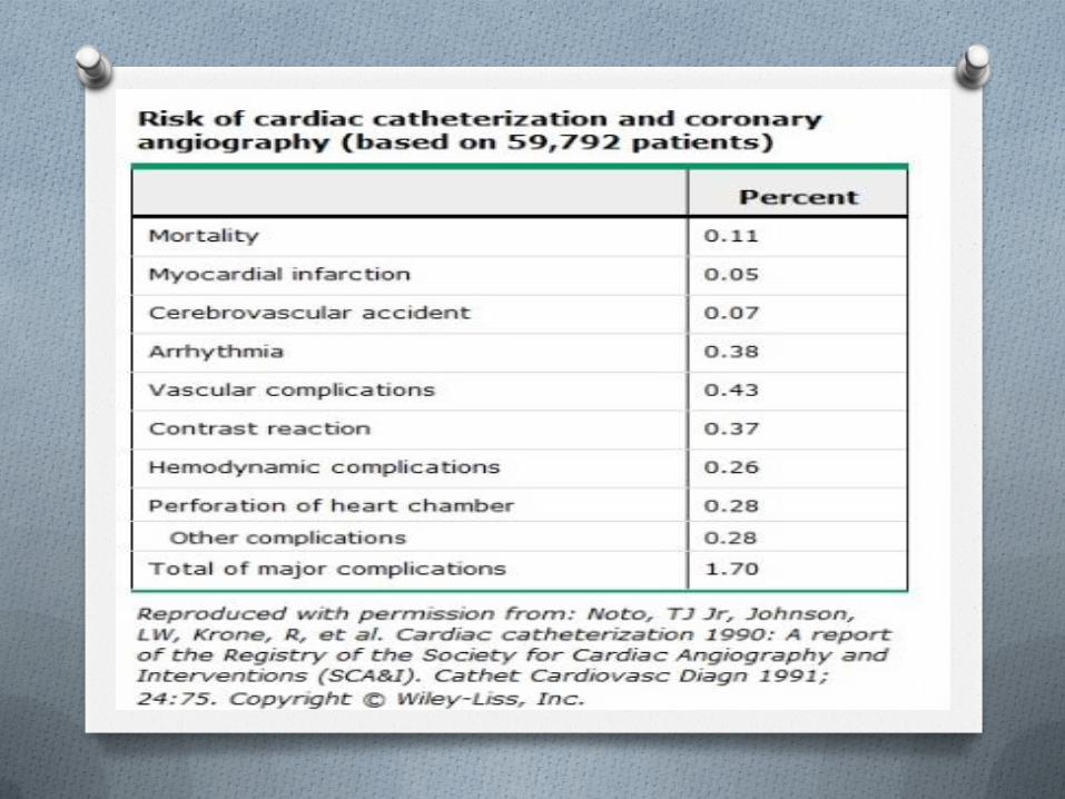
Major Complications
OMortality
OMyocardial infarction
OCerebrovascular accident
OArrhythmias
OVascular complications
OContrast reactions
OHemodynamic complications
OPerforation of heart chamber

Minor Complications
OAir embolus (0.1%).
OCholesterol embolization.
ONerve pain.
OLactic acidosis may develop in
diabetic patients taking
metformin.
O Infection.

Patients at Increased Risk for
Complications after Coronary
Arteriography
O Increased Medical Risk
O Increased Cardiac Risk
O Increased vascular Risk
O Risk of Death
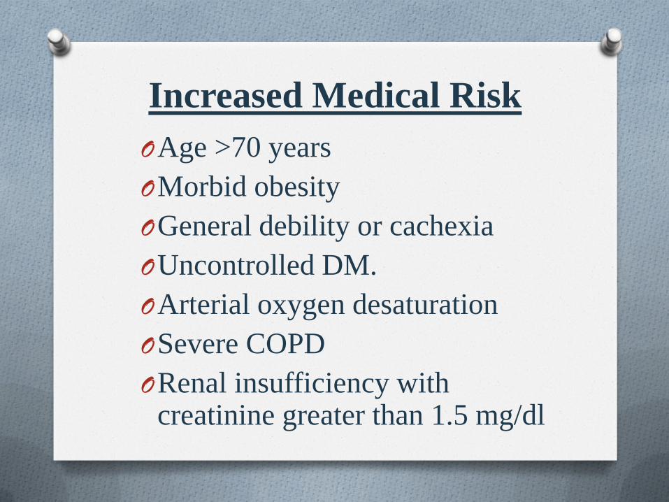
Increased Medical Risk
OAge >70 years
OMorbid obesity
OGeneral debility or cachexia
OUncontrolled DM.
OArterial oxygen desaturation
OSevere COPD
ORenal insufficiency with creatinine greater than 1.5 mg/dl

Increased Cardiac Risk
O Three-vessel coronary artery disease.
O Left main coronary artery disease.
O Functional class IV.
O Significant mitral or aortic valve disease or
mechanical prosthesis.
O Ejection fraction less than 35%.
O High-risk exercise treadmill testing
(hypotension or severe ischemia).
O Pulmonary hypertension.
O Pulmonary artery wedge pressure greater
than 25mmHg.

Increased vascular Risk
OAnticoagulation or bleeding diathesis.
OUncontrolled systemic HTN.
OSevere PVD.
ORecent stroke.
OSevere AR.
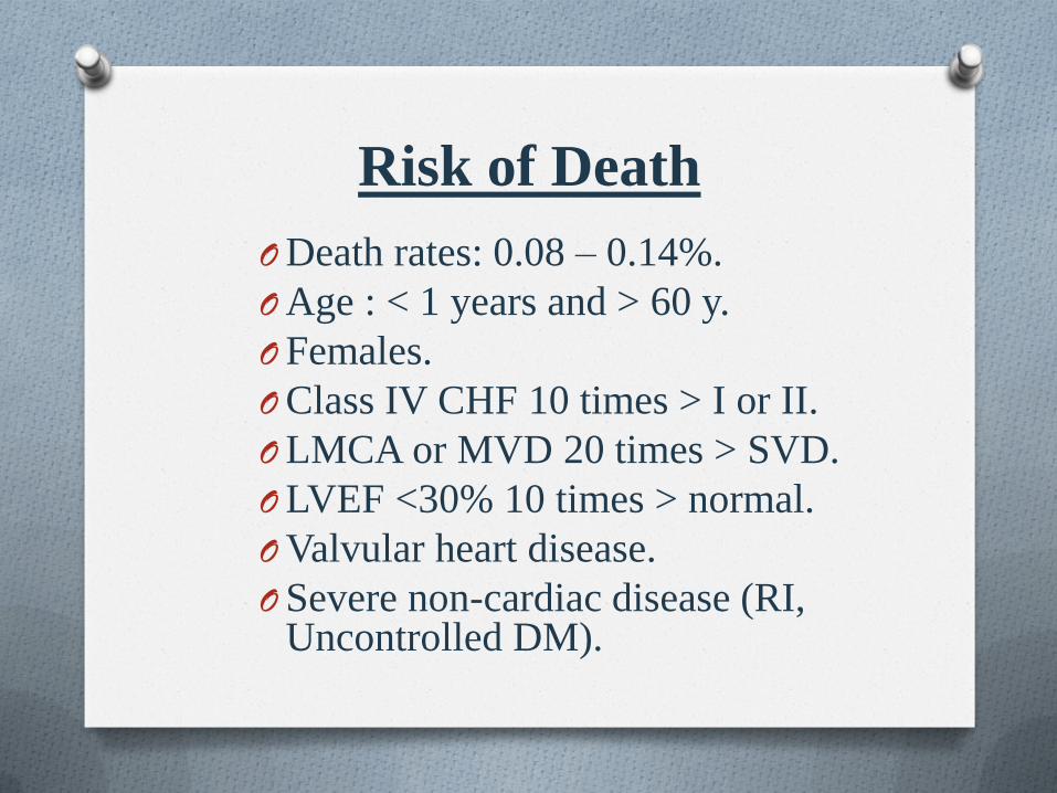
Risk of Death
ODeath rates: 0.08 – 0.14%.
OAge : < 1 years and > 60 y.
OFemales.
OClass IV CHF 10 times > I or II.
OLMCA or MVD 20 times > SVD.
OLVEF <30% 10 times > normal.
OValvular heart disease.
OSevere non-cardiac disease (RI, Uncontrolled DM).

AMI
O Incidence < 0.1 %.
O Caused mostly by coronary dissection , coronary embolization , rupture of atheromatous plaques & thrombosis.
Patients with ACS have a higher risk.
O Recently, incidence reduced by improvement in the equipment , operator skill, the use of more potent antithrombotic, beta blockers , statins & low osmolar contrast agents.

Cerebrovascular Complication O Incidence: 0.07- 0.1 %
O Caused by:
- Embolization (80%)
- Hemorrhage (20%)
Patients with AS have a higher risk
O Prevented by:
- Careful flushing & injection technique.
- Minimize time of guide wire in aortic root
- Carefully Wipe & immerse guide wires in heparinized saline before reintroduction.
Periprocedural stroke patients have poor outcome (in-hospital mortality about 32%)

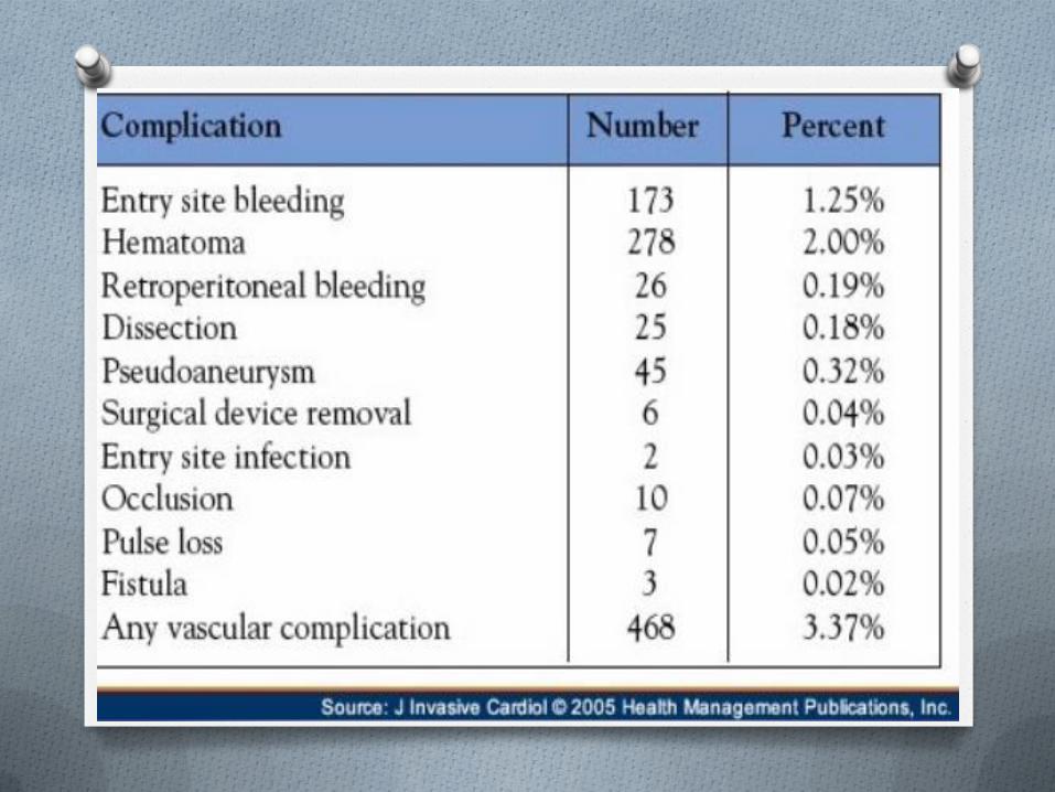
Vascular Complication A) Dissection of great vessels & coronaries.
B) Local complications at the site of puncture:
- Acute thrombosis.
- Distal embolization.
- Bleeding (femoral or retroperitoneal hematoma &
free bleeding at puncture site).
- Pseudoaneurysm.
- AV fistula.
- Local complications are less in radial approach.


Dissection & perforation of
great vessels O Incidence: 0.04 %.
O Caused by retrograde passing of wires & catheters through tortuous or stenotic arteries.
O Treatment:
Small retrograde dissections Sealed down by antegrade blood flow.
Large and flow limiting dissections :
- Immediate Proximal balloon inflation.
- Later Stenting ( great vessels) , CABG or ligation (coronaries) & pericardiocentesis (tamponade).

Femoral artery laceration
Free bleeding around the sheath.
O Treatment:
- Placement of upsized sheath.
- Manual pressure around the sheath till
procedure completed.
- Reverse anticoagulation & removal of the
sheath with compression for 30-60 min.
- Closure device.
- Not controlled Vascular surgery.

Femoral Hematoma
O More common than free bleeding.
O May cause femoral nerve compression
sensory or motor deficit which take weeks or
months to resolve.
O Spontaneous reabsorption within days.
O Prevented by accurate puncture , rapid
removal of the sheath within 2-4 hrs. &
effective manual compression (20-30 min) or
closure device.

Retroperitoneal hematoma Potentially life threatening
complication.
O Caused by high puncture
above inguinal lig.
O Clinically suspected
unexplained hypotension ,
ipsilateral flank pain &
hematocrit.
O Diagnosis: U/S & CT

Retroperitoneal hematoma
O Treatment :
- Conservative: bed rest, reversal of
anticoagulation , blood transfusion & manual
compression at the puncture site.
- Interventional: ipsilateral or contralateral
catheter approach localization of bleeding site
peripheral angioplasty balloon & covered
stent.

Arteriovenous fistula
O Caused by:
- low puncture site at the level of superficial femoral artery.
- Ongoing bleeding from arterial puncture site decompress into adjacent V. puncture site.
O Diagnosis: Thrill or continuous bruit over the puncture site confirmed by Doppler U/S or CT.
O Treatment: Surgical repair in about 2/3 of patients without spontaneous closure

Pseudoaneurysm
O Incidence: 0.1-0.2 % in diagnostic angio & 0.8-2.2 % in PTCA.
O Develops if hematoma forming a blood filled cavity continuous with arterial lumen.
O Blood flows in and out the cavity during systole and diastole.
O Mostly occur within first 3 days after removal of arterial sheath.
O Predisposing factors: Low puncture, brief compression, large pore sheath, aggressive antithrombotic, severe HTN, age > 65 yrs , RI , obesity & PVD.
O Diagnosis: Pulsatile mass with systolic bruit over the puncture site confirmed by Doppler U/S.

Pseudoaneurysm
O Complications:
- Rupture
- Distal embolization
- Local pain
- Neuropathy
- Local skin ischemia
- Infection
Pseudoaneurysm O Treatment :
Conservative: Aneurysm < 2 cm (serial U/S confirm cavity thrombosis).
Interventional: Aneurysm > 2 cm without surgical indication ( either by U/S guided compression or cavity injection of thrombin or collagen).
Surgical management: when aneurysm (very large, rapidly expanding, at the site of vascular anastomosis, causing skin necrosis or infected).


Arterial thrombosis
O Incidence: Rare.
O Predisposing factors: small vessel lumen,
PVD, DM, females, large sheath (IABP) &
prolonged procedures.
O C/P: pain, parasthesia, pallor, absent pulse &
coldness of LL.
O Prevention: frequent flushing, adequate
anticoagulation in prolonged procedures.
O Treatment: Percutaneous thrombectomy,
thrombolytic therapy & surgical consultation.


Arrhythmias
O Types:
A) Tachyarrhythmia:
- PVCs (common, induced by catheter introduction
in LV or RV & of no clinical significance)
- VT & VF
B) Bradyarrhythmia:
- Sinus bradycardia
- CHB.

Ventricular tachycardia &
Fibrillation O Causes:
- Excessive catheter manipulation & intracoronary injection of ionic high osmolar contrast especially in RCA.
- Prolonged catheter engagement inspite of damped pressure.
- Profound transmural ischemia or early MI.
- Incidence increases in patients with baseline prolonged QT interval.

Ventricular tachycardia &
Fibrillation
O Treatment:
-Immediate catheter repositioning.
- Hemodynamically stable VT IV lidocaine
or amiodarone.
- Hemodynamically unstable VT & VF DC.
- Mgso4 in patients with torsades.
Bradycardia
O Causes:
- Injection of ionic high osmolar dye especially
in RCA (forceful coughing enhances coronary
perfusion & help restoring normal rhythm).
- Vasovagal reactions (anxiety, pain, cardiac
perforation).
- Development of LBBB due to LV septal
scraping in a patient with pre-existing RBBB
CHB.

Bradycardia
O Prevention :
- Use of non ionic dye.
- adequate periprocedural hydration.
- adequate local anesthesia.
- sedation.
O Treatment:
- Forceful cough.
- IV atropine 0.5- 1 mg.
- IV fluids.
- Temporary pacing in resistant CHB.

Allergic reactions Precipitated by local anesthesia , contrast media , protamine sulfate.
Allergy to iodinated contrast agents
O Incidence: up to 1%
- Higher risk in patients with (past history of reactions , asthma , atopy & sea food allergy).
O Prevention:
- Premedication with steroids, antihistaminic & H2 blockers
- Use of non ionic dye.
O Treatment:
- IV epinephrine 1:10000 every min. until pulse restored.
- Rapid IV fluid infusion.
- IV vasopressors , hydrocortisone & atropine in bradycardia.

Contrast
induced nephropathy
(CIN)

Hemodynamic Complications
A. Hypotension:
Types & causes:
- Hypovolemic (blood loss, inadequate prehydration, excessive contrast induced diuresis).
- Cardiogenic (massive transmural ischemia, tamponade, arrhythmia & valvular regurge).
- Distributive ( contrast, vasovagal, nitrates & VD of inotropes).
Treatment:
According to the cause.

Hemodynamic Complications B. Volume overload:
Causes:
- Use of hypertonic contrast agent.
- Myocardial depression due to (contrast agent , massive ischemia & baseline LV dysfunction)
- Excessive prehydration
Treatment:
- Diuretics.
- Nitrates.
- Inotropes.

Cholesterol embolization
O Caused by catheter scraping of friable atheromatous plaques in aorta.
O C/P: according to the site of embolization
- Cerebral stroke
- Coronaries AMI
- Renal infarction or ARF
- Intestinal MVO
- Cutaneous digital cyanosis or gangrene.
O Prevention: ?? Preprocedural use of simvastatin
O Treatment: Supportive

Summary
O Proper patient selection, preparation and
attention to details efficiently reduce the
complications of coronary angiography.
O There is no routine procedures.
O Be careful with high risk patients.
O Never force the equipment.
O Catheters must fit the anatomy.
O Surgical pack-up is necessary.
O Patient safety is the first priority.
O Always remember that you are treating a
patient not a coronary vessel.
