comparative histology of the kidney of domestic …
TRANSCRIPT

COMPARATIVE HISTOLOGY OF THE KIDNEY
OF DOMESTIC ANIMALS
By
RAMCHANDRA PRASAD YADAVA
A THESIS
Submitted to the School of Advanced Graduate Studies of Michigan State University of Agriculture and Applied
Science in partial fulfillment of the requirementsfor the degree of
DOCTOR OF PHILOSOPHY
Department of Anatomy
1955

ProQuest Number: 10008665
All rights reserved
INFORMATION TO ALL USERS The quality of this reproduction is dependent upon the quality of the copy submitted.
In the unlikely event that the author did not send a complete manuscript and there are missing pages, these will be noted. Also, if material had to be removed,
a note will indicate the deletion.
uestProQuest 10008665
Published by ProQuest LLC (2016). Copyright of the Dissertation is held by the Author.
All rights reserved.This work is protected against unauthorized copying under Title 17, United States Code
Microform Edition © ProQuest LLC.
ProQuest LLC.789 East Eisenhower Parkway
P.O. Box 1346 Ann Arbor, Ml 4 8 1 0 6 - 1346

ABSTRACT
This study was undertaken to provide a more or less com
plete and up-to-date description of the microscopic structures of
the kidney of the domestic animals. Studies were made on sixty-
nine domestic animals of seven different species, the pig being
chosen as a type.
The tunica fibrosa of the kidney capsule in domestic animals
was two-layered except in the cat, where only one layer was p r e s
ent. The outer layer of the tunica fibrosa consisted of dense col
lagenous and a few elastic fibers and the inner layer was formed
by loose collagenous and reticular f ibers, with smooth muscle fibers
present in all animals except the cat. In the sheep and goat, and
to a le s se r extent in the ox, the muscle fibers were very numerous
and appeared to form a distinct muscular layer.
The j uxtamedullary renal corpuscles in the horse (191p), pig
(156|j.), dog (124p), and cat (106p) were larger than the cortical ones,
which were 1 7 8 i j l , 1 3 7 (j l , 122(x, and 96p, respectively. The juxtamedul-
la ry renal corpuscles in the ox (173p), sheep (147|a), and goat (15 7 (jl )
were smaller than the cortical ones, which were lBlp, 1S 3 pu, and 158(ji,
respectively. The juxtaglomeruli were larger than the cortical ones

in the pig, horse, dog, and cat; whereas in the ox the reverse was
true. In the sheep and goat no appreciable difference was noticed
between the size of the juxtamedullary glomeruli and those of the
cortical ones.
The juxtaglomerular apparatus in all animals contained some
spindle-shaped smooth muscle cells of the media of the afferent
arterio le along with the usual myoepitheloid cells. In the goat and
dog, the juxtaglomerular cells were more numerous than in the pig.
As compared with the pig (45p), the diameter of the proxi
mal convoluted tubule was greater in the horse (56p), ox (50p), and
goat (48(jl); equal in the sheep (45p); and smaller in the dog (39p)
and cat (41 p).
The brush border was found to form a cluster from each
individual cell in the proximal tubule of the pig kidney, whereas in
other animals they were uniformly arranged along the luminal border
of the cells. The basal striations were more distinct in the dog.
Fat globules were observed in the proximal tubule of the cat and
dog.
The maculae densae in all animals were single-layered except
in the horse, in which they were found to be stratified.
The papillary duct was lined by both simple and transitional
epithelium in all animals except the dog, in which the papillary duct

was lined by simple columnar epithelium only. Openings of the pap
illary duct on the papilla were observed in all animals studied.
The in tersti tial spaces of the kidney contained reticular f ibers ,
a few collagenous fibers , fibroblasts, histiocytes, and mast cells in
all animals except in the dog and cat, where mast cells were ab -
s ent .
The intertubular cell groups or Becher’s cells were present
in the interstitial spaces in the cortical region, especially in the
close neighborhood of Bowman's capsule, the cortical ar ter ies , and
arterio les in all animals except the cat, in which they were absent.
These cell groups were more numerous in the ox and horse than in
the pig, and less numerous in the sheep, goat, and dog. The cell
groups near the ar ter ies were larger than those located elsewhere.
The cells were mostly polygonal with faintly eosinophilic cytoplasm
and darkly stained nuclei. The cells were larger and more eosino
philic in the ox than those of the pig.
The cell unit of Goormaghtigh was found to be situated in the
triangular space between the afferent and efferent ar terio les, and the
macula densa in all animals without any species variation.
The following findings are thought to be reported for the f i rs t
time, or are contrary to the work of recent investigators:
iv

1. Smooth muscle cells in the capsule of the horse, goat,
pig, and dog.
2. Intertubular or Becher 's cells in the horse, ox, sheep,
goat, pig, and dog, but absent in the cat.
3. A. stratified macula densa in the horse.
4. Mast cells in the interstitial spaces of the kidney of all
species except the dog and cat.
5. Larger cortical than juxtamedullary renal corpuscles in
the ox, sheep, and goat.
6. Larger cortical than juxtamedullary glomeruli in the ox,
and equal or slightly larger ones in the sheep and goat.
7. Brush borders in clusters at the apices of the cells of
the proximal tubule in the pig.
8. Transitional epithelium extending varying distances into
the papillary ducts of all animals except the dog.
9. Papillary duct openings on the papilla of all animals in
vestigated .
10. The cell unit of Goormaghtigh in all the species in this
inv e stigation,

FRONTISPIECE
Photomicrograph of afferent (1) and efferent arterioles (Z), renal cor
puscle (3), unusual double maculae densae (4), cell unit of Goormagh-
tigh (5), Becher's cells (6), proximal tubule with basal striations (7),
and the distal tubule (8). H. and E. 420x. Six-year-old cow.

*f d f - ’ '.
& \}
kj
*
f
F R O N T I S P I E C E

8
££
FRONTISPIECE
Photomicrograph of afferent (1) and efferent arterioles (2), renal cor
puscle (3), unusual double maculae densae (4), cell unit of Goormagh-
tigh (5), Becher's cells (6), proximal tubule with basal striations (7),
and the distal tubule (8). H. and E. 420x. Six-year-old cow.
d
Y

F R O N T I S P I E C E

Dedicated to my P rofessor
Dr. M. L. Calhoun, D.V.M., M.S., Ph.D.,
Professo r and Head, Department of Anat
omy, College of Veterinary Medicine,
Michigan State University, whose devotion
to research, scientific achievements, and
rich human understanding have been a
never-ending source of inspiration.
v i i i

A C K N O W L E D G M E N T S
The author wishes to express his sincere gratitude and appre
ciation to Dr. M. Lois Calhoun, Professor and Head of the Depart
ment of Anatomy, for her patience, guidance, and advice during the
course of this investigation and the preparation of the manuscript;
to Dr. J. F reder ick Smithcors, Department of Anatomy, who gave so*
generously of his valuable time in reading the manuscript and giving
helpful suggestions; and to Dr. Esther M. Smith, Anatomy Department,
for her valuable suggestions in the preparation of the manuscript and
for her help in taking the photomicrographs. Thanks are also due to
Mrs. Ann L. Brooks, the department secretary , and other members
of the faculty and staff of the Department of Anatomy, for their help
ful cooperation.
Sincere appreciation is extended to Dr. L. F, Wolterink, P r o
fessor of Physiology and Pharmacology, for his efforts and construc
tive cri t icism of the manuscript, and Dr. W. O. Brinker, P rofessor
of Surgery and Medicine, for his helpful suggestions and assistance.
The author wishes to express his sincere gratitude and r e
spect to the late Dr. Frank Thorp, J r . , Professor of Animal Path-
ology, and a member of the author's guidance committee, for his
valuable help and suggestions.
i x

F or Ms aid in securing animals for this investigation, the
author is deeply indebted to P rofessor J. Lyman B ratz ler , Depart
ment of Animal Husbandry. Special thanks are extended to Dr.
William D. Baten, P rofesso r of Mathematics, for his valuable help
towards the statistical work of this investigation.
The author is also very thankful to Dr. Clifford C. Beck and
to Dr. Joyce L. Cathey, of the Department of Surgery and Medicine,
for their unusual cooperation, help, and permission to use the equip
ment and m ateria ls of the mastitis laboratory.
Many thanks are due to Dr. Clyde J. Douglass and Mrs. Errol
C. Benne for their helpful suggestions and assistance in histological
technique. Thanks are also due to Mr. Richare Moore, Dr. Chintamani
Singh, and Dr. Hans Ruhland for their invaluable assistance in many
German translations.
It is with deep and sincere appreciation that the author ex
presses his gratitude to Dr. Chester F. Clark, Dean of the College
of Veterinary Medicine, to Dr. Edward K. Sales, Professor and Head
of the Department of Surgery and Medicine, and to Dr. Albert R.
Drury, P rofesso r of Surgery and Medicine, for the appointment to a
graduate as sistantship and thus providing financial support to the author
which enabled him to pursue his studies in the United States.

V IT A
Ramchandra P rasad Yadava
candidate for the degree of
Doctor of Philosophy
Final examination: November 15, 1955; 2:00 p.m.Giltner Hall
Dissertation: Comparative Histology of the Kidney of DomesticAnimal s
Outline of Studies
Major subject: Anatomy
Minor subjects: Physiology; Surgery and Medicine
Biographical Items
Born: November 1, 1920, Patna, Bihar, India
Undergraduate Studies: Patna Science College and BiharVeterinary College, Patna, Bihar, India. Obtained the diploma in Veterinary Science from the Bihar Veterinary College in 1943.
Graduate Studies: Master of Science degree, Michigan StateCollege, East Lansing, Michigan, 1953-1954.
Experience: A member of the teaching staff of the BiharVeterinary College, Patna, Bihar, India, from 1943 to 1953. Held the positions of Demonstrator, Lecturer , and Assistant Professor at the Bihar Veterinary College, Patna, during the ten years of service to the institution. Appointed as Examiner in Anatomy by the Patna University in the year 1952. Granted extraordinary leave by the state government for advanced g rad uate studies in anatomy in the United States, February
x i

5, 1953. Graduate Assistantship in the Department of Surgery and Medicine since the fall of 1953.
Member of Bihar Veterinary Association; Phi Zeta; associate member of the Society of Sigma Xi; President of the Indian Students Association at Michigan State University .
x i i
T A B L E O F C O N T E N T S
P a g e
INTRODUCTION......................................................................................................................... 1
HISTORICAL BACKGROUND ................................................. 4
REVIEW OF LITERATURE ................................................................. 8
Gross Anatomy ............................................................................................................ 8
Microscopic Anatomy ............................................................................. 10
The Capsule ........................................................................................... 10
The Uriniferous Tubules ............................................................ 10
The nephron ......................................................................... 11
The renal c o r p u s c l e ................................................................. 11
The proximal convoluted tubule . . . 23
The loop of Henle ....................................... 27
The distal convoluted tubule ..................... 27
The collecting t u b u l e s ....................................... 3 0
The arched collecting tubules 30
The straight collecting tubules . . 31
The papillary ducts ............................................... 32
The basement membrane of the uriniferoustubules ..................... 33
The P a p i l l a ............ 33
x i i i

Pag e
The Interstitial S p a c e ........................................................................................... 34
Intertubular cell groups or Becher 's cells . . . . 34
Cell unit of Goormaghtigh or socle-p l a s m o d i u m ........................................................................................................ 35
Blood Vessels .................................................................................. 36
Circulation through the k i d n e y .................. 39
Arteriovenous anastomoses ........................................... 41
Lymphatic Vessels .............................................................................. 42
Nerves ........................................... 42
MATERIALS AND M E T H O D S ...................... 45
Source of Animals ............................................................................. 45
Techniques ............................................................................................................ 46
Fixation and Processing of the Tissues 46
Sectioning ............................................................................................................ 48
Staining and Mounting ......................................................................... 48
Methods of M e a s u r in g .......................... 48
RESULTS AND DISCUSSION..................... 50
Gross Anatomy ..................................................................... 50
Blood Vessels ...................................................................................... 52
Lymphatic Vessels ............................................... 54
Ne rv e s .............................................................................. 54
x i v

Page
Microscopic Anatomy ....................................................................................... 55
The Capsule ........................................................................................................ 55
The Uriniferous Tubules .................................................... 57
The nephron .............................................................................. 60
The renal c o r p u s c l e ................................................ 60
The proximal convoluted tubule . . . 69
The loop of Henle .............................. 73
The distal convoluted tubule...................................... 77
The collecting tubule ............................................... 82
The arched collecting tubule . 83
The straight collecting tubule .................. 83
The papillary duct ................................................................. 85
The basement membrane of the uriniferoustubule ................................................................................................................ 91
The P a p i l l a .............................................................................. 91
The Interstitial Connective Tissue ................................... 95
The intertubular cell groups or Becher 's cells . 98
Cell unit of Goormaghtigh or socleplasmodium 101
SUMMARY AND CONCLUSIONS .................................. 103
LITERATURE CITED ..................................................................... I l l
X V

LIST OF TABLES
T A B L E P a g e
I. Litera ture Review on Number, Size, andVolume of the G lo m e r u lu s ....................................................................... 13
II. Diameter of the Uriniferous Tubule,with Epithelial H e i g h t ............................................................................... 6l
x v i

LIST OF PLATES
P L A T E P a g e
I. Capsule of the kidney of the goat, showingthe capsula fibrosa and capsula adiposa .......................... 59
II. Kidney of the goat ........................................................................... 67
III. Kidney of the p i g .................................................................................... 72
IV. Kidney of the c a t ........................................................................... 75
V. Kidney of the horse ................................................. 81
VI. Papillary ducts in the dog k i d n e y ..................................... 88
VII. The papillary ducts in the ox kidney . . . 90
VIII. The papillary ducts in the cat kidney . . . . 93
IX. Kidney of the pig stained for reticularconnective tissue ..................................................................... 97
X. Kidney of the ox ....................................................................... 100
x v i i

INTRODUCTION
This investigation deals with a comparative study of the m i
croscopic s tructures of the kidney of the horse, ox, sheep, goat, pig,
dog, and cat. The pig has been chosen as a type because of the
morphological similarity of its kidney to that of the human species.
A detailed description of the microscopic structures of the kidney of
the pig has been given, and a comparison has been made with the
s tructures of the kidney of the rest of the animals mentioned above.
The microscopic structure of the kidney is of immense physi
ological and pathological value. There are many good textbooks of
histology in the English language that deal with the normal structure
of the human kidney. But there is none which describes the normal
structure of the kidney of the domestic animals, except an English
translation of Trautmann and Fiebigers ' German textbook, "Funda
mentals of the Histology of Domestic Animals." Although this book
was translated, revised, and published in 1952, there is no mention
of some of the very important structures of the kidney such as
juxtaglomerular apparatus, macula densa, and intertubular cells. In
certain places a description of the structures of the kidney has been
made without mentioning the species, which creates confusion.
1

2
Tereg (1911) contributed a valuable chapter on the kidney of
domestic animals in Ellenberger 's textbook, "Handbuch der Verglei-
chende mikroskopischen Anatomie der Haustiere ." But this was
written long ago and many new structures have been discovered since
then by various investigations on the kidney of man and laboratory
animal s .
Kunkel (1930) and Rytand (1938) made comparative studies of
the number and size of the glomeruli of the kidneys of various do- ■
mestic animals. These were valuable contributions, but the studies
remained confined to a very limited field.
Langham and Hallman (1939), and Eangham et al. (1942) made
extensive studies of the microscopic structure of the bovine kidney,
but they also left some of the important structures (juxtaglomerular
apparatus, macula densa, and intertubular cells) of the kidney un
touched.
Bloom (1954) gave a description of the normal structures of
the kidney of the dog and cat in his book, ’’Pathology of the Dog and
Cat." This description is based upon the work of various investi
gators, and certainly is of great value so far as the microscopic
s tructures of the kidneys of the dog and cat are concerned.
Nowhere in the l i terature has there been found a more or
less complete description of an up-to-date study of the comparative

3
histological s tructures of the kidney of the horse, ox, sheep, goat,
pig, dog, and cat. This investigation was undertaken to contribute
something towards this aspect of the science.
All microscopic studies were made under an ordinary light
microscope, and hence the description of ultramicroscopic s tructures
of the kidney of the domestic animals cannot be expected from this
work. Electron microscopic study is necessary to reveal such s truc
tures, and the author hopes that some investigators will undertake
that work in the near future. Fur ther work also remains to be done
on the reversa l of the Golgi element in the macula densa, the gran
ules of the juxtaglomerular cells, the nerve endings, and the lymph
vessels. In spite of these omissions the author hopes that this paper
will serve as a reference to biologists, physiologists, pathologists,
histologists, and students of anatomy.

HISTORICAL BACKGROUND
The study of the mammalian kidney goes as far back as the
fourth century B.C., when Aristotle (384-322 B.C.), a Greek philoso
pher, scientist, and founder of comparative anatomy, gave an i l lus
trated description of the mammalian uro-genital system in his
1'Historia animalium.n Aristotle called the kidney 11 the nephros"
and the pelvis of the kidney 1'the cavity of the nephros . '1 The ure ter
was called the ltduct from nephros to cy s t i s .1'
Aretaeus (early second century A..D.) gave a description of
the kidneys which has led many anatomists to suspect that Aretaeus
was aware of the existence of the papillary ducts (ducts of Bellini).
Aretaeus considered the kidneys to be true glands, and compared
them with the testes, probably because of the superficial similarity
in shape. A.retaeus stated ' ' the ir cavities are small and like sieves,
for the percolation of the urine; and these have attached to each of
the nervous canals, like reeds, which are inserted into the shoulders
of the bladder on each side; and the passage of urine from each side
of the kidneys to the bladder is equal.' '
4

5
Leonardo da Vinci (1452-1519), one of the greatest biological
investigators of all time and an a r t is t anatomist, made a valuable
sketch of the outline of the urino-genital system.
Berenger (1470-1530) was very much interested in the vascu
lar bed of the organs, and it was this in terest that led him to an
investigation of the form and function of the kidney. In his "Com-
mentaria cum amplissimis additionibus super anatomiam Mundini'1
Berenger mentioned that he injected the renal blood vessels.
Vesalius (1543) gave an illustrated description of the male
and female urino-genital system in his monograph "On the fabric
of the human body.IT Vesalius' treatment of the kidney is inadequate.
There is a crude discussion of the kidney in which the pelvis is
described and represented as divided in two by a sievelike structure.
Eustachius (1520-1574) attacked Vesalius for having re p re
sented the kidney of the dog instead of that of the human. Eusta
chius described for the f i rs t time the cortical substance of the kid
neys. He used magnifying lenses and injected vessels to aid his
study.
Ruini (1599) gave an excellent description of the kidney in
his monograph on the anatomy of the horse (A.natomia del Cavallo).
Ruysch (1638-1731), one of the most capable anatomists of
the period, became famous for his injection technique and studies

6
of the renal glomerulus. The renal glomerulus was named "Glom
erulus Ruyschiana" after him.
Bellini (1662) discovered the straight collecting tubules and
the papillary ducts of the kidney, and hence the papillary ducts are
also known as the ducts of Bellini. Malpighi (1659) went beyond this
by demonstrating that the kidney was composed of those pyramidal
masses of Bellini 's tubules. These pyramids of the kidney are hence
called pyramids of Malpighi. In addition, Malpighi observed the con
voluted tubules, and, with the aid of injected specimens, noted that
they commenced as inflated swellings or capsules containing a clus
ter of small blood vessels which in turn hang on the little ar ter ies
like ' 'apples on a t r e e . " These structures are now commonly r e
fe r red to as Malpighian corpuscles. Malpighi was one of the found
ers of microscopic anatomy.
F e r re in (1693-1769) described the small groups of straight
tubules radiating from the boundary zone of the renal pyramids into
the cortex as the medullary rays. These medullary rays were once
called F e r re in 's pyramids. F e r re in also described the convoluted
uriniferous tubules of the kidney.
Bertini (1772-1845) demonstrated for the f i rs t time the renal
columns which are inward extensions of the cortical structure of

7
the kidney, between the renal pyramids. Hence these renal columns
are also known as columns of Bertin.
Henle (1841) gave a detailed description of the loops of the
uriniferous tubules of the kidney in his ’’Allgemeine Anatomie."
These are called the loops of Henle.
Heidenhain (1834-1897) is remembered for his method of stain
ing the cells of the kidney by injecting indigo-carmine into the blood.
Working with little more than a microscope and a dissecting
needle, William Bowman (184Z) f i rs t drew a picture of the single
nephron that is remarkable close to present-day ideas of the renal
functional unit.
The above description of the historical events is based on the
work of Singer (1925), Castiglioni (1947), and Mettler (1947).

REVIEW OF LITERATURE
Gross Anatomy
Chauveau (1873) described the kidney of the ox as multi-
pyramidal with well-defined external lobation having fifteen to twenty
lobes in each kidney. Langham and Hallman (1939) found sixteen to
thirty-two lobes in each kidney of the ox.
Chauveau (1873), B rem er (1944), Patten (1953), and A.rey (1954)
refe r red to the structure of the mammalian kidneys during in tra
uterine life. These authors described the presence of multilobar
kidneys in the mammalian fetus with distinct superficial lobation.
Dixon (1931) described superficial lobation in the kidney of
a young child, and sometimes, though much less distinctly, in the
adult. Straus (1934) found the kidney of man to be multipyramidal
and that of apes unipyramidal.
Grahame (1944a) and Smith (1951) described the kidney of the
elephant as multipyramidal with distinct external lobation.
Elias (1944) and Sisson and Grossman (1953) described dis
tinct pyramids in the kidney of the pig, although the external s u r
face was quite smooth as in the case of unipyramidal kidneys.
8

9
Elias (1944) described the kidney of the horse, sheep, and
goat as a unilobar organ similar to that of rodents. In the German
shepherd dog, Elias stated that the pyramids of the kidney resembled
those of the pig. In a great Dane dog, the pyramids were closely
united; in the cat a simple, unilobar kidney with a simple pyramid
was present.
Sisson and Grossman (1953) stated that the longitudinal sec
tion of the kidney of the horse presented a nonpapillated appearance.
The inner central par t of the medulla was formed by a concave ridge
which was projected into the pelvis of the kidney. This projection
was called a renal crest. The renal c re s t presented a number of
small openings constituting the area cribrosa. In the ox these au
thors described distinct pyramids with their papillae projecting into
the calyces minores. The renal columns were also very distinct.
In the sheep, the renal cres t was formed by the fusion of twelve to
sixteen pyramids. Sisson and Grossman also described distinct
papillae in the kidney of the pig. In frontal sections of the dog
kidney, curved ridges proceeded dorsally and ventrally from the
renal cres t . Sections above or below the renal c re s t cut those
ridges in such a manner as to give the appearance of conical papil
lae, producing a false impression.

10
Microscopic Anatomy
The Capsule
Chauveau (1873) described the enveloping tunic of the kidney
of the horse as a fibrous membrane intimately united to the paren
chyma of the kidney into which it sent many prolongations.
Tereg (1911) described the capsule of the mammalian kidney
as composed of two easily separable layers. The inner layer was
devoid of blood vessels. The outer layer contained blood and lymph
vessels, and nerves. Tereg (1911) and Trautmann and Fiebiger (1952)
described the presence of smooth muscle fibers in the deeper p o r
tions of the capsule of the kidney of the ox and sheep.
Mollendorff (1930), Maximow and Bloom (1952), and Smith
and Copenhaver (1953) described the capsule of the human kidney as
being composed of collagenous fibers and a few elastic fibers. Greep
(1954) described the presence of a few smooth muscle cells in the
capsule of the human kidney in addition to the usual collagenous and
elastic fibers.
The Uriniferous Tubules
Greep (1954) described a uriniferous tubule as being composed
of two segments, a secretory portion, the nephron, having the function

11
of elaboration of urine, and a collecting portion, the collecting tubule,
to convey urine into the pelvis of the kidney. A. uriniferous tubule
in the human kidney was described as being 50 to 60 millimeters
long, the secretory portion being 30 to 40 millimeters in length.
The nephron
Huber (1932) teased out individual nephrons from adult rabbit
kidneys that had been subjected to acid maceration. Huber called
the nephron the renal tubule which was considered to be made up
of the renal corpuscle, the proximal convoluted portion with the
medullary loop, the distal convoluted portion, and the junctional tu
bule.
The renal corpuscle. The renal corpuscles of the mammalian
kidney were found to be spherical with a diameter varying between
1 0 0 (jl and 2 0 0 |j l (Huber, 1932, 1935). Langham and Hallman (1939)
observed the diameter of the renal corpuscle of an adult bovine kid
ney to be 2 1 6 [jl in a fixed and stained preparation. They noted that
the size of the renal corpuscle increased with the advancement of
age, reaching a maximum in a fully grown individual. Smith and
Copenhaver (1953) and Greep (1954) described the diameter of the
renal corpuscle of the human kidney as being close to 2 0 0 )j l .

12
The glomerulus of the mammalian kidney was studied from
time to time by various investigators. The results of their investi
gations showing the number, shape, size, and volume of the g lomer
uli a re given in Table I.
Shonyo and Mann (1944) noted that the glomeruli in the cortico-
medullary zone were usually larger than those located more periph-
e ra l ly .
The afferent arterio le of the guinea pig kidney was found by
Bensley (192-9) to be expanded into a sort of rese rvo ir after entering
the glomerulus. P r im ary branches were seen to ar ise from this
expanded sinus. Schloss (1946) observed a similar dilatation in the
afferent arterio le of the human kidney and called it "B ech e r ’s glom
erular s inus."
Dorello (1948) noted that the afferent arterio le in the kidney
of the pig was expanded and gave r ise to three primary branches,
each of which gave origin to three more branches.
Codden (1949) did not find any dilatation in the afferent a r
teriole of the human kidney after it entered the renal corpuscle.
He found a ramification of the afferent arteriole soon after it en
tered the renal corpuscle.
The glomerular capillary tuft was studied by Wilmer (1941)
with the help of celloidin corrosion injection technique. It was
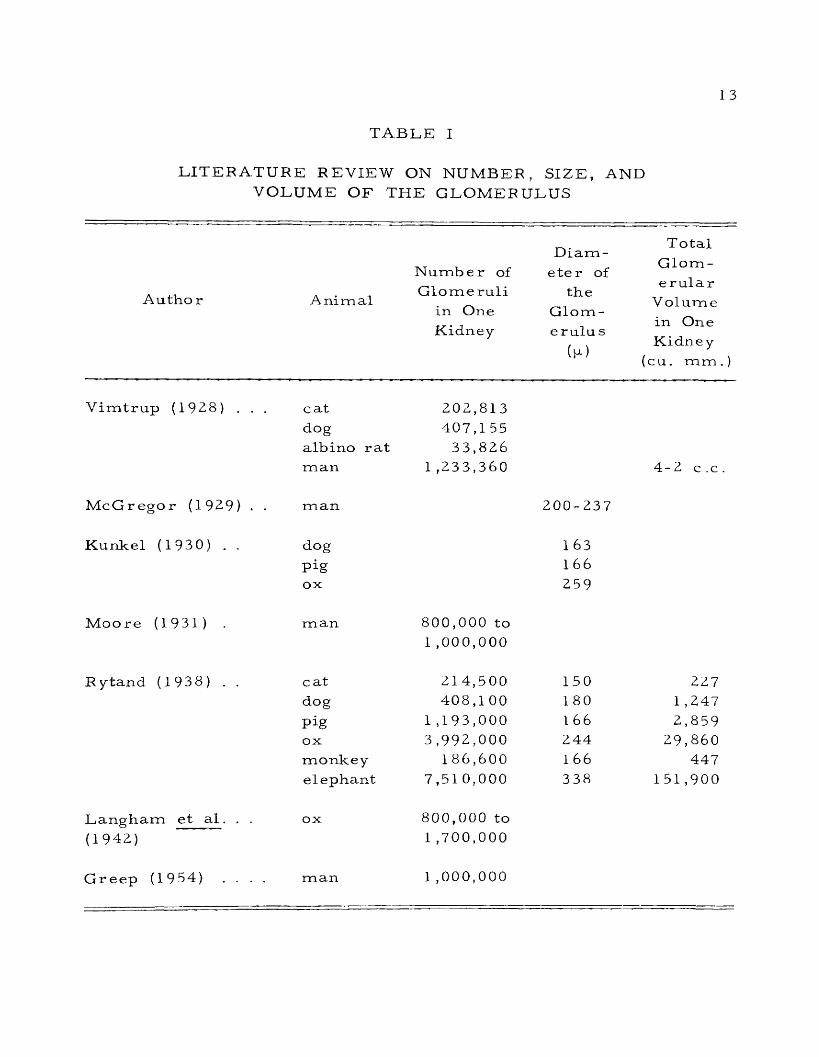
T A B L E I
LITERATURE REVIEW ON NUMBER, SIZE, AND VOLUME OF THE GLOMERULUS
Number ofDiam
eter of
T otal Glomerular
V olume in One Kidney
(cu. mm .)
Autho r AnimalGlomeruli
in One Kidney
the Glom - erulu s
(p)
Vimtrup (1928) . . . catdogalbino rat
202,813 407,1 55
33,826man 1 ,233,360 4-2 c .c .
McGregor (1929) . . man 200-237
Kunkel (1930) . . dog
Pigox
163166
259
Moo re (1931) man 800,000 to 1 ,000,000
Rytand (1938) . . cat 214,500 150 2Z7dog 408,1 00 180 1 ,247
Pig 1 ,193,000 1 6 6 2,859ox 3,992,000 244 29,860monkey 186,600 1 66 447elephant 7,51 0,000 338 151,900
Langham et a l . . . ox 800,000 to(1942) 1 ,700,000
Greep (1954) . . . . man 1 ,000,000

14
demonstrated that the afferent arterio le of the human kidney gave
p r im ary and corresponding secondary branches. It was noted that
detailed observations could not be made on approximately the f i rs t
two-thirds of the afferent ar ter io lar system, Wilmer did not observe
any anastomosis between the capillaries.
Smith (1951) found that the afferent arteriole of the human
kidney gave off two to four or more, rare ly up to ten, primary
branches, which in turn subdivided again, at times into as many as
fifty capillary loops. The primary branching gave the tuft a lobulated
structure
Trabucco and Marquez, (1952) did not find any evidence that
the glomerular tuft had any looping formations between the afferent
and efferent ar terio les. They noted that the glomerulus was formed
from one side of a single vessel bent on itself.
Boyer (1955) demonstrated that the human glomerulus was
neither a simple series of loops nor a skein of blind fingerlike
sacculations . According to Boyer the glomerular tuft was found to
be essentially a dense capillary bed with an afferent and efferent
channel. No simple loops were seen. Multiple anastomoses between
looping channels were seen to create a complex network which might
show short blind sacculations. Hall (1955) observed direct channels
in the glomerulus of the rat, cat, dog, rabbit, and man from the

15
afferent to the efferent end. of the lobule. These were thought to
function as by-passes for erythrocytes. The large capillaries were
seen to give rise medially to numerous small, short branches which
joined and divided freely to form a true capillary network; a com
plex but unified system of communicating, anastomosing capillaries
within the lobule.
The efferent arterio le of the guinea pig was found by Bensley
(1929) to proceed in a sinuous course toward the renal capsule at
f i rs t without giving off capillaries. It was found to terminate in a
plexus of capillaries around the convoluted tubules of the cortex.
The efferent vessels of the middle zone of the cortex, which were
usually supposed to be resolved immediately on emergence from the
glomeruli into the capillary vessels, were found to continue for some
distance as a discrete vessel. The efferent vessels of the juxta-
medullary region were much longer and formed the arteriole rectae
of the medulla.
Schloss (1946) observed in the human kidney a dilatation in
the efferent arterio le where it left the glomerulus. Edwards (1953)
demonstrated that in the human kidney 20 to 25 per cent of the ef
ferent arterio les of the glomeruli situated in the cortico-medullary
zone were short, thin-walled, and gave rise to a network of capil
la r ies . Such capillaries were found to supply blood to the parts of

16
all nephrons located in the cortico-medullary zone, whereas the long
efferent arterio les which were found to descend into the medulla
vascularized only the medullary parenchyma. The long efferent a r
terioles were found to comprise only 75 to 80 per cent of those
arising from the glomeruli in the cortico-medullary zone.
The in tercapillary or axial space of the normal glomerulus of
man and mammals was found by Bensley and Bensley (1930) to con
tain a small amount of connective tissue. They observed a layer of
reticular fibers surrounding the vessels at their entrance, with sparse
and delicate reticular fibers extending into the lobes of the glomer
ulus .
McManus et a l . (1951) made a histochemical study of the
human glomerulus and observed the presence of carbohydrate mate
rial in the intercapillary space of the normal human glomerulus.
Jones (1953) observed that the normal human glomerulus had
an interstitial connective tissue space, which contained connective
tissue cells. In childhood these cells were found to be less numer
ous than either the visceral epithelial layer of Bowman's capsule or
the endothelial cells, but by thirty years of age their number was
found to increase approximately to that of the endothelial and epi
thelial cells.

17
Ha.ll et al (195 3a) found the rat glomerulus to have either
exceedingly few in tersti tial cells and collagenous fibers or none at
all. Benedetti and Scapellato (1954) observed argyrophilic fibrils
and connective tissue cells in the stroma of the normal renal glom
erulus .
The juxtaglomerular apparatus was described by Goormaghtigh
(1939) as being composed mainly of smooth muscle cells, devoid of
myofibrils, called Mafibrillar c e l l s .M In the superficial cortical zone
of the normal kidney of the rabbit, these cells were found to show a
glandular cycle culminating in the formation of acidophil or basophil
secretion granules intermingled with minute vacuoles. These afibrillar
cells were observed to be in close contact with the lumen of the af
ferent arterio le or with the glomerular capillaries. In the case of
the dog, secretion granules were not found.
Edwards (1940) found in the mammalian kidney a periart io lar
pad incompletely surrounding the afferent arteriole and occupying a
region between the macula densa and the afferent and efferent a r
terioles at their junction with the glomerulus. This per iar ter io lar
pad was found to consist of a nestlike group of cells embedded in a
delicate, f ibri l la r network.
McManus (1942) defined the juxtaglomerular apparatus as a
group of apparently specialized structures in relation to and including

18
the afferent and efferent ar terio les of the glomerulus. He preferred
to use the term ' ' juxtaglomerular complex” instead of Goormaghtigh's
"juxtaglomerular appara tus.” His "complex" consisted of the juxta
glomerular apparatus and macula densa.
Graef (1943) reported medial hypertrophy and hyperplasia of
the arterio les in the case of a pregnant woman, possibly due to a
hormonal effect.
Schloss (1946) reported the existence of smooth muscle cells
in the area of the 1 *Polkissen1 ’ (juxtaglomerular apparatus of Goor-
maghtigh) in addition to the usual epithelioid cells. These muscle
cells contained myofibrils.
McManus (1947b) demonstrated the presence of granules in
the juxtaglomerular cells with the help of periodic acid Schiff reagent.
Des P rez (1948) used Masson's trichrome stain to demonstrate these
granules. These cells appeared large, pale - staining, with large,
prominent ovoid nuclei whose chromatin network was arranged in a
somewhat radial pattern. They were all afibrillar.
With the help of electron microscopy, Dalton (1951) demon
strated large cytoplasmic granules in the myoepithelial cells of the
juxtaglomerular apparatus in the kidney of the rat.
Gomori and Oltvanyi (1951) observed a nerve plexus among
the juxtaglomerular cells. Wilson (1952) developed a special staining

19
technique for demonstrating the granules of the juxtaglomerular cells.
The granules were stained deep purple, and nuclei light purple.
Maximow and Bloom (1952) noted the absence of juxtaglomer
ular cells in lower vertebrates and in children below the age of two
year s .
The function of the juxtaglomerular apparatus is very contro
versial . Goormaghtigh (1939, 1940, 1945a, 1945b, 1947, 1949, 1951)
believed that the afibrillar cells of the juxtaglomerular apparatus
had endocrine function. The endocrine activity of the afibrillar cells
was related to the production of the hypertensive substance present
in the ischemic kidney. It was suggested that in the normal condi
tion, the afibrillar cells regulated the tonus of the renal arterio les.
Kaufmann (1942), Dunihue (1947, 1949), and Hartroft and Hartroft
(1952) supported the view of Goormaghtigh that the juxtaglomerular
apparatus possessed an endocrine function.
Oberling (1944) did not find any elevation of blood p ressure
in children and adults whose kidneys possessed abundant granular
cells in the juxtaglomerular apparatus. Edwards (1945), Fox and
Jones (1945), and Schloss (1948) were unable to find any evidence in
support of Goormaghtigh's hypothesis of endocrine function of the
juxtaglomerular apparatus.

20
Graef and Proskauer (1945) and Becher (1949, 1950) thought
that the function of the juxtaglomerular apparatus might be to control
renal blood flow. Hartroft and Hartroft (1953) suggested that the
juxtaglomerular cells were involved in the hormonal regulation of
sodium metabolism and blood pressure .
The three primary membranes of the glomerulus were studied
by McGregor (1929). McGregor observed the presence of three mem
branes, epithelial, endothelial, and basement membrane, in the human
kidney. Bensley and Bensley (1930) found the glomerular epithelium
in human and small mammals to be a continuous layer over the whole
extent of the surface of the glomerulus and its lobes. It was com
posed of separate cells and did not constitute a syncytium. The
basement membrane was found to be structureless, supporting
the wall of the glomerular capillaries. The endothelium was also
found to be continuous.
Brem er (1938) demonstrated a protoplasmic film over the
glomerulus which had the property of storing dye granules after
vital staining. It did not form a continuous sheet. Only occasional
glomerular cells were found to store the vital dyes.
De Renyi (1941), using microdissection techniques, found the
epithelial lining noncontinuous. The cells resembled the pericytes
of other capillaries. The structure of the endothelium of the

21
capillary loops was different from the lining of the capillaries of
other organs.
McManus (1948a) observed that the basement membrane was
derived from Bowman's capsule and was attached to the arterio les
at the glomerular root. The capillary loops were also enclosed by
the basement membrane. Electron microscopy of the kidney revealed
a crenated appearance of the basement membrane which was studded
with minute projections (Gautier et a l ., 195 0).
Pease and Baker (1950) found a continuous epithelial layer,
a noncontinuous endothelial layer, and a basement membrane whose
external surface was covered by a system of interdigitating ridges.
Oberling et a l . (1951) and Dalton (1951) found the epithelial
layer carrying a system of many-branched villilike processes.
Hall et a l . (1953a) observed a continuous and highly porous
endothelial layer, a finely porous basement membrane, and an epi
thelial layer whose cells possessed inte rdigitating processes.
With the help of histochemical techniques and electron
micrographs, Rinehart et a l . (1953) found the surface epithelial
cells elaborating a cytoplasmic secretion which was in contact with
the basement membrane at many points. The basement membrane
was considered as being primarily a differentiated cytoplasmic prod
uct of the endothelial cells.

2 2
Simer et a l . (1953) did not find complete v isceral epithelial
and endothelial layers although every capillary was provided with a
f ibri l la r basement membrane.
Hall (1954) gave the ratio of endothelial to epithelial nuclear
count as 3:1. The basement membrane was found to be a complex
of three intimately related specialized structures under the electron
microscope (rat).
Pease (1955a) made an electron microscopic study of the
kidney of the rat and found that the epithelial cells of the glomeru
lar capillaries had long primary and secondary branches that ex
tended over and between the capillary loops and were applied directly
to the basement membrane. The basement membrane was found to
be continuous and divisible in three layers, the middle layer being
of high electron density. The endothelial cytoplasm was extremely
attenuated in most parts of the glomerular capillaries.
The secreting area of the glomerulus was found by Book (1936)
in a single typical human glomerulus to be 0.3813 square millimeter,
while Kirkman and Stowell (1942) found the area of a single albino
rat glomerulus to be 0.19 square millimeter.
Bowman's capsule was studied by Oleynik (1952). Oleynik
proposed to call Bowman's capsule "Shymlansky's capsule" as he
believed that Shymlansky published his results sixty years before Bowman.

23
Crabtree (1941) found in mice that the parietal layer of Bow
man's capsule was partly or completely composed of cuboidal cells.
These cuboidal cells were found to be identical with those of the
proximal convoluted tubule with respect to the ability to store gran
ules of trypan blue, a vital stain. Indirect evidence indicated that
single capsules could be changed from a squamous cell type to a
columnar cell type. After puberty the percentage of cuboidal cell
capsules began to increase. This occurred earlier in males than
in females, suggesting a response to an endocrine factor or factors.
Pease (1955b) made an electron microscopic study of the rat
kidney and found that the parietal cells of Bowman's capsule some
times had cytoplasmic processes suggestive of a rudimentary brush
b o rd e r .
The proximal convoluted tubule. Huber (1932, 1935) found the
length of the proximal convoluted tubule in the rabbit to be 9.4 m il
limeters and the diameter 50p to 60fjt. Grafflin (1939) noticed the
average number of turns in the proximal tubules in human kidney
to be forty. Huber observed in the rabbit 's proximal convoluted
tubule a relatively low epithelium with a wide lumen, or a high epi
thelium with a relatively narrow lumen. He observed that the cells
possessed a spherical nucleus, brush border, and mitochondrial gran
ules distributed in a manner to give distinct basal striations.

24
Grafflin (1942) noted a deposition of yellow to golden-brown
iron-containing pigment in the epithelium of the proximal tubule of
the r a t .
Mayer and Ottolenghi (1947) demonstrated a protrusion of
tubular epithelium into the space of Bowman’s capsule in the kid
neys of dogs and cats. Protruded tubular epithelium was continu
ous with the proximal tubule and was distinct from the capsular
epithelium, being cuboidal in shape.
Harman and Hogan (1949) found multinucleated giant epithelial
cells in the proximal convoluted tubules of human kidneys in 15.2
per cent of routine autopsies. Sulkin (1949) observed binucleated
cells in the various tubules of the normal kidney of the rat. Uni
la tera l nephrectomy showed a significant increase in the number of
binucleated cells.
Lowell et a l . (1953) found each nucleus of the cells of the
proximal convoluted tubule surrounded by a considerable body of
cytoplasm charged with well-preserved rodlets and granules of
mitochondrial character. The nuclei were unevenly spaced and
occasionally aggregated like the macula densa.
The ultra structure of the proximal convoluted tubules of the
mouse kidney as revealed by high resolution electron microscopy
was demonstrated by Sjostrand and Rhodin (1953). It was found that

25
the cell membrane appeared as a thin zone of condensed cytoplasm
toward the basement membrane. The mitochondria were found to
be rod-shaped. In the intermediate cell zone there were granules
and vacuoles .
The brush border was observed by Gautier and Bernhard
(1950) as being composed of round or polygonal tubules which orig
inated from the cytoplasm of the proximal tubule and ended in the
lumen as pores.
Sjostrand and Rhodin (1953) found the brush border consisted
of densely arranged cylindrical ducts closed toward the tubular lu
men.
Hall et a l . (1953b) noted that the brush border was composed
of individual tubular fibers having separate origins from the cell
surf ac e .
Pease (1955b) found that the brush border of the proximal
tubule was comprised of extensions of the apical cytoplasm. The
cytoplasm of the brush border had the same density as that of the
remainder of the cell, and similar granules might even be seen
within it.
F a t content of the proximal convoluted tubule was studied by
several investigators. Modell (1933) and Foote (1936) observed fat
in the proximal tubule of the cat. Foote and Grafflin (1938, 1942)

26
found fat in the proximal tubule of both cat and dog. Dallemagne
et a l . (1950), Silver (1951), and Platt (1951) demonstrated fat in the
proximal tubule of the dog. Foote and Grafflin observed that in the
cat the entire pars convoluta and the upper portion of the pars recta
of the proximal tubule were normally fat-laden, while the terminal
portion of the pars recta was fat-free. In the dog, on the other
hand, the reverse was true. The fat was confined solely to the
terminal portion of the pars recta.
Segmental differentiation in the proximal convoluted tubule was
made by several authors on the basis of the histology and physiology.
Foote (1936) and Foote and Grafflin (1938) determined the relative
lengths of the two segments (convoluted portion and straight portion)
of the proximal tubule in the cat and dog on the basis of fat content.
They described them as fat-laden and fa t-free segments of the prox
imal tubule .
Foote and Grafflin (1942) described the cells of the f i rs t
segment of the proximal tubule of the cat and dog as being highly
i r regular with a marked degree of interdigitation. The cell con
tours were more complicated at the lumen. The cell shapes were
more ir regular in the dog than in the cat. The cell contours of
the second segment were essentially rectilinear in both species.

27
Longley and F ishe r (1954) differentiated the two segments of
the proximal tubule with the help of staining techniques. Periodic
acid Schiff staining differentiated the two segments in the mouse,
rat, and cat, and the alkaline phosphatase technique differentiated
those segments in mouse, squirrel, cat, dog, and chicken.
The loop of Henle. Huber (1932, 1935) found the diameter of
the thin limb of Henle’s loop in the rabbit varying from 20p to 25p.
The thin segment was lined by squamous epithelium. The cells had
polygonal forms and relatively large nuclei. The length of the thin
segment varied from 1 to 15 millimeters.
Smith (1951) observed that in the human kidney short loops
of Henle were about seven times as numerous as long ones, and,
in some instances, where the glomeruli were located in the outer
cortex, the thin segment might be absent entirely. Where present,
the thin segment might occupy only the descending limb of Henle's
loop, or it might extend around the loop and for some distance up
the ascending limb. Smith noted the presence of the thin segment
only in mammals and in a small percentage of the nephron of birds.
The distal convoluted tubule. Huber (1932, 1935) found the
length of the distal convoluted tubule in rabbit kidneys varying from
3.6 to 7.8 mill imeters . The diameter was found to be between 30p
2 8
and. 40p. The distal tubule was lined by a low columnar or cuboidal
epithelium presenting a clear supranuclear zone of cytoplasm. The
cells were devoid of a brush border but the basal striations were
present, though not quite so distinct as in the case of the proximal
tubule .
Grafflin (1939) observed that the distal tubule of the human
nephron took a more direct course and in every case passed the
glomerulus near the afferent arteriole. The distal tubule was found
to make few turns until it arched to join the collecting tubule.
Trautmann and Fiebiger (1952) described the nuclei of the
distal tubule in the horse as being especially large.
Pease (1955b) made an extensive study of the tubular cells
of the rat kidney with the help of the electron microscope. It was
found that the apical ends of the cells of the distal tubule usually
had scattered short cytoplasmic processes suggestive of a rudi
mentary brush border.
The macula densa was demonstrated for the f irs t time by
Zimmermann (1933) in the mammalian kidney. Zimmermann gave
the name of macula densa because of crowding of the nuclei in the
distal convoluted tubule at the area of contact between the distal
tubule and the afferent arterio le of the glomerulus.

29
Edwards (1940) p referred to use the name ’’epithelial plaqueM
for macula densa. After an extensive study of the macula densa of
the mammalian kidney, he found the epithelial plaque in mammals,
birds, and frogs variably elliptical
It constituted one-half to two-thirds of the wall of the nephron
as seen in cross section in the kidney of man, rabbit, and rat, and
commonly one-half of this wall in the kidney of the whale, guinea
pig, cat, bird, and frog. He found the plaque was composed of stag
gered rows of columnar cells often markedly taller than those adja
cent and so close together that their nuclei were usually in contact
not only with others in the same row but also with those of the ad
jacent row. The cytoplasm of the cells was nongranular and neutro
philic or slightly basophilic and their nuclei were somewhat smaller,
contained more chromatin and a more conspicuous nucleolus than
those of the adjacent cells.
McManus (1943) and Okkels (1950) observed that the position
of the Golgi elements was reversed in the macula densa. The Golgi
element in the macula densa was situated towards the basal side of
the nucleus, whereas in the rest of the distal tubule it was situated
towards the luminal side of the nucleus.
McManus (1947a) reported the absence of a basement m em
brane in the macula densa. The cytoplasm of the cells of the macula

30
densa was separated from that of the juxtaglomerular apparatus only
by the cell membrane. This absence of the basement membrane was
thought to facilitate the exchange of materials between the cells of
the macula densa and the juxtaglomerular apparatus. Dina and Mar-
tuzzi (1952) found the macular tubular cells to be stratified and the
macula densa to be larger than the area of contact with the afferent
a r te r io le .
The function of the macula densa is not yet clear. Schloss
(1946) regarded the macula densa as a sensory area, probably a
chemo-receptor, influencing the contraction of the vessels. Becher
(1949, 1950) thought that the macula densa regulated the flow of
blood through the Malpighian bodies of the kidney. Okkels (1950)
suggested an angiotrophic role for the macula densa.
The collecting tubules
The collecting tubules are considered by all authors to be
comprised of the arched or initial collecting tubule, the straight
collecting tubules, and the papillary ducts or ducts of Bellini.
The arched collecting tubules. Huber (1932, 1935) found the
p rim ary collecting ducts of the cortex of the rabbit 's kidney united
in the periphery of the cortex to form collecting tubes which passed

31
through the cortex without receiving further branches. In the human
kidney it was observed that a limited number of primary collecting
tubules joined in the periphery of the cortex, and the collecting ducts
thus formed received further branches while passing through the c o r
tex. The collecting tubules were found, in general, lined by very
regular columnar cells, with clear protoplasm, devoid of specific
granules or striations and with relatively large nuclei of spherical
or ovoid forms. Trautmann and Fiebiger (1952) described the lin
ing epithelium of the connecting tubule in the domestic animals as
composed of polygonal, light-colored cells, distinguishable only by
their le s se r height from the cells of the collecting tubules.
Greep (1954) stated that the epithelial cells lining the arched
tubules of the human kidney were strictly cuboidal.
The straight collecting tubules. Greep (1954) stated that the
straight collecting tubules in the human kidney were located in the
medullary rays. These tubules were described as making up most
of the substance of the pyramids. The straight collecting tubules
were stated to fuse with one another in successive fashion in the
pyramids to form finally sixteen to twenty large papillary ducts.
The diameter of the tubule was 4 0 |jl in the medullary ray. The
diameter increased as the tubules fused. The epithelium was cuboidal

32
in the proximal portion, but the cells increased in height as the di
ameter increased. Trautmann and Fiebiger (1952) described the
presence of numerous fat droplets, especially in older domestic
an im als .
The papillary ducts . Lang ham e t a l . (1942) found that while
the epithelium of the papillary duct of the bovine kidney acquired a
tall columnar form the cells retained the other characteris tics of
those of the smaller collecting tubules.
Trautmann and Fiebiger (1952) described the epithelium in
the papillary ducts as two-layered, changing to transitional near the
opening into the pelvis.
Duran-Jorda (195 3) doubted the existence of openings of the
ducts of Bellini. After examining a few hundred routine sections
of the human kidney of all ages, and some serial sections of this
organ from different animals, especially the cat, which had only
one pyramid, the investigator was skeptical about accepting the
existence of openings of the ducts. He was of the opinion that in
the normal kidney the urine was dialysed through a papillary epi
thelium and not excreted directly into the renal pelvis by the ducts
of Bellini.

33
The basement membrane of the uriniferous tubules
Bensley and Bensley (1930) noted the presence of a thin
s tructure less basement membrane interposed between the epithelium
of the tubules and the reticular framework. This basement m em
brane was similar to but thinner than Bowman’s capsule.
McManus (1948b) studied the histochemical features of the
renal basement membrane, and suggested a passive role for it,
closely related to the activity of the tubular epithelium. McManus
thought that it was chitinous in nature.
Pease and Baker (1950) and Pease (1955b) found the s t ruc
ture of the basement membrane the same all along the uriniferous
tubules. They observed the connective tissue cells so few and far
between that they thought the basement membrane must be maintained
by the overlying epithelia rather than by fibroblasts. The basement
membrane appeared entirely homogeneous even under high resolution.
The Papilla
Langham et a l . (1942) observed transitional epithelium cover
ing the surface of the papilla in the bovine kidney. Maximow and
Bloom (1952) stated that in the human kidney the simple columnar
epithelium of the ducts of Bellini continued on to the surface of the

34
papilla in the area cribrosa . Trautmann and Fiebiger (1952) stated
that, with the exception of the pig and the goat, the transitional epi
thelium was replaced on the renal papilla by two-layered cuboidal
to columnar epithelium.
Vimtrup and Schmidt-Niels en (1952) observed that the papilla
of the kangaroo rat was relatively long and extended into the ureter.
The Interstitial Space
Kirkman (1943) found the presence of fibroblasts and m acro
phages in the in tersti tial space of the kidney of the albino rat. The
greatest concentration of macrophages and fibroblasts was found at
the level of that portion of the thick ascending limb of Henle's loop
which was lying in the intermediate zone of the medulla.
Intertubular cell groups or Becher 's cells
Schloss (1946) found the intertubular groups of cells in the
human kidney at the points where the connective tissue was abundant.
Neumann (1949) reported the lack of intertubular cell groups
in the vicinity of the glomeruli of the human kidney.
Becher (1949, 1950) found these cell-islets situated in the
human kidney in the immediate neighborhood of the afferent arterio les

35
and. other cortical ar ter ia l vessels and called them ' “paraportal cell-
islets .1 1
Regarding the function of these intertubular cell-groups,
Schloss (1946) suggested that those Becher cells which were com
pletely isolated and surrounded by connective tissue had an endocrine
function. Becher (1949, 1950) thought that those cell-groups might
regulate blood flow through the Malpighian bodies of the kidney.
Cell unit of Goormaghtigh or socleplasmodium
Schloss (1946), Neumann (1949), and Becher (1949, 1950) found
in the human kidney a cluster of small cells (cells of Goormaghtigh)
in the angle between afferent and efferent arterio les of the glomeru
lus. Becher (1950) regarded this cluster of small cells as an area
containing nerve receptors.
Ham (1953) described the presence of a curious little aggre
gation of small cells with pale nuclei between the macula densa and
the glomerulus proper, in the concavity between the afferent and the
efferent ar terio les. Ham was of the opinion that there was no c e r
tainty about their nature, function, or nomenclature.

36
Blood Vessels
MacCallam (1936) found the arterio lae rectae arising in the
dog and cat, exclusively from the vasa efferentia. No arteriolae
rectae verae were found in those animals. MacCallum (1939) and
Oliver (1939) found a f ew pathologically changed glomeruli in the
normal mammalian kidneys, and the number of such glomeruli was
found to increase with age and disease.
Fitzgera ld (1940) demonstrated the presence of two main renal
a r te r ie s supplying each kidney in 10 per cent of the horses and 5
per cent of the dogs examined.
Loomis and Jett-Jackson (1942) and Gouygou (1949) found
Ludwig’s branches (branches of interlobular arter ies that terminate
directly in the peritubular capillary plexus without the interposition
of a glomerulus) in the human kidney to occur in old age and in con
ditions of disease.
Grahame (1944a, 1944b) found the arteria l tree in the elephant
and camel similar to the general plan of the arterial distribution in
the kidneys of other mammals.
Cowdry (1950) observed that the individual interlobular a r
teries supplied definite parts of the kidney and did not anastomose
with their neighbors.

37
These ar te r ies were found to be typical ' 1 end -a r te r ie s1'; when
one was occluded the tissue depending on it suffered acutely. These
a r te r ie s were found to be occasionally anastomosed with branches of
the phrenic, adrenal, intercostal, or capsular ar ter ies .
P icard et a l . (1930) and P icard and Chambost (1952) observed
in the dog and cat a c ircular thickening situated at the origin of the
afferent arterio les of the juxtamedullary glomeruli. It appeared to
be valvular in nature.
Ruotolo (1950) described the presence of blocking mechanisms
of various types in the tunica intima and tunica media of the ar teries
of the human kidney.
Montaldo (1951) found a lozenge-shaped lumen of the mono-
podic branches of the arcuate and low interlobular arter ies at their
origin in the kidneys of man, dog, and rabbit.
Christensen (1952) described the presence of a few isolated
arterio lae rectae verae and Ludwig's arterio les in the dog's kidney.
Anastomosis of the afferent and efferent arterioles were also ob
served in a few instances.
Rotter (1952) considered the cushionlike thickenings of the
intima of the ar ter ies of the human kidney as normal. These were
regarded as arteriosclerotic changes by many investigators. These
thickenings were present in the form of a ring inside the branches

38
of the ar ter ies and were found to occur even in young children, but
increased in size and number with the age of the individual. They
were supposed to regulate blood supply and blood p ressure in the
corresponding vascular te r r i to r ies and to promote an economic dis
tribution of the blood.
Superficial veins of the mammalian kidneys were demonstrated
by Kazzaz and Shanklin (1951) with the help of vinylacetate injection
and corrosion technique. It was found that the dog's kidney had a
system of stellate veins which on the lateral side of the kidney were
drained into the interlobular veins and on the medial side directly
into the renal vein. The cat was found to have radially arranged
veins running along the surface of the kidney and emptying into the
renal vein. Numerous tributaries joined each side of the veins and
the deep veins connected them to the interlobular s . The human
kidney had a system of stellate veins similar to the dog but not so
well developed. The veins in the cow and sheep started as spurlike
projections and joined the interlobular veins.
Koester et a l . (1953) described the presence of cushions of
connective tissue which contained many large veins of a sinusoidal
nature, protruding into the vein lumina at the arcuate - interlobular
junction in the kidney of man and dog.

39
Circulation through the kidney
Bowman (1842) noted that all the blood of the renal artery
(with the exception of a small quantity distributed to the capsule,
surrounding fat, and the coats of the large vessels) entered the cap
illary tufts of the Malpighian bodies. F rom there, the blood was
found to pass into the capillary plexus surrounding the uriniferous
tubules, and finally left the organ through the branches of the renal
v e in .
Morison (192,6) studied the renal circulation in man, cat, dog,
rabbit, monkey, deer, sheep, and pig with special reference to its
finer circulation. It was noted that the parenchyma of the cortex
was supplied by the capillaries of the efferent glomerular vessels,
and in addition occasional nutrient branches from the interlobular
a r te r ie s . The interlobular ar tery might itself terminate by ramify
ing into a capillary plexus. The nonglomerular sources of nutrient
supply were thought to be negligible.
The medulla of the kidney was observed to receive blood
supply solely by arterio lae rectae spuriae. No evidence of the p re s
ence of the arterio lae rectae verae was found.
White (1939) made observations indicating the absence of
intermittance in normal dogs and rabbits as all of the glomeruli

40
were found open all the time. It was suggested that intermittanc e
might occur in extreme conditions such as severe hemorrhage or
sudden increment of circulating epinephrin.
Fox and Jones (1946) and Maegraith and Havard (1946) ob
served that in the human kidney, during active diuresis, the cortical
circulation was fully open, while in anuria the medullary circulation
was in action, and the cortical circulation was by-passed.
Heggie (1946, 1947) stated that the redistribution of blood flow
within the kidney of the rabbit could be maintained by a circulation
via the juxtamedullary glomeruli only. The cortex could remain
relatively avascular by vasoconstriction of the peripheral arteria l
vessels in response to afferent stimuli. Barclay et al. (1946, 1947)
supported the view of Heggie that by-passing could be effected, at
leas t in part, via the vasa recta.
Trueta et a l . (1946, 1948) made an extensive investigation on
circulation in the kidney of the rabbit and came to the conclusion
that in the experimental animal a renal circulation could be continued
through the medulla via the vasa recta while the cortex was function
ally ischemic. Under such conditions the flow of urine in the ureter
decreased or was entirely suppressed. Schlegel and Moses (1950)
studied circulation in the rabbit 's kidney, but were unable to confirm
the results of Trueta et a l . (1948) indicating a by-pass through the

41
j uxtamedullary glomeruli. More and Duff (1951) confirmed the find-
ing of Trueta et a l . (1948) that the only vessels through which blood
could circulate in sufficiently large amounts to by-pass the outer
cortex were those of the j uxtamedullary glomeruli, their efferent
ar ter io les and the corresponding vasa recta of the medulla in se
quence.
Moses and Schlegel (1952) found preservation of the juxta-
medullary circulation following ligation of the renal ar tery in the
rabbit. They observed that there was ischemia of the outer cortex
and the j uxtamedullary glomeruli and vasa recta of the medulla
contained blood. They also observed direct vascular connections
with vessels penetrating the capsule from the perirenal and perihilar
t issues. Stripping of the capsule and periu re tera l tissues resulted
in complete renal ischemia.
Arteriovenous anastomoses
Simkin et al. (1948) injected glass spheres 90p to 440fj. in
diameter into the renal ar ter ies of normal human kidneys post
mortem, and the existence of arteriovenous anastomoses was indi
cated by recovery from the renal vein of spheres many times greater
than the average diameter of a capillary. Similar experiments were
made on living anesthetized rabbits and dogs, and recovery of spheres

42
measuring 5 Op to 18Op in diameter was obtained from the venous
circulation, indicating the existence of arteriovenous anastomoses in
those animals Similar arteriovenous anastomoses were demonstrated
in the human kidney by B arr ie et a l . (1950) and Pompeiano (1951).
Christensen (1952) did not find arteriovenous anastomoses in the
canine kidney.
Lymphatic Vessels
P e i rce (1944) described the lymphatics which accompany the
large blood vessels in the kidney of the dog and rabbit. The cortical
and perirenal lymphatics were found to connect with each other.
Lymph capillary networks were seen in the immediate vincinity of
the Bowman's capsule of some glomeruli. Valves were lacking in
the renal parenchyma, but were present in the large trunks of the
renal sinus .
Rawson (1949) reported the absence of lymphatic channels in
the glomeruli, and about the afferent and efferent arterio les, and the
intertubular capillaries.
Nerv es
Bradford (1889) studied the innervation of the renal blood ves
sels of the dog and found both vaso-constrictor and vaso-dilator

43
f ibers , the fo rm er being well developed. No evidence was obtained
of the existence of constrictor f ibers in the vagus nerve supplying
the kidney.
Gruber (1933) concluded after review of l i terature that the
kidney received f ibers from the vagus nerve as well.
Oberling (1944) noted the existence of a highly developed
nerve plexus around the preglomerular portion of the afferent a r
teriole of the human kidney.
Szabo (1948) made an extensive review of l i te ra ture and
concluded that possibly vagal motor nerve endings were present
among the epithelial cells of the convoluted tubules.
In the kidney of the cat and rat, Harman and Davies (1948a,
1948b) demonstrated a rich nerve supply occurring in the renal sinus
in conjunction with a r te r ies , veins, and epithelium. Throughout the
parenchyma, adventitial components were found to penetrate the
glomerulus. The perivascular space of the glomerulus was p e r
vaded throughout with a fairly elaborate complex of nerve fibers.
Nerve endings were observed in the adventitia of the ar ter ies and
veins, in the media of arteriovenous anastomoses, in the epithelium
of the convoluted tubules, and in the perivascular tissue of the
g lomerulus.

44
Knoche (1950) observed nonmedullated nerve fibers in the
various vessels and a terminal reticulum in the convoluted tubules,
the glomeruli, the vessels, and the intertubular connective tissue.
Mitchell (1950a, 1950b, 1951) found the kidney of mammals
richly innervated. The medulla was found to have a scanty nerve
supply from the arcuate nerves accompanying the arteriolae rectae.
No nerves were found to enter the medulla directly from the main
interlobar nerves. Nerve fibers were found to terminate as free,
beaded filaments or as branched or fusiform endings between the
epithelial cells or on the basement membrane. Intrarenal ganglia
or nerve cells were confirmed.
Malmejac et a l . (1951) found the synapses of the vasoconstric
tor nerves of the renal vessels, in the ganglia of the solar plexus.

MATERIALS AND METHODS
Source of Animals
F or this study the kidneys from sixty-nine animals were used.
This group of animals included:
H o rse s : Ten (five castrated males and five females); all
adults; from a slaughterhouse at Grand Rapids, Michigan,
Cattle: Thirteen (nine castrated males and four females);
16 months to 8 years; from the meat laboratory, Animal Husbandry
Department, Michigan State University.
Sheep: Eleven (five males and six females); 3 to 24 months;
from the meat laboratory.
Goats: Six (four males and two females); 4 to 24 months;
from the dissection laboratory of the Department of Anatomy and
the autopsy laboratory of the Department of Pathology.
Pigs: Seven (four males and three females); 3 to 7 months;
from the meat laboratory.
Dogs: Nine (six males and three females); 2 to 36 months;
from the departments of Physiology and Surgery and Medicine.
45

46
Cats : Thirteen (two castrated males, three uncastrated males,
and eight females); 7 weeks to 5 years; from the departments of
Physiology, Paris ito logy, and Surgery and Medicine.
These animals were in good nutritional condition and appeared
to be free from disease.
T echniques
Fixation and Processing of the Tissues
The kidneys were split longitudinally soon after their removal
from the freshly killed animals, and were fixed in F.A.A. (Lavdows ky's)
mixture for four to six days. Small blocks were cut from the well-
fixed tissues for dehydration and further processing. The technique
of B arr ie (1953) for obtaining blocks from fixed kidney tissues which
insured anatomic continuity of the renal tubules in microscopic sec
tions was closely followed. The method took into account the gross
anatomy of the medulla so that the tubules and their accompanying
vessels could be followed continuously from the papilla to the cortico-
medullary junction.
Small blocks (about 1 centimeter wide and 0.5 centimeter
thick) were cut in the vertical plane of the straight collecting tubules
and the papillary ducts in such a manner as to include a portion of

47
tHe pelvis, the papilla, the medulla, the cortex, and the capsule of
the kidney. This plane gave a longitudinal section of the uriniferous
tubules. Sometimes, in case of large animals, these blocks were
divided transversely into two halves at the corticomedullary junction
to make them smaller for better sectioning. Blocks were also made
to show the cross sections of the cortical and medullary structures.
Four blocks were obtained from different parts of both kidneys of
each animal.
These blocks of tissues were then dehydrated and infiltrated
step by step as follows:
1. Normal butyl alcohol and 95 per cent ethyl alcohol (equal
p a r ts ) - -4 hours.
2. Normal butyl alcohol No. 1--4 hours.
3. Normal butyl alcohol No. Z--16 hours.
4. Normal butyl alcohol and 52° C paraffin (equal parts )--
2 h o u rs .
5. 52° C paraffin No. 1--2 hours.
6. 52° C paraffin No. 2—2 hours.
7. 56°C paraffin No. 3--18 hours.
The infiltrated tissues were then embedded in a mixture of
two parts of Tissuemat1 and one part of 56° C paraffin.
1 Fisher Scientific Company, Pittsburgh, Pennsylvania.
48
Sectioning
All sections were cat at seven microns.
Staining and Mounting
H a r r i s ’ hematoxylin and ethyl eosin were used as routine
stains. Weigert and Van Gieson's stains were used for elastic and
collagenous tissues and muscle fibers. Gridley's (1951) reticulum
stain was employed for the demonstration of reticular fibers.
The stained sections were mounted in gum damar.
Methods of Measuring
Measurements of the diameter of the renal corpuscle, glomer
ulus, proximal convoluted tubule, thin limb of Henle's loop, straight
collecting tubule, and papillary duct were made from random sam
ples of those structures seen under the light microscope. Measure
ments of the transverse diameter of the longitudinal section of the
renal corpuscle and glomerulus were made at their centers, and from
only those structures which appeared to be cut through the center in
the vertical plane. This was judged by the presence of both vascular
and urinary poles in the same section of the renal corpuscle.
49
Several tr ia ls were made with twenty as well as ten m eas
urements of the same structure in the same animal taken at different
places in the same as well as in different slides. It was found that
the average of ten measurements was as good as that of the twenty
measurements, and hence it was decided to take an average of ten
measurements of a particular structure from each animal. The
mean of the averages of all measurements in different animals of
the same species gave the final average measurement of that p a r
ticular structure in that species.

RESULTS AND DISCUSSION
Gross Anatomy
The mammalian kidneys are described as unilobar (unipyra-
midal) or multilobar (m ul tip y rami dal) according to the number of
lobes or pyramids present in one kidney. Some multilobar kidneys
present distinct superficial lobation while others do not.
The kidney of the pig is smooth, elongated, bean-shaped, and
without any superficial lobation. The length of the kidney is almost
twice the width. The hilus is located almost in the middle of the
medial border. The longitudinal section of the kidney presents a
distinct multipyramidal appearance with the papillae of the pyramids
projecting into the calyces minores. Some papillae are narrow and
conical while others are wide and flattened. The renal pelvis is
funnel-shaped, and divided into calyces majores.
The kidney of the horse is smooth and without external lo
bation. The left kidney is bean-shaped while the right kidney p re
sents a shape .which was described by Chauveau (1873) and Sisson
and Grossman (1953) as Mthe heart on a playing c a rd . '1 The left
kidney is longer and narrower than the right one. The location of
the hilus is like that of the pig. The kidney in longitudinal section
50
51
is found to be unilobar as the organ is not papillated and pyramids
were not evident. The inner central part of the medulla forms a
concave ridge which projects into the pelvis of the kidney. This
projection is described by Sisson and Grossman (1953) as the renal
cres t . The major and minor calyces are absent.
The kidney of the ox is elongated and presents a well-marked
superficial lobation similar to the mammalian fetal lobation. The
hilus in the right kidney is an extensive elliptical cavity located on
the anterior par t of the ventral surface near the medial border. In
the left kidney, the hilus presents a deep fissure which is situated
on the anterolateral par t of the dorsal surface. On section, the
renal pyramids are easily made out and the papillae are found to
project into the calyces minores. The calyces majores are present
but the pelvis is absent.
The kidneys of the sheep and the goat are smooth, bean-shaped,
and without external lobation. The situation of the hilus is like that
of the pig. The longitudinal section of the kidney presents a unilobar
appearance with the renal cre s t projecting into the pelvis. The major
and minor calyces are absent. Elias (1944) observed the multipapil-
lated appearance of the medulla in longitudinal section in a pair of
lamb kidneys. Elias assumed that sheep probably had compound
kidneys in which the individual lobes were closely united.

52
The kidneys of the dog are smooth and bean-shaped. The
hilus has the same location as that of the pig. The longitudinal
section of the kidney presents a unilobar appearance with the renal
c re s t projecting into the pelvis. The renal c res t presents curved
ridges. The major and minor calyces are absent. In the German
shepherd dog Elias (1944) observed multipapillated kidneys re sem
bling that of the pig.
The kidneys of the cat are smooth and bean-shaped. The
hilus is situated in the middle of the medial border. The section
of the kidney gives a very distinct unilobar appearance with a single
elongated papilla projecting into the pelvis. The major and minor
calyces are absent.
Blood Vessels
The renal ar tery enters the kidney at the hilum and splits
into a number of large branches, the interlobar ar ter ies . These
ascend the renal columns to a position level with the base of the
pyramids over which they send arching branches, the arcuate a r
ter ies . These ar ter ies do not anastomose with one another. F rom
the arcuate ar te r ie s numerous smaller branches, interlobular a r
te r ies , arising at regular intervals and at almost right angles to
the main vessels , extend radially between the medullary rays. These

53
arborize near the surface of the kidney and send some small branches
into the capsule. As the interlobular ar ter ies pass through the co r
tical labyrinth, numerous side stems arise , each of which is an af
ferent arterio le to a glomerulus. As the afferent arteriole enters
the renal corpuscle, it divides and subdivides to form the capillary
loops of the glomerulus. The loops recombine to form the efferent
arterio le . The efferent vessels from cortical glomeruli branch to
form a network that supplies the neighboring tubules and the kidney
capsule. The efferent vessels emerging from the juxtamedullary
glomeruli divide repeatedly, forming a tassel of long straight vessels
of near capillary size, the arteriolae rectae spuriae, which descend
into the medulla, supplying the tubular components of the pyramids.
Efferent vessels arising in the central zone of the cortex contribute
mainly to the blood supply of the convoluted tubules and also send
some branches into the medullary rays.
Small venules draining the capsule and the outer margin of
the cortex unite to form the stellate veins. These combine to form
the interlobular veins, which return along the path of the correspond
ing ar ter ies and receive numerous tributaries from anastomotic nets
around the cortical tubules. At the cortico medullary border the
interlobular veins join to form the arcuate veins, which extend across
the base of the pyramids. Venae rectae, which return blood from the

54
medulla, open into the arcuate veins. Through confluence of arcuate
veins the interlobar veins are formed which course between pyra
mids, in company with the corresponding ar te r ies , to the hilum of
the kidney where they join to form a main channel, the renal vein.
Lymphatic Vessels
There is an elaborate network of lymphatic vessels in the
cortex, but apparently none in the medulla. Large lymphatic chan
nels accompany the interlobar, arcuate, and interlobular vessels,
surrounding them in an i r regular anastomotic network, particularly
rich around the ar ter ies . A second plexus underlies the capsule,
and a third penetrates the perinephric fat, the last two communicat
ing freely with each other. The lymphatics do not enter the glomer
uli. All lymphatics drain into renal lymphatic vessels which leave
the kidney at the hilum and end in the la tera l aortic lymph nodes.
Valves are lacking in the lymph vessels of the renal parenchyma,
but are present in the large trunks of the renal sinus.
Ne rv es
The nerves of the kidney are derived from the renal plexus
of the sympathetic division of autonomic nervous sytem. The nerve
f ibers , which are mostly unmyelinated, enter the kidney with the
55
renal vessels and terminate among the afferent and efferent arterio les.
Nerve fibers penetrate the basement membrane to end adjacent to the
tubular cells, especially in the proximal segment. Others terminate
among the juxtaglomerular cells, in the parietal layers of Bowman's
capsule, and in the perivascular spaces of the glomerular tuft. The
vagus nerve is also generally considered to supply the kidney.
The above description of the gross anatomy of the kidney of
the domestic animals has been made short as the scope of this study
is confined to the microscopic structures only. Hence for a detailed
gross description of the organ, the work of Chauveau (1873), Reighard
and Jennings (1933), Zietzschmann et a l . (1943), and Sisson and G ross
man (1953) may be referred to.
Microscopic Anatomy
The Capsule
The outer surface of the kidney is covered with a delicate
fibrous capsule (tunica fibrosa) composed of collagenous and elastic
fibers. The capsule is easily lifted from the kidney when normal.
Any unusual adherence in this regard is indicative of a fibrous hy
perplasia throughout the kidney substance. Outside the tunica fibrosa
there is a fatty layer, the capsula adiposa. From the inner surface

5 6
of the capsule fine strands of loose connective tissue extend into the
kidney, forming the interstitial tissue.
The tunica fibrosa of the capsule of the kidney of the pig
was found to be thick and composed of two distinct layers. The
outer thick layer consisted of dense collagenous fibers and a few
elastic fibers. The inner thin layer was composed of loose collage
nous and reticular fibers. A few smooth muscle cells were also
observed in the inner layer of the tunica fibrosa. The reticular
fibers from the inner layers were found to penetrate the parenchyma
of the kidney. The internal surface of the inner layer presented a
corrugated appearance due to the impression left by the underlying
uriniferous tubules. The capsula adiposa was lying just outside the
tunica fibrosa and was found to contain blood vessels and nerves.
The capsule of the kidney of the horse, ox, sheep, goat, dog,
and cat was similar to that of the pig in general. The following
exceptions were noted:
Of the outer and inner layers of the tunica fibrosa, the inner
layer was better developed in the horse, both layers equally developed
and well demarcated in the sheep and goat, and the inner layer ab
sent in the cat.
In comparison with the pig all other domestic animals except
the dog and cat had more smooth muscle in the inner layer of the

57
tunica fibrosa. The ox, sheep, and goat contained so much smooth
muscle that it formed almost a distinct muscular layer in the deeper
portion of the capsule (Plate I). The dog had less than the pig, and
the cat had none.
Tereg (1911) and Trautmann and Fiebiger (1952) described
smooth muscle in the deeper portions of the capsule of the kidney
of the sheep and ox but did not mention amounts or refer to any
other species. Of the current authors of histology texts, Greep (1954)
was the only one to mention smooth muscle in the capsule of the hu
man kidney.
While all specimens observed presented some elastic fibers
in the outer layer, those of the sheep seemed to have the most.
Reticular fibers seemed to be least developed in the capsule
of the kidney of the cat.
The Uriniferous Tubules
The kidney is a compound tubular gland made up of a large
number of uriniferous tubules. Each uriniferous tubule is composed
of two segments, a secretory tubule, or nephron, and a collecting
tubule .

5 8
PLATE I
Capsule of the kidney of the goat, showing the capsula fibrosa and
capsula adiposa. Note the muscle cells in the inner layer of the
capsula fibrosa. H. and E. 1237x. Nine months old.
1 . Capsula adiposa
2. Capsula fibrosa: (a) outer layer; (b) inner layer
3. Proximal convoluted tubule
4. Distal convoluted tubule

59
X*
I
*»
omP L A T E I

S 8
PLATE I
Capsule of the kidney of the goat, showing the capsula fibrosa and
capsula adiposa. Note the muscle cells in the inner layer of the
capsula fibrosa. H. and E. 1237x. Nine months old.
1 . Capsula adiposa .
fr’2. Capsula fibrosa: (a) outer layer; (b) inner layer
3 . Sjtniyfjqqrial plotted tubule
4. Distal c<J& Sl-U.ted tubule

59
#•»•*0f
U
r »
*
P L A T E X

6 0
The nephr on
The nephron is the structural and functional unit of the kidney
and is the secretory portion of the uriniferous tubule. It consists of
the renal corpuscle, the proximal convoluted tubule, the loop of Henle,
and the distal convoluted tubule.
The renal corpuscle . Each uriniferous tubule begins in a
spherical expansion, known as Bowmanrs capsule, which encloses a
tuft of capillaries, the glomerulus. Bowman’s capsule and the glom
erulus together form the Malpighian or renal corpuscle. Each renal
corpuscle has a vascular pole, marked by the point where glomerular
vessels enter and leave, and nearly opposite to this is the urinary
pole, where the dilated uriniferous tubule invests the glomerulus to
form Bowman’s capsule.
The renal corpuscle of the pig kidney was found to be pear-
shaped. The average transverse diameters of the cortical and jux-
tamedullary renal corpuscles were 137p and 156p, respectively, as
shown in Table II. The size of the renal corpuscle was observed
to increase with the advancement of age, reaching maximum at the
adult stage.
In the horse, the shape of the renal corpuscle was s im
ilar to that of the pig. The transverse diameter of the cortical

6 1
T A B L E II
DIAMETER OF THE URINIFEROUS TUBULE, WITH EPITHELIAL HEIGHT
(in microns; average based on ten counts per animal)
R enal Corpuscle
Glome rulus
Specie s No.CorticalDi.1
J uxta- med- ullary
Di.
CorticalDi.
J uxta- med- ullary
Di.
Horse 1 0 178 191 159 166
Ox . . . 13 181 173 150 141
Sheep 1 1 153 147 1Z3 1Z3
Goat . 6 158 157 1Z7 1Z6
Pig . . 7 137 156 114 130
Dog . 9 1ZZ 1Z4 98 100
Cat . . 13 96 106 8Z 87
1Diameter .
ZEpithelial height.

62
T A B L E II ( C o n t in u e d )
P roximal Conv oluted
T ub ul e
Thin Segment of Henle 1 s
Loop
Distal Convoluted
T ub ul e
Straight Collecting
T ub ul e
PapillaryDuct
Di. E.H.2 Di. E .H. Di. E.H. Di. E.H. Di. E.H.
56 15 30 6 50 9 66 14 138 55
50 13 26 5 45 8 53 1 2 91 33
45 11 22 4 36 7 39 1 0 59 18
48 11 24 5 38 8 42 12 57 22
45 14 23 5 36 8 50 14 96 21
39 11 22 5 33 7 37 9 54 1 3
41 14 21 4 30 7 37 5 60 1 4

63
renal corpuscle was 178p and that of the juxtamedullary was
191m-.
The renal corpuscle of the ox was also pear-shaped and
had a diameter of 181p in the cortical region and 173p in the jux
tamedullary region of the kidney.
In the sheep, the shape of the renal corpuscle was similar
to that of the pig. The diameter of the cortical renal corpuscle
was 1 5 3 p and that of the juxtamedullary was 147(jl. In the goat,
the renal corpuscle was pear-shaped and the diameter in the co r
tical region was 158(jl and in the juxtamedullary region was 1 5 7 p .
In the dog and cat, the shape of the renal corpuscle was
more or less spherical. The diameter of the cortical and juxta
medullary renal corpuscles of the dog were l Z Z \ x and 124(j., r e
spectively. In the cat, the diameter of the cortical renal co r
puscle was 96p and that of the juxtamedullary was 106p.
Ham (1953) stated that the juxtamedullary corpuscle of the
human kidney was generally larger than those nearer the capsule,
probably because the juxtamedullary corpuscles were the f irs t to
form during development and hence were the oldest. In this inves
tigation it was observed that the above statement of Ham was also
true for the horse, pig, dog, and cat. In the case of the ox, sheep,
and goat it was found that the reverse was true. The juxtamedullary

64
renal corpuscles of the ox, sheep, and goat were smaller than the
cortical corpuscles.
The glomerulus consists of a tuft of capillaries supplied by
an afferent ar terio le and drained by an efferent arteriole. This
capillary tuft is formed by the abrupt division of the afferent a r
teriole which in turn subdivides again. The primary branching of
the capillaries near the arterio le tends to give the tuft a lobulated
structure. The visceral layer of Bowman's capsule is closely ap
plied to the capillary tuft.
Hall (1955) observed anastomoses between the capillaries of
the glomerulus of the cat, dog, and man. In this investigation, no
attempts were made to demonstrate such anastomoses between the
capillaries of the glomerulus of the animals under investigation.
The intercapillary or axial space of the glomerulus of the
pig kidney was observed to contain a small amount of connective
tissue. Collagenous and reticular fibers were found to surround
the capillary tuft of the glomerulus. Fibroblasts and histiocytes
were also observed in the axial space of the glomerulus.
Similar structures were also observed in the axial space of
the glomerulus of the horse, ox, sheep, goat, dog, and cat.
The three primary membranes of the glomerulus consists
of the visceral layer of Bowman’s capsule covering the glomerulus,

65
the endothelial layer lining the glomerular capillaries, and the base
ment membrane lying between the above two structures.
These three primary membranes of the glomerulus were
found to be present in the pig and other animals of this investigation.
The finer details of these three membranes could not be observed
with the ordinary light microscope (Plate II).
Bowman's capsule is formed by the deep invagination of the
expanded end of the proximal tubule. It is double layered. The
outer or parietal layer forms the wall of the capsule and the inner
or visceral layer envelops the loops of the capillaries of the g lomer
ulus. The capsular space situated between the two layers is contin
uous with the proximal convoluted tubule.
Both the parietal and the visceral layers of Bowman's capsule
in the kidney of the pig and other animals under investigation were
observed to have a flattened squamous epithelium. The capsular
space was found to be larger in the goat, ox, and sheep; smaller
in the horse and cat; and almost equal in the dog, as compared to
that of the pig.
The juxtaglomerular apparatus consists of the modified cells
of the media of the afferent arterio les of the glomerulus. Near the
entrance into the glomerulus, the smooth muscle cells of the affer
ent ar terio les become large, and pale- staining. Their nuclei, instead

() 6
PLATE II
Kidney of the goat. Note a single-layered macula densa and juxta
glomerular cells. H and E. 6l2x. Five months old.
1. Juxtaglomerular apparatus
2. Macula densa
3. Glomerulus
4. Parietal layer of Bowman’s c ap s ul e
5. Visceral layer of Bowman's capsule
6 . Endothelial cell of the glomerulus
7. Basement membrane of the glomerulus
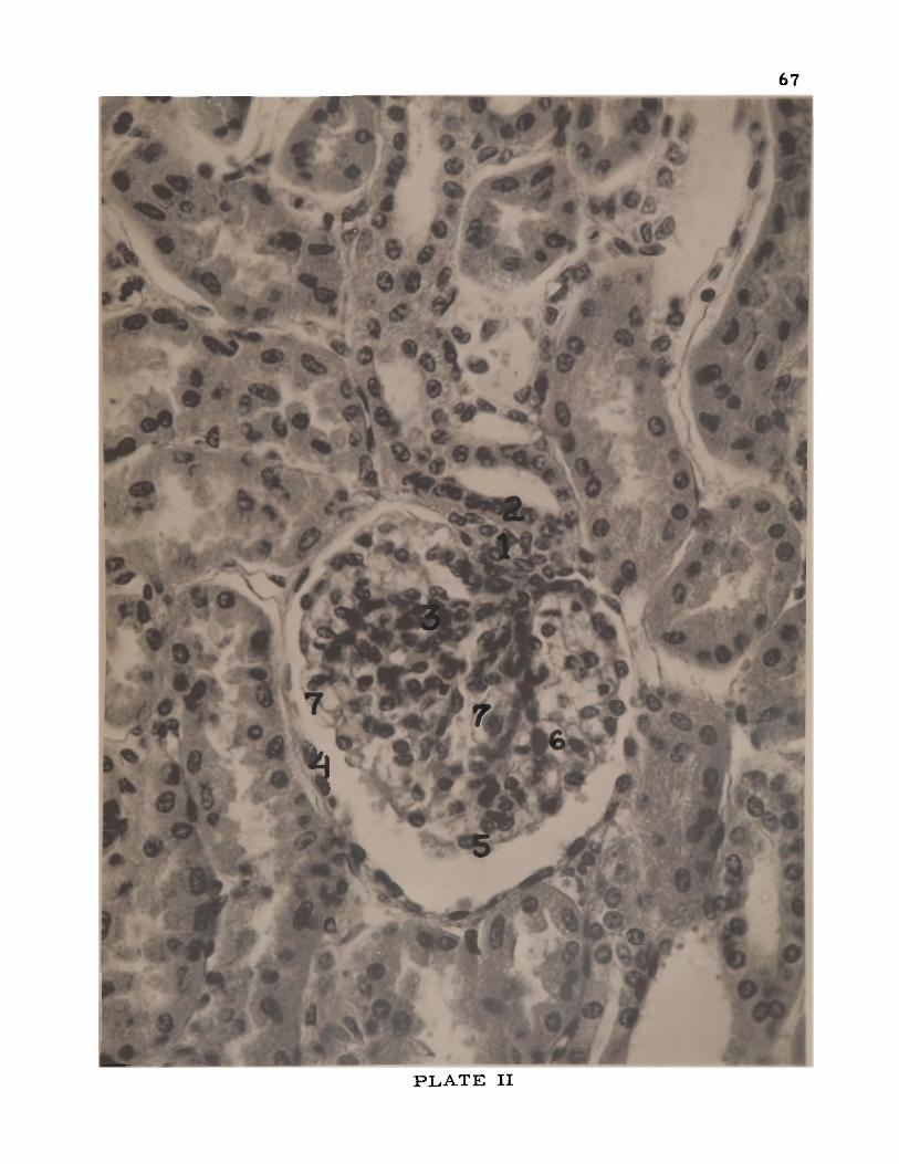
67
¥
**?.
<
*•6
%
*r©
p l a t e II
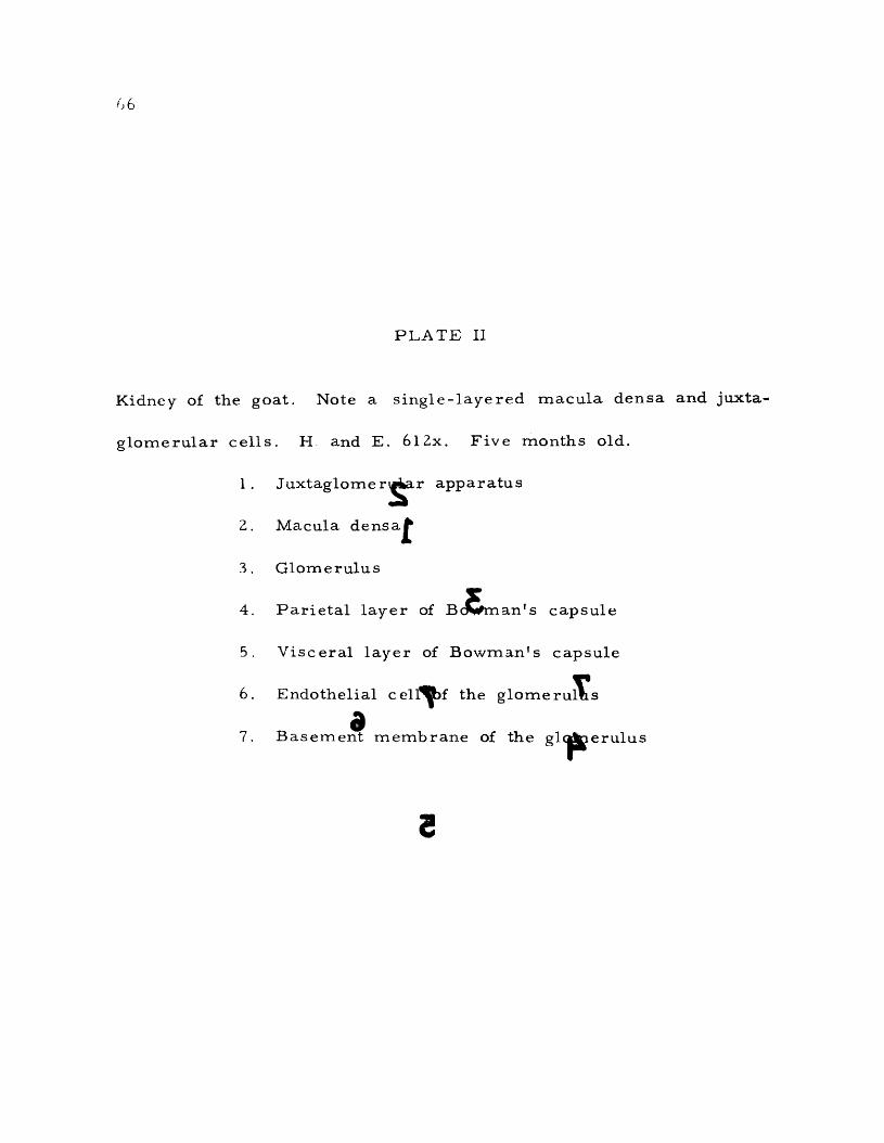
6 6
PLATE II
Kidney of the goat. Note a single-layered macula densa and juxta
glomerular cells. H and E. 6l2x. Five months old.
1. Juxtaglomerular apparatus
2. Macula densaj^
3. Glomerulus
4. Parietal layer of Bc^Sman's capsule
5. Visceral layer of Bowman's capsule
6. Endothelial cell^Btif the glome ru ^s
a7. Basement membrane of the glc^^erulus
3

0i * . ..
m *5 * *9
L I
19 ••«*. .-> " ^ \ <fSi
U " * • kv*' '
a
rI #
ad
P L A T E II

6 8
of being elongated, are rounded, and their cytoplasm, instead of con
taining myobibrils, usually contain granules. Moreover, the shape
of these cells is such that they resemble epithelial cells. These myo-
epithelioid cells are called juxtaglomerular cells. These cells thicken
the a r te r io la r wall and tend to form an asymmetrical cap on one side
of the glomerulus.
The nuclei of the juxtaglomerular cells in the pig kidney were
found to be round and somewhat deeply stained with hematoxylin and
eosin stains. They were found to be closely associated with the
macula densa. Along with these modified cells of the media some
of the spindle-shaped smooth muscle cells were also observed.
The structure of the juxtaglomerular apparatus of the horse,
ox, sheep, and cat were similar to that of the pig. In the case of
the goat and dog, the cells of the juxtaglomerular apparatus were
more numerous than those of the pig (Plate II).
Goormaghtigh (1951) stated that granules were absent in the
juxtaglomerular cells of the kidneys of the dog and man. According
to Maximow and Bloom (1952) the juxtaglomerular cells are absent
in children below the age of two years. In this investigation, juxta
glomerular cells were demonstrated in a two - month-old cat. Obser
vations were not made on young animals of the other species , so no
conclusions can be drawn regarding this point.

69
The proximal convoluted tubule. The proximal convoluted
tubule has the widest diameter of any portion of the nephron, and is
lined by simple cuboidal or truncated pyramidal cells having abun
dant protoplasm and large basally placed spherical nuclei. It is
characterized by the presence of a brush border, basal striations,
and the absence of clear cell boundaries. The brush border is the
cuticular covering of the free end of the cell lining the proximal
tubule. The basal striations are either the mitochondria or the
spaces between the mitochondria of the cells of the proximal tubule.
The proximal convoluted tubule pursues a looped and tortuous course
in the immediate vicinity of the renal corpuscle from which it orig
inates. This convoluted portion of the proximal tubule is called the
pars convoluta. The pars convoluta then enters the medullary ray,
becomes straight, and continues as the descending limb of the loop
of Henle. This straight portion of the proximal tubule is called
the pars recta, and constitutes the descending thick limb of Henle.
The s tructure of the pars recta is the same as that of the pars
convoluta of the proximal tubule.
The proximal tubule of the pig was found to have an average
diameter of 45p. It was lined by simple truncated pyramidal cells
whose average height was found to be 14p. The cytoplasm of the
cell was somewhat coarsely granular. These granules were more

70
or less evenly distributed, and hence basal striations were not d is
tinct. These grannies masked the cell boundaries, and therefore
they were not clear. The nuclei were usually basal, but in some
cells they were found to be situated in the middle or even towards
the apex of the cells. These nuclei were circular, and deeply
stained with hematoxylin and eosin stains. Each nucleus contained
a prominent nucleolus and some deeply stained chromatin granules.
The contour of the lumen of the proximal tubule was found to be
very i r regu lar . The brush border was prominent and formed a
separate cluster for each cell. These clusters were found to be
separated from one another (Plate III).
The general structures of the proximal tubule of the rest of
the animals under investigation were similar to those of the pig.
The diameter of the proximal tubule was greater in the
horse, ox, and goat; equal in the sheep; and smaller in the dog and
cat, in comparison to that of the pig (Table II). The height of the
cells lining the tubule was greater in the horse; equal in the cat;
and smaller in the ox, sheep, and dog, as compared to that of the
pig. The lumen of the proximal tubule was larger in the horse,
goat, ox, and sheep; almost equal in the dog; and smaller in the
cat. The contour of the lumen in the rest of the animals of this
investigation was more regular than that of the pig. Some of the

71
PLATE III
Kidney of the pig. Note the brash border forming a cluster on each
cell of the proximal tubule. H. and E. 2460x. Seven months old.
1 . Proximal tubule
2. Brush border
3. Distal tubule
4. Interstitial connective tissue cells
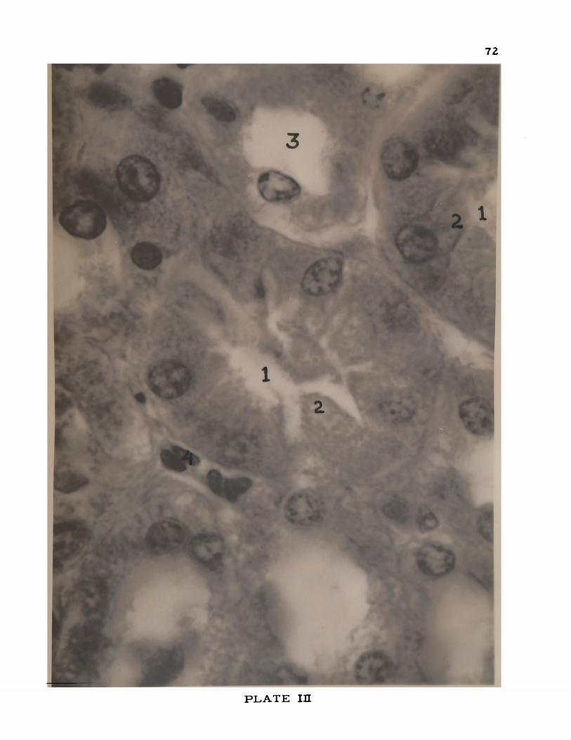
PLATE in

7 1
Z
PLATE III
Kidney of the pig. Note the brush border forming a cluster on each
cell of the proximal tubule. H. and E. 2460x. Seven months old.
1 . Proximal tubule
tZ . Brush border
3. Distal tubwlebSl
4. Interstitial connective tissue cells

72
P L A T E XU

73
nuclei of the cells of the proximal tubule were oval in the ox and
goat. The brush border was uniformly arranged along the apices
of the cells of the proximal tubule in the case of the rest of the
animals under investigation. They did not form clusters as was ob
served in the pig. Basal striations were more distinct in the dog
than in the pig. Fat globules were observed in the cells of the
proximal tubules of the cat and dog (Plate IV).
G rafflin (1942) noted a deposition of yellow to golden-brown
iron-containing pigment in the epithelium of the proximal tubule of
the rat. No such pigment was observed in any of the animals under
investigation.
The loop of Henle. The loop of Henle consists of a descend
ing and an ascending arm connected by a sharp bend or curve. It
is divided into a thick and a thin segment. The thick descending
arm of the loop of Henle is formed by the pars recta of the proxi
mal tubule; the thick ascending arm is formed by the pars recta
of the distal convoluted tubule. The loop of Henle of those tubules
arising from the outer cortical renal corpuscles passes only a re la
tively short distance into the medulla, the thin segment of this loop
being very short. On the other hand, the loop of Henle of the tubules
of the juxtamedullary renal corpuscle descends deeply into the medulla
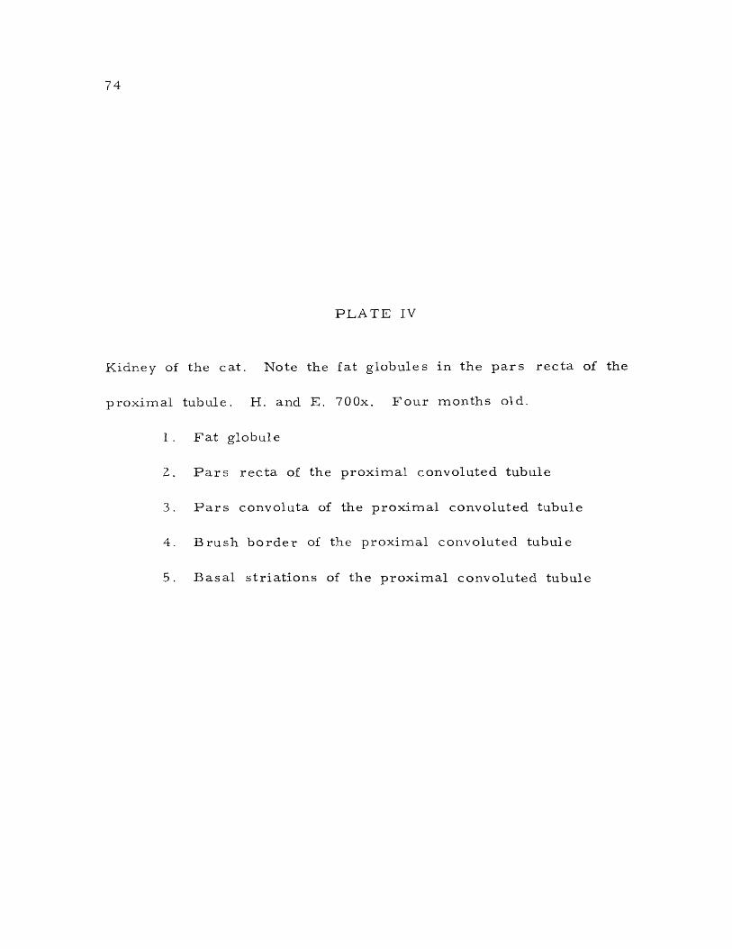
7 4
PLATE IV
Kidney of the cat. Note the fat globules in the pars recta of the
proximal tubule. H. and E. 700x. Four months old.
1 . Fat globule
2. Pars recta of the proximal convoluted tubule
3. Pars convoluta of the proximal convoluted tubule
4. Brush border of the proximal convoluted tubule
5. Basal striations of the proximal convoluted tubule

PLATE IV

7 4
IPLATE IV
Kidney of the cat. Note the fat globules in the pars recta of theIproximal tubule. H. and E. 700x. Four months old
S1 . Fat globule £
2. Pars recta of the proximal convoluted tubule
3. Pars convoluta of the proximal convoluted tubule
4. Brush border of the proximal convoluted tubule
5. Basal striations of the proximal convoluted tubule

75
P L A T E IV
76
often as far as the papilla. The greater part of the loop within the
medulla is made up of the thin segment. The epithelial lining of
the descending thick segment of Henle's loop resembles that of the
proximal tubule; the epithelial lining of the ascending tubule re sem
bles that of the distal tubule. The thin segment is smaller in di
ameter than either the proximal or distal tubules and is made up
of a flattened epithelium with nuclei which bulge into the lumen. The
protoplasm is less granular and therefore stains less eosinophilic
than either proximal or distal tubule. The transition from the thick
to thin segment of Henle's loop is abrupt.
The diameter of the thin segment of Henle's loop in the pig
kidney was found to be Z 3 \ x . It was lined by flattened epithelium
having an average height of 5p. The cells were markedly less
eosinophilic than those of the proximal tubule. The cytoplasm was
more or less clear with a spherical nucleus bulging into the lumen
of Henle's loop. This bulging of the nucleus gave a wavy contour
to the lumen of the tubule . .A brush border was absent and the
basal striations indistinct.
The general s tructure of the thin limb of Henle's loop of
all animals under investigation was similar to that of the pig. The
following few exceptions were noted:

7 7
The diameter of the thin limb of Henle's loop was greater in
the horse, ox, and. goat; and smaller in the sheep, dog, and cat, as
compared to that of the pig (Table II). The cell height was slightly
greater in the horse, and somewhat smaller in the sheep and cat,
the other animals having cells of equal height to those of the pig.
The lumen of the loop of Henle was wider than that of the pig, in
the horse, ox, sheep, and goat, and narrower in the dog, the cat
being equal.
The distal convoluted tubule. The distal convoluted tubule is
composed of two segments, the pars recta and the pars convoluta.
The pars recta forms the thick ascending limb of Henle's loop. The
thick segment of the ascending loop of Henle leaves the medullary
ray, becomes convoluted (pars convoluta), and returns to the glomer
ulus from which the nephron has its origin. The distal tubule, r e
turning to the neighborhood of the glomerulus, makes tangential con
tact over an extensive region with the afferent arteriole. In this
region of contact the nuclei of the distal convoluted tubule become
densely crowded and constitute the macula densa. There are fewer
convolutions in the distal than in the proximal tubule. The cells in
the distal tubule are cuboidal with spherical or ovoid nuclei. The
cells in the distal tubule are much less broad than those that line

78
Ihe proximal tubule and hence the nuclei appear more closely spaced
in the distal tubule. Cytoplasm is not nearly so acidophilic as the
proximal tubule. Faint basal striations are present although a brush
border is lacking in the distal tubule.
The diameter of the distal tubule in the pig kidney was found
to be 36p. The tubule was lined by cuboidal epithelium 8p in height.
The lumen was la rge r and its contour less ir regular than that of
the proximal convoluted tubule. The cytoplasm was granular and
less eosinophilic; the granules were finer than those of the proxi
mal tubule. The nuclei were spherical and basally placed although
a few oval nuclei were also noticed. These nuclei were closer to
each other than those of the proximal tubule. The nucleus presented
one or two prominent nucleoli and darkly stained coarse chromatin
granules. The cell boundaries were not clear. The brush border
was absent and the basal striations were not very distinct.
Except for a few differences, the distal tubule of all animals
under investigation was similar to that of the pig. Compared with
it, the diameter of the distal tubule was larger in the horse, ox,
and goat; equal in the sheep; and smaller in the dog and cat. By
a s imilar comparison the epithelial height was greater in the horse,
equal in the ox and goat; and smaller in the sheep, dog, and cat.

79
The lumen of the distal tubule was greater in the horse, ox, sheep,
and goat; and smaller in the dog and cat.
The macula densa in the pig constituted about half of the wall
of the distal convoluted tubule as seen in cross section. The cells
forming the macula densa were single-layered and appeared to be
slightly ta ller than the res t of the cells of the distal tubule. The
cytoplasm was faintly stained around the nucleus. The cells of the
macula densa were closely associated with the juxtaglomerular cells.
The cytoplasm of the cells contained very few fine granules. The
nucleus was spherical and contained a dark stained nucleolus and
coarse chromatin granules.
The macula densa in all animals under investigation was simi
lar to that of the pig except in the horse, which presented some spe
cial features. The macula densa in the horse kidney was stratified
although the stratifications were not distinct, as the nuclei were very
ir regularly crowded (Plate V). The height of the stratified epithelium
of the macula densa appeared to be more than double that of the
other cells lining the same tubule. The cells of the macula densa
were almost nongranular and did not take the stain, especially to
wards the base. The change from simple to stratified epithelium
of the macula densa of the horse was abrupt.

80
PLATE V
Kidney of the horse. Note the stratified macula densa. H. and E
400x. Adult.
1. Macula densa
Z. Straight collecting tubule

81
* £ > «
*3 <91< ;£jV
M*S2s' ■’
P L A T E V

8 0
Kidney
400x.
PLATE V
of the* horse. Note the stratified macula densa. H. and E1
Adult.
1 . Macula densa
Z, Straight collecting tubule
I

81
f t
v-7.T
ft
P L A T E V
82
McManus (1943) and Okkels (1950) dem onstrated the r e v e r se d
p o s it io n of the G olgi e lem en t in the m acula densa of the cat and
m an. G olgi e lem en ts w ere not studied in this work.
The co llec tin g tubule
The co llec tin g tubule c o n s is ts of the arched or initial c o l le c t
ing tubule, the straight co llecting tubule, and the papillary duct or
duct of B e l l in i .
The d ista l convoluted tubule i s joined d irectly to an arched
co llec tin g tubule, into which it d isch a rg es . T hese arched tubules are
short s tem s that branch from the s ides of the straight co llecting
tubules loca ted in the m edullary ray. The arched tubules are situ
ated in the co r t ica l labyrinth.
The stra ight co llecting tubules l ie side by side and make up
m o st of the substance of the pyram ids. The straight tubules join
with one another in s u c c e s s iv e fash ion in the pyram ids to form
f in a lly the la r g e papillary ducts. The d iam eter in c r e a se s as the
tubules jo in and it r ea ch es a m axim um in the papillary ducts. The
co llec tin g tubules are nearly always very regular in outline when
v iew ed in c r o s s section . The epithelial c e l ls lining the arched tu
bu les and the proxim al portion of the straight tubule are s tr ic t ly
83
cuboidal. The cells increase in height as the tubules increase in
diamete r .
The papillary ducts empty into the minor calyces through the
foramina situated on the surface of the papilla.
The arched collecting tubule. The arched collecting tubule
in the pig kidney was observed to have a greater diameter than the
distal tubule. It was lined by a simple cuboidal epithelium with quite
distinct cell boundaries. The lumen of the arched tubule was more
regular and wider than that of the distal tubule. The cytoplasm of
the cells lining the arched tubule was faintly stained and devoid of
characteris tic granules of the nephron. The nuclei were either
spherical or oval, and basally placed. Each nucleus contained a
nucleolus and some deeply stained chromatin granules.
Except for the diameter and cell height, other structures of
the arched collecting tubules of the horse, ox, sheep, goat, dog, and
cat resembled that of the pig- The diameter was greater in the
horse and ox, and smaller in the sheep, goat, dog, and cat. The
cell height was greater in the horse and smaller in the rest of the
animal s .
The straight collecting tubule. The general structure of the
straight collecting tubule of the pig kidney was similar to that of

84
the arched collecting tubule. The diameter of the straight collecting
tubule was 50p and the cell height was 14[jl. The lumen had a regu
lar contour and was wider than the arched collecting tubule. The
straight collecting tubule was lined by a simple cuboidal epithelium
which became somewhat wider and taller towards the papillary duct.
The cytoplasm was clear, faintly stained, and free from granules.
The nuclei were larger than the arched collecting tubule and each
contained a deeply stained nucleolus and a few coarse chromatin
granules. The cell boundaries were distinct.
The general structure of the straight collecting tubule in all
animals under investigation was similar to that of the pig. The
following differences were noted:
The diameter of the straight collecting tubule was greater in
the horse and ox; and smaller in the sheep, goat, dog, and cat
(Table II). The cell height was greater in the horse; and smaller
in the ox, sheep, goat, dog, and cat. The lumen of the tubule was
greater in the horse, ox, and cat; and smaller in the sheep, goat,
and dog. The cell boundaries were more distinct in the horse, ox,
goat, and dog; and less distinct in the sheep and cat.
Trautmann and Fiebiger (1952) noted the presence of numerous
fat droplets in the straight collecting tubules especially that of

8 5
the older domestic animals. No such fat droplets were observed in
the straight collecting tubules of the animals under investigation.
The papilla ry duct. The general structure of the papillary
duct of the pig kidney resembled that of the straight collecting tu
bule. The diameter and epithelial height of the papillary duct were
greater than those of the straight collecting tubule. The papillary
duct was lined by a simple tall columnar epithelium which became
transitional near the opening of the duct on the papilla. This t ran
sitional epithelium of the papillary duct continued as the epithelium
of the papilla. The change from simple to transitional type of epi
thelium in the papillary duct was found to be abrupt. The diameter
and the epithelial height of the papillary duct were found to be 9bp
and Zip, respectively (Table II). The tall columnar cells of the
papillary duct were faintly stained and nongranular. The nuclei were
large, spherical, and situated almost in the center of the cell. A
dark-staining nucleolus and chromatin granules were observed in
each nucleus. The cell boundaries, were distinct and the contour of
the lumen was regu la r .
The general structure of the papillary duct in all animals in
this investigation resembled that of the pig. The following exceptions
were noted:

8 6
The diameter of the papillary duct was greater in the horse
and smaller in the rest of the animals under investigation. The
epithelial height was greater in the horse, ox, and goat, and smaller
in the sheep, dog, and cat. In the horse, ox, goat, and sheep the
papillary duct was lined by a transitional epithelium for a greater
distance from its opening on the papilla than that of the pig. The
papillary duct of the dog kidney was lined by a simple columnar epi
thelium throughout its course. At the junction of the simple columnar
epithelium of the papillary duct and the transitional epithelium of
the papilla, a beaklike process of the transitional epithelium was
found in longitudinal section (Plate VI). The extension of the t ran
sitional epithelium in the papillary duct of the cat kidney was found
to be almost equal to that of the pig.
Langham and Hallman (1939) considered the transitional epi
thelium of the papillary duct in the bovine kidney as pathological.
This investigation has revealed that the transitional epithelium of
the papillary duct in bovine kidney is a normal one (Plate VII).
Langham et a l . (1942) found that only tall columnar cells lined the
papillary duct of the bovine kidney. From this study it has been
determined that transitional epithelium lines the papillary duct of
the bovine kidney for a distance of about 1500p from the opening of

8 7
PLATE VI
Papillary ducts in the dog kidney. H. and E. 320x. One year old.
1 . Simple columnar epithelium lining papillary ducts
2. Beaklike process at opening of the papillary duct
3. Oblique section of the papillary duct opening
4. Pelvis

^ * ’ A. '? A-«.t' A- • <* • >
'■mL* «* ** ____■- ■ “ * 3 _ ^ g i g - ' •- v ‘ ' /o • '•, • •" ■- v®s£
■ ’ • ** * ,-• •• * * . V • .% ■ - - <
v j *
4
£ & * . * v A • : ; f tr ¥ ■ * '
.':.7v • -

87
PLATE VI
Papillary ducts in the dog kidney. H. and E. 320x. One year old,
1 . Simple columnar epithelium lining papillary ducts
2. Beaklike process at opening of the papillary duct
3. Oblique section of the papillary duct opening
4. Pelvis
t

88
* j *
• ■SK. » d t ft ' J ® * 1 j * ^ A" "~ ^ / t 4 «>- *; • - ^ .3 ? 5* > * ■
f.-j'v *;s-- rasagfiHh iI ^ C * ^ V* A A-^^V ;- V.,*> %-• C *» * •» </ *V^ • :^#i(» • . • ’> r ^' Nfc. ' vS *> . . * * ~*r»r. a z - - .*
’<v
H v « > - *> - . . * * s j
BBS^— .., - c <*' '" i/ /-C— ' *
y j ' V e~ ^ 4 .«,« , r 3 K % r. ^ o ^ - ••
| V ■ ' ' . % ? < ; > r ^ r . - C *b . * 5 . , C ) .. '■' 7 ■ m oaCT
- <£-* v - n %
* <«/ z * ® • - * r t ^ * Z & "
7*5fc
l. * $ & ^V»S«I **%,
uV._ • «T '*5*$»>* • v ^ . ^ - sp«^»«fca> * 4
^ ^ *< v - ' - ; * ■ «’ W * . •> ^ C*‘^ I N
v , »>- **; - % . • a gJtg, • -v * « v& v \ V
X . A - » r ^ ^ ^
W ' *
P L A T E V I

89
PLATE VII
The papillary ducts in the ox kidney. H. and E. 320x. Nineteen
months old.
1. Transitional epithelium
2. Opening of papillary duct on the papilla

90
« -v • •!;. f *. ~ vVSi; v * C V %* t V • , v % < ' A
• ; / : ' . 4, v& " fck r1 - , ^ ' ^ v*j& K r „ «■'£*»* 1 Y * » - ‘ » ' * *••«••• . \
ru« w >?C >’> %••K 't*r&% ' I -; *> > $: i rA \ *> ' ** r • ' V '. i > * k ,
t ' ' . f V . ~ ^ ;► } » . , » W - - _' * >!% • ^ j»,‘ . v . -* .:*:
V 1>
■■ ^ ‘
* 2 j ‘v* *
**:■\
v* :’■«
.
**' r:^ ' i f #
*^4•» T * . '% . * 9* » •-' . V 4 * * V .. ,% Y ®
, \ > I % .^ ^ X v * * I I % ' *
* '*;■*
F*
V>. . :•••, >'••., $L* t v->— %?.-%*? S&K vfkllt *■» * w* -v .<RteV ?-# v* * 1 \ ' ■ • V • A
A » & ' '
i k ; -'
- * f In -l* *-V*V> >w•' *<C
4 v - ■>J V*C w
*2 0
* v ♦>tou-v
I vs ♦
-i«/’
j v r ® S P " ' - ' • '- 1 /« !?i' • r f 4mss., v + >*
■ I w 8 l f ] •aT.v4H•■*• V^;„. ^ r r : ,
# ' v . . ,# y j g p
i>
N 11f* *
' \ M«#.
P L A T E V II

89
tPLATE VII
The papillary ducts in the ox kidney. H. and E. 320x, Nineteen
rn onths old. ^
1 Transitional epithelium
2. Op ening of papillary duct on the papilla
r

90
i r i c * —
Sr>' , v
v * ^
T *
r* ’ ^ v ’ , vTV ^. .* 1 * C „ I&?, . , 7 u* *’ .„v » .w - s v . w . \
I -v^ V i ^"*% • t * " •" *
'•< 4 %•*V* -a ‘ * • -«
A r.40
« k ' ’ .«, J L \ V ,- V \ ~ ^ ' - i<v* V * t; * ^ *’~t" "* I "V - ?i ’ “ J. v ' ^ 4 **
*
".■/ .‘ *. ' W t f W j f o v ’•*v ■ * *' X *v5 ~ * ^ -.v .v .
' * 1 $ J T \L . — V •; '
*v'' . * t ' i v ’35 . ' *>-
• A * L * * *
. V " * f * * 4 * % >VjSj& <V C **% / V > VJ ;gj .* ^«-/3*.;i ^ » v &f1 >
^ ’'V) -' /<\» i * \ ■■ ®w 2i ti >t!i> -V vLv* 5o ^ ■**
, - ’V ! \r* *• .$&.» <> 5 > pi '» ' • v * \ . . r » v —-» > ? 3' • S v V ' -® SB ,:T»* 4 * i»sP• ^ *> 7 ■■? * j w ^ : * *■>K «< tfcl
TS'- "■ ' «
m 1
w V sm * ,r v ; >y ■
J & * ( '#
-«. ' r.'.'" ** < ' *9S& * -J“v v . 4<
>/#*..
pw S' i
■«*■» ^
- * > «
? V L Vj . s t i ■ ■?ftp <'k , v» i ‘4R, 3M41 V V. ^•wv - j«IBSk4J 'J®
« ,.v # • .* ^-fciV • -.
^ ' V , v4 V .4 . » ’*■
^ ^*r • ' I \SV 1
v *? \ ; c*v :
>‘\ . ' 4
, ty t* ■ *j V >
1 4> J, Of f j l ; » ■'■*» .5 / hTa * '*K S jf v :"
H ' V\ l * ° * , *r ® ^
^ > : ? / t . “ ....s ' % - . a » V J / ' f * ' *
. r 1 , 9*- *
%>- to' . w •
o
> > P p H
"W ' I
rm > f % %
P L A T E V n

91
the papillary duct at which point it changes to simple columnar epi
thelium .
Duran-Jorda (1953) could not observe the openings of the
papillary ducts in the cat kidney and hence came to the conclusion
that urine was dialysed through the papillary epithelium. The open
ings of the papillary duct in the cat kidney were observed in this
investigation (Plate VIII).
The basement membrane of the uriniferous tubule
The renal tubules and the excretory ducts are enveloped ex
ternally by a well-developed and distinct basement membrane.
The presence of the basement membrane of the uriniferous
tubule was observed in the pig kidney although details of its finer
s tructures could not be studied with the ordinary light microscope.
No difference was noted in the basement membrane of the uriniferous
tubules of other animals of this investigation.
The Papilla
The papilla is the apex of the renal pyramid, the base of
which is located at the corticomedullary junction of the kidney. The
surface of the papilla is perforated by foramina which are the open
ings of the papillary ducts. This perforated area of the papilla is
92
PLATE VIII
The papillary ducts in the cat kidney. H. and E. 220x. Seven weeks
old.
1 . Opening of papillary ducts
2. Papilla
3. Junction of transitional with simple columnar epithelium
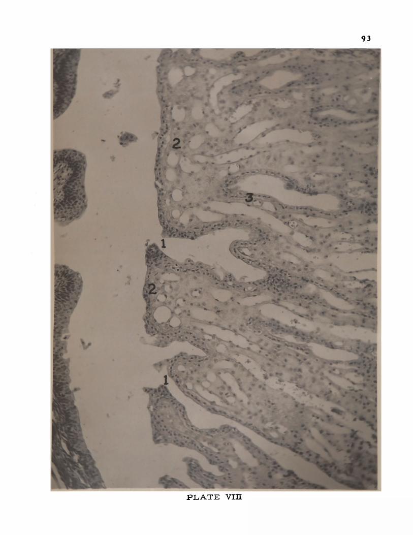
93
P L A T E Y in
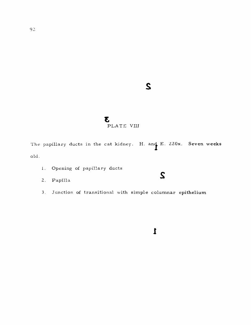
92
S
EPLATE VIII
The papillary ducts in the cat kidney. H. an j E. 220x. Seven weeks
old.
1 . Opening of papillary ducts
S2. Papilla
3. Junction of transitional with simple columnar epithelium
I

93
yi f
y* /V #*
v .
3 Vv
c • » 4 %■ s f^
<5 **• *’ ' * 4
3* . ^ ^
& * * ' .: ■ ' n ' A , . - -**»Sr>* *- /» » « •_ ^ . . r, — •v,
’V?.®-’ - \ £ *
y ^ S _ ' / - ^ j s 3 • * • :Q -* f% . A « x * * m _ « S f-4; * *’ <*•*■
hi' ii.<? *
-X >• -,•jri"* * * ’**►
jflr
T-'' — ' - * ■ % * J* •. r - - 2 .4
*s£u * * ~ *• * §•• *’* ■'£*•. -» .rf * >5
^ -*
■o'*. \ «✓ A
*>1■ ■-. * \ , ** t
^ ^ lEi-« • «
r" % * *
^ 1 .«
y ? ;% . • . . . • • . ' %>; . • v - • / . ■ ' > * .V*. : S s. _ *'«%.. .A « - _ -s* *'
- ^ > ♦
~ *1* * 'f^ j5 v . “ , I s‘ \ * v > ; .
^ *>
P L A T E VIH
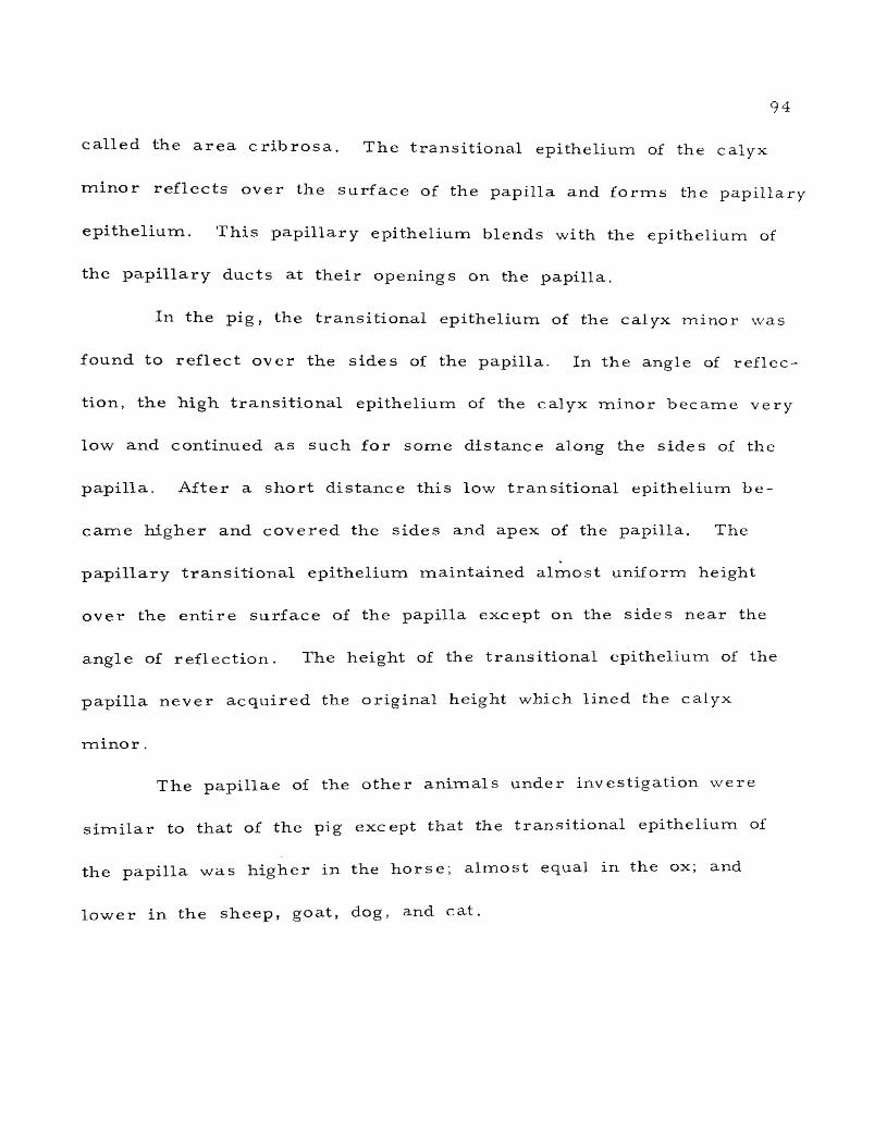
94
called the area cribrosa . The transitional epithelium of the calyx
rmnor reflects over the surface of the papilla and forms the papillary
epithelium. This papillary epithelium blends with the epithelium of
the papillary ducts at their openings on the papilla.
In the pig, the transitional epithelium of the calyx minor was
found to reflect over the sides of the papilla. In the angle of reflec
tion, the high transitional epithelium of the calyx minor became very
low and continued as such for some distance along the sides of the
papilla. After a short distance this low transitional epithelium be
came higher and covered the sides and apex of the papilla. The
papillary transitional epithelium maintained almost uniform height
over the entire surface of the papilla except on the sides near the
angle of reflection. The height of the transitional epithelium of the
papilla never acquired the original height which lined the calyx
minor .
The papillae of the other animals under investigation were
similar to that of the pig except that the transitional epithelium of
the papilla was higher in the horse; almost equal in the ox; and
lower in the sheep, goat, dog, and cat.

95
The Interstitial Connective Tissue
The in tersti t ial connective tissue is very scanty in the nor
mal mammalian kidney and consists of delicate reticular tissue which
forms a fine network between the tubules. The tissue is more abun
dant in the papillary portion of the medulla. Collagenous and e las
tic fibers are found only in the walls of the larger blood vessels.
Some loose connective tissue is present between the papillary ducts.
A. few strands of connective tissue enter the glomerulus along the
blood vessels. Fibroblasts and histiocytes are also found in the
in tersti tial space of the mammalian kidney. Proliferation of this
connective tissue throughout the kidney may play an important part
in disease.
The intersti tial space of the pig kidney was mainly occupied
by reticular fibers which extended from the capsule to the apex of
the papilla (Plate IX). Reticular fibers were found to be more
numerous near the blood vessels and in the interstitial spaces of
the medulla. Collagenous fibers were also found to surround the
large blood vessels especially at the corticomedullary junction. A
few very fine collagenous fibers were observed to invest the urinifer-
ous tubules and the Bowman’s capsule of the kidney. Fibroblasts,
histiocytes, and mast cells were also noted in the interstitial space
of the pig kidney.

96
PLATE IX
Kidney of the pig stained for reticular connective tissue. Note the
distribution of reticular fibers in the interstitial spaces of the paren
chyma and the glomerulus. Silver Stain (Gridley modification). 460x.
Seven months old.

97
P L A T E DC

96
PLATE IX
Kidney of the pig stained for reticular connective tissue. Note the
distribution of reticular fibers in the interstitial spaces of the paren
chyma and the glomerulus. Silver Stain (Gridley modification). 460x.
Seven months old.

PLATE IX

98
The in tersti tial connective tissue of the other animals under
investigation was similar to that of the pig except that the mast cells
were absent in the dog and cat.
The intertubular cell groups or BecherTs cells (Plate X)
The presence of intertubular cell groups in the human kidney
was reported by Schloss (1946), Neumann (1949), and Becher (1949,
195 0). These intertubular cell groups are situated in the human
kidney at the points where the connective tissue is abundant, in the
immediate neighborhood of the afferent arterio les, and other cortical
ar te r ia l vessels. These intertubular cell groups are also called
' 'paraportal cell i s le ts ’1 by Becher, The presence of intertubular
cell groups has not yet been reported in the domestic animals.
In the kidney of the pig, these intertubular cell groups were
observed in the interstitial spaces of the cortical region between
the uriniferous tubules. They were also found in the close vicinity
of Bowman's capsule and around the cortical ar teries . Those su r
rounding the ar ter ies usually were found to be larger. Some of the
smaller groups were found to be encapsulated. The cells did not
present a regular shape. Most of the cells were polygonal except a
few which were found to be oval. The cytoplasm was faintly eosino
philic and nongranular. The nucleus was smaller and darker than

99
PLATE X
Kidney of the ox. H. and E. 634x. Six years old.
1. Intertubular cell group or Becher's cells
Z. Interlobar artery
3. Proximal convoluted tubule
4. Distal convoluted tubule
5. Thin limb of Henlers loop

100
.a m i
&s
■*
: « * » ' ' 3
rr °
m \X '
9+6> » ■ A
O ko/4T«
% ,« * o '*V I •. *•<v
v &?•» **
•' • c * * .*■ 5 4 ’i
A W ZF* '
‘ * 1
o 4 » ■ ( ■ | . l ■■■ + < 9
\ \% h $«c
& <*
P L A T E X

99
2 £
PLATE X
Kidney of the ox. and E. 634x. Six years old,
1. Intertubular cell group or Becher's cells
2. Interlobar artery
3. Proximal convoluted tubul^
4. Distal convoluted tubule
5. Thin limb >f Henle's loop

100
** * i > * t i
•;>*>. . / - <» *> ^ .->• , . a ( r ■•-:£
“ . * *k;" f t ai* * <a T > - ; I -k * r J ' • .
^ ^ 3 jET «* Si®** W G * * T 7“ f_ >s? * > « A ‘m V * * ; w f to • £&%* *vA« X&w-'Jf *•'
a *<?&.V *4_rV
# - '
i*w s?.»fe««M MR -■
r - \ - •' ©SSP Sl «? , * 0 * ^
' /r* tfl *:3 *Ea r . *Pv®*© i. $*
ft^*" _ .. .
»l» - I'wm
m
&*P L A T E X

101
that of the surrounding tubules. The nuclei of these cells were gen
erally spherical, although a few oval nuclei were also noticed. They
were located either in the center or towards one corner of the cell.
The nucleus presented a characteristic appearance. Four or five
c re sc ent-shaped dark nucleoli were found to be present along the
periphery of the nucleus in close approximation to each other and
the nuclear membrane. A large deeply stained nucleolus was situ
ated at the center of the nucleus. The space between these nucleoli
was found to be unstained and free from chromatin granules.
These cell groups were found to be more numerous and better
developed in the ox and horse than those of the pig. In the ox, the
cells were more eosinophilic and contained more cytoplasm than
that of the pig (Plate X). These cell groups were less developed
in the sheep, goat, and dog, and absent in the cat.
Cell unit of Goormaghtigh or socleplasmodium (Frontispiece)
A cluster of small cells with pale nuclei between the macula
densa and the glomerulus proper, in the concavity between the affer
ent and the efferent arterio les of the mammalian kidneys can be
seen in microscopic sections. This cluster of small cells is called
the cell unit of Goormaghtigh, or Goormaghtigh* s cells.

1 0 2
In the pig kidney these cells were found to be closely asso
ciated with the juxtaglomerular cells and the cells of the macula
densa. Goormaghtigh cells of other animals of this investigation
were found to be similar to that of the pig.

SUMMARY AND CONCLUSIONS
1. Comparative microscopic studies were made on the kid
neys of sixty-nine domestic animals of seven different species, the
pig being chosen as a type. Tissues were routinely fixed in F.A.A.,
sectioned at seven microns, and stained with hematoxylin and eosin
stains. Additional stains were used to demonstrate special struc
tures.
2. The tunica fibrosa of the capsule of the kidney of the
domestic animals in this investigation was found to consist of two
layers, an outer and an inner, with the exception of the cat, where
the kidney capsule had only one layer, the inner layer being absent.
The outer layer of the tunica fibrosa consisted of dense collagenous
and a few elastic fibers, and the inner layer was formed by loose
collagenous and reticular fibers, with smooth muscle fibers present
in all animals except the cat. In the sheep and goat, and to a lesser
extent in the ox, the muscle fibers were very numerous, and appeared
to form a distinct muscular layer. No reports have been found which
mention the presence of smooth muscle cells in the capsule of horse,
goat, pig, and dog.
103
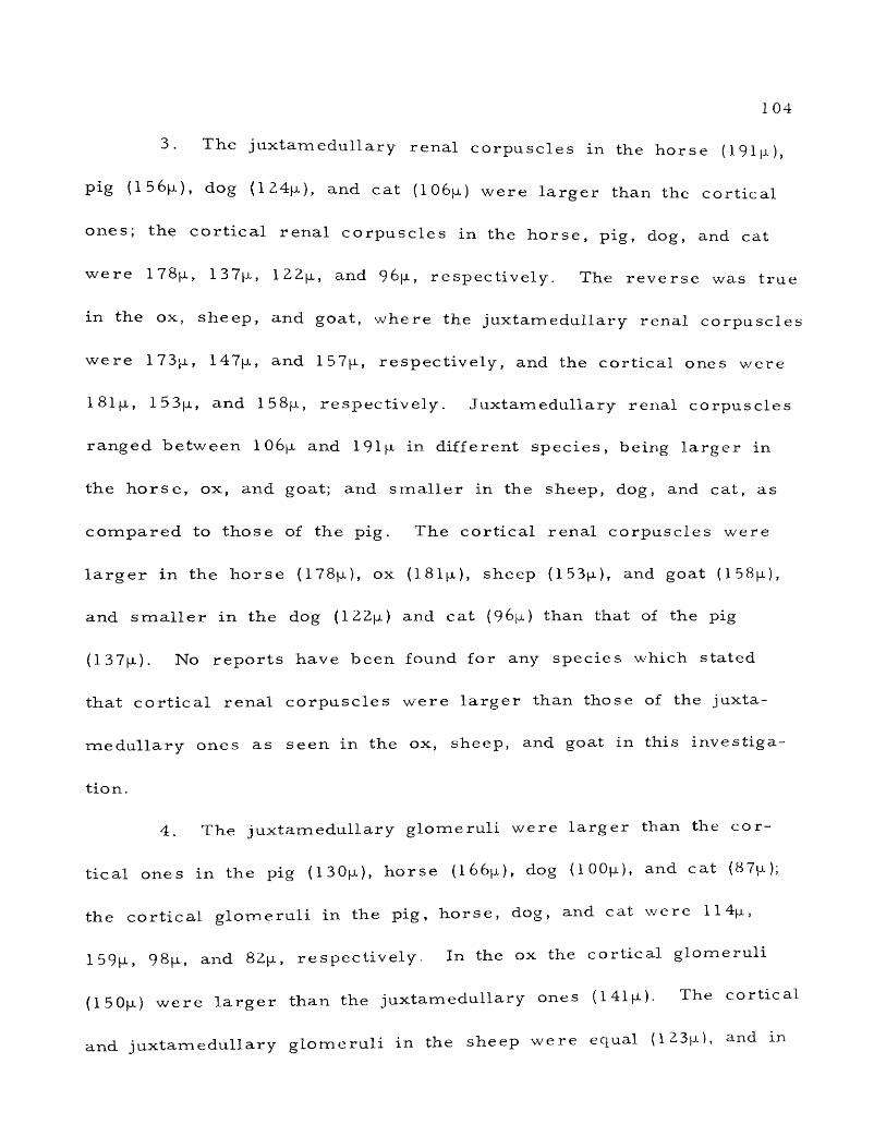
1 04
3. The juxtamedullary renal corpuscles in the horse (191 jjl ),
pig (1 56(a), dog (124p), and cat (10 6 jjl ) were larger than the cortical
ones, the cortical renal corpuscles in the horse, pig, dog, and cat
were 178(a, 137(a, IZZp, and 96[a, respectively. The reverse was true
in the ox, sheep, and goat, where the juxtamedullary renal corpuscles
were 173p, 147(a, and 157ja, respectively, and the cortical ones were
18lp, 153[a, and 158p, respectively. Juxtamedullary renal corpuscles
ranged between 106p and 191 |a in different species, being larger in
the horse, ox, and goat; and smaller in the sheep, dog, and cat, as
compared to those of the pig. The cortical renal corpuscles were
larger in the horse (178(a), ox (181 jjl ), sheep (153(a), and goat (158(a),
and smaller in the dog (122(a) and cat (96(a) than that of the pig
(137|u). N o reports have been found for any species which stated
that cortical renal corpuscles were larger than those of the juxta
medullary ones as seen in the ox, sheep, and goat in this investiga
tion.
4. The juxtamedullary glomeruli were larger than the cor
tical ones in the pig (130(a), horse (l66p), dog (100(a), and cat (87(a),
the cortical glomeruli in the pig, horse, dog, and cat were 11 4p,
159p> 98p, and 82p, respectively. In the ox the cortical glomeruli
(150(a) were larger than the juxtamedullary ones (141(a). The cortical
and juxtamedullary glomeruli in the sheep were equal (123(a), and m

1 05
the goat they were 127m- and 1 2 6 p , respectively. The juxtamedullary
glomeruli of the horse ( 1 6 6 p ) and ox (141(j.) were larger than those
of the pig ( 1 3 0 p ) , while in the sheep (1 2 3 p ) , goat ( 1 2 6 jj.), dog (lOOp),
and cat (87(j.) they were smaller. The cortical glomeruli of the
horse (1 59 p-), ox (150(j.), sheep (123(0.), and goat (127(o) were larger
than those of the pig (114[j.), while in the dog (9 8p ) and cat (82(x)
they were smaller.
5. The intercapillary space of the glomerulus of all animals
under investigation contained small amounts of collagenous and ret ic
ular fibers along with fibroblasts and histiocytes.
6. The capsular space of Bowman’s capsule was larger in
the goat, ox, and sheep; smaller in the horse and cat; and almost
equal in the dog, as compared to that of the pig.
7 . The juxtaglomerular apparatus in all animals in this
investigation contained some spindle-shaped smooth muscle cells
of the media of the afferent arteriole along with the usual myoepi-
thelioid cells, The juxtaglomerular apparatus of the horse, ox, sheep,
and cat was s imilar to that of the pig. In the goat and dog, the
juxtaglomerular cells were more numerous and better developed.
8. In comparison with the pig (45p), the diameter of the
proximal convoluted tubule was greater in the horse (56p), ox (50p),
and goat (48p); equal in the sheep (45p); and smaller in the dog (39p)

1 0 6
and cat (41p). The height of the cells lining the tubule (pig, 14p)
was g rea ter in the horse (15p); equal in the cat (14p); and smaller
in the ox (13p), sheep (1 1 ja.), goat (lip), and dog (lip). The brush
border was found to form a cluster from each individual cell in the
proximal tubule of the pig kidney, whereas in other animals of this
investigation they were uniformly arranged along the luminal border
of the cells. The basal striations were more distinct in the dog.
Fat globules were observed in the proximal tubule of the cat and
dog.
9. The diameter of the thin segment of the loop of Henle
ranged between 2lp and 30p in different species, being greater than
the pig in the horse, ox, and goat; and smaller than the pig in the
sheep, dog, and cat. The lumen was wider than the pig in the horse,
ox, sheep, and goat; equal to it in the cat; and narrower in the dog.
10. The diameter of the distal tubule ranged between 30p
and 5 Op in different species, being larger in the horse, ox, and goat;
equal to the pig in the. sheep; and smaller in the dog and cat. Com
pared to that of the pig (8p), the epithelial height was greater in the
horse (9p); equal in the ox (8p) and goat (8p); and smaller in the
sheep (7p), dog (7p), and cat (7p). The lumen of the distal tubule was
g reater in the horse, ox, sheep, and goat; and smaller in the dog and
c a t .

1 0 7
11. The macula densa in all animals in this investigation
was single-layered except in the horse, in which it was found to be
stratified and contained more faintly stained cells. No reports were
found regarding the stratified nature of the macula densa of the
h o r s e .
12. Compared with the pig (50p), the diameter of the straight
collecting tubule was greater in the horse (66p) and ox (53p); and
smaller in the sheep (39p), goat (42p), dog (37p), and cat (37p).
The cell height was equal in the horse (14p); and smaller in the
ox (12p), sheep (lOp), goat (12p), dog (9p), and cat (5p) than that
of the pig (14p). The lumen of the tubule was greater in the horse,
ox, and cat; smaller in the sheep, goat, and dog. The cell boundaries
were more distinct than the pig in the horse, ox, goat, and dog; and
less distinct in the sheep and cat.
13. The papillary duct was lined by a faintly stained simple
tall columnar epithelium which became transitional toward the opening
of the duct on the papilla in all animals of this investigation except
the dog. The diameter of the papillary duct ranged from 54p (dog)
to 138p (horse), the other species measuring as follows: pig, 96p;
ox, 91 p; cat, 6Op; sheep, 59p; goat, 57p. As compared with that of
the pig (21p), the epithelial height was greater in the horse (55p),
ox (33p), and goat (22p); and smaller in the sheep (I8p), dog (13p),

1 0 8
and cat (14p). In the horse, ox, goat, and sheep, the transitional
epithelium extended for a greater distance in the papillary ducts
from their openings on the papilla than in the pig. The papillary
duct of the dog kidney was lined only by a simple columnar epithe
lium throughout its course. The openings of the papillary ducts on
the papilla were found to be present in all animals of this investiga
tion. Langham et a l . (1942) found only simple columnar epithelium
lining the papillary ducts of the bovine kidney. In the present study
transitional epithelium extended about 15 00p into the papillary duct
in the specimens examined. Duran-Jorda (1953) doubted the existence
of the openings of the papillary ducts in the cat kidney and thought
that urine was dialysed through the papillary epithelium into the
pelvis. These openings of the papillary ducts of the cat kidney were
found to be present in this investigation.
14. The epithelium covering the papilla was found to be tran
sitional in all domestic animals in this study.
15. The intersti tial spaces were found to contain reticular
f ibers ; a few collagenous fibers around the tubules, Bowman s capsule,
and blood vessels; fibroblasts, histiocytes, and mast cells in all
animals in this investigation except in the dog and cat where mast
cells were absent. No reports were found regarding the presence of
mast cells in the kidney of any species including man and laboratory animals

1 09
1 6 . The intertubular cell groups or Becher's cells were
found to be present in the interstitial spaces in the cortical region,
especially in the close neighborhood of Bowman's capsule, the co r
tical a r te r ie s , and arterio les. The cell groups near the arteries
were la rge r than cell groups elsewhere. Some of the smaller groups
were found to be encapsulated. The cells were mostly polygonal with
faintly eosinophilic cytoplasm and darkly stained nuclei. In compari
son with the pig, these cell groups were found to be more numerous
in the ox and horse; less numerous in the sheep, goat, and dog; and
absent in the cat. In the ox the cells were larger and more eosino
philic than that of the pig. This is thought to be the f irs t report
regarding the presence of these cell groups in the kidney of domestic
animals .
17. The cell unit of Goormaghtigh was found in all animals
under study to be present in close association with the juxtaglomer
ular cells and the cells of the macula densa in the triangular space
formed by the afferent and efferent arterioles and the macula densa.
No species differences were observed. No reports were found con
cerning the presence of these cells in the kidney of large domestic
animals. Ham (1953) stated that there was as yet no certainty about
their nature, function, or nomenclature in the human kidney.

110
From the results of this investigation it is concluded that the
general plan of the structure of the kidney of all domestic animals
is the same except a few species variations.

LITERATURE CITED
Arey, L. B. 1954. Developmental Anatomy. W. B Saunders Company, Philadelphia.
Barclay, A. E., P. Daniel, K. J. Franklin, M. M. L. Prichard, and* Trueta. 1946. Records and findings obtained during studies
of the renal circulation in the rabbit with special reference to vascular short-circuiting and functional cortical ischaemia.J. Physiol. 105:27-28.
Barclay, J. A., W. T. Cooke, and R. A. Kenney. 1947. Lesser circulation of the kidney. Lancet, 252 (1 ): 3 07.
Barrie , H. J, 1953. Sections of the kidney, A method of ensuring anatomic continuity of the nephron in microscopic sections. Tech. Bull., 23:139-141.
Barrie , H. J., S. T. Klebanoff, and G. W. Cates. 1950. Directmedullary arterio les and arteriovenous anastomoses in the arcuate sponges of the kidney. Lancet, 258 (1):23.
Becher, H. 1949. Die gestaltlichen Grundlagen der Strombahn-steuerung am Gefasspol der Malpighischen Korperchen in der menschlichen Niere. Arztliche Forschung, 3:351-366 (abstracted in Excerpta Medica, Sec. 1, 4:109, 1950).
Becher, H. 1950. Der Regulations Apparat Zur Strombahnsteuerung am Gefasspol der Malpighischen Korperchen in der menschlichen Niere. Anat. Nachricht., 1:81-82 (abstracted in Excerpta Medica, Sec. 1, 4:541, 1950).
Benedetti, E. L., and L. Scapellato. 1954. Sulla struttura del’'me sangio11 di Zimmermann. Monitore Zool. Ital., 61:143- 158 (abstracted in Biological Abstracts, 29:1854, 1955).
Bensley, R. D. 1929. The efferent vessels of the renal glomerulus of mammals as mechanism for the control of glomerular ac
tivity and pressure . Am. J. Anat., 44:141-169.
1 11
112
Bensley, K. R., and R. D. Bensley. 1930. The structure of the renal corpuscle. Anat. Pec., 47:147-175.
Bloom, F. 1954. Pathology of the Dog and Cat; the Genitourinary System, with Clinical Considerations. American Veterinary Publications, Inc., Evanston.
Book, M. H. 1936. The secreting area of the glomerulus. J. Anat.,71:91-97.
Bowman, W. 1842. On the structure and use of the Malpighianbodies of the kidney; with observations on the circulationthrough that gland. Phil. Trans. Roy. Soc. Lond., 1 32:57-80 (republished in Med. Classics, 5:258-291, 1940).
Boyer, C. C. 1955. Plastic reconstruction of the human renal glomerulus from serial sections. Anat. R ec., 121:427.
Bradford, J. 1889. The innervation of the renal blood vessels.J. Physiol, 10:358.
B rem er , J. L. 1938. The protoplasmic films of the fat cell, the wall of the pulmonary alveolus and the renal glomerulus.Anat. Rec., 70:263-285,
B rem er , J, L. 1944, A Text-Book of Histology. Rewritten by H.L. Weatherford. The Blakiston Company, Philadelphia.
Castiglioni, A. 1947. A History of Medicine. Translated and edited by E. B. Krumbhaar. Alfred A. Knopf, New York.
Chauveau, A. 1873. The Comparative Anatomy of the Domesticated Animals. Translated and revised by G. Fleming. W. R.
Jenkins, New York.
Christensen, G. C. 1952. Circulation of blood through the canine kidney. Am. J. Vet. Res., 13:236-245.
Coddens, J. 1949. Het verloop van de bloedvaten in the glomerulus van de mensennier, Verh. Akad. Geneesk, 11.374-394 (ab stracted in Excerpta Medica, Sec. 1, 5:22-23, 1951).

1 13
Cowdry, E. V. 1950. A Text-Book of Histology. Lea and Febiger, Philadelphia.
Crabtree, C. 1941. The structure of Bowman's capsule as an index of age and sex variations in normal mice. Anat. Fee., 79:395-413.
Dallemagne, M. J . , M. A. Gerebtzoff, and E. Philippot. 1950. Fat deposits in the kidney in chronic intoxication of the dog by hexachlorocy clohexane. Science, 112:148.
Dalton, A. J. 1951. Structural details of some of the epithelial cell types in the kidney of the mouse as revealed by the electron microscope. J. Natl. Cancer Inst., 11:1163-1185 (abstracted in Excerpta Medica, Sec. 1, 7:358, 1953).
deRenyi, G. S. 1941. Studies on the structure of kidney glomeruli. Anat. Rec., 79:76-77.
DesPrez , J., J r . 1948. The juxtaglomerular apparatus of the hypertensive kidney. Am. J. Clin. Path., 18:953-960.
Dina, M. A., and M. Martuzzi. 1952. Rapporti t ra macula densa e polo vascolare del glomerulo renale, attraverso le sezioni seriate e la ricostruzione plastica. Boll. Soc. Ital. Biol. Sperim., 28:670-673 (abstracted in Excerpta Medica, Sec. 1,
7:498, 1953).
Dixon, A. F. 1931. The Uro-Genital System. Cunningham's Text- Book of Anatomy. Oxford University P ress , New York.
Dorello, P. 1948. II comportamento dell 'arteria afferente del glomerulo renale. Boll. Soc. Ital. Biol. Sperim., 24:396- 397 (abstracted in Excerpta Medica, Sec. 1, 4:174, 1950).
Dunihue, F. W. 1947. The juxtaglomerular apparatus in experimental hypotension. Am. J. Path., 23:906-907.
Dunihue, F. W. 1949. The effect of adrenal insufficiency and de- soxycorticosterone acetate on the juxtaglomerular apparatus.
Anat. Rec., 103:442-443.
1 1 4
Duran-Jorda, F. 1953. The renal ducts of Bellini; observations in newborn babies. Lancet, 264 (1): 7 2 7 .
Edwards, J. G. 1940. The vascular pole of the glomerulus in the kidney of vertebrates. Anat. Rec., 76:381-389.
Edwards, J. G. 1945. Human vascular pole structure of the glomerulus and renal hypertension. Fed. Proc., 4:19.
Edwards, J. G. 1953. Efferent arterioles in the human kidney.Anat. Rec., 115:391.
Elias, H. 1944. Histology of Domestic Animals. Middlesex University, Waltham, Massachusetts.
Fitzgerald, T. C. 1940. The renal circulation of domestic animalsA.m. J. Vet. Res., 1:89-95.
Foote, J. J. 1936. Segmental differentiation in the proximal convoluted tubule of the mammalian nephron. Proc. Soc. Exp. Biol, and Med., 34:196-198.
Foote, J. J ., and A. L. Grafflin. 1938. Quantitative measurementsof the fat-laden and fat-free segments of the proximal tubulein the nephron of the cat and dog. Anat. Rec., 72:169-179-
Foote, J. J., and A. L. Grafflin. 1942. Cell contours in the twosegments of the proximal tubule in the cat and dog nephron. A.m. J. Anat., 70:1-20.
Fox, T. F., and E. C. Jones. 1945. Internal secretions of thekidney. Lancet, 249 (2):747-748.
Fox, T. F., and E. C. Jones. 1946. The lesser circulation of kidney. Lancet, 251 (2):239.
Gautier, A., and W. Bernhard. 1950. Etude au microscope 6lectro- nique du rein de rat normal. Les tubes contournes et la bordure en brosse. Compt. Rend. Soc. Biol., 144.1479-148^.
Gautier, A., W. Bernhard, and C. Oberling. 1950. Sur I'existence d'un appareil lacunaire peri-capillaire du glomerule de Malpighi, revelee par le microscope el ectr onique. Compt. Rend.
Soc. Biol., 144:1605-1607.

115
Gomori, I. A., and Z . Oltvanyi. 1951. Adatok a vese juxtaglomeru- lar is kesziilekenek beidegzesehez es miikodesehez. Kiserletes Orvostudomany, 3:241-244 (abstracted in Excerpta Medica,Sec. 1, 7:313, 1953).
Goormaghtigh, N. 1939. Existence of an endocrine gland in the r e nal arterio les . Proc. Soc. Exp. Biol, and Med., 42:688-689.
Goormaghtigh, N. 1940. Histological changes in the ischemic kidney, with special reference to the juxtaglomerular apparatus. Am. J. Path., 16:409-416.
Goormaghtigh, 14. 1945a. Vascular and circulatory changes in renalcortex in the anuric c rush-syndrome. Pro. Soc. Exp. Biol, and Med., 59:303-305.
Goormaghtigh, N. 1945b. Facts in favor of an endocrine functionof the renal arterio les. J. Path, and Bact., 57:392-393.
Goormaghtigh, N. 1947. The renal arteriole changes in the anuricc rush-syndrome . Am. J. Path., 23:513-524.
Goormaghtigh, N. 1949. Considerations d’ordre anatomique surl'autonomic de la circulation renale. Progressus Medicinae, Istanbul, 3:91-110 (abstracted in Excerpta Medica, Sec. 1, 4:138-139, 1950).
Goormaghtigh, N. 1951. La fonction endocrine des arterioles renaleset sa signification. J. Urol. Med. Chir,, 57:467-471 (abstracted in Excerpta Medica, Sec. 1, 7:164, 1953).
Gouygou, C. 1949. Contribution a 1’etude du glomerule normal et pathologique (par injection arterielle). J. Urol. Paris , 55:774- 781 (abstracted in Excerpta Medica, Sec. 1, 6:80, 1952).
Graef, I. 1943. Medial hypertrophy of the renal arterioles in pregnancy. Am. J. Path., 19-121-133.
Graef, I., and G. G. Proskauer. 1945. Failure of pressor drugs to influence ' 'juxtaglomerular apparatus" in rats. Am. J.
Path., 21:779-786.
1 16
Grafflin, A. L. 1939. The normal, the acromegalic and the hyperplastic nephritic human nephron. Arch. Path., 27:691-716.
Grafflin, A. L. 1942. The storage and distribution of iron contain-pigment and the problem of segmental differentiation in
the proximal tubule of the rat nephron. Am, J. Anat., 70: 399-431.
Grahame, T. 1944a. The kidney of the Indian elephant. Vet. J., 100:97-102.
Grahame, T. 1944b. The ureter and arterial blood supply to the kidney of the camel. Vet. J., 100:257-261.
Greep, R. O. 1954. Urinary System. Greep's Histology. The Blakiston Company, Inc., New York.
Gridley, M. F. 1951. A modification of the silver impregnation method of staining reticular fibers. Am. J. Clin. Path., 21:897-899.
Gruber, C. M. 1933. The autonomic innervation of the genitourinary system. Physiol. Rev., 13:497-609.
Hall, B. V., E. Roth, and V. Johnson. 1953a. The ultramicroscopic structure and minute functional anatomy of the glomerulus. Anat. Rec., 115:315-316.
Hall, B. V., E. Roth, and V. Johnson. 1953b. Electron microscopy of the rat kidney. Anat. Rec., 115:427.
Hall, B. V. 1954. Observations on the number, size, and structure of the rat glomerular capillaries. Anat. Rec., 118:425.
Hall, B. V. 1955. The organization of the renal glomerulus intoindependent lobular systems of intercommunicating anastomos
ing capillaries. Anat. Rec., 121:433.
Ham, A. W. 1953. Histology. J. B. Lippincott Company, Philadel
phia .
Harman, P. J., and H. Davies. 1948a. Intrinsic nerves in the kid
ney of the cat and rat. Anat. Rec., 100:671.
117
Harman, P. J . , and H. Davies. 1948b. Intrinsic nerves in the mam- malian kidney. J. Comp. Neurol., 89:225-243.
Harman, J. W,, and J. M. Hogan. 1949. Multinucleated epithelialcells in the tubules of the human kidney. Arch. Path 47'29-36.
Hartroft, P. M., and W. S. Hartroft. 1952. The effects of dietaryfactors and administration of desoxycorticosterone acetate (DCA) on juxtaglomerular cells of the rat. Anat. Rec., 112: 341 .
Hartroft, P. M , and W. S. Hartroft. 1953. Studies on renal juxtaglomerular cells. J. Exp. Med., 97:415-428.
Heggie, J. F . 1946. Circulation in the kidney. Lancet, 251 (2):436.
Heggie, J. F. 1947. Renal circulation. Lancet, 253 (2):809-810.
Huber, G. C. 1932. Renal Tubules. Cowdry's Special Cytology.Paul B. Hoeber, Inc., New York. 2:933-977.
Huber, G. C. 1935. The form and structure of the mammalianrenal tubule. Berglund's "The Kidney in Health and Disease.” Lea and Febiger, Philadelphia.
Jones, D. B. 1953. Glomerulonephritis. Am. J. Path., 29:33-51.
Kaufmann, W. 1942. The Goormaghtigh cells in the normal anddiseased human kidney; their possible relationship to renal hypertension. Am. J. Path., 18:783-798.
Kazzaz, D. , and W. M. Shanklin. 1951. Comparative anatomy ofthe superficial vessels of the mammalian kidney demonstrated by plastic (vinyl acetate) injections and corrosion. J. Anat.,
85:163-165.
Kirkman, H. 1943. The number and distribution of macrophages and fibroblasts in kidneys of albino rats with emphasis on 25-day males. Am. J. Anat., 73:451-482.

118
Kirkman, H., and P. E. Stowell. 1942. Penal filtration surface in the albino rat. Anat. Pec., 82:373-392.
Knoche, H. 1950. Uber die feinere Innervation der Niere des Men- schen. Zeitschr. A.nat. Entwicklungsgeschichte, 115:97-114 (abstracted in Excerpta Medica, Sec. 1, 5:80, 1951).
K oester , H. L., H. G. Swann, and J. C. Locke. 1953. A morphological study of the arcuate-interlobar region of the renal venous system. Anat. Pec., 1 15:398-399.
Kunkel, P. A., J r. 1930. The number and size of the glomeruli in the kidney of several mammals. Johns Hopkins Hosp. Bull.,47:285-291.
Langham, P. F., and E. T. Hallman. 1939. The bovine kidney in health and disease. J. Am. Vet. Med. Assoc., 95:22-32.
Langham, P. F . , P. T. Ingle, and E. T. Hallman. 19 42. Further studies on the histology of the bovine kidney. Am. J. Vet,Res., 3:260-267.
Longley, J. B., and E. R. Fisher. 1954. Alkaline phosphatase and the periodic acid Schiff reaction in the proximal tubule of the vertebrate kidney: a study in segmental differentiation.Anat. Pec., 120:1-21.
Loomis, D., and C. E. Jett-Jackson. 1942. Plastic studies in abnormal renal architectur e . Arch. Path., 33:735-769.
Lowell, D. J., S. A. Greenspon, C. A. Krakower, and J. A. Bain.1953. Metabolic activity of renal cortical tubular epithelialcells. Am. J. Physiol., 172:709-717.
MacCallum, D. B. 1926. The arteria l blood supply of the mammaliankidney. Am. J. Anat., 38:153-175.
MacCallum, D. B. 1939. The bearing of degenerating glomeruli onthe problem of the vascular supply of the mammalian kidney.
Am. J. Anat., 65:69-103.
Maegraith, B., and P. E. Havard. 1946. Anoxia and renal function.
Lancet, 251 (2): 213-214.

119
Malmejac, J., G, Chardon, A. Gross, and G. Neverre. 1951. Etude analytique de la constitution du systeme vaso-constricteur renal. Tunisie med., 29:687-693 (abstracted in Excerpta Medica, Sec. 1, 7:476, 1953).
Maximow, A. A., and W. Bloom. 1952. A Text-Book of Histology.W. B. Saunders Company, Philadelphia.
Mayer, E., and L. A. Ottolenghi. 1947. Protrusion of tubularepithelium into the space of Bowman's capsule in kidneys of dogs and cats. Anat. Rec., 99:477-509.
McGregor, L . 1929. The finer histology of the normal glomerulus. Am. J. Path., 5:545-552.
McManus, J. F. A. 1942. The juxtaglomerular complex. Lancet,243 (2 ): 394- 3 96 .
McManus, J. F. A. 1943. Apparent reversal of position of theGolgi element in the renal tubule. Nature, 152:417.
McManus, J. F. A. 1947a. Further observations on the glomerular root of the vertebrate kidney. Quart. J. Micr. Sci., 88:39-44.
McManus, J. F. A. 1947b. The periodic acid routine applied tothe kidney. A.m. J. Path., 23:907-908.
McManus, J. F. A. 1948a. Structure of the glomerulus of the human kidney. Am. J. Path., 24:1259-1269-
McManus, J. F. A. 1948b. Histochemical features of the renalbasement membrane. Fed. Proc., 7:275-276.
McManus, J. F. A., C. H. Lupton, J r . , and L. S. Graham, J r. 1951.The demonstration of the intercapillary space of the human renal glomerulus. Anat. Rec., 110:57-63.
Mettle r , C. C. 1947. History of Medicine. Edited by F . A. Mettler. The Blakiston Company, Philadelphia.
Mitchell, G. A. G. 1950a. The nerve supply of the kidneys. Acta
Anatomic a, 10:1-37.

1 2 0
Mitchell, G. A.. G. 1950b. The renal nerves. Brit. J. Urol., 22:269-280 (abstracted in Excerpta Medica, Sec. 1, 6:120, 1952).
Mitchell, G. A. G. 1951. The intrinsic renal nerves. Acta Ana- tomica, 13:1-15.
Modell, W. 1933. Observations on the lipoids in the renal tubule of the cat, Anat. Rec., 57:13-27.
Mollendorff, W. von. 1930. Der Exkretionsapparat. Handbuch der Microskopischen Anatomie des Menschen. Julius Springer, Berlin. 7:1-328.
Montaldo, G. 1951. Par ticolar ita di insorgenza dei rami monopo- dici dai grossi tronchi juxtamidollari nel rene normale di uromo e di animali superiori. Boll. Soc. Ital. Biol. Sperim., 27:935-937 (abstracted in Excerpta Medica, Sec. 1, 6:226, 1952).
Moore, R. A. 1931. The total number of glomeruli in the normal human kidney. Anat. Rec., 48:153-168.
More, R. H., and G. L. Duff. 1951. The renal arterial vasculaturein man. A.m. J. Path., 27:95-117.
Morison, D. M. 1926. A study of the renal circulation, with specialreference to its finer distribution. Am. J. Anat., 37:53-93.
Moses, J. B., and J. U. Schlegel. 1952. Preservation of the juxta- medullary circulation following ligation of the renal artery in the rabbit. Anat. Rec., 114:149-156.
N e u m a n n , K. 1949- Q u a n t i t a t i v e r B e i t r a g z u r M o rp h o lo g ie d e rBecherschen oder intertubularen Zellgruppen der menschlichen Niere. Zeitschrift fur Zellforschung und Mikroskopische Anatomie, 34:520-546 (abstracted in Excerpta Medica, Sec. 1,
4:241, 1950).
Oberling, C. 1944. Fur ther studies on the preglomerular cellular
apparatus. Am. J. Path., 20:155-172.

121
Oberling, C., A. Gautier, and W. Bernhard. 1951. La structure
des capillaires glomerulaires vue au microscope electronique .P re s s e Med. P ar is , 59:938-940 (abstracted in Excerpta Medica, Sec. 1, 6:441, 1952).
Okkels, H. 1950, La zone angiotrope du segment III du tube uri- naire des mammiferes; observations cytologiques de la r e gion denommee "macula densa" de l 'appareil urinaire. Bull. Histo l. Physiol. Path., 27:145-148 (abstracted in Excerpta Medica, Sec. 1, 5:284, 1951).
Oleynik, S. E. 1952. On the discovery of Shymlansky's capsule(Russian Text). Klin. Med. (Mosk.), 30:89-91 (abstracted in Excerpta Medica, Sec. 1, 7:357, 1953).
Oliver, J. 1939. Architecture of the Kidney in Chronic Bright's Disease. P. B. Hoeber, Inc., New York.
Patten, B. M. 1953. Human Embryology. The Blakiston Company,Inc., New York.
Pease, D. C. 1955a. Electron microscopy of the vascular bed ofthe kidney cortex. Anat. Rec., 121:701-722.
Pease, D. C. 1955b. Electron microscopy of the tubular cells ofthe kidney cortex. Anat. Rec., 121:723-744.
Pease , D. C , and R. F. Baker. 1950. Electron microscopy of thekidney. Am. J. Anat., 87:349-389.
Peirce, E, C., II. 1944, Renal lymphatics. Anat. Rec., 90:315-
335.
Picard, D., V. Donnet, Chambost, and Brechet. 1950. A propos dela ' 'double circulation" renale: dispositifs valvulaires arteriels a la partie profonde du cortex. Compt. Rend. Soc. Biol. Paris ,
144: 1197-1198.
Picard, D., and Chambost. 1952. Sur les arterioles afferentes des glomerules juxta-medullaires . Nature des dispositifs de r e gulation situes a leur origine. Compt. Rend. Soc. Biol. Paris ,
146:581-582.

1 zz
Platt , H. 1951. Canine chronic nephritis. J. Comp. Path, and Therap., 61:140-149.
Pompeiano, O. 1951. Anastomosi arterio-venose nella corteccia del rene umano, Riv. Biol., 43:511-5Z7 (abstracted in Excerpta Medica, Sec. 1, 8:525, 1954).
Rawson, A. J. 1949. Distribution of the lymphatics of the humankidney as shown in a case of carcinomatous permeation.Arch. Path., 47:283-292.
Reighard, J., and H. S. Jennings. 19Z3. Anatomy of the cat. Henry Holt and Company, New York.
Rinehart, J. F ., M. G. Farquhar, H. C. Jung, and S. Abul-Haj, 1953. The normal glomerulus and its basic reactions in disease.A.m. J. Path., 29:21-31.
Rotter, W. 1952. Die Sperr- (Polster-BZW. Drossel) Arterien der Nieren des Menschen, Zeitschr. Zellforsch. mikr. Anat., 37:101-126.
Ruotolo, A. 1950. Contributo alio studio dei dispositivi di bloccoe dei corti circuiti nella circolazione del rene umano. AttiAccad. naz. Lincei, 8:622-629 (abstracted in Excerpta Medica, Sec. 1, 6:80, 1952).
Rytand, D. A. 19 38. The number and size of mammalian glomeruli as related to kidney and to body weight, with methods for their enumeration and measurement. Am. J. Anat., 62:507-
520.
Schlegel, J. U., and J. B. Moses. 1950. A method for visualization of kidney blood vessels applied to studies of the crush syndrome. Proc. Soc. Exp. Biol. Med., 74:832-837.
Schloss, G. 1946. Der R egulations - Apparat am Gefasspol desNierenkorperchens in der normalen menschlichen Niere.
Acta Anatomic a, 1:365-410.
Schloss, G. 1948. The juxtaglomerular E-cells of the rat kidneysin diuresis and antidiur e s i s , after adrenalectomy and hypo- physectomy, and in avitaminosis A, D, and E. Acta Ana-
tomica, 6:80-91.
1 2 3
Shonyo, E. S., and F. C. Mann. 1944. An experimental investigation of renal circulation. Arch. Path., 38:287-296.
Silver, M. L . 1951. Intracellular fat deposits in the kidney. Sci-ence , 113: 43.
Simer, P. H., A. Kirschbaum, and E. S. Mahon, 1953. Electronmicroscopic studies of the renal glomerular basement membrane in normal mice of the NH strain. Anat. Rec., 115:440.
Simkin, B., H. C. Bergman, H. Silver, and M. Prinzmetal. 1948.Renal arteriovenous anastomoses in rabbits, dogs, and human subjects. Arch. Int. Med., 81:115-125.
Singer, C. 1925. The Evolution of Anatomy. Alfred A. Knopf,New York.
Sisson, S., and J. D. Grossman. 1953. The Anatomy of the Domestic Animals. W. B. Saunders Company, Philadelphia.
Sjostrand, F. S., and J. Rhodin. 1953. The ultrastructure of theproximal convoluted tubules of the mouse kidney as revealed by high resolution electron microscopy. Exp. Cell. Res., 4:426-456.
Smith, H. W. 1951. The Kidney; Structure and Function in Health and Disease. Oxford University P ress , New York.
Smith, P. E., and W. M. Copenhaver. 1953. Bailey’s Text-Book of Histology. The Williams and Wilkins Company, Baltimore.
Straus, W. L ., J r . 1934. The structure of the primate kidney. J. Anat., 69:93-108.
Sulkin, N. M. 1949. Cytologic study of the remaining kidney following unilateral nephrectomy in the rat. Anat. Rec., 105:95-11 1.
Szabo, E. 1948. Innervation of the kidney and its practical significance. Acta Urologica, 2:31-41 (abstracted in Excerpta Med
ica, Sec. 1, 3:82, 1949).
124
T e r e g , J . 1911. D e r U r o p o e t i s c h e A.pparat . E l l e n b e r g e r H andbuchd e r v e r g l e i c h e n d e M i k r o s k o p i s c h e n A n a to m ie d e r H a u s t i e r e . P a u l P a r e y , B e r l i n .
Tra.bu.cco, A., and F. Marquez. 1952. Structure of the glomerular tuft. J. Urol., 67:235-255 (abstracted in Excerpta Medica,Sec. 1, 7:313, 1953).
Trautmann, A., and J. Fiebiger. 1952. Fundamentals of the Histology of Domestic Animals. Translated and revised by Habel, R. E., and E. L. Biberstein. Comstock Publishing Associates, Ithaca, New York.
Trueta, J., P. M. Daniel, A. E. Barclay, K. J. Franklin, and M. M.L. Prichard , 1946. Renal pathology in the light of recent neurovascular studies. Lancet, 251 (2):237-238.
Trueta, J., A. E. Barclay, P. M. Daniel, K. J. Franklin, and M. M.L. Prichard . 1948. Studies of the Renal Circulation. Charles C. Thomas, Springfield, Illinois.
Vimtrup, Bj. 1928. On the number, shape, structure and surface area of the glomeruli in the kidneys of man and mammals.A.m. J. Anat., 41:123-151.
Vimtrup, Bj., and B. Schmidt-Nielsen. 1952. The histology of the kidney of kangaroo rats. Anat. Rec., 114:515-528.
White, H. L. 1939. Observations indicating absence of glomerular intermittenc e in normal dogs and rabbits. A.m. J . Physiol.,
128:159-168.
Wilmer, H. A. 1941. The arrangement of the capillary tuft ofthe human glomerulus; an injection study. Anat. Rec., 80:
507-518.
Wilson, W. 1952. A new staining method for demonstrating the granules of the juxtaglomerular complex. Anat. Rec., 112:
497-507.
Zietzschmann, O., E. Ackerknecht, and H. Grau. 1943. Ellenberger- Baum Handbuch der vergleichenden Anatomie Der Haustiere.
Springer-V erlag , Berlin.

1 2 5
Zimmermann, K. W. 1933. Uber der Bau des Glomerulus der Saugerniere. Ztschr. mikr. anat. Forsch. 32:1 76-277.