histology urinary system -...
TRANSCRIPT

Histology Urinary system
By the end of the topic the learner should be able to:
• Describe the histological structure of the kidney.
• Illustrate the ultrastructure of the blood renal barrier.
• Illustrate the histological structure of the urinary passages.
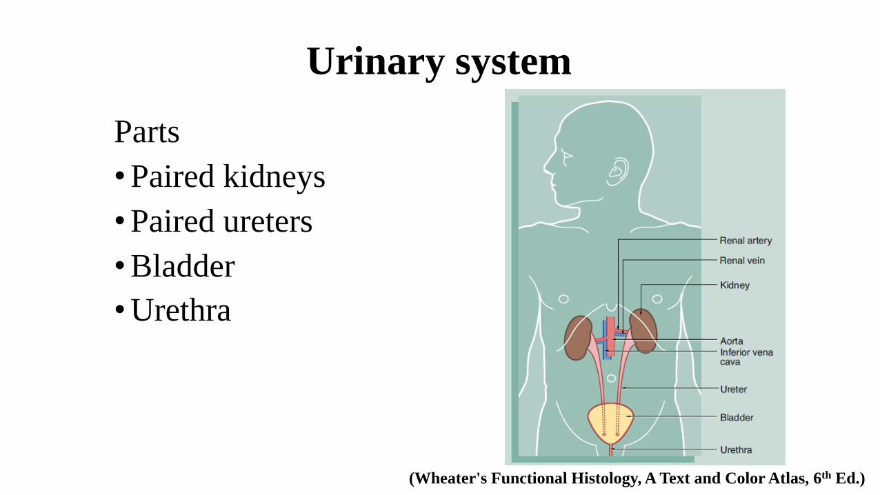
Urinary system
Parts
•Paired kidneys
•Paired ureters
•Bladder
•Urethra
(Wheater's Functional Histology, A Text and Color Atlas, 6th Ed.)
Kidney - Functions1. Controlling the water content of the body
2. Regulating the salt content of the blood
3. Regulating the extracellular fluid volume
4. Eliminating waste products, toxins and drugs; most importantly Urea
5. Controlling the acid-base balance of blood
6. Has a hormonal and metabolic function
• Secretion of Renin by juxtaglomerular cells to increase blood volume and
pressure
• Secretion of Erythropoietin that stimulates the production of erythrocytes in the bone marrow and thus regulates the oxygen-carrying capacity of the blood
• Conversion of Vitamin D, which regulates calcium balance, to an active form in the kidney
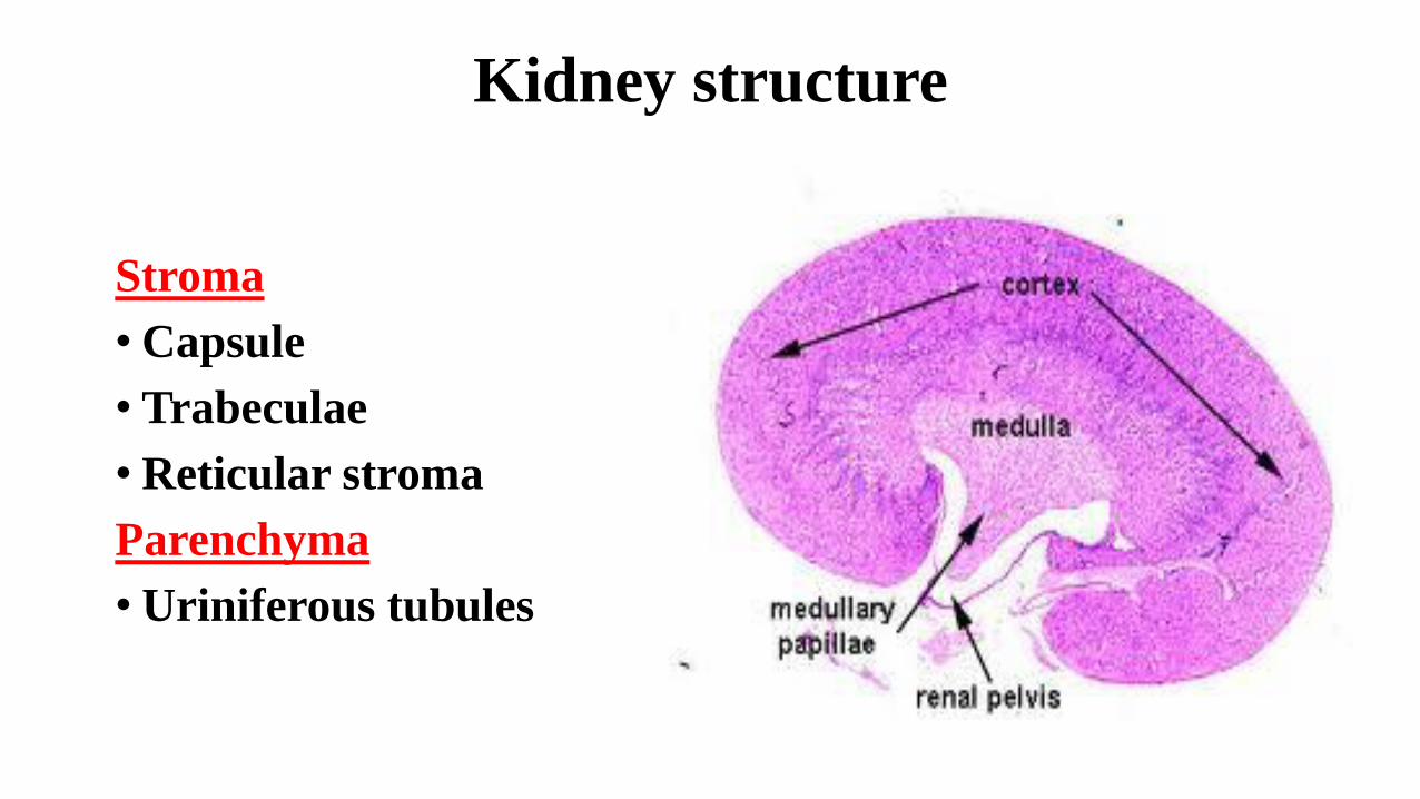
Kidney structure
Stroma
• Capsule
• Trabeculae
• Reticular stroma
Parenchyma
• Uriniferous tubules
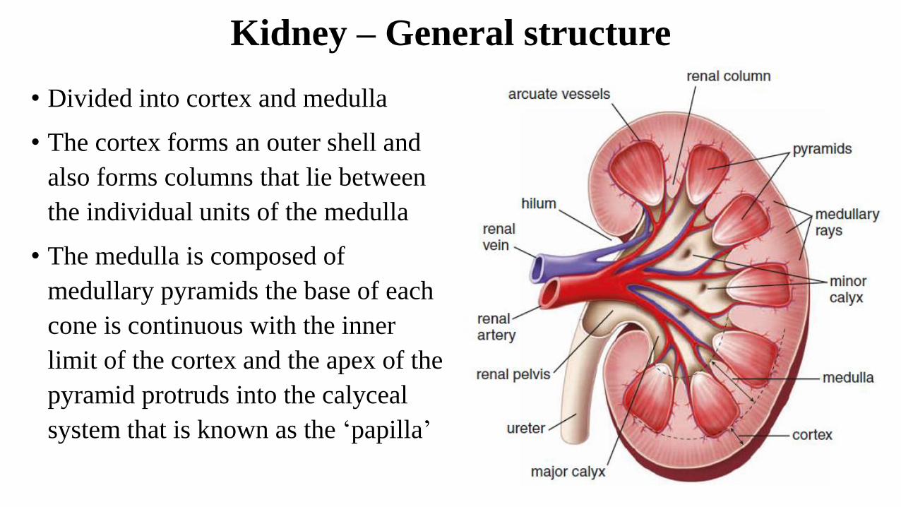
Kidney – General structure
• Divided into cortex and medulla
• The cortex forms an outer shell and
also forms columns that lie between
the individual units of the medulla
• The medulla is composed of
medullary pyramids the base of each
cone is continuous with the inner
limit of the cortex and the apex of the
pyramid protruds into the calyceal
system that is known as the ‘papilla’
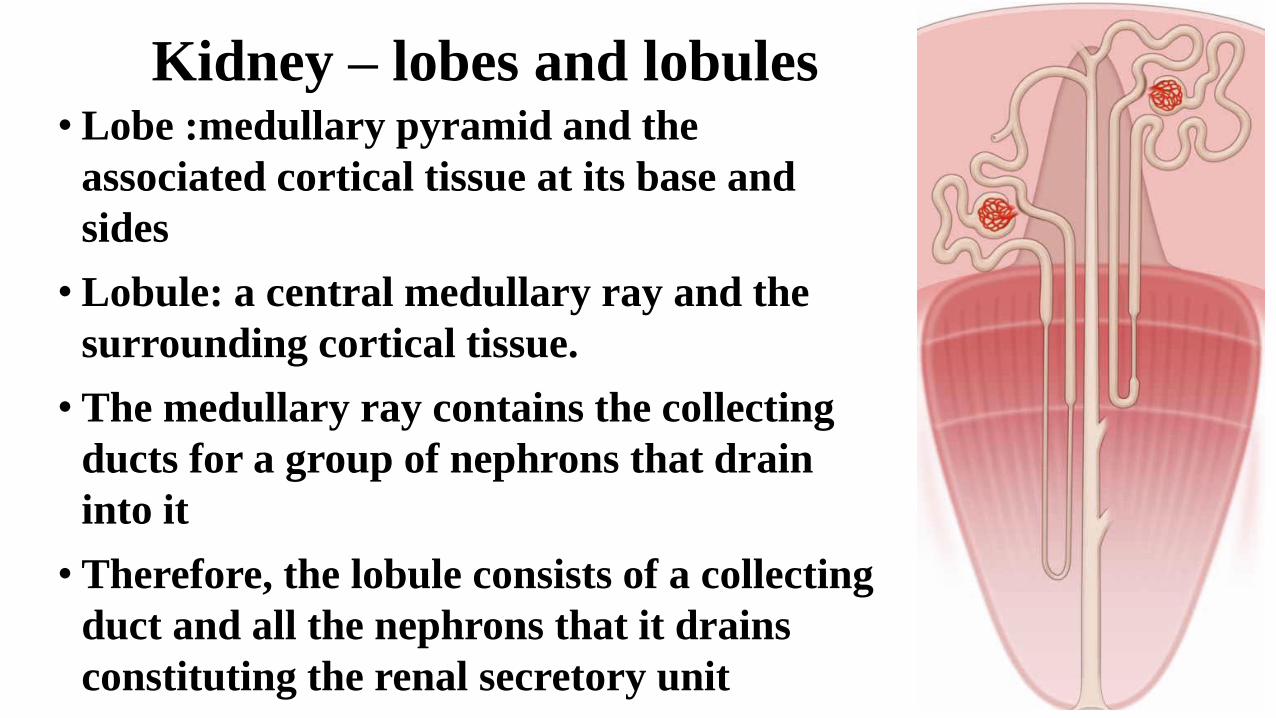
Kidney – lobes and lobules• Lobe :medullary pyramid and the
associated cortical tissue at its base and
sides
• Lobule: a central medullary ray and the
surrounding cortical tissue.
• The medullary ray contains the collecting
ducts for a group of nephrons that drain
into it
• Therefore, the lobule consists of a collecting
duct and all the nephrons that it drains
constituting the renal secretory unit
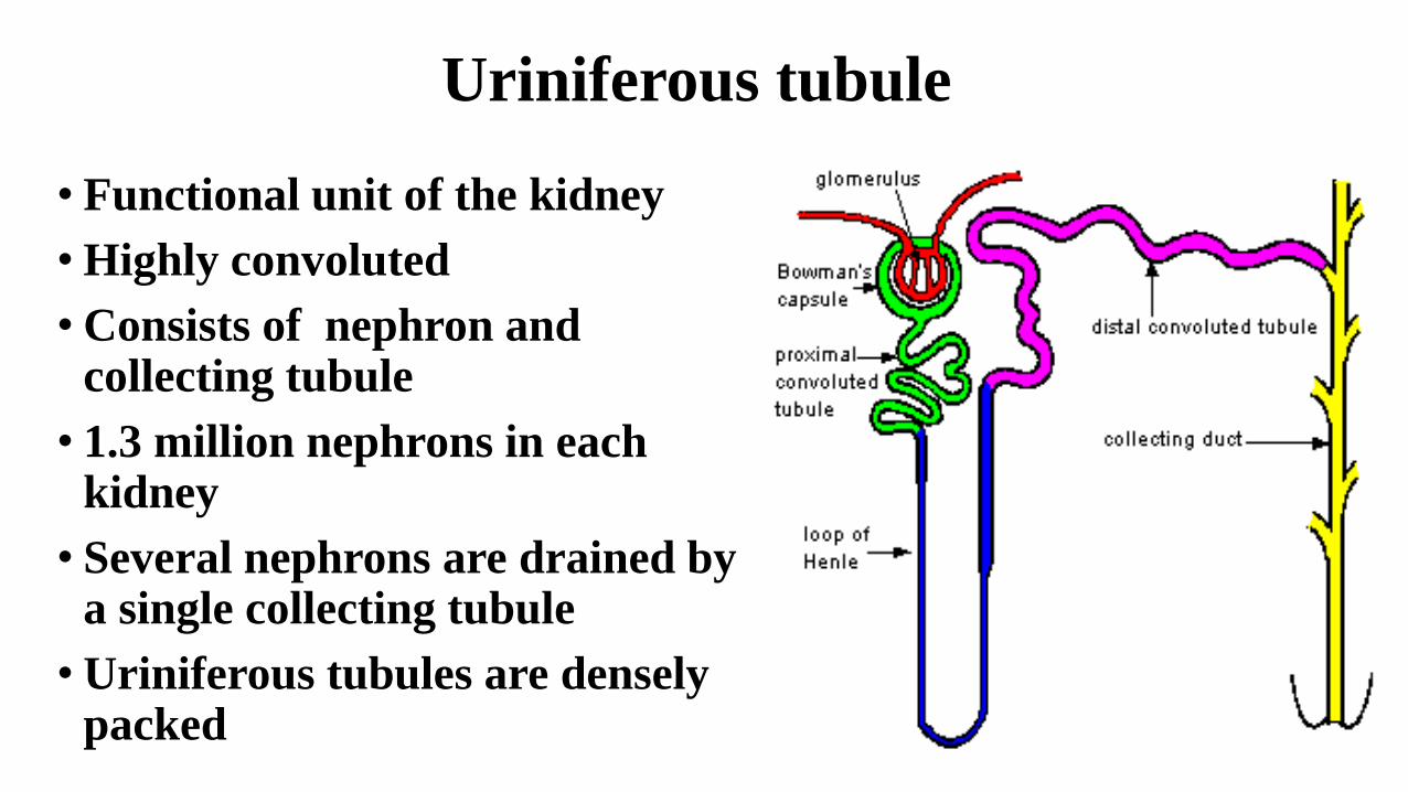
Uriniferous tubule
• Functional unit of the kidney
• Highly convoluted
• Consists of nephron and collecting tubule
• 1.3 million nephrons in each kidney
• Several nephrons are drained by a single collecting tubule
• Uriniferous tubules are densely packed
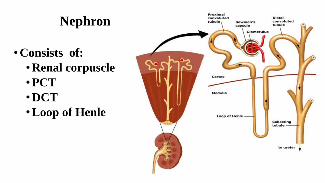
Nephron
•Consists of:
•Renal corpuscle
•PCT
•DCT
•Loop of Henle
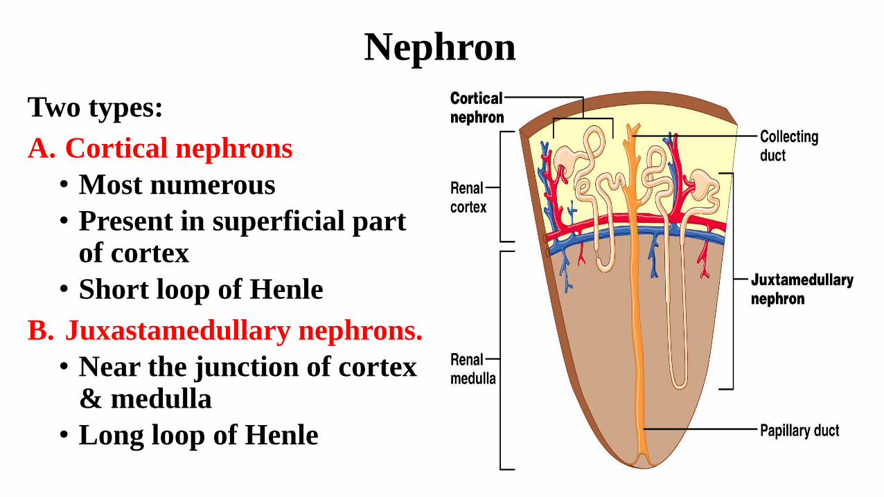
Nephron
Two types:
A. Cortical nephrons
• Most numerous
• Present in superficial part of cortex
• Short loop of Henle
B. Juxastamedullary nephrons.
• Near the junction of cortex & medulla
• Long loop of Henle
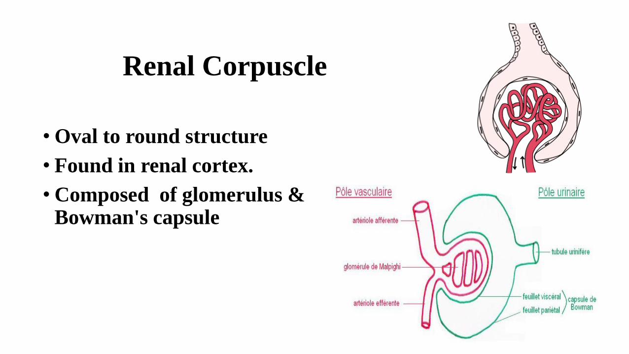
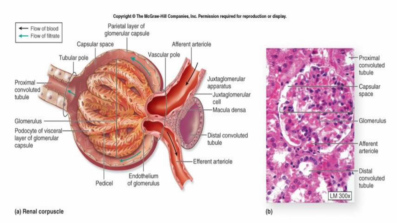
Renal Corpuscle
• Oval to round structure
• Found in renal cortex.
• Composed of glomerulus & Bowman's capsule
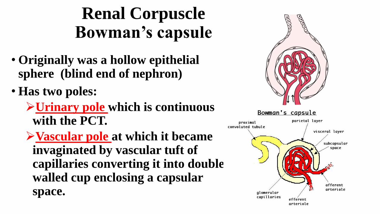
Renal Corpuscle Bowman’s capsule
• Originally was a hollow epithelial sphere (blind end of nephron)
• Has two poles:
Urinary pole which is continuous with the PCT.
Vascular pole at which it became invaginated by vascular tuft of capillaries converting it into double walled cup enclosing a capsular space.
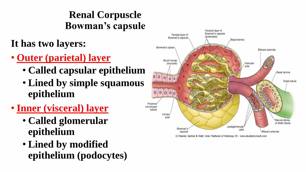
Renal Corpuscle Bowman’s capsule
It has two layers:
• Outer (parietal) layer
• Called capsular epithelium
• Lined by simple squamous epithelium
• Inner (visceral) layer
• Called glomerular epithelium
• Lined by modified epithelium (podocytes)
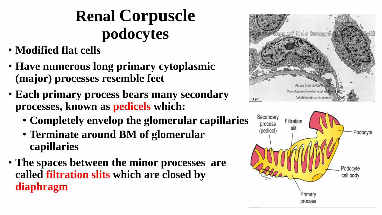
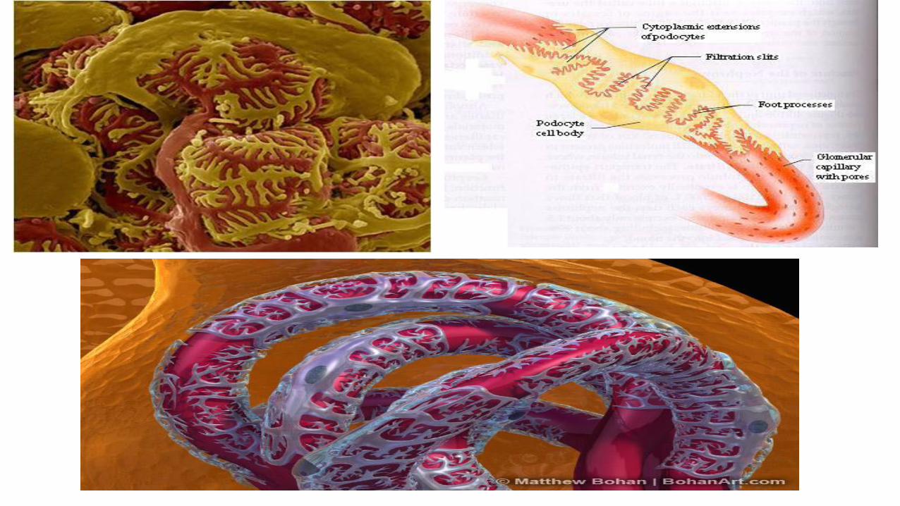
Renal Corpusclepodocytes
• Modified flat cells
• Have numerous long primary cytoplasmic (major) processes resemble feet
• Each primary process bears many secondary processes, known as pedicels which:
• Completely envelop the glomerular capillaries
• Terminate around BM of glomerular capillaries
• The spaces between the minor processes are called filtration slits which are closed by diaphragm
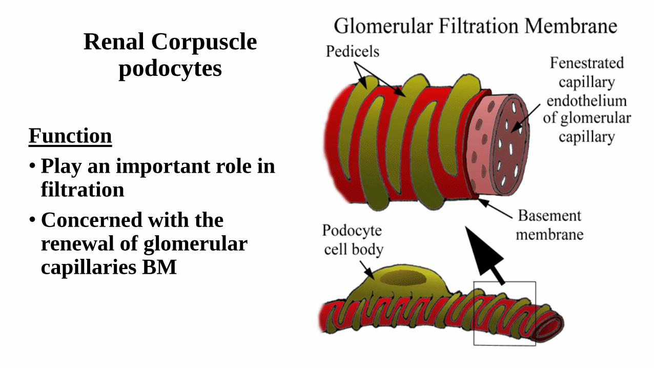
Renal Corpuscle podocytes
Function
• Play an important role in filtration
• Concerned with the renewal of glomerular capillaries BM
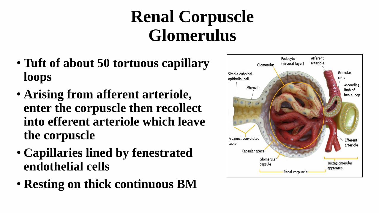
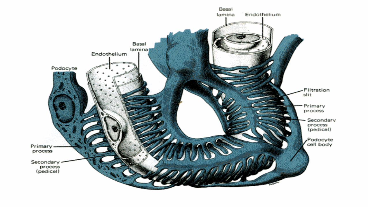
Renal Corpuscle Glomerulus
• Tuft of about 50 tortuous capillary loops
• Arising from afferent arteriole, enter the corpuscle then recollect into efferent arteriole which leave the corpuscle
• Capillaries lined by fenestrated endothelial cells
• Resting on thick continuous BM
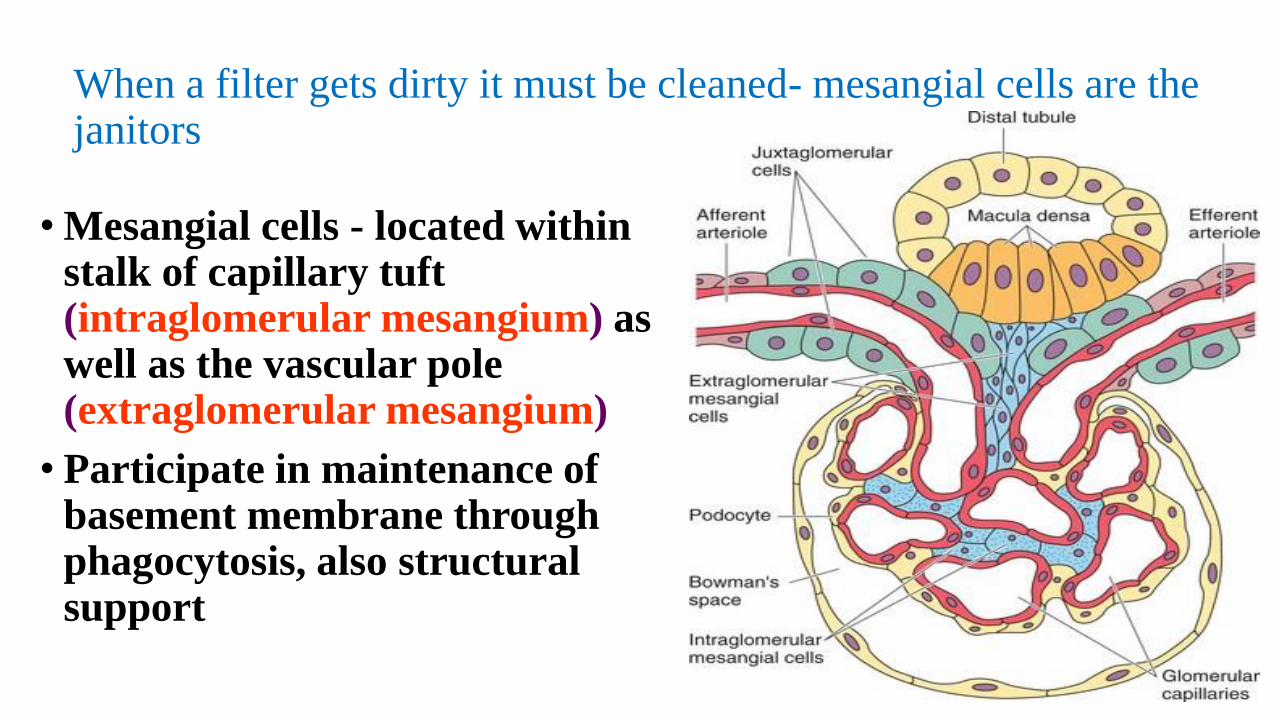
When a filter gets dirty it must be cleaned- mesangial cells are the janitors
• Mesangial cells - located within stalk of capillary tuft (intraglomerular mesangium) as well as the vascular pole (extraglomerular mesangium)
• Participate in maintenance of basement membrane through phagocytosis, also structural support
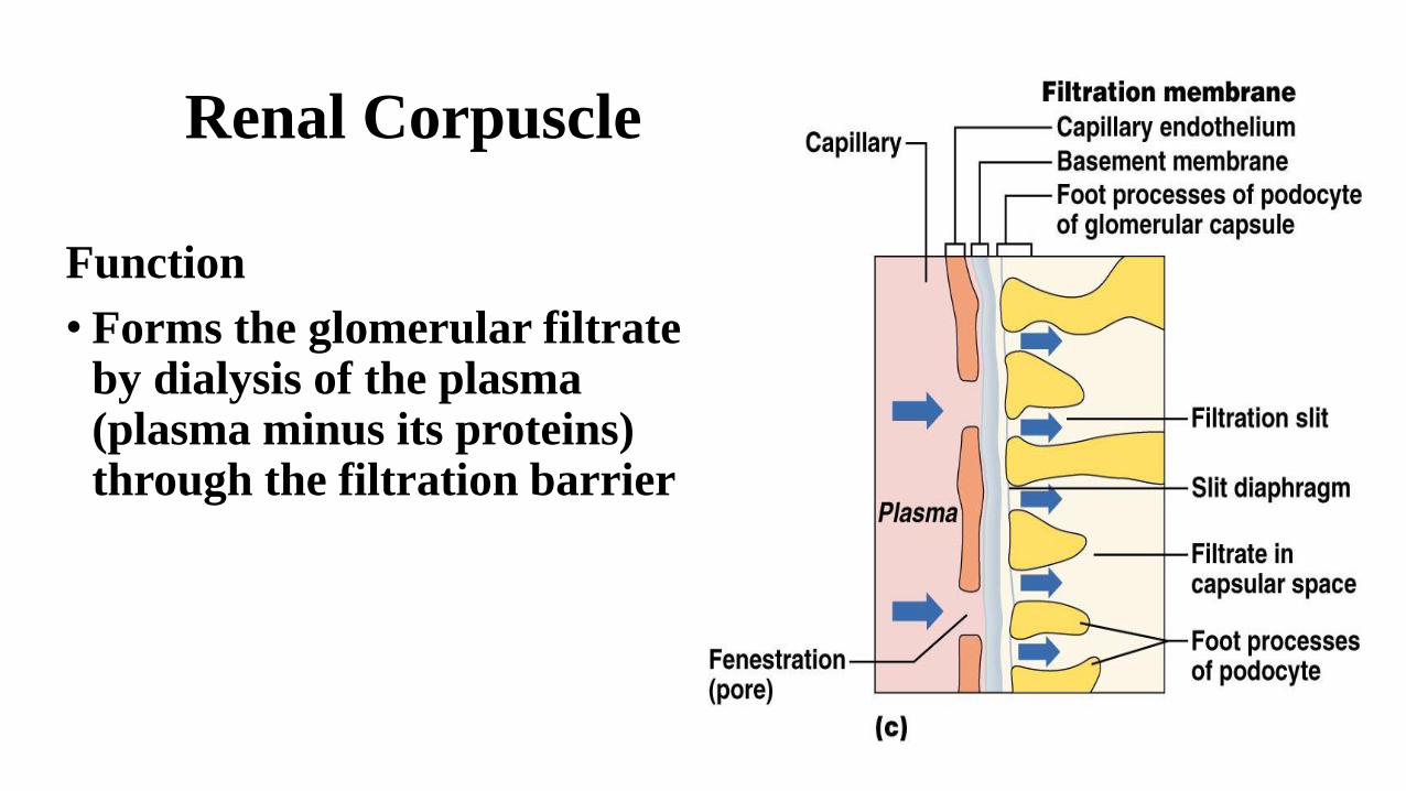
Renal Corpuscle
Function
• Forms the glomerular filtrate by dialysis of the plasma (plasma minus its proteins) through the filtration barrier
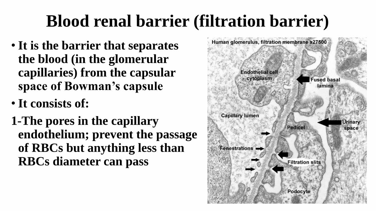
Blood renal barrier (filtration barrier)
• It is the barrier that separates the blood (in the glomerular capillaries) from the capsular space of Bowman’s capsule
• It consists of:
1-The pores in the capillary endothelium; prevent the passage of RBCs but anything less than RBCs diameter can pass
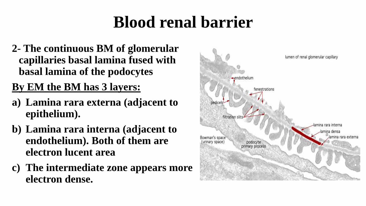
Blood renal barrier
2- The continuous BM of glomerularcapillaries basal lamina fused with basal lamina of the podocytes
By EM the BM has 3 layers:
a) Lamina rara externa (adjacent to epithelium).
b) Lamina rara interna (adjacent to endothelium). Both of them are electron lucent area
c) The intermediate zone appears more electron dense.
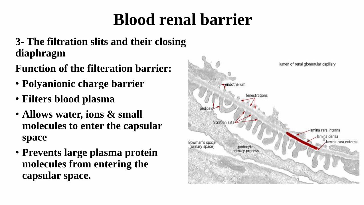
Blood renal barrier
3- The filtration slits and their closing diaphragm
Function of the filteration barrier:
• Polyanionic charge barrier
• Filters blood plasma
• Allows water, ions & small molecules to enter the capsular space
• Prevents large plasma protein molecules from entering the capsular space.
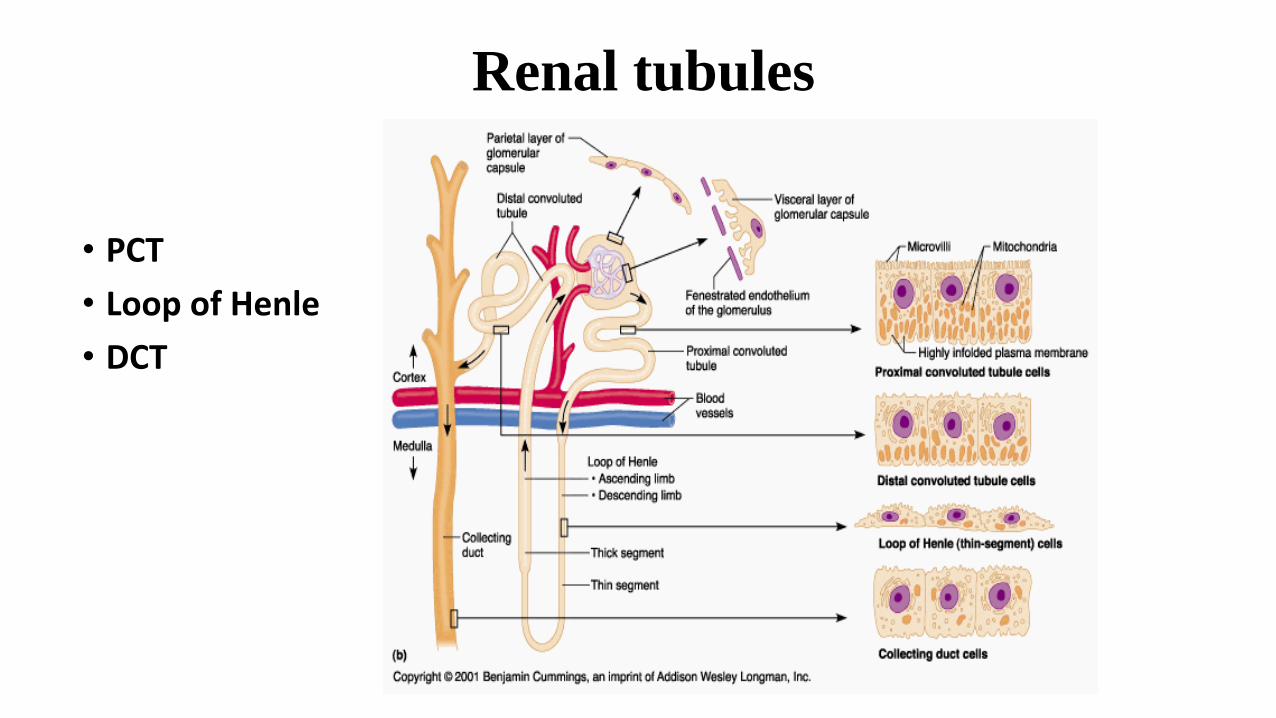
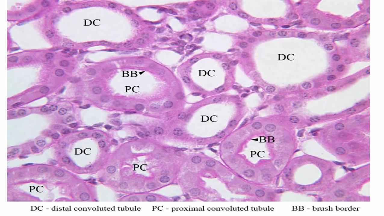
Renal tubules
• PCT
• Loop of Henle
• DCT
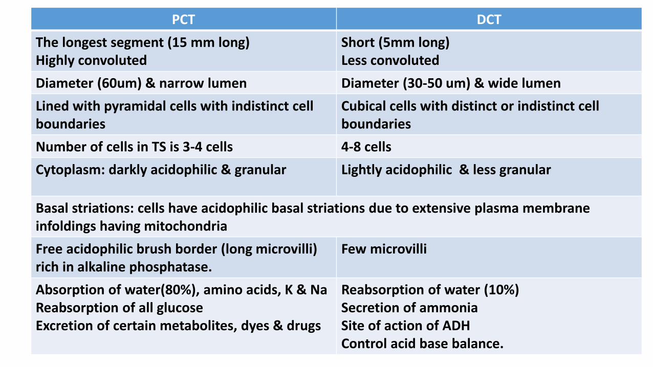
PCT DCT
The longest segment (15 mm long)Highly convoluted
Short (5mm long)Less convoluted
Diameter (60um) & narrow lumen Diameter (30-50 um) & wide lumen
Lined with pyramidal cells with indistinct cell boundaries
Cubical cells with distinct or indistinct cell boundaries
Number of cells in TS is 3-4 cells 4-8 cells
Cytoplasm: darkly acidophilic & granular Lightly acidophilic & less granular
Basal striations: cells have acidophilic basal striations due to extensive plasma membrane infoldings having mitochondria
Free acidophilic brush border (long microvilli) rich in alkaline phosphatase.
Few microvilli
Absorption of water(80%), amino acids, K & Na Reabsorption of all glucoseExcretion of certain metabolites, dyes & drugs
Reabsorption of water (10%)Secretion of ammoniaSite of action of ADHControl acid base balance.
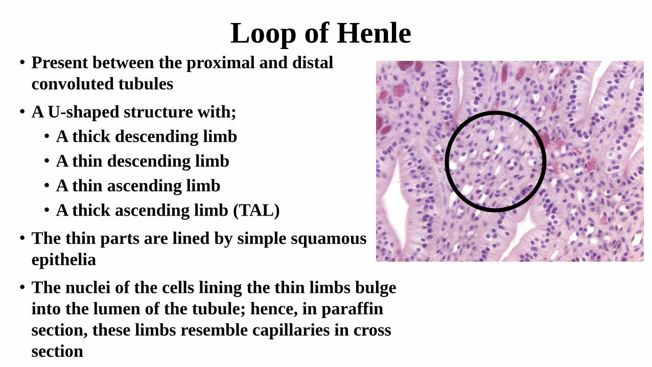
Loop of Henle• Present between the proximal and distal
convoluted tubules
• A U-shaped structure with;
• A thick descending limb
• A thin descending limb
• A thin ascending limb
• A thick ascending limb (TAL)
• The thin parts are lined by simple squamous
epithelia
• The nuclei of the cells lining the thin limbs bulge
into the lumen of the tubule; hence, in paraffin
section, these limbs resemble capillaries in cross
section
Connecting tubules
• The connecting tubule joins the distal convoluted tubule to the
collecting duct
• Connecting tubules of the subcapsular nephrons join directly to
the cortical collecting duct
• Connecting tubules from the midcortical and juxtamedullary
nephrons merge with other connecting tubules first to form an
arched connecting tubule before uniting with the cortical
collecting duct.
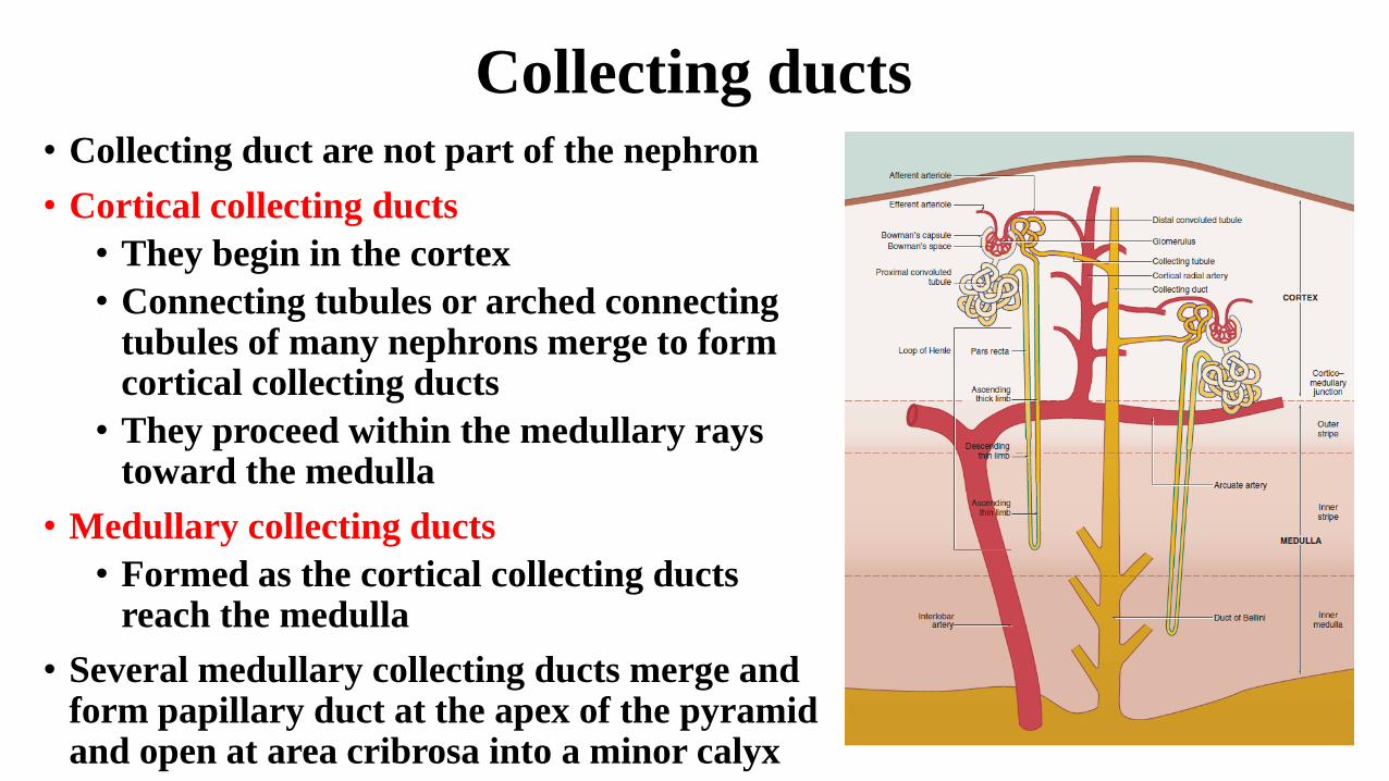
Collecting ducts• Collecting duct are not part of the nephron
• Cortical collecting ducts
• They begin in the cortex
• Connecting tubules or arched connecting tubules of many nephrons merge to form cortical collecting ducts
• They proceed within the medullary rays toward the medulla
• Medullary collecting ducts
• Formed as the cortical collecting ducts reach the medulla
• Several medullary collecting ducts merge and form papillary duct at the apex of the pyramid and open at area cribrosa into a minor calyx
Collecting ducts
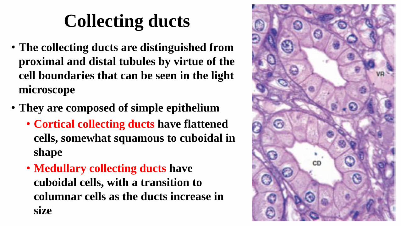
• The collecting ducts are distinguished from
proximal and distal tubules by virtue of the
cell boundaries that can be seen in the light
microscope
• They are composed of simple epithelium
• Cortical collecting ducts have flattened
cells, somewhat squamous to cuboidal in
shape
• Medullary collecting ducts have
cuboidal cells, with a transition to
columnar cells as the ducts increase in
size
Collecting ducts
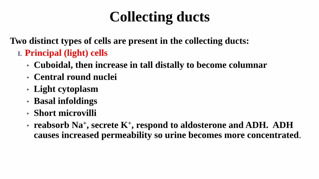
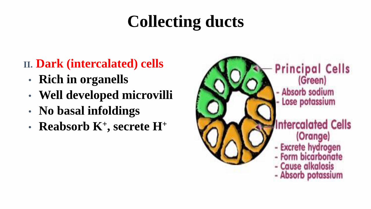
Two distinct types of cells are present in the collecting ducts:
I. Principal (light) cells
• Cuboidal, then increase in tall distally to become columnar
• Central round nuclei
• Light cytoplasm
• Basal infoldings
• Short microvilli
• reabsorb Na+, secrete K+, respond to aldosterone and ADH. ADH causes increased permeability so urine becomes more concentrated.
Collecting ducts
II. Dark (intercalated) cells
• Rich in organells
• Well developed microvilli
• No basal infoldings
• Reabsorb K+, secrete H+
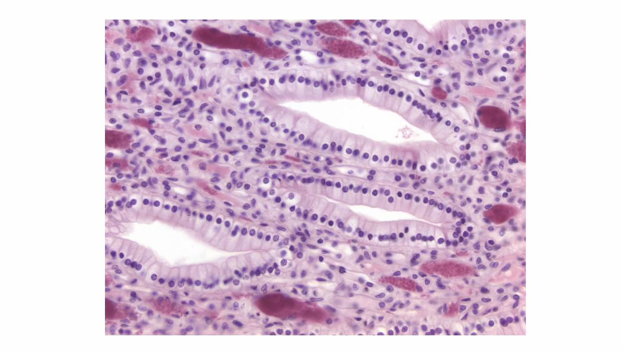
JUXTA-GLOMERULAR COMPLES
It is involved in the regulation of systemic blood pressure
It is located between glomerular afferent arteriole and distal
convoluted tubule of corresponding nephron
Composition
1- Macula densa:
It is an area of closely packed, specialised cells lining the DCT close
to the vascular pole
They are columnar, crowded with prominent deeply stained nuclei
It is sensitive to the concentration of sodium ions in the fluid within
the DCT

2- Juxta-glomerular (JG) cells (Renin producing cells):
They are modified smooth muscle cells of the afferent arteriole, small
numbers are present in the efferent arteriole.
It have features of myoepithelial cells with rounded nuclei and granular
cytoplasm
Contain mature and immature membrane –bound granules of the
enzyme renin
3- Polar cushion (Polkissen cells- Extraglomerular mesangial cells or
Lacis cells)
Formed of a mass of small cells with pale nuclei.
It is found in the triangular region between the afferent and
efferent arterioles at sides and macula densa at the base.
The apex of the triangle is formed by the glomerular mesangial
cells at the vascular pole
Three structures of the JG complex are in direct contact with each other
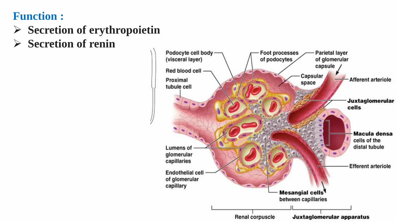
Function :
Secretion of erythropoietin
Secretion of renin

Ureter• Small muscular tubule.
• It carries urine from the renal pelvis to the urinary
bladder.
• Mucosa, muscularis, and adventitia.
• Mucosa consists of:
• Transitional epithelium
• loose connective tissue (lamina propria).
• Muscularis:
• Inner longitudinal and outer circular smooth
muscle layers.
• Difficult to distinguish.
• As it approaches the urinary bladder, the ureter
may also contain a third layer of smooth muscle.
• Adventitia : connective tissues, nerve fibers, and
blood vessels.
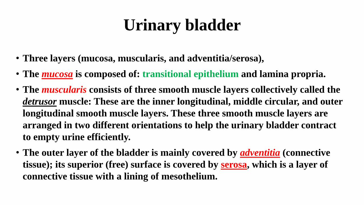
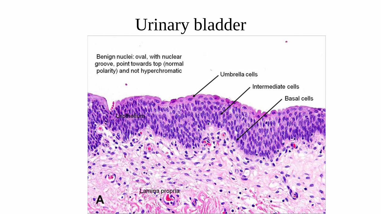
Urinary bladder
• Three layers (mucosa, muscularis, and adventitia/serosa),
• The mucosa is composed of: transitional epithelium and lamina propria.
• The muscularis consists of three smooth muscle layers collectively called the
detrusor muscle: These are the inner longitudinal, middle circular, and outer
longitudinal smooth muscle layers. These three smooth muscle layers are
arranged in two different orientations to help the urinary bladder contract
to empty urine efficiently.
• The outer layer of the bladder is mainly covered by adventitia (connective
tissue); its superior (free) surface is covered by serosa, which is a layer of
connective tissue with a lining of mesothelium.
Urinary bladder
Male urethra• The prostatic urethra:
Extends through the prostate gland and is lined by urothelium
(transitional epithelium )
• The membranous urethra:
• Passes through an external sphincter of striated muscle of the deep perineal pouch
• Lined by stratified columnar and pseudostratified columnar epithelium
• The spongy urethra:
• Enclosed within the erectile tissue of the penis
• Lined by stratified columnar and pseudostratified columnar epithelium, with stratified squamous epithelium distally
Female urethra
• The urethra is short, measuring 3 to 5 cm in length from the bladder
to the vestibule of the vagina.
• The lining epithelium is initially transitional epithelium, a
continuation of the bladder epithelium, but changes to stratified
squamous epithelium before its termination
• The lamina propria is a highly vascularized layer of connective tissue
• The urethra penetrates the urogenital diaphragm whose striated
muscle forms the external urethral sphincter