clinical workflows with the xnat imaging …...clinical workflows with the xnat imaging informatics...
TRANSCRIPT

Clinical workflows with the XNAT imaging informatics platform
Daniel Marcus, PhDWashington University School of Medicine
TACTICAL TutorialSeptember 20, 2018

Disclosures
• This work was supported in part by funding from the National Institutes of Health (4R01EB00935208, 1U24CA20485401, 1P30NS09857701) and the McDonnell Center for Higher Brain Function.
• Dr. Marcus has an ownership interest in Radiologics, Inc. and may financially benefit if the company is successful in marketing its products that are related to this research.
• Dr. Marcus has a financial interest in White Rabbit and may financially benefit if the company is successful in marketing its products that are related to this research.

I3CR – Integrative imaging informatics to enable cancer research

I3CR builds on XNAT. XNAT is…
…a feature rich…Archive, manage, process, view, and share imaging and related data.
…open…Open sourceOpen APIFree (though commercial support is available)Used by organizations around the world
…platform.Clinical/translational researchInstitutional repositoriesMulti-center studiesData sharing

AutomatedAnalytics
Integrated Search & Reporting
DICOMIntegration
Audit TrailUser Access Control
Case ReportForms
E-Signatures Dashboards Extensibility BulletproofSecurity
Top 10 XNAT features

Clinical use cases
1. Extract large data set from PACS
2. Process individual patient

Big data extraction
• Characteristics– Retrieve cases based on a list or query
– Requires scheduling
– Likely requires anonymization
– Data will be messy
– Batch process data
• Examples– Large retrospective data analysis
– Training data for machine learning applications

Individual patient
• Characteristics– Retrieve cases based real-time need
– Requires immediate retrieval
– No anonymization
– Data should comply with current protocol
– Process data immediately
• Examples– Insert novel analytic report into patient workflow
– Identify patients meeting study enrollment criteria

Individual patient

XNAT Container Service
Example: stereotactic radiosurgery imaging
• Patients with metastatic brain tumors undergo Gamma Knife radiosurgery
• Radiosurgery involves image-guided serial treatment of each lesion over months
• Current clinical limitations– Manual contouring of lesions is time consuming
– Serial time points are not spatially aligned
– Individual lesions are not tracked over time
– Imaging-based features are poorly leveraged
• Big Data workflow: retrieve list of patients, run batch pre-processing
• Individual patient workflow: retrieve individual patient, process, push results back to PACS

What we’re about to see
• XNAT 1.7.5• Event Service• Plugins
– DICOM Q/R plugin– ROI Collection data type plugin– Radiomic data type plugin– OHIF viewer plugin– Container Service plugin
• Containers: DICOM RT processor, RTlab, dcmtonii• Hosted on AWS • Data from CONDR project (~90 patients w/ brain mets &
GammaKnife plans

NOW’S A GOOD TIME FOR A DEMO

A production patient focused workflow
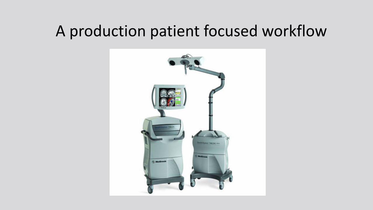
A production patient focused workflow

A production patient focused workflow

Future work
• Integration with RIS to extract radiology reports
• Integration with EMR to extract clinical data
• Improve UI/UX
• Improve workflows around patient privacy

Thanks from the I3CR team
Washington University
Dan Marcus (co-PI)
Rich Wahl (co-PI)
Cliff Robinson
Misha Milchenko
Pam Lamontagne, Rick Herrick, John Flavin, Matt Kelsey, Steve Moore, Mike McKay, Will Horton, Mark Florida, Charlie Moore
Institute for Cancer Research
Simon Doran (PI)
James Petts, James D’Arcy
Radiologics
Tim Olsen (PI)
Brian Holt, James Dickson, Patrick Clough, Jordan Woerndle

BONUS MATERIAL (IN CASE DEMO BREAKS)

Get stuff: Push from scanner

Get stuff: Pull from PACS

Do some stuff: Review acquisition protocol
<?xml version="1.0"
<iso:pattern
id="Acquisition">
<iso:rule context="scans">
<iso:assert
test="count(scan[@type=
mprage']) >= 1">
<nrgxsl:acquisition>
MRSession must have at
least 1 MPRAGE scan.
(etc…)

Do some stuff: Anonymize & Deface
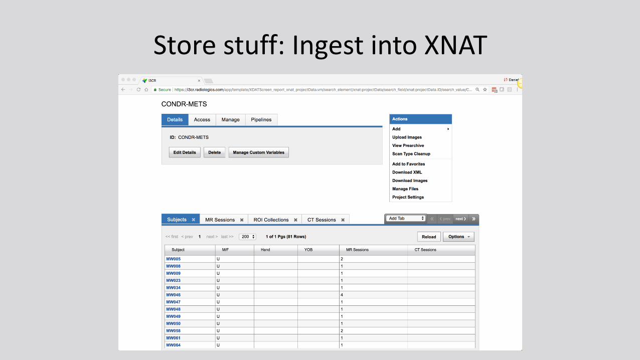
Store stuff: Ingest into XNAT

Store stuff: Ingest into XNAT

Store stuff: Ingest into XNAT

Store stuff: Ingest into XNAT

Do some more stuff: Convert RT Struct

Do some more stuff: Convert RT Struct

Do some more stuff: Convert RT Struct

Do some more stuff: Convert RT Struct

Do some more stuff: Convert RT Struct

Do some more stuff: Convert RT Struct
XNAT Event Service!

Do some more stuff: Convert RT Struct
XNAT Container Service!

Do some more stuff: Run analytics

Store stuff: Post files to archive

Store stuff: Post files to archive

Store stuff: Post features to archive

Store stuff: Post features to archive

Store stuff: Post features to archive

Use stuff: Get results to clinician

Use stuff: Get results to clinician