chapter 30 how animals move -...
TRANSCRIPT

Copyright © 2009 Pearson Education, Inc.
PowerPoint Lectures for
Biology: Concepts & Connections, Sixth Edition
Campbell, Reece, Taylor, Simon, and Dickey
Chapter 30 How Animals Move
Lecture by Edward J. Zalisko

Introduction: Man Versus Horse
Horses are well adapted for long-distance running
– Longer legs
– Lighter legs
– Increased oxygen-carrying capacity
Copyright © 2009 Pearson Education, Inc.

Humans have a more versatile body
– Humans run, crawl, swim, tumble, and throw
– Flexibility generally reduces efficiency
Copyright © 2009 Pearson Education, Inc.
Introduction: Man Versus Horse


MOVEMENT AND LOCOMOTION
Copyright © 2009 Pearson Education, Inc.

Animal movement is very diverse
Locomotion
– Active travel from place to place
– Requires energy to overcome friction and gravity
Copyright © 2009 Pearson Education, Inc.
30.1 Animals have evolved diverse means of locomotion

Swimming
– Supported by water
– But slowed by friction
Copyright © 2009 Pearson Education, Inc.
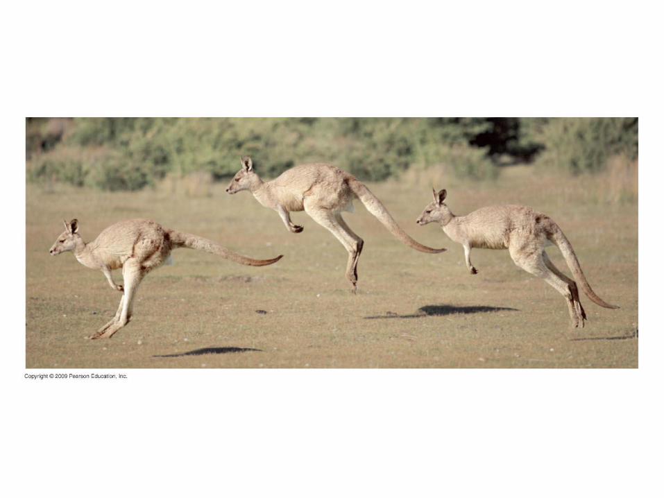
30.1 Animals have evolved diverse means of locomotion

Walking, hopping, or running
– Less affected by friction
– But must resist gravity
Copyright © 2009 Pearson Education, Inc.
30.1 Animals have evolved diverse means of locomotion


Burrowing or crawling
– Must overcome great friction
– May move by side-to-side undulations
– Or may move by a form of peristalsis
Copyright © 2009 Pearson Education, Inc.
30.1 Animals have evolved diverse means of locomotion

Head
Bristles
Longitudinalmusclerelaxed(extended)
Circularmusclecontracted
Circularmusclerelaxed
Longitudinalmusclecontracted
1
2
3

Flying
– Wings are airfoils that generate lift
– Flying is seen in birds, bats, and most insects
Copyright © 2009 Pearson Education, Inc.
30.1 Animals have evolved diverse means of locomotion


SKELETAL SUPPORT
Copyright © 2009 Pearson Education, Inc.

Skeletons provide
– Body support
– Movement by working with muscles
– Protection of internal organs
Copyright © 2009 Pearson Education, Inc.
30.2 Skeletons function in support, movement, and protection

Hydrostatic skeletons
– Fluid held under pressure in a closed body compartment
– Found in worms and cnidarians
Copyright © 2009 Pearson Education, Inc.
30.2 Skeletons function in support, movement, and protection


Exoskeletons
– Hard external cases
– Chitinous, jointed skeletons of arthropods
– Calcium carbonate shells of molluscs
Copyright © 2009 Pearson Education, Inc.
30.2 Skeletons function in support, movement, and protection


Shell
(exoskeleton)
Mantle
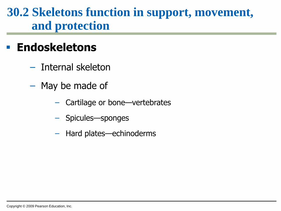
Endoskeletons
– Internal skeleton
– May be made of
– Cartilage or bone—vertebrates
– Spicules—sponges
– Hard plates—echinoderms
Copyright © 2009 Pearson Education, Inc.
30.2 Skeletons function in support, movement, and protection


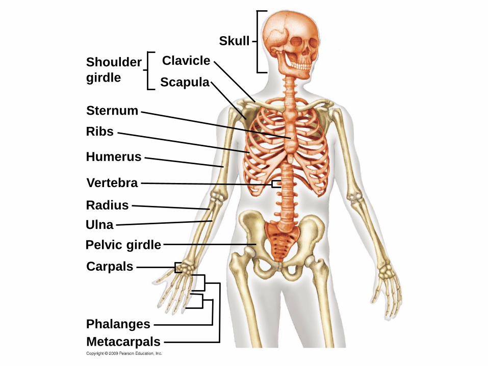
Human skeleton
– Axial skeleton
– Skull
– Vertebrae
– Ribs
– Appendicular skeleton
– Shoulder and pelvic girdles
– Arms and legs
Copyright © 2009 Pearson Education, Inc.
30.3 EVOLUTION CONNECTION: Vertebrate skeletons are variations on an ancient theme

Skull
Clavicle
Scapula
Shouldergirdle
Sternum
Ribs
Humerus
Vertebra
Radius
Ulna
Pelvic girdle
Carpals
Phalanges
Metacarpals
Femur
Patella
Tibia
Fibula
Tarsals
Metatarsals
Phalanges
Metacarpals
Phalanges
Carpals
Pelvic girdle
Ulna
Radius
Vertebra
Humerus
Ribs
Sternum
Skull
Clavicle
Scapula
Shoulder
girdle

Femur
Patella
Tibia
Fibula
Tarsals
Metatarsals
Phalanges

Intervertebraldiscs
Hip
bone
7 cervicalvertebrae
12 thoracic
vertebrae
5 lumbarvertebrae
Sacrum
Coccyx
Vertebrate bodies reveal variations of this basic skeletal arrangement
Master control (homeotic) genes
– Are active during early development
– Direct the arrangement of the skeleton
Vertebrates have evolved by changes in these master control genes
Copyright © 2009 Pearson Education, Inc.
30.3 EVOLUTION CONNECTION: Vertebrate skeletons are variations on an ancient theme

Gene expressionduring development
Python Chicken
Cervicalvertebrae
Thoracicvertebrae
Lumbarvertebrae
Hoxc6
Hoxc8
Hoxc6 and Hoxc8

Cartilage at the ends of bones
– Cushions joints
– Reduces friction of movements
Copyright © 2009 Pearson Education, Inc.
30.4 Bones are complex living organs

Bone cells
– Live in a matrix of
– Flexible protein fibers
– Hard calcium salts
– Are kept alive by
– Blood vessels
– Hormones
– Nerves
Copyright © 2009 Pearson Education, Inc.
30.4 Bones are complex living organs

Long bones have
– A fat-storing central cavity
– Spongy bone
– Located at the ends of bones
– Contains bone marrow, the site of blood cell production
Copyright © 2009 Pearson Education, Inc.
30.4 Bones are complex living organs

Cartilage
Spongy bone(contains redbone marrow)
Cartilage
Spongybone
Compactbone
Centralcavity
Yellowbone marrow
Bloodvessels
Fibrousconnectivetissue

Bone cells
– Repair bones
– Reshape bones throughout life
Broken bones
– Are realigned and immobilized
– Bone cells build new bone, healing the break
Copyright © 2009 Pearson Education, Inc.
30.5 CONNECTION: Healthy bones resist stress and heal from injuries

Osteoporosis is a bone disease characterized by
– Weak, porous bones
– Less likely if
– High levels of calcium in diet
– Regular exercise
– No smoking
Copyright © 2009 Pearson Education, Inc.
30.5 CONNECTION: Healthy bones resist stress and heal from injuries


30.6 Joints permit different types of movement
Joints allow limited movement of bones
Different joints permit various movements
Copyright © 2009 Pearson Education, Inc.

Humerus
Head ofhumerus
Scapula
Ball-and-socket joint
Ulna
Hinge joint
Ulna
Pivot joint
Radius

Head ofhumerus
Scapula
Ball-and-socket joint

Humerus
Ulna
Hinge joint

Ulna
Pivot joint
Radius

MUSCLE CONTRACTION AND MOVEMENT
Copyright © 2009 Pearson Education, Inc.

Muscles and bones interact to produce movement
Muscles can only contract
Copyright © 2009 Pearson Education, Inc.
30.7 The skeleton and muscles interact in movement

Antagonistic pairs of muscles
– Reverse actions
– Relengthen muscles
Copyright © 2009 Pearson Education, Inc.
30.7 The skeleton and muscles interact in movement

Biceps
Triceps
Biceps
Triceps
Tendon
Biceps contracted,triceps relaxed(extended)
Tricepscontracted,biceps relaxed
Muscle fibers
– Are cells
– Consist of bundles of myofibrils
Myofibrils contain overlapping
– Thick (myosin) filaments
– Thin (actin) filaments
Copyright © 2009 Pearson Education, Inc.
30.8 Each muscle cell has its own contractile apparatus

Sarcomeres are
– Repeating groups of thick and thin filaments
– The contractile unit—the fundamental unit of muscle action
Copyright © 2009 Pearson Education, Inc.
30.8 Each muscle cell has its own contractile apparatus

Muscle
Several muscle fibers
Single muscle fiber
(cell)
Plasma membrane
Myofibril
Nuclei
Z line
Light
band
Light
bandDark
band
Sarcomere
Thick
filaments
(myosin)
Thin
filaments
(actin)
Z line
SarcomereZ line

Muscle
Several muscle fibers
Single muscle fiber(cell)
Plasma membrane
Myofibril
Nuclei
Lightband
Lightband
Darkband
Sarcomere
Z line

Plasma membrane
Myofibril
Z line
Lightband
Lightband
Darkband
Sarcomere
Thickfilaments(myosin)
Thinfilaments(actin)
Z line
Sarcomere
Z line

The sliding-filament model explains muscle contraction
Copyright © 2009 Pearson Education, Inc.
30.9 A muscle contracts when thin filaments slide across thick filaments

Sarcomere
Dark bandZ Z
Relaxedmuscle
Contractingmuscle
Fully contractedmuscle
Contractedsarcomere

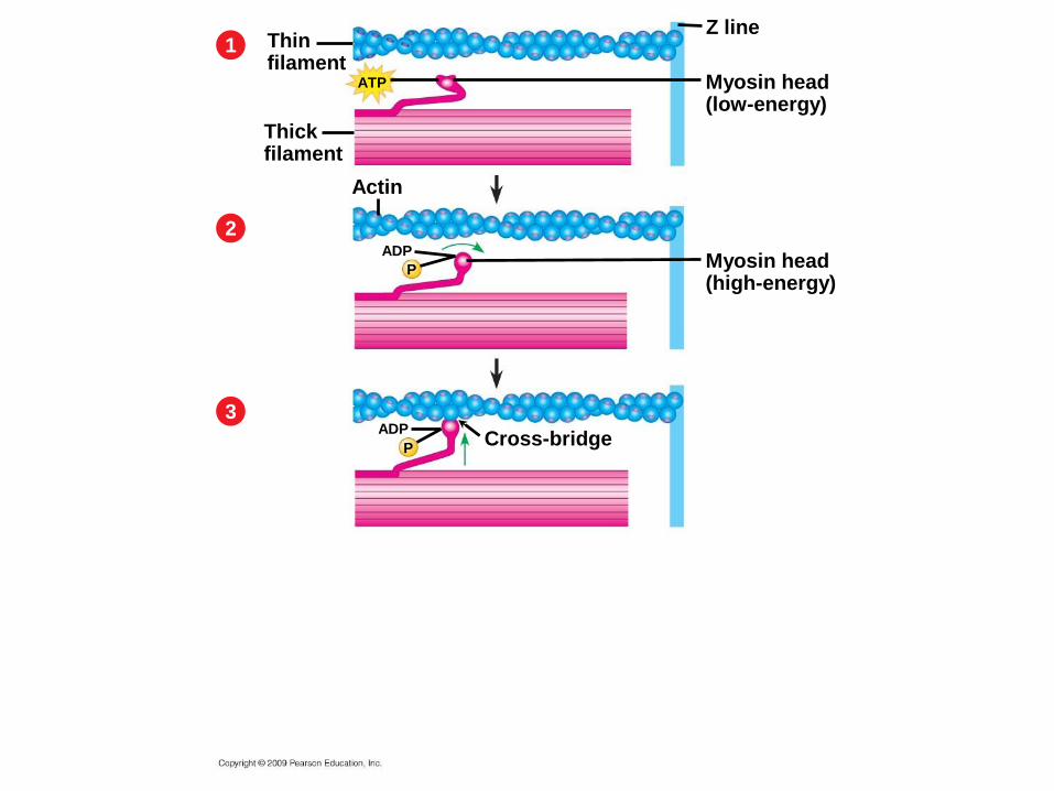
Myosin heads of the thick filaments
– Bind ATP and
– Extend to high-energy states
Myosin heads then
– Attach to binding sites on the actin molecules and
– Pull the thin filaments toward the center of the sarcomere
Copyright © 2009 Pearson Education, Inc.
30.9 A muscle contracts when thin filaments slide across thick filaments

Z line
Thick filament
Myosin head (low-energy configuration)
ATP
Thinfilament
Thinfilaments
Thickfilament
1
2
4
3
Actin
Myosin head (high-energy configuration)
ADP
P
Myosin head (low-energy configuration)
ADP
PCross-bridge
ADP+ P
Newpositionof Z lineThin filament moves
toward center of sarcomere.

Z line
Myosin head(low-energy)
ATP
Thinfilament
Thickfilament
1

Z line
Myosin head(low-energy)
ATP
Thinfilament
Thickfilament
1
2ADP
Myosin head(high-energy)
Actin
P
Z line
Myosin head(low-energy)
ATP
Thinfilament
Thickfilament
1
2ADP
Myosin head(high-energy)
Actin
P
ADP
PCross-bridge
3

Z line
Myosin head(low-energy)
ATP
Thinfilament
Thickfilament
1
2ADP
Myosin head(high-energy)
Actin
P
ADP
PCross-bridge
3
4
Myosin head(low-energy)
New positionof Z line
ADP + P

Motor neurons
– Carry action potentials
– That initiate muscle contractions
Copyright © 2009 Pearson Education, Inc.
30.10 Motor neurons stimulate muscle contraction

The axon of a motor neuron
– Forms synapses with a muscle
– At a neuromuscular junction
Copyright © 2009 Pearson Education, Inc.
30.10 Motor neurons stimulate muscle contraction

Action potentialMotor neuronaxon
Synapticterminal
Endoplasmicreticulum (ER)
Mitochondrion
SarcomerePlasma membrane
Myofibril
T tubule
Ca2+
releasedfrom ER

Acetylcholine
– Is released from the synaptic terminal of a motor neuron
– Diffuses to the plasma membrane of the muscle fiber
Copyright © 2009 Pearson Education, Inc.
30.10 Motor neurons stimulate muscle contraction

An action potential in a muscle fiber
– Passes along T tubules
– Into the center of muscle fiber
Calcium ions
– Are released from the endoplasmic reticulum
– Initiate muscle contraction by moving regulatory proteins away from the actin binding sites
Copyright © 2009 Pearson Education, Inc.
30.10 Motor neurons stimulate muscle contraction

Ca2+
Myosin-binding sites blocked
Myosin-binding site
Myosin-binding sites exposed
Troponin complexCa2+-binding sitesTropomyosin
Actin

A motor unit consists of
– A neuron
– The set of muscle fibers it controls
Copyright © 2009 Pearson Education, Inc.
30.10 Motor neurons stimulate muscle contraction

Bone
Muscle
Tendon
Muscle fibers(cells)
NucleiSynapticterminals
Motor neuroncell body
Motor neuronaxon
Spinal cord Motorunit 1
Motorunit 2
Nerve

Aerobic exercise provides most of the ATP used to power muscle movement during exercise
Aerobic exercise requires a steady supply of
– Glucose
– Oxygen
30.11 CONNECTION: Aerobic respiration supplies most of the energy for exercise
Copyright © 2009 Pearson Education, Inc.


Anaerobic exercise
– Generates ATP faster
– Is much less efficient at producing ATP
Copyright © 2009 Pearson Education, Inc.
30.12 CONNECTION: Muscle fiber characteristics affect athletic performance

Muscle fibers can be classified as
– Slow fibers
– Intermediate fibers
– Fast fibers
Most muscles have a combination of fiber types
The proportion of fiber types can be affected by exercise
Copyright © 2009 Pearson Education, Inc.
30.12 CONNECTION: Muscle fiber characteristics affect athletic performance


Muscles adapt to exercise by increasing
– Levels of myoglobin
– Number of mitochondria
– Number of capillaries going to muscles
Copyright © 2009 Pearson Education, Inc.
30.12 CONNECTION: Muscle fiber characteristics affect athletic performance

SlowIntermediateFast
100
80
60
40
20
0World-Class
Sprinter
World-Class
MarathonRunner
AverageActivePerson
AverageCouchPotato
Middle-DistanceRunner
ExtremeEndurance
Athlete


Sarcomere
ActinMyosin

Animal movement
requires bothmust overcome
forces of
gravity
and friction
hydrostatic
skeleton antagonistic
pairs
types are moveparts of
usually in
units ofcontraction
are
shorten as
(a) (b)
(c) (d)
(e)
made of called
endoskeletonmyosin pulls
actin filaments
sliding-filament
model

1. Compare the adaptations of humans and horses that increase speed
2. Describe the diverse methods of animal locomotion and the forces they must overcome
3. Describe the three main types of skeletons
4. Describe the complex structure of bone
5. Describe the causes of osteoporosis
6. Describe three types of joints
Copyright © 2009 Pearson Education, Inc.
You should now be able to

7. Describe the structure and arrangement of the filaments found in a muscle cell
8. Explain how a muscle cell contracts
9. Describe the role of calcium in a muscle contraction
10. Distinguish between aerobic and anaerobic exercise
11. Compare the structure and functions of different muscle fiber types
Copyright © 2009 Pearson Education, Inc.
You should now be able to