6 processes of digestion –ingestion –mechanical digestion chewing, swallowing peristalsis...
Post on 19-Dec-2015
234 views
TRANSCRIPT
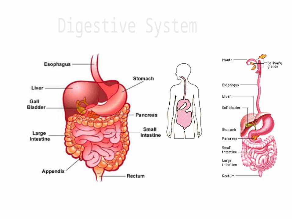
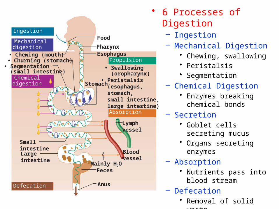
• 6 Processes of Digestion– Ingestion– Mechanical Digestion
• Chewing, swallowing• Peristalsis• Segmentation
– Chemical Digestion• Enzymes breaking
chemical bonds– Secretion
• Goblet cells secreting mucus
• Organs secreting enzymes
– Absorption• Nutrients pass into blood
stream– Defecation
• Removal of solid waste
FoodIngestion
PropulsionEsophagus
Stomach
PharynxMechanicaldigestion
Chemicaldigestion
• Chewing (mouth)• Churning (stomach)• Segmentation (small intestine)
Smallintestine Largeintestine
Defecation Anus
Feces
Bloodvessel
Lymphvessel
Absorption
• Swallowing (oropharynx)• Peristalsis (esophagus, stomach, small intestine, large intestine)
Mainly H2O
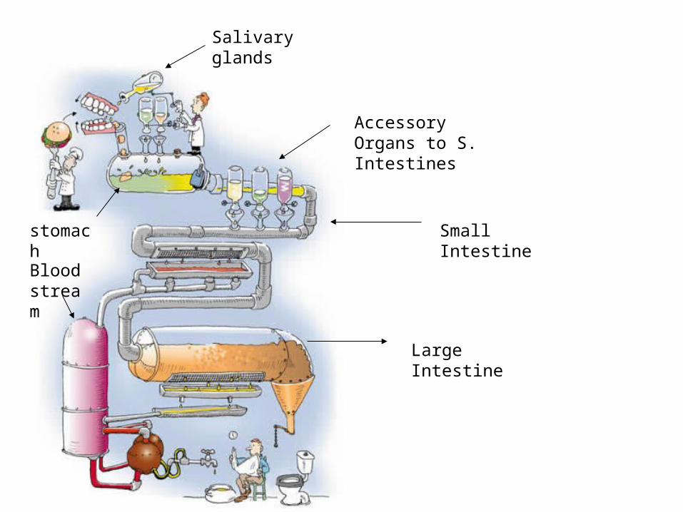
Salivary glands
stomach Small Intestine
Accessory Organs to S. Intestines
Large Intestine
Blood stream
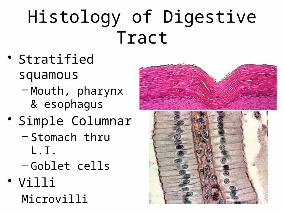
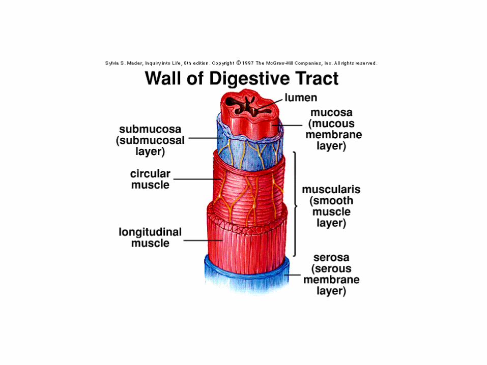
Histology of Digestive Tract
• Stratified squamous– Mouth, pharynx &
esophagus• Simple Columnar
– Stomach thru L.I.– Goblet cells
• VilliMicrovilli
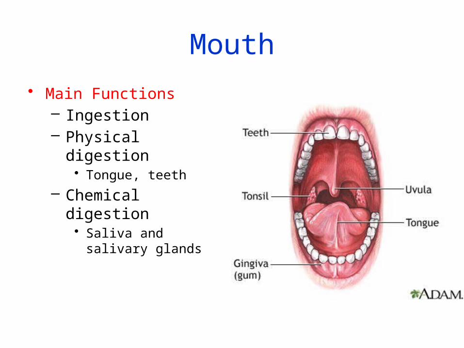
Mouth
• Main Functions– Ingestion– Physical digestion
• Tongue, teeth
– Chemical digestion• Saliva and salivary
glands
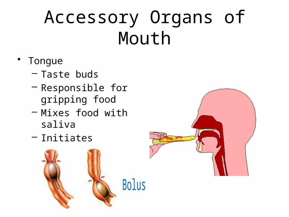
Accessory Organs of Mouth
• Tongue– Taste buds– Responsible for
gripping food– Mixes food with saliva– Initiates swallowing

Frommouth
(b) Segmentation: Nonadjacent segments of alimentary tract organs alternately contract and relax, moving the food forward then backward. Food mixing and slow food propulsion occurs.
(a) Peristalsis: Adjacent segments of alimentary tract organs alternately contract and relax, which moves food along the tract distally.
Peristalsis vs. Segmentation

Salivary GlandsParotid, submandibular and sublingual
• Functions– Produce saliva
• 99% water• Contains enzyme amylase• Mucin – forms mucus• Growth Factor
• Functions of Saliva– Cleanses mouth
• Growth factor and lysosomes– Dissolves food for taste– Moistens food; creates bolus– Enzyme amylase begins the
chemical digestion of starch

Amylase
• Starch (polysaccharide) disaccharides
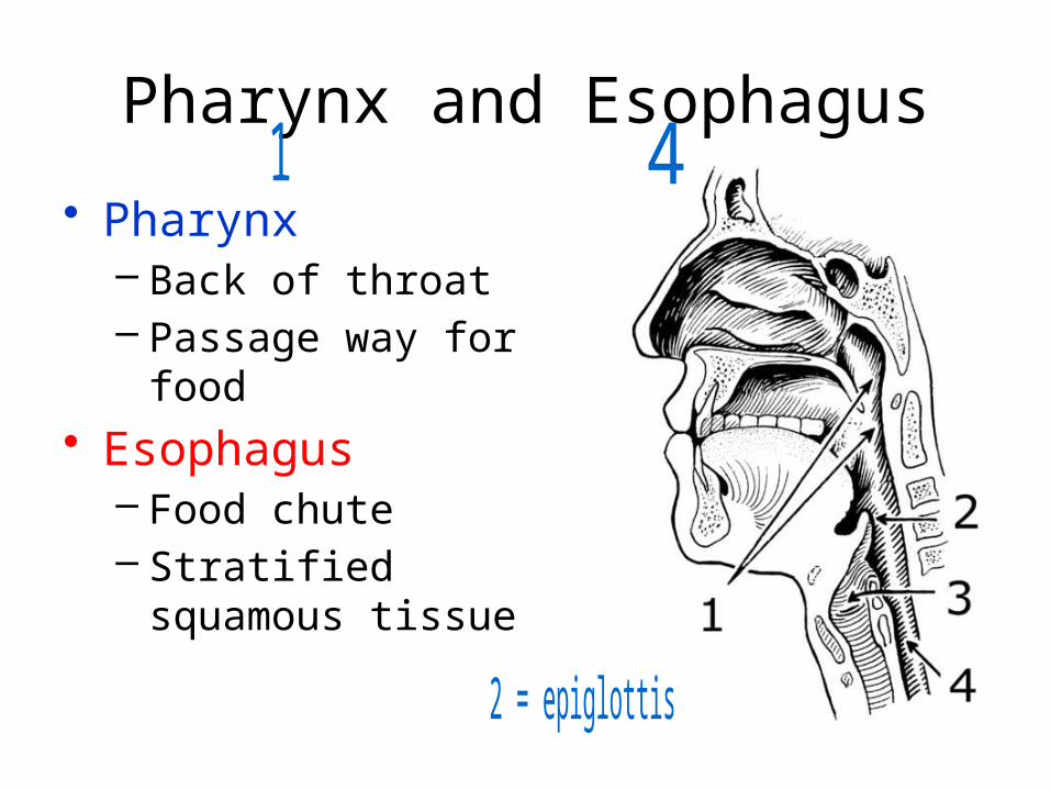
Pharynx and Esophagus
• Pharynx– Back of throat– Passage way for
food• Esophagus
– Food chute– Stratified squamous
tissue

Stomach
• Four Primary Functions– Temporary storage tank for food– Mechanically break down food
• Food is converted into creamy paste called chyme
– Chemical breakdown of protein begins– Production of intrinsic factor; necessary for
absorbing Vitamin B12.

Regions of the Stomach• Cardiac region
– Upper area around cardiac sphincter
• Fundus– Dome-shaped part of
stomach
• Body• Pylorus- inferior
portion• Pyloric sphincter:
controls the emptying of the stomach into the S.I.
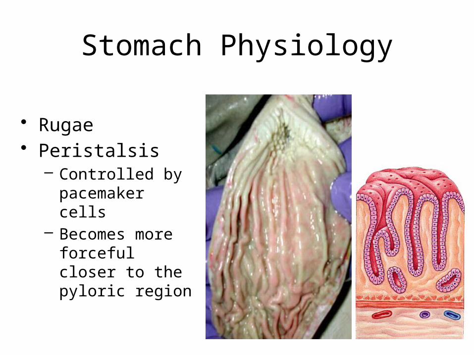
Stomach Physiology
• Rugae• Peristalsis
– Controlled by pacemaker cells
– Becomes more forceful closer to the pyloric region

• Gastric Pit– Gastric gland
• Secretes mucus and enzyme pepsinogen
• Parietal cells– Secrete HCl and
instrinsic factor– HCl is needed to
convert pepsinogen into pepsin
– Kills bacteria that is ingested with food
– Rennin
• Chief cells – produce pepsinogen; inactive form of pepsin
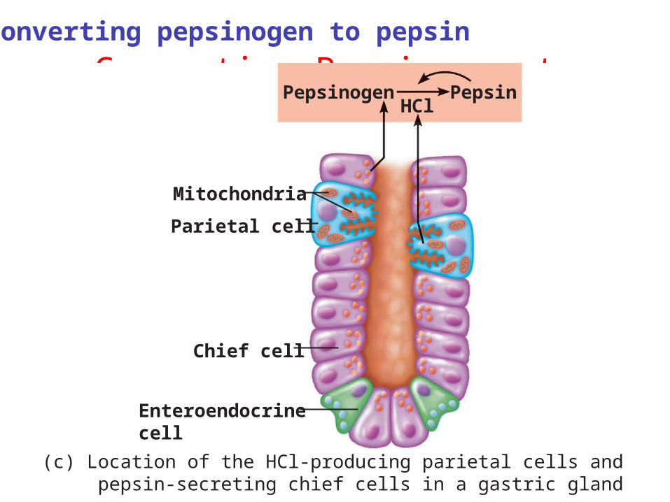
Converting Pepsinogen to Pepsin
(c) Location of the HCl-producing parietal cells and pepsin-secreting chief cells in a gastric gland
Pepsinogen
Mitochondria
PepsinHCl
Chief cell
Enteroendocrinecell
Parietal cell
Converting pepsinogen to pepsin

Regulation of Gastric Activity
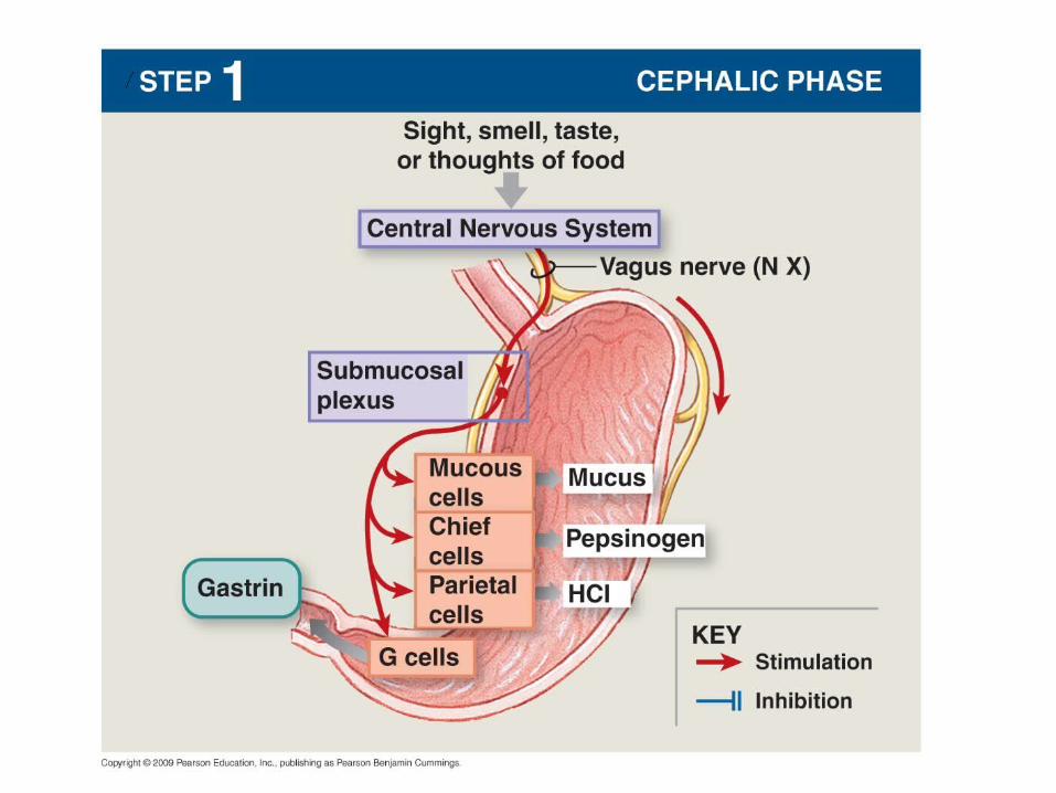
1. Cephalic phase – prepares stomach to receive food– Sight, smell, taste of food– Controlled by vagus nerve and
parasympathetic system– Only lasts a short time
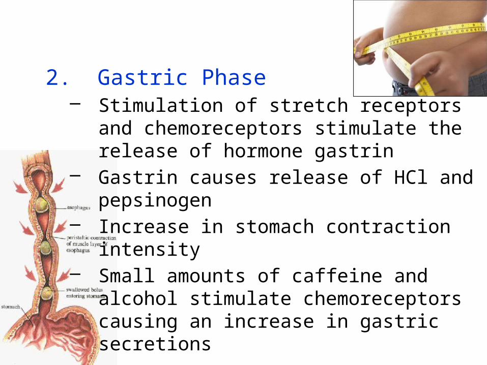
2. Gastric Phase– Stimulation of stretch receptors and
chemoreceptors stimulate the release of hormone gastrin
– Gastrin causes release of HCl and pepsinogen
– Increase in stomach contraction intensity– Small amounts of caffeine and alcohol
stimulate chemoreceptors causing an increase in gastric secretions

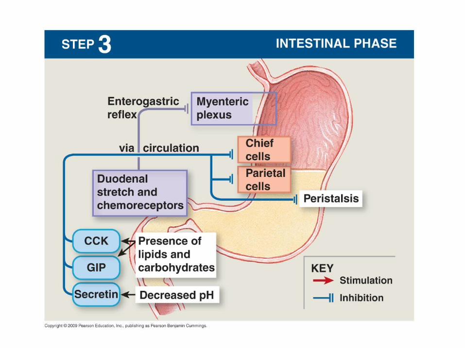
3. Intestinal phase– Begins when food starts to enter S.I.– Controls the rate of gastric emptying– Slows down gastric activities by releasing
three hormones• Cholecystokinin (CCK) • Secretin• Gastric inhibitory peptide (GIP)
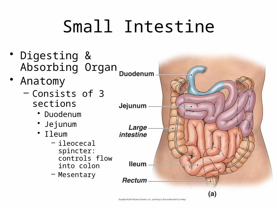
Small Intestine
• Digesting & Absorbing Organ
• Anatomy– Consists of 3
sections• Duodenum• Jejunum• Ileum
– ileocecal spincter: controls flow into colon
– Mesentary

Modifications for Absorption
• Folds– Mixes chyme
and slows down movement
• Villi– Fingerlike
projections increases surface area
• Microvilli
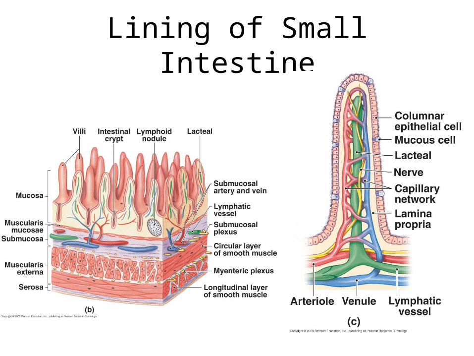
Lining of Small Intestine
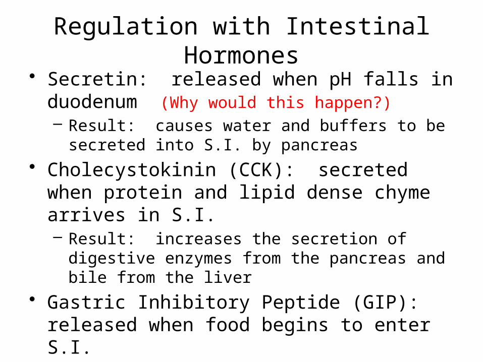
Regulation with Intestinal Hormones• Secretin: released when pH falls in duodenum
(Why would this happen?) – Result: causes water and buffers to be secreted into
S.I. by pancreas
• Cholecystokinin (CCK): secreted when protein and lipid dense chyme arrives in S.I.– Result: increases the secretion of digestive enzymes
from the pancreas and bile from the liver
• Gastric Inhibitory Peptide (GIP): released when food begins to enter S.I.– Result: inhibit digestive activity in stomach; causes
release of insulin from pancreas
1
2
3
4
5
6
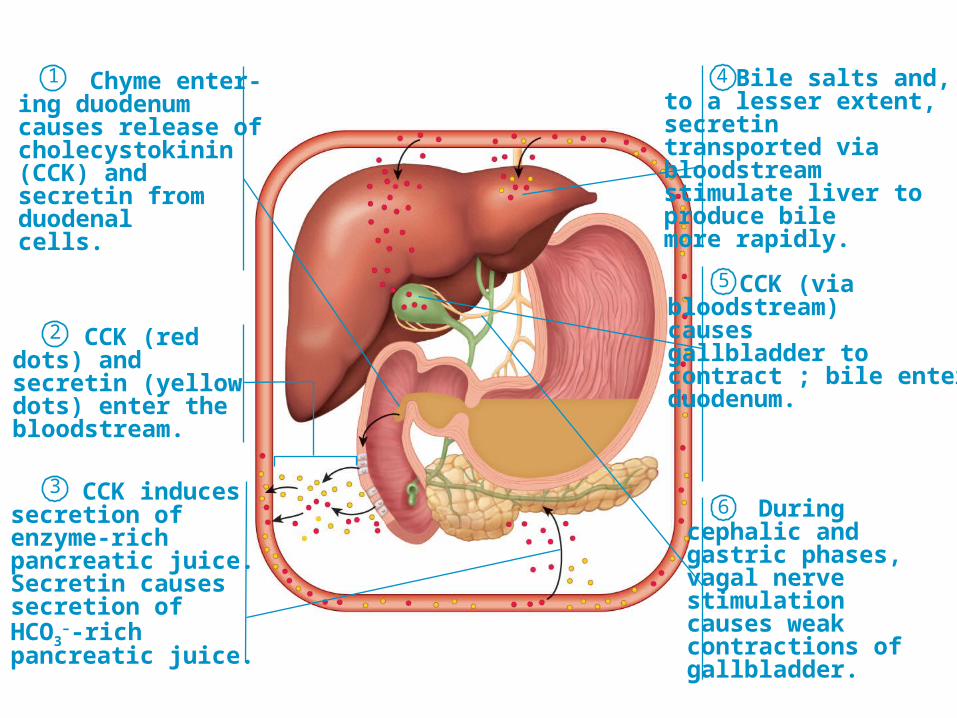
Chyme enter-ing duodenum causes release ofcholecystokinin (CCK) and secretin from duodenal cells.
CCK (red dots) and secretin (yellow dots) enter the bloodstream.
CCK induces secretion of enzyme-rich pancreatic juice. Secretin causes secretion of HCO3
–-rich pancreatic juice.
Bile salts and, to a lesser extent, secretin transported via bloodstream stimulate liver to produce bile more rapidly.
CCK (via bloodstream) causes gallbladder to contract ; bile enters duodenum.
During cephalic and gastric phases, vagal nerve stimulation causes weak contractions of gallbladder.
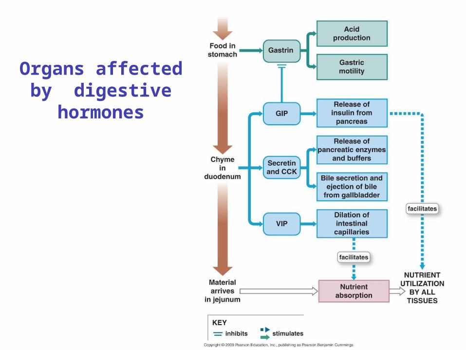
Organs affected by digestive hormones
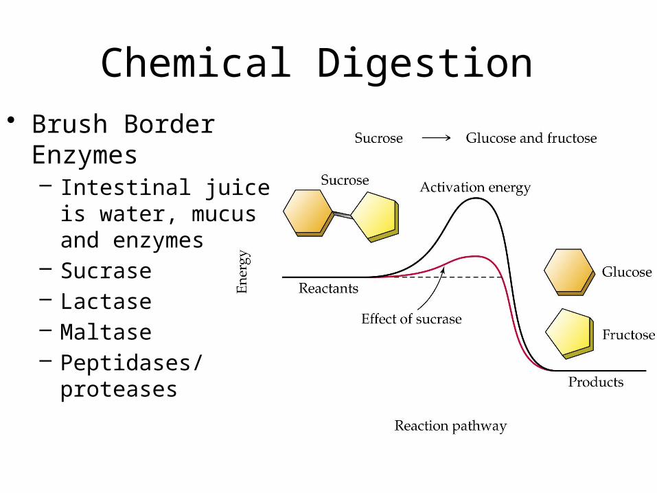
Chemical Digestion • Brush Border
Enzymes– Intestinal juice is
water, mucus and enzymes
– Sucrase– Lactase– Maltase– Peptidases/proteases
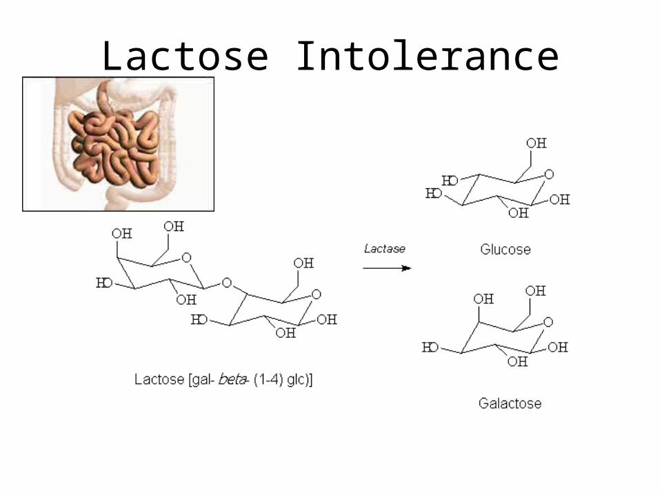
Lactose Intolerance
• Segmentation: movement of food through S.I.; moves very slowly so chemical digestion can occur
• Chemical Digestion:– Very few Enzymes come from S.I.– Need assistance from accessory organs
• Absorption: the building blocks of food are passed into blood stream by Active Transport– Most absorption is completed b4 it reaches the ileum
• Ilieum’s main function is to return bile salts to liver
– Mesentary
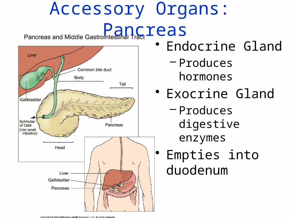
Accessory Organs: Pancreas• Endocrine Gland
– Produces hormones• Exocrine Gland
– Produces digestive enzymes
• Empties into duodenum

• Pancreatic duct fuses with Common bile duct and enters the duodenum
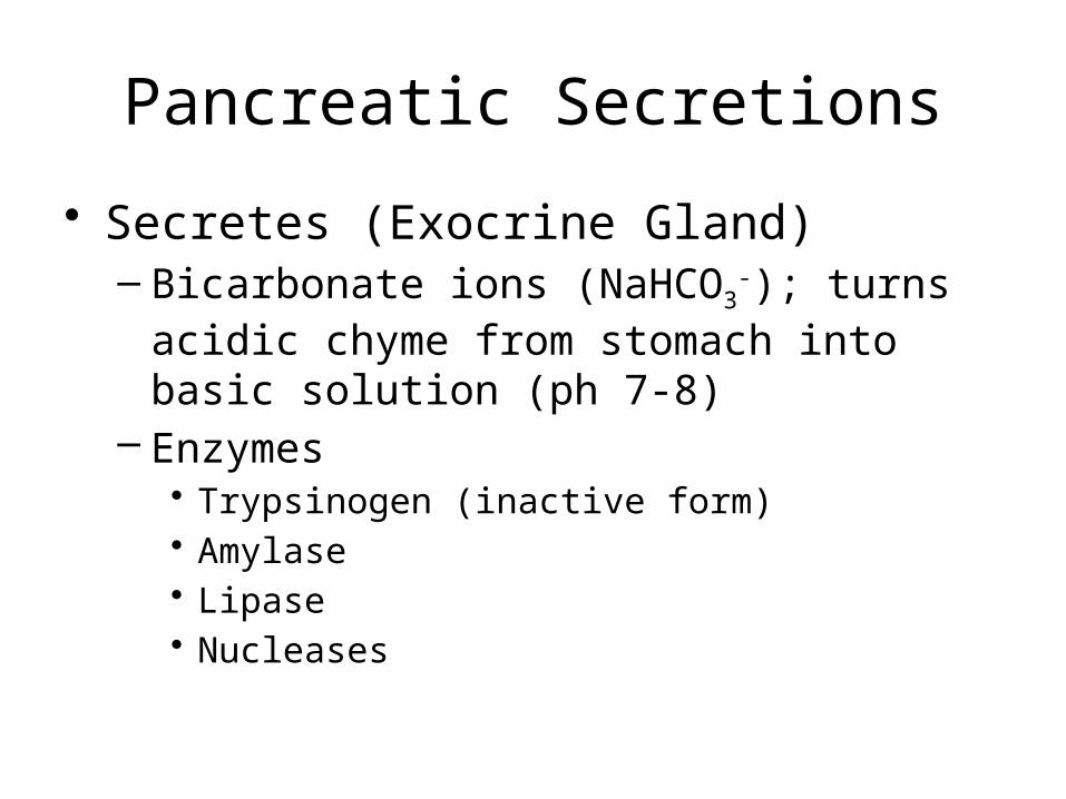
Pancreatic Secretions
• Secretes (Exocrine Gland)– Bicarbonate ions (NaHCO3
-); turns acidic chyme from stomach into basic solution (ph 7-8)
– Enzymes• Trypsinogen (inactive form)• Amylase• Lipase• Nucleases
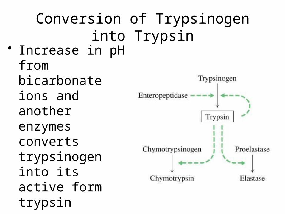
Conversion of Trypsinogen into Trypsin
• Increase in pH from bicarbonate ions and another enzymes converts trypsinogen into its active form trypsin
• Trypsin is a protease and continues protein digestion

Digestion of Lipids
•
Lipase breaks down lipids into fatty acids and glycerol.
Fatty acids and glycerol can then be absorbed into the blood stream
Bile assists in this process by physically breaking down fats into smaller droplets.
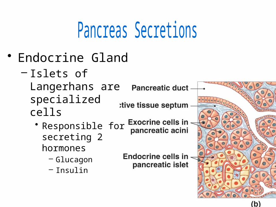
• Endocrine Gland– Islets of Langerhans
are specialized cells • Responsible for
secreting 2 hormones– Glucagon– Insulin

Anatomy of Liver and gall bladder• Liver
– Hepatic duct allows bile to leave the liver
• Gall Bladder– Lies under liver;
appears like a slightly inflated green balloon
– Stores bile• Bile leaves the liver via
the cystic duct• Cystic duct and hepatic
duct join to make Common Bile Duct.
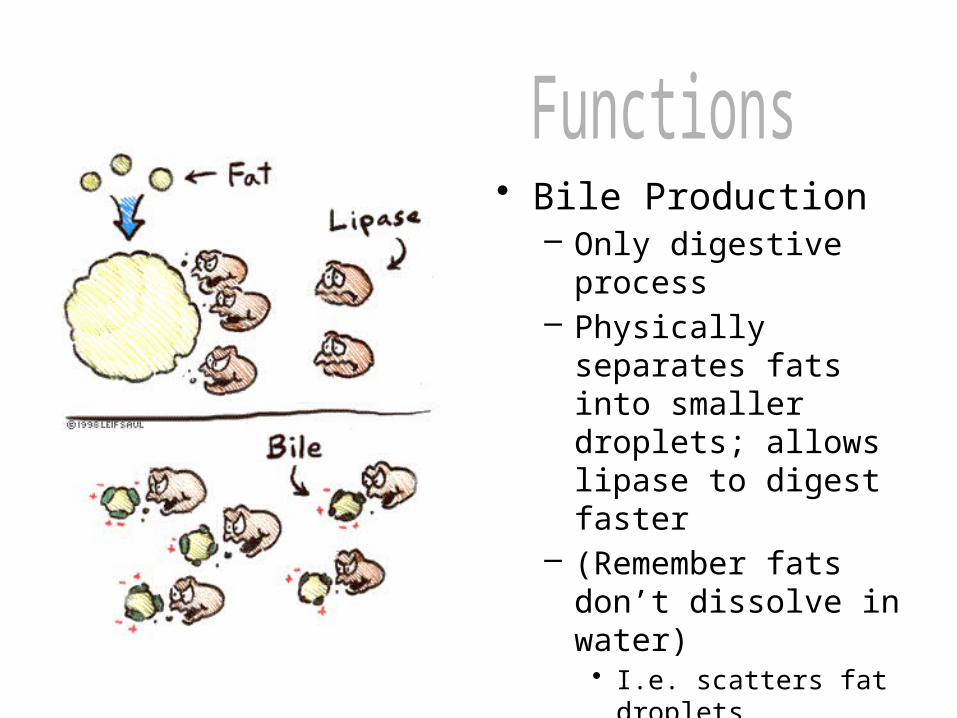
• Bile Production– Only digestive process– Physically separates
fats into smaller droplets; allows lipase to digest faster
– (Remember fats don’t dissolve in water)• I.e. scatters fat droplets
throughout solution
• Metabolic Regulation– Processes all nutrients absorbed into blood– Helps regulate cholesterol levels– Forms nonessential amino acids; byproduct is
ammonia (toxic)– Converts ammonia into urea (less toxic)– Stores glucose as glycogen (polysaccharide)– Stores fat soluble vitamins (A,D,E,K)– Detoxifies alcohol and drugs (both legal and illegal)
• Blood leaving liver has fewer waste products than the blood that enters it.

• Assists in Regulating Blood– Produces plasma
proteins– Conserves iron from
old RBC’s
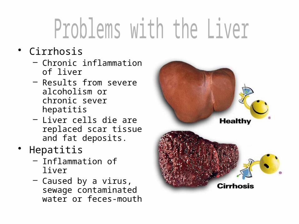
• Cirrhosis– Chronic inflammation of
liver– Results from severe
alcoholism or chronic sever hepatitis
– Liver cells die are replaced scar tissue and fat deposits.
• Hepatitis– Inflammation of liver– Caused by a virus, sewage
contaminated water or feces-mouth
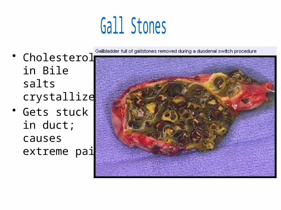
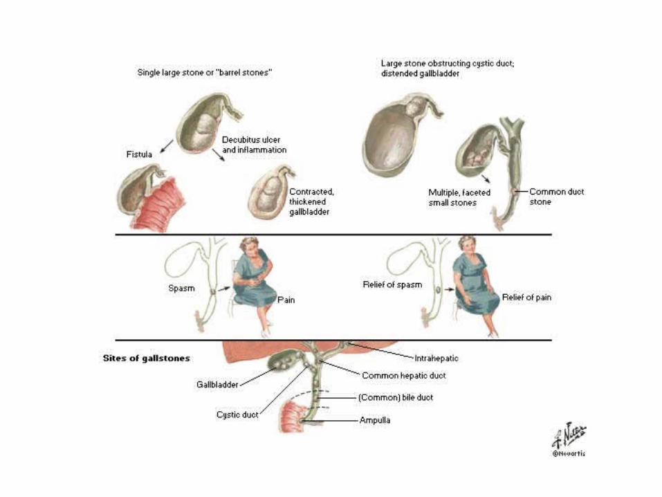
• Cholesterol in Bile salts crystallize;
• Gets stuck in duct; causes extreme pain
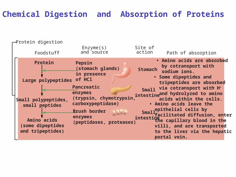
Protein digestion
• Amino acids are absorbed by cotransport with sodium ions.• Some dipeptides and tripeptides are absorbed via cotransport with H+
and hydrolyzed to amino acids within the cells.
+
• Amino acids leave the epithelial cells by facilitated diffusion, enter the capillary blood in the villi, and are transported to the liver via the hepatic portal vein.
Smallintestine
Smallintestine
Stomach
Foodstuff
Protein
Large polypeptides
Pepsin(stomach glands)in presence of HCl
Small polypeptides,small peptides
Pancreaticenzymes (trypsin, chymotrypsin,carboxypeptidase)
Amino acids(some dipeptidesand tripeptides)
Brush border enzymes(peptidases, proteases)
Path of absorptionEnzyme(s)and source
Site ofaction
Chemical Digestion and Absorption of Proteins
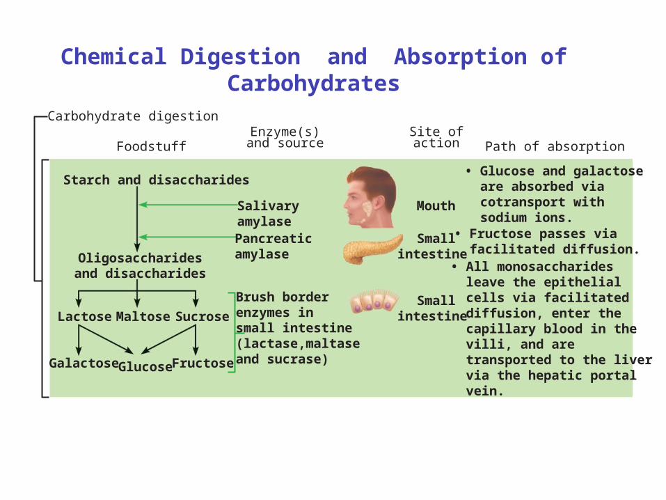
Carbohydrate digestion
• Glucose and galactose are absorbed via cotransport with sodium ions.• Fructose passes via facilitated diffusion.• All monosaccharides leave the epithelial cells via facilitated diffusion, enter the capillary blood in the villi, and are transported to the liver via the hepatic portal vein.
Starch and disaccharides
Oligosaccharidesand disaccharides
Lactose Maltose Sucrose
Glucose Fructose
Salivaryamylase
Mouth
Pancreaticamylase
Brush borderenzymes in small intestine(lactase,maltaseand sucrase)
Smallintestine
Smallintestine
Foodstuff
Galactose
Path of absorptionEnzyme(s)and source
Site ofaction
Chemical Digestion and Absorption of Carbohydrates
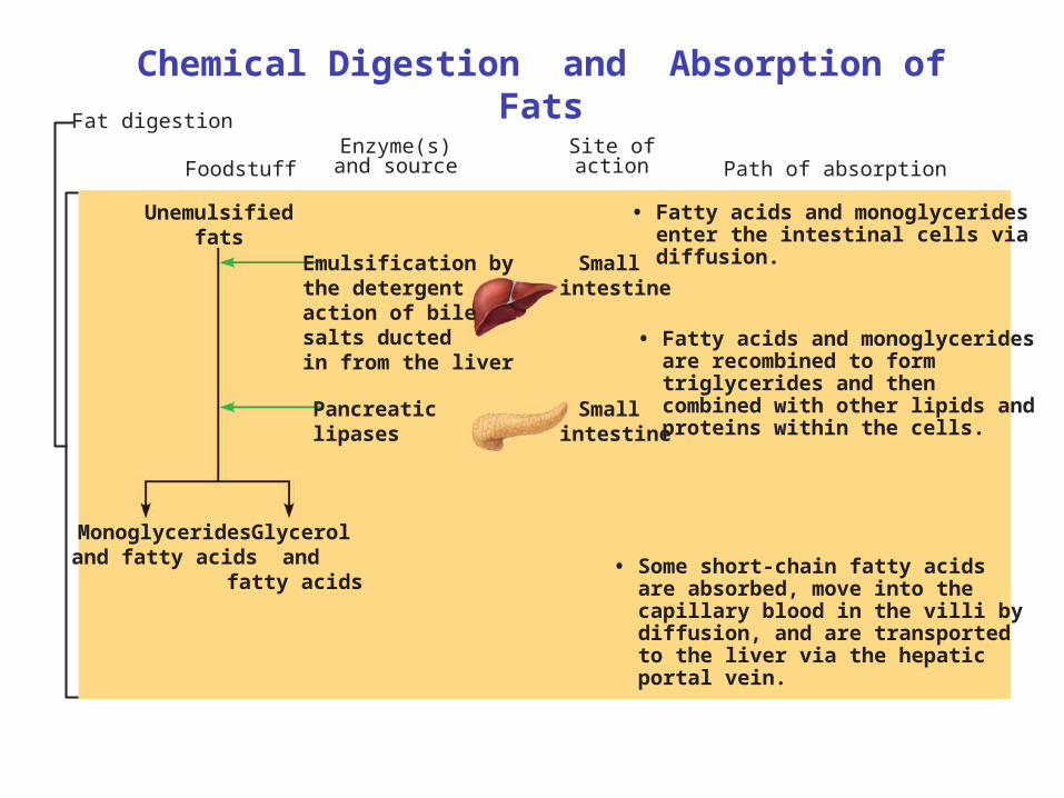
Fat digestion
Small intestine
Small intestine
Foodstuff
Unemulsifiedfats
Emulsification by the detergent action of bile salts ductedin from the liver
Pancreatic lipases
Monoglyceridesand fatty acids
Glyceroland
fatty acids
Path of absorptionEnzyme(s)and source
Site ofaction
• Fatty acids and monoglycerides enter the intestinal cells via diffusion.
• Fatty acids and monoglycerides are recombined to form triglycerides and then combined with other lipids and proteins within the cells.
• Some short-chain fatty acids are absorbed, move into the capillary blood in the villi by diffusion, and are transported to the liver via the hepatic portal vein.
Chemical Digestion and Absorption of Fats
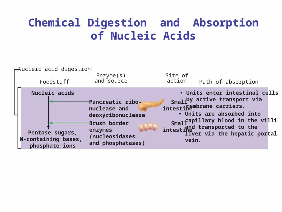
Nucleic acid digestion
• Units enter intestinal cells by active transport via membrane carriers.
• Units are absorbed into capillary blood in the villi and transported to the liver via the hepatic portal vein.
Smallintestine
Smallintestine
Foodstuff
Nucleic acids
Pancreatic ribo-nuclease and deoxyribonuclease
Brush borderenzymes(nucleosidasesand phosphatases)
Pentose sugars,N-containing bases,
phosphate ions
Path of absorptionEnzyme(s)and source
Site ofaction
Chemical Digestion and Absorption of Nucleic Acids
What remains in Small Intestine?
• Indigestible food materials (cellulose)• Lots of bacteria• Water• Passes through ileocecal valve into Large
Intestine
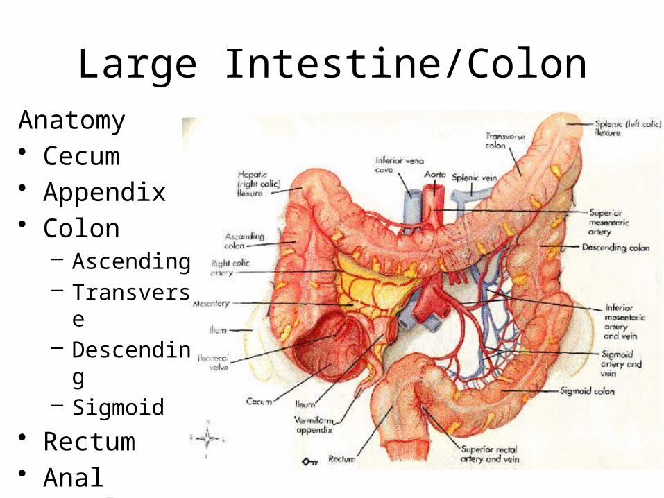
Large Intestine/ColonAnatomy• Cecum• Appendix• Colon
– Ascending– Transverse– Descending– Sigmoid
• Rectum• Anal Canal
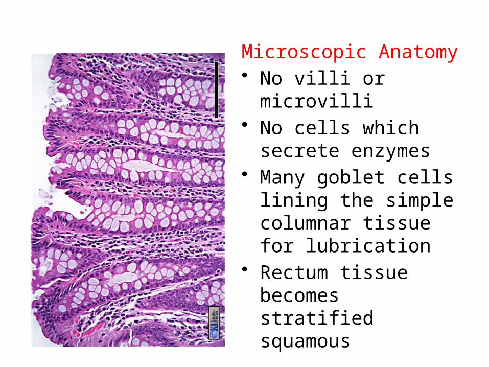
Microscopic Anatomy• No villi or microvilli• No cells which secrete
enzymes• Many goblet cells lining
the simple columnar tissue for lubrication
• Rectum tissue becomes stratified squamous
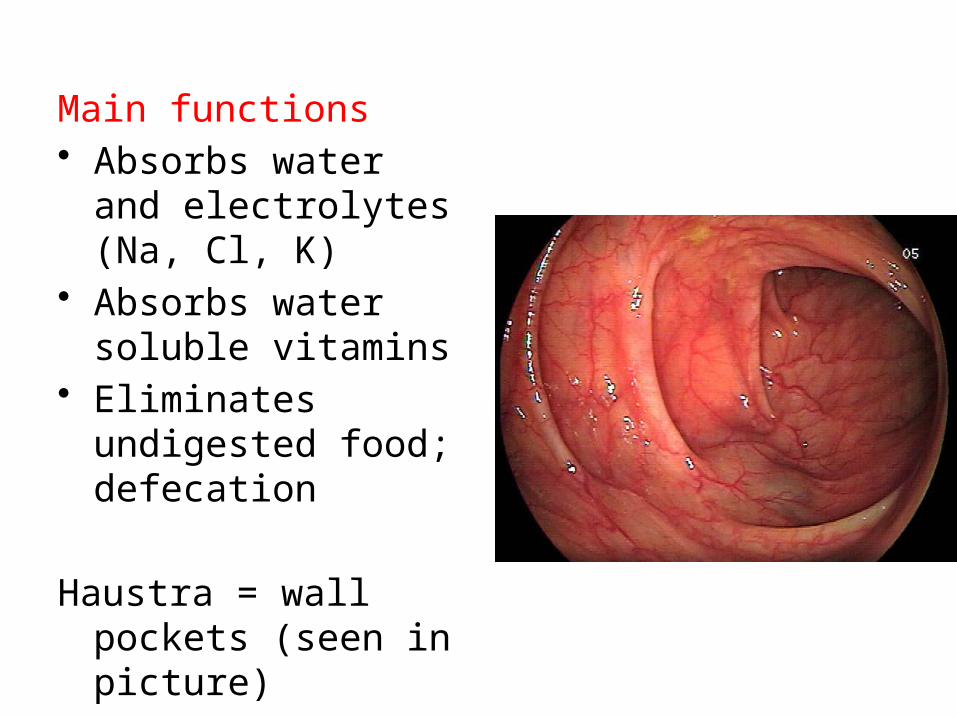
Main functions• Absorbs water and
electrolytes (Na, Cl, K)
• Absorbs water soluble vitamins
• Eliminates undigested food; defecation
Haustra = wall pockets (seen in picture)
Other Inhabitants• Bacteria
– Responsible for fermenting undigested food
– Product of these reactions is gas (H2, N2, CO2, H2S, CH4)
– Assist in synthesizing vitamin B and vitamin K.