cellular/molecular roleofestrogenreceptor and … · cellular/molecular roleofestrogenreceptor and...
TRANSCRIPT
Cellular/Molecular
Role of Estrogen Receptor � and � in PreservingHippocampal Function during Aging
Xiaoxia Han,1 Kristina K. Aenlle,2 Linda A. Bean,1 Asha Rani,1 Susan L. Semple-Rowland,1 Ashok Kumar,1
and Thomas C. Foster1
1Department of Neuroscience, McKnight Brain Institute, University of Florida, Gainesville, Florida 32610-0244, and 2Department of Veterans AffairsMedical Center, Geriatric Research, Education and Clinical Center, Miami, Florida 33125
The expression of the ER� and ER� estrogen receptors in the hippocampus may be important in the etiology of age-related cognitivedecline. To examine the role of ER� and ER� in regulating transcription and learning, ovariectomized wild-type (WT) and ER� and ER�knockout (KO) mice were used. Hippocampal gene transcription in young ER�KO mice was similar to WT mice 6 h after a single estradioltreatment. In middle-age ER�KO mice, hormone deprivation was associated with a decrease in the expression of select genes associatedwith the blood– brain barrier; cyclic estradiol treatment increased transcription of these select genes and improved learning in thesemice. In contrast to ER�KO mice, ER�KO mice exhibited a basal hippocampal gene profile similar to WT mice treated with estradiol and,in the absence of estradiol treatment, young and middle-age ER�KO mice exhibited preserved learning on the water maze. The preservedmemory performance of middle-age ER�KO mice could be reversed by lentiviral delivery of ER� to the hippocampus. These resultssuggest that one function of ER� is to regulate ER�-mediated transcription in the hippocampus. This model is supported by ourobservations that knockout of ER� under conditions of low estradiol allowed ER�-mediated transcription. As estradiol levels increasedin the absence of ER�, we observed that other mechanisms, likely including ER�, regulated transcription and maintained hippocampal-dependent memory. Thus, our results indicate that ER� and ER� interact with hormone levels to regulate transcription involved inmaintaining hippocampal function during aging.
IntroductionThe DNA binding domains of � and � estrogen receptors(ERs) are highly homologous (Katzenellenbogen and Korach,1997; Enmark and Gustafsson, 1999); however, estradiol-induced transcriptional activity differs because of divergenceof the ligand binding domain and disparities in the recruit-ment of coregulators (Nilsson et al., 2001; Harrington et al.,2003). Under low levels of estradiol, ER� transcriptional ac-tivity is greater than that of ER� (Barkhem et al., 1998;Pettersson et al., 2000). Moreover, ER� can heterodimerizewith ER� to inhibit ER�-mediated gene expression (Hall andMcDonnell, 1999; Pettersson et al., 2000; A. Gottfried-Blackmoreet al., 2007; Gonzales et al., 2008), indicating that regulation of geneexpression is dependent upon the ratio of ER� to ER� and the levelof estradiol (Foster, 2012).
Estradiol is neurotrophic (Gould et al., 1990; Woolley et al.,1990; Woolley and McEwen, 1992; Choi et al., 2003; Akama and
McEwen, 2003; Kretz et al., 2004; Rune and Frotscher, 2005; Jelkset al., 2007) and neuroprotective (Nilsen and Diaz Brinton, 2003;Dubal et al., 2006; Garcia-Segura et al., 2006; Aenlle and Foster,2010), functions that likely underlie the ability of estradiol topreserve cognitive function during aging (Foster, 2012). Interest-ingly, estradiol’s effects on cognitive aging are reduced in agingwomen (Sherwin, 2006) and rodents (Gibbs, 2000; Markham etal., 2002; Markowska and Savonenko, 2002; Foster et al., 2003;Sherwin, 2006; Bimonte-Nelson et al., 2006; Daniel et al., 2006;Talboom et al., 2008), and temporally correlated with alteredhippocampal expression of ER� and ER� (Tohgi et al., 1995;Adams et al., 2002; Mehra et al., 2005; Sharma and Thakur, 2006;Thakur and Sharma, 2007; Ishunina et al., 2007; Bohacek andDaniel, 2009). Thus, an age-related change in the ratio of ER�/ER� expression in the hippocampus may contribute to the re-duced ability of estradiol treatment to alter gene expression(Aenlle and Foster, 2010) and the reduction in the efficacy ofhormone therapy to ameliorate memory loss that often accom-panies advanced age (Foster, 2012).
In this study, we used ovariectomized ER�KO and ER�KOfemale mice to investigate how selective loss of one receptor sub-type affects behavior and gene expression profiles in aging mice.Our results are consistent with the idea that, under low estradiolconditions associated with female aging, activation of ER� helpsto regulate hippocampal gene expression and function. If ER�levels are reduced, our data suggest that ER� can compensate tomaintain hippocampal function provided that estradiol levels areincreased.
Received Oct. 19, 2012; revised Nov. 21, 2012; accepted Nov. 26, 2012.Author contributions: T.C.F. designed research; X.H., K.K.A., L.A.B., A.R., A.K., and T.C.F. performed research;
T.C.F. analyzed data; S.L.S.-R. and T.C.F. wrote the paper.This work was supported by National Institutes of Health Grants AG014979, AG037984, and AG036800, and the
Evelyn F. McKnight Brain Research Foundation. We thank Irina Madorsky, Jose Herrera, Gina Prado, Paul Huang, andKatrina Velez for technical assistance.
The authors declare no competing financial interests.Correspondence should be addressed to Dr. Thomas C. Foster, Department of Neuroscience, McKnight Brain
Institute, University of Florida, PO Box 100244, Gainesville, FL 32610-0244. E-mail: [email protected]:10.1523/JNEUROSCI.4937-12.2013
Copyright © 2013 the authors 0270-6474/13/332671-13$15.00/0
The Journal of Neuroscience, February 6, 2013 • 33(6):2671–2683 • 2671

Materials and MethodsMice. ER� �/� (ER�KO) (Lubahn et al., 1993)and ER� �/� (ER�KO) (Krege et al., 1998)mice were generated from heterozygous mousecolonies. The genotypes of the mice werescreened using PCR amplification as previ-ously described (Lubahn et al., 1993; Krege etal., 1998). Wild-type (WT) littermates pro-duced from the ER�KO and ER�KO breedingcolonies were combined into one WT group.Animals were housed 3–5 per cage and main-tained on 12:12 light/dark cycle (lights on at6:00 A.M.). All procedures involving animalsubjects were reviewed and approved by theInstitutional Animal Care and Use Committeeat the University of Florida and were per-formed in accordance with guidelines estab-lished by the U.S. Public Health ServicePolicy on Humane Care and Use of Labora-tory Animals.
Figure 1 shows the timeline for injections,behavioral training, and tissue collection foreach study, starting from the time of surgery.Young mice (WT: n � 27; ER�KO: n � 21;ER�KO: n � 25) were 4 months at the time ofsurgery. For examination of gene transcriptionprofiles, young mice were ovariectomized, and10 d after surgery were treated with a singleinjection of oil or estradiol. The hippocampi ofthese mice were collected 6 h later (Fig. 1A).The experimental groups included WT-EB(n � 7), WT-oil (n � 7), ER�KO-EB (n � 5), ER�KO-oil (n � 5),ER�KO-EB (n � 7), and ER�KO-oil (n � 5). To determine whetherknockout of either ER� or ER� influences hippocampal function inyoung mice, ER�KO (n � 11), ER�KO (n � 13), and WT (n � 13)mice were ovariectomized and their performance on the water mazetask was examined, starting 10 d after surgery and was repeated 4weeks later (Fig. 1B).
Middle-age mice (WT: n � 15; ER�KO: n � 23; ER�KO: n � 52) were13–14 months old at the time of surgery. To determine whether ER� andER� contribute to estradiol effects on behavior and gene expression dur-ing middle age, ER�KO and ER�KO mice were treated with either oil orestradiol. Treatments were initiated 1 week after surgery and were deliv-ered on two contiguous days of a 5 d cycle in accord with our previouslypublished work (Aenlle et al., 2009) (Fig. 1C). The experimental groupsincluded ER�KO-oil (n � 7), ER�KO-EB (n � 8), ER�KO-oil (n � 12),ER�KO-EB (n � 11), WT-oil (n � 15), and WT-EB (n � 14) littermates.Twenty-four hours after the last injection, hippocampi were collected forgene arrays and uterine tissue was collected and weighed. To determinewhether expression of ER� disrupts spatial learning, lentivirus encodingER� and GFP (n � 12) or GFP alone (n � 11) was bilaterally injected inthe hippocampus of middle-age ER�KO mice at the time of ovariectomy,and behavior was examined starting 5 weeks later (Fig. 1D).
Surgeries. Female mice (4 months and 13–14 months) were anesthe-tized (2 mg ketamine and 0.2 mg xylazine per 20 g of body weight), andthe ovaries were removed through a small abdominal midline incision.All mice received ad libitum access to food (Purina mouse chow) andwater until the surgery, after which time they were placed on casein-based chow (Cincinnati Lab Supply) that has lower levels of phytoestro-gens compared with soy-based chow.
Hippocampal lentiviral injections. To determine whether the behav-ioral changes observed in middle-age ER�KO mice could be attributed tothe absence of ER�, female ER�KO (13–14 months) were ovariectomizedand either pFin-EF1�-GFP-CMV-(FLAG)ER�-WPRE (ER�-FLAG) orpFIN-EF1�-GFP-WPRE (GFP) lentivirus (2 � 1012 transducing Us/�l) wasinjected bilaterally into the hippocampus 1.0 �l per side. Techniques forhippocampal injection of virus have previously been published (Foster et al.,2008; Zeier et al., 2009; Lee et al., 2012).
Estradiol treatments. Estradiol (17 �-estradiol benzoate, EB) (Sigma)was dissolved (0.1 mg/ml) in light mineral oil (Fisher Scientific) and wasinjected subcutaneously (5 �g) at the nape of the neck. In rodents,plasma estradiol levels rapidly increased within 1– 4 h after a single EBinjection (Woolley and McEwen, 1993; Sohrabji et al., 1994), and EB-induced transcriptional changes follow a similar time course (Hammer etal., 1986; Priest et al., 1995; Sohrabji et al., 1995; Too et al., 1999; Aenlle etal., 2009; Aenlle and Foster, 2010). Therefore, for young animals, a singleinjection was used to examine transcription 6 h after treatment. The doseand time point were selected to maintain consistency with our previousstudies examining the effects of estradiol on transcription (Aenlle et al.,2009; Aenlle and Foster, 2010).
To examine the effects of estradiol on behavior and transcription inmiddle-age mice, injections (5 �g) were delivered on two contiguousdays of a 5 d cycle and were initiated 1 week after surgeries in accord withour previously published work (Aenlle et al., 2009). Previous work indi-cated that repeated injections using these delivery parameters can im-prove spatial learning and memory in mice (Frick et al., 2002; Xu andZhang, 2006; Walf et al., 2008; Aenlle et al., 2009).
Water maze. Behavioral studies involving the water maze were performedin accord with our previously published work (Foster et al., 2008; Aenlle etal., 2009). All mice were first trained on the cue discrimination version of theMorris swim task using four training blocks, with three trials per block.Spatial training was initiated either 3 or 6 d after the completion of the cuediscrimination training, using methods that have been previously described(Foster et al., 2008; Aenlle et al., 2009). Briefly, spatial training was performedover a period of three consecutive days and consisted of four training blockswith three trials per block per day. The penultimate trial on days 2 and 3consisted of a probe trial that served as an index of learning. The probe trialwas performed by placing the mouse in the tank for 1 min without theplatform and recording both the time the animal spent in each quadrant ofthe tank and the number of times the animal crossed the location in the tankfrom which the platform had been removed. The spatial discriminationindex was computed using the formula (G � O)/(G � O), where G and Orepresent the percentage of time spent in the goal quadrant and quadrantopposite the goal, respectively.
RNA isolation and microarray analyses. Mice were anesthetized withCO2 and decapitated. The brain was quickly removed and placed in
Figure 1. Schedule of injections and behavioral testing. All time lines start with surgery to remove the ovaries. A, Young micewere ovariectomized and were treated 10 d later (arrowhead) with a single injection of oil (WT � 7, ER�KO � 5, ER�KO � 5) orestradiol (WT � 7, ER�KO � 5, ER�KO � 7). The hippocampi were collected 6 h after injection for examination of transcription.B, Young mice (WT � 13, ER�KO � 11, ER�KO � 13) were ovariectomized, and their performance on the cue (filled arrow)version of the water maze task was examined 10 d after surgery. Three days of training on the spatial version of the water maze(open arrows) was initiated on day 14 and again on day 43 after surgery. C, Middle-age mice were injected on 2 consecutive days(arrowheads) of every 5 d with oil (WT � 15, ER�KO � 7, ER�KO � 12) or estradiol (WT � 14, ER�KO � 8, ER�KO � 11),starting at the beginning of week 2 after ovariectomy. Arrows indicate the days of training on cue (filled arrow) and spatial (openarrows) tasks during week 5 and week 6, respectively, which was initiated 48 h after injection. At the end of training on week 6, themice were given two more series of injections and were killed for tissue collection 24 h after the last injection. D, Middle-age ER�KOmice received bilateral hippocampal injections of lentivirus to express ER�-FLAG and GFP (n � 12) or GFP alone (n � 11) at thetime of ovariectomy. Behavior was examined 5 weeks later.
2672 • J. Neurosci., February 6, 2013 • 33(6):2671–2683 Han et al. • Hippocampal Gene Expression in Knockout Mice

ice-cold artificial CSF. Both hippocampi were removed, frozen in liquidnitrogen, and stored at �80°C. Total RNA was isolated from each sampleusing a Qiagen RNeasy Lipid Tissue Mini Kit (Qiagen). RNA concentra-tion was determined using a spectrophotometer, and a subset of sampleswas examined using an Agilent 2100 Bioanalyzer (Agilent Technologies)to assess sample quality. Microarray hybridization was performed by theInterdisciplinary Center for Biotechnology Research Microarray Core,University of Florida, following the manufacturer’s protocol.
The first study examined the transcriptional response in young (4months) mice treated with estradiol. Mice were killed 6 h after injectionof either estradiol or oil, and hippocampi were prepared for gene tran-script analyses using Affymetrix Mouse 430 2.0 arrays (one array permouse) (Blalock et al., 2003; Aenlle and Foster, 2010). Normalizationand computation of gene expression values were performed using dChip(Li and Wong, 2001). The detection of signal (presence/absence) wasdetermined by MAS Version 5.0 (Affymetrix). The number of presentcalls for each probe was determined across all arrays using the criterionthat at least 80% of the chips within any single group (genotype andtreatment) had to exhibit a present call for that probe to be included insubsequent analyses. Using this criterion, 24,111 probes were selected forfurther analyses. Affymetrix GeneChip Mouse Gene 1.0 ST Arrays (onearray per mouse) were used for examination of transcription in middle-age mice, after cyclic injections of either estradiol or oil.
Quantitative PCR. Quantitative PCRs were performed using Gene Ex-pression Assay Mix TaqMan Universal PCR Master Mix (2�), and a 7300Real-Time PCR System with SDS Software Version 1.3.1 analysis soft-ware (Applied Biosystems). The PCR primers were designed to produceshort amplified products that crossed exon-exon junctions and mini-mized amplification of off-target sequences (ESR1: exons 4 and 5, 56 bp,NP_031982.1; ESR2 variant 1: exons 9 and 10, 69 bp, NP_997590.1; ESR2variant 2: exons 8 and 9, 69 bp, NP_034287.3; BTG2: exons 1 and 2, 102bp, NP_031596.1; NPAS4: exons 4 and 5, 62 bp, NP_705781.1). Eachsample was run in triplicate, and the values were normalized to GAPDHaccording to our previously described methods (Aenlle et al., 2009; Aen-lle and Foster, 2010).
Construction of lentiviral vectors. The lentiviral vector pFIN-WPREbackbone contained a 2 � 250 bp core chicken �-globin HS4 insulatorsequence in the 3�LTR and a woodchuck hepatitis post-transcriptionalregulatory element (WPRE) to enhance transgene expression (Semple-Rowland et al., 2007). A dual-promoter vector was constructed by insert-ing transgenes encoding GFP and FLAG tagged ER� into the multiplecloning site of the pFIN backbone. The transgenes were arranged head-to-tail so that transcription for both proceeded in the same direction andthe transgenes shared the same polyadenylation site located in dl.R re-gion of the 3� LTR. The EF1� (elongation factor-1 �) promoter wasamplified using PCR (5� (NotI), ATT GCG GCC GCT TTG GAG CTA A;5� (NotI), TTA GCG GCC GCC ACG ACA CCT GAA AT); and wasligated into pFIN-WPRE at the NotI site to create pFIN-EF1�-WPRE.GFP was amplified using PCR (5� (NheI), GCA GCT AGC CGC CACCAT GAG CAA; 5� (NheI-MluI-BsiWI), AAT GCT AGC ACG CGT CGTACG AGA GGC CTC AGT CAG); and was ligated into pFIN-EF1�-WPRE at the NheI site to create pFIN-EF1�-GFP-WPRE. The cytomeg-alovirus (CMV) promoter was amplified using PCR (5� (BsiWI), ATACGT ACG TAG TTC ATA GCC CAT ATA TGG; 5� (MluI-AsiSI-PacI),TAT ACG CGT GCG ATC GCT TAA TTA AGT AAG CAG TGG GTTCTC TAG T); and was ligated into pFIN-EF1�-GFP-WPRE using theBsiWI and MluI sites to create pFIN-EF1�-GFP-CMV-WPRE. Finally,the open-reading frame encoding human FLAG-tagged ER� was ampli-fied from pcDNA4/TO-FLAG:hERb (kind gift from Dr. Chegini) usingprimers that introduced PacI sites on the 5�- and 3�-ends of the codingregion (5�-ATA TTA ATT AAA AAC TTA AGC TTA CCG CCA TG;5�-ATA TTA ATT AAC CCT CTA GAT CAC TGA GAC). The PCRproduct (1679 bp) was then ligated into pFIN-EF1�-GFP-CMV-WPREat the PacI site to produce pFin-EF1�-GFP-CMV-(FLAG)ER�-WPRE.The integrity of FLAG-ER� in the final vector was verified by sequencing.
The functionality of (FLAG)ER� was confirmed using previously pub-lished methods (Foster et al., 2008). The ability of the receptor to respondto estradiol was examined by cotransfecting HEKT293 cells with pFin-EF1�-GFP-CMV-(FLAG)ER�-WPRE and ERE-TA-SEAP reporter plas-
mids and measuring changes in secreted placental alkaline phosphataseafter exposure to estradiol. The bicistronic lentiviral vector encodingGFP and (FLAG)ER� was packaged into lentivirus using a three plasmidpackaging system as previously described (Semple-Rowland et al., 2007).Viral titers were estimated using a Lenti-X qRT-PCR kit (Millipore Bio-science Research Reagents) and typically averaged 2 � 10 12 viral ge-nomes per milliliters.
Statistical analyses. For measures of behavior and uterine weight,ANOVAs were used to establish main effects and interactions. Follow-upANOVAs and Fisher’s protected least significant difference post hoc com-parisons ( p � 0.05) were used to identify significant comparisons. One-tailed Student’s t tests ( p � 0.05) were used to determine whether thediscrimination indices, calculated from the quadrant search behavior(Foster et al., 2003), were different from that expected by chance (i.e., adiscrimination index � 0). Two-tailed t tests ( p � 0.025) were used toidentify differentially expressed probes according to previously pub-lished work (Aenlle et al., 2009; Aenlle and Foster, 2010).
ResultsGene expression in the hippocampus of young adult ER�KOand ER�KO miceWe have previously shown that a single 5 �g estradiol injectionadministered to young mice produces robust changes in hip-pocampal gene expression 6 h after treatment (Aenlle and Foster,2010). To examine the role of ER� and ER� in regulating hip-pocampal gene expression, we examined the gene expressionprofiles of 4-month-old ER�KO and ER�KO female mice thatwere ovariectomized and subsequently treated with estradiol 10 dafter surgery and compared them with control animals that weretreated with oil vehicle (Fig. 1A).
Analysis of differential gene expression after estradiol treat-ment revealed that there were 1295 probes that were differentiallyexpressed between WT-EB and WT-oil-treated mice (Fig. 2A).Within the set of 1295 probes, 585 (45%) of these increased inexpression and 710 (55%) decreased in expression. Only fiveprobes showed a �2-fold increase in expression. ER� and ER�KO mice were less responsive to acute estradiol treatment thanWT animals as measured by changes in gene expression. Theexpression levels of 268 and 195 probes in the ER�KO-EB andER�KO-EB treatment groups, respectively, were altered com-pared genotype-matched oil treatment controls. None of thegene expression changes in the estradiol-treated ER�KO andER�KO mice were �2-fold. These results indicate that, com-pared with genotype-matched and oil-treated animals, youngER� and ER� KO mice are less responsive to estradiol treatmentthan WT mice.
To determine whether knockout of either the ER� or the ER�receptor alters gene expression, we examined gene expressionprofiles in ER�KO and ER�KO animals treated with oil or estra-diol and compared them with the gene expression profiles ofWT-oil-treated mice (Fig. 2B). ER�KO mice treated with oil ex-hibited very few differences in gene expression (183 probes) com-pared with the WT-oil controls. The expression levels of fiveER�KO-oil-treated probes increased �2-fold, three of whichrepresented the ER� gene (ESR1 Affymetrix probes: 1453145_at,1460591_at, and 1457877_at). This result suggests that func-tional knockout of the ER�, resulting from insertion of a neo-mycin cassette, induces a compensatory increase in ESR1transcription to counter loss of ER� receptor activity. Compari-son of ER�KO-EB-treated mice with WT-oil revealed 2368 dif-ferentially expressed probes. The level of expression of themajority of these probes increased in ER�KO-EB-treated mice(1583 increasing and 785 decreasing) (Fig. 2B). Of the probes thatshowed an increase in expression, 72 increased at least twofold
Han et al. • Hippocampal Gene Expression in Knockout Mice J. Neurosci., February 6, 2013 • 33(6):2671–2683 • 2673

and included the three probes for ESR1.No probe decreased by �2-fold. Collec-tively, these data show that ER�KO miceare responsive to estradiol treatments, aresult that supports the idea that ER� cancompensate for the loss of ER� (Fugger etal., 2000; Foster et al., 2008).
Unlike ER�KO mice, the expressionlevels of 1125 probes exhibited changes inER�KO mice treated with oil comparedwith WT-oil controls (Fig. 2B). The ma-jority of these probes (912, 81%) showedan increase in expression in ER�KO-oil-treated animals. Moreover, the expressionof 196 of these probes increased �2-fold,including the probe for the ER� gene(ESR2: 1426103_a_at); however, ER�probe was not analyzed further becausethe number of present calls for this probeacross all groups did not reach the criterion.Of the 213 probes that showed a decrease inexpression, none showed a decrease �2-fold. Comparisons of ER�KO-EB mice withWT-oil controls revealed that 1142 probeswere differentially expressed in theER�KO-EB mice (698 increasing and 444decreasing; Fig. 2B). Of these, the expressionof 3 probes increased and one decreasedwith magnitudes �2-fold. Thus, in contrastto ER�KO mice whose basal gene expres-sion levels were similar to WT animals,ER�KO mice exhibit a considerable shift inbasal gene expression, a shift that may reflectremoval of the inhibitory influences of ER�on ER�-mediated transcription (Petterssonet al., 2000; Williams et al., 2008).
To visualize the changes in basal and estradiol-induced geneexpression, the mean expression levels for probes in each groupwere normalized to the average expression levels of the corre-sponding probes in WT-oil-treated mice. WT probes whose ex-pression significantly increased in response to estradiol treatmentwere sorted by the magnitude of their increases, and the percent-age change values for the top 100 probes were plotted with thenormalized measures for ER�KO (Fig. 2C) or ER�KO (Fig. 2D)mice. The levels of basal gene expression in ER�KO-oil mice wereslightly higher than those in WT-oil mice (i.e., 100%). Estradioltreatment of ER�KO mice further increased gene expression sothat the levels of gene expression were similar to WT-EB (Fig.2C). Gene expression levels in ER�KO mice were always higherthan those in WT-oil mice regardless of treatment (Fig. 2D) andwere similar to those found in WT-EB mice. Indeed, in contrastto the estradiol-mediated increase in gene expression in WT andER�KO mice, a paired t test of the normalized values revealedthat expression of these genes was reduced [(t(99) � 4.94, p �0.0001) in ER�KO-EB (155 � 2% of WT-oil) compared withER�KO-oil mice (172 � 6% of WT-oil). Our analyses ofestradiol-induced changes in hippocampal gene expression areconsistent with reports of gene expression changes in other tis-sues responsive to estradiol (Lindberg et al., 2003; O’Lone et al.,2007) and suggest that, under normal conditions, ER� inhibitsER�-mediated transcription. The observation that ER�KO-oilmice exhibit a transcription profile similar to WT-EB mice isconsistent with this idea.
To determine whether basal gene expression patterns inER�KO and ER�KO mice influence hippocampal function,4-month-old ER�KO, ER�KO, and WT mice were ovariecto-mized and their performances on a cue discrimination watermaze task were examined 10 d after surgery. A repeated-measuresANOVA indicated that there was a decrease in distance to find theescape platform [F(3,102) � 7.73, p � 0.0001] over the four blocksof training, in the absence of a genotype difference (Fig. 3A).Examination of spatial discrimination over 3 d of training (ses-sion 1) indicated that there was a decrease in the escape pathlength [F(2,68) � 11.62, p � 0.0001] in the absence of a genotypedifference (Fig. 3B). Repeated-measures ANOVAs of the probetrial data for the penultimate trial on days 2 and 3 indicated thatthere was no training effect and no genotype difference for thediscrimination index scores or platform crossings (Fig. 3C, D).However, examination of training effects in each group indicatedthat ER�KO mice did show a training effect for platform crossing[F(1,12) � 5.44, p � 0.05]. To determine whether any group useda spatial search strategy, we compared the discrimination indexscores relative to chance (score � 0) performance using one-tailed t tests. Only ER�KO mice exhibited acquisition of a spatialsearch strategy, with a discrimination index score above chance(p � 0.05) at the end of training on day 3 (Fig. 3C).
To determine whether the duration of hormone deprivationresulting from ovariectomy influences spatial learning, ovariec-tomized ER�KO, ER�KO, and WT mice were retested on thespatial version of the water maze 1 month after the first trainingsession (session 2) using a new spatial location. Similar to the
Figure 2. Gene expression in young (4 months) WT and ERKO mice, 6 h after estradiol (WT � 7, ER�KO � 5, ER�KO � 7) oroil (WT � 7, ER�KO � 5, ER�KO � 5) treatment. A, Graphic summary of the total number of probes whose expression eithersignificantly increased (open) or decreased (filled) in WT-EB, ER�KO-EB, and ER�KO-EB mice compared with genotype-matchedoil-treated mice. B, Comparison of hippocampal gene expression in oil and estradiol-treated ERKO mice relative to WT-oil mice.ER�KO-oil-treated mice exhibited a negligible shift in basal gene expression and a robust shift in expression after estradioltreatment relative to WT-oil, suggesting that ER�KO mice remain responsive to estradiol treatment. In contrast, ER�KO-oil-treated mice exhibited a relatively large shift in basal gene expression, with the majority of probes (81%) showing increases inexpression. The numbers of probes in ER�KO treated with oil or estradiol that differed from WT-oil were similar. C, D, The meanexpression for each probe within each group was normalized to the expression level of the probe in WT-oil-treated mice, which wasset at 100%. The top 100 genes that showed an increase in expression in WT-EB mice were sorted by percentage change andplotted (solid line) along with their expression in ER�KO-oil (C, blue circles) and ER�KO-EB (C, red circles) and ER�KO-oil (D, bluecircles) and ER�KO-EB (D, red circles) mice. In general, expression of probes in for the ER�KO-oil group was slightly greater thanWT-oil (i.e., 100%) but less than in WT-EB. Expression increases observed in the ER�KO-EB group were similar to those observedin WT-EB mice, suggesting that ER�KO mice remain responsive to estradiol treatment. For ER�KO-oil and ER�KO-EB mice, probeexpression was greater than WT-oil and appeared to track expression in WT-EB mice, results suggesting that knockout of ER�permits expression of estradiol-sensitive genes in the absence of estradiol treatment.
2674 • J. Neurosci., February 6, 2013 • 33(6):2671–2683 Han et al. • Hippocampal Gene Expression in Knockout Mice

previous learning data, an effect of training [F(2,68) � 13.85, p �0.0001] on the escape path length was observed in the absence ofa genotype difference (Fig. 3B), and no training or genotype dif-ferences were observed for the discrimination index scores andplatform crossing. Examination of training effects in each geno-type indicated that only the ER�KO mice showed a training effectfor the discrimination index [F(1,12) � 6.88, p � 0.05] and forplatform crossings [F(1,12) � 8.08, p � 0.05]. Unlike the earliertests in which only the ER�KO acquired a spatial search strategy,one-tailed t tests indicated that ER�KO and WT mice acquired aspatial search strategy by day 2 and all groups performed abovechance for day 3 (Fig. 3C). Finally, examination of platformcrossings indicated that there was a significant genotype effect onday 3 [F(2,34) � 3.38, p � 0.05]. Post hoc tests indicated that theperformances of ER�KO mice were better than those observed inWT mice and that ER�KO mice exhibited a tendency (p � 0.085)to cross the platform more often than ER�KO mice. Together,these results indicate that ER�KO mice exhibited a modest, al-though significant, improvement in spatial learning comparedwith the other groups.
ER� and ER� influences on hippocampal function and geneexpression in middle-age miceThe effect of ovariectomy and estradiol treatment on hippocam-pal function varies across the life span (Foster, 2005). Previouswork indicates that cyclic injections of estradiol can improve spa-tial learning and memory in mice, including middle-age mice(Frick et al., 2002; Gresack and Frick, 2006; Xu and Zhang, 2006;Walf et al., 2008; Aenlle et al., 2009). To determine whether ER�and ER� contribute to the observed effects of estradiol on behav-ior and gene expression during middle age, ER�KO and ER�KOmice were ovariectomized at 13 months of age, treated with oil orestradiol, behaviorally tested, and killed to examine hippocampal
and uterine tissues (Fig. 1C). Uterineweight across experimental groups exhib-ited an interaction of genotype and treat-ment [F(2,61) � 5.1, p � 0.01] (Fig. 4). Posthoc analyses confirmed that estradioltreatment increased uterine weight in WTand ER�KO mice, but not in ER�KOmice (Lindberg et al., 2002). Furthermore,uterine weight was higher in ER�KO-oilmice compared with ER�KO-oil and WT-oil mice. These results are consistent withthe view that ER� induces uterine growthin response to estradiol treatment andthat ER� normally dampens ER� activ-ity (i.e., knockout of ER� promotesuterine growth in the absence of estra-diol treatment).
Cue discrimination task training wasperformed during week 5 after surgery,48 h after the fifth series of cyclic injec-tions of estradiol or oil (Fig. 1C). Signifi-cant effects of training [F(3,183) � 30.28p � 0.0001] and genotype [F(2,183) � 3.35,p � 0.05] were observed for the distancesswam to escape the pool during cue dis-crimination training (Fig. 5A). Post hoctests of genotype effects on swim distancecollapsed across training blocks, andtreatment groups indicated that the dis-tances swum by ER�KO and WT mice
were less than those swum by ER�KO mice. Separate ANOVAswithin each treatment group indicated a tendency (p � 0.055) forgenotype differences between oil-treated mice, and post hoc testson distance collapsed across training blocks indicated that theswim distances for ER�KO-oil mice were less than those forER�KO-oil mice (Fig. 5B). Together, these results indicate that,in the absence of estradiol treatment, middle-age ER�KO miceexhibit better cue discrimination learning than ER�KO mice.
Spatial discrimination training was initiated on week 6 aftersurgery, 48 h after the sixth series of cyclic injections of eitherestradiol or oil, and was continued for 3 consecutive days (Fig.1C). An ANOVA on escape path length revealed significant train-ing effects [F(2,122) � 22.42 p � 0.0001] and genotype effects[F(2,122) � 3.47, p � 0.05] (Fig. 5C). Post hoc tests of the genotypeeffects on escape path length collapsed across days and treatmentgroups indicated that the escape path lengths of ER�KO micewere shorter than those for ER�KO and WT mice (p � 0.05).ANOVAs within each treatment group revealed that there was aneffect of genotype in oil-treated mice [F(2,62) � 3.9, p � 0.05], andpost hoc tests of these mice indicated that the escape path lengthsfor ER�KO mice were less than those for ER�KO and WT mice(Fig. 5D). Thus, the effects of genotype on spatial discriminationlearning appear to be opposite to those observed for cue discrim-ination learning; in the absence of estradiol treatment, middle-age ER�KO mice exhibit better spatial discrimination learningthan ER�KO and WT mice, a difference that disappeared afterestradiol treatment.
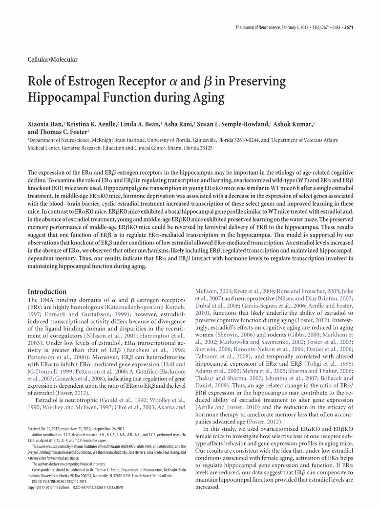
The ability of estradiol treatment to restore spatial discrimi-nation learning in ER�KO and WT mice to ER�KO performancelevels was confirmed by the probe trial data. Repeated-measuresANOVAs on the discrimination index measures for probe trialsdelivered as the penultimate trial on days 2 and 3 (Fig. 6A) indi-cated a training by treatment interaction [F(1,61) � 5.26, p � 0.05]
Figure 3. Water maze performance of young ERKO and WT mice. A, Mean path length to reach an escape platform over fourtraining blocks (three trials per block) for WT (filled circles, n � 13), ER�KO (gray circles, n � 11), and ER�KO (open circles, n �13). A decrease in path length was observed in the absence of a genotype effect. B, Mean path length to reach an escape platformduring spatial training. Training was provided in two sessions, starting 13 d after ovariectomy (session 1) and again 30 d afterovariectomy (session 2). For each session, a decrease in path length was observed in the absence of a genotype effect. C, Discrim-ination index calculated from performance on probe trials for WT (filled bars), ERKO (gray bars), and ERKO (open bars). Probe trialswere delivered as the penultimate trial on days 2 and 3 for each session. *Performance above chance and acquisition of a spatialsearch strategy. D, Platform crossing calculated from probe trial performance. #Significant difference between ER�KO and WTmice.
Han et al. • Hippocampal Gene Expression in Knockout Mice J. Neurosci., February 6, 2013 • 33(6):2671–2683 • 2675

and a trend for an interaction of training and genotype (p �0.08). Follow-up ANOVAs for each treatment group indicatedtraining effects for estradiol-treated mice [F(1,30) � 9.84, p �0.005], and an ANOVA for each day indicated a treatment effecton day 3 for the discrimination index (p � 0.05) that reflectedimproved performance of mice treated with estradiol. Examina-tion of training effects for each genotype indicated that onlyER�KO mice showed an improvement in performance [F(1,22) �5.37, p � 0.05]. Post hoc comparisons indicated that the perfor-mance of ER�KO mice on day 3 was significantly better than theperformance of the WT (p � 0.05; Fig. 6A). Finally, one-tailed ttests comparing search behavior to chance indicated that allestradiol-treated groups exhibited a search pattern focused on thegoal quadrant on day 3 (p � 0.05). For oil-treated mice, onlyER�KO-oil mice exhibited performances different from chance(Fig. 6A). A repeated-measures ANOVAon the number of platform crossings fordays 2 and 3 indicated an effect of training[F(1,61) � 7.16, p � 0.01] and a trend (p �0.09) for a genotype effect. Examinationof training effects in each genotype indi-cated that only ER�KO mice improvedperformance [F(1,22) � 4.81, p � 0.05].Post hoc comparisons of genotype differ-ences for each day indicated that ER�KOmice made more crossings than WT mice(p � 0.05) on day 3 (Fig. 6B). Thus, allgroups treated with estradiol appeared tohave acquired a spatial search strategy bythe third day of training, and the perfor-mances of ER�KO-oil mice were superiorto those of the other groups treated withoil (Fig. 6B).
For gene profiling, middle-age mice(4 – 6 mice per group) were examined 24 hafter the eighth series of estradiol or oilinjections (Fig. 1C) using AffymetrixGeneChip Mouse Gene 1.0 ST Arrays, onearray per mouse. First, we identified dif-ferentially expressed genes (p � 0.025)between oil and estradiol-treated micewithin each genotype (Fig. 7A). A total of2089 probes were found to be differen-tially expressed between WT-EB and WT-oil-treated mice, the majority of which (1324, 63%) exhibitedreduced expression. The expression level of one probe was re-duced by �2-fold, and the expression of 2 probes increased �2-fold. The number of probes exhibiting differential expression inER�KO-EB mice compared with ER�KO-oil mice was 916,half the number observed for WT mice. Although the majorityof probes for ER�KO-EB mice exhibited reduced expression(509, 56%), no probe was observed whose expression decreased�2-fold. In contrast, 31 probes exhibited a �2-fold increase inexpression. Interestingly, the basal expression levels of these 31probes in ER�KO-oil mice were lower than observed in WT-oilmice (Fig. 7B). These highly responsive probes have been associ-ated with the cells of the blood– brain barrier and choroid plexus(Lein et al., 2007) (Table 1). Thus, our analyses of middle-ageER�KO mice under low estradiol conditions suggest that there isa large shift in the expression of select genes associated with theblood– brain barrier and that their expression is restored to nor-mal levels by estradiol treatment. Finally, the changes in geneexpression for ER�KO-EB relative to ER�KO-oil mice were in-
termediate between WT and ER�KO mice, with 1681 probesshowing differential expression. In this case, 938 probes (56%)increased expression, and the increase for only one probe was�2-fold.
Next, we examined the differences in basal and estradiol-induced gene expression in ER�KO and ER�KO mice relative toWT-oil-treated mice (Fig. 8). ER�KO exhibited little change inthe number of differentially expressed genes after either oil orestradiol treatments. ER�KO-oil mice exhibited approximatelytwice as many differentially expressed probes as ER�KO-oil mice;estradiol treatment induced a fourfold increase in the number ofdifferentially expressed genes in ER�KO mice compared withER�KO mice (Fig. 8B). Five or fewer probes exhibited a foldchange �2 within each group. ER�KO and ER�KO mice do notexpress the full-length transcripts for ER� and ER�, respectively,because of a neomycin cassette insert that functionally knocks outthe gene. Therefore, it was notable that there was a threefoldincrease in the level of the transcript encoding the nonfunctionalER� (ESR2, gene ID 10401035) in ER�KO mice treated with
Figure 4. Uterine weight was increased by estradiol treatment (open bars) relative to oiltreatment (filled bars) in ER�KO and WT mice. The numbers in each bar indicate the number ofanimals in each group. *Significant ( p�0.05) differences between estradiol and oil treatment.#Significant difference between oil-treated WT and ER�KO animals relative to ER�KO oil-treated mice.
Figure 5. Water maze performance of middle-age ERKO and WT mice treated with estradiol (WT � 14, ER�KO � 8, ER�KO �11) or oil (WT � 15, ER�KO � 7, ER�KO � 12). A, Mean path length to reach an escape platform plotted over four training blocksduring cue discrimination training for ER�KO (gray circles and bars), ER�KO (open circles and bars), and WT mice (filled circles andbars). B, Mean path length (� SEM) across all cue discrimination training blocks grouped by treatment. C, Mean path length toreach an escape platform during spatial training. Spatial discrimination training was initiated on week 6 after surgery, 48 h afterthe previous injection of either estradiol or oil, and was continued for 3 consecutive days. D, Mean path length (� SEM) across alldays of spatial training grouped by treatment. #Significant differences from the ER�KO-oil group.
2676 • J. Neurosci., February 6, 2013 • 33(6):2671–2683 Han et al. • Hippocampal Gene Expression in Knockout Mice
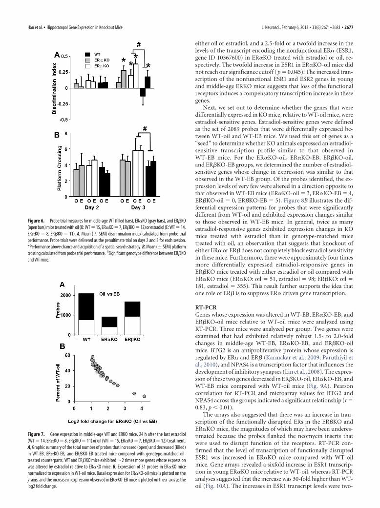
either oil or estradiol, and a 2.5-fold or a twofold increase in thelevels of the transcript encoding the nonfunctional ER� (ESR1,gene ID 10367600) in ER�KO treated with estradiol or oil, re-spectively. The twofold increase in ESR1 in ER�KO-oil mice didnot reach our significance cutoff (p � 0.045). The increased tran-scription of the nonfunctional ESR1 and ESR2 genes in youngand middle-age ERKO mice suggests that loss of the functionalreceptors induces a compensatory transcription increase in thesegenes.
Next, we set out to determine whether the genes that weredifferentially expressed in KO mice, relative to WT-oil mice, wereestradiol-sensitive genes. Estradiol-sensitive genes were definedas the set of 2089 probes that were differentially expressed be-tween WT-oil and WT-EB mice. We used this set of genes as a“seed” to determine whether KO animals expressed an estradiol-sensitive transcription profile similar to that observed inWT-EB mice. For the ER�KO-oil, ER�KO-EB, ER�KO-oil,and ER�KO-EB groups, we determined the number of estradiol-sensitive genes whose change in expression was similar to thatobserved in the WT-EB group. Of the probes identified, the ex-pression levels of very few were altered in a direction opposite tothat observed in WT-EB mice (ER�KO-oil � 3, ER�KO-EB � 4,ER�KO-oil � 0, ER�KO-EB � 5). Figure 8B illustrates the dif-ferential expression patterns for probes that were significantlydifferent from WT-oil and exhibited expression changes similarto those observed in WT-EB mice. In general, twice as manyestradiol-responsive genes exhibited expression changes in KOmice treated with estradiol than in genotype-matched micetreated with oil, an observation that suggests that knockout ofeither ER� or ER� does not completely block estradiol sensitivityin these mice. Furthermore, there were approximately four timesmore differentially expressed estradiol-responsive genes inER�KO mice treated with either estradiol or oil compared withER�KO mice (ER�KO: oil � 51, estradiol � 98; ER�KO: oil �181, estradiol � 355). This result further supports the idea thatone role of ER� is to suppress ER� driven gene transcription.
RT-PCRGenes whose expression was altered in WT-EB, ER�KO-EB, andER�KO-oil mice relative to WT-oil mice were analyzed usingRT-PCR. Three mice were analyzed per group. Two genes wereexamined that had exhibited relatively robust 1.5- to 2.0-foldchanges in middle-age WT-EB, ER�KO-EB, and ER�KO-oilmice. BTG2 is an antiproliferative protein whose expression isregulated by ER� and ER� (Karmakar et al., 2009; Paruthiyil etal., 2010), and NPAS4 is a transcription factor that influences thedevelopment of inhibitory synapses (Lin et al., 2008). The expres-sion of these two genes decreased in ER�KO-oil, ER�KO-EB, andWT-EB mice compared with WT-oil mice (Fig. 9A). Pearsoncorrelation for RT-PCR and microarray values for BTG2 andNPAS4 across the groups indicated a significant relationship (r �0.83, p � 0.01).
The arrays also suggested that there was an increase in tran-scription of the functionally disrupted ERs in the ER�KO andER�KO mice, the magnitudes of which may have been underes-timated because the probes flanked the neomycin inserts thatwere used to disrupt function of the receptors. RT-PCR con-firmed that the level of transcription of functionally disruptedESR1 was increased in ER�KO mice compared with WT-oilmice. Gene arrays revealed a sixfold increase in ESR1 transcrip-tion in young ER�KO mice relative to WT-oil, whereas RT-PCRanalyses suggested that the increase was 30-fold higher than WT-oil (Fig. 10A). The increases in ESR1 transcript levels were two-
Figure 7. Gene expression in middle-age WT and ERKO mice, 24 h after the last estradiol(WT � 14, ER�KO � 8, ER�KO � 11) or oil (WT � 15, ER�KO � 7, ER�KO � 12) treatment.A, Graphic summary of the total number of probes that increased (open) and decreased (filled)in WT-EB, ER�KO-EB, and ER�KO-EB-treated mice compared with genotype-matched oil-treated counterparts. WT and ER�KO mice exhibited 2 times more genes whose expressionwas altered by estradiol relative to ER�KO mice. B, Expression of 31 probes in ER�KO micenormalized to expression in WT-oil mice. Basal expression for ER�KO-oil mice is plotted on they-axis, and the increase in expression observed in ER�KO-EB mice is plotted on the x-axis as thelog2 fold change.
Figure 6. Probe trial measures for middle-age WT (filled bars), ER�KO (gray bars), and ER�KO(open bars) mice treated with oil (O: WT�15, ER�KO�7, ER�KO�12) or estradiol (E: WT�14,ER�KO � 8, ER�KO � 11). A, Mean (� SEM) discrimination index calculated from probe trialperformance. Probe trials were delivered as the penultimate trial on days 2 and 3 for each session.*Performance above chance and acquisition of a spatial search strategy. B, Mean (� SEM) platformcrossing calculated from probe trial performance. #Significant genotype difference between ER�KOand WT mice.
Han et al. • Hippocampal Gene Expression in Knockout Mice J. Neurosci., February 6, 2013 • 33(6):2671–2683 • 2677

fold and eightfold higher for middle-age ER�KO mice using genearrays or RT-PCR, respectively (Fig. 10B). Similarly, ESR2 tran-script levels were higher in ER�KO mice (Fig. 10C, D) relative toWT-oil, and the observed increases determined using RT-PCRwere greater than those obtained from the gene arrays.
Viral expression of ER� impairs spatial learning inER�KO miceOur experimental data indicate that, in the absence of estradioltreatment, middle-age ER�KO mice exhibit better spatial dis-crimination learning than ER�KO and WT mice. To furtherinvestigate this observation, we ovariectomized and bilaterally
Figure 8. Illustration of the total number of probes showing significant increases (open) anddecreases (filled) in ER�KO or ER�KO oil (ER�KO � 7, ER�KO � 12) and estradiol-treated(ER�KO � 8, ER�KO � 11) mice compared with WT-oil mice (n � 15). A, Compared with ER�KOmice, ER�KO mice exhibited more genes that were altered under oil conditions. Treatment withestradiol resulted in a further increase in the number of altered genes in ER�KO mice with littleevidence of an estradiol effect on gene expression in ER�KO mice. B, Analysis was limited to a subsetof genes that showed altered expression in WT-EB compared with WT-oil mice (i.e., estradiol-responsive genes). The number of estradiol-responsive genes is plotted for KO mice. Four times moregenes in ER�KO mice than in ER�KO mice exhibited changes that matched those observed in WT-EBunder oil and estradiol treatment conditions.
Figure 9. RT-PCR confirmation of the expression patterns observed for a subset of genes. Com-parisons of the expression levels of two genes, BTG2 and NPAS4, determined by microarray (openbars) for mice treated with oil (ER�KO-oil: n � 7, ER�KO-oil: n � 12) or estradiol (WT-EB: n � 14,ER�KO-EB: n�8, ER�KO-EB: n�11) and RT-PCR (filled bars, n�3 for each condition), illustratingthat expression was decreased by estradiol treatment in WT, ER�KO, and ER�KO-oil mice. Expressionis presented as percentage of WT-oil for microarray n � 15 and RT-PCR n � 3.
Table 1. Genes increased >2-fold by estradiol treatment in middle-aged ER�KO mice
Affymetrix probe gene ID Protein Symbol Fold
10356403 Potassium inwardly rectifying channel, subfamily J, member 13 Kcnj13 17.6710454192 Transthyretin TTR 11.6310436958 Chloride intracellular channel 6 Clic6 5.95410539393 Solute carrier family 4, sodium bicarbonate cotransporter, member 5 SLC4A5 5.0910419356 Orthodenticle homolog 2 (Drosophila) Otx2 4.4710351224 Coagulation factor V; similar to murine coagulation factor V LOC100048143 3.8610395389 Sclerostin domain containing 1 SOSTDC1 3.7410547191 Transmembrane protein 72 TMEM72 3.2610566034 Folate receptor 1 (adult) FOLR1 3.2210602033 Claudin 2 CLDN2 3.1210599422 RIKEN cDNA 1110059M19 gene 1110059M19Rik 2.9910577444 Defensin � 11 Defb11 2.9710402195 Tandem C2 domains, nuclear tc2n 2.8910451818 sulfotransferase family, cytosolic, 1C, member 2 Sult1c2 2.7410478048 Lipopolysaccharide binding protein lbp 2.6910586118 Calmodulin-like 4 calml4 2.6510527870 Klotho KL 2.6310543921 Solute carrier family 13 (sodium/sulfate symporters), member 4 Slc13a4 2.6010569008 Cytochrome c oxidase, subunit VIIIb COX8B 2.5510495712 ATP-binding cassette, subfamily A (ABC1), member 4 abca4 2.4410428619 Ectonucleotide pyrophosphatase/phosphodiesterase 2 ENPP2 2.4310381962 Angiotensin I converting enzyme (peptidyl-dipeptidase A) 1 ACE 2.4110362104 Solute carrier family 2 (facilitated glucose transporter), member 12 SLC2A12 2.4010344897 Sulfatase 1 Sulf1 2.3710373588 Retinol dehydrogenase 5 RDH5 2.3710517655 Phospholipase A2, group V; similar to phospholipase A2, group V LOC100048852 2.1910542993 Paraoxonase 3 PON3 2.1810440091 Collagen, type VIII, � 1 COL8A1 2.1410436947 Potassium voltage-gated channel, Isk-related subfamily, gene 2 KCNE2 2.1210569344 Insulin-like growth factor 2 IGF2 2.1010584653 C1q and tumor necrosis factor-related protein 5; membrane-type frizzled-related protein C1QTNF5 2.05
Genes in middle-age ER�KO-EB-treated mice that exhibited a �2-fold increase in expression relative to ER�KO-oil mice.
2678 • J. Neurosci., February 6, 2013 • 33(6):2671–2683 Han et al. • Hippocampal Gene Expression in Knockout Mice

injected the hippocampi of middle-age(13–14 months) ER�KO mice with lenti-virus encoding both ER�-FLAG and GFP(n � 12) or GFP alone (n � 11) to deter-mine whether expression of ER� in thehippocampus of these mice disruptedspatial learning. Training on the cue dis-crimination task was performed 5 weeksafter surgery. Expression of FLAG-taggedER� was detected using an anti-FLAGantibody. Consistent with our previousstudy in which we examined lentiviral-mediated expression of ER� in thehippocampus (Foster et al., 2008), histo-logical examination of the injected hip-pocampi indicated that the expression ofER�-FLAG and GFP was primarily local-ized to the hippocampus with limited ex-pression in the cortex along the needletrack (Fig. 11A). Intense expression of theviral transgenes was observed near the siteof injection; however, considerable ex-pression was observed several hundredmicrons on either side of the injection site(Fig. 11B). ER�-FLAG was largely ex-tranuclear in the cell bodies of pyramidaland granule cells with diffuse expressionin the apical dendritic regions (Fig. 11C,D). In contrast, GFP could be observedfilling the soma and dendritic processes(Fig. 11C).
Analyses of the behaviors of these miceshowed that there was a significant effectof training [F(3,63) � 15.46, p � 0.0001]for distance to escape the pool during cuediscrimination training in the absence ofan effect of viral injection (Fig. 12A). Spatialdiscrimination training was initiated 6weeks after surgery. An ANOVA on escapepath length indicated a significant effect oftraining block [F(2,42) � 27.28, p � 0.0001]and a tendency (p � 0.06) for an effect ofvirus injection (Fig. 12B). Post hoc tests foreach day indicated that ER�KO mice in-jected with virus encoding GFP (ER�KO-GFP) exhibited a significantly shorter pathlength on day 2 than ER�KO mice injectedwith virus encoding ER�-FLAG. Examina-tion of the probe trial discrimination indexscores indicated a tendency (p � 0.07) for atraining effect. The discrimination scoresfor ER�KO-GFP mice increased across days[F(1,10) � 7.35, p � 0.05], and a tendency fora treatment effect (p � 0.055) was observedfor day 3 with ER�KO-GFP mice showingincreased learning (Fig. 12C). A repeated-measures ANOVA for platform cross-ings revealed a treatment effect [F(1,21) �4.88, p � 0.05] that reflected an highernumber of platform crossings byER�KO-GFP mice compared with ER�-FLAG-expressing mice (Fig. 12D). To-gether, these results indicate that
Figure 10. RT-PCR confirmation of the expression pattern for ESR1 and ESR2 in ER�KO and ER�KO mice. A, The expression ofESR1 was increased in (A) young and (B) middle-age ER�KO mice. Note that the increase in ESR1 was considerably reduced inmiddle-age ER�KO mice. ESR2 expression was increased in (C) young and (D) middle-age ER�KO mice. A, Data are presented formicroarray probes 1435663_at (filled bars) and 1457877_at (open bars) for mice treated with oil (ER�KO-oil: n � 5, ER�KO-oil:n � 5) or estradiol (WT-EB: n � 7, ER�KO-EB: n � 5, ER�KO-EB: n � 7) and RT-PCR (gray bars, n � 3 for each condition). B–D,Data are presented for microarray (filled bars) for mice treated with oil (ER�KO-oil: n �7, ER�KO-oil: n �12) or estradiol (WT-EB:n � 14, ER�KO-EB: n � 8, ER�KO-EB: n � 11) and for RT-PCR (gray bars, n � 3 for each condition). All values are expressed asa fold change relative to the condition matched WT-oil (young microarray n � 7 and RT-PCR n � 3, middle-age microarray n �15 and RT-PCR n � 3). Error bars represent SEM.
Figure 11. Lentiviral-mediated expression of ER� and GFP in the hippocampus of ER�KO mice. A, Expression is largelylimited to the hippocampus and the needle track (arrow). Intense GFP expression (green) is observed in region CA1 nearthe injection site and was detectable several hundreds of microns from the injection site (box). B, Enlarged view of the boxshown in A. C, ER�-FLAG (red) and GFP (green) in the dentate gyrus showing perinuclear expression ER�-FLAG whileGFP could be observed filling dendrites. D, Expression of GFP (green) and ER�-FLAG (red) in region CA1. Scale bar: A,50 �m.
Han et al. • Hippocampal Gene Expression in Knockout Mice J. Neurosci., February 6, 2013 • 33(6):2671–2683 • 2679
expression of ER� in the hippocampusimpaired cognition under conditions oflow estradiol, and suggest that hip-pocampal ER� expression plays a role incognition in adults.
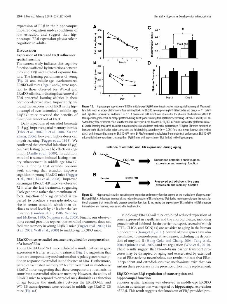
DiscussionExpression of ER� and ER� influencesspatial learningThe current study indicates that cognitivefunction is affected by interactions betweenER� and ER� and estradiol exposure his-tory. The learning performances of young(Fig. 3) and middle-age ovariectomizedER�KO-oil mice (Figs. 5 and 6) were supe-rior to those observed for WT-oil andER�KO-oil mice, indicating that removal ofER� preserved learning abilities in thesehormone-deprived mice. Importantly, wefound that expression of ER� in the hip-pocampi of ovariectomized, middle-ageER�KO mice reversed the benefits offunctional knockout of ER�.
Daily injections of estradiol benzoate(1–5 �g) improve spatial memory in mice(Frick et al., 2002; Li et al., 2004; Xu andZhang, 2006); however, higher doses canimpair learning (Fugger et al., 1998). Weconfirmed that estradiol injections (5 �g)can have lasting (48 –72 h) effects on cog-nition (Aenlle et al., 2009). In addition,estradiol treatment induced lasting mem-ory enhancement in middle-age ER�KOmice, a finding that extends previouswork showing that estradiol improvescognition in young ER�KO mice (Fuggeret al., 2000; Liu et al., 2008). Improvedlearning in ER�KO-EB mice was observed72 h after the last treatment, suggestinglikely genomic rather than membrane ef-fects. Injection of 5 �g estradiol is ex-pected to produce a supraphysiologicalrise in serum estradiol, which then de-clines to basal levels by 72 h after the lastinjection (Gordon et al., 1986; Woolleyand McEwen, 1993; Noppens et al., 2005). Finally, our observa-tions extend previous reports that estradiol treatment does notfacilitate memory in young ER�KO mice (Fugger et al., 2000; Liuet al., 2008; Walf et al., 2009) to middle-age ER�KO mice.
ER�KO mice: estradiol treatment required for compensationof a loss of ER�Young ER�KO and WT mice exhibited a similar pattern in geneexpression 6 h after estradiol treatment (Fig. 2), suggesting thatthere are compensatory mechanisms that regulate gene transcrip-tion in response to estradiol in the absence of ER�. Furthermore,estradiol facilitated memory 72 h after treatment in middle-ageER�KO mice, suggesting that these compensatory mechanismscontribute to estradiol effects on memory. However, the ability ofER�KO mice to respond to estradiol may diminish as a functionof age because the similarities between the ER�KO-EB andWT-EB transcriptomes were reduced in middle-age ER�KO-EBmice (Fig. 8A).
Middle-age ER�KO-oil mice exhibited reduced expression ofgenes expressed in capillaries and the choroid plexus, includinggenes involved in blood– brain barrier transport, several of which(TTR, CLIC6, and KCNJ13) are sensitive to aging in the humanhippocampus (Kang et al., 2011). Several of these genes have alsobeen linked to neurodegenerative diseases, including the deposi-tion of amyloid � (Hong-Goka and Chang, 2004; Tang et al.,2004; Quintela et al., 2009) and tau regulation (Woo et al., 2010).These results suggest that blood– brain barrier transport pro-cesses may be disrupted by aging and exacerbated by extendedloss of ER� activity; nevertheless, our results indicate that ER�-independent and estradiol-sensitive mechanisms exist that cansustain these processes in the presence of hormone replacement.
ER�KO mice: ER� regulation of transcription andhippocampal functionSuperior spatial learning was observed in middle-age ER�KOmice, an advantage that was negated by hippocampal expressionof ER�. This result suggests that knockout of ER� provided pro-
Figure 12. Hippocampal expression of ER� in middle-age ER�KO mice impairs water maze spatial learning. A, Mean pathlength to reach an escape platform over four training blocks for ER�KO mice expressing GFP (filled circles and bars, n � 11) or GFPand ER�-FLAG (open circles and bars, n � 12). A decrease in path length was observed in the absence of a treatment effect. B,Mean path length to reach an escape platform during 3 d of spatial training for ER�KO mice expressing GFP or GFP and ER�-FLAG.*A tendency for a treatment effect was the result of a decrease in the distance for ER�KO-GFP mice to reach the platform on day 2.C, Spatial learning measured as a discrimination index calculated from probe trial performance. #ER�KO-GFP mice exhibited anincrease in the discrimination index score across the 2 d of training. A tendency ( p � 0.055) for a treatment effect was observed forday 3, with increased learning for ER�KO-GFP mice. D, Platform crossing calculated from probe trial performance. ER�KO-GFPmice exhibited more platform crossings than ER�KO mice with expression of ER� limited to the hippocampus.
Figure 13. Hippocampal estradiol-sensitive gene expression and memory function depend on the relative level of expression ofER� and ER�. A, A decrease in estradiol and reduced expression of ER� relative to ER� during menopause disrupts the transcrip-tional processes that normally help preserve cognitive function. B, Increasing the expression of ER� relative to ER� preservestranscription and memory, even as estradiol levels decline.
2680 • J. Neurosci., February 6, 2013 • 33(6):2671–2683 Han et al. • Hippocampal Gene Expression in Knockout Mice

tection against cognitive decline. Furthermore, ER�KO miceexhibited a marked shift in basal transcription of estradiol-responsive genes (Figs. 2 and 8), suggesting that the shift in transcrip-tion may have contributed to preserved cognition in ER�KO mice. Asimilar finding has been reported in bone, such that aging ER�KOmice are protected from bone loss (Windahl et al., 2001) and showincreased transcription of estradiol-responsive genes (Lindberg etal., 2003). Furthermore, knockdown of ER� has been shown to re-duce the vulnerability of hippocampal cells to oxidative stress (Yanget al., 2009).
The increased basal transcription of estradiol-responsivegenes in the hippocampi of ER�KO-oil mice is likely the result ofseveral factors. Cell culture studies indicate that ER� antagonizesER�-mediated transcription (Hall and McDonnell, 1999;Pettersson et al., 2000; Gottfried-Blackmore et al., 2007). Simi-larly, ER�-associated transcriptional activity in bone (Lindberget al., 2003) and aortic tissue (O’Lone et al., 2007) is enhanced inER�KO mice. In brain regions that express both receptors, estradiol-induced transcription declines as the levels of ER� and ER� shift tofavor ER� (Gonzales et al., 2008). A second factor that influencesbasal transcription is the affinity of estradiol for ER� and ER�. Lowlevels of estradiol are synthesized in the hippocampus (Prange-Kielet al., 2003; Kretz et al., 2004) and low concentrations of estradiolfavor activation of ER� (Kuiper et al., 1997; Tremblay et al., 1997;Barkhem et al., 1998; Pettersson et al., 2000).Together, theseresults suggest that transcription of estradiol-sensitive genesin ER�KO-oil mice reflects a release of ER� inhibition ofER�-mediated transcription.
Interaction of ER� and ER� signaling pathwaysThe ERs knocked out in our mice were absent during fetal devel-opment. However, differences that we observed in cognition can-not be ascribed to developmental effects alone. First, superiorperformance of the ER�KO mice relative to the ER�KO and WTmice was limited to conditions of hormone deprivation, indicat-ing that changes in hormone levels were required to reveal theinteractions between ER� and ER�. Second, in middle-ageER�KO mice, lentiviral-mediated expression of ER� in the hip-pocampus impaired cognition, indicating that hippocampal ER�expression plays a role in cognition in adults. Indeed, loss of ER�expression in tissues other than the hippocampus may be a moresignificant problem for aging animals. At 2 years of age,ER�KO mice exhibit excessive weight gain and develop pituitarytumors and myeloproliferative disease, changes that suggest lossof ER� may induce an excessive growth response (Shim et al.,2003; Fan et al., 2010). Furthermore, there is evidence that brainregions outside the hippocampus may exhibit degeneration inaged ER�KO mice (Wang et al., 2001).
Interactions between ER� and ER� in younger animals mayprovide a mechanism for the regulation of hippocampal functionand gene expression in an environment of fluctuating hormonelevels (Foster, 2005, 2012). In young ER�KO mice, estradioltreatment decreased expression of a number of estradiol-responsive genes, an observation that suggests that excessive ER�activity can produce large-scale transcriptional changes in estra-diol responsive pathways. This feedback process may contributeto decreased ER� expression after administration of supraphysi-ological levels of estradiol (Iivonen et al., 2006). Previous workindicates that young ER�KO mice exhibit learning impairmentsand a decline in the expression of ER� when chronically treatedwith estradiol (Rissman et al., 2002), changes that could reflectthe absence of ER� regulation of ER� activity (Foster, 2012).Interestingly, we observed that transcription of nonfunctional
ESR1 and ESR2 increased in ER�KO and ER�KO mice, respec-tively, results consistent with the idea that the activities of thesereceptors play a role in regulating their transcription.
ER� and ER� over the life spanThe results indicate that maintenance of hippocampal functiondepends on a balance between estradiol levels and the relativelevel of expression of ER� and ER� (Fig. 13). Aging is associatedwith decreased expression of hippocampal ER� or the expressionof dominant-negative ER� splice variants that could reduce theability of estradiol treatments to preserve cognition (Tohgi et al.,1995; Adams et al., 2002; Mehra et al., 2005; Ishunina et al., 2007;Bohacek and Daniel, 2009). A decrease in estradiol and reducedexpression of ER� relative to ER� during menopause would bepredicted to act in concert to decrease transcriptional processesthat normally help preserve cognitive function (Fig. 13A). In-deed, ER� polymorphisms have been associated with greatermemory impairment during menopause as estradiol levels de-crease (Ji et al., 2000; Corbo et al., 2006; Olsen et al., 2006; Yaffe etal., 2009). Our results indicate that increasing the expression ofER� may preserve transcription and memory, even as estradiollevels decline (Fig. 13B). These results are consistent with mount-ing evidence that suggest that increased ER� expression is asso-ciated with improved learning and memory (Foster, 2012).Treatments to alter the expression of ERs within the hippocam-pus could provide an alternative to hormone replacement in pre-serving cognitive function.
ReferencesAdams MM, Fink SE, Shah RA, Janssen WG, Hayashi S, Milner TA, McEwen
BS, Morrison JH (2002) Estrogen and aging affect the subcellular distri-bution of estrogen receptor-� in the hippocampus of female rats. J Neu-rosci 22:3608 –3614. Medline
Aenlle KK, Foster TC (2010) Aging alters the expression of genes for neuro-protection and synaptic function following acute estradiol treatment.Hippocampus 20:1047–1060. CrossRef Medline
Aenlle KK, Kumar A, Cui L, Jackson TC, Foster TC (2009) Estrogen effectson cognition and hippocampal transcription in middle-aged mice. Neu-robiol Aging 30:932–945. CrossRef Medline
Akama KT, McEwen BS (2003) Estrogen stimulates postsynaptic density-95rapid protein synthesis via the Akt/protein kinase B pathway. J Neurosci23:2333–2339. Medline
Barkhem T, Carlsson B, Nilsson Y, Enmark E, Gustafsson J, Nilsson S (1998)Differential response of estrogen receptor � and estrogen receptor � topartial estrogen agonists/antagonists. Mol Pharmacol 54:105–112.Medline
Bimonte-Nelson HA, Francis KR, Umphlet CD, Granholm AC (2006) Pro-gesterone reverses the spatial memory enhancements initiated by tonicand cyclic oestrogen therapy in middle-aged ovariectomized female rats.Eur J Neurosci 24:229 –242. CrossRef Medline
Blalock EM, Chen KC, Sharrow K, Herman JP, Porter NM, Foster TC, Land-field PW (2003) Gene microarrays in hippocampal aging: statistical pro-filing identifies novel processes correlated with cognitive impairment.J Neurosci 23:3807–3819. Medline
Bohacek J, Daniel JM (2009) The ability of oestradiol administration to reg-ulate protein levels of oestrogen receptor � in the hippocampus and pre-frontal cortex of middle-aged rats is altered following long-term ovarianhormone deprivation. J Neuroendocrinol 21:640 – 647. CrossRef Medline
Choi JM, Romeo RD, Brake WG, Bethea CL, Rosenwaks Z, McEwen BS(2003) Estradiol increases pre- and post-synaptic proteins in the CA1region of the hippocampus in female rhesus macaques (Macaca mulatta).Endocrinology 144:4734 – 4738. CrossRef Medline
Corbo RM, Gambina G, Ruggeri M, Scacchi R (2006) Association of estro-gen receptor � (ESR1) PvuII and XbaI polymorphisms with sporadicAlzheimer’s disease and their effect on apolipoprotein E concentrations.Dement Geriatr Cogn Disord 22:67–72. CrossRef Medline
Daniel JM, Hulst JL, Berbling JL (2006) Estradiol replacement enhancesworking memory in middle-aged rats when initiated immediately after
Han et al. • Hippocampal Gene Expression in Knockout Mice J. Neurosci., February 6, 2013 • 33(6):2671–2683 • 2681
ovariectomy but not after a long-term period of ovarian hormone depri-vation. Endocrinology 147:607– 614. CrossRef Medline
Dubal DB, Rau SW, Shughrue PJ, Zhu H, Yu J, Cashion AB, Suzuki S, GerholdLM, Bottner MB, Dubal SB, Merchanthaler I, Kindy MS, Wise PM (2006)Differential modulation of estrogen receptors (ERs) in ischemic braininjury: a role for ER� in estradiol-mediated protection against delayed celldeath. Endocrinology 147:3076 –3084. CrossRef Medline
Enmark E, Gustafsson JA (1999) Oestrogen receptors: an overview. J InternMed 246:133–138. CrossRef Medline
Fan X, Gabbi C, Kim HJ, Cheng G, Andersson LC, Warner M, GustafssonJA (2010) Gonadotropin-positive pituitary tumors accompanied byovarian tumors in aging female ER� �/� mice. Proc Natl Acad SciU S A 107:6453– 6458. CrossRef Medline
Foster TC (2005) Interaction of rapid signal transduction cascades and geneexpression in mediating estrogen effects on memory over the life span.Front Neuroendocrinol 26:51– 64. CrossRef Medline
Foster TC (2012) Role of estrogen receptor � and � expression and signalingon cognitive function during aging. Hippocampus 22:656 – 669. CrossRefMedline
Foster TC, Sharrow KM, Kumar A, Masse J (2003) Interaction of age andchronic estradiol replacement on memory and markers of brain aging.Neurobiol Aging 24:839 – 852. CrossRef Medline
Foster TC, Rani A, Kumar A, Cui L, Semple-Rowland SL (2008) Viralvector-mediated delivery of estrogen receptor-� to the hippocampus im-proves spatial learning in estrogen receptor-� knock-out mice. Mol Ther16:1587–1593. CrossRef Medline
Frick KM, Fernandez SM, Bulinski SC (2002) Estrogen replacement im-proves spatial reference memory and increases hippocampal synaptophy-sin in aged female mice. Neuroscience 115:547–558. CrossRef Medline
Fugger HN, Cunningham SG, Rissman EF, Foster TC (1998) Sex differencesin the activational effect of ER� on spatial learning. Horm Behav 34:163–170. CrossRef Medline
Fugger HN, Foster TC, Gustafsson J, Rissman EF (2000) Novel effects ofestradiol and estrogen receptor � and � on cognitive function. Brain Res883:258 –264. CrossRef Medline
Garcia-Segura LM, Sanz A, Mendez P (2006) Cross-talk between IGF-I andestradiol in the brain: focus on neuroprotection. Neuroendocrinology84:275–279. CrossRef Medline
Gibbs RB (2000) Long-term treatment with estrogen and progesterone en-hances acquisition of a spatial memory task by ovariectomized aged rats.Neurobiol Aging 21:107–116. CrossRef Medline
Gonzales KL, Tetel MJ, Wagner CK (2008) Estrogen receptor (ER) � mod-ulates ER� responses to estrogens in the developing rat ventromedialnucleus of the hypothalamus. Endocrinology 149:4615– 4621. CrossRefMedline
Gordon MN, Osterburg HH, May PC, Finch CE (1986) Effective oral ad-ministration of 17 �-estradiol to female C57BL/6J mice through thedrinking water. Biol Reprod 35:1088 –1095. CrossRef Medline
Gottfried-Blackmore A, Croft G, McEwen BS, Bulloch K (2007) Transcrip-tional activity of estrogen receptors ER� and ER� in the EtC. 1 cerebellargranule cell line. Brain Res 1186:41– 47. CrossRef Medline
Gould E, Woolley CS, Frankfurt M, McEwen BS (1990) Gonadal steroidsregulate dendritic spine density in hippocampal pyramidal cells in adult-hood. J Neurosci 10:1286 –1291. Medline
Gresack JE, Frick KM (2006) Effects of continuous and intermittent estro-gen treatments on memory in aging female mice. Brain Res 1115:135–147.CrossRef Medline
Hall JM, McDonnell DP (1999) The estrogen receptor �-isoform (ER�) ofthe human estrogen receptor modulates ER� transcriptional activity andis a key regulator of the cellular response to estrogens and antiestrogens.Endocrinology 140:5566 –5578. CrossRef Medline
Hammer RE, Idzerda RL, Brinster RL, McKnight GS (1986) Estrogen regu-lation of the avian transferrin gene in transgenic mice. Mol Cell Biol6:1010 –1014. CrossRef Medline
Harrington WR, Sheng S, Barnett DH, Petz LN, Katzenellenbogen JA, Kat-zenellenbogen BS (2003) Activities of estrogen receptor �- and�-selective ligands at diverse estrogen responsive gene sites mediatingtransactivation or transrepression. Mol Cell Endocrinol 206:13–22.CrossRef Medline
Hong-Goka BC, Chang FL (2004) Estrogen receptors � and � in choroidplexus epithelial cells in Alzheimer’s disease. Neurosci Lett 360:113–116.CrossRef Medline
Iivonen S, Heikkinen T, Puolivali J, Helisalmi S, Hiltunen M, Soininen H,Tanila H (2006) Effects of estradiol on spatial learning, hippocampalcytochrome P450 19, and estrogen � and � mRNA levels in ovariecto-mized female mice. Neuroscience 137:1143–1152. CrossRef Medline
Ishunina TA, Fischer DF, Swaab DF (2007) Estrogen receptor � and itssplice variants in the hippocampus in aging and Alzheimer’s disease. Neu-robiol Aging 28:1670 –1681. CrossRef Medline
Jelks KB, Wylie R, Floyd CL, McAllister AK, Wise P (2007) Estradiol targetssynaptic proteins to induce glutamatergic synapse formation in culturedhippocampal neurons: critical role of estrogen receptor-�. J Neurosci27:6903– 6913. CrossRef Medline
Ji Y, Urakami K, Wada-Isoe K, Adachi Y, Nakashima K (2000) Estrogenreceptor gene polymorphisms in patients with Alzheimer’s disease, vas-cular dementia and alcohol-associated dementia. Dement Geriatr CognDisord 11:119 –122. CrossRef Medline
Kang HJ, Kawasawa YI, Cheng F, Zhu Y, Xu X, Li M, Sousa AM, Pletikos M,Meyer KA, Sedmak G, Guennel T, Shin Y, Johnson MB, Krsnik Z, MayerS, Fertuzinhos S, Umlauf S, Lisgo SN, Vortmeyer A, Weinberger DR, et al.(2011) Spatio-temporal transcriptome of the human brain. Nature 478:483– 489. CrossRef Medline
Karmakar S, Foster EA, Smith CL (2009) Estradiol downregulation of thetumor suppressor gene BTG2 requires estrogen receptor-� and the REAcorepressor. Int J Cancer 124:1841–1851. CrossRef Medline
Katzenellenbogen BS, Korach KS (1997) A new actor in the estrogen recep-tor drama: enter ER-�. Endocrinology 138:861– 862. CrossRef Medline
Krege JH, Hodgin JB, Couse JF, Enmark E, Warner M, Mahler JF, Sar M,Korach KS, Gustafsson JA, Smithies O (1998) Generation and repro-ductive phenotypes of mice lacking estrogen receptor �. Proc Natl AcadSci U S A 95:15677–15682. CrossRef Medline
Kretz O, Fester L, Wehrenberg U, Zhou L, Brauckmann S, Zhao S, Prange-Kiel J, Naumann T, Jarry H, Frotscher M, Rune GM (2004) Hippocam-pal synapses depend on hippocampal estrogen synthesis. J Neurosci 24:5913–5921. CrossRef Medline
Kuiper GG, Carlsson B, Grandien K, Enmark E, Haggblad J, Nilsson S,Gustafsson JA (1997) Comparison of the ligand binding specificity andtranscript tissue distribution of estrogen receptors � and �. Endocrinol-ogy 138:863– 870. CrossRef Medline
Lee WH, Kumar A, Rani A, Herrera J, Xu J, Someya S, Foster TC (2012)Influence of viral vector-mediated delivery of superoxide dismutase andcatalase to the hippocampus on spatial learning and memory during ag-ing. Antioxid Redox Signal 16:339 –350. CrossRef Medline
Lein ES, Hawrylycz MJ, Ao N, Ayres M, Bensinger A, Bernard A, Boe AF,Boguski MS, Brockway KS, Byrnes EJ, Chen L, Chen L, Chen TM, ChinMC, Chong J, Crook BE, Czaplinska A, Dang CN, Datta S, Dee NR,et al.(2007) Genome-wide atlas of gene expression in the adult mouse brain.Nature 445:168 –176. CrossRef Medline
Li J, Wong L (2001) Emerging patterns and gene expression data. GenomeInform 12:3–13. Medline
Li C, Brake WG, Romeo RD, Dunlop JC, Gordon M, Buzescu R, MagarinosAM, Allen PB, Greengard P, Luine V, McEwen BS (2004) Estrogen altershippocampal dendritic spine shape and enhances synaptic protein immu-noreactivity and spatial memory in female mice. Proc Natl Acad Sci U S A101:2185–2190. CrossRef Medline
Lin Y, Bloodgood BL, Hauser JL, Lapan AD, Koon AC, Kim TK, Hu LS, MalikAN, Greenberg ME (2008) Activity-dependent regulation of inhibitorysynapse development by Npas4. Nature 455:1198 –1204. CrossRefMedline
Lindberg MK, Weihua Z, Andersson N, Moverare S, Gao H, Vidal O, ErlandssonM, Windahl S, Andersson G, Lubahn DB, Carlsten H, Dahlman-Wright K,Gustafsson JA, Ohlsson C (2002) Estrogen receptor specificity for the effectsof estrogen in ovariectomized mice. J Endocrinol 174:167–178. CrossRefMedline
Lindberg MK, Moverare S, Skrtic S, Gao H, Dahlman-Wright K, GustafssonJA, Ohlsson C (2003) Estrogen receptor (ER)-� reduces ER�-regulatedgene transcription, supporting a “ying yang” relationship between ER�and ER� in mice. Mol Endocrinol (Baltimore) 17:203–208. CrossRefMedline
Liu F, Day M, Muniz LC, Bitran D, Arias R, Revilla-Sanchez R, Grauer S,Zhang G, Kelley C, Pulito V, Sung A, Mervis RF, Navarra R, Hirst WD,Reinhart PH, Marquis KL, Moss SJ, Pangalos MN, Brandon NJ (2008)Activation of estrogen receptor-� regulates hippocampal synaptic plastic-ity and improves memory. Nat Neurosci 11:334 –343. CrossRef Medline
2682 • J. Neurosci., February 6, 2013 • 33(6):2671–2683 Han et al. • Hippocampal Gene Expression in Knockout Mice

Lubahn DB, Moyer JS, Golding TS, Couse JF, Korach KS, Smithies O (1993)Alteration of reproductive function but not prenatal sexual developmentafter insertional disruption of the mouse estrogen receptor gene. ProcNatl Acad Sci U S A 90:11162–11166. CrossRef Medline
Markham JA, Pych JC, Juraska JM (2002) Ovarian hormone replacement toaged ovariectomized female rats benefits acquisition of the Morris watermaze. Horm Behav 42:284 –293. CrossRef Medline
Markowska AL, Savonenko AV (2002) Effectiveness of estrogen replace-ment in restoration of cognitive function after long-term estrogen with-drawal in aging rats. J Neurosci 22:10985–10995. Medline
Mehra RD, Sharma K, Nyakas C, Vij U (2005) Estrogen receptor � and �immunoreactive neurons in normal adult and aged female rat hippocam-pus: a qualitative and quantitative study. Brain Res 1056:22–35. CrossRefMedline
Nilsen J, Diaz Brinton R (2003) Mechanism of estrogen-mediated neuro-protection: regulation of mitochondrial calcium and Bcl-2 expression.Proc Natl Acad Sci U S A 100:2842–2847. CrossRef Medline
Nilsson S, Makela S, Treuter E, Tujague M, Thomsen J, Andersson G, EnmarkE, Pettersson K, Warner M, Gustafsson JA (2001) Mechanisms of estro-gen action. Physiol Rev 81:1535–1565. Medline
Noppens RR, Kofler J, Hurn PD, Traystman RJ (2005) Dose-dependentneuroprotection by 17�-estradiol after cardiac arrest and cardiopulmo-nary resuscitation. Crit Care Med 33:1595–1602. CrossRef Medline
O’Lone R, Knorr K, Jaffe IZ, Schaffer ME, Martini PG, Karas RH, BienkowskaJ, Mendelsohn ME, Hansen U (2007) Estrogen receptors � and � medi-ate distinct pathways of vascular gene expression, including genes in-volved in mitochondrial electron transport and generation of reactiveoxygen species. Mol Endocrinol (Baltimore) 21:1281–1296. CrossRefMedline
Olsen L, Rasmussen HB, Hansen T, Bagger YZ, Tanko LB, Qin G, Christian-sen C, Werge T (2006) Estrogen receptor � and risk for cognitive im-pairment in postmenopausal women. Psychiatr Genet 16:85– 88.CrossRef Medline
Paruthiyil S, Cvoro A, Tagliaferri M, Cohen I, Shtivelman E, Leitman DC(2011) Estrogen receptor � causes a G2 cell cycle arrest by inhibitingCDK1 activity through the regulation of cyclin B1, GADD45A, and BTG2.Breast Cancer Res Treat. 129:777–784. CrossRef Medline
Pettersson K, Delaunay F, Gustafsson JA (2000) Estrogen receptor � acts asa dominant regulator of estrogen signaling. Oncogene 19:4970 – 4978.CrossRef Medline
Prange-Kiel J, Wehrenberg U, Jarry H, Rune GM (2003) Para/autocrine reg-ulation of estrogen receptors in hippocampal neurons. Hippocampus13:226 –234. CrossRef Medline
Priest CA, Eckersell CB, Micevych PE (1995) Estrogen regulatespreproenkephalin-A mRNA levels in the rat ventromedial nucleus: tem-poral and cellular aspects. Brain Res Mol Brain Res 28:251–262. CrossRefMedline
Quintela T, Goncalves I, Baltazar G, Alves CH, Saraiva MJ, Santos CR (2009)17�-Estradiol induces transthyretin expression in murine choroid plexusvia an oestrogen receptor dependent pathway. Cell Mol Neurobiol 29:475– 483. CrossRef Medline
Rissman EF, Heck AL, Leonard JE, Shupnik MA, Gustafsson JA (2002) Dis-ruption of estrogen receptor � gene impairs spatial learning in femalemice. Proc Natl Acad Sci U S A 99:3996 – 4001. CrossRef Medline
Rune GM, Frotscher M (2005) Neurosteroid synthesis in the hippocampus:role in synaptic plasticity. Neuroscience 136:833– 842. CrossRef Medline
Semple-Rowland SL, Eccles KS, Humberstone EJ (2007) Targeted expres-sion of two proteins in neural retina using self-inactivating, insulatedlentiviral vectors carrying two internal independent promoters. Mol Vis13:2001–2011. Medline
Sharma PK, Thakur MK (2006) Expression of estrogen receptor (ER) � and� in mouse cerebral cortex: effect of age, sex and gonadal steroids. Neu-robiol Aging 27:880 – 887. CrossRef Medline
Sherwin BB (2006) Estrogen and cognitive aging in women. Neuroscience138:1021–1026. CrossRef Medline
Shim GJ, Wang L, Andersson S, Nagy N, Kis LL, Zhang Q, Makela S,Warner M, Gustafsson JA (2003) Disruption of the estrogen receptor� gene in mice causes myeloproliferative disease resembling chronicmyeloid leukemia with lymphoid blast crisis. Proc Natl Acad Sci U S A100:6694 – 6699. CrossRef Medline
Sohrabji F, Miranda RC, Toran-Allerand CD (1994) Estrogen differentially
regulates estrogen and nerve growth factor receptor mRNAs in adultsensory neurons. J Neurosci 14:459 – 471. Medline
Sohrabji F, Miranda RC, Toran-Allerand CD (1995) Identification of a pu-tative estrogen response element in the gene encoding brain-derived neu-rotrophic factor. Proc Natl Acad Sci U S A 92:11110 –11114. CrossRefMedline
Talboom JS, Williams BJ, Baxley ER, West SG, Bimonte-Nelson HA (2008)Higher levels of estradiol replacement correlate with better spatial mem-ory in surgically menopausal young and middle-aged rats. NeurobiolLearn Mem 90:155–163. CrossRef Medline
Tang YP, Haslam SZ, Conrad SE, Sisk CL (2004) Estrogen increases brainexpression of the mRNA encoding transthyretin, an amyloid � scavengerprotein. J Alzheimers Dis 6:413– 420; discussion 443– 449. Medline
Thakur MK, Sharma PK (2007) Transcription of estrogen receptor � and �in mouse cerebral cortex: effect of age, sex, 17�-estradiol and testoster-one. Neurochem Int 50:314 –321. CrossRef Medline
Tohgi H, Utsugisawa K, Yamagata M, Yoshimura M (1995) Effects of age onmessenger RNA expression of glucocorticoid, thyroid hormone, andro-gen, and estrogen receptors in postmortem human hippocampus. BrainRes 700:245–253. CrossRef Medline
Too CK, Giles A, Wilkinson M (1999) Estrogen stimulates expression ofadenine nucleotide translocator ANT1 messenger RNA in female rathearts. Mol Cell Endocrinol 150:161–167. CrossRef Medline
Tremblay GB, Tremblay A, Copeland NG, Gilbert DJ, Jenkins NA, Labrie F,Giguere V (1997) Cloning, chromosomal localization, and functionalanalysis of the murine estrogen receptor �. Mol Endocrinol (Baltimore)11:353–365. CrossRef Medline
Walf AA, Koonce CJ, Frye CA (2008) Estradiol or diarylpropionitrile ad-ministration to wild type, but not estrogen receptor � knockout, miceenhances performance in the object recognition and object placementtasks. Neurobiol Learn Mem 89:513–521. CrossRef Medline
Walf AA, Koonce C, Manley K, Frye CA (2009) Proestrous compared withdiestrous wild-type, but not estrogen receptor � knockout, mice havebetter performance in the spontaneous alternation and object recognitiontasks and reduced anxiety-like behavior in the elevated plus and mirrormaze. Behav Brain Res 196:254 –260. CrossRef Medline
Wang L, Andersson S, Warner M, Gustafsson JA (2001) Morphological ab-normalities in the brains of estrogen receptor � knockout mice. Proc NatlAcad Sci U S A 98:2792–2796. CrossRef Medline
Williams C, Edvardsson K, Lewandowski SA, Strom A, Gustafsson JA(2008) A genome-wide study of the repressive effects of estrogen re-ceptor � on estrogen receptor � signaling in breast cancer cells. On-cogene 27:1019 –1032. CrossRef Medline
Windahl SH, Hollberg K, Vidal O, Gustafsson JA, Ohlsson C, Andersson G(2001) Female estrogen receptor � �/� mice are partially protectedagainst age-related trabecular bone loss. J Bone Miner Res 16:1388 –1398.CrossRef Medline
Woo JM, Park SJ, Kang HI, Kim BG, Shim SB, Jee SW, Lee SH, Sin JS, Bae CJ,Jang MK, Cho C, Hwang DY, Kim CK (2010) Characterization ofchanges in global gene expression in the brain of neuron-specific enolase/human Tau23 transgenic mice in response to overexpression of Tau pro-tein. Int J Mol Med 25:667– 675. CrossRef Medline
Woolley CS, McEwen BS (1992) Estradiol mediates fluctuation in hip-pocampal synapse density during the estrous cycle in the adult rat. J Neu-rosci 12:2549 –2554. Medline
Woolley CS, McEwen BS (1993) Roles of estradiol and progesterone in reg-ulation of hippocampal dendritic spine density during the estrous cycle inthe rat. J Comp Neurol 336:293–306. CrossRef Medline
Woolley CS, Gould E, Frankfurt M, McEwen BS (1990) Naturally occurringfluctuation in dendritic spine density on adult hippocampal pyramidalneurons. J Neurosci 10:4035– 4039. Medline
Xu X, Zhang Z (2006) Effects of estradiol benzoate on learning-memorybehavior and synaptic structure in ovariectomized mice. Life Sci 79:1553–1560. CrossRef Medline
Yaffe K, Lindquist K, Sen S, Cauley J, Ferrell R, Penninx B, Harris T, Li R,Cummings SR (2009) Estrogen receptor genotype and risk of cognitiveimpairment in elders: findings from the Health ABC study. NeurobiolAging 30:607– 614. CrossRef Medline
Yang SH, Sarkar SN, Liu R, Perez EJ, Wang X, Wen Y, Yan LJ, Simpkins JW(2009) Estrogen receptor � as a mitochondrial vulnerability factor. J BiolChem 284:9540 –9548. CrossRef Medline
Zeier Z, Kumar A, Bodhinathan K, Feller JA, Foster TC, Bloom DC (2009)
Han et al. • Hippocampal Gene Expression in Knockout Mice J. Neurosci., February 6, 2013 • 33(6):2671–2683 • 2683

Fragile X mental retardation protein replacement restores hippocampalsynaptic function in a mouse model of fragile X syndrome. Gene Ther16:1122–1129. CrossRef Medline
2683a • J. Neurosci., February 6, 2013 • 33(6):2671–2683 Han et al. • Hippocampal Gene Expression in Knockout Mice