cellular/molecular myocilinisinvolvedinngr1/lingo-1
TRANSCRIPT

Cellular/Molecular
Myocilin Is Involved in NgR1/Lingo-1-MediatedOligodendrocyte Differentiation and Myelination of theOptic Nerve
Heung Sun Kwon,1 Naoki Nakaya,1 Mones Abu-Asab,2 Hong Sug Kim,3 and Stanislav I. Tomarev1
1Retinal Ganglion Cell Biology Section, Laboratory of Retinal Cell and Molecular Biology and 2Histology Core, National Eye Institute, National Institutes ofHealth (NIH), Bethesda, Maryland 20892, and 3Neuro-Oncology Branch, National Cancer Institute, NIH, Bethesda, Maryland 20892
Myocilin is a secreted glycoprotein that belongs to a family of olfactomedin domain-containing proteins. Although myocilin is detectedin several ocular and nonocular tissues, the only reported human pathology related to mutations in the MYOCILIN gene is primaryopen-angle glaucoma. Functions of myocilin are poorly understood. Here we demonstrate that myocilin is a mediator of oligodendrocytedifferentiation and is involved in the myelination of the optic nerve in mice. Myocilin is expressed and secreted by optic nerve astrocytes.Differentiation of optic nerve oligodendrocytes is delayed in Myocilin-null mice. Optic nerves of Myocilin-null mice contain reducedlevels of several myelin-associated proteins including myelin basic protein, myelin proteolipid protein, and 2�3�-cyclic nucleotide 3�-phosphodiesterase compared with those of wild-type littermates. This leads to reduced myelin sheath thickness of optic nerve axons inMyocilin-null mice compared with wild-type littermates, and this difference is more pronounced at early postnatal stages compared withadult mice. Myocilin also affects differentiation of oligodendrocyte precursors in vitro. Its addition to primary cultures of differentiatingoligodendrocyte precursors increases levels of tested markers of oligodendrocyte differentiation and stimulates elongation of oligoden-drocyte processes. Myocilin stimulation of oligodendrocyte differentiation occurs through the NgR1/Lingo-1 receptor complex. Myocilinphysically interacts with Lingo-1 and may be considered as a Lingo-1 ligand. Myocilin-induced elongation of oligodendrocyte processesmay be mediated by activation of FYN and suppression of RhoA GTPase.
Key words: astrocyte; differentiation; Lingo-1; myelin; myocilin; oligodendrocyte
IntroductionMYOCILIN (MYOC) was the first gene in which identified mu-tations were found to cause glaucoma, the second leading causeof blindness in the world (Adam et al., 1997, Stone et al., 1997).MYOC encodes a secreted glycoprotein and is expressed in ocularand several nonocular tissues. In the eye, MYOC expression hasbeen detected in the tissues responsible for aqueous humor pro-duction (ciliary body) and outflow (trabecular meshwork), aswell as in the iris, sclera, retinal pigmented epithelium, and opticnerve (Adam et al., 1997, Ortego et al., 1997, Stone et al., 1997,Tomarev et al., 2003). Available data suggest that expression ofmutated myocilin in the trabecular meshwork leads to the acti-vation of an unfolded protein response (Joe et al., 2003, Joe andTomarev, 2010, Zode et al., 2011) and increases sensitivity of cellsto oxidative stress (Joe and Tomarev, 2010). This may lead to
deterioration of trabecular meshwork function and elevation ofintraocular pressure. The pathological role of mutated myocilinin other ocular and nonocular tissues is less clear. Myoc-null micedo not show any detectable changes in intraocular pressure ordefects in the optic nerve head (Kim et al., 2001). However, Myoc-null mice exhibit reduced cortical bone thickness (Kwon et al.,2013b), a moderate reduction in the amount of dystrophin-associated syntrophin in the skeletal muscle (Joe et al., 2012), anddefects in sciatic nerve myelination (Kwon et al., 2013a) as com-pared with wild-type littermates. In the sciatic nerve, myocilin isexpressed in Schwann cells (Ohlmann et al., 2003) and localizedto the nodes of Ranvier where it interacts with gliomedin, neuro-fascin, and NrCAM, proteins essential for node formation andfunction (Eshed et al., 2005, Kwon et al., 2013a). Sciatic nerves ofMyoc-null mice have thinner myelin sheaths and partial disorga-nization of Ranvier nodes when compared with wild-type litter-mates (Kwon et al., 2013a).
In the CNS, including the optic nerve, oligodendrocytes play acritical role in the myelination of axons. Several regulators thatnegatively or positively regulate myelination in the CNS havebeen identified (Emery, 2010). One of the negative regulators ofmyelination is leucine-rich repeat and Ig domain-containing 1(Lingo-1), a transmembrane signaling protein expressed in botholigodendrocytes and neurons, but not in astrocytes (Mi et al.,2005). In neurons, Lingo-1 is a coreceptor of the Nogo receptor
Received Nov. 7, 2013; revised Jan. 24, 2014; accepted March 11, 2014.Author contributions: H.S. Kwon designed research; H.S. Kwon, N.N., and M.A.-A. performed research; H.S. Kim
contributed unpublished reagents/analytic tools; H.S. Kwon analyzed data; H.S. Kwon and S.I.T. wrote the paper.This work was supported by the Intramural Research Programs of the National Eye Institute, National Institutes
of Health. We thank Drs. Haohua Qian for help with flash visual evoked potentials recording and Thomas V. Johnsonfor critical reading of this manuscript.
The authors declare no competing financial interests.Correspondence should be addressed to Stanislav I. Tomarev, Building 6, Room 212, 6 Center Drive, National Eye
Institute, NIH, Bethesda, MD 20892. E-mail: [email protected]:10.1523/JNEUROSCI.4731-13.2014
Copyright © 2014 the authors 0270-6474/14/345539-13$15.00/0
The Journal of Neuroscience, April 16, 2014 • 34(16):5539 –5551 • 5539

complex that may mediate the inhibition of axon growth (Mi etal., 2004). We have recently shown that olfactomedin 1 (Olfm1),a protein belonging to the same superfamily as myocilin, interactswith the Nogo receptor complex preferentially binding NgR1.Olfm1 caused the inhibition of NgR1 signaling by interferingwith interaction between NgR1 and its coreceptors, p75NTR orLINGO-1 (Nakaya et al., 2012).
In the present communication, we investigated a role of myo-cilin in the optic nerve. Myoc-null mice show defects in opticnerve myelination. We demonstrate that myocilin is expressed inastrocytes and plays a role in differentiation of oligodendrocytesacting through the Lingo-1/NgR1 complex.
Materials and MethodsAnimals, plasmids, and antibodies. Mice were maintained in accordancewith guidelines set forth by the National Eye Institute Committee on theUse and Care of Animals. Myoc-null mice (B6/129 mixed genetic back-ground) have been described previously (Kim et al., 2001). Mice of eithersex were used in experiments. Human FLAG- and alkaline phosphatase(AP)-tagged myocilin, myocilin-�C, and myocilin-�N constructs havebeen described (Kwon et al., 2009). Antibodies were obtained from fol-lowing sources: HSC70 (Santa Cruz Biotechnology); MBP and Lingo-1(Abcam); neurofilament H, MBP, RhoA, Olig2, A2B5, and O4 (Milli-pore); Nogo, phosphor-Fyn, ErbB2, and ErbB3 (Cell Signaling Technol-ogy). Anti-mouse or rabbit IgG antibody conjugated to horseradishperoxidase were from GE. Alexa 488- or Alexa 594-conjugated anti-mouse, anti-rabbit, or anti-goat IgG was from Life Technologies. Anti-bodies against mouse myocilin were described previously (Malyukova etal., 2006). The human Lingo-1 extracellular domain sequence corre-sponding to residues 1–532 in the amino acid sequence of Lingo-1 wasamplified by PCR using pCMV-Lingo-1 (Thermo Scientific) as a tem-plate. Oligonucleotide primers 5�-CACAAGCTTATGCAGGTGAGCAAGAGG-3�and 5�-CACGGATTCCTCGCCCGGCTGGTTGGAGAT-3�were used for amplification. The amplified fragment was cloned into thepCMV14-FLAG vector, FLAG-tagged protein was expressed in CHOcells, and purified using Anti-FLAG affinity gel (Sigma).
Quantitative PCR. Total RNA was isolated from oligodendrocytes us-ing RNeasy mini-kit following manufacturer’s instructions (Qiagen).cDNA was synthesized using 1 �g of RNA as a template and SuperScriptOne-Step-RT-PCR System kit (Life Technologies) according to the man-ufacturer’s instructions (Life Technologies). cDNA was diluted (1:20)and the quantitative PCRs (qPCRs) were performed on an ABI7900 se-quence detection system (Applied Biosystems). Primers for qPCR wereas follows: Mbp, 5�-CCCGTGGAGCCGTGATC-3� and 5�-TCTTCAAACGAAAAGGGA-3�; Mog, 5�-ATGAAGGAGGCTACACCTGC-3� and5�-CAAGTGCGATGAGAGTCAGC-3�; Mag, 5�-AACCAGTATGGCCAGAGAGC-3� and 5�-GTTCCGGGTTGGATTTTACC-3�; Gapdh, 5�-CCCATCACCATCTTCCAGGAGCG-3� and 5�-CGGGAAGCTCACTGGCATGGCCT-3�. Gapdh was used for normalization. To quantifyingthe relative changes in gene expression, we used the 2 ���C
T method. Theaverage CT was calculated for the target genes and internal control(Gapdh) and the �CT (CT,target � CT,GAPDH) values were determined. Allreactions were performed in triplicate using three independent samples.
Dorsal root ganglia cultures. Dissociated mouse dorsal root ganglia(DRG) cultures were grown in Neurobasal medium (Life Technologies)supplemented with B27 (Life Technologies) as described previously (Po-liak et al., 2003, Eshed et al., 2005). Briefly, DRG of postnatal day 5 (P5)mice were trypsinized, seeded on poly-D-lysine/laminin 2-well cultureslides (BD Bioscience) or poly-D-lysine-coated 13 mm slides (Sigma),and grown in Neurobasal medium, insulin, transferrin, and sodium sel-enite supplement, 0.2% BSA, 4 mg/ml D-glucose (Sigma), GlutaMAX(Life Technologies), 50 ng/ml NGF (Sigma), and antibiotics.
Immunoprecipitation and Western blots. Protein samples from tissuesor cells were dissolved with RIPA lysis buffer (Sigma) containing 10 mM
DTT and protease inhibitor (Sigma). After sonication for 30 s threetimes, the samples were denatured at 100°C for 5 min. The denaturedsamples were analyzed using a 4 –12% polyacrylamide Bis-Tris gel (LifeTechnologies). After electrophoresis, the proteins were transferred to a
PVDF membrane (Life Technologies). The membrane was blocked with5% skim milk in Tris-buffered saline. Primary and secondary antibodiesand SuperSignal West Dura Chemiluminescent Substrate (ThermoFisher Scientific) were used to detect proteins. All Western blotting ex-periments were repeated at least three times.
Primary astrocyte and oligodendrocyte cultures. Mouse optic nerve as-trocytes were derived from the anterior portions of C57BL/6 mouse opticnerves. The posterior pole of the eye was dissected, and the optic nervehead was freed from sclera and other neighboring tissues. The optic nervehead was sliced sagittally and the anterior portion of the optic nerve wascarefully dissected from the prelaminar and postlaminar regions under adissection microscope. Two or three explants of the anterior region wereobtained from each eye. The explants were put into T-25 cm 2 plastictissue culture flasks, which had been conditioned with DMEM/F-12 sup-plemented with 10% FBS. The first passage cells were characterized byimmunostaining with antibodies against GFAP and Olig2 to identifyastrocytes and oligodendrocytes, respectively. Astrocytes were selectedfrom primary cultures by growing cells for 1 week in modified astrocyte-defined, serum-free medium (ADM; Clonetics) containing forskolin tosuppress fibroblast growth. More than 95% of cells in second-passagecultures were positive for GFAP. They were grown to 60 – 80% conflu-ency and starved in serum-free medium for 1 week before being used forexperiments.
Immature oligodendrocytes were isolated using the MACS procedurefollowing the manufacturer’s instructions (Miltenyi Biotec). Optic nerveand brain tissues were collected from two to three P5–P7 mice for eachisolation. For oligodendrocyte selection, cells were incubated withanti-O4 monoclonal antibodies magnetic beads in MACS BSA buffer(Miltenyi Biotec) for 15 min at 4°C. Cells were applied to type MS MiniMACS columns in the presence of a strong magnetic field as recom-mended by the manufacturer. Columns were washed four times withstaining buffer, followed by elution with MACS buffer in the absence ofmagnetic field. A plunger was applied for elution. The purity of isolatedcells was assessed by immunostaining with O4 antibodies.
Immunohistochemistry. Frozen sections and free-floating sections werestained as previously described (Kwon et al., 2013a) with slight modifi-cations. Tissues were permeabilized in 0.3% Triton X-100 (Sigma) in PBSfor 30 min, washed in PBS, then blocked in 4% normal serum in PBS for20 min, and incubated with primary antibodies in 2% normal serum at4°C overnight. The sections were washed with 0.3% Triton X-100 in PBSand then incubated with secondary antibodies raised in goat and conju-gated to Alexa 488 or 594 for 1 h at room temperature (RT). Nuclei werecounterstained with 0.5 �g/ml DAPI and mounted in Vectashield (Vec-tor Laboratories). Staining without primary antibodies was used as anegative control.
Coimmunoprecipitation. Optic nerve lysates were cleared by centrifu-gation at 16,000 � g for 15 min, immunoprecipitated with antibodiesagainst myocilin or Lingo-1 at 4°C overnight, and then incubated withprotein-A agarose (Roche) at RT for 1 h. Bound proteins were eluted fromagarose beads by boiling in SDS-PAGE sample buffer and analyzed by West-ern blotting using indicated antibodies. HEK-293 cells were transientlytransfected with Lingo-1 and NgR1 using Lipofectamine 2000 (Life Technol-ogies) and seeded in 6-well culture dishes. Cells were washed with PBS andlysed in lysis buffer 48 h after transfection. Cleared lysates were subjected toimmunoprecipitation with Lingo-1 antibodies and then incubated withProtein-G magnetic beads (Life Technologies). Immunoprecipitates wereanalyzed by Western blotting using indicated antibodies.
RhoA assay. GST-Rhotekin binding domain and GST-PAK bindingdomain were obtained from Millipore. Small GTPase activities weremeasured as described previously (Ren et al., 1999). Briefly, progenitorand differentiated oligodendrocytes were lysed in 300 �l of 25 mM
HEPES, pH 7.5, containing 1% Igepal CA-630, 150 mM NaCl, 10 mM
MgCl2, 1 mM EDTA, and 1% glycerol. Cell lysates (200 –500 �g) wereclarified at 100,000 � g for 15 min and incubated for 40 min with 20 �gof GST fusion proteins containing the Rhotekin binding domain (forRhoA assay) bound to glutathione-Sepharose beads (Millipore). Sampleswere washed with lysis buffer and then immunoblotted with anti-RhoA.
AP binding assay. AP-tagged fusion protein expression constructs weretransfected into HEK-293 cells to generate conditioned medium (CM)
5540 • J. Neurosci., April 16, 2014 • 34(16):5539 –5551 Kwon et al. • Myocilin Participates in Myelination of the Optic Nerve
containing AP-fusion proteins. The culture medium was changed to thefresh serum-free medium 24 h after transfection, CM was harvested24 – 48 h later, filtered through a 0.22 �m filter, and stored at �80°C untiluse. Absolute concentration and integrity of AP-tagged myocilin wasdetermined by Western blotting using samples with a known amount ofpurified myocilin. COS-7 cells were transfected with Lingo-1, NgR1, orvector plasmids and incubated with AP-myocilin containing CM for 90min at RT 48 h after transfection. Cells were washed five times, fixed bytreatment with 60% acetone, 3% formaldehyde, and 20 mM HEPES, pH7.5, for 30 s and surface binding was visualized using nitro blue tetrazo-lium (NBT) and 5-bromo-4-chloro-3�-indolyphosphate (BCIP) as APsubstrates following the manufacturer’s instructions (GenHunter). Theimages of stained cells were obtained with a dissection microscope (ZeissSTEMI SV-11). For quantitative analysis of the activity of cell-bound AP,1-Step PNPP (Pierce) was added to the fixed cells and the absorbance at405 nm in the supernatant was measured using a microplate reader (Bio-Rad Model-680).
Recording of flash visual evoked potentials. Flash visual evoked poten-tials (fVEPs) were recorded as described previously (Goto et al., 2001).Briefly, mice were kept in a dark room for 30 min and prepared underdim red illumination. Mice were anesthetized with an intraperitonealinjection of 5 �l/g body weight of ketamine (20 mg/ml) and xylazine (2mg/ml) mixture. The pupil was dilated with 2.5% phenylephrine HCl,and the animals were placed on a heating pad to maintain body temper-ature. fVEPs were recorded using a needle electrode placed on the scalpoverlying the visual cortex. Similar needle electrodes inserted under thecheek and the back skin served as reference and ground leads, respec-tively. The luminance of the flash device was 200 cd-s/m 2. Responseswere amplified 1–1000 Hz, and the responses to 100 successive flashespresented at a rate of 1 Hz were averaged in each mouse; the data wereacquired using an Espion system (Diagnosys).
Electron microscopy. Mice were anesthetized using a lethal dose of ket-amine/xylazine injected intraperitoneally. Anesthetized animals wereperfused with a fixative containing 4% paraformaldehyde, 2.5% glutar-aldehyde, 0.13 N NaH4PO4, and 0.11 M NaOH, pH 7.4. Perfused tissueswere fixed in PBS-buffered 2.5% glutaraldehyde and 0.5% osmium te-troxide, dehydrated, and embedded into Spurr’s epoxy resin. Ultrathinsections (90 nm) were made, double-stained with uranyl acetate and leadcitrate, and viewed in a JEOL JEM 1010 transmission electron micro-scope equipped with digital imaging camera. The g-ratio was determinedby dividing the circumference of an axon (without myelin) by the cir-cumference of the same axon including myelin. Six wild-type and sevenMyoc-null mice were analyzed.
Assessment of length of oligodendrocyte processes. Primary oligodendro-cytes were grown for 8 d at 37°C in 5% CO2, subsequently fixed in 4%paraformaldehyde in PBS for 10 min, washed twice with PBS, and storedin 0.05% sodium azide (Sigma) in PBS at 4°C. Baseline media were sup-plemented with myocilin (1 �g/ml) when indicated to test its effect onoligodendrocyte process outgrowth. Oligodendrocyte process out-growth per cell was assessed as follows. Given a directory containing pairsof images corresponding to nuclear (DAPI) and MBP images, the pluginopened the images in a nuclear and a neuronal stack. Images were firstpreprocessed to optimize uniformity of illumination and contrast in theinput images. This step consisted of subtraction of a background imageto reduce artifacts generated by the acquisition system as well as contrastenhancement, rolling ball radius background subtraction, despeckling,and a Gaussian blur by the commands built into ImageJ. These prepro-cessing steps were first performed on a small subset of images to allowchoosing the threshold to be applied to the oligodendrocyte process out-growth images. The outgrowth stack was then skeletonized and the por-tions of the skeleton corresponding to the cell body were removed bysubtracting the oligodendrocyte nuclei stack. The total length of the pro-cesses was estimated by measuring the area covered by the skeleton ineach image and oligodendrocyte nuclei were counted. The results tablewas saved to a text file and the oligodendrocyte nuclei and skeleton stacksas image stacks for later visual inspection.
Lentivirus production and infection. cDNA was inserted into Gatewayentry vector pCR8/GW/TOPO and subsequently into pLenti4/TO/V5-DEST. Lentivirus was produced in 293FT cells with a packaging mix
(ViraPower mix; Life Technologies). Plasmids were transfected withLipofectamine and Plus reagent (Life Technologies). After 3 h, mediawas changed with 5% FBS. Virus-laden supernatant was collected at72 h. The supernatant was filtered and concentrated by ultracentrif-ugation, and viral titer was determined by serial dilution. Mouseoligodendrocytes were plated into 2-well PDL-coated plates in SATOmodified medium containing 5 mg/ml insulin, 50 mg/ml transferrin, 1.6mg/ml putrescine, 0.3 mg/ml 3,3�,5Triiodo-L-thyronine sodium salt, 0.4mg/ml thyroxine, 10 ng/ml PDGF, and 10 ng/ml FGF for 2 d. Themedium was replaced with SATO medium without PDGF, containingfull-length LINGO-1 lentivirus, or control lentivirus at 5 MOI. Cells wereincubated for 24 h, virus-containing medium was removed, and cells werefed with medium containing 3,3�,5-Triiodo-L-thyronine sodium salt, to in-duce differentiation.
Statistical analysis. Each experimental condition had between four andsix explants per n, with a minimum of n � 3 for each experimentalcondition. Outgrowth measurements for each experimental conditionwere pooled and averaged. Error bars are presented as mean � SEM.Two-tailed ANOVA was performed on the raw pooled data, and statisti-cal significance was determined by a two-tailed Student’s t test or Bon-ferroni multiple comparisons post hoc test was used when comparison ofthree or more treatment groups was made (IBM SPSS Statistics 17).
ResultsMyocilin is expressed in optic nerve astrocytesIt has been reported that myocilin is detected in the optic nerve ofadult rats (Ohlmann et al., 2003), pigs (Noda et al., 2000), andhumans (Karali et al., 2000, Clark et al., 2001, Ricard et al., 2001).
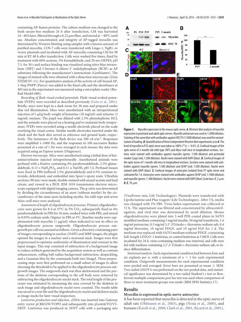
Figure 1. Myocilin expression in the mouse optic nerve. A, Western blot analysis of myocilinexpression in postnatal and adult optic nerves. Myocilin antiserum was used in 1:2000 dilution.Staining of the same blot with antibodies against HSC70 (1:5000 dilution) was used for normal-ization of loading. B, Quantification of three independent Western blot experiments as in A. Thelevel of myocilin in P25 optic nerve was taken as 100% (**p � 0.01). C, Confocal images of theoptic nerve of 2-month-old wild-type (WT) and Myoc-null mice in longitudinal sections. Sec-tions were stained with antibodies against myocilin (green; 1:500 dilution) and perinodalmarker Caspr (red; 1:200 dilution). Nuclei were stained with DAPI (blue). D, Confocal images ofthe optic nerve of 1-month-old mice in longitudinal sections. Sections were stained with anti-bodies against myocilin (green; 1:500 dilution) and GFAP (red; 1:500 dilution). Nuclei werestained with DAPI (blue). E, Confocal images of astrocytes isolated from P7 optic nerve andcultivated for 7 d. Astrocytes were stained with antibodies against GFAP (red; 1:1000 dilution)and myocilin (green; 1: 600 dilution). Nuclei were stained with DAPI (blue).Scale bars: C, 2 �m;D, E, 10 �m.
Kwon et al. • Myocilin Participates in Myelination of the Optic Nerve J. Neurosci., April 16, 2014 • 34(16):5539 –5551 • 5541

In the mouse optic nerve, myocilin pro-tein was detected starting from P6 and itslevel progressively increased between P6and P25. However, myocilin level in theoptic nerve of adult mice was 40% lowerthan at P25 (Fig. 1A,B). To analyze myo-cilin localization in the optic nerve, westained longitudinal sections of 1-month-old optic nerve with antibodies againstmyocilin and GFAP, an astrocyte marker.Similar to its distribution in the adult ratoptic nerve (Ohlmann et al., 2003), myo-cilin showed the most intensive stainingin stellate-shaped cells and was partiallycolocalized with GFAP (Fig. 1D). Myoci-lin was also expressed in astrocytes iso-lated from the optic nerve of P7 mice andcultivated for 7 d (Fig. 1E). In culturedastrocytes, myocilin showed a typical pe-rinuclear staining similar to its distribu-tion in trabecular meshwork cells andoptic nerve head human astrocytes(Lutjen-Drecoll et al., 1998, Clark et al.,2001).
We (Kwon et al., 2013a) and others(Ohlmann et al., 2003) have demon-strated that myocilin is expressed in thesciatic nerve where it localizes to Schwanncells and concentrates at the nodes ofRanvier. In the optic nerve, myocilin isnot concentrated at nodes of Ranvier asshown by immunostaining of the longitu-dinal sections of the optic nerve with an-tibodies against myocilin and perinodalmarker Caspr (Menegoz et al., 1997, Peleset al., 1997; Fig. 1C). Since Myoc-null mu-tation led to profound changes in myeli-nation of the sciatic nerve and its structure
Figure 2. Reduced levels of myelin proteins in the optic nerve of Myoc-null mice and effects of myocilin on differentiation of oligodendrocyte precursors. A, Western blot analysis of optic nervelysates of adult wild-type (WT) and Myoc-null mice using MBP (1:1000 dilution), PLP (1:1,000 dilution), CNPase (1:1000 dilution), and MPZ (1: 1000 dilution) antibodies. Staining of the same blotswith antibodies against HSC70 (1:5000 dilution) was used for normalization of loading. B, Quantification of three independent Western blot experiments as in A. The levels of corresponding proteinsin the optic nerve of wild-type mice were taken as 100% (*p � 0.05; **p � 0.01). C, MBP levels in the optic nerve lysates of wild-type and Myoc-null mice of different ages (P5–P21) as judged byWestern blot analysis. Staining of the same blot with antibodies against HSC70 (1:5000 dilution) was used for normalization of loading. D, E, Confocal images of P13 wild-type and Myoc-null opticnerves in longitudinal sections. Sections were stained with antibodies against MBP (1:500 dilution) or CNPase (1:500 dilution). Nuclei were stained with DAPI (blue). Scale bars: 10 �m.
Figure 3. Reduced level of MBP in the cerebellum of P30 Myoc-null mice. A, Confocal images of wild-type (WT) and Myoccerebellum. Sections were stained with antibodies against myocilin (red; 1:200 dilution and calbindin D, Purkinje cell marker,green; 1: 400 dilution). Nuclei were stained with DAPI (blue). Scale bar, 20 �m. B, Confocal images of wild-type and Myoccerebellum. Sections were stained with antibodies against MBP (1: 200 dilution). Nuclei were stained with DAPI (blue). Scale bar,20 �m. C, E, Western blot analysis of P30 (C) and P60 (E) cerebellum lysates of wild-type and Myoc-null mice using MBP antibodies(1: 1000 dilution). Staining of the same blot with antibodies against HSC70 (1:5000 dilution) was used for normalization of loading.D, F, Quantification of four independent Western blot experiments as in C and E, respectively. The levels of MBP in the cerebellumof wild-type mice were taken as 100% (**p � 0.01).
5542 • J. Neurosci., April 16, 2014 • 34(16):5539 –5551 Kwon et al. • Myocilin Participates in Myelination of the Optic Nerve

(Kwon et al., 2013a), we investigated the effects of myocilin dele-tion on the optic nerve in greater detail.
Differentiation of oligodendrocytes is delayed inMyoc-null miceMyoc deletion led to profound changes in the levels of severalmyelin-associated proteins and reduced the thickness of myelinsheaths in sciatic nerve (Kwon et al., 2013a). Therefore, we testedthe levels of MBP, myelin proteolipid protein (PLP), and 2�3�-cyclic nucleotide 3�-phosphodiesterase (CNPase), the major pro-teins of the CNS myelin sheath, and MPZ (P0), one of the majorproteins of the PNS myelin sheath, in the optic nerve of 2-month-old Myoc-null and wild-type mice. The levels of MPZ and CN-Pase were significantly reduced, while the levels of MBP and PLPwere moderately reduced in Myoc-null mice compared with wild-type littermates (Fig. 2A,B). Immunostaining of P13 optic nervewith antibodies against MBP and CNPase also showed a reducedimmunofluorescence of the Myoc-null as compared with wild-type samples (Fig. 2D,E). In the sciatic nerve, a difference in theMBP levels between Myoc-null and wild-type mice was morepronounced at early postnatal stages than in adults (Kwon et al.,2013a). A similar pattern was observed in the optic nerve: P13–P21 Myoc-null mice showed a more dramatic reduction in thelevels of MBP compared with wild-type littermates than adultpairs (Fig. 2C). Moreover, age-dependent difference in the MBPlevels between Myoc-null and wild-type littermates was also ob-served in cerebellum, another site of myocilin expression in theCNS. Staining of P30 brain section with antibodies against myo-cilin demonstrated that it was localized in Purkinje cells (Fig. 3A).Similar to previously published data (Golan et al., 2008), MBPwas detected in the white matter tracts of the cerebellum at thisage (Fig. 3B). The level of MBP was significantly reduced in thecerebellum of P30 Myoc-null mice as compared with wild-typelittermates (Fig. 3B–D). The MBP level was only slightly reducedin the cerebellum of P60 Myoc-null mice as compared with wild-type, but this difference was not statistically significant (Fig.3E,F).
Transcription factor Olig2 is one of the major transcriptionfactors in oligodendrocyte lineage specification during develop-ment and promotes oligodendrocyte differentiation (for review,seeMeijer et al., 2012). Western blotting and immunofluores-cence analyses demonstrated that the levels of Olig2 were reduced
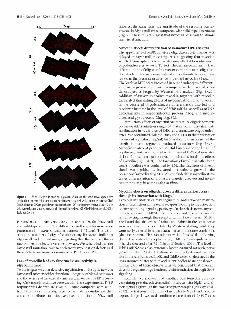
in P25 and adult optic nerve of Myoc-null mice as compared withtheir wild-type littermates (Fig. 4). This difference was more pro-nounced in P25 than in 2-month-old mice. In the course of opticnerve development, oligodendrocyte precursor cells (OPCs) mi-grate from the optic chiasm to the optic nerve head. The migra-tion process starts at embryonic day 17.5 and comes tocompletion around P28 (Pernet et al., 2008). Our previous datasuggested that myocilin may stimulate trabecular meshwork andNIH3T3 cell migration (Kwon and Tomarev, 2011). Since de-layed migration of oligodendrocytes could contribute to the ob-served reduction in the levels of myelin proteins at early postnataldays, we tested whether myocilin absence affects migration ofOPCs. We tested postnatal stages because myocilin could be de-tected in the optic nerve only after P6 (Fig. 1A). Longitudinalsection of wild-type and Myoc-null P8 –P25 optic nerves wasstained with antibodies against Olig2. Although the levels ofOlig2 immunofluorescence were reduced in Myoc-null samplesas compared with their wild-type littermates, the distribution ofOlig2-positive cells was similar in both cases (Fig. 5). This indi-cates that migration of oligodendrocytes is not dramatically af-fected in the optic nerve of Myoc-null mice. Together, theseresults suggest that differentiation of oligodendrocytes may bedelayed in vivo in the absence of myocilin.
Hypomyelination of the optic nerve in Myoc-null miceThe reduction in the levels of myelin-associated proteins impliespossible defects in the myelination of the optic nerve in Myoc-nullmice. Therefore, we analyzed effects of myocilin deficiency on themyelination of the optic nerve at P13 and at P60. Electron mi-croscopy (EM) examination of the optic nerve showed that thethickness of myelin was reduced in Myoc-null mice comparedwith wild-type littermates (Fig. 6). For quantitative analysis ofmyelin thickness, we measured g-ratios of myelinated axons (in-ner axon diameter/total fiber diameter of myelinated axons) closeto the optic nerve head. A significant increase in the averageg-ratio was observed in the optic nerve of P13 Myoc-null micecompared with their wild-type littermates (0.75 � 0.041 vs0.63 � 0.017, respectively, p � 0.05; Fig. 6C). In contrast, theg-ratio only slightly changed in the optic nerve of P60 Myoc-nulland wild-type mice (0.72 � 0.014 vs 0.68 � 0.017, respectively;Fig. 6F). When such measurements were made close to the chi-asm, results were very similar: 0.74 � 0.065 versus 0.63 � 0.021 at
Figure 4. A, Confocal images of P25 wild-type (WT) and Myoc-null optic nerves in transverse sections. Sections were stained with antibodies against neurofilament H (green; 1:500 dilution) andOlig2 (red; 1:200 dilution). Nuclei were stained with DAPI (blue). Scale bar, 20 �m. B, Western blot analysis of optic nerve lysates of P25 and 2-month-old wild-type and Myoc-null mice using Olig2antibodies (1: 500 dilution). Staining of the same blot with antibodies against HSC70 (1:5000 dilution) was used for normalization of loading. C, Quantification of three independent Western blotexperiments as in B. The levels of Olig2 in the optic nerve of wild-type mice were taken as 100% (**p � 0.01).
Kwon et al. • Myocilin Participates in Myelination of the Optic Nerve J. Neurosci., April 16, 2014 • 34(16):5539 –5551 • 5543
P13 and 0.71 � 0.064 versus 0.67 � 0.045 at P60 for Myoc-nulland wild-type samples. The differences in the g-ratio were morepronounced in axons of smaller diameter (�3 �m). The ultra-structure and periodicity of compact myelin were similar inMyoc-null and control mice, suggesting that the reduced thick-ness of myelin reflects fewer myelin wraps. We concluded that theMyoc-null mutation leads to optic nerve myelination defects andthese defects are more pronounced at P13 than at P60.
Loss of myocilin leads to abnormal visual activity inMyoc-null miceTo investigate whether defective myelination of the optic nerve inMyoc-null mice modifies functional integrity of visual pathwaysand the activity of the central visual system, we used fVEP record-ing. One-month-old mice were used in these experiments. fVEPresponse was delayed in Myoc-null mice compared with wild-type littermates indicating impaired salutatory conduction thatcould be attributed to defective myelination in the Myoc-null
mice. At the same time, the amplitude of the response was in-creased in Myoc-null mice compared with wild-type littermates(Fig. 7). These results suggest that myocilin loss leads to abnor-mal visual function.
Myocilin affects differentiation of immature OPCs in vitroThe appearance of MBP, a mature oligodendrocyte marker, wasdelayed in Myoc-null mice (Fig. 2C), suggesting that myocilinsecreted from optic nerve astrocytes may affect differentiation ofoligodendrocytes in vivo. To test whether myocilin may affectdifferentiation of oligodendrocytes in vitro, immature oligoden-drocytes from P5 mice were isolated and differentiated in culturefor 8 d in the presence or absence of purified myocilin (1 �g/ml).The levels of MBP were increased in oligodendrocytes differenti-ating in the presence of myocilin compared with untreated oligo-dendrocytes as judged by Western blot analysis (Fig. 8A,B).Addition of antiserum against myocilin together with myocilineliminated stimulating effects of myocilin. Addition of myocilinin the course of oligodendrocyte differentiation also led to amoderate increase in the level of MBP mRNA, as well as mRNAencoding myelin oligodendrocyte protein (Mog) and myelin-associated glycoprotein (Mag; Fig. 8C).
Stimulatory effects of myocilin on immature oligodendrocyteprecursor differentiation suggested that myocilin may stimulatemyelination in cocultures of DRG and immature oligodendro-cytes. We cocultured isolated DRG and OPCs in the presence orabsence of myocilin (1 �g/ml) for 3 weeks and then measured thelength of myelin segments produced in cultures (Fig. 9A,B).Myocilin treatment produced 3-fold increase in the length ofmyelin segments as compared with untreated DRG cultures. Ad-dition of antiserum against myocilin reduced stimulating effectsof myocilin (Fig. 9A,B). The formation of myelin sheath after 4weeks in culture was confirmed by EM. The thickness of myelinsheath was significantly increased in cocultures grown in thepresence of myocilin (Fig. 9C). We concluded that myocilin stim-ulates differentiation of immature oligodendrocytes and myeli-nation not only in vivo but also in vitro.
Myocilin effects on oligodendrocyte differentiation occursthrough its interaction with Lingo-1Extracellular molecules may regulate oligodendrocyte matura-tion by interaction with several receptors leading to the activationof corresponding signaling pathways. In the sciatic nerve, myoci-lin interacts with ErbB2/ErbB3 receptors and may affect myeli-nation acting through this receptor family (Kwon et al., 2013a).We found that the levels of ErbB1 and ErbB2 in the optic nervewere very low and not detectable by Western blotting, while theywere easily detectable in the sciatic nerve in the same conditions(data not shown). This is consistent with published data showingthat in the postnatal rat optic nerve, ErbB1 is downregulated andis hardly detected after P21 (Liu and Neufeld, 2004). The level ofErbB4 mRNA was also extremely low in cultured rat optic nerve(Martínez et al., 2004). Additional experiments showed that, un-like in the sciatic nerve, ErbB2 and ErbB3 were not detected in theimmunoprecipitates with myocilin antibodies (data not shown).On the basis of these observations we concluded that myocilindoes not regulate oligodendrocyte differentiation through ErbBsignaling.
Recently, we showed that another olfactomedin domain-containing protein, olfactomedin1, interacts with NgR1 and af-fects signaling through the Nogo receptor complex (Nakaya et al.,2012). To test possible binding of myocilin to NgR1 and its core-ceptor, Lingo-1, we used conditioned medium of COS-7 cells
Figure 5. Effects of Myoc deletion on migration of OPCs in the optic nerve. Optic nervelongitudinal 10-�m-thick longitudinal sections were stained with antibodies against Olig2(1:200 dilution). OPCs migrated from the optic chiasm (OC) starting from embryonic day 17.5 inwild-type mice and stopped migrating in the optic nerve head (ONH) by P4 (Pernet et al., 2008).Scale bar, 20 �m.
5544 • J. Neurosci., April 16, 2014 • 34(16):5539 –5551 Kwon et al. • Myocilin Participates in Myelination of the Optic Nerve

transiently transfected with a construct encoding full-lengthmyocilin, N-terminal and C-terminal domains of myocilin fusedto AP. Similar amounts of fusion proteins (2 nM; Fig. 10B) wereadded to COS-7 cells that were transfected with plasmids encod-ing the indicated cell-surface proteins. The activity of AP boundto cell membrane was visualized after 8 h of incubation as de-scribed in Materials and Methods (Fig. 10A). Both full-lengthmyocilin and the N-terminal domain of myocilin but notC-terminal domain of myocilin demonstrated binding to thetested receptors indicating that the N-terminal domain of myo-cilin is critical for the interaction with Lingo-1 and NgR1. Theaffinities of myocilin for Lingo-1 and NgR1 were estimated on thebasis of binding of increased amounts of the myocilin-AP proteinto COS-7 cells expressing corresponding constructs. The calcu-
lated Kd were very similar for the con-structs tested and were 24 –26 nM (Fig.10C,D). Interaction of myocilin andLingo-1 in the optic nerve in vivo was con-firmed by coimmunoprecipitation ofthese proteins from the optic nerve lysates(Fig. 10E,F).
It has been shown that Lingo-1 is a po-tent negative regulator of oligodendrocytedifferentiation and axon regeneration (Miet al., 2004, 2005). To test whether stimu-lation of oligodendrocyte differentiationby myocilin treatment may occur throughinhibition of Lingo-1 signaling, we in-fected OPCs differentiating into oligo-dendrocytes with Lingo-1 lentivirus in thepresence or absence of myocilin. Lingo-1lentivirus infection led to approximatelytwofold increase of Lingo-1 level in differ-entiating oligodendrocytes after 10 d inculture (Fig. 11A). This led to approxi-mately twofold decrease in the MBP level(Fig. 11A,B). Addition of myocilin 3 d af-ter Lingo-1 lentivirus infection and subse-quent incubation for an additional 7 drestored the MBP level to the level exceed-ing the control one, but this level waslower than the MBP level in oligodendro-cytes that were treated with myocilinwithout Lingo-1 lentivirus infection (Fig.11A,B). Immunostaining of oligodendro-cyte cultures differentiating in differentconditions confirmed Western blottingresults: Lingo-1 infection reduced thelevel of MBP immunofluorescence, whiletreatment with myocilin restored the levelof MBP fluorescence to the level excidingthe control one (Fig. 11C). The solubleLingo-1 ectodomain was shown to reverseinhibitory effects of full-lengthLingo-1 onoligodendrocyte differentiation (Jepsonet al., 2012). Indeed, addition of moderateconcentrations of soluble Lingo-1 (0.2�g/ml) increased the MBP level in the dif-ferentiating oligodendrocytes, while si-multaneous addition of soluble Lingo-1and myocilin produced stimulatory effectexceeding the stimulatory effects of myo-cilin or soluble Lingo-1 when they were
added separately (Figs. 11D,E, 8A). On the basis of these exper-iments we concluded that myocilin effects on oligodendrocytedifferentiation are mediated by its interaction with the Lingo-1receptor.
Myocilin induces elongation of oligodendrocyte processes viaRhoA-Fyn signalingDifferentiation of oligodendrocytes in culture is accompanied bythe growth of cell processes (Hardy and Reynolds, 1991, Pfeifferet al., 1993). Addition of myocilin (1 �g/ml) to differentiatingOPCs increased the length of their processes as compared withuntreated differentiating OPCs, while the addition of antibodiesagainst myocilin together with myocilin eliminated the stimulat-ing effect of myocilin (Fig. 12A). To quantify stimulation effect of
Figure 6. Myoc-null mice show defects in organization of optic nerves. Electron micrographs of the optic nerve cross sections ofP13 (A, B) and P60 (D, E) wild-type (WT) and Myoc-null mice. Typical images are shown. Myoc-null mice showed a lower averageaxon diameter compared with wild-type animals. A, D, Asterisks mark unmyelinated axons or axon with a reduced myelin sheath.Five pairs of wild-type and Myoc-null mice were used in these experiments. Scale bars: A, D, 2 �m; B, E, 100 nm. C, F, g-ratios (axondiameter/fiber diameter of myelinated axons) for the sciatic nerve of P13 (C) and 2-month-old (F ) wild-type and Myoc-null mice.
Figure 7. A, VEPs recorded in 1-month-old wild-type (WT) and Myoc-null mice. The data represent a typical recording. B, C,Quantification of data from five independent pairs of wild-type and Myoc-null littermates (***p � 0.005).
Kwon et al. • Myocilin Participates in Myelination of the Optic Nerve J. Neurosci., April 16, 2014 • 34(16):5539 –5551 • 5545

myocilin, we stained differentiated oligo-dendrocytes with antibodies against O4,an oligodendrocyte marker, and mea-sured the length of the processes as de-scribed in Materials and Methods (Fig.12B,C). Quantification of these experi-ments demonstrated that addition ofmyocilin increased the process length by45% after 8 d of differentiation in culture(Fig. 12C).
It has been demonstrated that stimula-tion of oligodendrocyte process elonga-tion by blocking Lingo-1 functions mayinclude suppression of RhoA-GTP (Mi etal., 2005). Similarly, addition of solubleLingo-1, myocilin, or their combinationdecreased the levels of RhoA-GTP in dif-ferentiating oligodendrocytes (Fig. 13A–D). It has been shown that activity ofRhoA-GTPase is regulated by Fyn kinase.Reduced levels of RhoA-GTPase and in-creased levels of phosphorylated Fyn(pFYN) are correlated with oligodendro-cyte differentiation (Mi et al., 2005).Likewise, addition of myocilin to differen-tiating OPCs increased the levels of pFYN(Fig. 13E,F), while the level of pFYN wasreduced in the optic nerve of Myoc-nullmice as compared with their wild-type lit-termates (Fig. 13G,H). These data suggestthat myocilin-induced elongation of oli-godendrocyte processes may be mediatedby activation of FYN and suppression ofRhoA-GTPase.
DiscussionAxon myelination is critical for a rapid sa-lutatory impulse conduction and protec-tion against axonal damage in both theCNS and PNS of vertebrates. The correcttiming of myelination is essential for thenormal development of neurons. Themultilayered myelin sheaths around large-caliber axons consist of spiral wraps of theplasma membrane of specialized glialcells, Schwann cells in the PNS, and oligo-dendrocytes in the CNS. Signals control-ling myelination and the regulation ofmyelination differ between the CNS andPNS (Brinkmann et al., 2008, Taveggia etal., 2010). In the PNS, axon-derived factorneuregulin 1 (Nrg1) is one of the key fac-tors regulating myelination (Michailov etal., 2004, Taveggia et al., 2005). Nrg1 actsby activating a family of tyrosine kinaseErbBs with ErbB2-ErbB3 playing a criticalrole in the sciatic nerve (Birchmeier,2009). However, the role of Nrg1 in myelination of the CNSremains controversial (Brinkmann et al., 2008). Other extracel-lular ligands and secreted molecules as well as intrinsic controlof oligodendrocyte differentiation may play critical functionsin the myelination of the CNS (Emery, 2010). Data reportedhere together with our published data (Kwon et al., 2013a)
indicate that myocilin plays a role in myelination of both theCNS and PNS.
Myocilin has multiple functions in ocular and nonocular tis-sues and may act through different signaling pathways. Previ-ously, we demonstrated that myocilin may act as a modulator ofthe Wnt signaling pathway and its overexpression in the eye angle
Figure 8. A, Western blot analysis of OPCs that were differentiated for 6 d in the presence (1 �g/ml) or absence of myocilin.Antiserum against myocilin was added together with myocilin to oligodendrocytes in Myocilin/antiserum lane. B, Quantification ofthree independent Western blot experiments as in A. The level of MBP in OPCs differentiating in the absence of myocilin was takenas 100% (**p � 0.01). C, Relative levels of indicated mRNAs in oligodendrocytes differentiating in the absence or presence ofmyocilin (1 �g/ml) for 8 d as judged by qRT-PCR analysis. GAPDH mRNA was used for normalization. The mean level of expressionof corresponding mRNAs in the absence of myocilin was taken as one arbitrary unit (**p � 0.01).
Figure 9. Myocilin stimulates oligodendrocyte-induced myelination in DRG cultures. A, DRG were cocultured with oligoden-drocytes in the presence (1 �g/ml) or absence of myocilin for 3 weeks and then stained with antibodies against neurofilament H(green; 1:500 dilution) and MBP (red; 1:200 dilution). The length of MBP-stained fragments was measured as described inMaterials and Methods. Scale bar, 10 �m. B, Quantification of the results shown in A. Length of MPB stained segments is shown inarbitrary units (**p � 0.01). C, EM analysis of DRG myelination by oligodendrocytes. DRG and oligodendrocytes were coculturedfor 4 weeks in the absence or presence of myocilin (1 �g/ml). The asterisk marks the myelin sheath. Ax, Axon. Typical images areshown. Scale bar, 5 nm.
5546 • J. Neurosci., April 16, 2014 • 34(16):5539 –5551 Kwon et al. • Myocilin Participates in Myelination of the Optic Nerve

tissues of transgenic mice stimulated accumulation of �-cateninin these tissues (Kwon et al., 2009). In skeletal muscle, myocilin ispart of the dystrophin-associated protein complex. It interacts with�1-syntrophin, a cytoplasmic component of the dystrophin-associated protein complex. Overexpression of myocilin in trans-genic mice leads to a redistribution of some proteins in thedystrophin-associated protein complex and to muscle size in-crease compared with those in wild-type littermates suggestingthat myocilin is one of the regulators of muscle hypertrophy (Joeet al., 2012). Myocilin also stimulates osteogenic differentiationof mesenchymal stem cells, which was associated with activationof the p38, Erk1/2, and JNK MAP kinase signaling pathways andwith upregulated expression of the osteogenic transcription fac-tors Runx2 and Dlx5. Cortical bone thickness and trabecular vol-ume, as well as the expression level of osteopontin, a knownfactor of bone remodeling and osteoblast differentiation, werereduced dramatically in the femurs of Myoc-null mice comparedwith wild-type mice (Kwon et al., 2013a).
Our recent data demonstrated that myocilin is expressed inSchwann cells and preferentially concentrated at the nodes ofRanvier in the sciatic nerve (Kwon et al., 2013b). It is interestingto note that another olfactomedin domain-containing protein,gliomedin, has also been detected at the PNS nodes where it wasconcentrated in Schwann cell microvilli (Eshed et al., 2005).Myocilin colocalizes with gliomedin in the nodal region of sciaticnerves and interacts with gliomedin via the N-terminal domainof myocilin that lacks the olfactomedin domain. We suggestedthat myocilin and gliomedin may perform complimentary func-tions in the PNS, though the effects of the Myoc-null mutationappeared to be more severe than for gliomedin-null mutation. Inthe sciatic nerve, myocilin affects myelination, is involved in theorganization of the nodes, and may act through the ErbB signal-ing pathway. Here we demonstrate that myocilin is also involvedin myelination of the optic nerve. However, there are remarkabledifferences in the mechanisms of myocilin action in myelinationof PNS and CNS. Unlike the sciatic nerve, myocilin is not ex-
Figure 10. Secreted myocilin-AP binds to Lingo-1 and NgR1 on the surface of COS-7 cells. A, Myocilin-AP binds to Lingo-1, NgR1, and their combination on the surface of COS-7 cells. COS-7 cells transfectedwith receptor-expressing constructs indicated in each part. Two days later, cells were incubated with 2 nM myocilin-AP and stained for bound AP. In a control sample, COS-7 cells were transfected with the emptyvector. Scale bar, 10 �m. B, Western blot analysis of the level of AP-fusion proteins added to COS-7 cells transfected with constructs shown in A. Antiserum against AP was used in 1:1000 dilution. C, D, Bindingof the increasing concentration of myocilin-AP to Lingo-1 (C) and NgR1 (D). These data were used for Kd calculation. E, F, Coimmunoprecipitation of myocilin with Lingo-1 from the extracts of optic nerve. Extractswere treated with antibodies against myocilin (1:1000 dilution) or Lingo-1 (1:1000 dilution), and protein complexes were immunoprecipitated using protein G beads, eluted from the beads, separated bySDS-PAGE, and then probed with antibodies against Lingo-1 or myocilin (1:1000). Bottom shows Western blot of lysates (10% input) before immunoprecipitation.
Kwon et al. • Myocilin Participates in Myelination of the Optic Nerve J. Neurosci., April 16, 2014 • 34(16):5539 –5551 • 5547

Figure 12. Myocilin stimulates elongation of oligodendrocyte processes. A, Differentiation of OPCs in vitro for 8 d. Myocilin (1 �g/ml) or myocilin together with myocilin antiserum was added tooligodendrocytes after plating. Scale bar, 20 �m. B, OPCs were grown as in A and then stained with antibodies against O4 (1:200 dilution). The length of oligodendrocyte processes was measuredusing the NeuriteTracer program as described in Materials and Methods. Scale bar, 10 �m. C, Process length (measured in arbitrarily units) of control and myocilin-treated oligodendrocyte precursordifferentiating as in B. **p � 0.01.
Figure 11. Myocilin affects oligodendrocyte differentiation through interaction with Lingo-1. A, Western blot analysis of oligodendrocytes differentiating after Lingo-1 lentivirus (LN-Lingo-1)infection in the absence or presence of myocilin for 10 d. Myocilin (1 �g/ml) was added where shown 3 d after Lingo-1 lentivirus infection. Antibodies against MBP, Lingo-1, and HSC70 were usedin 1:1000, 1:500, and 1:2000 dilutions, respectively. B, Quantification of three Western blots as in A. The MBP level in oligodendrocytes differentiating without addition of Lingo-1 and myocilin wastaken as one arbitrary unit (**p � 0.01). C, Confocal images of OPCs differentiating into oligodendrocytes after LN-Lingo-1 infection and myocilin treatment (1 �g/ml) for 10 d as in A.Oligodendrocytes were stained with antibodies against MBP (1:500 dilution) and O4 (1:200 dilution). Nuclei were stained with DAPI (blue). Scale bar, 10 �m. D, Western blot analysis ofoligodendrocytes differentiating in the absence or presence of soluble Lingo-1 (sLingo-1, 0.2 �g/ml) and myocilin (0.5 �g/ml) for 10 d. Antibodies against MBP and HSC70 were used in 1:1000 and1:2000 dilutions, respectively. E, Quantification of three Western blots as in D (**p � 0.01).
5548 • J. Neurosci., April 16, 2014 • 34(16):5539 –5551 Kwon et al. • Myocilin Participates in Myelination of the Optic Nerve

pressed in myelinating cells (oligodendrocytes) of the optic nervebut in another type of glial cells, astrocytes. This conclusion issupported by data on transcriptosomes of astrocytes, neurons,and oligodendrocytes from developing and mature mouse fore-brain (Cahoy et al., 2008). According to these data, myocilin isenriched in astrocytes as compared with oligodendrocytes andneurons.
Myocilin is secreted from astrocytes and affects differentiationof oligodendrocytes. This was observed both in vivo, when wecompared wild-type and Myoc-null optic nerves, and in vitro,when we compared differentiation of OPCs in the presence andabsence of myocilin. Moreover, in the optic nerve, myocilin ac-tion may occur through the NgR1/Lingo-1 receptor complex.Myocilin binds to both NgR1 and Lingo-1 as judged by AP bind-ing assay, while related protein, Olfm1, binds only to NgR1 butnot to Lingo-1 in the same test (Nakaya et al., 2012). Lingo-1 is amultifunctional protein that is not only a negative regulator ofoligodendrocyte differentiation but is also involved in neuronalsurvival and axon regeneration. Lingo-1 is a coreceptor of NgR1complex that modulates the activity of myelin inhibitors partici-pation in the regulation of axon growth (Mi et al., 2004, Zhang etal., 2009). It has been shown that Lingo-1 inhibits oligodendro-cyte terminal differentiation through intercellular interactionsand is capable of a self-association in trans. It was also suggestedthat Lingo-1 acts as both a ligand and a receptor and that a dis-ruption of homophilic interaction could lead to an increase inmyelination (Jepson et al., 2012). Myocilin may be considered asthe first Lingo-1 ligand. We suggest that interaction of myocilinwith Lingo-1 may inhibit self-association of Lingo-1 and thuspromote differentiation of oligodendrocytes and myelination.
Although myocilin may stimulate trabecular meshwork andNIH3T3 cell migration acting through the integrin-focal adhe-sion kinase (FAK)-serine/threonine kinase (AKT) signaling path-way (Kwon and Tomarev, 2011), myocilin deletion does notdramatically affect migration of optic nerve OPCs. Similarly, sin-
gle knock-out of Nogo A or MAG induced a delay in differentia-tion of optic nerve oligodendrocytes but did not affect migrationof OPCs (Pernet et al., 2008).
Myocilin interacts with ErbB2/3 receptors with the affinitysimilar to that for Lingo-1 (Kwon et al., 2013b). However, onlyLingo-1 but not ErbB2/3 was detected in the immunoprecipitatesafter immunoprecipitation of optic nerve lysates with myocilinantibodies, reflecting the fact that Lingo-1 and NgR1 are moreabundant in the optic nerve than ErbB receptors and may indi-cate that the NgR/Lingo-1 receptor complex but not the ErbB2/3complex is essential for myocilin action in the optic nerve. Myo-cilin interaction with the NgR/Lingo-1 complex led to a reduc-tion of RhoA GTPase activation associated with an increase of thelength of oligodendrocyte processes. Similarly, it has been shownthat a reduction of Lingo-1 results in an increase of the length ofoligodendrocyte processes, while overexpression of Lingo-1 hasan opposite effect (Mi et al., 2005).
Adult Myoc-null mice did not show any obvious defects in theoptic nerve head (Kim et al., 2001). However, myocilin absenceled to a reduced myelination of the optic nerve in Myoc-null miceas compared with wild-type littermates and this reduction wasmore pronounced at early postnatal stages than in adult animals.Similar observations were also made in the sciatic nerve of Myoc-null mice (Kwon et al., 2013a). We believe that differentiation ofoptic nerve oligodendrocytes is delayed in Myoc-null mice com-pared with their wild-type littermates. A delay in the differentia-tion of oligodendrocytes was also observed in the cerebellum ofP30 Myoc-null mice as judged by the MBP level. The levels ofMBP were comparable in the cerebellum of P60 Myoc-null andwild-type mice. A similar delay in the differentiation of oligoden-drocytes in the cerebellum at early postnatal days with a recoveryat later postnatal stages was described in Nogo A-null mice (Per-net et al., 2008). A delay in oligodendrocyte differentiation atearly postnatal stages with subsequent recovery in adults was alsoreported in the optic nerve of semaphorin 6A-null mice (Bernard
Figure 13. Myocilin effects on RhoA-GTPase and pFYN. A–D, RhoA-GTP levels in control oligodendrocytes and oligodendrocytes treated with soluble Lingo-1 (sLingo-1; 0.2 �g /ml) and myocilin(1 �g/ml) for 8 d. Rho inhibitor CT04 (1.0 �g/ml) was used for positive control in A. HSC70 was used for normalization. Quantifications of three Western blots are shown in B and D. E, Western blotanalysis of Fyn phosphorylation in oligodendrocyte treated with myocilin for 8 d as in A. Antibodies against Fyn and phosphorylated Fyn used were used in 1:1000 and 1:500 dilution, respectively.G, Fyn phosphorylation in the optic nerve of wild-type (WT) and Myoc-null mice (P30). F, H, Quantification of three independent Western blots as in E and G. HSC70 was used for normalization.
Kwon et al. • Myocilin Participates in Myelination of the Optic Nerve J. Neurosci., April 16, 2014 • 34(16):5539 –5551 • 5549

et al., 2012) and in some neurological disorders including amouse model of autism (Pacey et al., 2013). Reduced myelinationin Myoc-null mice was accompanied by a modified visual func-tion: a delayed fVEP response and the increased amplitude of theresponse (Fig. 7). The delay of fVEP was not surprising since it hasbeen shown that the latency of the fVEP reflects the amount ofdemyelination in the visual pathway (You et al., 2011). The am-plitude of fVEP usually correlates with axonal damage and itsincrease in Myoc-null mice compared with wild-type littermateswas somewhat unexpected. However, it was shown that the cor-relation between axonal damage and amplitude was weaker thanthat between demyelination and latency and it was suggested thatthe function of the lost axons may be partially compensated in thevisual system at the cortical level (You et al., 2011).
In conclusion, we have identified myocilin as a novel positiveregulator of oligodendrocyte differentiation in the optic nerve. Inthe absence of myocilin, oligodendrocyte differentiation is de-layed in vivo. Although addition of purified myocilin stimulatedoligodendrocyte differentiation in vitro, further studies are nec-essary to elucidate whether similar effects could be observed invivo. Moreover, interaction of myocilin with the NgR1/Lingo-1complex opens a possibility that myocilin may be involved in theregulation of axon growth and regeneration.
ReferencesAdam MF, Belmouden A, Binisti P, Brezin AP, Valtot F, Bechetoille A, Das-
cotte JC, Copin B, Gomez L, Chaventre A, Bach JF, Garchon HJ (1997)Recurrent mutations in a single exon encoding the evolutionarily con-served olfactomedin-homology domain of TIGR in familial open-angleglaucoma. Hum Mol Genet 6:2091–2097. CrossRef Medline
Bernard F, Moreau-Fauvarque C, Heitz-Marchaland C, Zagar Y, Dumas L,Fouquet S, Lee X, Shao Z, Mi S, Chedotal A (2012) Role of transmem-brane semaphorin Sema6A in oligodendrocyte differentiation and myeli-nation. Glia 60:1590 –1604. CrossRef Medline
Birchmeier C (2009) ErbB receptors and the development of the nervoussystem. Exp Cell Res 315:611– 618. CrossRef Medline
Brinkmann BG, Agarwal A, Sereda MW, Garratt AN, Muller T, Wende H,Stassart RM, Nawaz S, Humml C, Velanac V, Radyushkin K, Goebbels S,Fischer TM, Franklin RJ, Lai C, Ehrenreich H, Birchmeier C, Schwab MH,Nave KA (2008) Neuregulin-1/ErbB signaling serves distinct functionsin myelination of the peripheral and central nervous system. Neuron59:581–595. CrossRef Medline
Cahoy JD, Emery B, Kaushal A, Foo LC, Zamanian JL, Christopherson KS,Xing Y, Lubischer JL, Krieg PA, Krupenko SA, Thompson WJ, Barres BA(2008) A transcriptome database for astrocytes, neurons, and oligoden-drocytes: a new resource for understanding brain development and func-tion. J Neurosci 28:264 –278. CrossRef Medline
Clark AF, Kawase K, English-Wright S, Lane D, Steely HT, Yamamoto T,Kitazawa Y, Kwon YH, Fingert JH, Swiderski RE, Mullins RF, HagemanGS, Alward WL, Sheffield VC, Stone EM (2001) Expression of the glau-coma gene myocilin (MYOC) in the human optic nerve head. FASEB J15:1251–1253. Medline
Emery B (2010) Regulation of oligodendrocyte differentiation and myelina-tion. Science 330:779 –782. CrossRef Medline
Eshed Y, Feinberg K, Poliak S, Sabanay H, Sarig-Nadir O, Spiegel I, Berming-ham JR Jr, Peles E (2005) Gliomedin mediates Schwann cell-axon inter-action and the molecular assembly of the nodes of Ranvier. Neuron 47:215–229. CrossRef Medline
Golan N, Adamsky K, Kartvelishvily E, Brockschnieder D, Mobius W, SpiegelI, Roth AD, Thomson CE, Rechavi G, Peles E (2008) Identification ofTmem10/Opalin as an oligodendrocyte enriched gene using expressionprofiling combined with genetic cell ablation. Glia 56:1176 –1186.CrossRef Medline
Goto Y, Furuta A, Tobimatsu S (2001) Magnesium deficiency differentiallyaffects the retina and visual cortex of intact rats. J Nutr 131:2378 –2381.Medline
Hardy R, Reynolds R (1991) Proliferation and differentiation potential ofrat forebrain oligodendroglial progenitors both in vitro and in vivo. De-velopment 111:1061–1080. Medline
Jepson S, Vought B, Gross CH, Gan L, Austen D, Frantz JD, Zwahlen J, LoweD, Markland W, Krauss R (2012) LINGO-1, a transmembrane signalingprotein, inhibits oligodendrocyte differentiation and myelinationthrough intercellular self-interactions. J Biol Chem 287:22184 –22195.CrossRef Medline
Joe MK, Tomarev SI (2010) Expression of myocilin mutants sensitizes cellsto oxidative stress-induced apoptosis. Implication for glaucoma patho-genesis. Am J Pathol 176:2880 –2890. CrossRef Medline
Joe MK, Sohn S, Hur W, Moon Y, Choi YR, Kee C (2003) Accumulation ofmutant myocilins in ER leads to ER stress and potential cytotoxicity inhuman trabecular meshwork cells. Biochem Biophys Res Commun 312:592– 600. CrossRef Medline
Joe MK, Kee C, Tomarev SI (2012) Myocilin interacts with syntrophins andis member of dystrophin-associated protein complex. J Biol Chem 287:13216 –13227. CrossRef Medline
Karali A, Russell P, Stefani FH, Tamm ER (2000) Localization of myocilin/trabecular meshwork–inducible glucocorticoid response protein in thehuman eye. Invest Ophthalmol Vis Sci 41:729 –740. Medline
Kim BS, Savinova OV, Reedy MV, Martin J, Lun Y, Gan L, Smith RS, TomarevSI, John SW, Johnson RL (2001) Targeted disruption of the Myocilingene (Myoc) suggests that human glaucoma-causing mutations are gainof function. Mol Cell Biol 21:7707–7713. CrossRef Medline
Kwon HS, Tomarev SI (2011) Myocilin, a glaucoma-associated protein, pro-motes cell migration through activation of integrin-focal adhesion ki-nase-serine/threonine kinase signaling pathway. J Cell Physiol 226:3392–3402. CrossRef Medline
Kwon HS, Lee HS, Ji Y, Rubin JS, Tomarev SI (2009) Myocilin is a modula-tor of Wnt signaling. Mol Cell Biol 29:2139 –2154. CrossRef Medline
Kwon HS, Johnson TV, Joe MK, Abu-Asab M, Zhang J, Chan CC, Tomarev SI(2013a) Myocilin mediates myelination in the peripheral nervous systemthrough ErbB2/3 signaling. J Biol Chem 288:26357–26371. CrossRefMedline
Kwon HS, Johnson TV, Tomarev SI (2013b) Myocilin stimulates osteogenicdifferentiation of mesenchymal stem cells through MAPK signaling. J BiolChem 288:16882–16894. CrossRef Medline
Liu B, Neufeld AH (2004) Activation of epidermal growth factor receptorsdirects astrocytes to organize in a network surrounding axons in the de-veloping rat optic nerve. Dev Biol 273:297–307. CrossRef Medline
Lutjen-Drecoll E, May CA, Polansky JR, Johnson DH, Bloemendal H, NguyenTD (1998) Localization of the stress proteins alpha B-crystallin and tra-becular meshwork inducible glucocorticoid response protein in normaland glaucomatous trabecular meshwork. Invest Ophthalmol Vis Sci 39:517–525. Medline
Malyukova I, Lee HS, Fariss RN, Tomarev SI (2006) Mutated mouse andhuman myocilins have similar properties and do not block general secre-tory pathway. Invest Ophthalmol Vis Sci 47:206 –212. CrossRef Medline
Martínez JC, Malave C, Bosch I, Castillo C, Nunez J, Villegas GM, Villegas R(2004) A real-time quantitative PCR comparative study between rat op-tic and sciatic nerves: determination of neuregulin-1 mRNA levels. BrainRes Mol Brain Res 130:49 – 60. CrossRef Medline
Meijer DH, Kane MF, Mehta S, Liu H, Harrington E, Taylor CM, Stiles CD,Rowitch DH (2012) Separated at birth? The functional and moleculardivergence of OLIG1 and OLIG2. Nat Rev Neurosci 13:819 – 831.CrossRef Medline
Menegoz M, Gaspar P, Le Bert M, Galvez T, Burgaya F, Palfrey C, Ezan P,Arnos F, Girault JA (1997) Paranodin, a glycoprotein of neuronal para-nodal membranes. Neuron 19:319 –331. CrossRef Medline
Mi S, Lee X, Shao Z, Thill G, Ji B, Relton J, Levesque M, Allaire N, Perrin S,Sands B, Crowell T, Cate RL, McCoy JM, Pepinsky RB (2004) LINGO-1is a component of the Nogo-66 receptor/p75 signaling complex. Nat Neu-rosci 7:221–228. CrossRef Medline
Mi S, Miller RH, Lee X, Scott ML, Shulag-Morskaya S, Shao Z, Chang J, ThillG, Levesque M, Zhang M, Hession C, Sah D, Trapp B, He Z, Jung V,McCoy JM, Pepinsky RB (2005) LINGO-1 negatively regulates myelina-tion by oligodendrocytes. Nat Neurosci 8:745–751. CrossRef Medline
Michailov GV, Sereda MW, Brinkmann BG, Fischer TM, Haug B, BirchmeierC, Role L, Lai C, Schwab MH, Nave KA (2004) Axonal neuregulin-1regulates myelin sheath thickness. Science 304:700 –703. CrossRefMedline
Nakaya N, Sultana A, Lee HS, Tomarev SI (2012) Olfactomedin 1 interactswith the nogo a receptor complex to regulate axon growth. J Biol Chem287:37171–37184. CrossRef Medline
5550 • J. Neurosci., April 16, 2014 • 34(16):5539 –5551 Kwon et al. • Myocilin Participates in Myelination of the Optic Nerve

Noda S, Mashima Y, Obazawa M, Kubota R, Oguchi Y, Kudoh J, MinoshimaS, Shimizu N (2000) Myocilin expression in the astrocytes of the opticnerve head. Biochem Biophys Res Commun 276:1129 –1135. CrossRefMedline
Ohlmann A, Goldwich A, Flugel-Koch C, Fuchs AV, Schwager K, Tamm ER(2003) Secreted glycoprotein myocilin is a component of the myelinsheath in peripheral nerves. Glia 43:128 –140. CrossRef Medline
Ortego J, Escribano J, Coca-Prados M (1997) Cloning and characterizationof subtracted cDNAs from a human ciliary body library encoding TIGR, aprotein involved in juvenile open angle glaucoma with homology to my-osin and olfactomedin. FEBS Lett 413:349 –353. CrossRef Medline
Pacey LK, Xuan IC, Guan S, Sussman D, Henkelman RM, Chen Y, ThomsenC, Hampson DR (2013) Delayed myelination in a mouse model of frag-ile X syndrome. Hum Mol Genet 22:3920 –3930. CrossRef Medline
Peles E, Nativ M, Lustig M, Grumet M, Schilling J, Martinez R, Plowman GD,Schlessinger J (1997) Identification of a novel contactin-associatedtransmembrane receptor with multiple domains implicated in protein-protein interactions. EMBO J 16:978 –988. CrossRef Medline
Pernet V, Joly S, Christ F, Dimou L, Schwab ME (2008) Nogo-A and myelin-associated glycoprotein differently regulate oligodendrocyte maturationand myelin formation. J Neurosci 28:7435–7444. CrossRef Medline
Pfeiffer SE, Warrington AE, Bansal R (1993) The oligodendrocyte and itsmany cellular processes. Trends Cell Biol 3:191–197. CrossRef Medline
Poliak S, Salomon D, Elhanany H, Sabanay H, Kiernan B, Pevny L, StewartCL, Xu X, Chiu SY, Shrager P, Furley AJ, Peles E (2003) Juxtaparanodalclustering of Shaker-like K� channels in myelinated axons depends onCaspr2 and TAG-1. J Cell Biol 162:1149 –1160. CrossRef Medline
Ren XD, Kiosses WB, Schwartz MA (1999) Regulation of the small GTP-binding protein Rho by cell adhesion and the cytoskeleton. EMBO J 18:578 –585. CrossRef Medline
Ricard CS, Agapova OA, Salvador-Silva M, Kaufman PL, Hernandez MR(2001) Expression of myocilin/TIGR in normal and glaucomatous pri-mate optic nerves. Exp Eye Res 73:433– 447. CrossRef Medline
Stone EM, Fingert JH, Alward WL, Nguyen TD, Polansky JR, Sunden SL,Nishimura D, Clark AF, Nystuen A, Nichols BE, Mackey DA, Ritch R,Kalenak JW, Craven ER, Sheffield VC (1997) Identification of a genethat causes primary open angle glaucoma. Science 275:668 – 670.CrossRef Medline
Taveggia C, Zanazzi G, Petrylak A, Yano H, Rosenbluth J, Einheber S, Xu X,Esper RM, Loeb JA, Shrager P, Chao MV, Falls DL, Role L, Salzer JL(2005) Neuregulin-1 type III determines the ensheathment fate of axons.Neuron 47:681– 694. CrossRef Medline
Taveggia C, Feltri ML, Wrabetz L (2010) Signals to promote myelin forma-tion and repair. Nat Rev Neurol 6:276 –287. CrossRef Medline
Tomarev SI, Wistow G, Raymond V, Dubois S, Malyukova I (2003) Geneexpression profile of the human trabecular meshwork: NEIBank se-quence tag analysis. Invest Ophthalmol Vis Sci 44:2588 –2596.CrossRef Medline
You Y, Klistorner A, Thie J, Graham SL (2011) Latency delay of visualevoked potential is a real measurement of demyelination in a rat model ofoptic neuritis. Invest Ophthalmol Vis Sci 52:6911– 6918. CrossRefMedline
Zhang Z, Xu X, Zhang Y, Zhou J, Yu Z, He C (2009) LINGO-1 interacts withWNK1 to regulate nogo-induced inhibition of neurite extension. J BiolChem 284:15717–15728. CrossRef Medline
Zode GS, Kuehn MH, Nishimura DY, Searby CC, Mohan K, Grozdanic SD,Bugge K, Anderson MG, Clark AF, Stone EM, Sheffield VC (2011) Re-duction of ER stress via a chemical chaperone prevents disease pheno-types in a mouse model of primary open angle glaucoma. J Clin Invest121:3542–3553. CrossRef Medline
Kwon et al. • Myocilin Participates in Myelination of the Optic Nerve J. Neurosci., April 16, 2014 • 34(16):5539 –5551 • 5551