cellular/molecular ... · cellular/molecular activationofsynapticgroupiimetabotropicglutamate...
TRANSCRIPT

Cellular/Molecular
Activation of Synaptic Group II Metabotropic GlutamateReceptors Induces Long-Term Depression at GABAergicSynapses in CNS Neurons
Zheng-Quan Tang,1 Yu-Wei Liu,1* Wei Shi,1,2* Emilie Hoang Dinh,1 William R. Hamlet,1,2 Rebecca J. Curry,1,2
and Yong Lu1,2
1Department of Anatomy and Neurobiology, College of Medicine, Northeast Ohio Medical University, Rootstown, Ohio 44272, and 2School of BiomedicalSciences, Kent State University, Kent, Ohio 44240
Metabotropic glutamate receptor (mGluR)-dependent homosynaptic long-term depression (LTD) has been studied extensively at gluta-matergic synapses in the CNS. However, much less is known about heterosynaptic long-term plasticity induced by mGluRs at inhibitorysynapses. Here we report that pharmacological or synaptic activation of group II mGluRs (mGluR II) induces LTD at GABAergic synapseswithout affecting the excitatory glutamatergic transmission in neurons of the chicken cochlear nucleus. Coefficient of variation andfailure rate analysis suggested that the LTD was expressed presynaptically. The LTD requires presynaptic spike activity, but does notrequire the activation of NMDA receptors. The classic cAMP-dependent protein kinase A signaling is involved in the transductionpathway. Remarkably, blocking mGluR II increased spontaneous GABA release, indicating the presence of tonic activation of mGluR II byambient glutamate. Furthermore, synaptically released glutamate induced by electrical stimulations that concurrently activated both theglutamatergic and GABAergic pathways resulted in significant and constant suppression of GABA release at various stimulus frequencies(3.3, 100, and 300 Hz). Strikingly, low-frequency stimulation (1 Hz, 15 min) of the glutamatergic synapses induced heterosynaptic LTD ofGABAergic transmission, and the LTD was blocked by mGluR II antagonist, indicating that synaptic activation of mGluR II induced theLTD. This novel form of long-term plasticity in the avian auditory brainstem may play a role in the development as well as in temporalprocessing in the sound localization circuit.
IntroductionLong-term depression (LTD), a persistent reduction in synapticefficacy, can be triggered by pharmacological and/or synapticactivation of metabotropic glutamate receptors (mGluRs) in theCNS (Collingridge et al., 2010; Castillo et al., 2011). Among thethree groups of mGluRs, mGluR II is widely expressed at bothexcitatory and inhibitory synapses in the CNS (Ohishi et al.,1993a,b, 1998). A large body of research has shown that activa-tion of mGluR II induces LTD (mGluR II–LTD) at glutamatergicsynapses in many brain regions, such as hippocampal CA3 (Ko-bayashi et al., 1996; Yokoi et al., 1996; Domenici et al., 1998;Tzounopoulos et al., 1998), hippocampal CA1 (Manahan-Vaughan, 1997; Li et al., 2002), medial perforant path– dentategyrus (Huang et al., 1997, 1999; Kulla et al., 1999; Poschel andManahan-Vaughan, 2005), prefrontal cortex (Otani et al., 1999,2002; Huang et al., 2007), perirhinal cortex (McCaffery et al.,
1999; Cho et al., 2000), entorhinal cortex (Wang et al., 2012),corticostriatal (Kahn et al., 2001), amygdala (Lin et al., 2000), andnucleus accumbens (Robbe et al., 2002). In striking contrast, tothe best of our knowledge, nothing is known as to whether acti-vation of mGluR II can induce heterosynaptic long-term plastic-ity at inhibitory synapses.
In the avian sound localization circuit, cochlear nucleus mag-nocellularis (NM) represented a highly likely place for the pres-ence of mGluR II-induced heterosynaptic LTD of inhibitorysynapses because of its highly specialized morphological arrange-ment of excitatory and inhibitory synapses. NM neurons (ho-molog to mammalian bushy cells) have few or no short dendrites(Jhaveri and Morest, 1982). From the auditory nerve, each NMneuron receives only a few (one to three) large glutamatergicsynapses called the end bulbs of Held (Ryugo and Parks, 2003),which cover the majority (�60%) of the somatic area (Parks,1981). GABAergic synaptic terminals, primarily originating fromthe ipsilateral superior olivary nucleus (SON) (Yang et al., 1999;Burger et al., 2005), form bouton-like puncta that impinge ontoNM somata and are presumably located near glutamatergic ter-minals (Code et al., 1989; Lachica et al., 1994). Heterosynapticinteractions between the glutamatergic and GABAergic pathwaysto NM bear functional implications. For example, GABA regu-lates glutamate release via presynaptic GABAB receptors ensuringhigh-frequency firing capability of NM neurons (Otis and Trussell,1996; Brenowitz et al., 1998). Conversely, synaptically released glu-
Received Jan. 15, 2013; revised Aug. 28, 2013; accepted Aug. 31, 2013.Author contributions: Y.L. designed research; Z.-Q.T., Y.-W.L., W.S., E.H.D., W.R.H., R.J.C., and Y.L. performed
research; Z.-Q.T., Y.-W.L., W.S., E.H.D., W.R.H., R.J.C., and Y.L. analyzed data; Z.-Q.T. and Y.L. wrote the paper.This work was supported by National Institute on Deafness and Other Communication Disorders Grant
R01DC008984 (Y.L.). We thank Dr. Joshua Gittelman for helpful discussion and comments.*Y.-W.L. and W.S. contributed equally to this work.The authors declare no competing financial interests.Correspondence should be addressed to Yong Lu at the above address. E-mail: [email protected]:10.1523/JNEUROSCI.0202-13.2013
Copyright © 2013 the authors 0270-6474/13/3315964-14$15.00/0
15964 • The Journal of Neuroscience, October 2, 2013 • 33(40):15964 –15977

tamate could “spillover” to activate mGluRs on the inhibitory termi-nals and modulate GABAergic transmission (Lu, 2007). Previously,we found that the mGluR II agonist DCG-IV [(2S,2�R,3�R)-2-(2�,3�-dicarboxycyclopropyl)glycine] suppressed GABAergic synaptic re-sponses, and the recovery was incomplete after washout of theagonist up to 20 min (Lu, 2007). Thus, we hypothesized that activa-tion of mGluR II induced LTD at GABAergic synapses in NM neu-rons. Here, we provide evidence to support this hypothesis. Ourfinding represents a novel form of heterosynaptic plasticity in theCNS.
Materials and MethodsElectrophysiology. Brainstem slices (300 �m in thickness) were preparedfrom white leghorn chick embryos (E17–E20) of both sexes, as describedpreviously (Tang et al., 2011). The ice-cold artificial CSF (ACSF) used fordissecting and slicing the brain tissue contained the following (in mM):250 glycerol, 3 KCl, 1.2 KH2PO4, 20 NaHCO3, 3 HEPES, 1.2 CaCl2, 5MgCl2, and 10 glucose, pH 7.4 (when gassed with 95% O2 and 5% CO2).The procedures have been approved by the Institutional Animal Careand Use Committee at Northeast Ohio Medical University and are inaccordance with National Institutes of Health policies on animal use.Slices were incubated at 34 –36°C for �1 h in normal ACSF containingthe following (in mM): 130 NaCl, 26 NaHCO3, 3 KCl, 3 CaCl2, 1 MgCl2,1.25 NaH2PO4, and 10 glucose, pH 7.4. For recording, slices were trans-ferred to a 0.5 ml chamber mounted on a Zeiss Axioskop 2 FS Plusmicroscope with a 40� water-immersion objective and infrared differ-ential interference contrast optics. The chamber was continuously super-fused with ACSF (�2 ml/min) by gravity. Recordings were performed at34 –36°C.
Patch pipettes were drawn on an Electrode Puller PP-830 (Narishige)to 1–2 �m tip diameter using borosilicate glass micropipettes (innerdiameter of 0.86 mm, outer diameter of 1.60 mm; VWR Scientific). Theelectrodes had resistances between 2 and 4 M� when filled with a solu-tion containing the following (in mM): 105 K-gluconate, 35 KCl, 5 EGTA,10 HEPES, 1 MgCl2, 4 ATP-Mg, and 0.3 GTP-Na, pH 7.2 (adjusted withKOH and osmolarity between 280 and 290 mOsm/L). The Cl � concen-tration (37 mM) in the internal solution approximated the physiologicalCl � concentration (Monsivais and Rubel, 2001) in NM neurons. Theliquid junction potential was 10 mV, and data were corrected accord-ingly. Voltage- and current-clamp experiments were performed with anAxoPatch 200B and an AxoClamp 2B amplifier, respectively (MolecularDevices). Voltage-clamp recordings were obtained at a holding potentialof �70 mV, and current-clamp recordings were obtained at the restingmembrane potential (RMP). Data were low-pass filtered at 3–10 kHz anddigitized with a Data Acquisition Interface ITC-18 (Instrutech) at 20kHz. Recording protocols were written and run using the acquisition andanalysis software AxoGraph X (AxoGraph Scientific).
Extracellular stimulation was performed using concentric bipolarelectrodes with a tip core diameter of 127 �m (World Precision Instru-ments). The stimulating electrodes were placed using a Micromanipula-tor NMN-25 (Narishige) and were positioned at an area lateral to theNM, in which the auditory nerve fibers and the GABAergic fibers fromthe SON mix and travel together into the NM. Such placement of thestimulating electrodes could evoke one of the following responses in aparticular recorded NM neuron under normal ACSF perfusion: EPSConly, IPSC only, or EPSC plus IPSC. The observation of the responses inthe third category depends on the intensity of the stimulus. In someneurons, low-intensity stimuli evoked EPSC only, and higher-intensitystimuli evoked both EPSC and IPSC. In other neurons, low-intensitystimuli evoked IPSC only, and higher intensity stimuli evoked both IPSCand EPSC. The selection of cells depends on the experimental design. Forthe experiments in which the modulatory effects of mGluRs were studiedby application of exogenous agonists or antagonists, results were col-lected from neurons in which the responses under investigation (IPSCsor EPSCs) were evoked. For the experiments in which the effects ofsynaptically released glutamate on the IPSCs were studied, only neuronsfrom which both EPSC and IPSC could be evoked were used. Squareelectric pulses (duration of 200 �s) were delivered through a Stimulator
A320RC (World Precision Instruments). Optimal stimulation parameterswere selected for each cell to give reliable postsynaptic responses. IPSCs wererecorded (once every 30 s, for �1 h) in the presence of DNQX (50 �M), anantagonist for AMPA receptors. EPSCs and EPSPs were recorded in thepresence of GABAA receptor (GABAAR) antagonist SR95531 [2-(3-carboxypropyl)-3-amino-6-(4-methoxyphenyl)pyridazinium bromide] (10�M) and glycinergic receptor antagonist strychnine (1 �M). Because of thefluctuation in amplitude of individual IPSCs, the first 20 IPSCs before drugapplication were averaged, the averaged value was treated as the control,and each IPSC was normalized to this averaged value and plotted againsttime. Graphs were made in Igor (Wavemetrics). Means and SEM arereported. Statistical differences were determined by t test or ANOVA posthoc Fisher’s test.
All chemicals and drugs were obtained from Sigma, except for DCG-IV,LY341495 [(2S)-2-amino-2-[(1S,2S)-2-carboxycycloprop-1-yl]-3-(xanth-9-yl) propanoic acid], LY354740 [(�)-2-aminobicyclo[3.1.0]-hexane-2,6-dicarboxylatemonohydrate],3,5-DHPG[(RS)-3,5-dihydroxyphenylglycine],L-AP-4, SQ22536 [9-(tetrahydro-2�-furyl)adenine], KT5720 [(9S,10R,12R)-2,3,9,10,11,12-hexahydro-10-hydroxy-9-methyl-1-oxo-9,12-epoxy-1H-diindolo[1,2,3-fg:3�,2�,1�-kl]pyrrolo[3,4-i][1,6]benzodiazocine-10-carboxylicacid hexyl ester], and CPPG [(RS)-�-cyclopropyl-4-phosphono-phenylyglycine], which were obtained from Tocris Bioscience. Drugs werebath applied using a gravity-driven perfusion system.
Group II mGluR immunostaining and Western blot. All reagents forimmunostaining were purchased from Sigma unless otherwise indicated.Chick embryos (E13–E18), 1- or 2-d-old post-hatch chicks (P1–P2), and1-week-old hatchlings (P7–P9) were used to study the expression ofmGluR II using a specific polyclonal antibody against both mGluR2 andmGluR3 (Abcam). Embryos were decapitated, and the brains were im-mediately immersed in a fixative solution of 4% paraformaldehyde(PFA), pH 7.4, whereas hatched animals were deeply anesthetized withFatal-Plus (Vortech Pharmaceuticals) and transcardially perfused withPFA. The brains were dissected out, postfixed in PFA for 2 h at roomtemperature (RT) and then overnight (O/N) at 4°C, rinsed thoroughly inPBS (pH 7.4), and vibratome sliced (50 �m in thickness).
The free-floating sections were rinsed in PBS. Endogenous peroxidaseactivity was quenched for 30 min in 3% H2O2 (in 80% methanol–PBS),and nonspecific binding sites were blocked for 2 h in 5% normal goatserum (in 0.5% Triton X-100 –PBS). For antigen retrieval, sections wereheated (70 –90°C) in sodium citrate buffer (0.05 M) and 0.9% NaCl solu-tion for 60 min. Sections were incubated with the primary antibody(1:500 in 2% bovine serum albumin, 0.5% Triton X-100 –PBS, O/N atRT), followed by incubation with the biotinylated anti-rabbit secondaryantibody (1:400; Abcam) for 2 h at RT. The signal was amplified using anavidin– biotin– horseradish peroxidase-based system (Vector Laborato-ries). Sections were rinsed in Tris-buffered saline (TBS), pH 7.4, beforenickel-diaminobenzidine reaction, mounted on slides, and allowed to airdry (O/N at RT). Finally, slices were dehydrated by an ascending ethanolseries, cleared in xylene, and coverslipped using Permount mountingmedia (Thermo Fisher Scientific). Negative control experiments wereperformed with omission of the primary antibody. Images were takenwith a high-resolution CCD camera system (Spot camera; DiagnosticInstruments) mounted on an Olympus Provis AX70 microscope.
All reagents for Western blot analysis were purchased from ThermoFisher Scientific unless otherwise stated. Eight chicks (P1–P3) weredeeply anesthetized by isoflurane inhalation (Aerrane; Baxter Interna-tional) and rapidly decapitated. The brains were immediately dissectedout in ice-cold ACSF. Tissues of interest were collected into vials filledwith lysis buffer: 10 mM Tris HCl, 100 mM NaCl, 1 mM EDTA, 1 mM
EGTA, and 1% SDS, pH 6.8. After homogenizing the tissues, total pro-teins were extracted by centrifugation, and their concentration was de-termined by BCA assay (Thermo Fisher Scientific). Proteins were loaded(50 �g/lane) and separated by 10% SDS-PAGE and then wet transferredonto PVDF membranes (100 V, 1 h), which were blocked in 5% skimmilk and TBS–Tween 20 (TBS-T), pH 7.5 (for 1 h at RT), before incuba-tion with anti-mGluR2/3 antibody (1:1000 in blocking buffer, O/N at4°C). Membranes were then rinsed three times (10 min each) in TBS-T(and before all subsequent steps), incubated 1 h with horseradishperoxidase-conjugated goat anti-rabbit-IgG (1:3000; Bio-Rad), pro-
Tang et al. • mGluR II–LTD at Inhibitory Synapses J. Neurosci., October 2, 2013 • 33(40):15964 –15977 • 15965
cessed for chemiluminescent reaction (5 min; ECL kit; Thermo FisherScientific), and developed on film.
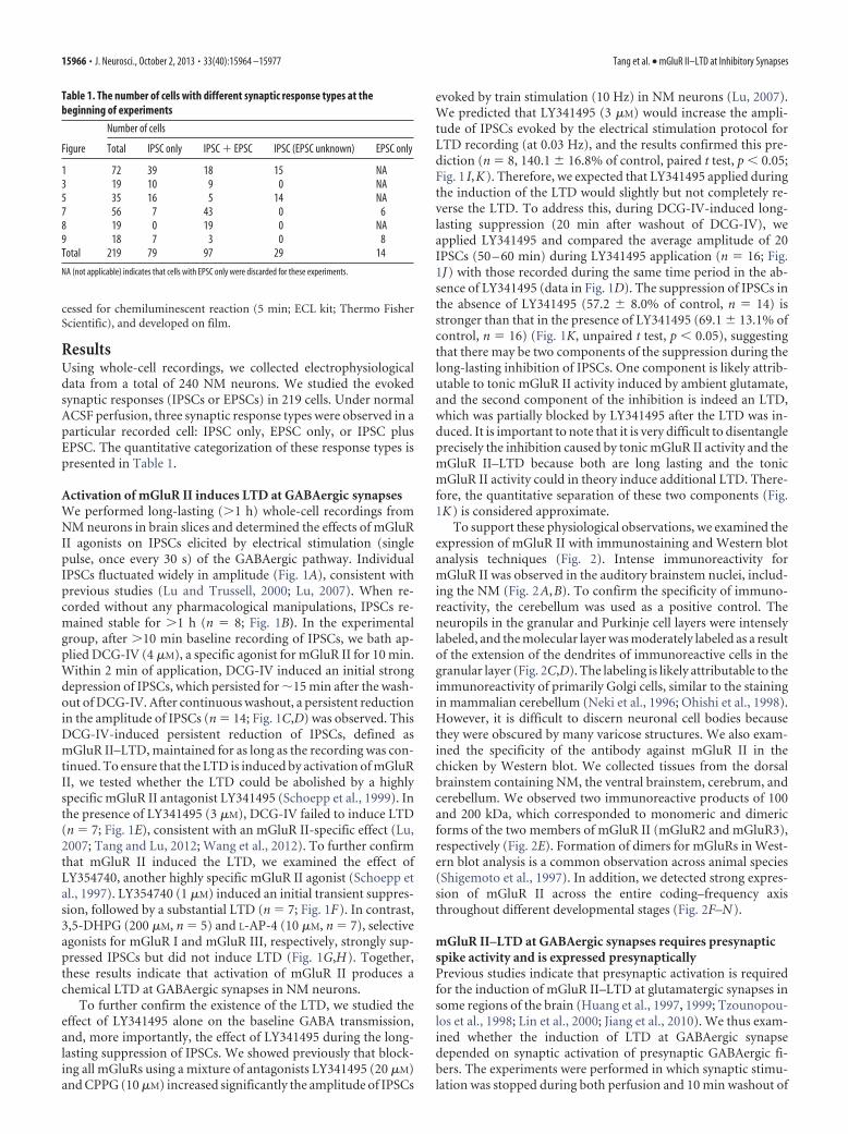
ResultsUsing whole-cell recordings, we collected electrophysiologicaldata from a total of 240 NM neurons. We studied the evokedsynaptic responses (IPSCs or EPSCs) in 219 cells. Under normalACSF perfusion, three synaptic response types were observed in aparticular recorded cell: IPSC only, EPSC only, or IPSC plusEPSC. The quantitative categorization of these response types ispresented in Table 1.
Activation of mGluR II induces LTD at GABAergic synapsesWe performed long-lasting (�1 h) whole-cell recordings fromNM neurons in brain slices and determined the effects of mGluRII agonists on IPSCs elicited by electrical stimulation (singlepulse, once every 30 s) of the GABAergic pathway. IndividualIPSCs fluctuated widely in amplitude (Fig. 1A), consistent withprevious studies (Lu and Trussell, 2000; Lu, 2007). When re-corded without any pharmacological manipulations, IPSCs re-mained stable for �1 h (n � 8; Fig. 1B). In the experimentalgroup, after �10 min baseline recording of IPSCs, we bath ap-plied DCG-IV (4 �M), a specific agonist for mGluR II for 10 min.Within 2 min of application, DCG-IV induced an initial strongdepression of IPSCs, which persisted for �15 min after the wash-out of DCG-IV. After continuous washout, a persistent reductionin the amplitude of IPSCs (n � 14; Fig. 1C,D) was observed. ThisDCG-IV-induced persistent reduction of IPSCs, defined asmGluR II–LTD, maintained for as long as the recording was con-tinued. To ensure that the LTD is induced by activation of mGluRII, we tested whether the LTD could be abolished by a highlyspecific mGluR II antagonist LY341495 (Schoepp et al., 1999). Inthe presence of LY341495 (3 �M), DCG-IV failed to induce LTD(n � 7; Fig. 1E), consistent with an mGluR II-specific effect (Lu,2007; Tang and Lu, 2012; Wang et al., 2012). To further confirmthat mGluR II induced the LTD, we examined the effect ofLY354740, another highly specific mGluR II agonist (Schoepp etal., 1997). LY354740 (1 �M) induced an initial transient suppres-sion, followed by a substantial LTD (n � 7; Fig. 1F). In contrast,3,5-DHPG (200 �M, n � 5) and L-AP-4 (10 �M, n � 7), selectiveagonists for mGluR I and mGluR III, respectively, strongly sup-pressed IPSCs but did not induce LTD (Fig. 1G,H). Together,these results indicate that activation of mGluR II produces achemical LTD at GABAergic synapses in NM neurons.
To further confirm the existence of the LTD, we studied theeffect of LY341495 alone on the baseline GABA transmission,and, more importantly, the effect of LY341495 during the long-lasting suppression of IPSCs. We showed previously that block-ing all mGluRs using a mixture of antagonists LY341495 (20 �M)and CPPG (10 �M) increased significantly the amplitude of IPSCs
evoked by train stimulation (10 Hz) in NM neurons (Lu, 2007).We predicted that LY341495 (3 �M) would increase the ampli-tude of IPSCs evoked by the electrical stimulation protocol forLTD recording (at 0.03 Hz), and the results confirmed this pre-diction (n � 8, 140.1 16.8% of control, paired t test, p 0.05;Fig. 1 I,K). Therefore, we expected that LY341495 applied duringthe induction of the LTD would slightly but not completely re-verse the LTD. To address this, during DCG-IV-induced long-lasting suppression (20 min after washout of DCG-IV), weapplied LY341495 and compared the average amplitude of 20IPSCs (50 – 60 min) during LY341495 application (n � 16; Fig.1J) with those recorded during the same time period in the ab-sence of LY341495 (data in Fig. 1D). The suppression of IPSCs inthe absence of LY341495 (57.2 8.0% of control, n � 14) isstronger than that in the presence of LY341495 (69.1 13.1% ofcontrol, n � 16) (Fig. 1K, unpaired t test, p 0.05), suggestingthat there may be two components of the suppression during thelong-lasting inhibition of IPSCs. One component is likely attrib-utable to tonic mGluR II activity induced by ambient glutamate,and the second component of the inhibition is indeed an LTD,which was partially blocked by LY341495 after the LTD was in-duced. It is important to note that it is very difficult to disentangleprecisely the inhibition caused by tonic mGluR II activity and themGluR II–LTD because both are long lasting and the tonicmGluR II activity could in theory induce additional LTD. There-fore, the quantitative separation of these two components (Fig.1K) is considered approximate.
To support these physiological observations, we examined theexpression of mGluR II with immunostaining and Western blotanalysis techniques (Fig. 2). Intense immunoreactivity formGluR II was observed in the auditory brainstem nuclei, includ-ing the NM (Fig. 2A,B). To confirm the specificity of immuno-reactivity, the cerebellum was used as a positive control. Theneuropils in the granular and Purkinje cell layers were intenselylabeled, and the molecular layer was moderately labeled as a resultof the extension of the dendrites of immunoreactive cells in thegranular layer (Fig. 2C,D). The labeling is likely attributable to theimmunoreactivity of primarily Golgi cells, similar to the stainingin mammalian cerebellum (Neki et al., 1996; Ohishi et al., 1998).However, it is difficult to discern neuronal cell bodies becausethey were obscured by many varicose structures. We also exam-ined the specificity of the antibody against mGluR II in thechicken by Western blot. We collected tissues from the dorsalbrainstem containing NM, the ventral brainstem, cerebrum, andcerebellum. We observed two immunoreactive products of 100and 200 kDa, which corresponded to monomeric and dimericforms of the two members of mGluR II (mGluR2 and mGluR3),respectively (Fig. 2E). Formation of dimers for mGluRs in West-ern blot analysis is a common observation across animal species(Shigemoto et al., 1997). In addition, we detected strong expres-sion of mGluR II across the entire coding–frequency axisthroughout different developmental stages (Fig. 2F–N).
mGluR II–LTD at GABAergic synapses requires presynapticspike activity and is expressed presynapticallyPrevious studies indicate that presynaptic activation is requiredfor the induction of mGluR II–LTD at glutamatergic synapses insome regions of the brain (Huang et al., 1997, 1999; Tzounopou-los et al., 1998; Lin et al., 2000; Jiang et al., 2010). We thus exam-ined whether the induction of LTD at GABAergic synapsedepended on synaptic activation of presynaptic GABAergic fi-bers. The experiments were performed in which synaptic stimu-lation was stopped during both perfusion and 10 min washout of
Table 1. The number of cells with different synaptic response types at thebeginning of experiments
Number of cells
Figure Total IPSC only IPSC � EPSC IPSC (EPSC unknown) EPSC only
1 72 39 18 15 NA3 19 10 9 0 NA5 35 16 5 14 NA7 56 7 43 0 68 19 0 19 0 NA9 18 7 3 0 8Total 219 79 97 29 14
NA (not applicable) indicates that cells with EPSC only were discarded for these experiments.
15966 • J. Neurosci., October 2, 2013 • 33(40):15964 –15977 Tang et al. • mGluR II–LTD at Inhibitory Synapses

DCG-IV. As shown in Figure 3A, DCG-IV application withoutpresynaptic stimulation of the GABAergic afferents failed to in-duce LTD (n � 7). This observation further supports the idea thatDCG-IV can be washed out rapidly (Breakwell et al., 1997; Lu andRubel, 2005). Therefore, the results argue against the possibilitythat the mGluR II–LTD may be caused by incomplete washout ofDCG-IV and thus residual occupancy of mGluR II duringwashout.
Given the mixed synaptic responses in the cells collected un-der normal ACSF perfusion for this experiment (four cells withIPSC plus EPSC and three cells with IPSC only; Fig. 3A), it isdifficult to draw a firm conclusion on whether the GABAergic orglutamatergic presynaptic spike activity is important for the in-duction of the LTD. However, in the cells from which only IPSCswere evoked in ACSF, DCG-IV application without afferentstimulation did not induce LTD of IPSCs. This observation sug-gests that presynaptic spike activity of the GABAergic terminals isrequired for the induction of the LTD. To further test this, weasked whether a presynaptic Ca 2� increase in the GABAergicterminals or a postsynaptic Ca 2� increase in NM neurons is re-quired for the LTD. The rationale to investigate Ca 2� signaling is
that the requirement of spike activity in long-term plasticity isgenerally associated with action potential-induced Ca 2� increaseat the loci in which the plasticity is induced. We increased theconcentration of Ca 2� chelator EGTA inside the recording elec-trodes from 5 to 15 mM. If a postsynaptic Ca 2� increase wererequired for the LTD induction, then LTD would not be formedin the presence of high EGTA. We found that DCG-IV inducedLTD of IPSCs as it did in the presence of a lower concentration ofEGTA (n � 7 cells; Fig. 3D), indicating that postsynaptic Ca 2�
signaling is not required for the LTD induction. To test whetherthe LTD requires presynaptic Ca 2� signaling in the GABAergicterminals, we incubated brain slices in the membrane-permeableCa 2� chelator BAPTA-AM at a moderate concentration (10 �M)for 30 – 60 min for BAPTA to be accumulated inside the cells andto eliminate Ca 2� increase in the presynaptic terminals (Lin et al.,2000). During the IPSC recordings, a lower concentration ofBAPTA-AM (5 �M) was continuously applied in the bath. Wechose not to use too high concentrations of BAPTA-AM nor toolong incubation time, which could diminish synaptic responses,dramatically leading to difficulty of studying very small IPSCs.Because BAPTA-AM treatment does not selectively target
Figure 1. mGluR II agonists induce LTD at GABAergic synapses. A, B, Individual IPSCs recorded once every 30 s varied widely in amplitude, but the averaged amplitude remained relatively constantin 80-min recordings (n � 8). C, D, Bath-application of an mGluR II agonist DCG-IV (4 �M) induced an initial transient suppression, followed by LTD of IPSCs (n � 14). E, LY341495 (3 �M), anantagonist for mGluR II, eliminated the effects of DCG-IV (n � 7). F, Another mGluR II agonist, LY354740 (1 �M), also induced an initial transient suppression and LTD of IPSCs (n � 7). G, H, mGluRI agonist (3,5-DHPG, 200 �M; n � 5) or mGluR III agonist (L-AP-4, 10 �M; n � 7) produced an initial suppression but not LTD of IPSCs in NM neurons. I, LY341495 (3 �M) slightly increased theamplitude of IPSCs evoked at 0.03 Hz (n � 8). J, Application of LY341495 (3 �M) during the induction of LTD partially reversed the plasticity (n � 16), suggesting the presence of a low level of tonicinhibition mediated by ambient glutamate-activated mGluR II. K, Averaged amplitude of IPSCs under the conditions of LY341495 (I ), DCG-IV (D), and LY341495 during LTD induction (J ), normalizedto their respective controls. The red dashed lines indicate the baseline (100% of control). Cells were voltage clamped at �70 mV. Means SEM are shown in this and subsequent figures. *p 0.05(t test).
Tang et al. • mGluR II–LTD at Inhibitory Synapses J. Neurosci., October 2, 2013 • 33(40):15964 –15977 • 15967

Figure 2. mGluR II is expressed in NM neurons throughout the entire tonotopic axis across different ages. A, Immunohistochemistry revealed expression of mGluR II in auditory brainstem nuclei.NA, Nucleus angularis; NL, nucleus laminaris. B, Part of the NM shown at a higher amplification. C, D, Expression of mGluR II in the cerebellar cortex used as a positive control. The neuropils in thegranular and Purkinje cell layers were intensely labeled, and the molecular layer was moderately labeled as a result of the extension of the dendrites of immunoreactive cells (primarily Golgi cells)in the granular layer. E, Western blot confirmed the specificity of the antibody against mGluR II in chicken brain tissues. The two immunoreactive products of 100 and 200 kDa correspond tomonomeric and dimeric forms of mGluR2/3, respectively. F–N, Expression of mGluR II in different characteristic frequency regions (LF, MF, and HF indicate low, middle, and high frequency,respectively) in E18 (n � 2 animals; F–H ), 1- or 2-d-old post-hatch chicks (P1–P2, n � 5 animals; I–K ), and 1-week-old chicks (P7–P9, n � 2 animals; L–N ).
15968 • J. Neurosci., October 2, 2013 • 33(40):15964 –15977 Tang et al. • mGluR II–LTD at Inhibitory Synapses

GABAergic terminals, we applied the LTD recording protocol tocells that showed IPSCs only in ACSF. DCG-IV induced muchsmaller LTD of IPSCs after BAPTA incubation (Fig. 3E), suggest-ing that an action potential-induced presynaptic Ca 2� increase inthe GABAergic terminals is possibly required for the LTD.
In different areas of the brain, mGluR II can be located pre-synaptically or postsynaptically (Nicoletti et al., 2011). To iden-tify the locus of the expression of mGluR II–LTD at GABAergicsynapses in NM neurons, we performed analyses on failure rateand coefficient of variation (CV) of IPSCs, under the conditionsof control, during DCG-IV (initial suppression), and after wash-out of DCG-IV (LTD induction). Changes in failure rate and CVindicate alterations in presynaptic release probability (Faber andKorn, 1991). We observed a significant increase in failure rate(Fig. 3B; control, 5.0 2.1%; DCG-IV, 60.7 6.0%, p 0.0001;LTD, 24.2 7.0%, p 0.05; n � 14) both during the initialsuppression and the LTD of IPSCs, suggesting a presynapticmechanism of mGluR II action. There was also a significant de-crease in normalized 1/CV 2 during DCG-IV and LTD (Fig. 3C;DCG-IV, 38.8 14.5% of the control, p 0.0001; LTD, 60.2 7.8% of the control, p 0.01; n � 14). These data indicate thatthe LTD results from a persistent reduction in the release proba-bility at the GABAergic synapses. To further confirm the presyn-aptic expression of mGluR II–LTD at GABAergic synapses, weexamined the effects of DCG-IV on the intrinsic excitability ofNM neurons. We observed little effect of DCG-IV on normalizedRMP, current threshold, action potential height and width, andinput resistance (Rin) under either current-clamp (n � 7; Fig.4A–F) or voltage-clamp (n � 14; Fig. 4G) recordings, presum-ably excluding postsynaptic mechanisms. Therefore, the induc-tion of LTD at GABAergic synapses in NM neurons is primarilypresynaptic, consistent with the presynaptic expression of
mGluR II–LTD at glutamatergic synapses in some regions of thebrain (Lin et al., 2000; Poschel and Manahan-Vaughan, 2005;Wang et al., 2012).
Although these physiological results and our previous data(Lu, 2007) demonstrate presynaptic actions of mGluR II, theanatomy data (Fig. 2) showed primarily somatic rather thanpunctate labeling of mGluR II. It is well known that Ca 2� signal-ing of NM neurons is regulated by multiple postsynaptic mGluRs(Rubel and Fritzsch, 2002), including mGluR II (Lu and Rubel,2005). Therefore, the explanation for this discrepancy may bethat the expression of mGluR II on GABAergic terminals ismasked by robust postsynaptic immunoreactivity.
mGluR II–LTD at GABAergic synapses is independent ofNMDA receptors and involves cAMP/PKA signalingBecause DCG-IV at a high concentration (10 –15 �M) activatesNMDA receptors (NMDARs; Wilsch et al., 1994; Breakwell et al.,1997) and activation of NMDARs is required for some forms oflong-term plasticity at GABAergic synapses in the brain (Lien etal., 2006; Castillo et al., 2011), we determined whether pharma-cological blockade of NMDARs affected the mGluR II–LTD ofGABAergic synapses in NM. As shown in Figure 5A, bath appli-cation of the NMDAR antagonist APV (50 �M) did not affect theLTD (n � 6), indicating that the expression of the LTD does notrequire activation of NMDARs.
We next investigated the signaling mechanisms underlyingmGluR II–LTD at GABAergic synapses. mGluR II are known to
Figure 3. The mGluR II–LTD requires presynaptic spike activity and is expressed presynap-tically. A, DCG-IV (4 �M) application without electrical stimulation of the GABAergic afferentsfailed to induce LTD (n �7). B, C, mGluR II–LTD was accompanied by changes in failure rate andthe CV of IPSCs. DCG-IV increased the failure rate ( p � 0.0169, n � 14) and decreased 1/CV 2
( p � 0.0063, n � 14) of IPSCs during LTD, indicating presynaptic actions of mGluR II. D, DCG-IV(4 �M) induced approximately the same level of LTD with high EGTA in recording electrodes(n � 7). E, BAPTA-AM treatment (10 �M, 30 – 60 min incubation followed by perfusion at alower concentration, 5 �M, throughout the recording) of the brain slices essentially eliminatedDCG-IV-induced LTD of IPSCs (n � 5). *p 0.05, **p 0.01, ***p 0.001 (ANOVA post hocFisher’s test).
Figure 4. mGluR II agonists do not change the excitability of NM neurons. A, Representativemembrane potential recordings in response to prolonged (200 ms) somatic current injectionsunder the conditions of control, DCG-IV (4 �M), and LTD (45 min after termination of DCG-IVapplication). The action potential (labeled with the symbol #) waveform under the controlcondition is shown at larger scales. B–F, Normalized RMPs, current thresholds, action potential(AP) height and width, and Rin remained essentially unchanged between control and during/after DCG-IV application (n � 7), presumably excluding postsynaptic mechanisms of the LTDinduction by mGluR II. G, Rin of NM neurons obtained under voltage-clamp recordings remainedunchanged (n � 14) during LTD induction. *p 0.05 (ANOVA post hoc Fisher’s test).
Tang et al. • mGluR II–LTD at Inhibitory Synapses J. Neurosci., October 2, 2013 • 33(40):15964 –15977 • 15969

couple to Gi-proteins, and activation of Gi-proteins results indecreased activity of adenylyl cyclase (AC) and subsequent re-duction in cAMP and suppression of PKA (Pin and Acher, 2002).To determine whether AC activity is required for mGluR II–LTD,we incubated brain slices in ACSF containing the AC inhibitorSQ22536 (50 �M) for 1 h and then repeated the LTD protocolduring which SQ22536 was present. Under these conditions,DCG-IV (4 �M) failed to induce LTD (n � 8; Fig. 5B). The initialsuppression of IPSCs was not affected, possibly because it wasmediated by a membrane-delimited pathway whereby theG-protein �� complex inhibits voltage-gated Ca 2� channels(Herlitze et al., 1996). We then determined whether PKA is in-volved in mGluR II–LTD. Application of KT5720 (1 �M), a selec-tive PKA inhibitor, led to a progressive reduction in IPSCs, andthe effects stabilized within 1 h (n � 9; Fig. 5C). Therefore, weincubated slices in ACSF containing KT5720 (1 �M) for 1 h andthen repeated the LTD protocol in the presence of KT5720.KT5720 completely abolished the LTD without blocking the ini-tial depression (n � 12; Fig. 5D). Together, our findings demon-strate that mGluR II–LTD at GABAergic synapses is associatedwith inhibition of AC and cAMP-dependent PKA, consistentwith the mechanisms underlying mGluR II-induced LTD at someglutamatergic synapses (Tzounopoulos et al., 1998; Huang et al.,1999, 2007; Lin et al., 2000; Robbe et al., 2002; Bellone et al.,2008).
Activation of mGluR II by endogenous glutamate andsynaptically induced heterosynaptic LTD at GABAergicsynapsesAlthough the above results demonstrate that activation of mGluRII by exogenous agonists induced an initial suppression followedby LTD at GABAergic synapses, a more physiologically relevantquestion is whether mGluR II can be activated by endogenousglutamate. Presynaptic mGluR II can be activated by ambientglutamate (Chu and Moenter, 2005), synaptically released gluta-mate, or glutamate spillover from neighboring synapses (Mitch-ell and Silver, 2000; Tang et al., 2009). To find out whethermGluR II can be activated by ambient glutamate, we examined
the effects of LY341495 on sIPSCs in NM neurons. LY341495(150 nM) increased the frequency (control, 0.66 0.09 Hz;LY341495, 1.17 0.24 Hz; n � 9, p 0.01; Fig. 6) withoutaffecting the amplitude (control, �124.0 9.4 pA; LY341495,�112.5 10.4 pA; n � 9, p � 0.05), suggesting that backgroundambient glutamate tonically activates presynaptic mGluR II,leading to suppression of spontaneous GABA release.
We next asked whether activation of mGluR II by endogenousglutamate could induce LTD at GABAergic synapses. To addressthis, we concurrently activated both the glutamatergic andGABAergic pathways evoking a synaptic current typically with afast and a slow component (Fig. 7A,B). The fast component wasabolished by DNQX (50 �M), whereas the slow component wasabolished by SR95531 (10 �M), a selective GABAAR antagonist.We then examined whether low-frequency stimulation (LFS; 1Hz, for 15 min) applied to the two pathways induced LTD. TheLFS protocol is commonly used to induce homosynaptic mGluRII–LTD at glutamatergic synapses (Kobayashi et al., 1996; Yokoiet al., 1996; Manahan-Vaughan, 1997; Domenici et al., 1998;Huang et al., 1999; Cho et al., 2000; Li et al., 2002). In the presenceof APV (50 �M), LFS induced a robust LTD (n � 11; Fig. 7C). Wenext determined whether LFS-induced LTD was attributable toactivation of mGluR II by synaptically released glutamate. Be-cause LY341495 can affect spontaneous GABA release (Fig. 6), wethus incubated the slices in ACSF containing LY341495 (200 nM)for 10 min before recordings of IPSCs started to eliminate theeffects of tonic mGluR II activated by ambient glutamate. In thepresence of LY341495, LFS failed to induce LTD (n � 13; Fig.7D), suggesting that the LTD was induced by mGluR II activatedby synaptically released glutamate. It is conceivable that the ma-jor source of glutamate needed to induce the LTD is from theauditory nerve, because the electrical stimulation applied to thesecells coactivated both the excitatory and inhibitory inputs. How-ever, it is still unknown whether ambient glutamate or otheractivity-independent pools of the transmitter is sufficient to in-
Figure 5. The LTD is NMDAR independent and mediated by the classic cAMP/PKA signalingpathway. A, The LTD persisted in the presence of the NMDAR antagonist APV (50 �M, n � 6). B,Blocking AC by SQ22536 (50 �M, n � 8) eliminated the LTD without affecting the initial inhi-bition. C, KT5720 (1 �M, n � 9), an antagonist for PKA, gradually reduced IPSCs, and the effectsstabilized in �1 h. D, Incubation of brain slices for 1 h in ACSF containing KT5720 (1 �M, n �12) occluded the induction of LTD.
Figure 6. Ambient glutamate is sufficient in activating mGluR II. A, Representative sIPSCsrecorded under control (ctr) and LY341495 (LY; 150 nM) application. Shown on the right are theaveraged traces under control and drug conditions (106 and 145 events, respectively). B, C,Cumulative fractions of the frequency (Freq) and amplitude (Amp) of sIPSCs show thatLY341495 increased the frequency without affecting the amplitude of sIPSCs (n �9). The insetsshow the averaged frequency and amplitude of sIPSCs (n � 9). IEI: Inter-event interval. **p 0.01 (t test).
15970 • J. Neurosci., October 2, 2013 • 33(40):15964 –15977 Tang et al. • mGluR II–LTD at Inhibitory Synapses

duce LTD. To test this, we intentionally recorded from NM neu-rons in which only IPSCs were evoked and examined whether theLFS induced LTD in the absence of glutamate release from theauditory nerve innervating the recorded cells. As expected,the LFS elicited a short-term suppression but not an LTD ofIPSCs (Fig. 7E), suggesting that spike activity-dependent gluta-mate release from the auditory nerve is necessary for the induc-tion of the LTD.
To confirm that the LFS does not alter glutamatergic trans-mission, which could in turn impact synaptic inhibition, wetested the effect of the LFS on evoked EPSCs in NM neurons. TheLFS (1 Hz, 15 min) did not affect the EPSCs of NM neurons (Fig.7F, n � 6 cells, paired t test, p � 0.05), indicating that the amountof glutamate release during the LFS protocol is relatively con-stant, and there is no significant synaptic depression of the excit-atory transmission at the stimulus interval used (1 s). This isconsistent with previous findings that this synapse has the capa-bility of releasing glutamate at high probability, and synaptic
depression does not emerge until the stimulus pulse interval isbelow 10 ms (i.e., stimulus frequency of 100 Hz; Zhang and Trus-sell, 1994). Therefore, it is conceivable that a stabilized amount ofmGluR II is constantly activated by synaptically released gluta-mate during the LFS protocol, inducing LTD of IPSCs.
An equally important issue is whether mGluR II activationaffects the excitatory transmission during the LTD inductionprotocol, because mGluR II could alter glutamate release and inturn affect the GABAergic transmission. We found that DCG-IV(4 �M) did not change the amplitude of EPSCs in NM neurons(Fig. 7G, n � 6 cells, p � 0.05). Otis and Trussell (1996) reportedthat ()-1-amino-1,3-cyclopentanedicarboxylic acid (200 �M),a generic mGluR agonist that presumably activates all membersof the three groups of mGluRs, did not change EPSCs in NMneurons. This is an unusual observation because mGluRs func-tion as autoreceptors modulating glutamate release in many glu-tamate synapses in the CNS. Our results further confirmed theobservation by Otis and Trussell (1996) and specifically demon-
Figure 7. Synaptically released glutamate induces heterosynaptic mGluR II–LTD at GABAergic synapses. A, A schematic drawing showing the experimental paradigm and possible mechanismsunderlying mGluR II–LTD of GABA release. B, Concurrent activation of the glutamatergic and GABAergic pathways. The EPSC was blocked by DNQX (50 �M). C, An LFS (1 Hz, for 15 min) induced LTDof IPSCs (n � 11). D, The LTD was blocked by LY341495 (150 nM; n � 13), excluding electrical LTD and confirming chemical LTD induced by mGluR II. E, In cells in which only IPSCs were evokedintentionally, LFS elicited a short-term suppression but not an LTD of IPSCs (n � 7). F, The same LFS protocol did not change EPSCs in NM neurons (n � 6). The three EPSC traces under the barindicating LFS were individual traces obtained from different time points throughout the LFS application. G, H, Neither DCG-IV (4 �M, n � 6) nor LY341495 (LY; 0.2 �M, n � 5) altered the amplitudeof EPSCs in NM neurons.
Tang et al. • mGluR II–LTD at Inhibitory Synapses J. Neurosci., October 2, 2013 • 33(40):15964 –15977 • 15971

strated that mGluR II did not modulate glutamate release at NM.In addition, the mGluR II antagonist LY341495 (0.2 �M) did notaffect the EPSCs of NM neurons (Fig. 7H, n � 5 cells, p � 0.05).Together, these data (Fig. 7C–H) demonstrate that LFS of theglutamatergic pathway activates presynaptic mGluR II onGABAergic terminals and induces heterosynaptic LTD atGABAergic synapses, without modulating the excitatory inputsof NM neurons.
Finally, we tested whether synaptically released glutamate ac-tivates mGluR II, suppressing evoked GABA release in a stimulusfrequency-dependent manner. The glutamatergic inputs to NMneurons are mediated by morphologically specialized presynap-tic synapses, the end bulbs of Held, which cover the majority ofthe somata (Parks, 1981). With activation of the glutamatergicsynapses, a high level of glutamate could be released and spilled toactivate mGluRs on GABAergic terminals. We examined the ef-fects of LY341495 on IPSCs evoked by train stimulations at dif-ferent stimulus frequencies (3.3, 100, or 300 Hz). As expected,blocking mGluR II with LY341495 (10 nM) increased the ampli-tude of IPSCs (Fig. 8A–G). Because IPSCs evoked at 100 and 300Hz summated temporally forming a plateau during the stimula-
tion and a long decay time course, we also analyzed the IPSC arearepresenting the total charge (product of time and current). Boththe amplitude and the area of IPSCs showed a significant increasewhen mGluR II were blocked, demonstrating that synapticallyreleased glutamate could activate mGluR II. Interestingly, theextent of activation of mGluR II and their modulatory strengthon IPSCs were stimulus frequency independent (Fig. 8H), re-flecting the high affinity of mGluR II in binding with glutamateand their readiness of being activated in NM neurons. In contrast,blocking mGluR III by CPPG (50 nM) significantly increasedIPSCs at both stimulus frequencies of 3.3 Hz (n � 7) and 100 Hz(n � 3), and the latter effect was significantly stronger than theformer (data not shown).
In CA1 of the hippocampus, the form of long-term plasticity isdependent on stimulus frequency (Dudek and Bear, 1992). Be-cause the LFS at 1 Hz elicited LTD and endogenous activity ofmGluR II was detected at various stimulus frequencies (3.3, 100,and 300 Hz) in NM neurons, a logical question to ask next iswhether the mGluR II–LTD is also stimulus frequency depen-dent. To address this, we tested whether a high-frequency stimu-lus (100 Hz, 9 s) that coactivated both the excitatory and
Figure 8. Synaptically released glutamate activates mGluR II independent of stimulus frequency. Train stimulations that caused concurrent activation of the glutamatergic and GABAergicpathways were used. A, Superimposed original current traces evoked by a train stimulation at 3.3 Hz (A1) and averaged IPSCs (A2) obtained under conditions of control, the mGluR II antagonistLY341495 (10 nM), and wash. A nearly complete recovery of the responses is observed after the washout. B, LY341495 (LY) significantly increases the normalized amplitude of IPSCs regardless of thepresence of transmission failures (n � 9; dark gray: failures included, p 0.001; light gray: failures excluded, p 0.001). Post hoc Fisher’s analyses revealed significant differences in normalizedIPSC amplitude between control (ctr) and LY341495. C, Consistently, LY341495 also significantly increases the normalized area (representing charge) of IPSCs (n � 9; dark gray: failures included,p 0.01; light gray: failures excluded, p 0.001). D, E, LY341495 significantly increases the normalized amplitude (dark filled bars, p � 0.0116) and area (open bars, p 0.05) of IPSCs elicitedby train stimulation (100 Hz, 20 pulses) (n � 3). F, G, LY341495 significantly increases the normalized amplitude (dark solid bars, p 0.0001) and area (open bars, p 0.0001) of IPSCs elicited bytrain stimulation (300 Hz, 20 pulses; n � 7). H, No significant differences in the effects of LY341495 on the amplitude and area of IPSCs are detected under different stimulus frequencies. *p 0.05,**p 0.01, ***p 0.001 (ANOVA post hoc Fisher’s test).
15972 • J. Neurosci., October 2, 2013 • 33(40):15964 –15977 Tang et al. • mGluR II–LTD at Inhibitory Synapses

inhibitory inputs to NM neurons induced LTD. The chosen stim-ulus frequency is physiologically relevant for both the excitatoryinputs (Warchol and Dallos, 1990; Fukui et al., 2006) and theinhibitory inputs (Lachica et al., 1994; Coleman et al., 2011). The
duration of the stimulation (9 s) matchedthe number of pulses (900) in the LFS pro-tocol (1 Hz, 15 min), and the high-frequency stimulation protocol was usedto characterize the glutamatergic andGABAergic responses. NM cells can read-ily sustain firing in vivo at rates at andabove 100 Hz. However, in slice prepara-tions, electrical stimulation at 100 Hzrapidly diminished synaptic currents.Prominent synaptic depression of EPSCsoccurred within the first 10 pulses, and bythe end of 1 s of the recording, EPSCs di-minished to the minimal level (n � 10;Fig. 9A1,B). Correspondingly, NM neu-rons were driven to fire action potentialsat the beginning of the train stimulation,followed by subthreshold EPSPs (n � 5;Fig. 9A2). Similar results were obtainedfor IPSCs. Because of their slower kinetics,IPSCs temporally summated when stimu-lated at 100 Hz and decayed to a level closeto the baseline in 9 s (n � 8; Fig. 9C).Despite the apparent fast depletion ofboth transmitters in response to the trainstimulation, concurrent activation of theglutamatergic and GABAergic pathwayswith the train stimulation (100 Hz, 900pulses) caused a transient facilitation ofIPSCs recorded with single-pulse stimula-tion. However, the train stimulation didnot induce any long-term plasticity of theIPSCs (n � 8; Fig. 9D). The transient fa-cilitation may be interpreted as a result ofa large GABA release from replenishedvesicle pools through a feedback mecha-nism after the train stimulation. The lackof long-term plasticity of the IPSCs sug-gests that the LTD may be stimulus fre-quency and duration dependent.
DiscussionWe report a novel form of heterosynapticlong-term plasticity: mGluR II–LTD atGABAergic synapses in the central audi-tory system. This mGluR II–LTD can beinduced by pharmacological or synapticactivation of mGluR II. Below we discussthe rigorous evidence we presented to es-tablish the validity of the LTD, followedby assessment of the signaling pathwayand functional implication.
Establishment of mGluR II–LTD atGABAergic synapsesSince its discovery (Palmer et al., 1997),chemical LTD has been studied exten-sively at glutamatergic synapses (Col-lingridge et al., 2010). However, therehave been debates on whether chemical
LTD is caused by incomplete washout of the agonists (Wostrackand Dietrich, 2009). The following results demonstrate that theLTD we observed is not an artifact of agonist treatment. First,
Figure 9. In slice preparations, a high-frequency electrical stimulation (100 Hz) rapidly diminishes synaptic currents. A1, EPSCsof one NM neuron in response to a train stimulation (100 Hz, 100 pulses). The first and the last 10 responses are shown at a largertimescale. By the end of the recording (duration of 1 s), EPSCs diminish to a minimal level. A2, Correspondingly, in current-clamprecordings, a postsynaptic NM cell was driven by the same stimulus protocol to fire action potentials (APs) only at the beginning ofthe stimulation. A total of 10 and 5 cells were recorded in voltage- and current-clamp mode, respectively. B, Normalized EPSCamplitudes plotted against stimulus pulse number (n � 10). C1, C2, IPSCs of two representative NM neurons in response to a trainstimulation (100 Hz, 1000 pulses). The first and the last 10 responses are shown at a larger timescale. Because of their slowerkinetics, IPSCs temporally summated. The summated responses diminished to a level close to the baseline (indicated by the dashedline) in �10 s. Individual IPSCs vary widely in their amplitudes, resulting in mixed short-term synaptic facilitation and depression.D, Concurrent activation of the glutamatergic and GABAergic pathways with train stimulation (100 Hz, 900 pulses) does not induceany long-term plasticity of the IPSCs (n � 8). Stimulus artifacts in the EPSC and IPSC recordings are blanked for clarity.
Tang et al. • mGluR II–LTD at Inhibitory Synapses J. Neurosci., October 2, 2013 • 33(40):15964 –15977 • 15973

application of the mGluR II antagonist LY341495 after washoutof DCG-IV partially reversed the LTD (Fig. 1 J,K), indicating thepresence of an LTD insensitive to LY341495. The partial sensitiv-ity of the LTD to LY341495 may be explained by constitutiveactivity of mGluR II in the absence of agonists (Lodge et al.,2013). Second, DCG-IV application without synaptic stimula-tion of the GABAergic afferents fails to induce LTD (Fig. 3A).Chelating Ca 2� in presumably all neuronal compartments usingBAPTA-AM essentially eliminated the LTD, whereas chelatingCa 2� in postsynaptic cells using high EGTA did not (Fig. 3D,E),suggesting that both mGluR II activation and Ca 2� signaling inthe GABAergic terminals are required. Third, blocking mGluR II(Figs. 1E, 7D) or candidate molecules suspected to be involved inthe LTD pathway eliminates the LTD (Fig. 5B,D). The reversibil-ity of the initial inhibition also indicates complete washout ofDCG-IV. Fourth, physiologically released glutamate but notGABA induced LTD of GABA release (Fig. 7C,E), confirmingchemical LTD induced by heteroreceptors and excluding electri-cal LTD induced by direct low-frequency activation of the GABAafferents (Gaiarsa et al., 2002). Finally, neither mGluR II agonistnor the LFS protocol affects glutamatergic transmission in NM(Fig. 7F,G), indicating that the LTD is formed at the GABAergicsynapses rather than caused by altered glutamate release.
Endogenous glutamate readily activates mGluR II in NM neu-rons (Figs. 6-8), possibly because mGluR II have high affinity ofbinding glutamate (Cartmell and Schoepp, 2000). Glutamate re-quired for the endogenous activation of mGluR II on the GABAe-rgic terminals in NM could originate from several sources, suchas ambient glutamate present in the extracellular space, synapticrelease from the auditory nerve, and spillover from neighboringglutamatergic synapses or activity-independent release from glialcells. The observation that mGluR II antagonist enhanced thefrequency without affecting the amplitude of sIPSC indicates thatpresynaptic mGluR II are activated tonically by ambient gluta-mate and exert a continued control of the basal level of GABArelease. Blocking mGluR II increased the amplitude of IPSCs,regardless of stimulus frequencies (3.3, 100, or 300 Hz), indicat-ing that mGluR II could be activated by synaptically releasedglutamate and probably reached saturation at low stimulus fre-quencies. Consequently, LFS applied to coactivate the glutamateand GABA pathways was able to trigger LTD, strongly indicatingthat synaptic activation of mGluR II caused a heterosynapticLTD. Figure 10 schematically depicts the three components of theinhibition caused by mGluR II on the GABAergic transmission inNM. Ambient glutamate activates a proportion of the high-affinity mGluR II, suppressing the spontaneous GABA release.Similar tonic mGluR activity has been observed in dopamineneurons in the substantia nigra (Wang et al., 2005). When exog-enous mGluR II agonists were added, a strong inhibition of IPSCswas produced. After washout of the agonists, the response recov-ered partially and remained constant afterward, suggesting thepresence of LTD.
Presynaptic expression and the cAMP/PKA signaling ofthe LTDThe majority of mGluR II–LTD at glutamatergic synapses is ex-pressed presynaptically (Bellone et al., 2008), whereas postsynap-tic expression of mGluR II–LTD at some glutamatergic synapseswas also reported (Huang et al., 1999; Cho et al., 2000; Otani etal., 2002). Several lines of evidence show that the induction ofmGluR II–LTD of GABAergic synapses in NM is expressed pre-synaptically. First, mGluR II–LTD induced by either DCG-IV orLFS was accompanied by the increase in CV of IPSC amplitude
and failure rate of GABAergic responses. Second, the induction ofmGluR II–LTD by DCG-IV was associated with minimal changesof postsynaptic neural excitability. Third, treatment of BAPTA-AM, but not postsynaptically loaded EGTA, prevented mGluRII–LTD, suggesting that presynaptic Ca 2� signaling is involved.Finally, blocking mGluR II increased the frequency without af-fecting the amplitude of sIPSCs. Therefore, the mGluR II–LTD isprimarily caused by a presynaptic persistent reduction in theprobability of GABA release.
Multiple molecular mechanisms account for long-termchanges at GABAergic synapses (Castillo et al., 2011). Being pri-marily coupled to Gi-proteins, mGluR II are negatively linked toAC, leading to a reduction in the intracellular cAMP levels andinhibition of PKA (Pin and Acher, 2002), although PKC is in-volved in some cases (Otani et al., 1999; Kahn et al., 2001). Con-sequently, mGluR II–LTD at glutamatergic synapses is expressedthrough common signaling mechanisms involving cAMP/PKA(Tzounopoulos et al., 1998; Robbe et al., 2002; Chevaleyre andCastillo, 2003; Zhao and Tzounopoulos, 2011). The mGluR II–LTD in NM neurons shares the same induction and expressionmechanisms, because blocking AC or PKA abolished the LTD.Therefore, inhibition of cAMP/PKA activity may be a signaltransduction mechanism by which mGluR II–LTD can be in-duced at both glutamatergic and GABAergic synapses. Other fea-tures of mGluR II–LTD at GABAergic synapses appear to besimilar to mGluR II–LTD at glutamatergic synapses describedpreviously in hippocampal CA3 (Tzounopoulos et al., 1998) andamygdala (Lin et al., 2000). Both forms of mGluR II–LTD requirepresynaptic spiking activity but do not require activation ofNMDARs.
Functional implicationsLong-term plasticity is commonly expressed in higher-orderbrain structures, such as the cerebral and cerebellar cortex, in-cluding the auditory cortex (Schreiner and Winer, 2007), inwhich synaptic plasticity is implicated in normal auditory signal-ing processing (Oda et al., 1998; Froemke et al., 2007) and hearingloss (Xu et al., 2010; Sanes and Kotak, 2011). In the past decade,such long-term plasticity has also been revealed in lower auditorybrainstem (Tzounopoulos and Kraus, 2009), such as in the lateral
Figure 10. Schematic drawing depicting the three components of the inhibition caused bymGluR II on the GABAergic transmission in NM neurons. Ambient glutamate activates somemGluR II, suppressing the spontaneous release of GABA. Application of an exogenous mGluR IIagonist produces a large inhibition of IPSCs, which consists of a reversible component andthe LTD.
15974 • J. Neurosci., October 2, 2013 • 33(40):15964 –15977 Tang et al. • mGluR II–LTD at Inhibitory Synapses

superior olive (Kotak and Sanes 2000, 2002; Kotak et al., 2001;Chang et al. 2003) and in the dorsal cochlear nucleus (Fujino andOertel, 2003; Tzounopoulos et al., 2004, 2007; Zhao and Tzou-nopoulos, 2011). However, in the auditory brainstem structuresresponsible for precise timing coding, such long-term plasticitywas thought to be absent based on the assumption that high-fidelity information transfer was so critical for temporal process-ing that long-term plasticity of neuronal properties could becounterproductive. Our results challenge this view by establish-ing that synaptic activation of mGluR II can form long-termmodulatory action on GABAergic inputs in neurons that performprecise timing coding in the auditory brainstem.
Physiological functions of long-term plasticity at inhibitorysynapses remain primarily elusive (Castillo et al., 2011). mGluR-dependent LTD has been implicated in synaptic remodeling dur-ing development, especially at synapses in which such LTD is agedependent (Li et al., 2002). For instance, mGluR I/II–LTD ofglutamate synapses is accompanied by synaptic elimination atCA3–CA1 synapses in hippocampus (Shinoda et al., 2005), andmGluR I–LTD contributes to synaptic elimination of climbingfiber synapses on cerebellar Purkinje neurons (Ichise et al., 2000).In the auditory system, LTD at inhibitory synapses may contrib-ute to synaptogenesis and synaptic refinement (Kotak and Sanes,2000; Kotak et al., 2001). NM neurons experience dramatic den-dritic pruning during development, with extensive dendritic ar-borization in early embryos (E8 –E13) and few short dendrites inlate embryos (E16 –E20; Jhaveri and Morest, 1982). mGluR-mediated modulation of the GABAergic transmission is develop-mentally regulated during the period of dendritic pruning (Tangand Lu, 2012). Whether and how mGluR II–LTD of GABAergicsynapses is involved in development, or in dendritic retractionafter afferent deprivation (Wang and Rubel, 2012), remain to beinvestigated. In mature animals, we propose that such LTD pre-vents GABA spikes induced by overactivation of the GABAergicinput (Lu and Trussell, 2001). Such a role supports our previ-ously proposed model in which tonic activity of mGluRs regu-lates the basal level of GABA release, whereas a use-dependentactivation of metabotropic GABA receptors provides a feedbackcontrol of GABA release in NM, ensuring high fidelity of phase-locking and precise temporal processing of sounds (Lu, 2007).Furthermore, NM neurons receive high rate inputs from the au-ditory nerve in vivo (Warchol and Dallos, 1990; Fukui et al.,2006), and a possible high level of tonic mGluR II activity wouldmaintain the LTD at a constant state. Therefore, regulation oftonic mGluR II activity would be physiologically important fortemporal coding, because variations in GABA release may gener-ate patterned neuronal responses encoding dynamic temporalfeatures of sound. Such regulation can be achieved at multiplestages along mGluR II signaling pathways by multiple G-protein-coupled receptors, which merits additional investigation to revealthe sophisticated yet synergetic actions of various neuromodula-tors in controlling synaptic strength in time-coding neurons.
ReferencesBellone C, Luscher C, Mameli M (2008) Mechanisms of synaptic depression
triggered by metabotropic glutamate receptors. Cell Mol Life Sci 65:2913–2923. CrossRef Medline
Breakwell NA, Huang L, Rowan MJ, Anwyl R (1997) DCG-IV inhibits syn-aptic transmission by activation of NMDA receptors in area CA1 of rathippocampus. Eur J Pharmacol 322:173–178. CrossRef Medline
Brenowitz S, David J, Trussell L (1998) Enhancement of synaptic efficacy bypresynaptic GABAB receptors. Neuron 20:135–141. CrossRef Medline
Burger RM, Cramer KS, Pfeiffer JD, Rubel EW (2005) Avian superior oli-
vary nucleus provides divergent inhibitory input to parallel auditorypathways. J Comp Neurol 481:6 –18. CrossRef Medline
Cartmell J, Schoepp DD (2000) Regulation of neurotransmitter release bymetabotropic glutamate receptors. J Neurochem 75:889 –907. CrossRefMedline
Castillo PE, Chiu CQ, Carroll RC (2011) Long-term plasticity at inhibitorysynapses. Curr Opin Neurobiol 21:328 –338. CrossRef Medline
Chang EH, Kotak VC, Sanes DH (2003) Long-term depression of synapticinhibition is expressed postsynaptically in the developing auditory sys-tem. J Neurophysiol 90:1479 –1488. CrossRef Medline
Chevaleyre V, Castillo PE (2003) Heterosynaptic LTD of hippocampalGABAergic synapses: a novel role of endocannabinoids in regulating ex-citability. Neuron 38:461– 472. CrossRef Medline
Cho K, Kemp N, Noel J, Aggleton JP, Brown MW, Bashir ZI (2000) A newform of long-term depression in the perirhinal cortex. Nat Neurosci3:150 –156. CrossRef Medline
Chu Z, Moenter SM (2005) Endogenous activation of metabotropic gluta-mate receptors modulates GABAergic transmission to gonadotropin-releasing hormone neurons and alters their firing rate: a possible localfeedback circuit. J Neurosci 25:5740 –5749. CrossRef Medline
Code RA, Burd GD, Rubel EW (1989) Development of GABA immunore-activity in brainstem auditory nuclei of the chick: ontogeny of gradients interminal staining. J Comp Neurol 284:504 –518. CrossRef Medline
Coleman WL, Fischl MJ, Weimann SR, Burger RM (2011) GABAergic andglycinergic inhibition modulate monaural auditory response propertiesin the avian superior olivary nucleus. J Neurophysiol 105:2405–2420.CrossRef Medline
Collingridge GL, Peineau S, Howland JG, Wang YT (2010) Long-term de-pression in the CNS. Nat Rev Neurosci 11:459 – 473. CrossRef Medline
Domenici MR, Berretta N, Cherubini E (1998) Two distinct forms of long-term depression coexist at the mossy fiber-CA3 synapse in the hippocam-pus during development. Proc Natl Acad Sci U S A 95:8310 – 8315.CrossRef Medline
Dudek SM, Bear MF (1992) Homosynaptic long-term depression in areaCA1 of hippocampus and effects of N-methyl-D-aspartate receptor block-ade. Proc Natl Acad Sci U S A 89:4363– 4367. CrossRef Medline
Faber DS, Korn H (1991) Applicability of the coefficient of variationmethod for analyzing synaptic plasticity. Biophys J 60:1288 –1294.CrossRef Medline
Froemke RC, Merzenich MM, Schreiner CE (2007) A synaptic memorytrace for cortical receptive field plasticity. Nature 450:425– 429. CrossRefMedline
Fujino K, Oertel D (2003) Bidirectional synaptic plasticity in thecerebellum-like mammalian dorsal cochlear nucleus. Proc Natl Acad SciU S A 100:265–270. CrossRef Medline
Fukui I, Sato T, Ohmori H (2006) Improvement of phase information atlow sound frequency in nucleus magnocellularis of the chicken. J Neuro-physiol 96:633– 641. CrossRef Medline
Gaiarsa JL, Caillard O, Ben-Ari Y (2002) Long-term plasticity at GABAergicand glycinergic synapses: mechanisms and functional significance.Trends Neurosci 25:564 –570. CrossRef Medline
Herlitze S, Garcia DE, Mackie K, Hille B, Scheuer T, Catterall WA (1996)Modulation of Ca 2� channels by G-protein beta gamma subunits. Nature380:258 –262. CrossRef Medline
Huang CC, Yang PC, Lin HJ, Hsu KS (2007) Repeated cocaine administra-tion impairs group II metabotropic glutamate receptor-mediated long-term depression in rat medial prefrontal cortex. J Neurosci 27:2958 –2968.CrossRef Medline
Huang L, Killbride J, Rowan MJ, Anwyl R (1999) Activation of mGluRIIinduces LTD via activation of protein kinase A and protein kinase C in thedentate gyrus of the hippocampus in vitro. Neuropharmacology 38:73–83. CrossRef Medline
Huang LQ, Rowan MJ, Anwyl R (1997) mGluR II agonist inhibition of LTPinduction, and mGluR II antagonist inhibition of LTD induction, in thedentate gyrus in vitro. Neuroreport 8:687– 693. CrossRef Medline
Ichise T, Kano M, Hashimoto K, Yanagihara D, Nakao K, Shigemoto R,Katsuki M, Aiba A (2000) mGluR1 in cerebellar Purkinje cells essentialfor long-term depression, synapse elimination, and motor coordination.Science 288:1832–1835. CrossRef Medline
Jhaveri S, Morest DK (1982) Sequential alterations of neuronal architecturein nucleus magnocellularis of the developing chicken: a Golgi study. Neu-roscience 7:837– 853. CrossRef Medline
Tang et al. • mGluR II–LTD at Inhibitory Synapses J. Neurosci., October 2, 2013 • 33(40):15964 –15977 • 15975

Jiang B, Huang S, de Pasquale R, Millman D, Song L, Lee HK, Tsumoto T,Kirkwood A (2010) The maturation of GABAergic transmission in vi-sual cortex requires endocannabinoid-mediated LTD of inhibitory inputsduring a critical period. Neuron 66:248 –259. CrossRef Medline
Kahn L, Alonso G, Robbe D, Bockaert J, Manzoni OJ (2001) Group 2metabotropic glutamate receptors induced long term depression inmouse striatal slices. Neurosci Lett 316:178 –182. CrossRef Medline
Kobayashi K, Manabe T, Takahashi T (1996) Presynaptic long-term depres-sion at the hippocampal mossy fiber–CA3 synapse. Science 273:648 – 650.CrossRef Medline
Kotak VC, Sanes DH (2000) Long-lasting inhibitory synaptic depression isage- and calcium-dependent. J Neurosci 20:5820 –5826. Medline
Kotak VC, Sanes DH (2002) Postsynaptic kinase signaling underlies inhib-itory synaptic plasticity in the lateral superior olive. J Neurobiol 53:36 –43. CrossRef Medline
Kotak VC, DiMattina C, Sanes DH (2001) GABAB and Trk receptor signal-ing mediates long-lasting inhibitory synaptic depression. J Neurophysiol86:536 –540. Medline
Kulla A, Reymann KG, Manahan-Vaughan D (1999) Time-dependent in-duction of depotentiation in the dentate gyrus of freely moving rats: in-volvement of group 2 metabotropic glutamate receptors. Eur J Neurosci11:3864 –3872. CrossRef Medline
Lachica EA, Rubsamen R, Rubel EW (1994) GABAergic terminals in nu-cleus magnocellularis and laminaris originate from the superior olivarynucleus. J Comp Neurol 348:403– 418. CrossRef Medline
Li ST, Kato K, Tomizawa K, Matsushita M, Moriwaki A, Matsui H, MikoshibaK (2002) Calcineurin plays different roles in group II metabotropic glu-tamate receptor- and NMDA receptor-dependent long-term depression.J Neurosci 22:5034 –5041. Medline
Lien CC, Mu Y, Vargas-Caballero M, Poo MM (2006) Visual stimuli-induced LTD of GABAergic synapses mediated by presynaptic NMDAreceptors. Nat Neurosci 9:372–380. CrossRef Medline
Lin HC, Wang SJ, Luo MZ, Gean PW (2000) Activation of group II metabo-tropic glutamate receptors induces long-term depression of synaptictransmission in the rat amygdala. J Neurosci 20:9017–9024. Medline
Lodge D, Tidball P, Mercier MS, Lucas SJ, Hanna L, Ceolin L, Kritikos M,Fitzjohn SM, Sherwood JL, Bannister N, Volianskis A, Jane DE, Borto-lotto ZA, Collingridge GL (2013) Antagonists reversibly reverse chemi-cal LTD induced by group I, group II and group III metabotropicglutamate receptors. Neuropharmacology 74:135–146. CrossRef Medline
Lu T, Trussell LO (2000) Inhibitory transmission mediated by asynchro-nous transmitter release. Neuron 26:683– 694. CrossRef Medline
Lu T, Trussell LO (2001) Mixed excitatory and inhibitory GABA-mediatedtransmission in chick cochlear nucleus. J Physiol 535:125–131. CrossRefMedline
Lu Y (2007) Endogenous mGluR activity suppresses GABAergic transmis-sion in avian cochlear nucleus magnocellularis neurons. J Neurophysiol97:1018 –1029. Medline
Lu Y, Rubel EW (2005) Activation of metabotropic glutamate receptors in-hibits high-voltage-gated calcium channel currents of chicken nucleusmagnocellularis neurons. J Neurophysiol 93:1418 –1428. CrossRefMedline
Manahan-Vaughan D (1997) Group 1 and 2 metabotropic glutamate recep-tors play differential roles in hippocampal long-term depression andlong-term potentiation in freely moving rats. J Neurosci 17:3303–3311.Medline
McCaffery B, Cho K, Bortolotto ZA, Aggleton JP, Brown MW, Conquet F,Collingridge GL, Bashir ZI (1999) Synaptic depression induced by phar-macological activation of metabotropic glutamate receptors in theperirhinal cortex in vitro. Neuroscience 93:977–984. CrossRef Medline
Mitchell SJ, Silver RA (2000) Glutamate spillover suppresses inhibition byactivating presynaptic mGluRs. Nature 404:498 –502. CrossRef Medline
Monsivais P, Rubel EW (2001) Accommodation enhances depolarizing in-hibition in central neurons. J Neurosci 21:7823–7830. Medline
Neki A, Ohishi H, Kaneko T, Shigemoto R, Nakanishi S, Mizuno N (1996)Metabotropic glutamate receptors mGluR2 and mGluR5 are expressed intwo non-overlapping populations of Golgi cells in the rat cerebellum.Neuroscience 75:815– 826. CrossRef Medline
Nicoletti F, Bockaert J, Collingridge GL, Conn PJ, Ferraguti F, Schoepp DD,Wroblewski JT, Pin JP (2011) Metabotropic glutamate receptors: fromthe workbench to the bedside. Neuropharmacology 60:1017–1041.CrossRef Medline
Oda Y, Kawasaki K, Morita M, Korn H, Matsui H (1998) Inhibitory long-term potentiation underlies auditory conditioning of goldfish escape be-haviour. Nature 394:182–185. CrossRef Medline
Ohishi H, Shigemoto R, Nakanishi S, Mizuno N (1993a) Distribution of themessenger RNA for a metabotropic glutamate receptor, mGluR2, in thecentral nervous system of the rat. Neuroscience 53:1009 –1018. CrossRefMedline
Ohishi H, Shigemoto R, Nakanishi S, Mizuno N (1993b) Distribution of themRNA for a metabotropic glutamate receptor (mGluR3) in the rat brain:an in situ hybridization study. J Comp Neurol 335:252–266. CrossRefMedline
Ohishi H, Neki A, Mizuno N (1998) Distribution of a metabotropic gluta-mate receptor, mGluR2, in the central nervous system of the rat andmouse: an immunohistochemical study with a monoclonal antibody.Neurosci Res 30:65– 82. CrossRef Medline
Otani S, Auclair N, Desce JM, Roisin MP, Crepel F (1999) Dopamine recep-tors and groups I and II mGluRs cooperate for long-term depressioninduction in rat prefrontal cortex through converging postsynaptic acti-vation of MAP kinase. J Neurosci 19:9788 –9802. Medline
Otani S, Daniel H, Takita M, Crepel F (2002) Long-term depression in-duced by postsynaptic group II metabotropic glutamate receptors linkedto phospholipase C and intracellular calcium rise in rat prefrontal cortex.J Neurosci 22:3434 –3444. Medline
Otis TS, Trussell LO (1996) Inhibition of transmitter release shortens theduration of the excitatory synaptic current at a calyceal synapse. J Neuro-physiol 76:3584 –3588. Medline
Palmer MJ, Irving AJ, Seabrook GR, Jane DE, Collingridge GL (1997) Thegroup I mGlu receptor agonist DHPG induces a novel form of LTD in theCA1 region of the hippocampus. Neuropharmacology 36:1517–1532.CrossRef Medline
Parks TN (1981) Changes in the length and organization of nucleus lami-naris dendrites after unilateral otocyst ablation in chick embryos. J CompNeurol 202:47–57. CrossRef Medline
Pin JP, Acher F (2002) The metabotropic glutamate receptors: structure,activation mechanism and pharmacology. Curr Drug Targets CNS Neu-rol Disord 1:297–317. CrossRef Medline
Poschel B, Manahan-Vaughan D (2005) Group II mGluR-induced longterm depression in the dentate gyrus in vivo is NMDA receptor-independent and does not require protein synthesis. Neuropharmacology49 [Suppl 1]:1–12. CrossRef
Robbe D, Bockaert J, Manzoni OJ (2002) Metabotropic glutamate receptor2/3-dependent long-term depression in the nucleus accumbens is blockedin morphine withdrawn mice. Eur J Neurosci 16:2231–2235. CrossRefMedline
Rubel EW, Fritzsch B (2002) Auditory system development: primary audi-tory neurons and their targets. Annu Rev Neurosci 25:51–101. CrossRefMedline
Ryugo DK, Parks TN (2003) Primary innervation of the avian and mamma-lian cochlear nucleus. Brain Res Bull 60:435– 456. CrossRef Medline
Sanes DH, Kotak VC (2011) Developmental plasticity of auditory corticalinhibitory synapses. Hear Res 279:140 –148. CrossRef Medline
Schoepp DD, Johnson BG, Wright RA, Salhoff CR, Mayne NG, Wu S, Cock-erman SL, Burnett JP, Belegaje R, Bleakman D, Monn JA (1997)LY354740 is a potent and highly selective group II metabotropic gluta-mate receptor agonist in cells expressing human glutamate receptors.Neuropharmacology 36:1–11. CrossRef Medline
Schoepp DD, Jane DE, Monn JA (1999) Pharmacological agents acting atsubtypes of metabotropic glutamate receptors. Neuropharmacology 38:1431–1476. CrossRef Medline
Schreiner CE, Winer JA (2007) Auditory cortex mapmaking: principles,projections, and plasticity. Neuron 56:356 –365. CrossRef Medline
Shigemoto R, Kinoshita A, Wada E, Nomura S, Ohishi H, Takada M, Flor PJ,Neki A, Abe T, Nakanishi S, Mizuno N (1997) Differential presynapticlocalization of metabotropic glutamate receptor subtypes in the rat hip-pocampus. J Neurosci 17:7503–7522. Medline
Shinoda Y, Kamikubo Y, Egashira Y, Tominaga-Yoshino K, Ogura A (2005)Repetition of mGluR-dependent LTD causes slowly developing persistentreduction in synaptic strength accompanied by synapse elimination.Brain Res 1042:99 –107. CrossRef Medline
Tang ZQ, Lu Y (2012) Development of GPCR modulation of GABAergictransmission in chicken nucleus laminaris neurons. PloS One 7:e35831.CrossRef Medline
15976 • J. Neurosci., October 2, 2013 • 33(40):15964 –15977 Tang et al. • mGluR II–LTD at Inhibitory Synapses

Tang ZQ, Gao H, Lu Y (2009) Control of a depolarizing GABAergic input inan auditory coincidence detection circuit. J Neurophysiol 102:1672–1683.CrossRef Medline
Tang ZQ, Dinh EH, Shi W, Lu Y (2011) Ambient GABA-activated tonicinhibition sharpens auditory coincidence detection via a depolarizingshunting mechanism. J Neurosci 31:6121– 6131. CrossRef Medline
Tzounopoulos T, Kraus N (2009) Learning to encode timing: mechanismsof plasticity in the auditory brainstem. Neuron 62:463– 469. CrossRefMedline
Tzounopoulos T, Janz R, Sudhof TC, Nicoll RA, Malenka RC (1998) A rolefor cAMP in long-term depression at hippocampal mossy fiber synapses.Neuron 21:837– 845. CrossRef Medline
Tzounopoulos T, Kim Y, Oertel D, Trussell LO (2004) Cell-specific, spiketiming-dependent plasticities in the dorsal cochlear nucleus. Nat Neuro-sci 7:719 –725. CrossRef Medline
Tzounopoulos T, Rubio ME, Keen JE, Trussell LO (2007) Coactivation ofpre- and postsynaptic signaling mechanisms determines cell-specificspike-timing-dependent plasticity. Neuron 54:291–301. CrossRefMedline
Wang L, Kitai ST, Xiang Z (2005) Modulation of excitatory synaptic trans-mission by endogenous glutamate acting on presynaptic group II mGluRsin rat substantia nigra compacta. J Neurosci Res 82:778 –787. CrossRefMedline
Wang S, Chen X, Kurada L, Huang Z, Lei S (2012) Activation of group IImetabotropic glutamate receptors inhibits glutamatergic transmission inthe rat entorhinal cortex via reduction of glutamate release probability.Cereb Cortex 22:584 –594. CrossRef Medline
Wang Y, Rubel EW (2012) In vivo reversible regulation of dendritic pattern-
ing by afferent input in bipolar auditory neurons. J Neurosci 32:11495–11504. CrossRef Medline
Warchol ME, Dallos P (1990) Neural coding in the chick cochlear nucleus.J Comp Physiol A Neuroethol Sens Neural Behav Physiol 166:721–734.Medline
Wilsch VW, Pidoplichko VI, Opitz T, Shinozaki H, Reymann KG (1994)Metabotropic glutamate receptor agonist DCG-IV as NMDA receptoragonist in immature rat hippocampal neurons. Eur J Pharmacol 262:287–291. CrossRef Medline
Wostrack M, Dietrich D (2009) Involvement of Group II mGluRs in mossyfiber LTD. Synapse 63:1060 –1068. CrossRef Medline
Xu H, Kotak VC, Sanes DH (2010) Normal hearing is required for the emer-gence of long-lasting inhibitory potentiation in cortex. J Neurosci 30:331–341. CrossRef Medline
Yang L, Monsivais P, Rubel EW (1999) The superior olivary nucleus and itsinfluence on nucleus laminaris: a source of inhibitory feedback for coin-cidence detection in the avian auditory brainstem. J Neurosci 19:2313–2325. Medline
Yokoi M, Kobayashi K, Manabe T, Takahashi T, Sakaguchi I, Katsuura G,Shigemoto R, Ohishi H, Nomura S, Nakamura K, Nakao K, Katsuki M,Nakanishi S (1996) Impairment of hippocampal mossy fiber LTD inmice lacking mGluR2. Science 273:645– 647. CrossRef Medline
Zhang S, Trussell LO (1994) Voltage clamp analysis of excitatory synaptictransmission in the avian nucleus magnocellularis. J Physiol 480:123–136.Medline
Zhao Y, Tzounopoulos T (2011) Physiological activation of cholinergic in-puts controls associative synaptic plasticity via modulation of endocan-nabinoid signaling. J Neurosci 31:3158 –3168. CrossRef Medline
Tang et al. • mGluR II–LTD at Inhibitory Synapses J. Neurosci., October 2, 2013 • 33(40):15964 –15977 • 15977