cellular/molecular task
TRANSCRIPT
Cellular/Molecular
TASK-2 Channels Contribute to pH Sensitivity ofRetrotrapezoid Nucleus Chemoreceptor Neurons
Sheng Wang,1,2* Najate Benamer,3,6* Sebastien Zanella,4* Natasha N. Kumar,1 Yingtang Shi,1 Michelle Bevengut,4
David Penton,3,6 Patrice G. Guyenet,1 Florian Lesage,5,6 Christian Gestreau,4† Jacques Barhanin,3,6†
and Douglas A. Bayliss1†
1Department of Pharmacology, University of Virginia School of Medicine, Charlottesville, Virginia 22908, 2Department of Physiology, Hebei MedicalUniversity, Shijiazhuang, Hebei, 050017, China, 3Universite de Nice-Sophia Antipolis, Centre National de la Recherche Scientifique (CNRS), Laboratoire dePhysioMedecine Moleculaire (LP2M), Formation de Recherche en Evolution (FRE) 3472, Unite de Formation et de Recherche (UFR) Sciences, Parc Valrose,06108 Nice, France, 4Aix-Marseille-Universite, CNRS, Centre de Recherche en Neurobiologie et Neurophysiologie de Marseille–Unite Mixte de Recherche(UMR) 7286, 13344 Marseille, France, 5Universite de Nice-Sophia Antipolis, CNRS, Institut de Pharmacologie Moleculaire, UMR7275, Sophia Antipolis,06560 Valbonne, France, and 6Laboratories of Excellence, Ion Channel Science and Therapeutics, France
Phox2b-expressing glutamatergic neurons of the retrotrapezoid nucleus (RTN) display properties expected of central respiratory chemo-receptors; they are directly activated by CO2 /H � via an unidentified pH-sensitive background K � channel and, in turn, facilitate brain-stem networks that control breathing. Here, we used a knock-out mouse model to examine whether TASK-2 (K2P5), an alkaline-activatedbackground K � channel, contributes to RTN neuronal pH sensitivity. We made patch-clamp recordings in brainstem slices from RTNneurons that were identified by expression of GFP (directed by the Phox2b promoter) or �-galactosidase (from the gene trap used forTASK-2 knock-out). Whereas nearly all RTN cells from control mice were pH sensitive (95%, n � 58 of 61), only 56% of GFP-expressingRTN neurons from TASK-2�/� mice (n � 49 of 88) could be classified as pH sensitive (�30% reduction in firing rate from pH 7.0 to pH7.8); the remaining cells were pH insensitive (44%). Moreover, none of the recorded RTN neurons from TASK-2�/� mice selected basedon �-galactosidase activity (a subpopulation of GFP-expressing neurons) were pH sensitive. The alkaline-activated background K �
currents were reduced in amplitude in RTN neurons from TASK-2�/� mice that retained some pH sensitivity but were absent frompH-insensitive cells. Finally, using a working heart– brainstem preparation, we found diminished inhibition of phrenic burst amplitudeby alkalization in TASK-2�/� mice, with apneic threshold shifted to higher pH levels. In conclusion, alkaline-activated TASK-2 channelscontribute to pH sensitivity in RTN neurons, with effects on respiration in situ that are particularly prominent near apneic threshold.
IntroductionCentral respiratory chemoreceptors sense CO2/H� levels within theCNS and regulate the activity of the respiratory network to stabilizebrain and arterial PCO2
/pH at physiological levels. Several candidatecellular chemosensors have been identified within the caudal brain-stem, but the molecular basis for CO2/H� sensitivity has not been
determined in any of those cell types (Nattie and Li, 2006; Guyenet etal., 2010; Feldman et al., 2013).
The Phox2b-expressing, glutamatergic neurons of the retrotrap-ezoid nucleus (RTN) have characteristics expected of central respi-ratory chemoreceptor neurons. These RTN neurons show a robustchemosensitivity, both in vivo and in vitro (Mulkey et al., 2004; Stor-netta et al., 2006; Onimaru et al., 2012; Wang et al., 2013), and, whenselectively activated by optogenetic approaches, RTN neurons stim-ulate breathing in anesthetized and conscious animals (Abbott et al.,2009, 2011; Kanbar et al., 2010). Conversely, inhibition or ablationof RTN neurons depresses ventilatory responses to CO2 in vivo andraises the CO2 threshold for breathing (Dubreuil et al., 2008;Takakura et al., 2008; Marina et al., 2010; Ramanantsoa et al., 2011).Moreover, the human condition of central congenital hypoventila-tion syndrome (CCHS), which is characterized by life-threateningdefects in respiratory chemosensitivity, is attributable to a polyala-nine expansion in the Phox2b transcription factor (Amiel et al.,2009; Carroll et al., 2010). Importantly, a mouse knock-in modelthat incorporates the human Phox2b mutation leads to selectiveablation of glutamatergic neurons in RTN and complete disruptionof the respiratory chemoreflex at birth (Dubreuil et al., 2008; Ra-manantsoa et al., 2011).
Received June 9, 2013; revised July 26, 2013; accepted Aug. 5, 2013.Author contributions: S.W., S.Z., N.N.K., Y.S., P.G.G., C.G., J.B., and D.A.B. designed research; S.W., N.B., S.Z.,
N.N.K., Y.S., and D.P. performed research; F.L. contributed unpublished reagents/analytic tools; S.W., N.B., S.Z.,N.N.K., Y.S., M.B., P.G.G., F.L., C.G., J.B., and D.A.B. analyzed data; S.Z., N.N.K., P.G.G., C.G., J.B., and D.A.B. wrote thepaper.
This work was supported by National Institutes of Health Grants HL74011 (P.G.G.) and HL108609 (D.A.B.) andFrench National Agency for Research Grants ANR RESPITASK (J.B., C.G.) and ANR-11-LABX-0015-01 (J.B., .F.L.).
*S.W., N.B., and S.Z. contributed equally to this work.†C.G., J.B., and D.A.B. contributed equally to this work.This article is freely available online through the J Neurosci Author Open Choice option.Correspondence should be addressed to one of the following: Christian Gestreau, Aix Marseille Universite, CNRS,
CRN2M-UMR7286, 13344 Marseille Cedex 15, Marseille, France, E-mail: [email protected]; JacquesBarhanin, Universite de Nice-Sophia Antipolis, CNRS, Laboratoire de PhysioMedecine Moleculaire (LP2M), FRE 3472,UFR Sciences, Parc Valrose, 06108 Nice Cedex, France, E-mail: [email protected]; or Douglas A.Bayliss, Department of Pharmacology, 1340 Jefferson Park Avenue, University of Virginia School of Medicine,Charlottesville, VA 22908, E-mail: [email protected].
DOI:10.1523/JNEUROSCI.2451-13.2013Copyright © 2013 the authors 0270-6474/13/3316033-12$15.00/0
The Journal of Neuroscience, October 9, 2013 • 33(41):16033–16044 • 16033
The RTN neurons possess an intrinsic chemosensitivity that isattributable, at least in part, to a pH-sensitive background K�
current (Mulkey et al., 2004; Lazarenko et al., 2009; Wang et al.,2013). Pharmacological experiments and studies using knock-out mice appear to exclude TASK-1 and TASK-3 channels (K2P3and K2P9), two acid-sensitive members of the two-pore domain(K2P) family of background K� channels initially proposed forthis role (Mulkey et al., 2007). Strikingly, a different member ofthe K2P family, the alkaline-activated TASK-2 channel (K2P5), isexpressed in the RTN neurons that are selectively lost in micebearing the CCHS mutation (Gestreau et al., 2010). In addition,TASK-2 knock-out mice presented with a blunted ventilatoryresponse to CO2, although measurements of pH-sensitive K�
currents in RTN neurons were not provided (Gestreau et al.,2010).
Here, we used patch-clamp electrophysiology, histochemis-try, single-cell RT-PCR (scPCR), and an in situ working heart–brainstem preparation to test directly the effect of TASK-2channel deletion on the pH sensitivity of individual Phox2b-expressing RTN neurons and on CO2 modulation of central re-spiratory output. We found that TASK-2 channels indeedcontribute to pH sensitivity in Phox2b-expressing RTN neurons,and they are fully responsible for the pH-sensitive backgroundK� current in nearly half of those cells. The diminished RTNneuronal pH sensitivity was reflected in blunted effects of lower-ing CO2 on central respiratory drive in an in situ preparationfrom TASK-2�/� mice, and a correspondingly higher pH levelwas required to eliminate respiratory neural output. Thus,TASK-2 channels represent a molecular substrate for pH sensingin RTN respiratory chemoreceptor neurons.
Materials and MethodsAnimals of either sex were used for all experiments in accordance withAnimal Care and Use Guidelines from the National Institutes of Healthand in accordance with French national legislation (Directive JO 87-848)and the European Communities Council (Directive 2010/63/EU, 74). Allanimal protocols were approved by either the Animal Care and UseCommittee of the University of Virginia or the local ethics committeeDirection Departementale de la Protection des Populations, Prefecturedes Bouches du Rhone (with permit numbers 13-06 and 13-227 deliveredto M.B. and C.G., respectively). Animals had access to food and water adlibitum and were exposed to 12 h light/dark cycles.
TASK-2 Knockout and Phox2b–GFP mice. We used previously de-scribed TASK-2 knock-out mice and a BAC transgenic mouse line tocharacterize effects of TASK-2 deletion on pH sensitivity in the Phox2b-expressing population of RTN neurons. The TASK-2 knock-out line wasprovided by K. Mitchell and W. C. Skarnes (University of California,Berkeley, Berkeley, CA) and backcrossed onto the C57BL6/J geneticbackground for 10 generations (Gestreau et al., 2010). The mice weregenerated using an exon trapping approach in which the exon trap in-corporated a �-galactosidase expression construct into the TASK-2 locus(Leighton et al., 2001), allowing localization of TASK-2 expression bystaining for �-galactosidase enzyme activity. For some experiments, theTASK-2 knock-out mice were crossed with a line of BAC transgenic mice,produced by the GENSAT (Gene Expression Nervous System Atlas)group, in which GFP expression is directed selectively to Phox2b-expressing neurons (Lazarenko et al., 2009). The derivation and salientproperties of these so-called Jx99 mice have been described previously(Lazarenko et al., 2009). In short, virtually all GFP-expressing RTN neu-rons in these mice express Phox2b, a marker for respiratory chemosen-sitive neurons (Lazarenko et al., 2009). Moreover, we find that �95% ofGFP-expressing neurons recorded from the RTN in brainstem slicesfrom these mice are pH sensitive (Lazarenko et al., 2009). By crossingthese two lines to obtain TASK2–Jx99 mice, we could use GFP expressionto identify RTN chemoreceptor neurons in a manner that does not relyon either TASK-2 expression or pH-dependent functional properties.
Electrophysiological recordings in brainstem slices. Transverse brainstemslices were prepared from neonatal mouse pups (P4 –P12) after rapiddecapitation, either with or without anesthesia (ketamine and xylazine at375 and 25 mg/kg, i.m.). Brainstems were removed, and slices (300 �m)were cut in the coronal plane with a vibrating microslicer (DSK 1500E,Dosaka or Vibratome, Microm) in ice-cold sucrose-containing solution(in mM: 260 sucrose, 3 KCl, 5 MgCl2, 1 CaCl2, 1.25 NaH2PO4, 26NaHCO3, 10 glucose, and 1 kynurenic acid) or in artificial CSF (aCSF)solution (in mM: 118 NaCl, 6 KCl, 1.25 NaH2PO4, 25 NaHCO3, 2 CaCl2,1 MgCl2, and 25 glucose). Slices were incubated for 30 min to 1 h at 37°Cand subsequently at room temperature in aCSF (described above) or innormal Ringer’s solution containing the following (in mM): 130 NaCl,3 KCl, 2 MgCl2, 2 CaCl2, 1.25 NaH2PO4, 26 NaHCO3, and 10 glucose.All cutting and incubation solutions were bubbled with 95% O2 and5% CO2.
We targeted RTN neurons for patch-clamp recordings from coronalbrainstem slices in chambers on fixed-stage fluorescence microscopesequipped with infrared Nomarski optics (Carl Zeiss AxioExaminer andOlympus Optical BX51WI). RTN neurons were identified either by GFPexpression (in the TASK2–Jx99 line) or by use of fluorescein di-�-D-galactopyranoside (20 �M FDG; Sigma), a fluorogenic substrate for�-galactosidase activity (in the TASK-2 modified lines). For recordings,slices were superfused with either (1) HEPES-based buffer (in mM: 140NaCl, 3 KCl, 2 MgCl2, 2 CaCl2, 10 HEPES, 10 glucose, with pH adjustedbetween 7.0 and 8.0 by addition of HCl or NaOH) or (2) HCO3
�-buffered aCSF in which solutions were bubbled with 85% O2 and 5%CO2 (balance N2) to attain a pH of 7.4, with acidosis (pH 7.1) andalkalosis (pH 7.6) obtained by raising or decreasing CO2 to 10 or 3%,respectively.
Recordings were performed in either cell-attached or whole-cell con-figurations at room temperature using pClamp, a Multiclamp amplifier,and a Digidata 1440A analog-to-digital converter (all from MolecularDevices) or the Patchmaster and Fitmaster programs and an EPC9 am-plifier (all from HEKA Elektronik). Borosilicate patch electrodes (3– 6M�) were filled with the following (in mM): 120 KCH3SO3 (or KCl), 4NaCl, 1 MgCl2, 0.5 CaCl2, 10 HEPES, 10 EGTA, 3 Mg-ATP, and 0 – 0.3GTP-Tris, pH adjusted to 7.2 with KOH. All recordings were made in thepresence of strychnine (1–30 �M), bicuculline (10 –20 �M), and 6-cyano-7-nitroquinoxaline-2,3-dione (10 –20 �M) in the bath solution to blockfast excitatory and inhibitory synaptic transmission.
Cell-attached recordings of RTN neuronal firing were made undervoltage clamp at a holding potential of �60 mV (Perkins, 2006), andrecordings of membrane potential and action potential discharge wereobtained by whole-cell current clamp (Mulkey et al., 2004; Lazarenko etal., 2009, 2010). Firing rate histograms were generated by integratingaction potential discharge in 10 s bins using Spike2 software (CambridgeElectronic Design). We determined pH sensitivity of individual RTNneurons by plotting firing rate versus bath pH and calculating the pHvalue at which firing rate was reduced to half of that obtained at pH 7.0(pH50) using linear regression analysis (Excel) (Lazarenko et al., 2009).The pH-sensitive currents in RTN neurons were characterized underwhole-cell voltage clamp; compensation for series resistance and cellcapacitance was obtained using the amplifier circuits. We obtainedsteady-state current–voltage ( I–V) relationships from a holding poten-tial of �60 mV under control conditions and after bath acidification oralkalization (Mulkey et al., 2004); in some cases, tetrodotoxin (0.1 �M),4-aminopyridine (10 mM), tetraethylammonium (3 mM), and barium(10 �M) were added to the bath. The I–V curve of the pH-sensitivecurrent was obtained in individual cells by digital subtraction, normal-ized to cell capacitance (from the amplifier circuit), and averaged forpresentation.
Working heart– brainstem or “in situ” preparation. The arterially per-fused working heart– brainstem or “in situ” preparation of mice (P30 –P40) was used as described previously (Paton, 1996; Gestreau et al., 2005;Stettner et al., 2008). In this preparation, the brainstem is well oxygen-ated, has a normal pH, and generates a eupneic pattern of respiratorymotor activity (Dutschmann et al., 2000; Wilson et al., 2001). Mice weredeeply anesthetized with isoflurane (1-chloro-2,2,2-trifluoroethyl-dif-luoromethylether; Baxter). Once respiration was depressed and the ani-
16034 • J. Neurosci., October 9, 2013 • 33(41):16033–16044 Wang et al. • TASK-2 Mediates pH Sensing in RTN Chemoreceptors

mal ceased to respond to noxious pinch to the tail or a hindpaw, it wastransected below the diaphragm, decerebrated at the precollicular level,and transferred into an ice-cooled (5°C) aCSF (see below for composi-tion) that was equilibrated with 95% O2 and 5% CO2. The skin and thelungs were removed. The left phrenic nerve was separated and cut at thelevel of the diaphragm and prepared for recording. The preparation wasthen transferred to a recording chamber. The descending aorta was can-nulated and perfused with aCSF at 30°C containing the following (inmM): 125 NaCl, 3 KCl, 1.25 KH2PO4, 2.5 CaCl2, 1.25 MgSO4, 25NaHCO3, and 10 D-glucose (1.25% Ficoll) using a peristaltic pump(Watson-Marlow). The perfusate was oxygenated, and the pH was main-tained at 7.35 by gassing the aCSF with a 90% O2 and 5% CO2 (balancenitrogen). The perfusate was filtered and passed through bubble traps toremove gas bubbles. The perfusate leaking from the preparation wascollected and recirculated after reoxygenation. Cardiac activity returnedwithin seconds and rhythmic contractions of respiratory muscles withina few minutes after onset of reperfusion. Respiratory-related movementswere abolished by injecting 250 �l of saline (intravenous) containingvecuronium bromide (30 �g/ml; Organon). After paralysis, the perfu-sion flow was adjusted to obtain a clearly identifiable three-phase respi-ratory pattern. The preparations were deemed stable if such respiratorypattern was maintained for at least 30 min. Flow rates were 16 –22 ml/min, and they generated a perfusion pressure of 40 –70 mmHg as mea-sured through a double lumen catheter connected to the aortic perfusioncannula.
Respiratory motor nerve activity was recorded from the central end ofthe phrenic nerve via a suction electrode. The activity was amplified(2000 –10,000), filtered (0.1–3 kHz), rectified, and integrated (time con-stant of 100 ms). All data were digitized (sampling rate of 10 kHz) usinga Digidata interface and stored on a computer using pClamp 10 software(Molecular Devices).
To test the chemosensitive response of the in situ preparations frommice, the levels of CO2 were either increased or decreased from the con-trol level of 5% producing changes in the pH but not in the O2 level (90%in the gas mixture) using a gas mixer (CWE). The gas levels were con-trolled by using a gas analyzer (Golf120; Vigaz). The following tests wereused: (1) hypercapnic acidic aCSF equilibrated with 7% CO2, pH 7.2, or9% CO2, pH 7.1, as a model for respiratory acidosis; and (2) hypocapnicalkaline aCSF equilibrated with 3% CO2, pH 7.55, or 2% CO2, pH 7.8, asa model for respiratory alkalosis. In some cases, levels of CO2 were de-creased as low as 0.9%, pH 8.35, to determine the apneic threshold. Toassess breathing activity, phrenic inspiratory burst frequency (cycles perminutes) and amplitude (relative to amplitude at 9% CO2) were deter-mined during the last minute of the control period and each test. Severaltests were run in each preparation with order randomized to avoid time-dependent effects.
Histochemical analysis of TASK-2 expression. Localization of TASK-2expression was examined in Phox2b-expressing RTN neurons of theTASK2–Jx99 line by staining for �-galactosidase activity (from theTASK-2 gene trap construct) and immunohistochemistry for GFP (fromthe Phox2b BAC transgene) (Lazarenko et al., 2009; Gestreau et al.,2010); we also costained for tyrosine hydroxylase (TH) to distinguishRTN chemoreceptor neurons from nearby Phox2b-expressing C1 adren-ergic neurons (Stornetta et al., 2006). Young (P11–P24) wild-type(TASK-2�/�–Jx) and TASK-2 knock-out (TASK-2�/�–Jx) mice weredeeply anesthetized with ketamine and xylazine (200 and 14 mg/kg, i.p.)and perfused transcardially with PBS (0.1 M), pH 7.4, followed by 4%paraformaldehyde (in 0.1 M phosphate buffer, pH 7.4). The brain wasdrop-fixed for 1 h and then sectioned (30 �m; 1:3 series) with a vibratingmicrotome (VT1000S; Leica). Free-floating brainstem sections werestained with 5-bromo-4-chloro-3-indolyl-�-D-galactopyranoside (X-gal) for 24 h at 37°C, as described previously (Gestreau et al., 2010).Thereafter, the tissue was blocked for 1 h in Tris-buffered saline (TBS)containing 10% normal horse serum (NHS), 0.3% Triton X-100, and0.05% merthiolate. Tissue sections were incubated for 48 h at 4°C withprimary antibodies [chicken anti-GFP (1:1000; GFP-1020; Aves Labs)and sheep anti-TH (1:1000; AB1542; Millipore)] diluted in TBS contain-ing 1% NHS, 0.1% Triton X-100, and 0.05% merthiolate. The sectionswere rinsed with TBS and incubated for 1 h with donkey anti-sheep Cy3
and donkey anti-chicken Alexa Fluor 488 (1:400; Jackson ImmunoRe-search) in secondary buffer (1% NHS/TBS). Sections were mounted us-ing Vectashield mounting media (Vector Laboratories) and viewedunder bright-field (X-gal) and epifluorescence (GFP, TH) optics using aZ.1 Axioimager (Carl Zeiss) with a computer-controlled stage (Neuro-lucida software version 10; MicroBrightField Bioscience). The rostrocau-dal distribution of GFP-positive (GFP �), TH �, and X-gal � neurons inthe RTN region was mapped for each tissue section, and the popula-tion of putative chemoreceptor RTN neurons [i.e., GFP �, non-catecholaminergic, TH-negative (TH �) neurons] was identified withinthe marginal layer ventrolateral to the facial motor nucleus (Stornetta etal. 2006). To combine data across mice, tissue sections were alignedrelative to the most rostral section containing the inferior olivary nucleusand the genu of the facial nerve (according to Paxinos and Franklin,2004). The rostrocaudal distribution of GFP � and X-gal � neurons wasmapped, and the proportion of GFP �/TH � RTN neurons that were alsoX-gal � was quantified using NeuroExplorer software (MicroBrightFieldBioscience).
scPCR from acutely dissociated GFP-expressing RTN neurons. Neuronswere acutely dissociated from neonatal (P7–P10) brainstem slices pre-pared from Jx99 Phox2b-GFP mice, essentially as described previously(Wang et al., 2013). In short, transverse brainstem slices were prepared asdescribed above and incubated for 15 min at room temperature in PIPESbuffer (in mM: 120 NaCl, 5 KCl, 1 CaCl2, 1 MgCl2, 25 D-glucose, 20PIPES, 100% O2; Kay and Wong, 1986) and then for 60 min at 33°C inPIPES containing trypsin (type XI, 0.5 mg/ml; Sigma). After enzymatictreatment, slices were rinsed and maintained in PIPES buffer at roomtemperature for �60 min. Slices were transferred to DMEM buffer (In-vitrogen), and the RTN region was identified and excised under afluorescence-equipped dissecting microscope (Carl Zeiss DiscoveryV20). The tissue was triturated gently in DMEM buffer using a series offire-polished Pasteur pipettes (600, 300, and 150 �m, inner diameter),and the DMEM/neuron suspension was placed in a recording chamberon a fixed-stage fluorescence microscope (Carl Zeiss AxioExaminer).
scPCR was performed on dissociated RTN neurons (Lazarenko et al.,2010; Wang et al., 2013). Individual GFP fluorescent cells were aspiratedinto pipettes containing 10� RT buffer and RNaseOUT (Superscript III;Invitrogen) and expelled (�1 �l) into sterile tubes containing dNTPs,BSA, RNaseOUT, MgCl2, oligo-dT, and random hexamers. The pre-RTmixture was incubated at 65°C, first-strand cDNA synthesis was per-formed with Superscript III Reverse Transcriptase, and RNA was di-gested with RNase H and cDNA stored at �20°C. Two rounds ofconventional PCR (GoTaq; Promega) used pairs of gene-specific, intron-spanning, outside and nested primer pairs. Primers for Phox2b, GAPDH,VGlut2, TH, and glutamic acid decarboxylase (GAD1) were describedpreviously (Lazarenko et al., 2010; Wang et al., 2013); new primers wereprepared for TASK-2 (forward outside, AAGATCCTACAGGTGGT-GTCTGAT; reverse outside, GGTTCACACCGGCCACAAAGT; forwardnested,CATCACCACCATCGGTTATGGCAA;reversenested,ACACGTGATCTGAGCCTTCCTCA). Amplicons obtained from each of theprimer sets were of the predicted size and were also verified by directsequencing. We included a no-template negative control for each PCRreaction (H2O and/or bath solution), and amplification of GAPDHmRNA served as a positive control for each cell.
Data acquisition and analyses. Results are presented as mean � SEM.Data were analyzed statistically using ANOVA or Student’s t test, asindicated; post hoc pairwise comparisons used the Bonferroni’s or Holm–Sidak methods. Differences were considered significant at p 0.05.
ResultsTASK-2 deletion blunts or eliminates RTN neuronalpH sensitivityWe tested the hypothesis that alkaline-activated TASK-2 chan-nels contribute to the intrinsic pH sensitivity of RTN chemore-ceptor neurons. To this end, we used whole-cell and cell-attachedpatch recordings to determine effects of changes in bath pH onthe firing activity of RTN neurons in brainstem slices derivedfrom global TASK-2 knock-out mice (TASK-2�/�) and their
Wang et al. • TASK-2 Mediates pH Sensing in RTN Chemoreceptors J. Neurosci., October 9, 2013 • 33(41):16033–16044 • 16035

control littermates (TASK-2�/�). To identify RTN chemorecep-tor neurons independent of their pH sensitivity, we crossed theTASK-2 knock-out mice with a line of BAC transgenic mice inwhich the Phox2b promoter drives GFP expression in chemosen-sitive cells of the RTN (Lazarenko et al., 2009; Gestreau et al.,2010).
As depicted for a GFP-expressing RTN neuron from a controlTASK-2�/� mouse studied under whole-cell current clamp (Fig.1A), bath acidification from pH 7.3 to pH 7.0 caused membranedepolarization with increased spontaneous discharge, whereasalkalization to pH 7.5 hyperpolarized and silenced the neuron;membrane potential and firing rate recovered to baseline levelswhen the cell was returned to the initial bath solution at pH 7.3.As shown in Figure 1B, qualitatively similar responses to changesin pH were also observed in some RTN neurons from TASK-2�/�
mouse, but the effects appeared blunted, especially during bathalkalization. Thus, whereas this GFP-expressing RTN neuron de-polarized and increased firing during acidification to pH 7.0, thedischarge slowed only modestly with strong alkalization and thecell maintained its discharge even at pH 8.0. Analysis of averagedeffects of pH on membrane potential in RTN neurons fromTASK-2�/� and TASK-2�/� indicated there was a significantgenotype-dependent difference, with alkaline-induced hyperpo-larization less pronounced in cells deleted for TASK-2 (Fig. 1C;F(2,60) � 5.6, p 0.01).
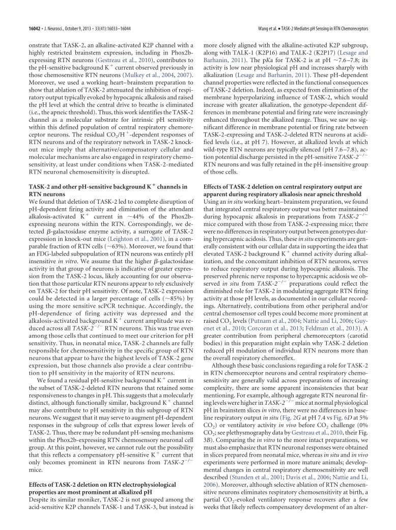
To study a large population of cells under recording condi-tions that preserve intracellular contents, we used the cell-attached configuration to determine effects of bath pH on thefiring activity of RTN neurons in slices from TASK-2�/� andTASK-2�/� mice (Fig. 2). We found that GFP-expressing RTNneurons from wild-type mice were nearly universally sensitive tochanges in pH under these conditions (Fig. 2A), as describedpreviously (Lazarenko et al., 2009). However, we found thatmany cells from TASK-2�/� mice showed only modest changesin firing rate as bath pH was altered (Fig. 2B) or they did not altertheir discharge over a wide range of pH changes (Fig. 2C).
To characterize the pH dependence of firing in individualRTN neurons, we calculated the percentage decrease in firingbetween pH 7.0 and pH 7.8 (Figs. 2A–C) and plotted the fre-quency distribution of those values for cells from TASK-2�/� andTASK-2�/� mice (Fig. 2D). The majority of RTN neurons fromTASK-2�/� mice showed 100% decrease in firing over this pHrange (i.e., they were silenced at pH 7.8). In contrast, the responseof cells from TASK-2�/� mice was much more variable, withmany RTN neurons showing relatively little effect (30% de-crease in firing), some with modest sensitivity (40 –90% decreasein firing), and only very few that stopped firing entirely. To pro-vide a consistent criterion for defining whether a cell was pHsensitive, we chose a cutoff value of �30% decrease in firing frompH 7.0 to pH 7.8 (Fig. 2D). As shown in Figure 2E, this standard
Figure 1. Effects of pH changes on membrane potential and firing rate in RTN neurons from TASK-2�/� and TASK-2�/� mice. A, B, Representative whole-cell current-clamp recordings ofmembrane potential (bottom traces) and the associated firing rate (10 s bins; top plots) from GFP-expressing RTN neurons in brainstem slices from TASK-2�/� (A) and TASK-2�/� (B) mice duringexposure to bath solutions of varying pH. Note that, whereas RTN neurons from both genotypes depolarized and increased firing during acidification (from pH 7.3 to pH 7.0), the effect of alkalizationon membrane potential and firing rate was more pronounced at pH 7.5 in the wild-type cell; even with stronger alkalization (to pH 8.0), the TASK-2�/� neuron continued to discharge. C, Therelationship between membrane potential and bath pH for the subgroup of TASK-2�/� and TASK-2�/� RTN neurons studied under current-clamp conditions (n � 10 and 6); data were fitted bylinear regression. There was a genotype-dependent difference in effects of pH on membrane potential (F(2,60) � 5.6, p 0.01).
16036 • J. Neurosci., October 9, 2013 • 33(41):16033–16044 Wang et al. • TASK-2 Mediates pH Sensing in RTN Chemoreceptors
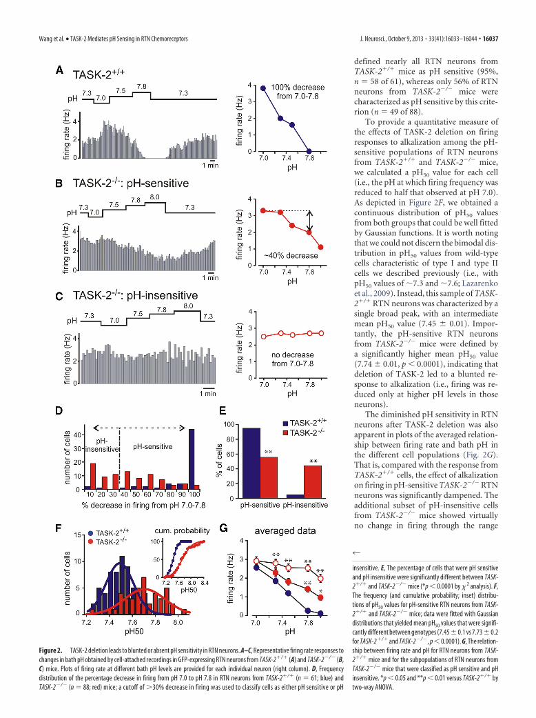
defined nearly all RTN neurons fromTASK-2�/� mice as pH sensitive (95%,n � 58 of 61), whereas only 56% of RTNneurons from TASK-2�/� mice werecharacterized as pH sensitive by this crite-rion (n � 49 of 88).
To provide a quantitative measure ofthe effects of TASK-2 deletion on firingresponses to alkalization among the pH-sensitive populations of RTN neuronsfrom TASK-2�/� and TASK-2�/� mice,we calculated a pH50 value for each cell(i.e., the pH at which firing frequency wasreduced to half that observed at pH 7.0).As depicted in Figure 2F, we obtained acontinuous distribution of pH50 valuesfrom both groups that could be well fittedby Gaussian functions. It is worth notingthat we could not discern the bimodal dis-tribution in pH50 values from wild-typecells characteristic of type I and type IIcells we described previously (i.e., withpH50 values of �7.3 and �7.6; Lazarenkoet al., 2009). Instead, this sample of TASK-2�/� RTN neurons was characterized by asingle broad peak, with an intermediatemean pH50 value (7.45 � 0.01). Impor-tantly, the pH-sensitive RTN neuronsfrom TASK-2�/� mice were defined bya significantly higher mean pH50 value(7.74 � 0.01, p 0.0001), indicating thatdeletion of TASK-2 led to a blunted re-sponse to alkalization (i.e., firing was re-duced only at higher pH levels in thoseneurons).
The diminished pH sensitivity in RTNneurons after TASK-2 deletion was alsoapparent in plots of the averaged relation-ship between firing rate and bath pH inthe different cell populations (Fig. 2G).That is, compared with the response fromTASK-2�/� cells, the effect of alkalizationon firing in pH-sensitive TASK-2�/� RTNneurons was significantly dampened. Theadditional subset of pH-insensitive cellsfrom TASK-2�/� mice showed virtuallyno change in firing through the range
Figure 2. TASK-2 deletion leads to blunted or absent pH sensitivity in RTN neurons. A–C, Representative firing rate responses tochanges in bath pH obtained by cell-attached recordings in GFP-expressing RTN neurons from TASK-2�/� (A) and TASK-2�/� (B,C) mice. Plots of firing rate at different bath pH levels are provided for each individual neuron (right column). D, Frequencydistribution of the percentage decrease in firing from pH 7.0 to pH 7.8 in RTN neurons from TASK-2�/� (n � 61; blue) andTASK-2�/� (n � 88; red) mice; a cutoff of �30% decrease in firing was used to classify cells as either pH sensitive or pH
4
insensitive. E, The percentage of cells that were pH sensitiveand pH insensitive were significantly different between TASK-2�/� and TASK-2�/� mice (*p 0.0001 by � 2 analysis). F,The frequency (and cumulative probability; inset) distribu-tions of pH50 values for pH-sensitive RTN neurons from TASK-2�/� and TASK-2�/� mice; data were fitted with Gaussiandistributions that yielded mean pH50 values that were signifi-cantly different between genotypes (7.45 � 0.1 vs 7.73 � 0.2for TASK-2�/� and TASK-2�/�, p 0.0001). G, The relation-ship between firing rate and pH for RTN neurons from TASK-2�/� mice and for the subpopulations of RTN neurons fromTASK-2�/� mice that were classified as pH sensitive and pHinsensitive. *p 0.05 and **p 0.01 versus TASK-2�/� bytwo-way ANOVA.
Wang et al. • TASK-2 Mediates pH Sensing in RTN Chemoreceptors J. Neurosci., October 9, 2013 • 33(41):16033–16044 • 16037

from pH 7.0 to pH 7.8, with a slightly sharper decrease in dis-charge evident only at pH 8.0. Note that the averaged firing ratewas not different among any of these groups at pH 7.0, but therelationships diverged at higher pH values at which alkaline-activated TASK-2 channels are most active. Because nearly equalpopulations of RTN neurons were defined as pH sensitive or pHinsensitive in TASK-2�/� mice (Fig. 2E), the aggregate firing re-sponse from the entire population of TASK-2-deleted RTN neu-rons falls approximately midway between those two firing versuspH curves (data not shown) and is significantly higher than thatfor TASK-2�/� cells (p 0.0001 by two-way ANOVA). At pH7.3, the combined average firing rate for pH-sensitive and pH-insensitive TASK-2�/� RTN neurons was greater than that forTASK-2�/� cells (2.6 � 0.1 vs 1.9 � 0.2 Hz, p 0.01), and thisdifference was further enhanced at more alkalized pH levels.
Together, these data indicate that pH sensitivity is generallydiminished across all RTN neurons from TASK-2 knock-outmice and that TASK-2 channels are required for pH sensitivity in�44% of those cells.
A pH-sensitive background K � current is reduced inamplitude or absent in RTN neurons from TASK-2�/� miceWe previously described a pH-sensitive background K� currentin RTN neurons with characteristics not unlike those of thealkaline-activated TASK-2 channel (Mulkey et al., 2004, 2007;Lazarenko et al., 2010). Thus, after assessing the pH sensitivity inGFP-expressing RTN neurons from TASK-2�/� (n � 12) andTASK-2�/� (n � 19) mice in the cell-attached configuration, weobtained whole-cell access and used voltage-clamp recordings toexamine pH-sensitive currents in those cells (Fig. 3). As shown inFigure 3A for a cell from a TASK-2�/� mouse, acidification of thebath solution (from pH 7.3 to pH 7.0) caused an inward shift inholding current at �60 mV that was accompanied by a decreasein conductance, whereas bath alkalization (from pH 7.0 to pH8.0) resulted in an outward shift in holding current with an in-crease in conductance. We obtained qualitatively similar resultsfrom the subgroup of RTN neurons from TASK-2�/� mice thatshowed firing responses that were characterized as pH sensitive(data not shown). However, in the pH-insensitive population ofRTN neurons obtained from TASK-2�/� mice (n � 7), there waslittle effect of bath alkalization on holding current or conduc-tance, as exemplified in the cell of Figure 3B.
We determined I–V relationships for the pH-sensitive currentin each of these groups of RTN neurons, obtained by digital sub-traction of currents in pH 7.0 from those in pH 8.0 (and normal-ized to cell capacitance, i.e., current density). In TASK-2�/� cells,the averaged alkaline-activated current was weakly rectifying,with a reversal potential near the predicted value for EK (approx-imately �95 mV); this is typical of the pH-sensitive backgroundK� current reported previously for wild-type RTN neurons. Inthe pH-sensitive RTN neurons from TASK-2�/� mice, the I–Vwas similar in shape to that of TASK-2�/� cells but diminished inamplitude. In contrast, there was essentially no alkaline-activatedcurrent in those pH-insensitive TASK-2�/� neurons that failed toalter their discharge in response to changes in bath pH. Thesedata suggest that TASK-2 channels are required for the alkaline-activated background K� current in the subset of pH-insensitiveRTN neurons; in the other RTN neurons from TASK-2�/� mice,although TASK-2 channels contribute to the overall backgroundK� current, currents from other background K� channels con-tribute to pH sensitivity.
Most chemosensitive RTN neurons express TASK-2, withapproximately half the population expressing higher levelsThe data presented to this point suggest a prominent, even nec-essary, role for TASK-2 channels in the pH sensitivity of nearlyhalf the population of Phox2b (and GFP)-expressing RTN neu-rons and a more modest but still measurable contribution in theother half of those cells. In light of these results, we sought toexamine whether there is a differential distribution of TASK-2expression in RTN neurons.
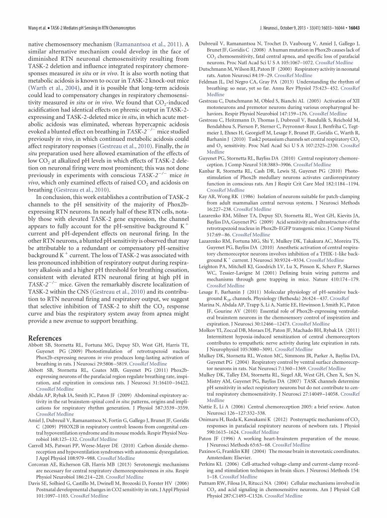
There are no well established TASK-2 antibodies available forimmunohistochemistry. Therefore, we used X-gal staining as asurrogate for TASK-2 expression in RTN neurons, taking advan-tage of the �-galactosidase cassette that was integrated into theTASK-2 locus by the gene trap construct in this knock-out mouseline (Leighton et al., 2001; Gestreau et al., 2010); as expected,X-gal staining was not observed in tissue from TASK-2�/�–Jxmice (data not shown). The Phox2b-expressing chemosensitivepopulation of RTN neurons was defined in these mice by immu-nohistochemical detection of GFP expression from the Phox2bBAC transgene, absence of staining for TH, and by their anatom-ical localization (Stornetta et al., 2006; Lazarenko et al., 2009). Asshown in Figure 4A, we detected overlapping expression of GFP
Figure 3. Alkaline-activated background K � current is diminished or absent in TASK-2�/� RTN neurons. Voltage-clamp recordings were obtained from GFP-expressing RTN neurons during bathacidification and alkalization; before gaining whole-cell access, each cell was first characterized as pH sensitive or pH insensitive based on firing responses to pH changes in the cell-attachedconfiguration (Fig. 2). A, In a pH-sensitive TASK-2�/� RTN neuron, acidification decreased holding current (top trace) and conductance (bottom trace) and alkalization caused a reversible outwardshift in current with an increase in conductance. B, In a pH-insensitive TASK-2�/� RTN neuron, changes in bath pH had little effect on holding current or conductance. C, Averaged I–V relationshipof the pH-sensitive current density (pH 8.0 minus pH 7.0) for RTN neurons from TASK-2�/� mice (n � 13) and for RTN neurons from TASK-2�/� mice that were classified as pH sensitive (n � 12)and pH insensitive (n � 7). Note that the weakly rectifying alkaline-activated K � current seen in TASK-2�/� neurons was reduced in pH-sensitive RTN neurons from TASK-2�/� mice and absentin pH-insensitive TASK-2�/� RTN neurons. **p 0.01 versus TASK-2�/� by two-way ANOVA.
16038 • J. Neurosci., October 9, 2013 • 33(41):16033–16044 Wang et al. • TASK-2 Mediates pH Sensing in RTN Chemoreceptors
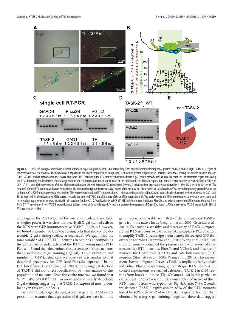
and X-gal in the RTN region of the rostral ventrolateral medulla.At higher power, it was clear that nearly all X-gal-stained cells inthe RTN were GFP immunoreactive (GFP�, �98%). However,we found a number of GFP-expressing cells that showed no de-tectable X-gal staining (yellow arrowheads). We quantified thetotal number of GFP�/TH� neurons in sections encompassingthe entire rostrocaudal extent of the RTN in young mice (P11–P24, n � 5) and then determined the percentage of those neuronsthat also showed X-gal staining (Fig. 4B). The distribution andnumber of GFP-labeled cells we observed was similar to thatdescribed previously for GFP (and Phox2b) expression in theJx99 line of mice (Lazarenko et al., 2009), indicating that deletionof TASK-2 did not affect specification or maintenance of thispopulation of neurons. Over the entire nucleus, we found that63 � 5.4% of GFP�/TH� neurons showed clearly detectableX-gal staining, suggesting that TASK-2 is expressed most prom-inently in this group of cells.
As mentioned, X-gal staining is a surrogate for TASK-2 ex-pression; it assumes that expression of �-galactosidase from the
gene trap is comparable with that of the endogenous TASK-2gene from the native locus (Leighton et al., 2001; Gestreau et al.,2010). To provide a sensitive and direct assay of TASK-2 expres-sion in RTN neurons, we used a nested, multiplex scPCR reactionto amplify TASK-2 transcripts from acutely dissociated GFP flu-orescent neurons (Lazarenko et al., 2010; Wang et al., 2013); wesimultaneously confirmed the presence of two markers of che-mosensitive RTN neurons, Phox2b and VGlut2, and absence ofmarkers for GABAergic (GAD1) and catecholaminergic (TH)neurons (Stornetta et al., 2006; Wang et al., 2013). The experi-ment shown in Figure 4C reveals TASK-2 amplicons in five of sixindividual Phox2b-expressing, glutamatergic RTN neurons. Incontrol experiments, we verified deletion of TASK-2 in RTN neu-rons from knock-out mice (Fig. 4D, lanes 1– 6); in this particularexperiment, TASK-2 was simultaneously detected in two of threeRTN neurons from wild-type mice (Fig. 4D, lanes 7–9). Overall,we detected TASK-2 expression in 85% of the RTN neuronstested by scPCR (n � 52 of 61; Fig. 4E), a greater fraction thanobtained by using X-gal staining. Together, these data suggest
Figure 4. TASK-2 is strongly expressed in a subset of Phox2b-expressing RTN neurons. A, Photomicrographs of histochemical staining for X-gal (left) and GFP and TH (right) in the RTN region ofthe rostroventrolateral medulla. The boxed region depicted in the lower-magnification image (top) is shown at greater magnification (bottom). Note that, among the double-positive neurons(GFP �/X-gal �; white arrowheads), there were also some GFP � neurons in the RTN that were not stained with X-gal (yellow arrowheads). B, Top, Schematic of the brainstem region containingthe RTN, delimiting the landmarks used to align sections for cell counts. Bottom, Quantification of the total number of Phox2b-expressing chemoreceptor neurons in each section (defined asGFP �/TH �) and of the percentage of those RTN neurons that also showed detectable X-gal staining. Overall, �-galactosidase expression was observed in �63% (321 � 40 of 504 � 25 RTNneurons) of these RTN neurons, with an even fractional distribution throughout the rostrocaudal extent of the nucleus. 7n, Facial nerve; VII, facial nucleus; VRG, ventral respiratory group; NA, nucleusambiguus. C, scPCR from a representative sample of GFP-expressing dissociated RTN neurons (lanes 1– 6) revealed expression of Phox2b and VGlut2 in all cells tested, with no evidence for GAD1 andTH, as expected for chemosensitive RTN neurons. Notably, we detected TASK-2 in all but one of those RTN neurons (lane 5). The positive control GAPDH transcript was universally detectable, andno-template negative controls were included in all reactions (nt, lane 7). D, Verification by scPCR of TASK-2 deletion from individual Phox2b- and VGlut2-expressing RTN neurons obtained fromTASK-2�/� mice (lanes 1– 6); TASK-2 expression was evident in two of three wild-type RTN neurons processed concurrently. E, Quantification of scPCR data revealed TASK-2 expression in 85% ofRTN neurons (n � 52/61).
Wang et al. • TASK-2 Mediates pH Sensing in RTN Chemoreceptors J. Neurosci., October 9, 2013 • 33(41):16033–16044 • 16039
that TASK-2 expression can be detected in the majority of RTNneurons but that levels of TASK-2 expression may be higherin the subgroup of cells that are recognized by detectable�-galactosidase activity from the TASK-2 locus in this knock-outmouse line.
TASK-2 channels are required for pH sensitivity in those RTNneurons with high levels of TASK-2 expressionTo this point, our data indicate that a subgroup of GFP-expressing RTN neurons we tested require TASK-2 expressionfor their pH sensitivity (�44%), and a comparable populationshowed detectable expression of �-galactosidase from theTASK-2 locus (�63%). This suggested the possibility that mea-surable �-galactosidase activity might identify the specific sub-population of RTN neurons for which TASK-2 expression iscritical for pH sensitivity.
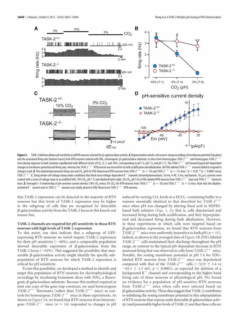
To test this possibility, we developed a method to identify andtarget this population of RTN neurons for electrophysiologicalrecordings by incubating brainstem slices with FDG, a fluoro-genic �-galactosidase substrate. Because this method required atleast one copy of the gene trap construct, we used heterozygousTASK-2�/� littermates (rather than TASK-2�/� mice) as con-trols for homozygous TASK-2�/� mice in these experiments. Asshown in Figure 5A, we found that RTN neurons from heterozy-gous TASK-2�/� mice (n � 14) responded to changes in pH
induced by varying CO2 levels in a HCO3�-containing buffer in a
manner essentially identical to that described for TASK-2�/�
mice when pH was changed by altering fixed acid in HEPES-based bath solution (Figs. 1, 2); that is, cells depolarized andincreased firing during bath acidification, and they hyperpolar-ized and decreased firing during bath alkalization. However,in these experiments in which cells were targeted based on�-galactosidase expression, we found that RTN neurons fromTASK-2�/� mice were uniformly insensitive to bath pH (n � 11).Indeed, as shown in the averaged data of Figure 5B, FDG-labeledTASK-2�/� cells maintained their discharge throughout the pHrange, in contrast to the typical pH-dependent decrease in RTNneuronal firing that was observed in cells from TASK-2�/� mice.Notably, the resting membrane potential at pH 7.4 for FDG-labeled RTN neurons from TASK-2�/� mice was depolarizedcompared with that of the TASK-2�/� cells (�52.9 � 1.4 vs�65.1 � 1.5 mV, p 0.0001), as expected for deletion of abackground K� channel and corresponding to the higher basalfiring rate of these neurons at physiological pH. We foundno evidence for a population of pH-sensitive RTN neuronsfrom TASK-2�/� mice when cells were selected based on�-galactosidase activity. These data suggest that TASK-2 contributesto stabilizing the resting membrane potential in this subpopulationof RTN neurons that express easily detectable �-galactosidase activ-ity (and presumably higher levels of TASK-2) and that these cells are
Figure 5. TASK-2 deletion ablates pH sensitivity in all RTN neurons selected for �-galactosidase activity. A, Representative whole-cell current-clamp recordings of membrane potential (top plots)and the associated firing rate (bottom traces) from RTN neurons stained with FDG, a fluorogenic �-galactosidase substrate, in slices from heterozygous TASK-2�/� and homozygous TASK-2�/�
mice during exposure to bath solutions equilibrated with different levels of CO2 (3, 5, and 10%, corresponding to pH 7.6, pH 7.4, and pH 7.1). The TASK-2�/� cell showed typical pH-dependentchanges in membrane potential and firing rate, whereas the TASK-2�/� RTN neuron was insensitive to both acidification and alkalization. All FDG-labeled TASK-2�/� neurons failed to respond tochanges in pH. B, The relationship between firing rate and CO2 (pH) for FDG-fluorescent RTN neurons from TASK-2�/� (n � 14) and TASK-2�/� (n � 11) mice. *p 0.05, **p 0.0001 versusTASK-2�/�. C, Using whole-cell voltage clamp under conditions that block most voltage-dependent K � channels (tetraethylammonium, 10 mM; 4-AP, 3 mM; and barium, 10 �M), currents wereevoked with a series of voltage steps in an acidified (left, 10% CO2, pH 7.1) and alkalized bath (right, 3% CO2, pH 7.6) in FDG-labeled RTN neurons from TASK-2�/� (top) and TASK-2�/� (bottom)mice. D, Averaged I–V relationship of pH-sensitive current density (10% CO2 minus 3% CO2) for RTN neurons from TASK-2�/� (n � 10) and TASK-2�/� (n � 5) mice. Note that the alkaline-activated K � current seen in TASK-2�/� neurons was totally absent in FDG-fluorescent TASK-2�/� RTN neurons.
16040 • J. Neurosci., October 9, 2013 • 33(41):16033–16044 Wang et al. • TASK-2 Mediates pH Sensing in RTN Chemoreceptors

strictly dependent on TASK-2 channels for their pH sensitivity. Con-sistent with this, whereas �-galactosidase-expressing TASK-2�/�
neurons showed a robust pH-sensitive and voltage-independent K�
current, this current was not seen at all in FDG-labeled RTN neuronsfrom TASK-2�/� mice (Fig. 5C,D). Thus, TASK-2�/� RTN neuronsselected based on �-galactosidase activity appear to be functionallyequivalent to the pH-insensitive subset of Phox2b-expressingTASK-2�/� cells that appear to depend critically on TASK-2 for theirpH sensitivity.
The respiratory chemoreflex is blunted in a working heart–brainstem preparation from TASK-2�/� miceOur data indicate that RTN neurons from TASK-2�/� mice showa blunted response to alkaline pH, i.e., firing rate is approximatelyequal to that of cells from TASK-2�/� mice at relatively acidifiedpH levels but better maintained at more alkalized pH levels (Fig.2F). To test for respiratory chemosensitivity in a more intactpreparation over a wide range of pH/CO2, we used the workingheart– brainstem preparation. In this in situ preparation, CO2
and bicarbonate levels in the brainstem perfusate are controlledand phrenic nerve activity serves as a measure of central respira-tory outflow. As reported previously (Abdala et al., 2009; Marinaet al., 2010), we found that respiratory acidosis evoked by increas-ing inspired CO2 from 5 to 7 or 9% caused an increase in phrenicburst amplitude with little effect on phrenic burst frequency (Fig.6A–C). The acidosis-induced increase in phrenic burst amplitudewas similar in preparations from TASK-2�/�, TASK-2�/�, andTASK-2�/� littermates (Fig. 6C). Conversely, respiratory alkalo-sis obtained by decreasing CO2 from 5 to 3 or 2% caused a de-crease in phrenic burst amplitude. The alkalosis-induceddecrease in phrenic burst amplitude was blunted in prepara-tions from TASK-2�/� mice compared with TASK-2-
expressing littermates (Fig. 6 A, C), with a significantly smallereffect evident at 3% CO2 (52.4 � 4.2% for TASK-2�/� vs31.6 � 7.6% for TASK-2�/�) and at 2% CO2 (42.6 � 5.5% forTASK-2�/� vs 21.2 � 6.6% for TASK-2�/� and 8.7 � 5.8% forTASK-2�/�). An additional decrease in phrenic burst fre-quency was observed at 2% CO2, but this was not significantlydifferent between genotypes (Fig. 6B).
In a subset of experiments (Fig. 6D), we progressively loweredCO2 levels to determine the effect of TASK-2 deletion on theapneic threshold (i.e., the pH of the perfusate at which respiratoryoutflow was eliminated). Note that the apneic threshold occurs atmore alkalized levels in situ compared with in vivo preparations(Takakura et al., 2008; Molkov et al., 2011), and, in some in situpreparations from TASK-2�/� mice, we did not observe a cessa-tion of phrenic activity even at the lowest CO2 levels tested (Fig.6D); for those cases, we reported the lowest pH value measured asthe apneic threshold. As shown in Figure 6E, the pH at apneic thresh-old was significantly higher in preparations from TASK-2�/� (pH8.2 � 0.05, n � 6) compared with TASK-2�/� littermate controls(pH 7.9 � 0.05, n � 7, p 0.01) and TASK-2�/� mice (pH 7.8 �0.04, n � 6, p 0.001). Thus, deletion of alkaline-activated TASK-2channel is associated with diminished respiratory responses to hy-pocapnic alkalosis in situ.
DiscussionPhox2b-expressing, glutamatergic neurons of the RTN arecentral respiratory chemosensors, i.e., they provide a CO2-dependent excitatory drive to brainstem respiratory networks.The pH sensitivity of these neurons is, at least in part, an intrinsicproperty (Wang et al., 2013), but the molecular basis for pHsensing has not been defined (Guyenet et al., 2010; Feldman et al.,2013). In this paper, we used a knock-out mouse model to dem-
Figure 6. TASK-2 deletion blunts effects of alkalization on respiratory-like neural output in an in situ preparation. A, A working heart– brainstem preparation was used to examine effects ofrespiratory acidosis (7% CO2; top traces) and respiratory alkalosis (2% CO2; bottom traces) on integrated phrenic nerve activity in TASK-2�/� (left) and TASK-2�/� (right) mice. Phrenic nerveamplitude was reversibly increased in 7% CO2 and decreased in 2% CO2, but the inhibition by 2% CO2 was less pronounced in TASK-2�/� mice. B, For both genotypes, respiratory frequency wasunaffected by changes in CO2 from 3 to 9%; frequency decreased significantly only when CO2 was reduced to �2%. C, Effects on phrenic nerve amplitude of raising CO2 (from 5 to 7 or 9%) were notdifferent between TASK-2�/� mice and TASK-2�/� mice; however, decreases in phrenic nerve amplitude associated with lowering CO2 (to 3 and 2%) were relatively blunted in TASK-2�/� mice.*p 0.05, **p 0.001 versus TASK-2�/�; ‡p 0.05 versus TASK-2�/� by two-way ANOVA. D, Apneic threshold (i.e., the pH when phrenic nerve activity was eliminated) was determined bysequentially lowering CO2 levels in preparations from TASK-2�/� (left) and TASK-2�/� (right) mice. Note the more modest effect of respiratory alkalosis on phrenic nerve discharge in theTASK-2�/� exemplar. E, Averaged values of pH at apneic threshold were significantly higher in preparations from TASK-2�/� mice. **p 0.001 versus TASK-2�/�; ‡‡p 0.01 versus TASK-2�/�
by one-way ANOVA with sample size indicated.
Wang et al. • TASK-2 Mediates pH Sensing in RTN Chemoreceptors J. Neurosci., October 9, 2013 • 33(41):16033–16044 • 16041
onstrate that TASK-2, an alkaline-activated K2P channel with ahighly restricted brainstem expression, including in Phox2b-expressing RTN neurons (Gestreau et al., 2010), contributes tothe pH-sensitive background K� current observed previously inthose chemosensitive RTN neurons (Mulkey et al., 2004, 2007).Moreover, we used a working heart– brainstem preparation toshow that ablation of TASK-2 attenuated the inhibition of respi-ratory output typically evoked by hypocapnic alkalosis and raisedthe pH level at which the central drive to breathe is eliminated(i.e., the apneic threshold). Thus, this work identifies the TASK-2channel as a molecular substrate for intrinsic pH sensitivitywithin this defined population of central respiratory chemore-ceptor neurons. The residual CO2/H�-dependent responses ofRTN neurons and of the respiratory network in TASK-2 knock-out mice imply that alternative/compensatory cellular andmolecular mechanisms are also engaged in respiratory chemo-sensitivity, at least under conditions when TASK-2-mediatedRTN neuronal chemosensitivity is disrupted.
TASK-2 and other pH-sensitive background K � channels inRTN neuronsWe found that deletion of TASK-2 led to complete disruption ofpH-dependent firing activity and elimination of the attendantalkalosis-activated K� current in �44% of the Phox2b-expressing neurons within the RTN. Correspondingly, we de-tected �-galactosidase enzyme activity, a surrogate of TASK-2expression in knock-out mice (Leighton et al., 2001), in a com-parable fraction of RTN cells (�63%). Moreover, we found thatan FDG-labeled subpopulation of RTN neurons was entirely pHinsensitive in vitro. We assume that the higher �-galactosidaseactivity in that group of neurons is indicative of greater expres-sion from the TASK-2 locus, likely accounting for our observa-tion that those particular RTN neurons appear to rely exclusivelyon TASK-2 for their pH sensitivity. Of note, TASK-2 expressioncould be detected in a larger percentage of cells (�85%) byusing the more sensitive scPCR technique. Accordingly, thepH-dependence of firing activity was depressed and thealkalosis-activated background K � current amplitude was re-duced across all TASK-2�/� RTN neurons. This was true evenamong those cells that continued to meet our criterion for pHsensitivity. Thus, in neonatal mice, TASK-2 channels are fullyresponsible for chemosensitivity in the specific group of RTNneurons that appear to have the highest levels of TASK-2 geneexpression, but those channels also provide a clear contribu-tion to pH sensitivity in the majority of RTN neurons.
We found a residual pH-sensitive background K� current inthe subset of TASK-2-deleted RTN neurons that retained someresponsiveness to changes in pH. This suggests that a molecularlydistinct, although functionally similar, background K� channelmay also contribute to pH sensitivity in this subgroup of RTNneurons. We suggest that it may serve to augment pH-dependentresponses in the subgroup of cells that express lower levels ofTASK-2. Thus, there may be redundant pH-sensing mechanismswithin the Phox2b-expressing RTN chemosensory neuronal cellgroup. At this point, however, we cannot rule out the possibilitythat this reflects a compensatory pH-sensitive K� current thatonly becomes prominent in RTN neurons from TASK-2�/�
mice.
Effects of TASK-2 deletion on RTN electrophysiologicalproperties are most prominent at alkalized pHDespite its similar moniker, TASK-2 is not grouped among theacid-sensitive K2P channels TASK-1 and TASK-3, but instead is
more closely aligned with the alkaline-activated K2P subgroup,along with TALK-1 (K2P16) and TALK-2 (K2P17) (Lesage andBarhanin, 2011). The pKa for TASK-2 is at pH �7.6 –7.8; itsactivity is low near physiological pH and increases sharply withalkalization (Lesage and Barhanin, 2011). These pH-dependentchannel properties were reflected in the functional consequencesof TASK-2 deletion. Indeed, as expected from elimination of themembrane hyperpolarizing influence of TASK-2, which wouldincrease with greater alkalization, the genotype-dependent dif-ferences in membrane potential and firing rate were increasinglyenhanced throughout the alkalized range. Thus, we saw no sig-nificant difference in membrane potential or firing rate betweenTASK-2-expressing and TASK-2-deleted RTN neurons at acidi-fied levels (i.e., at pH 7). However, at alkalized levels at whichwild-type RTN neurons are typically silenced (pH 7.6 –7.8), ac-tion potential discharge persisted in the pH-sensitive TASK-2�/�
RTN neurons and was fully retained in the pH-insensitive groupof those cells.
Effects of TASK-2 deletion on central respiratory output areapparent during respiratory alkalosis near apneic thresholdUsing an in situ working heart– brainstem preparation, we foundthat integrated central respiratory output was better maintainedduring hypocapnic alkalosis in preparations from TASK-2�/�
mice compared with those from TASK-2-expressing mice; therewere no differences in respiratory output between genotypes dur-ing hypercapnic acidosis. Thus, these in situ experiments are gen-erally consistent with our cellular data in supporting the idea thatelevated TASK-2 background K� channel activity during alkal-ization, and the concomitant inhibition of RTN neurons, servesto reduce respiratory output during hypocapnic alkalosis. Thepreserved phrenic nerve response to hypercapnic acidosis we ob-served in situ from TASK-2�/� preparations could reflect thediminished role for TASK-2 in modulating aggregate RTN firingactivity at those pH levels, as documented in our cellular record-ings. Alternatively, contributions from other peripheral and/orcentral chemosensor cell types could become more prominent atraised CO2 levels (Putnam et al., 2004; Nattie and Li, 2006; Guy-enet et al., 2010; Corcoran et al., 2013; Feldman et al., 2013). Agreater contribution from peripheral chemoreceptors (carotidbodies) in this preparation might explain why TASK-2 deletionreduced pH modulation of individual RTN neurons more thanthe overall respiratory chemoreflex.
Although these basic conclusions regarding a role for TASK-2in RTN chemoreceptor neurons and central respiratory chemo-sensitivity are generally valid across preparations of increasingcomplexity, there are some apparent inconsistencies that bearmentioning. For example, although aggregate RTN neuronal fir-ing levels were higher in TASK-2�/� mice at normal physiologicalpH in brainstem slices in vitro, there were no differences in base-line respiratory output in situ (Fig. 2G at pH 7.4 vs Fig. 6D at 5%CO2) or ventilatory activity in vivo before CO2 challenge (0%CO2; see plethysmography data by Gestreau et al., 2010, their Fig.3B). Comparing the in vitro to the more intact preparations, wemust also emphasize that RTN neuronal responses were obtainedin slices prepared from neonatal mice, whereas in situ and in vivoexperiments were performed in more mature animals; develop-mental changes in central respiratory chemosensitivity are welldescribed (Stunden et al., 2001; Davis et al., 2006; Nattie and Li,2006). Moreover, although selective ablation of RTN chemosen-sitive neurons eliminates respiratory chemosensitivity at birth, apartial CO2-evoked ventilatory response recovers after a fewweeks that likely reflects compensatory development of an alter-
16042 • J. Neurosci., October 9, 2013 • 33(41):16033–16044 Wang et al. • TASK-2 Mediates pH Sensing in RTN Chemoreceptors
native chemosensory mechanism (Ramanantsoa et al., 2011). Asimilar alternative mechanism could develop in the face ofdiminished RTN neuronal chemosensitivity resulting fromTASK-2 deletion and influence integrated respiratory chemore-sponses measured in situ or in vivo. It is also worth noting thatmetabolic acidosis is known to occur in TASK-2 knock-out mice(Warth et al., 2004), and it is possible that long-term acidosiscould lead to compensatory changes in respiratory chemosensi-tivity measured in situ or in vivo. We found that CO2-inducedacidification had identical effects on phrenic output in TASK-2-expressing and TASK-2-deleted mice in situ, in which acute met-abolic acidosis was eliminated, whereas hypercapnic acidosisevoked a blunted effect on breathing in TASK-2�/� mice studiedpreviously in vivo, in which continued metabolic acidosis couldaffect respiratory responses (Gestreau et al., 2010). Finally, the insitu preparation used here allowed examination of the effects oflow CO2 at alkalized pH levels in which effects of TASK-2 dele-tion on neuronal firing were most prominent; this was not donepreviously in experiments with conscious TASK-2�/� mice invivo, which only examined effects of raised CO2 and acidosis onbreathing (Gestreau et al., 2010).
In conclusion, this work establishes a contribution of TASK-2channels to the pH sensitivity of the majority of Phox2b-expressing RTN neurons. In nearly half of these RTN cells, nota-bly those with elevated TASK-2 gene expression, the channelappears to fully account for the pH-sensitive background K�
current and pH-dependent effects on neuronal firing. In theother RTN neurons, a blunted pH sensitivity is observed that maybe attributable to a redundant or compensatory pH-sensitivebackground K� current. The loss of TASK-2 was associated withless pronounced inhibition of respiratory output during respira-tory alkalosis and a higher pH threshold for breathing cessation,consistent with elevated RTN neuronal firing at high pH inTASK-2�/� mice. Given the remarkably discrete localization ofTASK-2 within the CNS (Gestreau et al., 2010) and its contribu-tion to RTN neuronal firing and respiratory output, we suggestthat selective inhibition of TASK-2 to shift the CO2 responsecurve and bias the respiratory system away from apnea mightprovide a new avenue to support breathing.
ReferencesAbbott SB, Stornetta RL, Fortuna MG, Depuy SD, West GH, Harris TE,
Guyenet PG (2009) Photostimulation of retrotrapezoid nucleusPhox2b-expressing neurons in vivo produces long-lasting activation ofbreathing in rats. J Neurosci 29:5806 –5819. CrossRef Medline
Abbott SB, Stornetta RL, Coates MB, Guyenet PG (2011) Phox2b-expressing neurons of the parafacial region regulate breathing rate, inspi-ration, and expiration in conscious rats. J Neurosci 31:16410 –16422.CrossRef Medline
Abdala AP, Rybak IA, Smith JC, Paton JF (2009) Abdominal expiratory ac-tivity in the rat brainstem-spinal cord in situ: patterns, origins and impli-cations for respiratory rhythm generation. J Physiol 587:3539 –3559.CrossRef Medline
Amiel J, Dubreuil V, Ramanantsoa N, Fortin G, Gallego J, Brunet JF, GoridisC (2009) PHOX2B in respiratory control: lessons from congenital cen-tral hypoventilation syndrome and its mouse models. Respir Physiol Neu-robiol 168:125–132. CrossRef Medline
Carroll MS, Patwari PP, Weese-Mayer DE (2010) Carbon dioxide chemo-reception and hypoventilation syndromes with autonomic dysregulation.J Appl Physiol 108:979 –988. CrossRef Medline
Corcoran AE, Richerson GB, Harris MB (2013) Serotonergic mechanismsare necessary for central respiratory chemoresponsiveness in situ. RespirPhysiol Neurobiol 186:214 –220. CrossRef Medline
Davis SE, Solhied G, Castillo M, Dwinell M, Brozoski D, Forster HV (2006)Postnatal developmental changes in CO2 sensitivity in rats. J Appl Physiol101:1097–1103. CrossRef Medline
Dubreuil V, Ramanantsoa N, Trochet D, Vaubourg V, Amiel J, Gallego J,Brunet JF, Goridis C (2008) A human mutation in Phox2b causes lack ofCO2 chemosensitivity, fatal central apnea, and specific loss of parafacialneurons. Proc Natl Acad Sci U S A 105:1067–1072. CrossRef Medline
Dutschmann M, Wilson RJ, Paton JF (2000) Respiratory activity in neonatalrats. Auton Neurosci 84:19 –29. CrossRef Medline
Feldman JL, Del Negro CA, Gray PA (2013) Understanding the rhythm ofbreathing: so near, yet so far. Annu Rev Physiol 75:423– 452. CrossRefMedline
Gestreau C, Dutschmann M, Obled S, Bianchi AL (2005) Activation of XIImotoneurons and premotor neurons during various oropharyngeal be-haviors. Respir Physiol Neurobiol 147:159 –176. CrossRef Medline
Gestreau C, Heitzmann D, Thomas J, Dubreuil V, Bandulik S, Reichold M,Bendahhou S, Pierson P, Sterner C, Peyronnet-Roux J, Benfriha C, Tegt-meier I, Ehnes H, Georgieff M, Lesage F, Brunet JF, Goridis C, Warth R,Barhanin J (2010) Task2 potassium channels set central respiratory CO2
and O2 sensitivity. Proc Natl Acad Sci U S A 107:2325–2330. CrossRefMedline
Guyenet PG, Stornetta RL, Bayliss DA (2010) Central respiratory chemore-ception. J Comp Neurol 518:3883–3906. CrossRef Medline
Kanbar R, Stornetta RL, Cash DR, Lewis SJ, Guyenet PG (2010) Photo-stimulation of Phox2b medullary neurons activates cardiorespiratoryfunction in conscious rats. Am J Respir Crit Care Med 182:1184 –1194.CrossRef Medline
Kay AR, Wong RK (1986) Isolation of neurons suitable for patch-clampingfrom adult mammalian central nervous systems. J Neurosci Methods16:227–238. CrossRef Medline
Lazarenko RM, Milner TA, Depuy SD, Stornetta RL, West GH, Kievits JA,Bayliss DA, Guyenet PG (2009) Acid sensitivity and ultrastructure of theretrotrapezoid nucleus in Phox2b-EGFP transgenic mice. J Comp Neurol517:69 – 86. CrossRef Medline
Lazarenko RM, Fortuna MG, Shi Y, Mulkey DK, Takakura AC, Moreira TS,Guyenet PG, Bayliss DA (2010) Anesthetic activation of central respira-tory chemoreceptor neurons involves inhibition of a THIK-1-like back-ground K � current. J Neurosci 30:9324 –9334. CrossRef Medline
Leighton PA, Mitchell KJ, Goodrich LV, Lu X, Pinson K, Scherz P, SkarnesWC, Tessier-Lavigne M (2001) Defining brain wiring patterns andmechanisms through gene trapping in mice. Nature 410:174 –179.CrossRef Medline
Lesage F, Barhanin J (2011) Molecular physiology of pH-sensitive back-ground K2P channels. Physiology (Bethesda) 26:424 – 437. CrossRef
Marina N, Abdala AP, Trapp S, Li A, Nattie EE, Hewinson J, Smith JC, PatonJF, Gourine AV (2010) Essential role of Phox2b-expressing ventrolat-eral brainstem neurons in the chemosensory control of inspiration andexpiration. J Neurosci 30:12466 –12473. CrossRef Medline
Molkov YI, Zoccal DB, Moraes DJ, Paton JF, Machado BH, Rybak IA (2011)Intermittent hypoxia-induced sensitization of central chemoreceptorscontributes to sympathetic nerve activity during late expiration in rats.J Neurophysiol 105:3080 –3091. CrossRef Medline
Mulkey DK, Stornetta RL, Weston MC, Simmons JR, Parker A, Bayliss DA,Guyenet PG (2004) Respiratory control by ventral surface chemorecep-tor neurons in rats. Nat Neurosci 7:1360 –1369. CrossRef Medline
Mulkey DK, Talley EM, Stornetta RL, Siegel AR, West GH, Chen X, Sen N,Mistry AM, Guyenet PG, Bayliss DA (2007) TASK channels determinepH sensitivity in select respiratory neurons but do not contribute to cen-tral respiratory chemosensitivity. J Neurosci 27:14049 –14058. CrossRefMedline
Nattie E, Li A (2006) Central chemoreception 2005: a brief review. AutonNeurosci 126 –127:332–338.
Onimaru H, Ikeda K, Kawakami K (2012) Postsynaptic mechanisms of CO2
responses in parafacial respiratory neurons of newborn rats. J Physiol590:1615–1624. CrossRef Medline
Paton JF (1996) A working heart-brainstem preparation of the mouse.J Neurosci Methods 65:63– 68. CrossRef Medline
Paxinos G, Franklin KBJ (2004) The mouse brain in stereotaxic coordinates.Amsterdam: Elsevier.
Perkins KL (2006) Cell-attached voltage-clamp and current-clamp record-ing and stimulation techniques in brain slices. J Neurosci Methods 154:1–18. CrossRef Medline
Putnam RW, Filosa JA, Ritucci NA (2004) Cellular mechanisms involved inCO2 and acid signaling in chemosensitive neurons. Am J Physiol CellPhysiol 287:C1493–C1526. CrossRef Medline
Wang et al. • TASK-2 Mediates pH Sensing in RTN Chemoreceptors J. Neurosci., October 9, 2013 • 33(41):16033–16044 • 16043

Ramanantsoa N, Hirsch MR, Thoby-Brisson M, Dubreuil V, Bouvier J, Ruf-fault PL, Matrot B, Fortin G, Brunet JF, Gallego J, Goridis C (2011)Breathing without CO2 chemosensitivity in conditional Phox2b mutants.J Neurosci 31:12880 –12888. CrossRef Medline
Stettner GM, Zanella S, Huppke P, Gartner J, Hilaire G, Dutschmann M(2008) Spontaneous central apneas occur in the C57BL/6J mouse strain.Respir Physiol Neurobiol 160:21–27. CrossRef Medline
Stornetta RL, Moreira TS, Takakura AC, Kang BJ, Chang DA, West GH,Brunet JF, Mulkey DK, Bayliss DA, Guyenet PG (2006) Expression ofPhox2b by brainstem neurons involved in chemosensory integration inthe adult rat. J Neurosci 26:10305–10314. CrossRef Medline
Stunden CE, Filosa JA, Garcia AJ, Dean JB, Putnam RW (2001) Develop-ment of in vivo ventilatory and single chemosensitive neuron responses tohypercapnia in rats. Respir Physiol 127:135–155. CrossRef Medline
Takakura AC, Moreira TS, Stornetta RL, West GH, Gwilt JM, Guyenet PG
(2008) Selective lesion of retrotrapezoid Phox2b-expressing neuronsraises the apnoeic threshold in rats. J Physiol 586:2975–2991. CrossRefMedline
Wang S, Shi Y, Shu S, Guyenet PG, Bayliss DA (2013) Phox2b-expressingretrotrapezoid neurons are intrinsically responsive to H � and CO2.J Neurosci 33:7756 –7761. CrossRef Medline
Warth R, Barriere H, Meneton P, Bloch M, Thomas J, Tauc M, Heitzmann D,Romeo E, Verrey F, Mengual R, Guy N, Bendahhou S, Lesage F, Poujeol P,Barhanin J (2004) Proximal renal tubular acidosis in TASK2 K �
channel-deficient mice reveals a mechanism for stabilizing bicarbonatetransport. Proc Natl Acad Sci U S A 101:8215– 8220. CrossRef Medline
Wilson RJ, Remmers JE, Paton JF (2001) Brain stem PO2 and pH of theworking heart-brain stem preparation during vascular perfusion withaqueous medium. Am J Physiol Regul Integr Comp Physiol 281:R528 –R538. Medline
16044 • J. Neurosci., October 9, 2013 • 33(41):16033–16044 Wang et al. • TASK-2 Mediates pH Sensing in RTN Chemoreceptors