cellular/molecular inflammasome-inducedil-1 ... · cellular/molecular inflammasome-inducedil-1...
TRANSCRIPT

Cellular/Molecular
Inflammasome-Induced IL-1� Secretion in Microglia IsCharacterized by Delayed Kinetics and Is Only PartiallyDependent on Inflammatory Caspases
Saskia M. Burm,1 Ella A. Zuiderwijk-Sick,1 Anke E.J. ‘t Jong,1 Céline van der Putten,1 Jennifer Veth,1 Ivanela Kondova,2
and X Jeffrey J. Bajramovic1
1Alternatives Unit, 2Animal Science Department, Biomedical Primate Research Centre, 2288 GJ Rijswijk, The Netherlands
Inflammasomes are multiprotein complexes that link pathogen recognition and cellular stress to the processing of the proinflammatorycytokine interleukin-1� (IL-1�). Whereas inflammasome-mediated activation is heavily studied in hematopoietic macrophages anddendritic cells, much less is known about microglia, resident tissue macrophages of the brain that originate from a distinct progenitor. Todirectly compare inflammasome-mediated activation in different types of macrophages, we isolated primary microglia and hematopo-ietic macrophages from adult, healthy rhesus macaques. We analyzed the expression profile of NOD (nucleotide-binding oligomerizationdomain)-like receptors, adaptor proteins, and caspases and characterized inflammasome activation and regulation in detail. We heredemonstrate that primary microglia can respond to the same innate stimuli as hematopoietic macrophages. However, microglial re-sponses are more persistent due to lack of negative regulation on pro-IL-1� expression. In addition, we show that while caspase 1, 4, and5 activation is pivotal for inflammasome-induced IL-1� secretion by hematopoietic macrophages, microglial secretion of IL-1� is onlypartially dependent on these inflammatory caspases. These results identify key cell type-specific differences that may aid the develop-ment of strategies to modulate innate immune responses in the brain.
Key words: IL-1�; inflammasome; inflammatory caspases; macrophages; microglia
IntroductionMicroglia are the resident tissue macrophages of the CNS. Likeother macrophages, they express multiple receptors of the innateimmune system, including Toll-like receptors (TLRs), NOD(nucleotide-binding oligomerization domain)-like receptors(NLRs), C-type lectin receptors, and retinoic acid-induciblegene-I-like receptors (Olson and Miller, 2004; Zuiderwijk-Sick etal., 2007; Furr et al., 2008; Shah et al., 2008; Shi et al., 2012).Recent studies have implicated NLR-mediated activation of mi-croglia in several neurodegenerative (Salminen et al., 2008;Heneka et al., 2013) and infectious brain diseases (Hafner-Bratkovic et al., 2012; Jamilloux et al., 2013), providing an impe-tus for more detailed studies of this receptor family.
NLRs can sense disturbances in cellular homeostasis caused byamongst others pathogens, large protein aggregates, and neigh-boring cell death. To date, 23 NLRs have been described for hu-
mans (Martinon et al., 2009). Ligand recognition by NOD1 andNOD2 can directly induce transcription of proinflammatory cy-tokines and chemokines via NF�B and IRF3 signaling (Ting et al.,2010). Other NLRs, such as NALP1 (NACHT-, LRR-, and PYD-containing protein 1), NALP3, NALP7, AIM2 (absent in mela-noma 2), and IPAF (ICE-protease activating factor) can formmultiprotein complexes called inflammasomes. These receptorscan either directly or indirectly via the adaptor proteins ASC(apoptosis-associated speck-like protein containing a caspase re-cruitment domain) or CARDINAL (CARD inhibitor of NF-�B-activating ligands) interact with inflammatory caspases andactivate them. In turn, activated caspases can process precursorproteins, such as pro-IL-1�, to their bioactive and secreted forms.Thereby NLRs link perturbances in cellular homeostasis to theproduction of proinflammatory cytokines (Martinon et al., 2009;Latz, 2010; Khare et al., 2012).
Inflammasome-mediated activation of microglia is involvedin both infectious (Chang et al., 2012; Kaushik et al., 2012; Lee etal., 2013) and noninfectious (Halle et al., 2008; Abulafia et al.,2009; Meissner et al., 2010) neurological diseases. Microglia ex-press various components of the inflammasome, includingNALP1, NALP3, ASC, and caspase 1 (Abulafia et al., 2009; Jamil-loux et al., 2013; Shi et al., 2013). In addition, microglia can beinduced to express pro-IL-1�, which can be processed to bioac-tive and secreted IL-1� in response to pathogens, protein aggre-gates, and more general cellular stressors, such as ATP andreactive oxygen species (Halle et al., 2008; Abulafia et al., 2009;
Received June 19, 2014; revised Oct. 31, 2014; accepted Nov. 4, 2014.Author contributions: S.M.B. and J.J.B. designed research; S.M.B., E.A.Z.-S., A.E.J.t.J., C.v.d.P., J.V., and I.K. per-
formed research; I.K. contributed unpublished reagents/analytic tools; S.M.B., E.A.Z.-S., A.E.J.t.J., and J.J.B. analyzeddata; S.M.B. and J.J.B. wrote the paper.
This work was supported by the Dutch MS Research Foundation (MS12-805). We thank T. Haaksma for experttechnical assistance. We thank Dr. E. Remarque for expert assistance with the statistical analyses and Drs. R. Bontrop,L.A. ‘t Hart, M.G. Netea, and J.M. van Noort for critically reading the manuscript.
The authors declare no competing financial interests.Correspondence should be addressed to Jeffrey J. Bajramovic, PhD, Alternatives Unit, Biomedical Primate Re-
search Centre, Lange Kleiweg 161, 2288 GJ Rijswijk, The Netherlands. E-mail: [email protected]:10.1523/JNEUROSCI.2510-14.2015
Copyright © 2015 the authors 0270-6474/15/350678-10$15.00/0
678 • The Journal of Neuroscience, January 14, 2015 • 35(2):678 – 687
Terada et al., 2010; Hanamsagar et al., 2011; Wu et al., 2013;Walsh et al., 2014).
Although microglia resemble hematopoietic macrophagesboth in phenotype and function, it has recently been uncoveredthat they originate from a different progenitor (Ginhoux et al.,2010). Furthermore, there are indications that regulation of sig-naling by innate immune receptors is different in microglia (Xiaoet al., 2013), and microglia have been reported to specificallyemploy caspases 3/7 and 8 during inflammatory conditions (Bur-guillos et al., 2011). To directly compare inflammasome-mediated activation in different types of macrophages in anoutbred system with close resemblance to humans, we isolatedprimary microglia and hematopoietic macrophages from adult,healthy rhesus macaques. We analyzed the expression profile ofNLRs, adaptor proteins, and caspases and characterized inflam-masome activation and regulation in detail. Our data revealimportant cell type-specific differences pertaining to the negativeregulation of pro-IL-1� expression as well as to the inflammasome-induced enzymatic processing of pro-IL-1�.
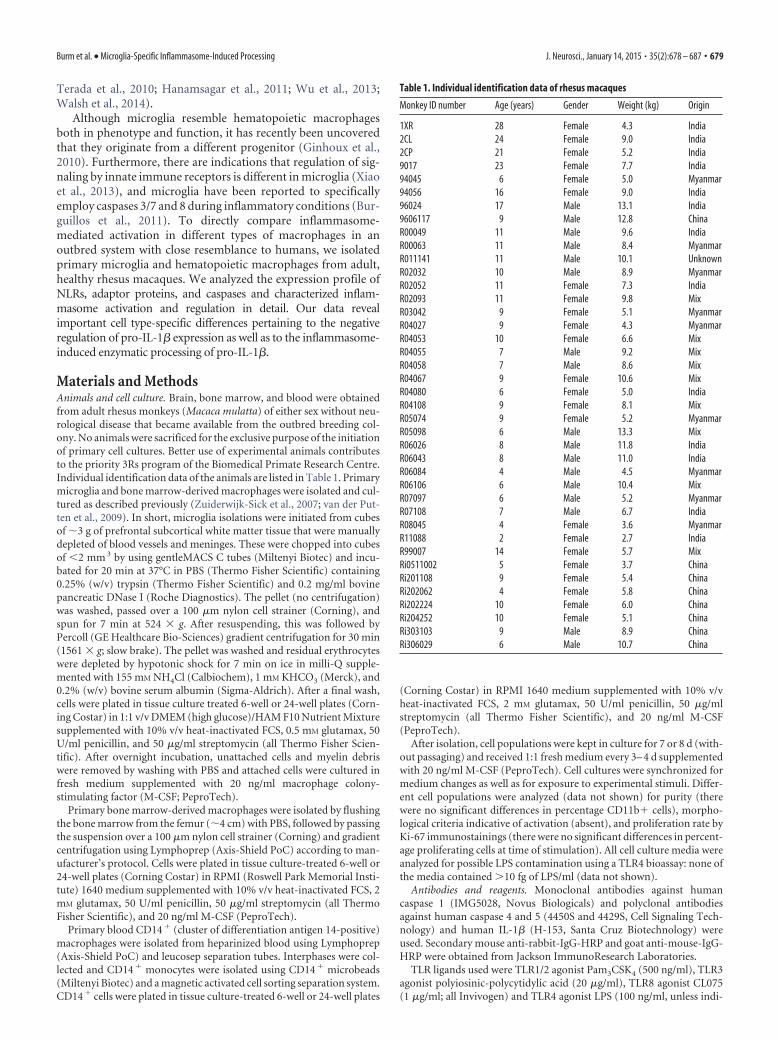
Materials and MethodsAnimals and cell culture. Brain, bone marrow, and blood were obtainedfrom adult rhesus monkeys (Macaca mulatta) of either sex without neu-rological disease that became available from the outbred breeding col-ony. No animals were sacrificed for the exclusive purpose of the initiationof primary cell cultures. Better use of experimental animals contributesto the priority 3Rs program of the Biomedical Primate Research Centre.Individual identification data of the animals are listed in Table 1. Primarymicroglia and bone marrow-derived macrophages were isolated and cul-tured as described previously (Zuiderwijk-Sick et al., 2007; van der Put-ten et al., 2009). In short, microglia isolations were initiated from cubesof �3 g of prefrontal subcortical white matter tissue that were manuallydepleted of blood vessels and meninges. These were chopped into cubesof �2 mm 3 by using gentleMACS C tubes (Miltenyi Biotec) and incu-bated for 20 min at 37°C in PBS (Thermo Fisher Scientific) containing0.25% (w/v) trypsin (Thermo Fisher Scientific) and 0.2 mg/ml bovinepancreatic DNase I (Roche Diagnostics). The pellet (no centrifugation)was washed, passed over a 100 �m nylon cell strainer (Corning), andspun for 7 min at 524 � g. After resuspending, this was followed byPercoll (GE Healthcare Bio-Sciences) gradient centrifugation for 30 min(1561 � g; slow brake). The pellet was washed and residual erythrocyteswere depleted by hypotonic shock for 7 min on ice in milli-Q supple-mented with 155 mM NH4Cl (Calbiochem), 1 mM KHCO3 (Merck), and0.2% (w/v) bovine serum albumin (Sigma-Aldrich). After a final wash,cells were plated in tissue culture treated 6-well or 24-well plates (Corn-ing Costar) in 1:1 v/v DMEM (high glucose)/HAM F10 Nutrient Mixturesupplemented with 10% v/v heat-inactivated FCS, 0.5 mM glutamax, 50U/ml penicillin, and 50 �g/ml streptomycin (all Thermo Fisher Scien-tific). After overnight incubation, unattached cells and myelin debriswere removed by washing with PBS and attached cells were cultured infresh medium supplemented with 20 ng/ml macrophage colony-stimulating factor (M-CSF; PeproTech).
Primary bone marrow-derived macrophages were isolated by flushingthe bone marrow from the femur (�4 cm) with PBS, followed by passingthe suspension over a 100 �m nylon cell strainer (Corning) and gradientcentrifugation using Lymphoprep (Axis-Shield PoC) according to man-ufacturer’s protocol. Cells were plated in tissue culture-treated 6-well or24-well plates (Corning Costar) in RPMI (Roswell Park Memorial Insti-tute) 1640 medium supplemented with 10% v/v heat-inactivated FCS, 2mM glutamax, 50 U/ml penicillin, 50 �g/ml streptomycin (all ThermoFisher Scientific), and 20 ng/ml M-CSF (PeproTech).
Primary blood CD14 � (cluster of differentiation antigen 14-positive)macrophages were isolated from heparinized blood using Lymphoprep(Axis-Shield PoC) and leucosep separation tubes. Interphases were col-lected and CD14 � monocytes were isolated using CD14 � microbeads(Miltenyi Biotec) and a magnetic activated cell sorting separation system.CD14 � cells were plated in tissue culture-treated 6-well or 24-well plates
(Corning Costar) in RPMI 1640 medium supplemented with 10% v/vheat-inactivated FCS, 2 mM glutamax, 50 U/ml penicillin, 50 �g/mlstreptomycin (all Thermo Fisher Scientific), and 20 ng/ml M-CSF(PeproTech).
After isolation, cell populations were kept in culture for 7 or 8 d (with-out passaging) and received 1:1 fresh medium every 3– 4 d supplementedwith 20 ng/ml M-CSF (PeproTech). Cell cultures were synchronized formedium changes as well as for exposure to experimental stimuli. Differ-ent cell populations were analyzed (data not shown) for purity (therewere no significant differences in percentage CD11b� cells), morpho-logical criteria indicative of activation (absent), and proliferation rate byKi-67 immunostainings (there were no significant differences in percent-age proliferating cells at time of stimulation). All cell culture media wereanalyzed for possible LPS contamination using a TLR4 bioassay: none ofthe media contained �10 fg of LPS/ml (data not shown).
Antibodies and reagents. Monoclonal antibodies against humancaspase 1 (IMG5028, Novus Biologicals) and polyclonal antibodiesagainst human caspase 4 and 5 (4450S and 4429S, Cell Signaling Tech-nology) and human IL-1� (H-153, Santa Cruz Biotechnology) wereused. Secondary mouse anti-rabbit-IgG-HRP and goat anti-mouse-IgG-HRP were obtained from Jackson ImmunoResearch Laboratories.
TLR ligands used were TLR1/2 agonist Pam3CSK4 (500 ng/ml), TLR3agonist polyiosinic-polycytidylic acid (20 �g/ml), TLR8 agonist CL075(1 �g/ml; all Invivogen) and TLR4 agonist LPS (100 ng/ml, unless indi-
Table 1. Individual identification data of rhesus macaques
Monkey ID number Age (years) Gender Weight (kg) Origin
1XR 28 Female 4.3 India2CL 24 Female 9.0 India2CP 21 Female 5.2 India9017 23 Female 7.7 India94045 6 Female 5.0 Myanmar94056 16 Female 9.0 India96024 17 Male 13.1 India9606117 9 Male 12.8 ChinaR00049 11 Male 9.6 IndiaR00063 11 Male 8.4 MyanmarR011141 11 Male 10.1 UnknownR02032 10 Male 8.9 MyanmarR02052 11 Female 7.3 IndiaR02093 11 Female 9.8 MixR03042 9 Female 5.1 MyanmarR04027 9 Female 4.3 MyanmarR04053 10 Female 6.6 MixR04055 7 Male 9.2 MixR04058 7 Male 8.6 MixR04067 9 Female 10.6 MixR04080 6 Female 5.0 IndiaR04108 9 Female 8.1 MixR05074 9 Female 5.2 MyanmarR05098 6 Male 13.3 MixR06026 8 Male 11.8 IndiaR06043 8 Male 11.0 IndiaR06084 4 Male 4.5 MyanmarR06106 6 Male 10.4 MixR07097 6 Male 5.2 MyanmarR07108 7 Male 6.7 IndiaR08045 4 Female 3.6 MyanmarR11088 2 Female 2.7 IndiaR99007 14 Female 5.7 MixRi0511002 5 Female 3.7 ChinaRi201108 9 Female 5.4 ChinaRi202062 4 Female 5.8 ChinaRi202224 10 Female 6.0 ChinaRi204252 10 Female 5.1 ChinaRi303103 9 Male 8.9 ChinaRi306029 6 Male 10.7 China
Burm et al. • Microglia-Specific Inflammasome-Induced Processing J. Neurosci., January 14, 2015 • 35(2):678 – 687 • 679

cated otherwise; from Escherichia coli serotype O26:B6; Sigma-Aldrich).Inflammasome-activating agents used were monosodium urate (MSU)crystals (150 �g/ml; Invivogen), ATP (5 mM), and silica (500 �g/ml;Sigma-Aldrich).
Fluorochrome inhibitor of caspase assays (FLICA; AbD Serotec) todetermine specific caspase 1 activity were performed according to man-ufacturer’s instructions. Caspase inhibitors specific for caspase 1 (Z-YVAD-FMK; 10 – 40 �M), caspase 4 (Z-LEVD-FMK; 10 – 40 �M), andcaspase 5 (Z-WEHD-FMK; 10 – 40 �M) were from BioVision. Concen-trations of inflammasome-activating agents and caspase inhibitors werebased on earlier studies (Zuiderwijk-Sick et al., 2007; Halle et al., 2008;Zhou et al., 2011).
RNA isolation, cDNA synthesis, and real-time PCRs. Total cellular RNAwas isolated using TriReagent (Sigma-Aldrich) or the RNeasy minikit(Qiagen) according to manufacturer’s protocol. Subsequently, 1 �g oftemplate mRNA was reverse transcribed into cDNA with the RevertAidFirst Strand cDNA synthesis kit (Thermo Fisher Scientific) according tomanufacturer’s protocol. Probes for real-time PCRs were designed usingthe Universal Probe Library design center (Roche Applied Science). Real-time quantitative PCRs were performed on the CFX96 real-time PCRdetection system (Bio-rad Laboratories) using primer (Thermo FisherScientific) and probe (Exiqon ProbeLibrary, Roche) combinations listedin Table 2, and iTaq Universal Probes Supermix (Biorad). Relative geneexpression was standardized to �-actin using the Pfaffl method (Pfaffl,2001).
Cytokine analysis. Sandwich ELISA kits for human IL-1� (R&D Sys-tems) were used for quantification of IL-1� in cell culture supernatantsaccording to manufacturer’s instructions.
Western blot analysis. Culture supernatants were collected and residualcrystals or cellular debris was removed by short centrifugation at 12,000� g. To remove large proteins, 1:1 v/v acentonitrile (ICN Biomedicals)was added, followed by 30 min incubation at room temperature andshort centrifugation at 12,000 � g. Thereafter, supernatants were con-centrated using 10 kDa microcentrifuge tubes (Merck). Concentrateswere collected and standardized to volume.
Cell lysates were prepared in mammalian protein extraction reagentsupplemented with Halt protease and phosphatase inhibitor mixture(Thermo Fisher Scientific) according to manufacturer’s instructions.Cell lysates were standardized to protein concentrations that were deter-mined using Bradford assays (Thermo Fisher Scientific).
Culture supernatants and cell lysates were separated on 12% Bis/Trisgel in NuPage MOPS [3-(N-morpholino)propanesulfonic acid] buffer(Thermo Fisher Scientific). Proteins were transferred to nitrocellulosemembranes (GE Healthcare Bio-Sciences) by semidry blotting. Mem-branes were probed with indicated primary and secondary antibodies,and developed with chemiluminescence (Thermo Fisher Scientific).
Statistics. All data are depicted as means � SD. Graphpad Prism 6(Graphpad Software, 2014, version 6.0e) for Macintosh was used forgraphical representations. Statistical analyses were performed using Mi-crosoft Excel for Mac (Microsoft, 2011, version 14.4.1) and the R statis-tical package (R Development Core Team, 2009, version 3.02).
ResultsWe first assessed mRNA expression levels of different inflam-masome components in primary microglia, bone marrow-derived macrophages (BMDMs), and blood CD14�-derivedmacrophages (CD14Ms; Fig. 1A). mRNA transcripts for NLRfamily members NALP1–NALP3, AIM2, IPAF, NAIP (neuronalaptosis inhibitory protein), MALT (mucosa-associated lymphoidtissue lymphoma-translocation gene), and NOD1–NOD4 weredetectable in all cell types, while transcripts for NALP4 –NALP14were below detection levels. In addition, all cell types expressedtranscripts for the adaptor proteins ASC, CARDINAL, EBP1 (endbinding protein 1), and PKR (double-stranded RNA-activatedprotein kinase) as well as transcripts for caspase 1, 3–5, 7, and 8.Expression levels of adaptor protein- and caspase-encoding tran-
scripts were relatively abundant when compared with levels ofNLR-encoding transcripts.
The expression profiles are consistent with the notion that allthese cell types can form functional inflammasomes. Yet, com-parison of relative mRNA expression levels of microglia,BMDMs, and CD14Ms (Fig. 1B) revealed interesting differences.Where mRNA expression levels in BMDMs and CD14Ms were
Table 2. Overview of primer/probe combinations used for real-time quantitativePCR
Genename
Universalprobenumber
Forward primer(5�¡ 3�)
Reverse primer(5�¡ 3�)
Ampliconlength (nt)
AIM2 3 agcctgagcagaaacagagg ccataactggcaaacagtctttc 78ASC 65 gaactggacctgcaaggact tcctccaccaggtaggactg 66�-Actin 63 gcccagcacgatgaagat cgccgatccacacagagta 65CARDINAL 81 tgacgattgggtttggttc agccactgttcatggtgct 67CASP1 4 ccaggacattaaaataaggaa
attgtccaaaaacctttacagaaggatctc 77
CASP3 68 tggaattgatgcgtaatgtt tggctcagaagcacacaaac 73CASP4 26 ttccgggcaattgaaaatgg tgcaagctgtactaatgaaggtg 85CASP5 80 ttgctttctgttcttcaacacc tgaagatggagccctttg 66CASP7 25 gctgacttcctcttcgccta caaaccaggagcctcttcct 76CASP8 55 catggaccacagtaacaagga gccatagatgatgcccttgt 73CD14 74 gcatcctgcttgttgctg tcgtccagctcacaaggtt 77EBP1 62 gacgaggcagctgagttga ttccgaagtagaaatccctctct 88IL-1� 6 aataacctggaggccatcg gctaaaaggtgccgacctg 69IL-1R� 16 tgcctgtcctgtgtcaagtc cgcttgtcctgttttctgttc 95IL-6 40 acaaaagtcctgatccagttcc gtcatgtcctgcagccact 131IL-8 4 tctgtgtaaacatgacttccaagc cactccttggcaaaactgc 96IPAF 18 aagtgaaccctgtgaccttga accaaattgtgaagattctgagc 96MALT 4 ccagactcagttcactgcaaaa gcaatgagaggtttcccaac 130MYD88 80 gcaaggaatgtgacttccaga gatggggatcagtcgcttc 77NAIP 6 gacagcgtggtggaaattg gttgtccagtgctcgaaagaaa 129NALP1 45 catcctgcctgccaactca cctcagctcctgcctcatct 75NALP2 11 caccctccagacactccg cagtatcaataatcagttgtgggttg 104NALP3 74 cacctgttgtgcaatctgaag gcaagatcctgacaacacgc 74NALP4 87 cggtcctggtatacctgatgct tcagagatgtattcacagcac 67NALP5 14 gcctctcagtgatgccttg tgatgccacagtcctcca 71NALP6 75 tctcgaggcaccacaaaaca gactttgcagtgggacagc 111NALP7 30 gctggactggacagactgc tccttgcagctgaggtagaac 66NALP8 1 ccctgaagaaccctgactgt agcagatagaggtgaacagg 69NALP9 7 ctggacgaaggctcaggaag cagggacgggaagacaggtt 110NALP10 40 gaggggtttgagtcccaag cgtggggagtgtatgtctcc 64NALP11 1 ccttaatgatatttcggaaaggattc gcagtcgagatataggacaactt 93NALP12 83 gcctaggggaatgtgtcaac gggtttgagtgctccttcac 70NALP13 80 tcagcttgtaacctcaagtatc caaggccaggtcctgacagc 75NALP14 82 ttgagatatccaaactgtaacattca catttttatcagtctttggttgcag 117NOD1 24 acaacaatctcaacgactacgg cagtgatctggtttacgctgag 90NOD2 74 gactacaactctgtgggtgacatt tgagatattgttatcgcgcaaat 93NOD3 34 aggtcggcaaggacttctc acacagcttctcgtgggtgt 71NOD4 38 catcagagctgtgggtcctc caggtacttcttggccaactcta 75PKR 13 aaaacacagaattgacggaaaga tcaagttttgccaatgctttt 96Pro-IL-1� 10 aaagcttggtgatgtctggtc ggacatggagaacaccacttg 89Pro-IL-18 87 gccaactctggctgctaaa cagcagccatctttattcctg 135TGF� 31 actactacgccaaggaggtcac tgcttgaacttgtcatagatttcg 73TLR2 1 cggcctgtggtacatgaaa atgtccctgttgggagctt 78TLR4 69 aatcccctgaggcatttagg tcaattgtctggatttcacacc 92TNF� 79 aagcctgtagcccatgttgt gctggttatctgtcagctcca 112
Overview of primer/probe combinations used for real-time quantitative PCR, including sequences (5� ¡ 3�) offorward and reverse primer, human Universal Probe Library number of corresponding probe, and amplicon length(in nucleotides (nt)). AIM2, Absent in melanoma 2; ASC, apoptosis-associated speck-like protein containing acaspase recruitment domain; CARDINAL (CARD8), CARD inhibitor of NF-�B-activating ligands; CASP, caspase; CD14,cluster of differentiation antigen 14; EBP, end binding protein; IPAF, ICE-protease activating factor; MALT, mucosa-associated lymphoid tissue lymphoma-translocation gene; MyD88, myeloid differentiation primary response 88;NAIP, neuronal apoptosis inhibitor protein; NALP, NACHT-, LRR-, and PYD-containing protein; NOD, nucleotide-binding oligomerization domain; PKR , double stranded RNA-activated protein kinase; TGF, transforming growthfactor; TLR, Toll-like receptor; TNF, tumor-necrosis factor.
680 • J. Neurosci., January 14, 2015 • 35(2):678 – 687 Burm et al. • Microglia-Specific Inflammasome-Induced Processing

A
B
C
D
Figure 1. mRNA expression profile of inflammasome components in primary rhesus microglia, BMDMs, and CD14Ms. A, mRNA expression levels of NLRs, adaptor proteins, and caspases in primaryrhesus microglia, BMDMs, and CD14Ms. mRNA expression levels of genes of interest (GOI) are expressed relative to reference gene [�-actin (�-ACT)] mRNA expression levels. Data are representedas means � SD. NALP4-NALP11, NALP13, and NALP14 mRNA expression levels were below detection limits and low expression levels of NALP12 prohibited reliable quantification of all samples. B,Relative mRNA expression levels compared between microglia and BMDMs, between microglia and CD14Ms, and between CD14Ms and BMDMs. C, Fold increases in relative mRNA expression levelsof NLRs, adaptor proteins, and caspases after 16 h exposure to LPS (100 ng/ml) in microglia, BMDMs, and CD14Ms. Data are represented as mean values. D, Relative mRNA expression levels after 16 hexposure to LPS (100 ng/ml) compared between microglia and BMDMs, between microglia and CD14Ms, and between CD14Ms and BMDMs. *p � 0.05, **p � 0.01; ANOVA with Tukey’s HSDcorrection.
Burm et al. • Microglia-Specific Inflammasome-Induced Processing J. Neurosci., January 14, 2015 • 35(2):678 – 687 • 681

comparable, microglia expressed significantly lower levels ofAIM2, NOD1, NOD3, caspase 1, 3, 4, and 5-encoding mRNAtranscripts compared to BMDMs. In addition, levels of caspase 4and 5-encoding mRNA transcripts were lower in microglia whencompared with CD14Ms, although this was not significant.
As in vitro inflammasome activation is most often monitoredin cells that have been primed with a TLR agonist (Martinon etal., 2002; Netea et al., 2009; Zhou et al., 2011), we also analyzedmRNA expression levels after overnight exposure to the TLR4agonist LPS. Comparison to nonstimulated cells revealed LPS-induced changes in mRNA expression levels of many inflam-masome components in all cell types (Fig. 1C). Priming ofmicroglia significantly enhanced expression levels of PKR andcaspase 1, 4, and 5 transcripts. Comparison of relative expressionlevels between cell types after LPS priming revealed that themRNA expression profile of microglia now more closely resem-bled that of hematopoietic macrophages (Fig. 1D). The expres-sion levels of transcripts encoding caspase 1, 3, 4, and 5 were nowsimilar in all macrophages, but LPS-primed microglia still ex-pressed markedly lower levels of NOD3-encoding transcriptscompared with BMDMs and CD14Ms.
For functional analysis of inflammasome-mediated activationin microglia and BMDMs, cells were primed with different TLRagonists before triggering the inflammasome with silica, MSUcrystals, or ATP (Martinon et al., 2006; Zhou et al., 2011). Prim-ing of microglia and BMDMs by exposure to TLR1/2, TLR4, orTLR8 agonists strongly induced pro-IL-1�-encoding mRNA ex-pression levels, whereas exposure to a TLR3 agonist was muchless effective (Fig. 2A). Consistent with literature (Halle et al.,2008; Netea et al., 2009), secretion of IL-1� protein in response toTLR priming alone was �500 pg/ml or below detection levelsboth in microglia as well as in BMDMs (Fig. 2B). Exposure ofmicroglia and BMDMs that were primed with the TLR4 agonistLPS to inflammasome activators strongly induced processing andsecretion of IL-1� (Fig. 2C,D). Whereas the physiological rele-vance of MSU and ATP for microglia is clear (Shi et al., 2003;Rock and Kono, 2008; Euser et al., 2009; Hanamsagar et al., 2011;Maetzler et al., 2011; McFarland et al., 2013; Martins et al., 2014),it is less likely that they will encounter silica. However, as silicaand MSU were more potent in triggering inflammasome-mediated activation in microglia than ATP, we chose to continueour studies with these stimuli.
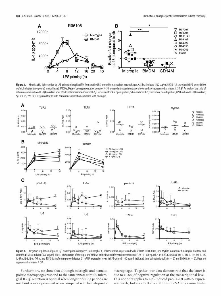
We first characterized the influence of priming on infla-mmasome-mediated activation by varying the length of thepriming period before inflammasome activation. In microglia,silica-induced IL-1� secretion was detectable from 2 h after LPSpriming onwards and increased gradually for �12 h after LPSpriming (Fig. 3A). Priming for �16 h led to lower levels of se-creted IL-1�. In BMDMs, silica-induced IL-1� secretion was de-tected as early as 1 h after LPS priming. Much higher levels ofsecreted IL-1� were obtained if BMDMs were primed with LPSfor 6 – 8 h, whereas priming for �8 h led to lower levels of secretedIL-1� (Fig. 3A). Similar differences in kinetics were foundbetween microglia and CD14Ms and also applied to MSU-induced inflammasome activation. Analysis of the ratio ofinflammasome-induced secretion of IL-1� after 16 h of LPSpriming to inflammasome-induced secretion of IL-1� after 4 h ofLPS priming confirmed that these kinetics differ significantly be-tween microglia and hematopoietic macrophages (Fig. 3B).
We examined whether the different kinetics of IL-1� secretionin microglia could be attributed to differences in sensitivity toLPS-induced signaling. However, basal expression levels ofTLR2-, TLR4-, CD14-, and MyD88 (myeloid differentiation pri-
mary response 88)-encoding transcripts were similar for micro-glia and other macrophages (Fig. 4A). In addition, priming ofmicroglia and BMDMs with different concentrations of LPS re-sulted in similar differences in IL-1� secretion profiles betweenmicroglia and BMDMs (Fig. 4B). Finally, we assessed pro-IL-1�-encoding mRNA levels in more detail after LPS priming (Fig. 4C).Pro-IL-1� mRNA transcripts were strongly induced already after1 h of LPS priming in both microglia and BMDMs. However,while pro-IL-1� mRNA transcript levels were downregulated al-ready 4 h after LPS priming in BMDMs, they remained high inmicroglia. Analysis of other cytokine-encoding mRNA tran-scripts after priming with LPS demonstrates that the lack of neg-ative regulation on pro-IL-1� transcripts also applied to IL-1�and IL-8 (Fig. 4C). Other LPS-inducible transcripts like those ofIL-1R� (IL-1 receptor antagonist), IL-6, and TNF-� (tumor-necrosis factor �) were subject to regulation in both microglia aswell as in BMDMs.
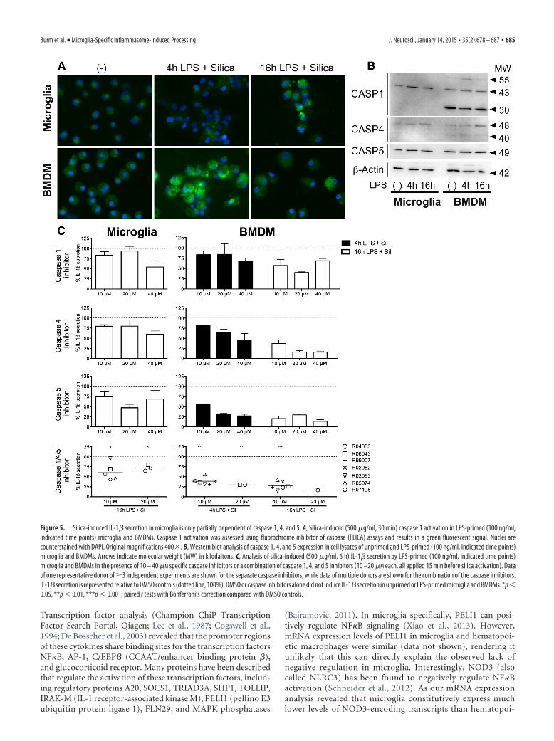
To examine the processing and secretion of IL-1� in moredetail, we first monitored the activation of caspase 1, the principalinflammasome-activated caspase described (Martinon andTschopp, 2004). To our surprise, silica-induced caspase 1 activa-tion could not be detected in microglia that were primed withLPS for 4 h. Only after 16 h of LPS priming some caspase 1activation could be observed (Fig. 5A). This was in marked con-trast to BMDMs, where robust silica-induced caspase 1 activationwas detectable 4 h after LPS priming, while longer priming re-sulted in decreased caspase 1 activation. Western blot analysis ofcaspase 1 expression revealed that although both microglia andBMDMs expressed caspase 1, BMDMs expressed an additionalisoform at 30 kDa and an additional high molecular-weight bandat 55 kDa, indicative of a protein complex (Fig. 5B). In addition,Western blot analysis revealed that both microglia and BMDMexpressed approximately similar amounts of the inflammatorycaspases 4 and 5 under basal conditions and after LPS priming.Similar to caspase 1, BMDMs expressed an additional isoform ofcaspase 4 at 40 kDa. To assess their functional involvement in thesecretion of IL-1�, we specifically inhibited caspase 1, 4, and 5before activating the inflammasome (Fig. 5C). In LPS-primedmicroglia and BMDMs, inhibition of caspase 1 inhibited silica-induced IL-1� secretion by �46 and 59% respectively. Inhibitionof caspase 4 and 5 inhibited silica-induced IL-1� secretion inmicroglia by �41 and 53% respectively, compared with �84 and86% inhibition in BMDMs respectively. Whereas inhibition ofcaspase 4 and 5 in BMDMs was clearly dose dependent, this was notthe case for microglia, suggesting that exposure to even higher con-centrations of caspase inhibitors would not lead to a further reduc-tion in silica-induced IL-1� secretion in microglia. This could not bedirectly tested, as the necessary dissolvent controls affected cellularhomeostasis (data not shown). However, simultaneous inhibition ofcaspase 1, 4, and 5 inhibited silica-induced IL-1� secretion in micro-glia by �38%, whereas this inhibited silica-induced IL-1� secretionin BMDMs by �84%. Together these data indicate that microgliaare less dependent on inflammatory caspases than BMDMs for thesecretion of silica-induced IL-1�.
DiscussionAlthough microglia have long been considered similar to othermyeloid macrophages, it is becoming more and more apparentthat they differ in many respects, as illustrated by the distinct rolesthat resident microglia and peripheral macrophages play in CNSinjury (Raivich and Banati, 2004; Greenhalgh and David, 2014).Differences in regulation of innate immune responses (Xiao et al.,2013) and alternative use of caspases have been described for
682 • J. Neurosci., January 14, 2015 • 35(2):678 – 687 Burm et al. • Microglia-Specific Inflammasome-Induced Processing

microglia in earlier studies (Burguillos et al., 2011). Our data nowidentify new and important cell type-specific differences ininflammasome-mediated responses.
Results from this study demonstrate that microglia and he-matopoietic macrophages are in principle endowed with sim-ilar inflammasome machinery. Published or online datasetson gene expression profiles of rodent microglia and macro-phages (Chiu et al., 2013; Hickman et al., 2013; Butovsky et al.,2014) did not reveal differences in expression levels of inflam-
masome components between microglia and hematopoieticmacrophages and are thus in line with most of our results.However, we report here that there are relatively large differ-ences for two transcripts in particular. Resting microglia ex-press less caspase 4 and 5 than resting BMDMs and CD14Ms(Fig. 1A). As caspase 4 and 5 are not encoded for in rodents,which express the orthologue caspase 11 (Martinon andTschopp, 2004), this is a new finding that underlines the addedvalue of studies using primate material.
Figure 2. TLR-mediated priming of microglia and BMDMs induces the expression of pro-IL-1�-encoding mRNA, but processing and secretion of IL-1� is measured only after inflammasomeactivation. A, Relative pro-IL-1�-encoding mRNA expression levels in primary rhesus microglia and BMDMs. Cells were exposed for 16 h to TLR1/2 (PAM3CSK4, 500 ng/ml), TLR3 (polyiosinic-polycytidylic acid, 20 �g/ml), TLR4 (LPS, 100 ng/ml), and TLR8 (CL075, 1 �g/ml) agonists. B, Levels of secreted IL-1� by TLR-primed microglia and BMDMs. C, Levels of secreted IL-1� byLPS-primed (100 ng/ml, 4 h) microglia and BMDMs exposed to inflammasome inducers silica (500 �g/ml, 6 h), MSU (150 �g/ml, 6 h), or ATP (5 mM, 6 h). *p � 0.05, **p � 0.01; paired t tests withBonferroni’s correction compared with unstimulated cells (-). D, Western blot analysis of pro-IL-1� and cleaved IL-1� protein in cell lysates (lys) and supernatant (sup) of LPS-primed (100 ng/ml,4 h) microglia and BMDMs that were exposed to silica (500 �g/ml, 6 h), MSU (150 �g/ml, 6 h), or ATP (5 mM, 6 h). Arrows indicate molecular weight (MW) in kilodaltons.
Burm et al. • Microglia-Specific Inflammasome-Induced Processing J. Neurosci., January 14, 2015 • 35(2):678 – 687 • 683
Furthermore, we show that although microglia and hemato-poietic macrophages respond to the same innate stimuli, micro-glial IL-1� secretion is optimal when longer priming periods areused and is more persistent when compared with hematopoietic
macrophages. Together, our data demonstrate that the latter isdue to a lack of negative regulation at the transcriptional level.This not only applies to LPS-induced pro-IL-1� mRNA expres-sion levels, but also to IL-1� and IL-8 mRNA expression levels.
Figure 3. Kinetics of IL-1� secretion by LPS-primed microglia differ from that by LPS-primed hematopoietic macrophages. A, Silica-induced (500 �g/ml, 6 h) IL-1� secretion in LPS-primed (100ng/ml, indicated time points) microglia and BMDMs. Data of one representative donor of �3 independent experiments are shown and are represented as mean � SD. B, Analysis of the ratio ofinflammasome-induced IL-1� secretion after 16 h to inflammasome-induced IL-1� secretion after 4 h. Open symbols, Silica-induced IL-1� secretion; closed symbols, MSU-induced IL-1� secretion;*p � 0.05, **p � 0.01; paired t tests with Bonferroni’s correction compared with microglia.
Figure 4. Negative regulation of pro-IL-1� transcription is impaired in microglia. A, Relative mRNA expression levels of TLR2, TLR4, CD14, and MyD88 in unprimed microglia, BMDMs, andCD14Ms. B, Silica-induced (500 �g/ml, 6 h) IL-1� secretion of microglia and BMDMs primed with different concentrations of LPS (4 –500 ng/ml, 4 or 16 h). C, Relative pro-IL-1�, IL-1�, pro-IL-18,IL-1R�, IL-8, IL-6, TNF�, and TGF� (transforming growth factor �) mRNA expression levels in LPS-primed (100 ng/ml, indicated time points) microglia (n 3) and BMDMs (n 3). Data arerepresented as mean � SD.
684 • J. Neurosci., January 14, 2015 • 35(2):678 – 687 Burm et al. • Microglia-Specific Inflammasome-Induced Processing
Transcription factor analysis (Champion ChiP TranscriptionFactor Search Portal, Qiagen; Lee et al., 1987; Cogswell et al.,1994; De Bosscher et al., 2003) revealed that the promoter regionsof these cytokines share binding sites for the transcription factorsNF�B, AP-1, C/EBP� (CCAAT/enhancer binding protein �),and glucocorticoid receptor. Many proteins have been describedthat regulate the activation of these transcription factors, includ-ing regulatory proteins A20, SOCS1, TRIAD3A, SHP1, TOLLIP,IRAK-M (IL-1 receptor-associated kinase M), PELI1 (pellino E3ubiquitin protein ligase 1), FLN29, and MAPK phosphatases
(Bajramovic, 2011). In microglia specifically, PELI1 can posi-tively regulate NF�B signaling (Xiao et al., 2013). However,mRNA expression levels of PELI1 in microglia and hematopoi-etic macrophages were similar (data not shown), rendering itunlikely that this can directly explain the observed lack ofnegative regulation in microglia. Interestingly, NOD3 (alsocalled NLRC3) has been found to negatively regulate NF�Bactivation (Schneider et al., 2012). As our mRNA expressionanalysis revealed that microglia constitutively express muchlower levels of NOD3-encoding transcripts than hematopoi-
Figure 5. Silica-induced IL-1� secretion in microglia is only partially dependent of caspase 1, 4, and 5. A, Silica-induced (500 �g/ml, 30 min) caspase 1 activation in LPS-primed (100 ng/ml,indicated time points) microglia and BMDMs. Caspase 1 activation was assessed using fluorochrome inhibitor of caspase (FLICA) assays and results in a green fluorescent signal. Nuclei arecounterstained with DAPI. Original magnifications 400�. B, Western blot analysis of caspase 1, 4, and 5 expression in cell lysates of unprimed and LPS-primed (100 ng/ml, indicated time points)microglia and BMDMs. Arrows indicate molecular weight (MW) in kilodaltons. C, Analysis of silica-induced (500 �g/ml, 6 h) IL-1� secretion by LPS-primed (100 ng/ml, indicated time points)microglia and BMDMs in the presence of 10 – 40 �M specific caspase inhibitors or a combination of caspase 1, 4, and 5 inhibitors (10 –20 �M each, all applied 15 min before silica activation). Dataof one representative donor of �3 independent experiments are shown for the separate caspase inhibitors, while data of multiple donors are shown for the combination of the caspase inhibitors.IL-1� secretion is represented relative to DMSO controls (dotted line, 100%). DMSO or caspase inhibitors alone did not induce IL-1� secretion in unprimed or LPS-primed microglia and BMDMs. *p�0.05, **p � 0.01, ***p � 0.001; paired t tests with Bonferroni’s correction compared with DMSO controls.
Burm et al. • Microglia-Specific Inflammasome-Induced Processing J. Neurosci., January 14, 2015 • 35(2):678 – 687 • 685
etic macrophages, NOD3 is an interesting candidate for futurestudies.
Various studies using primary microglia describe inflam-masome-induced IL-1� secretion in response to protein aggre-gates and pathogens as caspase 1 dependent (Halle et al., 2008;Meissner et al., 2010; Terada et al., 2010; Hanamsagar et al., 2011;Hafner-Bratkovic et al., 2012; Jamilloux et al., 2013; Lee et al.,2013; Walsh et al., 2014). Our results confirm that microglialsecretion of IL-1� is dependent on caspase 1, but also demon-strate that this dependence is only partial. In addition, we dem-onstrate that although caspase 4 and 5 are involved in bothmicroglia and hematopoietic macrophages, their relative con-tribution to inflammasome-induced IL-1� secretion appearsto be less important in microglia. Our data on the simultane-ous inhibition of caspase 1, 4, and 5 are in line with this notion.It has been reported that activation of caspase 4 and 5 canpotentiate the activity of caspase 1 (Martinon et al., 2002;Sollberger et al., 2012) and this process might well be lessefficient in microglia.
Overall our data show that microglia are less dependent thanBMDMs on inflammatory caspases for the processing and secre-tion of IL-1� and suggest that they also employ other mecha-nisms. Alternative mechanisms that have been described forIL-1� processing and secretion include activation of a noncanonicalcaspase 8 inflammasome (Gringhuis et al., 2012; Antonopoulos etal., 2013) and inflammasome-independent mechanisms, such asmatrix metalloproteinases (Schonbeck et al., 1998), cathepsins(Edye et al., 2013), and serine proteases (Joosten et al., 2009;Stehlik, 2009; Karmakar et al., 2012; Cassel et al., 2014). Prelim-inary results from our laboratory indicate that inhibition ofcaspase 8 does not affect silica-induced IL-1� secretion in eithermicroglia or BMDMs (data not shown). The relative contribu-tion of other pathways in microglial IL-1� secretion remains tobe investigated.
This study, describing cell type-specific differences in the neg-ative regulation of pro-IL-1� expression and in the enzymaticprocessing of pro-IL-1�, concurs with recently described funda-mental differences in inflammasome-mediated activation ofmonocytes and macrophages (Netea et al., 2009). Whether suchdifferences are cell inherent (Ginhoux et al., 2010) or induced byprolonged exposure to the neuronal microenvironment is cur-rently unknown. It also remains to be demonstrated how thedifferences described here translate to tissue-specific responses tochronic or acute cellular stress in vivo. Currently, inhibition ofcaspase 1 activation is considered a therapeutic strategy to reduceinflammation in neuroinflammatory diseases (Rabuffetti et al.,2000). Our results suggest that this strategy might only be par-tially effective on microglia. To develop therapeutic strategiesthat target IL-1� processing and secretion in microglia specifi-cally, it is important to delineate the additional mechanisms usedby microglia for the secretion of IL-1�.
ReferencesAbulafia DP, de Rivero Vaccari JP, Lozano JD, Lotocki G, Keane RW, Dietrich
WD (2009) Inhibition of the inflammasome complex reduces the in-flammatory response after thromboembolic stroke in mice. J Cereb BloodFlow Metab 29:534 –544. CrossRef Medline
Antonopoulos C, El Sanadi C, Kaiser WJ, Mocarski ES, Dubyak GR (2013)Proapoptotic chemotherapeutic drugs induce noncanonical processingand release of IL-1beta via caspase-8 in dendritic cells. J Immunol 191:4789 – 4803. CrossRef Medline
Bajramovic JJ (2011) Regulation of innate immune responses in the centralnervous system. CNS Neurol Disord Drug Targets 10:4–24. CrossRefMedline
Burguillos MA, Deierborg T, Kavanagh E, Persson A, Hajji N, Garcia-Quintanilla A, Cano J, Brundin P, Englund E, Venero JL, Joseph B (2011)Caspase signalling controls microglia activation and neurotoxicity. Na-ture 472:319 –324. CrossRef Medline
Butovsky O, Jedrychowski MP, Moore CS, Cialic R, Lanser AJ, Gabriely G,Koeglsperger T, Dake B, Wu PM, Doykan CE, Fanek Z, Liu L, Chen Z,Rothstein JD, Ransohoff RM, Gygi SP, Antel JP, Weiner HL (2014)Identification of a unique TGF-�-dependent molecular and functionalsignature in microglia. Nat Neurosci 17:131–143. CrossRef Medline
Cassel SL, Janczy JR, Bing X, Wilson SP, Olivier AK, Otero JE, Iwakura Y,Shayakhmetov DM, Bassuk AG, Abu-Amer Y, Brogden KA, Burns TL,Sutterwala FS, Ferguson PJ (2014) Inflammasome-independent IL-1beta mediates autoinflammatory disease in Pstpip2-deficient mice. ProcNatl Acad Sci U S A 111:1072–1077. CrossRef Medline
Chang J, Yang L, Kouadir M, Peng Y, Zhang S, Shi F, Zhou X, Yin X, Zhao D(2012) Antibody-mediated inhibition of integrin alpha5beta1 blocksneurotoxic prion peptide PrP106 –126-induced activation of BV2 micro-glia. J Mol Neurosci 48:248 –252. CrossRef Medline
Chiu IM, Morimoto ET, Goodarzi H, Liao JT, O’Keeffe S, Phatnani HP,Muratet M, Carroll MC, Levy S, Tavazoie S, Myers RM, Maniatis T(2013) A neurodegeneration-specific gene-expression signature ofacutely isolated microglia from an amyotrophic lateral sclerosis mousemodel. Cell Rep 4:385– 401. CrossRef Medline
Cogswell JP, Godlevski MM, Wisely GB, Clay WC, Leesnitzer LM, Ways JP,Gray JG (1994) NF-kappa B regulates IL-1 beta transcription through aconsensus NF-kappa B binding site and a nonconsensus CRE-like site.J Immun 153:712–723. Medline
De Bosscher K, Vanden Berghe W, Haegeman G (2003) The interplay be-tween the glucocorticoid receptor and nuclear factor-kappaB or activatorprotein-1: molecular mechanisms for gene repression. Endocr Rev 24:488 –522. CrossRef Medline
Edye ME, Lopez-Castejon G, Allan SM, Brough D (2013) Acidosis drivesdamage-associated molecular pattern (DAMP)-induced interleukin-1 se-cretion via a caspase-1-independent pathway. J Biol Chem 288:30485–30494. CrossRef Medline
Euser SM, Hofman A, Westendorp RG, Breteler MM (2009) Serum uric acidand cognitive function and dementia. Brain 132:377–382. CrossRef Medline
Furr SR, Chauhan VS, Sterka D Jr, Grdzelishvili V, Marriott I (2008) Char-acterization of retinoic acid-inducible gene-I expression in primary mu-rine glia following exposure to vesicular stomatitis virus. J Neurovirol14:503–513. CrossRef Medline
Ginhoux F, Greter M, Leboeuf M, Nandi S, See P, Gokhan S, Mehler MF,Conway SJ, Ng LG, Stanley ER, Samokhvalov IM, Merad M (2010) Fatemapping analysis reveals that adult microglia derive from primitive mac-rophages. Science 330:841– 845. CrossRef Medline
Greenhalgh AD, David S (2014) Differences in the phagocytic responseof microglia and peripheral macrophages after spinal cord injury andits effects on cell death. J Neurosci 34:6316 – 6322. CrossRef Medline
Gringhuis SI, Kaptein TM, Wevers BA, Theelen B, van der Vlist M, BoekhoutT, Geijtenbeek TB (2012) Dectin-1 is an extracellular pathogen sensorfor the induction and processing of IL-1beta via a noncanonical caspase-8inflammasome. Nat Immunol 13:246 –254. CrossRef Medline
Hafner-Bratkovic I, Bencina M, Fitzgerald KA, Golenbock D, Jerala R (2012)NLRP3 inflammasome activation in macrophage cell lines by prion pro-tein fibrils as the source of IL-1beta and neuronal toxicity. Cell Mol LifeSci 69:4215– 4228. CrossRef Medline
Halle A, Hornung V, Petzold GC, Stewart CR, Monks BG, Reinheckel T,Fitzgerald KA, Latz E, Moore KJ, Golenbock DT (2008) The NALP3inflammasome is involved in the innate immune response to amyloid-beta. Nat Immunol 9:857– 865. CrossRef Medline
Hanamsagar R, Torres V, Kielian T (2011) Inflammasome activation andIL-1beta/IL-18 processing are influenced by distinct pathways in micro-glia. J Neurochem 119:736 –748. CrossRef Medline
Heneka MT, Kummer MP, Stutz A, Delekate A, Schwartz S, Vieira-Saecker A,Griep A, Axt D, Remus A, Tzeng TC, Gelpi E, Halle A, Korte M, Latz E,Golenbock DT (2013) NLRP3 is activated in Alzheimer’s disease andcontributes to pathology in APP/PS1 mice. Nature 493:674 – 678.CrossRef Medline
Hickman SE, Kingery ND, Ohsumi TK, Borowsky ML, Wang LC, Means TK,El Khoury J (2013) The microglial sensome revealed by direct RNA se-quencing. Nat Neurosci 16:1896 –1905. CrossRef Medline
Jamilloux Y, Pierini R, Querenet M, Juruj C, Fauchais AL, Jauberteau MO,
686 • J. Neurosci., January 14, 2015 • 35(2):678 – 687 Burm et al. • Microglia-Specific Inflammasome-Induced Processing
Jarraud S, Lina G, Etienne J, Roy CR, Henry T, Davoust N, Ader F (2013)Inflammasome activation restricts Legionella pneumophila replication inprimary microglial cells through flagellin detection. Glia 61:539 –549.CrossRef Medline
Joosten LA, Netea MG, Fantuzzi G, Koenders MI, Helsen MM, Sparrer H,Pham CT, van der Meer JW, Dinarello CA, van den Berg WB (2009)Inflammatory arthritis in caspase 1 gene-deficient mice: contributionof proteinase 3 to caspase 1-independent production of bioactiveinterleukin-1beta. Arthritis Rheum 60:3651–3662. CrossRef Medline
Karmakar M, Sun Y, Hise AG, Rietsch A, Pearlman E (2012) Cutting edge:IL-1beta processing during Pseudomonas aeruginosa infection is medi-ated by neutrophil serine proteases and is independent of NLRC4 andcaspase-1. J Immunol 189:4231– 4235. CrossRef Medline
Kaushik DK, Gupta M, Kumawat KL, Basu A (2012) NLRP3 inflam-masome: key mediator of neuroinflammation in murine Japanese en-cephalitis. PloS One 7:e32270. CrossRef Medline
Khare S, Dorfleutner A, Bryan NB, Yun C, Radian AD, de Almeida L, Roja-nasakul Y, Stehlik C (2012) An NLRP7-containing inflammasome me-diates recognition of microbial lipopeptides in human macrophages.Immunity 36:464 – 476. CrossRef Medline
Latz E (2010) The inflammasomes: mechanisms of activation and function.Curr Opin Immunol 22:28 –33. CrossRef Medline
Lee HM, Kang J, Lee SJ, Jo EK (2013) Microglial activation of the NLRP3inflammasome by the priming signals derived from macrophages infectedwith mycobacteria. Glia 61:441– 452. CrossRef Medline
Lee W, Mitchell P, Tjian R (1987) Purified transcription factor AP-1 interactswith TPA-inducible enhancer elements. Cell 49:741–752. CrossRef Medline
Maetzler W, Stapf AK, Schulte C, Hauser AK, Lerche S, Wurster I, SchleicherE, Melms A, Berg D (2011) Serum and cerebrospinal fluid uric acidlevels in lewy body disorders: associations with disease occurrence andamyloid-beta pathway. J Alzheimers Dis 27:119 –126. CrossRef Medline
Martinon F, Tschopp J (2004) Inflammatory caspases: linking an intracel-lular innate immune system to autoinflammatory diseases. Cell 117:561–574. CrossRef Medline
Martinon F, Burns K, Tschopp J (2002) The inflammasome: a molecularplatform triggering activation of inflammatory caspases and processing ofproIL-beta. Mol Cell 10:417– 426. CrossRef Medline
Martinon F, Petrilli V, Mayor A, Tardivel A, Tschopp J (2006) Gout-associated uric acid crystals activate the NALP3 inflammasome. Nature440:237–241. CrossRef Medline
Martinon F, Mayor A, Tschopp J (2009) The inflammasomes: guardians ofthe body. Annu Rev Immunol 27:229 –265. CrossRef Medline
Martins I, Wang Y, Michaud M, Ma Y, Sukkurwala AQ, Shen S, Kepp O,Metivier D, Galluzzi L, Perfettini JL, Zitvogel L, Kroemer G (2014) Mo-lecular mechanisms of ATP secretion during immunogenic cell death.Cell Death Differ 21:79 –91. CrossRef Medline
McFarland NR, Burdett T, Desjardins CA, Frosch MP, Schwarzschild MA(2013) Postmortem brain levels of urate and precursors in Parkinson’s dis-ease and related disorders. Neurodegener Dis 12:189–198. CrossRef Medline
Meissner F, Molawi K, Zychlinsky A (2010) Mutant superoxide dismutase1-induced IL-1beta accelerates ALS pathogenesis. Proc Natl Acad SciU S A 107:13046 –13050. CrossRef Medline
Netea MG, Nold-Petry CA, Nold MF, Joosten LA, Opitz B, van der Meer JH,van de Veerdonk FL, Ferwerda G, Heinhuis B, Devesa I, Funk CJ, MasonRJ, Kullberg BJ, Rubartelli A, van der Meer JW, Dinarello CA (2009)Differential requirement for the activation of the inflammasome for pro-cessing and release of IL-1beta in monocytes and macrophages. Blood113:2324 –2335. CrossRef Medline
Olson JK, Miller SD (2004) Microglia initiate central nervous system innateand adaptive immune responses through multiple TLRs. J Immunol 173:3916 –3924. CrossRef Medline
Pfaffl MW (2001) A new mathematical model for relative quantification inreal-time RT-PCR. Nucleic Acids Res 29:e45. CrossRef Medline
Rabuffetti M, Sciorati C, Tarozzo G, Clementi E, Manfredi AA, Beltramo M(2000) Inhibition of caspase-1-like activity by Ac-Tyr-Val-Ala-Asp-chloromethyl ketone induces long-lasting neuroprotection in cerebralischemia through apoptosis reduction and decrease of proinflammatorycytokines. J Neurosci 20:4398 – 4404. Medline
Raivich G, Banati R (2004) Brain microglia and blood-derived macro-phages: molecular profiles and functional roles in multiple sclerosis andanimal models of autoimmune demyelinating disease. Brain Res BrainRes Rev 46:261–281. CrossRef Medline
Rock KL, Kono H (2008) The inflammatory response to cell death. AnnuRev Pathol 3:99 –126. CrossRef Medline
Salminen A, Ojala J, Suuronen T, Kaarniranta K, Kauppinen A (2008)Amyloid-beta oligomers set fire to inflammasomes and induce Alzhei-mer’s pathology. J Cell Mol Med 12:2255–2262. CrossRef Medline
Schneider M, Zimmermann AG, Roberts RA, Zhang L, Swanson KV, Wen H,Davis BK, Allen IC, Holl EK, Ye Z, Rahman AH, Conti BJ, Eitas TK, KollerBH, Ting JP (2012) The innate immune sensor NLRC3 attenuates Toll-likereceptor signaling via modification of the signaling adaptor TRAF6 and tran-scription factor NF-kappaB. Nat Immunol 13:823–831. CrossRef Medline
Schonbeck U, Mach F, Libby P (1998) Generation of biologically active IL-1beta by matrix metalloproteinases: a novel caspase-1-independent path-way of IL-1 beta processing. J Immunol 161:3340 –3346. Medline
Shah VB, Huang Y, Keshwara R, Ozment-Skelton T, Williams DL, Keshvara L(2008) Beta-glucan activates microglia without inducing cytokine pro-duction in Dectin-1-dependent manner. J Immunol 180:2777–2785.CrossRef Medline
Shi F, Yang L, Kouadir M, Yang Y, Wang J, Zhou X, Yin X, Zhao D (2012) TheNALP3 inflammasome is involved in neurotoxic prion peptide-induced mi-croglial activation. J Neuroinflammation 9:73. CrossRef Medline
Shi F, Yang L, Kouadir M, Yang Y, Ding T, Wang J, Zhou X, Yin X, Zhao D(2013) Prion protein participates in the regulation of classical and alternativeactivation of BV2 microglia. J Neurochem 124:168–174. CrossRef Medline
Shi Y, Evans JE, Rock KL (2003) Molecular identification of a danger signalthat alerts the immune system to dying cells. Nature 425:516 –521.CrossRef Medline
Sollberger G, Strittmatter GE, Kistowska M, French LE, Beer HD (2012)Caspase-4 is required for activation of inflammasomes. J Immunol 188:1992–2000. CrossRef Medline
Stehlik C (2009) Multiple interleukin-1beta-converting enzymes contribute toinflammatory arthritis. Arthritis Rheum 60:3524–3530. CrossRef Medline
Terada K, Yamada J, Hayashi Y, Wu Z, Uchiyama Y, Peters C, Nakanishi H(2010) Involvement of cathepsin B in the processing and secretion ofinterleukin-1beta in chromogranin A-stimulated microglia. Glia 58:114 –124. CrossRef Medline
Ting JP, Duncan JA, Lei Y (2010) How the noninflammasome NLRs functionin the innate immune system. Science 327:286–290. CrossRef Medline
van der Putten C, Zuiderwijk-Sick EA, van Straalen L, de Geus ED, Boven LA,Kondova I, Ijzerman AP, Bajramovic JJ (2009) Differential expression ofadenosine A3 receptors controls adenosine A2A receptor-mediated inhi-bition of TLR responses in microglia. J Immunol 182:7603–7612.CrossRef Medline
Walsh JG, Reinke SN, Mamik MK, McKenzie BA, Maingat F, Branton WG,Broadhurst DI, Power C (2014) Rapid inflammasome activation in mi-croglia contributes to brain disease in HIV/AIDS. Retrovirology 11:35.CrossRef Medline
Wu Z, Sun L, Hashioka S, Yu S, Schwab C, Okada R, Hayashi Y, McGeer PL,Nakanishi H (2013) Differential pathways for interleukin-1beta pro-duction activated by chromogranin A and amyloid beta in microglia.Neurobiol Aging 34:2715–2725. CrossRef Medline
Xiao Y, Jin J, Chang M, Chang JH, Hu H, Zhou X, Brittain GC, Stansberg C,Torkildsen Ø, Wang X, Brink R, Cheng X, Sun SC (2013) Peli1 pro-motes microglia-mediated CNS inflammation by regulating Traf3 degra-dation. Nat Med 19:595– 602. CrossRef Medline
Zhou R, Yazdi AS, Menu P, Tschopp J (2011) A role for mitochondria inNLRP3 inflammasome activation. Nature 469:221–225. CrossRefMedline
Zuiderwijk-Sick EA, van der Putten C, Bsibsi M, Deuzing IP, de Boer W,Persoon-Deen C, Kondova I, Boven LA, van Noort JM, ‘t Hart BA, AmorS, Bajramovic JJ (2007) Differentiation of primary adult microglia alterstheir response to TLR8-mediated activation but not their capacity as APC.Glia 55:1589 –1600. CrossRef Medline
Burm et al. • Microglia-Specific Inflammasome-Induced Processing J. Neurosci., January 14, 2015 • 35(2):678 – 687 • 687