case 11.1 recurrent cold sores case 11.2 nickel dermatitis
TRANSCRIPT
Essentials of Clinical Immunology, Sixth Edition. Helen Chapel, Mansel Haeney, Siraj Misbah, and Neil Snowden.© 2014 John Wiley & Sons, Ltd. Published 2014 by John Wiley & Sons, Ltd.
Case 11.1 Recurrent cold sores
A 38-year-old woman had been troubled since the age of 8 by recurrent cold sores. Several times each year she would develop a distinctive tingling sensation around her nose or lips, followed several hours later by localized formation of small blisters which crusted, became more painful and gradually cleared over several days. The attacks were often provoked by exposure to strong sunlight. She had a history of troublesome hay fever but was otherwise well. She was able to prevent attacks by use of a high-factor sun-block and treat any breakthrough episodes with prompt use of aciclovir cream at the onset of symptoms.
Case 11.2 Nickel dermatitis
A 47-year-old woman presented with a 3-week history of an acute rash that started beneath her watch. Two weeks later, a further patch appeared at the umbilicus. She had previously noted that she could not wear cheap earrings without triggering a rash on her ear lobes. There was no past medical history of note and no personal or family history of atopy. On examination, two patches of dermatitis were seen over the presenting areas. The appearances were suggestive of nickel-induced contact dermatitis corresponding to nickel in the watch and on a jeans stud. She was patch-tested to a battery of commonly implicated agents (Table 11.2): strongly positive results were induced by nickel sulphate and cobalt chloride only. The final diagnosis was nickel dermatitis, which cleared spontaneously following avoidance of nickel-containing articles.
Table 11.2 Some agents responsible for allergic contact dermatitis
Agent Examples of exposure
Metals Nickel Clasps, necklaces, watch-straps
Chromate Cement (building site workers)
Cobalt*
Medications ‘Para’-group chemicals Benzocaine-type anaesthetics, sulphonamide antibiotics, PABA-containing substances (e.g. sunscreens) and oral hypoglycaemic agents(sulphonylureas)
Phenothiazines Phenothiazine-based antihistamines
Neomycin Topical antibiotics
Plastics Epoxyresins, acrylates Construction industry, glues
Rubber Accelerators Tyre industry, rubber gloves, shoes, clothing, household ‘grips’, etc.
Plants Poison ivy (USA only)
Primula
Chrysanthemum
Geranium
Cosmetics Perfumes
Preservatives
Lanolin
*Source of cobalt sensitivity is usually obscure but it may exist as a co-sensitivity with nickel (metal) or chromate (cement).PABA, Para-amino benzoic acid.

Case Figure 11.2a Allergic contact dermatitis to nickel: a characteristic site (due to contact with the stud fastener of jeans)
Case Figure 11.2b Nickel-induced contact dermatitis on the temple, related to wearing spectacles.
Case 11.3 Atopic eczema
A 5-year-old boy had developed an itchy rash on his trunk and feet at the age of 18 months. This waxed and waned over the next 3 years and gradually came to involve predominantly his flanks, popliteal and antecubital fossae. He had mild asthma requiring occasional bronchodilators only. His mild atopic eczema was treated with bland emollient creams and occasionally 1% hydrocortisone. His prognosis was good and his skin problems resolved over the next few years.

Case Figure 11.3 Eczema on infant’s ankles, similar to Case Figure 11.2 when young.
Case 11.4 Pemphigus vulgaris
A 46-year-old woman presented with a generalized, blistering rash of 4 weeks’ duration. Her trunk was mainly affected. On examination, there was extensive blistering and large areas of denuded skin. Ulcers were also present in her mouth. The provisional diagnosis was pemphigus vulgaris.
Laboratory investigations showed a normal haemoglobin, full blood count and biochemical profile. Her serum contained antibodies reacting strongly with the cell surfaces of keratinocytes in the epidermis. Direct immunofluorescent examination of a biopsy of normal skin taken from a site adjacent to one bulla showed deposition of IgG around the keratinocytes, giving a ‘chicken-wire’ appearance (see Fig. 11.6). These findings are characteristic of pemphigus vulgaris. She was treated with methyl prednisolone initially, reducing to prednisolone 40 mg/day when new blister formation ceased. She has been regularly followed for 2 years, during which therapy has been gradually reduced to maintenance levels and no new bullae have appeared.
Fig. 11.6 Characteristic findings on direct immunofluorescent examination of skin biopsies.
Dermis
Autoantibodies to epidermalintercellular cement
Autoantibodies to basementmembrane zone
Coarse granular deposits ofIgA in dermal papilla
Granular deposits of Ig andcomplement alongbasement membrane zone
Pemphigus
PemphigoidBullousskindiseases
Dermatitisherpetiformis
Lupuserythematosus—systemic andcutaneous
Epidermis
Finding on direct immunofluorescenceDisorder

Case Figure 11.4 Pemphigus Vulsaris. The fragile blisters in pemphigus rarely remain intact so erosions are more commonly seen (a). In almost all Case Figures, there will be erosions in the oral cavity, seen here on the soft palate (b).

Case 11.5 Hereditary angioedema
Daniel, a 14-year-old boy, presented with a 6-month history of recurrent episodes of swelling of his lips, eyes and tongue (Fig. 11.9). The swellings came on suddenly, grew over a period of 15–20 min, and lasted from 12 to 48 h. They were not itchy but tended to give a prickly sensation. There was no obstruction of airways or abdominal pain during the attacks, which were often associated with intercurrent infection. Urticaria was absent. His sister, aged 21 years, had suffered from an identical problem for 4 years. Physical examination was normal.
The clinical story was typical of angioedema and the family history suggested that this might be hereditary angioedema (HAE). Blood samples taken for complement analysis during remission showed a normal C3 (0.85 g/l), but a rather low C4 of 0.12 g/l (NR 0.2–0.4) and a C1 inhibitor (Cī-INH) level of 0.06 g/l (NR 0.18–0.26); these findings were consistent with the diagnosis of HAE. When the tests were repeated during a subsequent attack of angioedema, the C3 concentration was unchanged but the C4 level was extremely low at 0.04 g/l. Daniel was started on treatment with danazol. Although his Cī- INH level only rose to 0.14 g/l, he had no further attacks of HAE. What is not clear is why neither parent has a history of HAE, since the condition is inherited in an autosomal dominant fashion.
Fig. 11.9 Severe periorbital angioedema.
Case Figure 11.5 Swollen eyelids due to angioedema.

Case 11.6 Urticarial vasculitis
A 41-year-old woman presented with itchy lumps on her legs of 5 weeks’ duration. The lumps lasted 4–5 days. She had a history of asthma from childhood and was being treated with an array of drugs. As far as she knew, she had never ‘reacted’ to any of these drugs. On examination, her legs showed palpable, purpuric lesions and areas of urticaria. General examination was otherwise normal. The skin lesions were those of urticarial vasculitis.
Investigations showed a low haemoglobin (107 g/l) with a hypochromic blood film. Her white cell count was 12.2 × 10⁹/l, with a raised eosinophil count of 1.56 × 10⁹/l; the erythrocyte sedimentation rate was 45 mm/h. Serum immunoglobulins were normal but her complement levels were low: C3 was 0.55 g/l (NR 0.8–1.4) and C4 was 0.15 g/l (NR 0.2–0.4). Antinuclear and anti-dsDNA antibodies, antineutrophil cytoplasmic antibodies, cryoglobulins and rheumatoid factor were not detected. Biopsy of an acute lesion showed histological features of vasculitis, with deposition of C3 in the deep dermal blood vessels on direct immunofluorescence. She was treated empirically with prednisolone. Her vasculitis improved considerably but no underlying cause was ever found.
Case Figure 11.6 Urticaria – marked wheals with surrounding erythema.
Case 11.7 Mixed cryoglobulinaemia
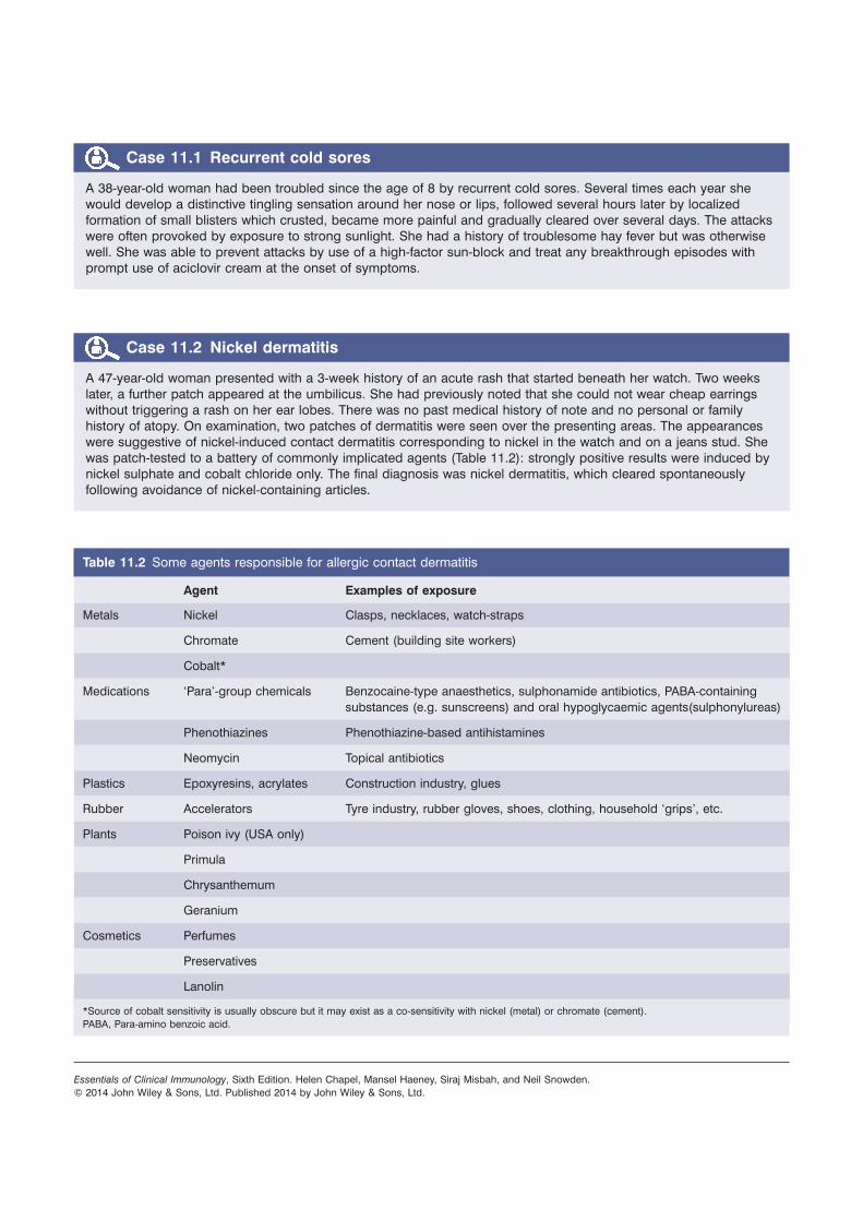
A 45-year-old woman presented with ankle oedema due to nephrotic syndrome. In the preceding 5 years, she had experienced several episodes of a purpuric, erythematous, papular rash on the legs, accompanied by a bilateral arthropathy of the knees and ankles. A biopsy of the rash had shown features of vasculitis which had responded to systemic steroids. She now had a non-selective proteinuria of 10 g/day and a creatinine clearance of 74 ml/min. Serum alanine aminotransferase (ALT) was increased at 140 U/ml (NR < 50). Rheumatoid factor was detectable to a titre of 1/1280 but antinuclear antibodies were negative. Hepatitis B surface antigen was absent but antibodies to hepatitis C and hepatitis C viral RNA were detected in her serum. The serum immunoglobulins, measured at room temperature, were: IgG 2.10 g/l (NR 7.2–19.0); IgA 0.85 g/l (NR 0.8–5.0); and IgM 2.80 g/l (NR 0.5–2.0). Complement levels were abnormal, with a C3 of 0.80 g/l (NR 0.8–1.4) and a C4 of 0.02 g/l (NR 0.2–0.4). The very low C4 level raised the suspicion of cryoglobulinaemia. A warm sample of her serum contained a mixed cryoglobulin, composed of a monoclonal IgM and polyclonal IgG. A skin biopsy showed scattered deposits of IgM, IgG and C3 in dermal blood vessels. The histology of a renal biopsy showed membranoproliferative glomerulonephritis: on direct immunofluorescence, granular deposits of IgM and IgG were seen along the epithelial basement membrane. The final diagnosis was mixed cryoglobulinaemia secondary to chronic hepatitis C infection with cutaneous vasculitis, arthropathy and membranoproliferative glomerulonephritis. No risk factors for hepatitis C infection were identified; she was treated with a good response.

Case Figure 11.7a Palpable purpura due to small vessel cutaneous vasculitis in a patient with cryoglobulinaemia.
Case Figure 11.7b Serum; the left tube is at room temperature; the right tube has been in the 4°C incubator and the cryoglobulin has precipitated out and settled to the bottom of the tube.
(c)RT
3YC
IgM
k
IgG
Case Figure 11.7c Warm electrophoresis (at 37°C) of the serum shows a marked monoclonal band in the mid-gamma region – immunofixation shows this band to be monoclonal IgM of kappa type complexed with polyclonal IgG.
Case Figure 11.7d H&E staining of the renal biopsy of a similar patient shows eosinophilic material – the cryoglobulin – in the glomerulus which corresponds to the IgG fluorescent staining on the right.
Case 11.8 Subacute cutaneous lupus erythematosus
A 34-year-old woman presented with a 2-year history of a waxing and waning rash on her neck and arms. She had noticed that the rash was likely to flare following exposure to bright sunlight. Her general health was good, although in the last few months she had developed flitting pain and stiffness in the small joints of her hands. She had no history of Raynaud’s phenomenon, mouth ulcers or eye trouble. On examination, there was an extensive skin rash involving the neck and arms and extending onto the face. The rash was red, raised above the surrounding skin and tended to form ring-like patterns with scaly margins (Fig. 11.11). There was no evidence of vasculitis and there were no other abnormal physical signs. The skin appearances were those of subacute cutaneous lupus erythematosus (SCLE); there was no clinical evidence of systemic involvement.
Laboratory investigations showed a normal haemoglobin, white cell count, blood creatinine and urinalysis. Antinuclear antibodies were detected at a titre of 1/100 and antibodies to Ro were detected by precipitation. Serum C3 and C4 levels were normal. A biopsy of affected skin on the neck showed typical changes of SCLE. Direct immunofluorescence demonstrated granular deposits of C3 and IgG along the basement membrane zone in the affected skin. She was treated with sunscreens, topical steroids and hydroxychloroquine. Over a period of several months, the skin lesions became less frequent but still flared after sun exposure.

Case Figure 11.8a Discoid lupus erythematosus: Well defined, scaly erythematous plaques (a). Dilated, plugged follicles may be seen on close inspection, particularly in the ears. Chronic DLE results in scarring which is often atrophic with postinflammatory hypopigmentation or hyperpigmntation as shown (b). Hyperpigmentation is commoner in racially pigmented skin.
Fig. 11.11 Typical changes of subacute cutaneous lupus erythematosus on the neck.

Case 11.9 Systemic sclerosis
The patient first developed Raynaud’s phenomenon in her early 20 s. In cold weather, her hands and toes became white and painful and then turned blue; when the circulation returned, it was accompanied by extreme redness and pain (see Fig. 11.13). Several years after developing Raynaud’s phenomenon, she noticed some tethering and thickening of her skin, starting in the hands but eventually affecting her face and mouth. On one occasion, an ulcer on her right index finger discharged ‘tiny pieces of chalk’. At the age of 54, she developed dysphagia: she could swallow food only if she took fluids with it. At the age of 56, diarrhoea became a problem. Barium studies showed pseudodiverticulae typical of systemic sclerosis, a hiatus hernia, an atonic oesophagus and stomach, and a dilated, distorted proximal jejunum.
When reassessed at the age of 59, her heart, chest and abdomen were normal. She showed marked sclerodactyly and typical skin changes of scleroderma. Soft-tissue, calcified nodules were present on her fingers, forearms and over the patellae. Telangiectasia was evident on the hands, face and lips. Over the following 2 years she became increasingly short of breath, with marked ankle oedema and worsening of the diarrhoea. Lung function tests showed a restrictive defect with a reduction in transfer factor. A computed tomography scan of the thorax showed no evidence of pulmonary fibrosis but an electrocardiogram and echocardiogram suggested right-ventricular strain secondary to pulmonary hypertension associated with limited systemic sclerosis. There was no biochemical evidence of renal or liver disease.
She has taken part in controlled trials of new treatments for systemic sclerosis: none has worked. She has now been referred to a specialist pulmonary hypertension unit for assessment as to whether she might benefit from treatment with bosentan, an endothelin 1 antagonist to prevent progression to pulmonary fibrosis.
Fig. 11.13 Raynaud’s phenomenon in limited systemic sclerosis.

Case Figure 11.9a Well-demarcated digital ischaemia due to Raynaud’s phenomenon.
Case Figure 11.9b Ulcerated calcinotic nodule on the finger tip of a patient with limited cutaneous systemic sclerosis. Slide courtesy of Martin Pattrick.
Case Figure 11.9c Barium swallow showing dilated baggy atonic oesophagus in limited systemic sclerosis. Slide courtesy of Martin Pattrick.

Case Figure 11.9e Infarction of the finger tips due to severe Raynaud’s in limited systemic sclerosis.
Case Figure 11.9d Well established sclerodactyly in late stage systemic sclerosis or MCTD. Slide courtesy of Martin Pattrick.