carcinoma vagina
TRANSCRIPT

Vaginal CancerVaginal Cancer
Dr Nabeel YahiyaDr Nabeel Yahiya
Junior resident in RadiotherapyJunior resident in Radiotherapy
Kottayam Medical CollegeKottayam Medical College
Vaginal CancerVaginal Cancer Rare tumor representing only 1-2% of all gynecologic Rare tumor representing only 1-2% of all gynecologic
malignanciesmalignancies
80-90% are metastatic80-90% are metastatic
cervix or endometrium. cervix or endometrium.
Metastatic cancer from the vulva, ovaries, Metastatic cancer from the vulva, ovaries, choriocarcinoma, rectosigmoid, and bladder, renal cell choriocarcinoma, rectosigmoid, and bladder, renal cell carcinoma, carcinoma, melanoma, and breast cancer are less common, and breast cancer are less common
Mean age of patients with primary vaginal cancer is 60-65 Mean age of patients with primary vaginal cancer is 60-65 yearsyears

Introduction Introduction
The vagina is a muscular dilatable tubular structure The vagina is a muscular dilatable tubular structure averaging 7.5 cm in length that extends from the averaging 7.5 cm in length that extends from the cervix to the vulvacervix to the vulva

Relations Relations

Relations Relations
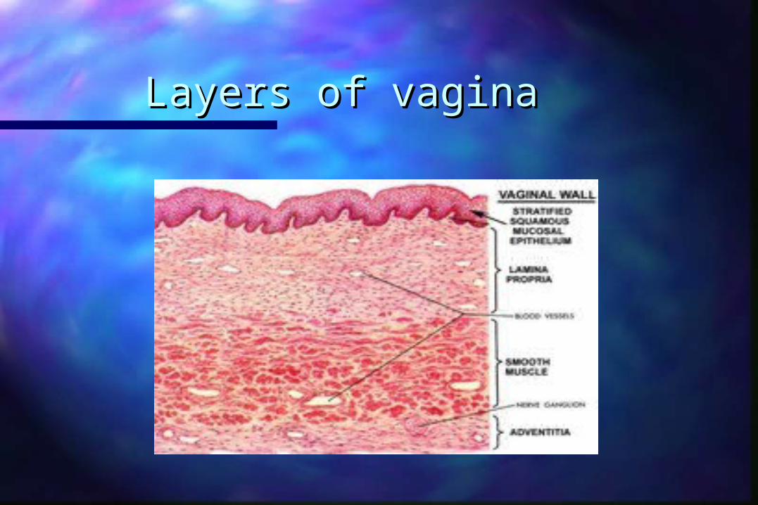
Layers of vaginaLayers of vagina

Blood supplyBlood supply

Lymphatic Drainage of VaginaLymphatic Drainage of Vagina

Lymphatics Lymphatics
Vaginal Cancer: Predisposing Vaginal Cancer: Predisposing FactorsFactors
HPV infectionHPV infection Low socioeconomic statusLow socioeconomic status History of genital wartsHistory of genital warts Vaginal discharge or irritationVaginal discharge or irritation Previously abnormal Pap smearPreviously abnormal Pap smear Early hysterectomyEarly hysterectomy Previous pelvic radiation (?)Previous pelvic radiation (?) In-utero exposure to DESIn-utero exposure to DES
Vaginal Cancer precursorsVaginal Cancer precursors
Hallmark of VAINHallmark of VAIN– cytologic atypia-Pleomorphisim, irreg nuclear cytologic atypia-Pleomorphisim, irreg nuclear
contours and chromatin clumpingcontours and chromatin clumping– Abnormal maturation Abnormal maturation
– nuclear enlargementnuclear enlargement
10-30% progress to Vaginal Ca10-30% progress to Vaginal Ca
Vaginal Cancer precursorsVaginal Cancer precursors
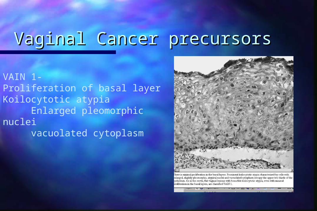
VAIN 1-Proliferation of basal layerKoilocytotic atypia
Enlarged pleomorphic nucleivacuolated cytoplasm
Vaginal Cancer precursorsVaginal Cancer precursors
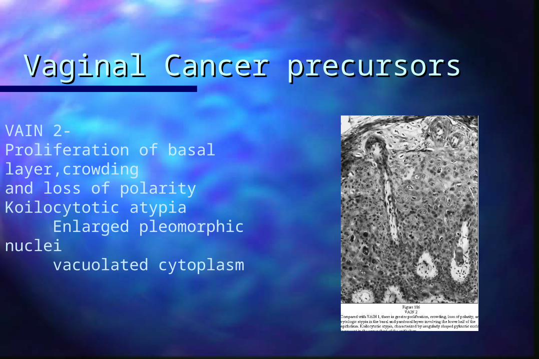
VAIN 2-Proliferation of basal layer,crowding and loss of polarityKoilocytotic atypia
Enlarged pleomorphic nucleivacuolated cytoplasm
Vaginal Cancer precursorsVaginal Cancer precursors
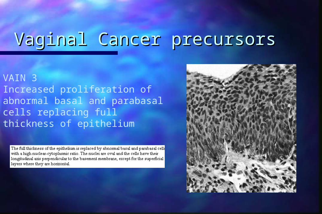
VAIN 3Increased proliferation of abnormal basal and parabasal cells replacing fullthickness of epithelium
Vaginal Cancer precursorsVaginal Cancer precursors
VAIN 3VAIN 3– usually occurs in upper third of vagina and usually occurs in upper third of vagina and
is multifocal and diffuse in half the cases.is multifocal and diffuse in half the cases.– 1/3 of patients have a hx/o CIN1/3 of patients have a hx/o CIN– CIN coexists w/ VAIN in 10-20% of ptsCIN coexists w/ VAIN in 10-20% of pts– Colposcopic findings are similar to those of Colposcopic findings are similar to those of
CIN (aceto white epithelium with CIN (aceto white epithelium with punctations and mosaic patterns)punctations and mosaic patterns)
Vaginal Cancer precursorsVaginal Cancer precursors
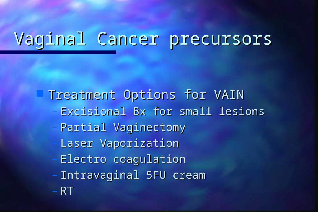
Treatment Options for VAINTreatment Options for VAIN– Excisional Bx for small lesionsExcisional Bx for small lesions– Partial VaginectomyPartial Vaginectomy– Laser VaporizationLaser Vaporization– Electro coagulationElectro coagulation– Intravaginal 5FU creamIntravaginal 5FU cream– RT RT

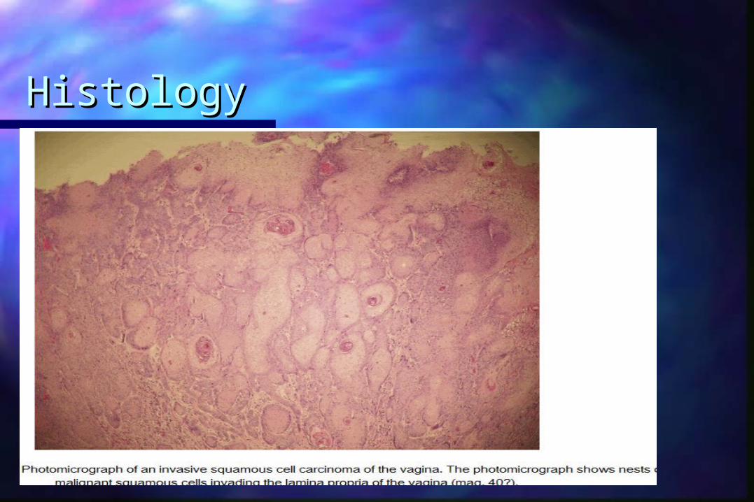
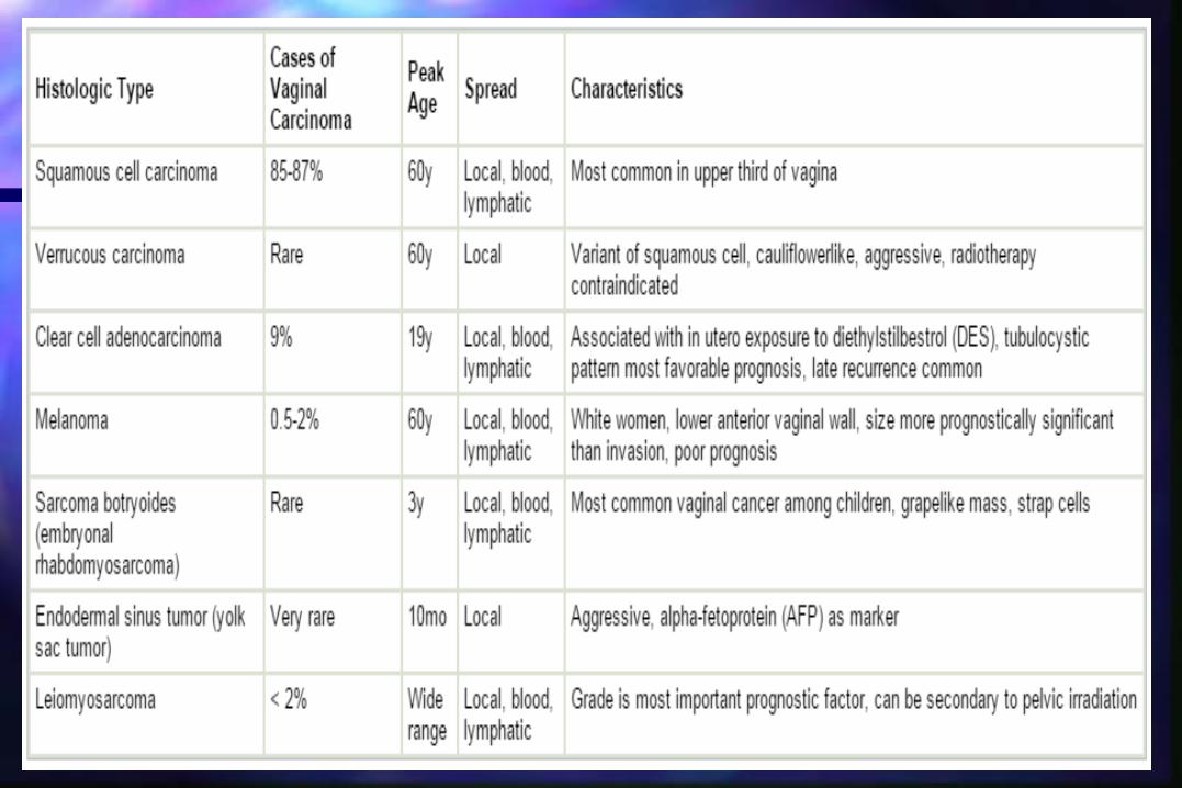
Pathology Pathology Invasive squamous cell carcinoma is found in 75% to 95% Invasive squamous cell carcinoma is found in 75% to 95%
of primary vaginal carcinomasof primary vaginal carcinomas
The majority of these lesions tend to be nonkeratinizing The majority of these lesions tend to be nonkeratinizing and moderately differentiated.and moderately differentiated.
The well-differentiated lesions may demonstrate The well-differentiated lesions may demonstrate keratinization, manifested by squamous pearls and keratinization, manifested by squamous pearls and intracellular bridgesintracellular bridges
Grossly, these tumors may manifest as nodular, ulcerated, Grossly, these tumors may manifest as nodular, ulcerated, indurated, exophytic, or endophytic lesionsindurated, exophytic, or endophytic lesions
Histology Histology

Histologically, keratinizing, nonkeratinizing, basaloid, Histologically, keratinizing, nonkeratinizing, basaloid, warty, and verrucous variants have been describedwarty, and verrucous variants have been described
Vaginal Adenosis and Vaginal Adenosis and AdenocarcinomaAdenocarcinoma
Adenocarcinoma is found in 5% to 10% of all vaginal Adenocarcinoma is found in 5% to 10% of all vaginal cancerscancers
The non–clear cell adenocarcinoma frequently arises The non–clear cell adenocarcinoma frequently arises in the submucosa. in the submucosa.
When a biopsy of a vaginal lesion reveals When a biopsy of a vaginal lesion reveals adenocarcinoma, it is important to look for a primary adenocarcinoma, it is important to look for a primary lesion, such as endometrial cancer, elsewherelesion, such as endometrial cancer, elsewhere
Vaginal adenosis defines the abnormal presence of Vaginal adenosis defines the abnormal presence of glandular epithelium in the vagina, which is normally glandular epithelium in the vagina, which is normally devoid of glandular elementsdevoid of glandular elements
The glandular epithelial cells may line glands in the The glandular epithelial cells may line glands in the submucosa or cover or replace surface squamous submucosa or cover or replace surface squamous cells and are usually located near the surface cells and are usually located near the surface epitheliumepithelium
adenosis is the most common histological adenosis is the most common histological abnormality in women exposed to DES in utero, it is abnormality in women exposed to DES in utero, it is not strictly confined to this populationnot strictly confined to this population
The classic gross appearance of adenosis is red, The classic gross appearance of adenosis is red, velvety, grapelike clusters in the vaginavelvety, grapelike clusters in the vagina
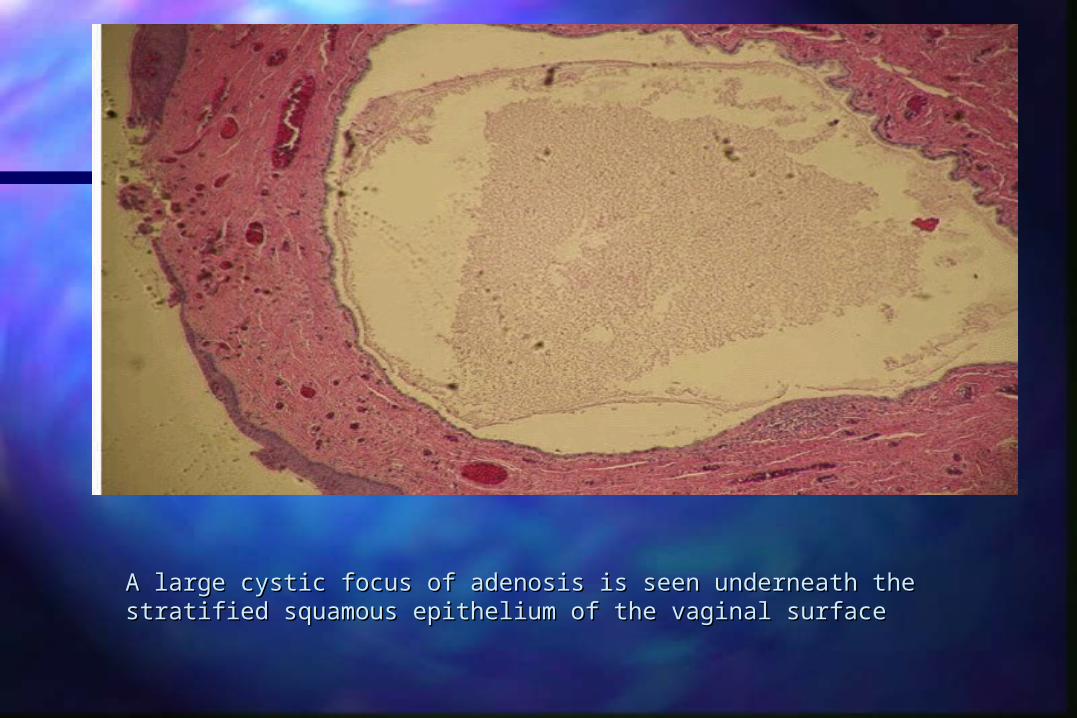
A large cystic focus of adenosis is seen underneath the stratified A large cystic focus of adenosis is seen underneath the stratified squamous epithelium of the vaginal surface squamous epithelium of the vaginal surface

Adenosis is associated with 97% of vaginal clear cell Adenosis is associated with 97% of vaginal clear cell cancercancer
DES-associated CCA has a predilection for the upper DES-associated CCA has a predilection for the upper third of the vagina and the ectocervixthird of the vagina and the ectocervix
The most common histologic pattern is tubulocystic The most common histologic pattern is tubulocystic followed by a solid pattern. followed by a solid pattern.
The most common cells noted are the clear cell, The most common cells noted are the clear cell, hobnail cell, and endometrioid cellhobnail cell, and endometrioid cell
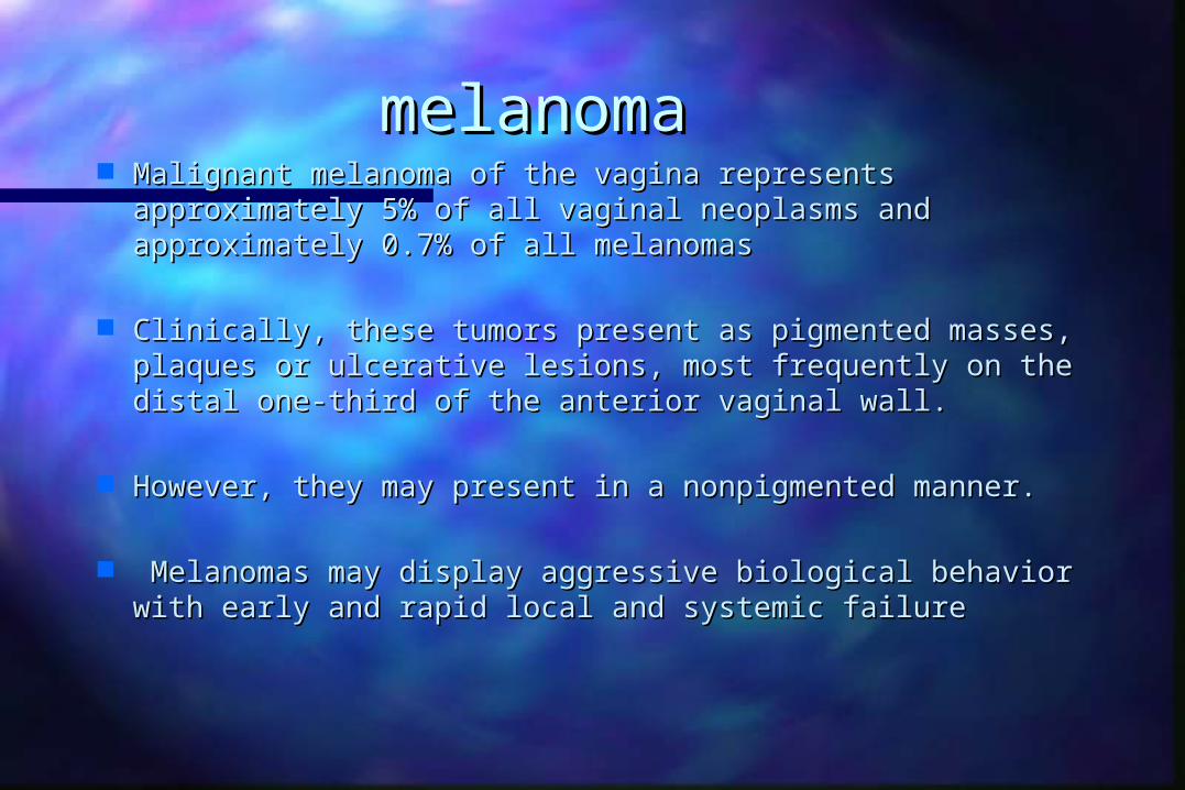
melanomamelanoma Malignant melanoma of the vagina represents Malignant melanoma of the vagina represents
approximately 5% of all vaginal neoplasms and approximately 5% of all vaginal neoplasms and approximately 0.7% of all melanomasapproximately 0.7% of all melanomas
Clinically, these tumors present as pigmented masses, Clinically, these tumors present as pigmented masses, plaques or ulcerative lesions, most frequently on the plaques or ulcerative lesions, most frequently on the distal one-third of the anterior vaginal wall. distal one-third of the anterior vaginal wall.
However, they may present in a nonpigmented manner.However, they may present in a nonpigmented manner.
Melanomas may display aggressive biological behavior Melanomas may display aggressive biological behavior with early and rapid local and systemic failure with early and rapid local and systemic failure
Sarcoma Sarcoma
LeiomyosarcomasLeiomyosarcomas endometrial stromal sarcomasendometrial stromal sarcomas malignant mixed mullerian tumorsmalignant mixed mullerian tumors rhabdomyosarcomas are the major types of primary rhabdomyosarcomas are the major types of primary
vaginal sarcomasvaginal sarcomas
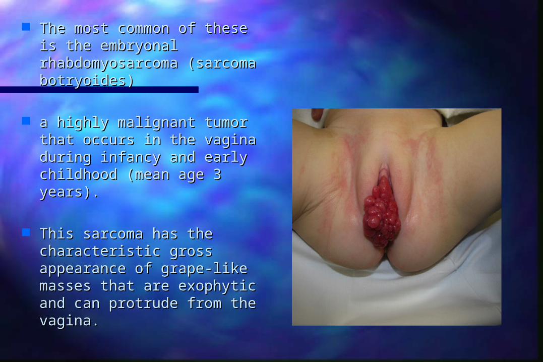
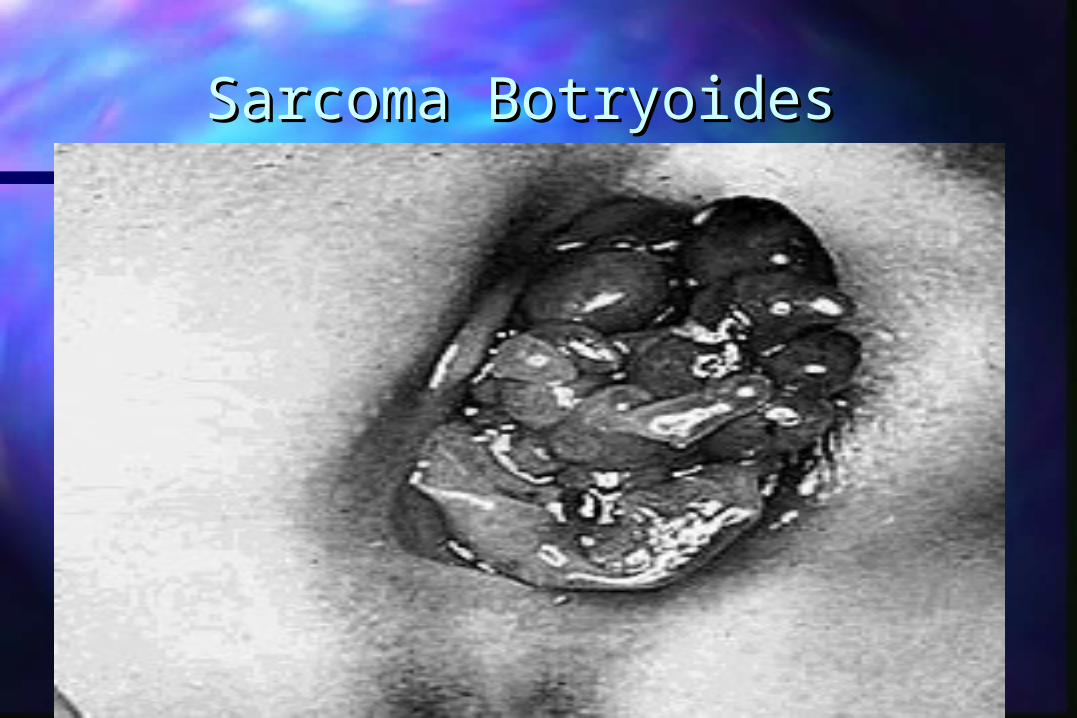
The most common of these The most common of these is the embryonal is the embryonal rhabdomyosarcoma rhabdomyosarcoma (sarcoma botryoides)(sarcoma botryoides)
a highly malignant tumor a highly malignant tumor that occurs in the vagina that occurs in the vagina during infancy and early during infancy and early childhood (mean age 3 childhood (mean age 3 years). years).
This sarcoma has the This sarcoma has the characteristic gross characteristic gross appearance of grape-like appearance of grape-like masses that are exophytic masses that are exophytic and can protrude from the and can protrude from the vagina. vagina.
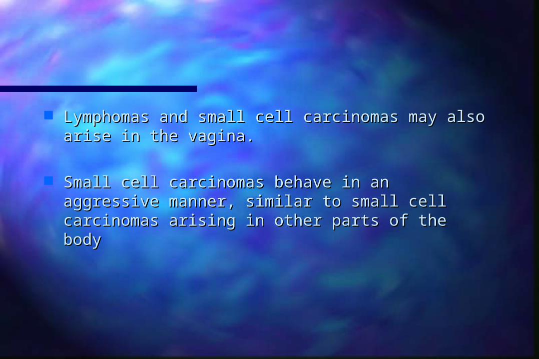
Lymphomas and small cell carcinomas may also Lymphomas and small cell carcinomas may also arise in the vagina. arise in the vagina.
Small cell carcinomas behave in an aggressive Small cell carcinomas behave in an aggressive manner, similar to small cell carcinomas arising in manner, similar to small cell carcinomas arising in other parts of the bodyother parts of the body
Natural History and Patterns Natural History and Patterns of Spreadof Spread
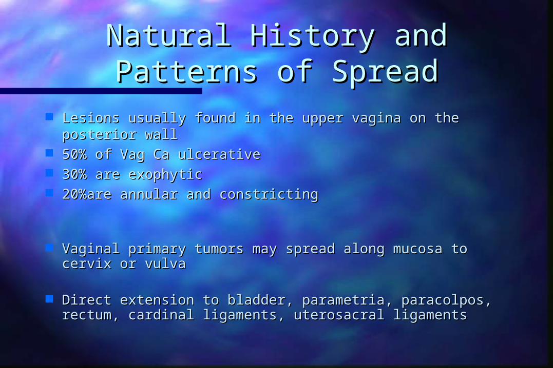
Lesions usually found in the upper vagina on the posterior Lesions usually found in the upper vagina on the posterior wallwall
50% of Vag Ca ulcerative50% of Vag Ca ulcerative 30% are exophytic 30% are exophytic 20%are annular and constricting20%are annular and constricting
Vaginal primary tumors may spread along mucosa to cervix Vaginal primary tumors may spread along mucosa to cervix or vulvaor vulva
Direct extension to bladder, parametria, paracolpos, rectum, Direct extension to bladder, parametria, paracolpos, rectum, cardinal ligaments, uterosacral ligamentscardinal ligaments, uterosacral ligaments

Natural History and Patterns Natural History and Patterns of Spreadof Spread
Any of the nodal groups may be involved regardless Any of the nodal groups may be involved regardless of the location of the tumorof the location of the tumor
Inguinal nodes most often involved if lesion is in the Inguinal nodes most often involved if lesion is in the lower 1/3 of the vaginalower 1/3 of the vagina
Clinically apparent inguinal node mets seen in 5-20% Clinically apparent inguinal node mets seen in 5-20% of patientsof patients
Incidence of pelvic nodes varies with stage and Incidence of pelvic nodes varies with stage and location of the tumorlocation of the tumor

Clinical PresentationClinical Presentation
Abnormal vaginal bleedingAbnormal vaginal bleeding– 50-75% of patients with primary tumors50-75% of patients with primary tumors
Discharge Discharge
DysuriaDysuria
PainPain

Diagnostic Work-upDiagnostic Work-up
Complete history and physicalComplete history and physical
Speculum examination and palpation of the vaginaSpeculum examination and palpation of the vagina
Bimanual pelvic and rectovaginal examinationBimanual pelvic and rectovaginal examination
Pap smear, colposcopy, directed biopsiesPap smear, colposcopy, directed biopsies

Diagnostic Work-upDiagnostic Work-up
CystoscopyCystoscopy ProctosigmoidoscopyProctosigmoidoscopy Chest X-rayChest X-ray IVPIVP Barium enemaBarium enema Computed TomographyComputed Tomography MRIMRI
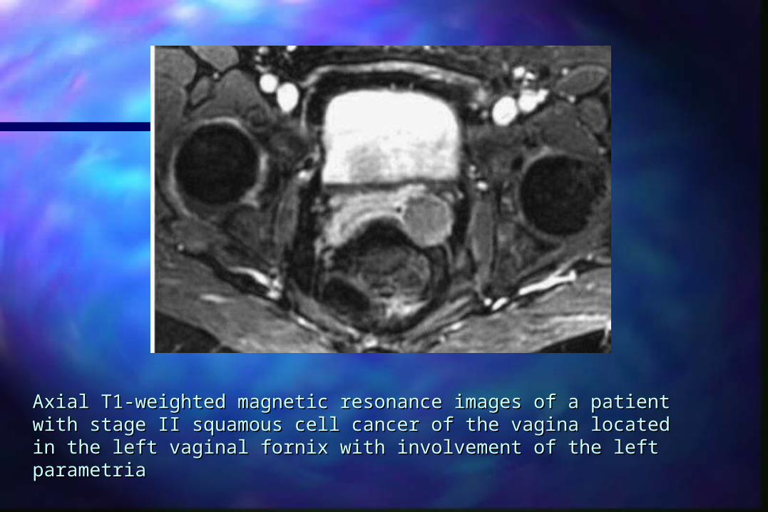
Axial T1-weighted magnetic resonance images of a patient with stage Axial T1-weighted magnetic resonance images of a patient with stage II squamous cell cancer of the vagina located in the left vaginal fornix II squamous cell cancer of the vagina located in the left vaginal fornix with involvement of the left parametriawith involvement of the left parametria
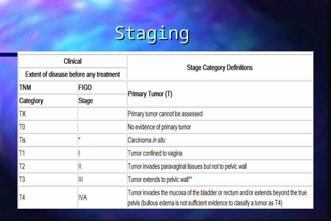
StagingStaging


5 Year Survival 5 Year Survival
0
10
20
30
40
50
60
70
80
Stage I Stage I I Stage I I I Stage IV

Natural History and Patterns Natural History and Patterns of Failureof Failure
Stage IStage I– 10-20% pelvic recurrence, 10-20% distant10-20% pelvic recurrence, 10-20% distant
Stage IIStage II– 35% pelvic recurrence, 22% distant 35% pelvic recurrence, 22% distant
Stage IIIStage III– 25-45% pelvic recurrence, 23% distant25-45% pelvic recurrence, 23% distant
Stage IVStage IV– 58% pelvic recurrence, 30% distant58% pelvic recurrence, 30% distant
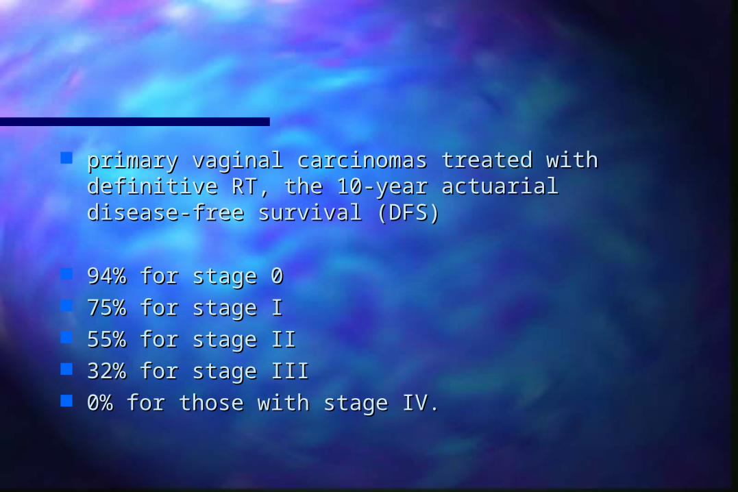
primary vaginal carcinomas treated with definitive primary vaginal carcinomas treated with definitive RT, the 10-year actuarial disease-free survival (DFS)RT, the 10-year actuarial disease-free survival (DFS)
94% for stage 094% for stage 0 75% for stage I75% for stage I 55% for stage II55% for stage II 32% for stage III32% for stage III 0% for those with stage IV.0% for those with stage IV.
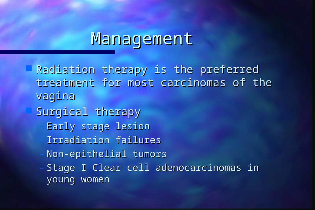
ManagementManagement
Radiation therapy is the preferred Radiation therapy is the preferred treatment for most carcinomas of the treatment for most carcinomas of the vaginavagina
Surgical therapySurgical therapy– Early stage lesionEarly stage lesion– Irradiation failuresIrradiation failures– Non-epithelial tumorsNon-epithelial tumors– Stage I Clear cell adenocarcinomas in young Stage I Clear cell adenocarcinomas in young
womenwomen

ManagementManagement SurgerySurgery
– Wide local excision reserved for carcinoma insitu or Wide local excision reserved for carcinoma insitu or small superficially invasive lesions that r well small superficially invasive lesions that r well demarcateddemarcated
– Stage I tumors of the middle or upper third of vagina Stage I tumors of the middle or upper third of vagina treated with radical hysterovaginectomy and PLNDtreated with radical hysterovaginectomy and PLND
– Stage I tumors of the lower third of vagina which may Stage I tumors of the lower third of vagina which may encroach on the vulva treated with radical encroach on the vulva treated with radical vulvovaginectomy and bilat. groin node dissectionvulvovaginectomy and bilat. groin node dissection
– Pelvic exenteration possible for more invasive lesionsPelvic exenteration possible for more invasive lesions
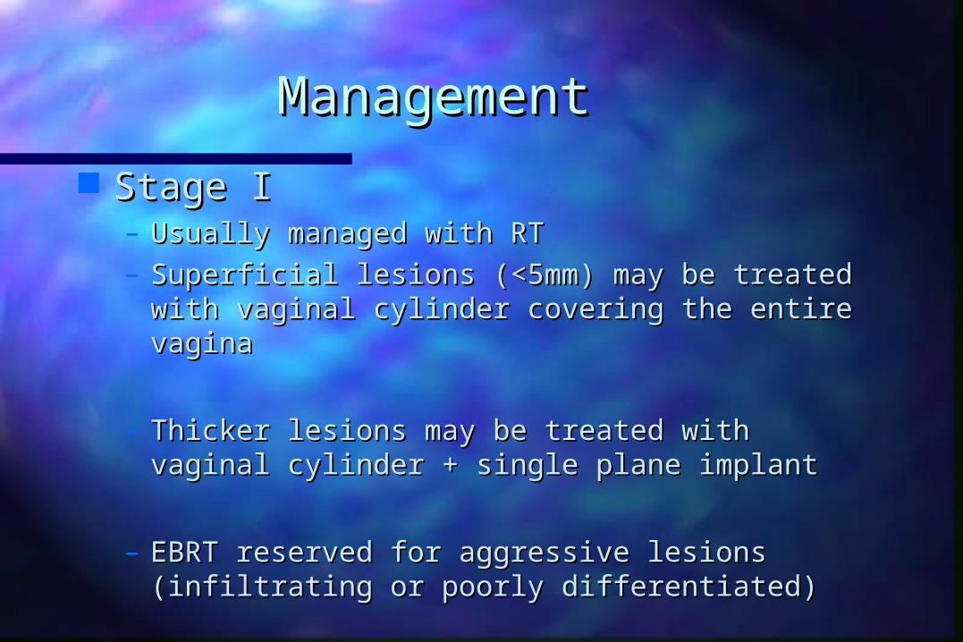
ManagementManagement
Stage IStage I– Usually managed with RTUsually managed with RT– Superficial lesions (<5mm) may be treated with Superficial lesions (<5mm) may be treated with
vaginal cylinder covering the entire vaginavaginal cylinder covering the entire vagina
– Thicker lesions may be treated with vaginal Thicker lesions may be treated with vaginal cylinder + single plane implant cylinder + single plane implant
– EBRT reserved for aggressive lesions (infiltrating EBRT reserved for aggressive lesions (infiltrating or poorly differentiated)or poorly differentiated)
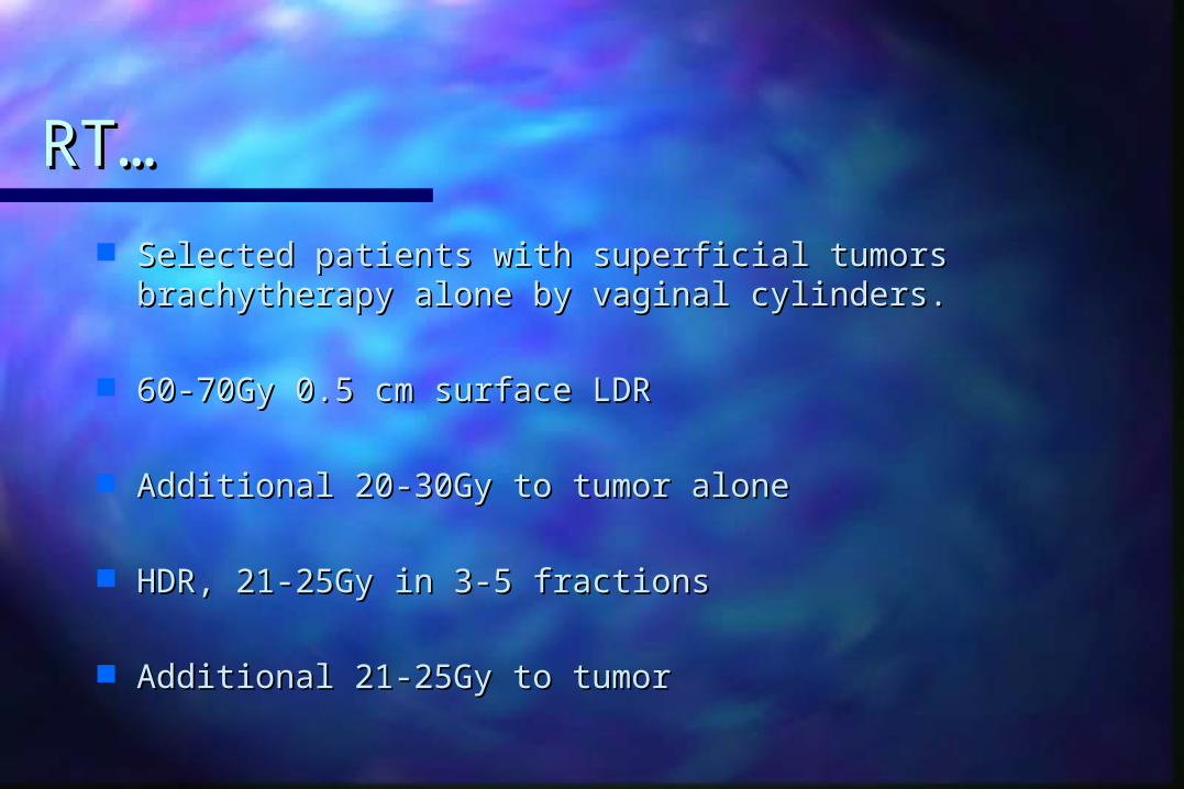
RT…RT…
Selected patients with superficial tumors Selected patients with superficial tumors brachytherapy alone by vaginal cylinders.brachytherapy alone by vaginal cylinders.
60-70Gy 0.5 cm surface LDR60-70Gy 0.5 cm surface LDR
Additional 20-30Gy to tumor aloneAdditional 20-30Gy to tumor alone
HDR, 21-25Gy in 3-5 fractions HDR, 21-25Gy in 3-5 fractions
Additional 21-25Gy to tumor Additional 21-25Gy to tumor

RT..RT..
Combination of ICA and IBT in lesions thicker than Combination of ICA and IBT in lesions thicker than 5mm5mm
Vaginal cylinder delivers 45Gy LDR or 21-25Gy by Vaginal cylinder delivers 45Gy LDR or 21-25Gy by HDR 0.5 cm vaginal mucosaHDR 0.5 cm vaginal mucosa
Additional therapy with interstitial BT to tumor Additional therapy with interstitial BT to tumor volumevolume
25-35Gy LDR25-35Gy LDR

Stage 1 RT..Stage 1 RT..
Combination of EBRT n BT for more aggressive stage Combination of EBRT n BT for more aggressive stage 1 with greater infiltration and poor differentiation1 with greater infiltration and poor differentiation
Recent trend towards combination Recent trend towards combination
Possible under estimation of submucosal disease or Possible under estimation of submucosal disease or nodal statusnodal status

Stage 2Stage 2
Radiation is the primary optionRadiation is the primary option
EBRT + BTEBRT + BT
EBRT 45-50.4GyEBRT 45-50.4Gy
Boost to tumor volume with BT to total dose of 75-Boost to tumor volume with BT to total dose of 75-80Gy80Gy
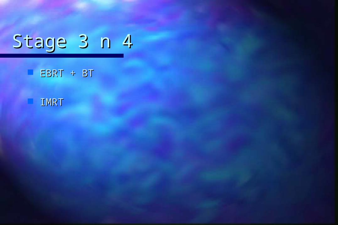
Stage 3 n 4Stage 3 n 4
EBRT + BTEBRT + BT
IMRT IMRT
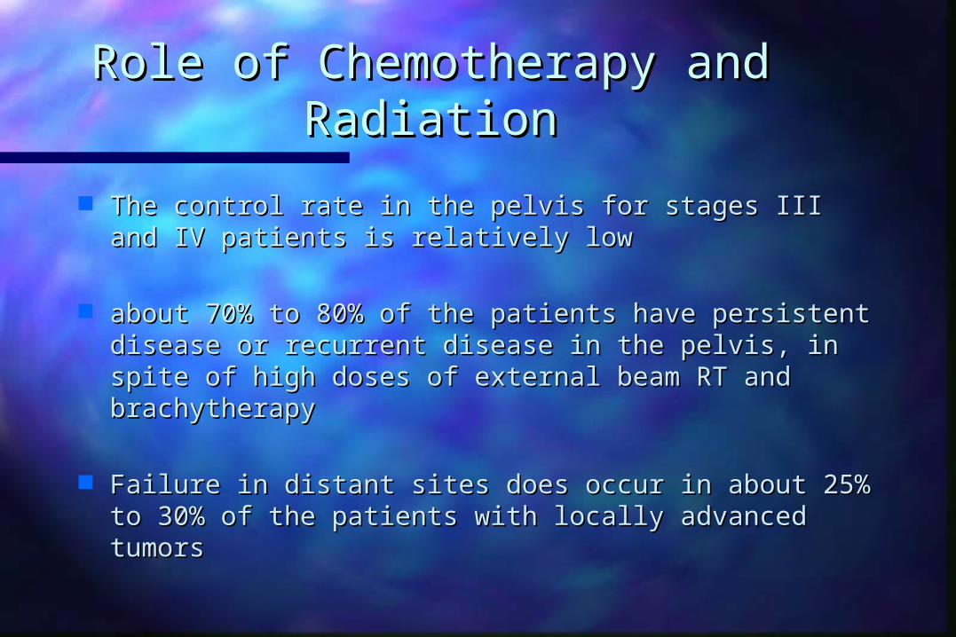
Role of Chemotherapy and Role of Chemotherapy and RadiationRadiation
The control rate in the pelvis for stages III and IV The control rate in the pelvis for stages III and IV patients is relatively lowpatients is relatively low
about 70% to 80% of the patients have persistent about 70% to 80% of the patients have persistent disease or recurrent disease in the pelvis, in spite of disease or recurrent disease in the pelvis, in spite of high doses of external beam RT and brachytherapyhigh doses of external beam RT and brachytherapy
Failure in distant sites does occur in about 25% to Failure in distant sites does occur in about 25% to 30% of the patients with locally advanced tumors30% of the patients with locally advanced tumors

Therefore, there is a need for better approaches to Therefore, there is a need for better approaches to the management of advanced disease such as the the management of advanced disease such as the use of concomitant chemoradiotherapyuse of concomitant chemoradiotherapy
Agents such as 5-FU, mitomycin-C, and cisplatin Agents such as 5-FU, mitomycin-C, and cisplatin have shown promise when combined with RThave shown promise when combined with RT
Advanced cervical cancer has improvement in Advanced cervical cancer has improvement in locoregional control, overall survival, and disease-locoregional control, overall survival, and disease-free survival for patients receiving cisplatin-based free survival for patients receiving cisplatin-based chemotherapy concurrently with RTchemotherapy concurrently with RT
This was interpolated in to therapy of vaginal cancer. This was interpolated in to therapy of vaginal cancer.

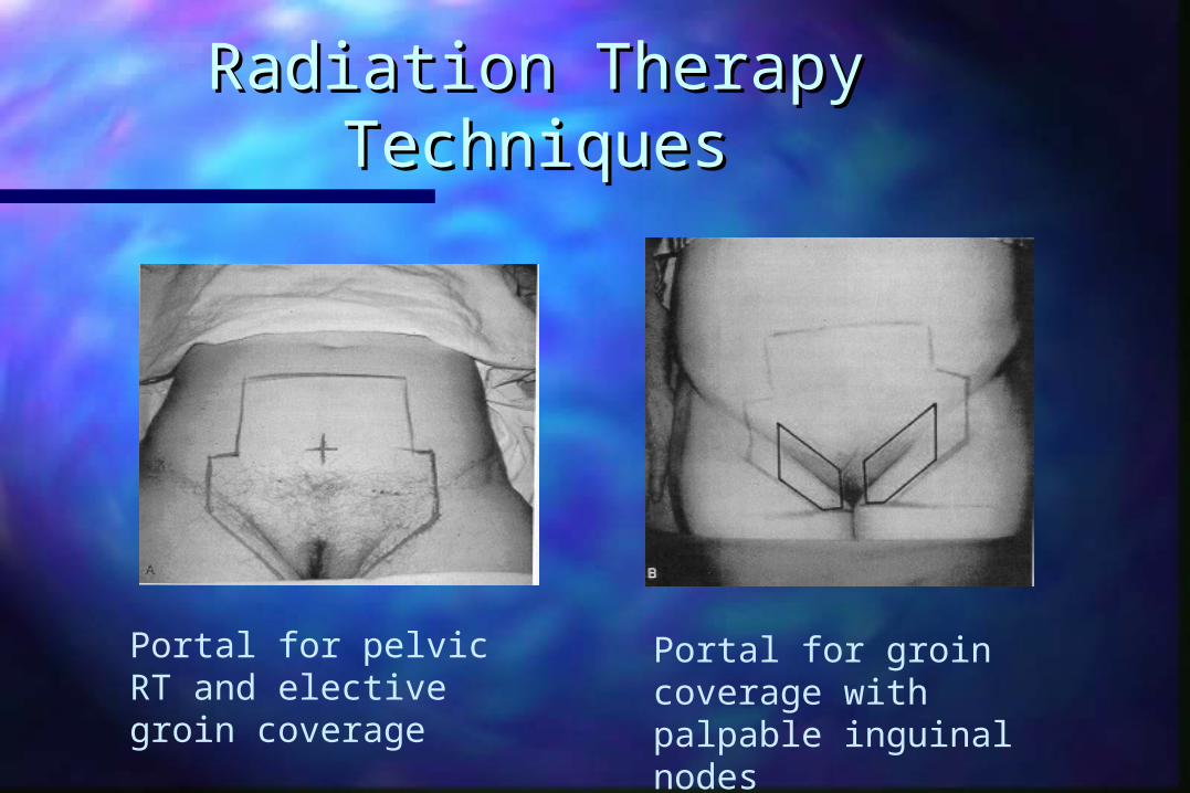
Radiation Therapy TechniquesRadiation Therapy Techniques
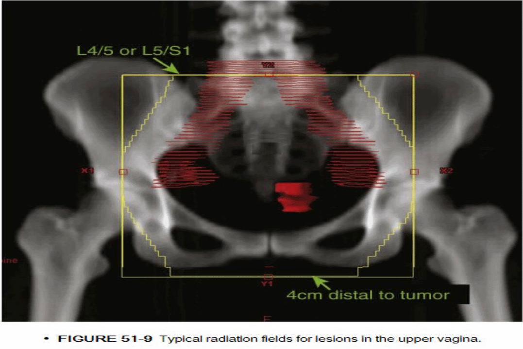
EBRT delivered through AP:PA portals or using 4 field EBRT delivered through AP:PA portals or using 4 field “box technique”“box technique”
It should ensure coverage of vagina common illiac, It should ensure coverage of vagina common illiac, external illiac, hypogastric, and obturator nodeexternal illiac, hypogastric, and obturator node
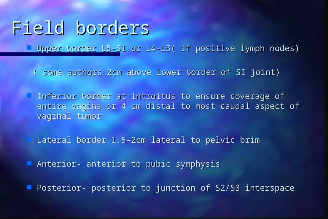
Field bordersField borders Upper border L5-S1 or L4-L5( if positive lymph nodes)Upper border L5-S1 or L4-L5( if positive lymph nodes)
( some authors 2cm above lower border of SI joint)( some authors 2cm above lower border of SI joint)
Inferior border at introitus to ensure coverage of entire Inferior border at introitus to ensure coverage of entire vagina or 4 cm distal to most caudal aspect of vaginal vagina or 4 cm distal to most caudal aspect of vaginal tumortumor
Lateral border 1.5-2cm lateral to pelvic brimLateral border 1.5-2cm lateral to pelvic brim
Anterior- anterior to pubic symphysisAnterior- anterior to pubic symphysis
Posterior- posterior to junction of S2/S3 interspace Posterior- posterior to junction of S2/S3 interspace

Inguinal nodes should be electively covered (4500-Inguinal nodes should be electively covered (4500-5000cGy) for tumors of the lower 1/3 of vagina5000cGy) for tumors of the lower 1/3 of vagina
Additional 1500cGy (4-5cm depth) delivered for Additional 1500cGy (4-5cm depth) delivered for palpable inguinal nodespalpable inguinal nodes
Radiation Therapy TechniquesRadiation Therapy Techniques
Portal for pelvic RT and elective groin coverage
Portal for groin coverage with palpable inguinal nodes

Portal to include inguinal nodesPortal to include inguinal nodes

EBRT doseEBRT dose
45-50.4 Gy IN 25-28 fractions45-50.4 Gy IN 25-28 fractions
Boost depend on size n site of lesionBoost depend on size n site of lesion
Vaginal apex > 5mm EBRT OR IBT boostVaginal apex > 5mm EBRT OR IBT boost
< 5mm - ICA< 5mm - ICA

Mid and distal s treated with IBT Mid and distal s treated with IBT
If extensive disease treated with EBRTIf extensive disease treated with EBRT
Total dose of 70-80GyTotal dose of 70-80Gy

Brachytherapy Brachytherapy
Intracavitary brachytherapyIntracavitary brachytherapy
VAIN and highly selective minimally invasiveVAIN and highly selective minimally invasive
Boost after EBRT lesion < 5mm total dose 70-80GyBoost after EBRT lesion < 5mm total dose 70-80Gy
LDR or HDRLDR or HDR
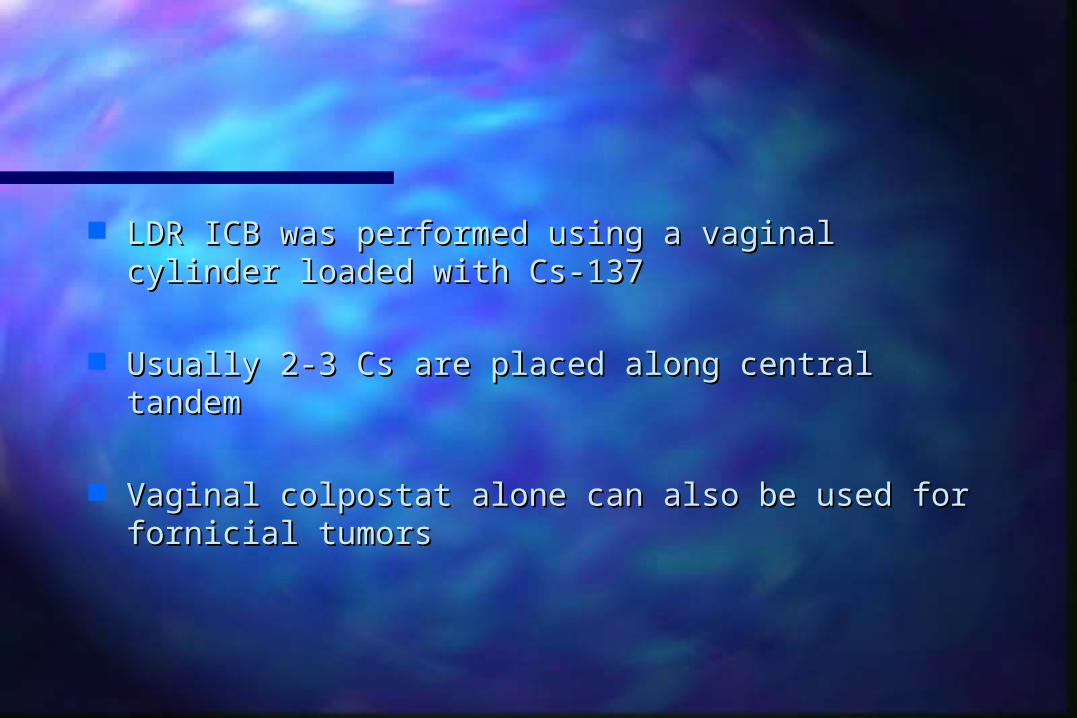
LDR ICB was performed using a vaginal cylinder LDR ICB was performed using a vaginal cylinder loaded with Cs-137loaded with Cs-137
Usually 2-3 Cs are placed along central tandemUsually 2-3 Cs are placed along central tandem
Vaginal colpostat alone can also be used for fornicial Vaginal colpostat alone can also be used for fornicial tumorstumors

HDR-ICB is typically performed using Ir-192 HDR-ICB is typically performed using Ir-192
EBRT 45-50.4Gy followed by HDR 20-28Gy in 3-4 EBRT 45-50.4Gy followed by HDR 20-28Gy in 3-4 fractionsfractions

Applicators Applicators

Applicators..Applicators..
Shielded Cylindrical ApplicatorShielded Cylindrical Applicator

Interstitial brachytherapyInterstitial brachytherapy
ICB not suitable > 5mm ICB not suitable > 5mm
Vaginal cylinder fails to deliver sufficient coverage to Vaginal cylinder fails to deliver sufficient coverage to paravaginal tissueparavaginal tissue
Boost after EBRTBoost after EBRT

IBT..IBT.. Routine preoperative assessment under anaesthesia to Routine preoperative assessment under anaesthesia to
assess diseaseassess disease
If MRI is available it s superior to assess the thickness If MRI is available it s superior to assess the thickness
Patient is positioned in dorsal lithotomy positionPatient is positioned in dorsal lithotomy position
A speculam and digital examination allows assessment A speculam and digital examination allows assessment of vaginal width, tumor size and location, amount and of vaginal width, tumor size and location, amount and thickness of residual parametrial or paravaginal diseasethickness of residual parametrial or paravaginal disease

IBT..IBT..
A sterile set up is used at time of insertionA sterile set up is used at time of insertion
A foley catheter is placed for bladder drainageA foley catheter is placed for bladder drainage
Radio opaque markers can be kept for tumor Radio opaque markers can be kept for tumor delineationdelineation

Templates..Templates..
Template systems are available to secure the Template systems are available to secure the position of needles in the target volumesposition of needles in the target volumes
Syed- Neblette Syed- Neblette
Modified Syed- Neblette Modified Syed- Neblette
Martinez-Universal Perineal ImplantMartinez-Universal Perineal Implant

IBT..IBT..
These system consist of a perineal template, a These system consist of a perineal template, a vaginal cylinder obturator, and hollow guides for vaginal cylinder obturator, and hollow guides for loading radionuclide sources.loading radionuclide sources.
So through opening in the template needles can be So through opening in the template needles can be insertedinserted
Goal is to cover GTV with 1-2 cm marginGoal is to cover GTV with 1-2 cm margin


IBT..IBT..
It s optimal to place needle under image guidanceIt s optimal to place needle under image guidance
Laparoscopic. CT, USG, MRILaparoscopic. CT, USG, MRI
TRUSTRUS
With MRI n CT 3D Image based brachytherapy can be With MRI n CT 3D Image based brachytherapy can be donedone
Increasing tumor control and decreasing toxicityIncreasing tumor control and decreasing toxicity

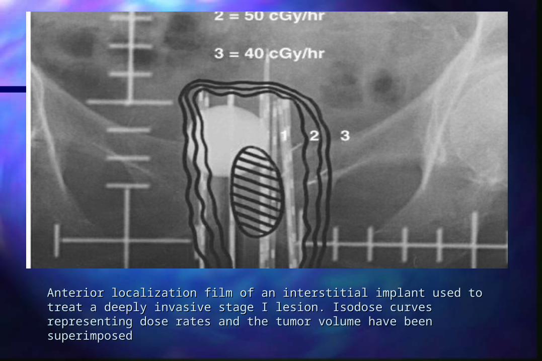
Anterior localization film of an interstitial implant used to treat a deeply Anterior localization film of an interstitial implant used to treat a deeply invasive stage I lesion. Isodose curves representing dose rates and the invasive stage I lesion. Isodose curves representing dose rates and the tumor volume have been superimposedtumor volume have been superimposed
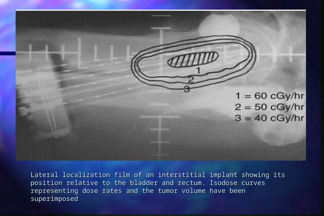
Lateral localization film of an interstitial implant showing its position Lateral localization film of an interstitial implant showing its position relative to the bladder and rectum. Isodose curves representing dose relative to the bladder and rectum. Isodose curves representing dose rates and the tumor volume have been superimposedrates and the tumor volume have been superimposed

TRUS GUIDEDTRUS GUIDED
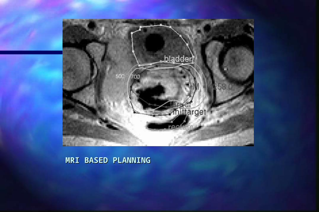
MRI BASED PLANNINGMRI BASED PLANNING

IBTIBT
HDR is preferredHDR is preferred
Limiting exposure to care givers and ability to Limiting exposure to care givers and ability to optimize dose distribution by 3D image based optimize dose distribution by 3D image based planningplanning
Permanent implant with Au-198 n I-125 reported in Permanent implant with Au-198 n I-125 reported in elderly patientselderly patients
Management of rare Management of rare histologieshistologies
Small Cell CarcinomaSmall Cell Carcinoma
Poor prognosis (85% die in first year)Poor prognosis (85% die in first year)– Reasonable local control may be obtained with surgery Reasonable local control may be obtained with surgery
or irradiation followed by systemic chemoor irradiation followed by systemic chemo
– EPEP
– Cyclophosphamide, Adriamycin, Vincristine (CAV) X 12 Cyclophosphamide, Adriamycin, Vincristine (CAV) X 12 cycles (some prior to initiation of RT)cycles (some prior to initiation of RT)
– Doses of RT similar to SCCADoses of RT similar to SCCA
ManagementManagement
RhabdomyosarcomaRhabdomyosarcoma– Generally treated with a combination of surgery, Generally treated with a combination of surgery,
RT, and chemotherapyRT, and chemotherapy
– Vincristine, Dactinomycin, Cyclophosphamide Vincristine, Dactinomycin, Cyclophosphamide (VAC) X 1-2 years effective adjuvant treatment for (VAC) X 1-2 years effective adjuvant treatment for stage 1 dzstage 1 dz
– Local excision + interstitial/intracavitary RT + Local excision + interstitial/intracavitary RT + systemic chemo has replaced radical pelvic systemic chemo has replaced radical pelvic surgery as therapy of choicesurgery as therapy of choice
Sarcoma BotryoidesSarcoma Botryoides

Sarcoma BotryoidesSarcoma Botryoides
Strap cell
ManagementManagement
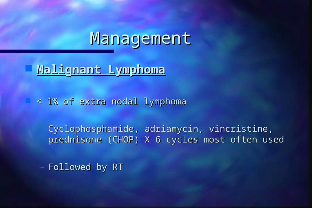
Malignant LymphomaMalignant Lymphoma
< 1% of extra nodal lymphoma< 1% of extra nodal lymphoma
– Cyclophosphamide, adriamycin, vincristine, Cyclophosphamide, adriamycin, vincristine, prednisone (CHOP) X 6 cycles most often usedprednisone (CHOP) X 6 cycles most often used
– Followed by RTFollowed by RT

Clear Cell Adenocarcinoma Clear Cell Adenocarcinoma and DES Exposureand DES Exposure
Incidence is between 0.14 to 1.4/1000 women Incidence is between 0.14 to 1.4/1000 women exposed to DESexposed to DES
Median age at diagnosis 19 yearsMedian age at diagnosis 19 years
Lesions found mainly in the upper 1/3 of the anterior Lesions found mainly in the upper 1/3 of the anterior vaginal wallvaginal wall
90% of patients with early stage disease (I and II) at 90% of patients with early stage disease (I and II) at diagnosisdiagnosis

ManagementManagement Clear Cell AdenocarcinomaClear Cell Adenocarcinoma
– Surgery for stage I lesions has advantage of ovarian Surgery for stage I lesions has advantage of ovarian preservation and better vaginal function following preservation and better vaginal function following skin graftskin graft
– Vaginectomy, radical hysterectomy PLND, paraaortic Vaginectomy, radical hysterectomy PLND, paraaortic LNBx (frozen section of distal margin)LNBx (frozen section of distal margin)
– Intracavitary or transvaginal radiation can be used Intracavitary or transvaginal radiation can be used for small lesionsfor small lesions
– More extensive lesions: EBRTMore extensive lesions: EBRT

Clear cell adenocarcinomaClear cell adenocarcinoma
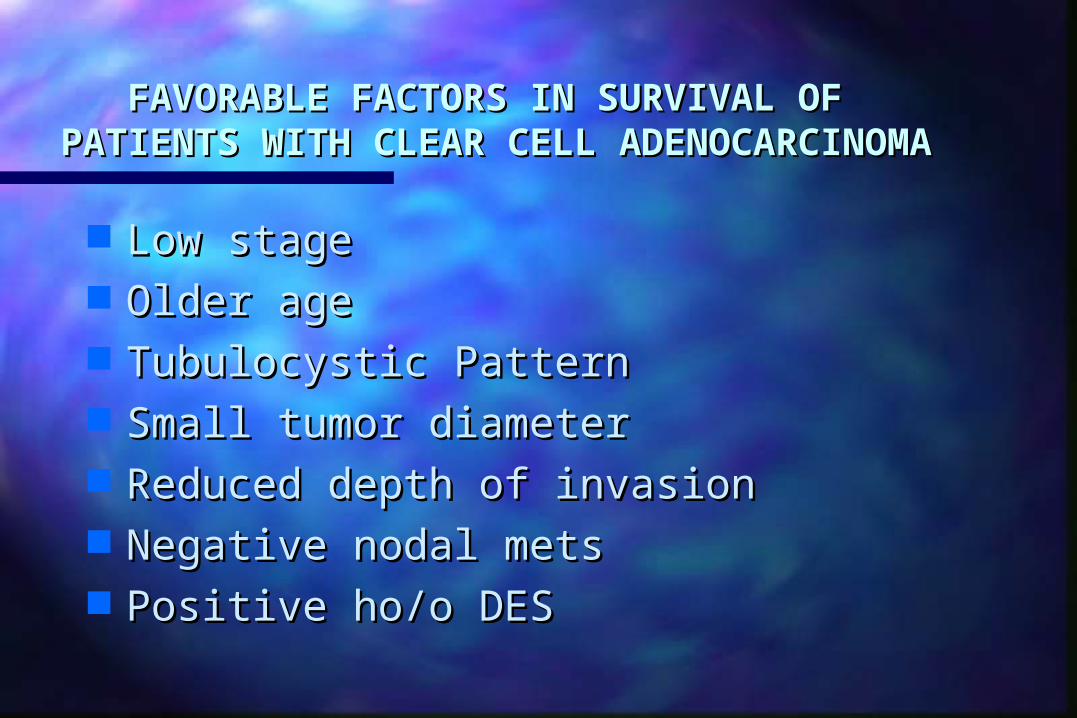
FAVORABLE FACTORS IN SURVIVAL OFFAVORABLE FACTORS IN SURVIVAL OF PATIENTS WITH CLEAR CELL PATIENTS WITH CLEAR CELL
ADENOCARCINOMAADENOCARCINOMA
Low stageLow stage Older ageOlder age Tubulocystic PatternTubulocystic Pattern Small tumor diameterSmall tumor diameter Reduced depth of invasionReduced depth of invasion Negative nodal metsNegative nodal mets Positive ho/o DESPositive ho/o DES

Melanoma Melanoma
Wide local excisionWide local excision
Radical surgeryRadical surgery
Radiation Radiation
Overall survival s poor 5-20%Overall survival s poor 5-20%

SummarySummary Superficial stage I lesions may be treated with RT or Superficial stage I lesions may be treated with RT or
radical hysterovaginectomyradical hysterovaginectomy
Stage II-IVA treated with WPRT and brachytherapyStage II-IVA treated with WPRT and brachytherapy
Role of chemotherapy in advanced SCCA presently Role of chemotherapy in advanced SCCA presently unknown unknown
Pelvic failures and distant metastases occur in 1/2 of Pelvic failures and distant metastases occur in 1/2 of pts with advanced dzpts with advanced dz

The EndThe End