btca, a class ia type iii chaperone, interacts with the ... · homology modeling server (...
TRANSCRIPT

BtcA, A Class IA Type III Chaperone, Interacts with theBteA N-Terminal Domain through a Globular/Non-Globular MechanismChen Guttman1, Geula Davidov1, Adi Yahalom4, Hadassa Shaked4, Sofiya Kolusheva2, Ronit Bitton2,3,
Shiran Barber-Zucker1, Jordan H. Chill4*, Raz Zarivach1*
1 Departments of Life Sciences and the National Institute for Biotechnology in the Negev (NIBN), Ben-Gurion University of the Negev, Be’er Sheva, Israel, 2 Ilse Katz
Institute for Nanoscale Science and Technology, Ben-Gurion University of the Negev, Be’er Sheva, Israel, 3 Department of Chemical Engineering, Ben Ben-Gurion University
of the Negev, Be’er Sheva, Israel, 4 Department of Chemistry, Bar Ilan University, Ramat Gan, Israel
Abstract
Bordetella pertussis, the etiological agent of ‘‘whooping cough’’ disease, utilizes the type III secretion system (T3SS) to delivera 69 kDa cytotoxic effector protein, BteA, directly into the host cells. As with other T3SS effectors, prior to its secretion BteAbinds BtcA, a 13.9 kDa protein predicted to act as a T3SS class IA chaperone. While this interaction had been characterizedfor such effector-chaperone pairs in other pathogens, it has yet to be fully investigated in Bordetella. Here we provide thefirst biochemical proof that BtcA is indeed a class IA chaperone, responsible for the binding of BteA’s N-terminal domain. Webring forth extensive evidence that BtcA binds its substrate effector through a dual-interface binding mechanismcomprising of non-globular and bi-globular interactions at a moderate micromolar level binding affinity. We demonstratethat the non-globular interactions involve the first 31 N-terminal residues of BteA287 and their removal leads todestabilization of the effector-chaperone complex and lower binding affinities to BtcA. These findings represent animportant first step towards a molecular understanding of BteA secretion and cell entry.
Citation: Guttman C, Davidov G, Yahalom A, Shaked H, Kolusheva S, et al. (2013) BtcA, A Class IA Type III Chaperone, Interacts with the BteA N-Terminal Domainthrough a Globular/Non-Globular Mechanism. PLoS ONE 8(12): e81557. doi:10.1371/journal.pone.0081557
Editor: David M. Ojcius, University of California Merced, United States of America
Received July 31, 2013; Accepted October 23, 2013; Published December 2, 2013
Copyright: � 2013 Guttman et al. This is an open-access article distributed under the terms of the Creative Commons Attribution License, which permitsunrestricted use, distribution, and reproduction in any medium, provided the original author and source are credited.
Funding: The research leading to these results has received funding from the EU Seventh Framework Programme (FP7/2007–2013 under grant agreementu239182 and u227764 (P-CUBE)). The funders had no role in study design, data collection and analysis, decision to publish, or preparation of the manuscript.
Competing Interests: The authors have declared that no competing interests exist.
* E-mail: [email protected] (RZ); [email protected] (JHC)
Introduction
A large number of plant, animal and human gram negative
pathogens utilize the type III secretion system (T3SS) for virulence
and host immunomodulation purposes. Bordetella pertussis, the
causative agent of the ‘‘whooping cough’’, is one such pathogen in
which the T3SS is essential for its virulence and persistence in the
lower respiratory tract [1,2]. The T3SS is a multi-component
system composed of an injectisome apparatus, a protruding
needle-like hollow superstructure anchored into the double
membrane of gram negative pathogens and which serves as a
means for specialized proteins (effector protein) to be secreted
directly into the host cells [3]. Within the bacterial cytosol effector
proteins are commonly escorted or chaperoned by non-secreted
proteins termed class I chaperones. These chaperons are further
sub classified to class IA and IB so to differentiate between single
effector binder and multi-effector binder (also termed multi-cargo).
These chaperons are typically small (14–17 kDa) acidic heart-
shaped homodimers. Albeit sharing little sequence similarity,
chaperons from various pathogens maintain the same structure
composed of a 5-strand beta sheet framed by a two-helix bundle
and one alpha-helix on either side [3–5]. Biochemical and
structural studies revealed that chaperons bind to their cognate
effector via a dedicated chaperon binding domain (CBD)
positioned 20–150 residues from the effector’s N-terminal end
[5,6]. Binding was shown to be mediated via a loosely sequence-
conserved beta strand motif (henceforth ‘‘beta-motif’’) formed at
the effector CBD which interacts with a hydrophobic pocket
located on each chaperon subunit such that the effector CBD is
wrapped around the chaperon dimer [3–5]. Alternatively, the
chaperon can bind one beta-motif (‘‘non-globular’’ binding) and
an additional binding is mediated via a distal non-conserved site
on the effector surface (‘‘globular’’ binding) which in some cases is
relayed by other functional domains [6–8].
To date, only one effector-chaperon duo has been identified in
Bordetella, BteA and BtcA [2,9]. BteA and BtcA are located
approximately 2.5 Mb from the T3SS gene locus bsc and their
expression has been shown to be coordinated, as befits a class I
chaperone-effector pair [2]. BteA, a 69 kDa protein, is a highly
cytotoxic agent leading to rapid non-apoptotic cell death in
various infected cells upon delivery in a T3SS-dependent manner.
BteA cytotoxicity is mediated via its C-terminal domain while the
N-terminal domain, characterized by an ellipsoid bi-pyramidial
dumb-bell shape, is responsible for both BteA localization to lipid
rafts within the cell host cytosol and its interaction with its cognate
chaperone, BtcA [2,9,10]. To this date there is no information
regarding the biochemical properties and structure of BtcA, and
the manner in which it interacts with BteA. In this study we
address these questions by combining homology modeling,
biochemical and biophysical experiments, showing that BtcA is a
PLOS ONE | www.plosone.org 1 December 2013 | Volume 8 | Issue 12 | e81557
dimer which interacts with the BteA N-terminal (residues 1–287,
BteA287) in a specific manner via three independent interfaces
with a dissociation constant in the low micromolar range. Through
the use of various biochemical techniques and NMR experiments
we successfully mapped the BtcA binding site upon BteA287, and
show that one interface is located at the N-termini which is
predicted to be non-globular in nature and responsible for the
complex stabilization, while the second interface, predicted to be
globular, is located at the middle of the N-terminal domain and
confers the main binding strength and interface. The third
interface was detected in between the non-globular and globular
interaction, highlighting the extensive binding of BtcA to BteA287.
We thus provide a first biochemical characterization of the
BtcA:BteA complex, a key contributor to Bordetella toxicity.
Materials and Methods
Homology modeling and multiple sequence analysis ofBtcA
The BtcA amino acid sequence was submitted to the CPH
homology modeling server (http://www.cbs.dtu.dk/services/
CPHmodels/) and a monomer model (residues 6–115) was
proposed based on the SycE structure (PDB accession code
1JYA)[11]. Since SycE’s template was chosen by the CPH server,
we sought to manually fit the model to a slightly different class IA
fold, represented by SycT’s symmetry-derived dimer structure
(PDB accession code 2BHO [12]). The structure quality was
validated through several homology modeling servers [13–15].
The dimer model of BtcA was then manually adjusted for
minimizing clashes and subjected to minimization function via
Swiss PDB viewer version 4.0.4 [16]. Structural superposition for
calculating the root mean square distance (RMSD) between
structures was performed by the SwissPDB viewer alternate
domain fitting function. Images of BtcA, SycE and SycT models
were visualized using PyMOL [17]. Multiple sequence alignments
(MSA) of BtcA and BteA287 were performed using Jalview with
the Muscle algorithm [18,19].
Cloning, expression and purification of BtcAThe cloning of BtcA into pET28(+) was described previously
[2]. The ligated plasmid was transformed into E. coli BL21
Codon+ competent bacteria cells after which selected colonies
were grown to mid-exponential phase. At this point expression of
the proteins was induced by addition of isopropyl b-D-1-
thiogalactopyranoside (IPTG) to 1 mM final concentration for
18 hr at 25uC. Cells were collected by centrifugation at 6000 rpm
for 7 min at 4uC after which the pellet was resuspended with
binding buffer (20 mM imidazole, 300 mM NaCl, 20 mM Tris
pH 8, 0.02% Triton X-100). The cells were lysed by French Press
(Thermo Scientific, Asheville, NC) and centrifuged at 45,000 rpm
for 45 min at 4uC. Batch purification was conducted by applying
the supernatant to buffer equilibrated Ni-NTA beads (Novagen),
followed by 3 washing steps using Econo-Column (Bio-Rad,
Hercules, CA): buffer 1 (50 ml of 300 mM NaCl, 20 mM Tris
pH 8, 20 mM imidazole), buffer 2 (50 ml of 600 mM NaCl,
20 mM Tris, pH 8, 30 mM imidazole) and buffer 3 (50 ml of
300 mM NaCl, 20 mM Tris pH 8, 40 mM imidazole). Elution
was performed in the presence of elution buffer (350 mM NaCl,
20 mM Tris pH 8, 300 mM imidazole) after which eluted samples
of BtcA were concentrated and injected onto a Superdex 75 26/60
column (GE healthcare, Little Chalfont, UK) pre-equilibrated with
BtcA buffer (20 mM Tris pH 8, 350 mM NaCl). Selected peaks
were fractionated, pooled and concentrated to 12 mg/ml.
MALDI-TOF/MS analysisMatrix was prepared by dissolving sinapinic acid (Sigma-
Aldrich, Rehovot, Israel) in TA (33% Acetonitrile, 0.1% TFA) to
saturation. The protein samples were mixed with the matrix at
10:1 and 100:1 v/v matrix:sample ratios. Each mixture (1 ml) was
dispensed on the MALDI target plate and dried at ambient
temperature. Samples were analyzed on a Reflex IV (Bruker
Daltonics, Bremen, Germany) MALDI-TOF mass spectrometer
using 337 nm radiation from a nitrogen laser. The spectra of BtcA
and BtcA-BteA287 complex were recorded in linear mode within
a mass range from m/z 1,000 to 22,000 and 20,000 to 150,000,
respectively.
Circular dichroism analysisCircular dichroism measurements were conducted with a J750
Spectropolarimeter (JascoInc, Mary’s Court, Easton, USA). A
BtcA sample was prediluted to 0.2 mg/ml in buffer containing
50 mM NaCl, 20 mM Tris pH 8 and measured with a 0.1 cm
optical path Suprasil quartz cuevette (Hellma GMBH & Co.,
Mullheim, Germany). Spectra profiles of the samples were
measured at a wavelength range of 200–260 nm at ambient
temperature with bandwidth set to 1 nm, scan speed set to
10 nm?min21 and a time constant of 4 seconds. Secondary
structure content was predicted through the use of K2D algorithm
via Didchroweb online server [20,21].
Analytical size exclusion chromatography, molecularweight determination and model fitting
Purified BtcA (29 mg/ml) was loaded onto a Suprdex 75 10/
300 (GE Healthcare, Little Chalfont, UK) equilibrated with
20 mM Tris buffer pH 8, 350 mM NaCl, 20 mM EDTA and
elution volume was monitored via absorbance at 280 nm. A
calibration curve was generated by plotting the elution volume of a
protein standard kit (GE healthcare, Little Chalfont, UK) against
their known molecular weight. The elution volume of BtcA was
used to extract the molecular weight from the established curve.
Cross linking assaysFor crosslinking assays a 3 mM solution of ethylene glycol
bis[succinimidylsuccinate] (EGS, Thermo Scientific Pierce) was
serially diluted 1:1 with PBS. Protein samples (BtcA and BtcA-
BteA287 complex) were diluted to approximately 4 mg/ml in PBS
and were incubated with the different EGS concentrations for 30
minutes at RT. The reactions were quenched by addition of 1 M
Tris pH 8, followed by buffer exchange to 10 m M Tris 8, 5 mM
NaCl (for MALDI-TOF analysis) or supplemented with sample
buffer (for SDS-PAGE analysis).
Characterization of the BtcA-BteA287 interaction byprotection from trypsinization
BteA287 and BtcA were mixed at ,1:3 molar ratio (2 mg/ml
and 3 mg/ml, respectively) and incubated at 37uC for 30 and 60
minutes. Samples containing the complex and either the chaperon
or effector were subjected to limited proteolysis by addition of
1:6000 Trypsin (Sigma-Aldrich, Israel). Reaction was carried out
at RT for 30 and 60 minutes after which proteolysis was quenched
by addition of sample buffer. The samples were resolved via 17.5%
SDS-polyacrylamide gel and stained by Coomassie Blue stain.
Newly-appearing bands were identified by the Edman degradation
assay (Biological Services department, Weizmann Institute of
Science, Rehovot, Israel).
BtcA Interacts with BteA N-Terminal Domain
PLOS ONE | www.plosone.org 2 December 2013 | Volume 8 | Issue 12 | e81557
Determining the BtcA stoichiometry by SEC-RALSPurified BtcA (525 mg) were loaded onto Superdex 200 column
(10/300, GE Healthcare) connected to a triple detector array
(TDA) model 305 (Viscotek Ltd., Houston, TX) which consisted of
a static light scattering cell with a photodiode dectector at 90u for
right angle light scattering (RALS), a deflection refractometer (Ri),
as well as a photometer. The column was equilibrated with buffer
(20 mM Tris pH 8.0, 350 mM NaCl, 20 mM EDTA). All data
were acquired using the Omnisec software (Viscotek Ltd.,
Houston, TX). Bovine serum albumin (Sigma) was used for
TDA internal constants calibration. The incremental refractive
index, dn/dc, was set to 0.185. The RALS data in combination
with the concentration as determined with the deflection
refractometer provided an estimation of the molecular mass.
Microscale thermophoresis (MST)The proteins BteA287 and BteA32-287 were labeled by using
the protein labeling kit BLUE-NHS (NT-495 blue fluorescent dye)
according to the manufacturer’s instructions. BtcA was brought to
200 and 100 mM concentration (BteA287 and BteA32-287,
respectively) and was serially 1:1 diluted in 20 mM Tris 8,
350 mM NaCl, 20 mM EDTA and 0.05% Tween-20. Labeled
BteA287 and BteA32-287 were mixed at 1:1 (v/v) and incubated
for 10 minutes at RT. Samples were loaded on hydrophilic silicon
capillaries (K004 MonolithTM) and MST measurements were
performed during 30 seconds on a Monolith NT.115 (NanoTem-
per Technologies, Munich, Germany) at 20uC (blue LED power
set to 50% and infrared laser power set to 40% power). Data of 3
independent measurements were averaged, analyzed and fitted in
Origin 8 software (OriginLab, Guangzhou, P.R. China) using the
logistic dose-response algorithm.
SAXS data collectionPrior to performing the SAXS experiments all protein samples
were subjected to SEC purification to eliminate products of
complex formation or aggregation. BtcA, BtcA:BteA287 and
BtcA:BteA32-287 samples were diluted each with 20 mMTris 8,
350 mM NaCl, 20 mM Tris 8, 300 mM NaCl and 10 mM Tris 8,
300 mMNaCl, respectively. SAXS measurements were performed
at the French national synchrotron facility SOLEIL, on the
SWING beamline. The incident beam energy was 12 keV. The
sample to detector (Aviex CCD) distance was set to 1892 mm,
covering a q-range of 0.004–0.7 A21. All experiments were
temperature controlled at 25uC. Typically 55 successive frames
of 0.5 s each were recorded for both protein solution and its
corresponding buffer. Each frame was first angularly averaged and
the final spectrum and experimental error were obtained by
averaging over all frames and subtracting the pure solvent
spectrum from the sample spectrum. Intensities were scaled using
the scattering of water [22].
SAXS data analysis and envelope modelThe radius of gyration (rg) was evaluated using the Guinier
approximation [23]. The GNOM program was used to obtain the
Pair-distance distribution functions, the corresponding maximum
dimension of protein complexes (Dmax)and to determine the value
for rg from the entire scattering profile [24]. Ab initio envelopes
were generated by the program DAMMIN (Svergun, 1999) using
atomic radii set to the dummy atom packing radius determined by
DAMMIN without imposing symmetry operation [24]. Several
DAMMIN runs were performed for every sample and an averaged
dummy ball model (DBM) was generated by DAMAVER [25].
The generated DBMs were manually fitted on BtcA homology
model via the Coot software [26] and visualized by PyMOL [27].
NMR spectroscopyNMR samples of BteA287 were prepared as previously
described [10]. In order to study the binding mode of BtcA to
BteA, a 0.4 mM sample of uniformly labeled 2H, 13C, 15N-
BteA287 in 20 mM phosphate buffer (pH 7.5), 100 mM NaCl and
7% 2H2O was placed in a Wilmad NMR tube (Wilmad Labglass,
Vineland, NJ, USA). Experiments were conducted at a static
magnetic field of 16.4 T and using a Bruker DRX700
spectrometer equipped with a cryogenic TCI probe and z-
gradients. Assignment of BteA287 backbone resonances was
performed using a suite of TROSY-based triple resonance
experiments, including the HNCO and HN(CA)CO, HN(CO)CA
and HNCA, and HN(CO)CACB and HNCACB [28,29]. Each
pair of spectra provided intra- and inter-residual connectivities
which together allowed the assignment of a system of peaks to a
particular 1H-15N moiety along the BteA287 sequence. To follow
the interaction between BteA287 and BtcA a series of 2D-1H,15N-
TROSY-HSQC (tr-HSQC) spectra were recorded for the sample
before addition of BtcA and at five different BtcA concentrations
corresponding to BteA:BtcA ratios of 1:0.25, 1:0.47, 1:0.73, 1:1
and 1:1.43, with BtcA molar ratios referring to the dimeric
protein. A tr-HNCO spectrum was recorded for the final sample
to facilitate the identification of peaks. Each spectrum was
acquired for 30 minutes at 303 K. BtcA was added from a 25–
30 mg/ml stock in 20 mM Tris buffer (pH 8.0) and 350 mM
NaCl, leading to final concentrations of 0.18 and 0.26 mM of
BteA and BtcA, respectively. Addition of similar amounts of
equivalent buffer had a negligible effect on the spectrum. Spectra
were processed using the TopSpin 2.1 package (Bruker BioSpin,
Karlsruhe, Germany).
Results
Bioinformatics analysis suggests BtcA is a class IAchaperone
One of the hallmarks of the T3SS class IA chaperones is their
structure of a five stranded b-sheet flanked by three a-helices on
either side [3,4,30]. We speculated that if BtcA is a class IA
chaperone its structure will be similar to other class IA chaperons.
For this purpose, we generated a homology model through the use
of CPH modeling server [31]. The modeling server chose SycE’s
deposited structure as a template and generated an initial
monomeric model of BtcA (Figure 1A and Materials and
Methods). The generated model received a high score of reliability
(10.9) and exhibited an excellent fit to the structure of SycE
(RMSD 1.17 A), demonstrating the typical a/b mix structure of
class IA chaperones. Since T3SS chaperones are typically
associated as homodimers [3,5,30], we modeled a BtcA dimer
on to the determined structure of SycT (Figure 1B) [12] with good
results (RMSD 1.65 A), further supporting our notion that BtcA is
a class IA chaperon.
We extended our bioinformatic analysis to the dimeric form of
BtcA to examine its homology to another chaperone-effector
complex. We superimposed the BtcA model on SipA-InvB
complex structure and demonstrated that the BtcA model indeed
has a conserved b-strand binding pocket, or ‘‘b-pocket’’ described
by Lilic et al [6] (Figure 1C, top panel; RMSD 1.75 A).
Furthermore, we were able to show that the b-pocket could
enable the fitting of the N-terminal fragment of SipA (residues 23–
44, PDB code 2FM8). The multiple sequence alignment of several
class IA chaperons highlights the conservation of several key
BtcA Interacts with BteA N-Terminal Domain
PLOS ONE | www.plosone.org 3 December 2013 | Volume 8 | Issue 12 | e81557

Figure 1. Comparison of BtcA model and sequence to known class I chaperons. Superposition of monomer (A) and dimer (B) BtcAhomology models over the determined structures of SycE (1JYA, [11]) and SycT (2BHO, [12]), respectively. (C) Superposition of BtcA model over thedetermined InvB:SipA complex, focusing on the b-pocket secondary structure, highlighting key residues with and without the presence of yellow-colored SipA’s b-motif (lower and upper panel, respectively). (D) MSA of BtcA and other class I chaperons demonstrating conservation among keyresidues that form the b-pocket motif (marked by green dots).doi:10.1371/journal.pone.0081557.g001
BtcA Interacts with BteA N-Terminal Domain
PLOS ONE | www.plosone.org 4 December 2013 | Volume 8 | Issue 12 | e81557
residues, mostly leucines, which could be traced back to the BtcA
model (Figure 1D). We conclude that according to bioinformatics
and homology modeling BtcA is a T3SS class IA chaperon
characterized by two b-pockets in its dimer form.
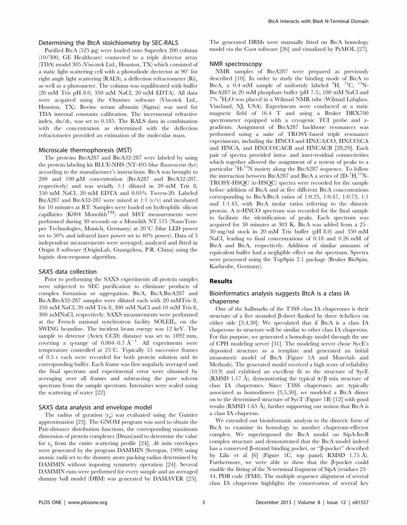
BtcA is a soluble protein characterized by a prominent a/b fold
For the purpose of biochemical characterization and verification
of our BtcA homology model we expressed and purified BtcA
using Ni2+-affinity chromatography and size exclusion chroma-
tography. A large scale expression and purification has generated
large quantities of BtcA at over 95% purity (Figure 2A). Matrix-
assisted laser desorption/ionization time-of-flight mass spectrom-
etry analysis (MALDI/TOF-MS) produced a major peak which
corresponds to the mass of a monomer (14.5 kDa with His6 tag)
further verifying the purity of the protein and the validity of the
protein purification scheme (Figure 2B). The circular dichroism
(CD) curve of BtcA at room temperature (Figure 2C, black line)
exhibited a double minimum at 208 nm and 222 nm. Analysis of
this curve suggests contributions of both a-helical (37%) and b-
strand (26%) conformations. These figures are similar to the values
obtained from the model of BtcA, in which 28 and 33 residues of
the total 111 chaperone-modelled residues adopt a-helical and b-
strand conformations, respectively.
BtcA is a homodimerSince class IA chaperons readily form homodimers we analyzed
BtcA’s oligomeric tendency through biochemical and biophysical
approaches. The size exclusion chromatogram (SEC) of BtcA
shows it to elute earlier than expected for a BtcA dimer, exhibiting
an elution volume (15.79 ml) highly similar to that of the 44 kDa
ovalbumin (15.78 ml) (Figure 3A, black line). Although this would
suggest trimeric stoichiometry for BtcA, elution in gel filtration
columns is strongly influenced by size and, to a lesser extent, by
non-specific interactions with the matrix. To determine the size of
BtcA more accurately we performed a cross-linking experiment on
purified BtcA followed by SDS-PAGE analysis (Figure 3B). The
gel exhibits an EGS dose-response pattern of BtcA shifting onto a
Figure 2. BtcA purification and fold characterization. (A) SDS-PAGE analysis of BtcA Ni2+-affinity chromatography (AC) and size exclusionchromatography (SEC). Lanes are labeled as follows: M, marker; W, whole cell lysate, P, pellet; FT, flow through; E, elution from AC column; S, elutionfrom SEC column; F, concentrated BtcA sample. (B) MALDI/TOF-MS analysis of purified BtcA with corresponding masses (in Daltons) indicated abovemain peaks. (C) Circular dichroism curve for BtcA measured in 50 mM NaCl, 20 mM Tris pH 8 buffer conditions at 25uC.doi:10.1371/journal.pone.0081557.g002
BtcA Interacts with BteA N-Terminal Domain
PLOS ONE | www.plosone.org 5 December 2013 | Volume 8 | Issue 12 | e81557

covalently formed SDS-resistant dimer (marked by black arrow).
Notably, this is the highest molecular weight species observed in
the experiment. Furthermore, SEC-coupled right angle light
scattering (RALS) analysis of BtcA yielded a calculated average
molecular weight of ,32 kDa, supporting our notion that BtcA is
a homodimer (Figure S1). The observed variation of the calculated
molecular weight across BtcA’s peak is possibly due to a high-MW
contaminant peak eluting just before BtcA. In summary, we
conclude that BtcA forms a native homodimer, suggested by the
SEC results to be slightly extended in shape.
BtcA binds BteA’s N-terminal at intermediate bindingaffinity
Previous publications have demonstrated that BtcA interacts
with BteA through the latter’s first 130 residues (chaperone
binding domain or CBD) via a yet-to-be determined mechanism
and at unknown molecular stoichiometry. We first evaluated the
affinity and stochiometry of binding of recombinant BteA287 to
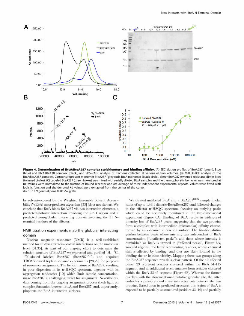
BtcA by analytical size exclusion chromatography (Figure 4A).
Addition of BtcA to BteA samples induced a pronounced shift in
the elution profile of the latter, indicating the formation of a higher
molecular weight species (green curve versus black curve),
identified by SDS-PAGE to contain both BteA287 and BtcA.
MALDI-TOF analysis of this complex verified our observation
that BtcA binds to BteA287 (Figure 4B), showing evidence of 1:1
and 1:2 BteA287:BtcA ratios, as well as some dimeric BteA287.
At this stage we employed microscale thermophoresis (MST) to
quantify the strength of BtcA’s binding to BteA287 (Figure 4C).
We have mixed labeled BteA287 with decreasing concentrations of
BtcA and the measured thermophoresis was fitted with a sigmoid
curve (R2,0.98), extrapolating the dissociation value as
6.6 mM61.1. We thus conclude that BtcA binds to BteA’s N-
terminal with medium strength and a stoichiometry of 2:1 and 1:1.
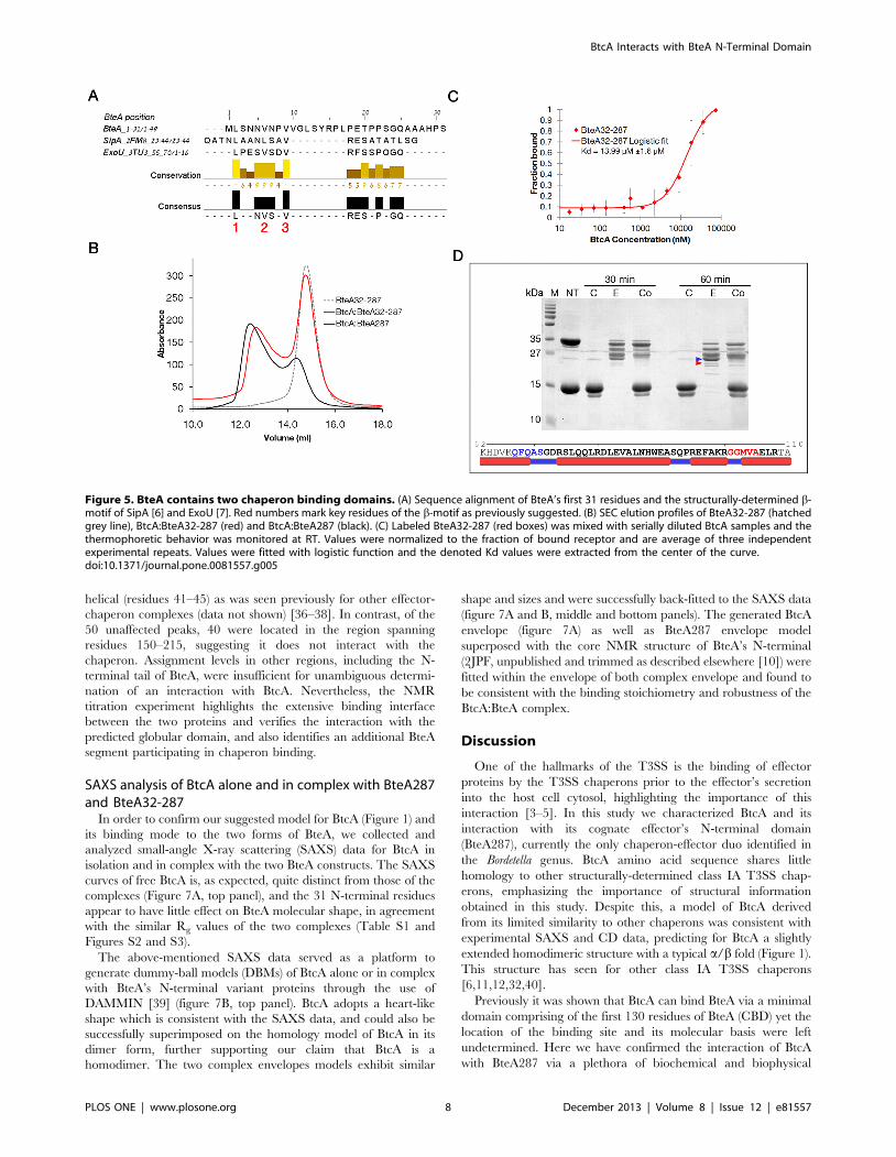
BtcA binds BteA287 via two chaperon binding sitesHaving established the formation of the BtcA:BteA287 com-
plex, we proceeded to explore the mechanisms by which BtcA
binds its effector. Previous publications have demonstrated that
chaperones bind their respective effectors through a beta motif
located at the effector’s N-terminal domain [6,32]. We postulated
that the first 31 residues of BteA might harbor such a b-motif
especially since our homology model indicated that BtcA might
exhibit two b-motif binding pockets (Figure 1C). The multiple
sequence alignment (MSA) between the first 130 residues of BteA287
(the minimal domain shown to bind BtcA) and the beta motifs
residues of SipA and ExoU exhibits strong conservation of leucine
and valine residues within the determined beta motif residues of
SpcU and ExoU (Figure 5A). Furthermore, the general motif
identified by Lilic et al [6], [L/M/I/F]XXX[L,V]XX[V,L,I,Q,H], is
not found elsewhere in the CBD of BteA. Thus we hypothesized that
removal of the first 31 residues will modulate binding of BtcA to
BteA287. For this purpose we analyzed a complex between BteA32-
287, an effector construct lacking the first 31 residues, and BtcA by
analytical SEC and compared it to the BteA287:BtcA complex
elution profile at similar chaperone:effector molar ratios (Figure 5B).
Strikingly, even in the presence of BtcA over half of the truncated
effector is found in the free state, suggesting the shorter BteA binds
BtcA with lower affinity, in agreement with findings for complex of
InvB with either SipA1-270 or SipA32-264 [6]. Further supporting
this conclusion is the finding that the MST-derived dissociation values
afforded a two-fold and statistically significant reduction in affinity
associated with the removal of the first 31 N-terminal residues, from
6.6 mM to 14 mM (R2,0.98, Figure 5C). Thus, there is compelling
evidence that these 31 residues, containing the putative b-motif, play
a role in binding of effector and chaperon.
Formation of a complex between BtcA and truncated BteA287
clearly indicates that an additional binding interface is involved in
their interaction. This was confirmed by subjecting BteA287, BtcA
and the formed complex to a time-limited trypsinization assay in
which the proteolytic products were separated via SDS-PAGE
(Figure 5D). We identified a 26 kDa fragment observed in the
effector lane yet absent in the complex lane, suggesting a digestion
site which is masked by the chaperon-effector interface. This site
was mapped to the segment spanning residues 67-108 (Figure 5D)
which is within the previously determined CBD region. Further-
more, we have found that this fragment is expected to be on the
surface of the protein since most of the residues were predicted to
Figure 3. Size and oligomeric characterization of BtcA. (A) SEC elution profiles of BtcA (black), aldolase, ribonuclease (grey) and ovalbumin(green) on a Superdex 200 10-300 column. (B) SDS-PAGE analysis of BtcA crosslinked with increasing amounts of EGS. Arrow marks the expected bandsize of BtcA’s dimer population.doi:10.1371/journal.pone.0081557.g003
BtcA Interacts with BteA N-Terminal Domain
PLOS ONE | www.plosone.org 6 December 2013 | Volume 8 | Issue 12 | e81557
be solvent-exposed by the Weighted Ensemble Solvent Accessi-
bility (WESA) meta-predictor algorithm [33] (data not shown). We
conclude that BtcA binds BteA287 via two interaction elements, a
predicted-globular interaction involving the CBD region and a
predicted non-globular interacting domain involving the 31 N-
terminal residues of the effector.
NMR titration experiments map the globular interactingdomain
Nuclear magnetic resonance (NMR) is a well-established
method for studying protein-protein interactions on the molecular
level [34,35]. As part of our ongoing effort to determine the
solution structure of BteA287 we expressed and purified 2H, 13C,15N-labeled labeled BteA287 (BteA287DCN) and acquired
TROSY-based triple-resonance experiments [28,29] for purposes
of resonance assignment. The helical nature of BteA287, resulting
in poor dispersion in its tr-HSQC spectrum, together with its
aggregation tendencies [10] which limit sample concentration,
make BteA287 a challenging target for assignment. Nevertheless,
data coming from the ongoing assignment process sheds light on
complex formation between BtcA and BteA287, and, importantly,
pinpoints the BteA interaction surfaces.
We titrated unlabeled BtcA into a BteA287DCN sample (molar
ratios of up to 1.43:1 dimeric-BtcA:BteA287) and followed changes
in the effector tr-HSQC spectrum, focusing on outlying peaks
which could be accurately monitored in the two-dimensional
experiment (Figure 6A). Binding of BtcA results in widespread
intensity loss of BteA287 peaks, suggesting that the two proteins
form a complex with intermediate (micromolar) affinity charac-
terized by an extensive interaction surface. The titration distin-
guishes between peaks whose intensity was independent of BtcA
concentration (‘‘unaffected peaks’’), and those whose intensity is
diminished as BtcA is titrated in (‘‘affected peaks’’, Figure 6A,
zoomed regions), the latter representing residues, whose chemical
shift is affected by binding, and thus are likely located in the
binding site or its close vicinity. Mapping these two groups along
the BteA287 sequence reveals a clear pattern. Of the 40 affected
peaks, 29 represent residues clustered within the BteA 61-115
segment, and an additional seven emanate from residues clustered
within the BteA 33-45 segment (Figure 6B). Whereas the former
overlaps with the aforementioned putative globular site, the latter
embodies a previously unknown interaction site between the two
proteins. Based upon its predicted structure, this region of BteA is
expected to be partially unstructured (residues 33–40) and partially
Figure 4. Determination of BtcA:BteA287 complex stoichiometry and binding affinity. (A) SEC elution profiles of BteA287 (green), BtcA(blue) and BtcA:BteA28 complex (black), and SDS-PAGE analysis of fractions collected at various elution volumes. (B) MALDI-TOF analysis of theBtcA:BteA287 complex. Cartoons represent monomer BteA287 (grey rod), BtcA monomer (black circle), dimer BteA287 (twinned rods) and dimer BtcA(twinned circles). (C) Labeled BteA287 (green boxes) was mixed with serially diluted BtcA samples and the thermophoretic behavior was monitored atRT. Values were normalized to the fraction of bound receptor and are average of three independent experimental repeats. Values were fitted withlogistic function and the denoted Kd values were extracted from the center of the curve.doi:10.1371/journal.pone.0081557.g004
BtcA Interacts with BteA N-Terminal Domain
PLOS ONE | www.plosone.org 7 December 2013 | Volume 8 | Issue 12 | e81557
helical (residues 41–45) as was seen previously for other effector-
chaperon complexes (data not shown) [36–38]. In contrast, of the
50 unaffected peaks, 40 were located in the region spanning
residues 150–215, suggesting it does not interact with the
chaperon. Assignment levels in other regions, including the N-
terminal tail of BteA, were insufficient for unambiguous determi-
nation of an interaction with BtcA. Nevertheless, the NMR
titration experiment highlights the extensive binding interface
between the two proteins and verifies the interaction with the
predicted globular domain, and also identifies an additional BteA
segment participating in chaperon binding.
SAXS analysis of BtcA alone and in complex with BteA287and BteA32-287
In order to confirm our suggested model for BtcA (Figure 1) and
its binding mode to the two forms of BteA, we collected and
analyzed small-angle X-ray scattering (SAXS) data for BtcA in
isolation and in complex with the two BteA constructs. The SAXS
curves of free BtcA is, as expected, quite distinct from those of the
complexes (Figure 7A, top panel), and the 31 N-terminal residues
appear to have little effect on BteA molecular shape, in agreement
with the similar Rg values of the two complexes (Table S1 and
Figures S2 and S3).
The above-mentioned SAXS data served as a platform to
generate dummy-ball models (DBMs) of BtcA alone or in complex
with BteA’s N-terminal variant proteins through the use of
DAMMIN [39] (figure 7B, top panel). BtcA adopts a heart-like
shape which is consistent with the SAXS data, and could also be
successfully superimposed on the homology model of BtcA in its
dimer form, further supporting our claim that BtcA is a
homodimer. The two complex envelopes models exhibit similar
shape and sizes and were successfully back-fitted to the SAXS data
(figure 7A and B, middle and bottom panels). The generated BtcA
envelope (figure 7A) as well as BteA287 envelope model
superposed with the core NMR structure of BteA’s N-terminal
(2JPF, unpublished and trimmed as described elsewhere [10]) were
fitted within the envelope of both complex envelope and found to
be consistent with the binding stoichiometry and robustness of the
BtcA:BteA complex.
Discussion
One of the hallmarks of the T3SS is the binding of effector
proteins by the T3SS chaperons prior to the effector’s secretion
into the host cell cytosol, highlighting the importance of this
interaction [3–5]. In this study we characterized BtcA and its
interaction with its cognate effector’s N-terminal domain
(BteA287), currently the only chaperon-effector duo identified in
the Bordetella genus. BtcA amino acid sequence shares little
homology to other structurally-determined class IA T3SS chap-
erons, emphasizing the importance of structural information
obtained in this study. Despite this, a model of BtcA derived
from its limited similarity to other chaperons was consistent with
experimental SAXS and CD data, predicting for BtcA a slightly
extended homodimeric structure with a typical a/b fold (Figure 1).
This structure has seen for other class IA T3SS chaperons
[6,11,12,32,40].
Previously it was shown that BtcA can bind BteA via a minimal
domain comprising of the first 130 residues of BteA (CBD) yet the
location of the binding site and its molecular basis were left
undetermined. Here we have confirmed the interaction of BtcA
with BteA287 via a plethora of biochemical and biophysical
Figure 5. BteA contains two chaperon binding domains. (A) Sequence alignment of BteA’s first 31 residues and the structurally-determined b-motif of SipA [6] and ExoU [7]. Red numbers mark key residues of the b-motif as previously suggested. (B) SEC elution profiles of BteA32-287 (hatchedgrey line), BtcA:BteA32-287 (red) and BtcA:BteA287 (black). (C) Labeled BteA32-287 (red boxes) was mixed with serially diluted BtcA samples and thethermophoretic behavior was monitored at RT. Values were normalized to the fraction of bound receptor and are average of three independentexperimental repeats. Values were fitted with logistic function and the denoted Kd values were extracted from the center of the curve.doi:10.1371/journal.pone.0081557.g005
BtcA Interacts with BteA N-Terminal Domain
PLOS ONE | www.plosone.org 8 December 2013 | Volume 8 | Issue 12 | e81557

approaches and determined the binding stoichiometry to be 2:1
(chaperon:effector), as was seen for other chaperon:effector
complexes [6,36,40,41], as well as 1:1 (as shown by MALDI-
TOF). However, we find the BtcA:BteA287 complex to differ from
their counterparts in other T3SS systems in its micromolar affinity,
which is 2–3 orders of magnitude weaker than previously-
published chaperon:effector determined affinities [7]. This result
was obtained from MST data and confirmed by the NMR
titration results. The micromolar affinity of this complex implies
that the release of BteA287 from BtcA is more favorable in
comparison to the above-mentioned chaperon:effector complexes.
The mechanism through which chaperons bind to and stabilize
their cognate effectors is of much interest in the field of T3SS and
until now was not known for the Bordetella secretion system.
Chaperons bind their cognate effectors via two possible mecha-
nisms, one involving binding of the effector’s two b-motif (‘‘non-
globular interaction’’) to the dimeric chaperon and the other
involving the combined binding of one non-globular interaction
and one globular interaction. The latter is mediated in many cases
Figure 6. NMR-monitored titration reveals key BteA287 residues involved in the binding of BtcA. (A) Superposition of the tr-1H-15NHSQC spectra of a BteA287DCN sample in the absence of BtcA (blue) and in the presence of increasing BtcA concentrations, 110 mM (orange), 160 mM(green) and 270 mM (red). Below are detailed views of four cross-peaks marked by arrow heads. (B) The BteA287 amino acid sequence showingassigned backbone resonances (underline), affected (bold red) and unaffected residues (bold black). Arrowheads correspond to peaks shown indetail. Highlighted in yellow are the currently known non-globular (residues 2–10) and globular (residues 67–108) interaction domains.doi:10.1371/journal.pone.0081557.g006
BtcA Interacts with BteA N-Terminal Domain
PLOS ONE | www.plosone.org 9 December 2013 | Volume 8 | Issue 12 | e81557

via a domain which has a dedicated function within the host cell
[6]. To determine which mechanism governs the interaction of
BtcA with BteA’s N-terminal domain, we utilized complementary
bioinformatics and empirical approaches. Our study identified two
possible b-pockets for BtcA, as observed for other class IA
chaperons, yet only one possible b-motif peptide was identified at
the N-terminal region of BteA which is within the secretion signal
peptide [2,9,10]. Since T3SS effector-chaperon complexes in-
volves at least two interaction sites, we speculated that at least one
additional site should be located C-terminal to the b-motif yet
within the CBD boundaries. For this purpose we utilized a
protection assay comparing BtcA:BteA287 complex to each of the
components alone, enabling the identification of the second
interaction site located at CBD’s midsection (residues 67–108).
While the suggested N-termini b-motif is predicted to be mostly
unfolded, similar to other T3SS effector’s determined b-motif, the
second motif of BteA287 is predicted to be folded as interchanging
alpha helices and short loops, which are characteristic of a
globular domain [6–8]. Furthermore, NMR titration experiments
interpreted in the context of our ongoing backbone assignment
identified key residues located at the BtcA binding interface. Most
notable was finding an additional binding surface including
residues 33–45 located between the N-terminal signal peptide
and the CBD. Precedents for this additional binding region, which
highlights the extensive nature of the chaperon:effector interface
[6,7,36–38,40,42], have been previously observed for a number of
effector-chaperon complexes [36–38]. In other BteA regions data
was insufficient to unequivocally determine their involvement in
binding, although it strongly suggests a contribution of the N-
terminal signal peptide to the observed affinity (data not shown).
We anticipate that completion of the BteA287 assignment will
yield a much-needed global fold for the effector and serve as a
foundation for determining its structure. However, even at this
stage the NMR data draw a compelling picture as to the nature of
the chaperon:effector complex. Line shape behavior is highly
consistent with an intermediate-affinity complex, and affected
outlying peaks in the two-dimensional spectra were clustered in the
binding regions in a statistically significant manner.
In light of this NMR-based mapping of the binding site and the
possible role of the first 50 effector residues it is interesting to
reconsider the binding affinities found for wild-type and truncated
BteA. The SEC-RALS and MST experiments demonstrate,
qualitatively and quantitatively, respectively, that while a weaker
complex was formed by the truncated BteA32-287 variant, the
predicted globular domain is the main mediator and driving force
leading to the interaction between BtcA to BteA287. Although the
difference in calculated dissociation constant suggested by MST
(,40%) may not fully account for the observed chromatogram
shift, this discrepancy may originate from the intermediate affinity
of the complex and inherent methodological differences. The
absence of the first 31 N-terminal residues obviously obviates their
contribution to binding, but also may perturb the fold of BteA
Figure 7. SAXS analysis of free and complexed BtcA. (A) Experimental data (circles) and the corresponding back-fitted dummy-ball model(DBM). (B) Dummy-ball envelope models (light azure) with superimposed models of BtcA (red cartoon) and BteA287 (orange cartoon, 2JPF). TheDBMs of BtcA and BteA287 (blue and green mesh, respectively) have been superimposed over the DBMs of the BtcA:BteA287 and BtcA:BteA32-287complexes.doi:10.1371/journal.pone.0081557.g007
BtcA Interacts with BteA N-Terminal Domain
PLOS ONE | www.plosone.org 10 December 2013 | Volume 8 | Issue 12 | e81557

residues 33-45 and affect their ability to interact with the
chaperon. Further studies will be necessary to further illuminate
this aspect of complex formation.
Further indication of the intimate nature of the BtcA:BteA
complex was offered by the SAXS data, allowing a comparison of
Rg values for free and complexed BteA. The effector Rg increased
from 3.1 [10] to 4.6 nm upon binding of BtcA, even though the
chaperon Rg was estimated at 2.9 nm. This relatively small
increase in size is consistent with our suggested model of an
extensive interaction surface between the two proteins. The similar
Rg values observed for the two BteA N-terminal variants in
complex with BtcA suggests that the flexible N-terminal tail
contributes little to the envelope shape and size of both complexes.
This might be due to the different binding position the two
variants adopt once they are bound by BtcA.
In conclusion, we present here an in-depth structural and
biochemical characterization of effector-chaperon in Bordetella,
contributing and strengthening to the currently accepted dogma of
a binding site which combines interactions with predicted globular
and non-globular regions, as was suggested by Lilic et al [6].
Besides providing first direct information for the only known
chaperon:effector system in Bordetella, this study also lays the
groundwork for further and more detailed structural studies of this
intriguing protein-protein complex.
Supporting Information
Figure S1 SEC-RALS analysis of BtcA. Solid and dotted
lines represent absorbance at 280 nm and right angle light
scattering (RALS)-determined molecular weight, respectively.
(TIF)
Figure S2 Guinier plots of SAXS data.(TIF)
Figure S3 Overlap of SAXS data of BteA287 and BteA32-287 complex with BtcA. SAXS data of BtcA in complex with
either BteA287 (green) or BteA32-287 (blue) demonstrate the
differences in scattering patterns.
(TIF)
Table S1 Summary of SAXS geometric data.
(DOCX)
Acknowledgments
We would like to thank Dr. Melissa Graewert (EMBL, Hamburg,
Germany) for assistance with the SEC-RALS experiments. We would like
also to thank Dr. Hadas Hawlena (BGU, Beer-sheba, Israel) for assistance
with the statistics calculations.
Author Contributions
Conceived and designed the experiments: CG SK RB JC RZ. Performed
the experiments: CG GD AY HS SK RB SBZ JC RZ. Analyzed the data:
CG SK RB JC RZ. Contributed reagents/materials/analysis tools: JC RZ.
Wrote the paper: CG SK RB JC RZ.
References
1. Shrivastava R, Miller JF (2009) Virulence factor secretion and translocation by
Bordetella species. Curr Opin Microbiol 12: 88–93.
2. Panina EM, Mattoo S, Griffith N, Kozak NA, Yuk MH, et al. (2005) A genome-
wide screen identifies a Bordetella type III secretion effector and candidate
effectors in other species. Mol Microbiol 58: 267–279.
3. Galan JE, Wolf-Watz H (2006) Protein delivery into eukaryotic cells by type III
secretion machines. Nature 444: 567–573.
4. Cornelis GR (2006) The type III secretion injectisome. Nat Rev Microbiol 4:
811-825.
5. Ghosh P (2004) Process of protein transport by the type III secretion system.
Microbiol Mol Biol Rev 68: 771–795.
6. Lilic M, Vujanac M, Stebbins CE (2006) A common structural motif in the
binding of virulence factors to bacterial secretion chaperones. Mol Cell 21: 653–
664.
7. Halavaty AS, Borek D, Tyson GH, Veesenmeyer JL, Shuvalova L, et al. (2012)
Structure of the Type III Secretion Effector Protein ExoU in Complex with Its
Chaperone SpcU. PLoS One 7: e49388.
8. Gendrin C, Contreras-Martel C, Bouillot S, Elsen S, Lemaire D, et al. (2012)
Structural basis of cytotoxicity mediated by the type III secretion toxin ExoU
from Pseudomonas aeruginosa. PLoS Pathog 8: e1002637.
9. French CT, Panina EM, Yeh SH, Griffith N, Arambula DG, et al. (2009) The
Bordetella type III secretion system effector BteA contains a conserved N-
terminal motif that guides bacterial virulence factors to lipid rafts. Cell Microbiol
11: 1735–1749.
10. Guttman C, Davidov G, Shaked H, Kolusheva S, Bitton R, et al. (2013)
Characterization of the N-Terminal Domain of BteA: A Bordetella Type III
Secreted Cytotoxic Effector. PLoS One 8: e55650.
11. Birtalan S, Ghosh P (2001) Structure of the Yersinia type III secretory system
chaperone SycE. Nat Struct Biol 8: 974–978.
12. Locher M, Lehnert B, Krauss K, Heesemann J, Groll M, et al. (2005) Crystal
structure of the Yersinia enterocolitica type III secretion chaperone SycT. J Biol
Chem 280: 31149–31155.
13. McGuffin LJ, Buenavista MT, Roche DB (2013) The ModFOLD4 server for the
quality assessment of 3D protein models. Nucleic Acids Res 41: W368–W372.
14. Cristobal S, Zemla A, Fischer D, Rychlewski L, Elofsson A (2001) A study of
quality measures for protein threading models. BMC Bioinformatics 2: 5.
15. Benkert P, Tosatto SC, Schomburg D (2008) QMEAN: A comprehensive
scoring function for model quality assessment. Proteins 71: 261–277.
16. Guex N, Diemand A, Peitsch MC (1999) Protein modelling for all. Trends
Biochem Sci 24: 364–367.
17. Delano W (2002) The PyMOL Molecular Graphics System. In: S . Carlos,
editor editors. : DeLano Scientific.
18. Edgar RC (2004) MUSCLE: multiple sequence alignment with high accuracy
and high throughput. Nucleic Acids Res 32: 1792–1797.
19. Waterhouse AM, Procter JB, Martin DM, Clamp M, Barton GJ (2009) Jalview
Version 2—a multiple sequence alignment editor and analysis workbench.
Bioinformatics 25: 1189–1191.
20. Whitmore L, Wallace BA (2004) DICHROWEB, an online server for protein
secondary structure analyses from circular dichroism spectroscopic data. Nucleic
Acids Res 32: W668–673.
21. Andrade MA, Chacon P, Merelo JJ, Moran F (1993) Evaluation of secondary
structure of proteins from UV circular dichroism spectra using an unsupervised
learning neural network. Protein Eng 6: 383–390.
22. Carn F, Guyot S, Baron A, Perez J, Buhler E, et al. (2012) Structural Properties
of Colloidal Complexes between Condensed Tannins and PolysaccharideHyaluronan. Biomacromolecules 13: 751–759.
23. Guinier A, Fournet G (1955) Small-angle scattering of X-rays. New York: Wiley.
24. Svergun D (1992) Determination of the regularization parameter in indirect-
transform methods using perceptual criteria. Journal of Applied Crystallography25: 495–503.
25. Volkov VV, Svergun DI (2003) Uniqueness of ab initio shape determination in
small-angle scattering. Journal of Applied Crystallography 36: 860–864.
26. Emsley P, Cowtan K (2004) Coot: model-building tools for molecular graphics.
Acta Crystallogr D Biol Crystallogr 60: 2126–2132.
27. DeLano WL (2002) The PyMOL Molecular Graphics System. San Carlos:
DeLano Scientific.
28. Salzmann M, Pervushin K, Wider G, Senn H, Wuthrich K (1998) TROSY in
triple-resonance experiments: New perspectives for sequential NMR assignmentof large proteins. Proceedings of the National Academy of Sciences 95: 13585–
13590.
29. Salzmann M, Wider G, Pervushin K, Senn H, Wuthrich K (1999) TROSY-type
Triple-Resonance Experiments for Sequential NMR Assignments of Large
Proteins. Journal of the American Chemical Society 121: 844–848.
30. Dean P (2011) Functional domains and motifs of bacterial type III effector
proteins and their roles in infection. FEMS Microbiol Rev 35: 1100–1125.
31. Nielsen M, Lundegaard C, Lund O, Petersen TN (2010) CPHmodels-3.0—
remote homology modeling using structure-guided sequence profiles. Nucleic
Acids Res 38: W576–581.
32. Janjusevic R, Quezada CM, Small J, Stebbins CE (2012) Structure of the
HopA1(21-102)-ShcA Chaperone-Effector Complex of Pseudomonas syringae
Reveals Conservation of a Virulence Factor Binding Motif from Animal to Plant
Pathogens. J Bacteriol 195: 658–664.
33. Chen H, Zhou H-X (2005) Prediction of solvent accessibility and sites of
deleterious mutations from protein sequence. Nucleic Acids Research 33: 3193–3199.
BtcA Interacts with BteA N-Terminal Domain
PLOS ONE | www.plosone.org 11 December 2013 | Volume 8 | Issue 12 | e81557

34. Zuiderweg ER (2002) Mapping protein-protein interactions in solution by NMR
spectroscopy. Biochemistry 41: 1–7.35. Kleckner IR, Foster MP (2010) An introduction to NMR-based approaches for
measuring protein dynamics. Biochim Biophys Acta 1814: 942–968.
36. Stebbins CE, Galan JE (2001) Maintenance of an unfolded polypeptide by acognate chaperone in bacterial type III secretion. Nature 414: 77–81.
37. Schubot FD, Jackson MW, Penrose KJ, Cherry S, Tropea JE, et al. (2005)Three-dimensional structure of a macromolecular assembly that regulates type
III secretion in Yersinia pestis. J Mol Biol 346: 1147–1161.
38. Vogelaar NJ, Jing X, Robinson HH, Schubot FD (2010) Analysis of the crystalstructure of the ExsC.ExsE complex reveals distinctive binding interactions of
the Pseudomonas aeruginosa type III secretion chaperone ExsC with ExsE andExsD. Biochemistry 49: 5870–5879.
39. Svergun DI (1999) Restoring low resolution structure of biological macromol-
ecules from solution scattering using simulated annealing. Biophys J 76: 2879–
2886.
40. Vujanac M, Stebbins CE (2013) Context-dependent protein folding of a
virulence peptide in the bacterial and host environments: structure of an SycH-
YopH chaperone-effector complex. Acta Crystallogr D Biol Crystallogr 69: 546–
554.
41. Letzelter M, Sorg I, Mota LJ, Meyer S, Stalder J, et al. (2006) The discovery of
SycO highlights a new function for type III secretion effector chaperones. Embo J
25: 3223–3233.
42. Birtalan SC, Phillips RM, Ghosh P (2002) Three-dimensional secretion signals in
chaperone-effector complexes of bacterial pathogens. Mol Cell 9: 971–980.
BtcA Interacts with BteA N-Terminal Domain
PLOS ONE | www.plosone.org 12 December 2013 | Volume 8 | Issue 12 | e81557