bone marrow failure update - pathology · bone marrow failure update andrea m. sheehan, md ... many...
TRANSCRIPT
Bone Marrow Failure Update
Andrea M. Sheehan, MD Associate Professor of Pathology & Immunology
Director of Hematopathology
August 7, 2014

The Maroon Bells

Pine Creek Cookhouse

View from the Pine Creek Cookhouse
Bone Marrow Failure
Final common endpoint rather than a distinct disease entity Congenital, environmental, genetic influences
Starting point for investigation as to why it happened
Congenital (constitutional) or acquired Can present at any age, but inherited/constitutional
usually present in childhood or young adulthood
Single lineage or multiple lineages Some initially single lineage but then progress into
multilineage with pancytopenia and more extensive marrow hypoplasia
Bone Marrow Failure
Important to consider constitutional syndromes Treatment may differ, especially if the child goes
to BMT Radiation sensitivity
Many have increased risk of MDS, leukemias and/or solid tumors – will need surveillance
Early onset of abnormal blood/bone marrow findings, family history, dysmorphic physical findings, ethnicity may be helpful, often genetic or other testing (chromosomal breakage, telomere length) will be confirmatory
Inherited Bone Marrow Failure
Most germline inherited disorders are single gene disorders, inheritance patterns vary
Autosomal recessive
Autosomal dominant
X-linked recessive
Many have disease specific genetic mutations identified, although can be variable within any particular disease, and often not all genes/mutations known
Bone Marrow Failure
Although peripheral blood changes may be present (high MCV for example), circulating cells usually have normal function
Many of the inherited syndrome have increased risk of MDS and/or acute leukemia Some have increased risk of solid tumors also
Variable genetic expressivity and penetrance within any given disorder

Inherited Bone Marrow Failure
Many patients have associated dysmorphic features or physical abnormalities (skeletal, others) identified in early infancy or at birth, but many patients will not necessarily have the obvious physical manifestations
Some develop over time and may become more obvious in later childhood, young adulthood (dyskeratosis congenita)
Cytopenias, marrow failure may be first manifestation of the disorder
Inherited Bone Marrow Failure
If anemic, may have evidence of fetal type hematopoiesis
Macrocytosis
Increased Hgb F (F cells)
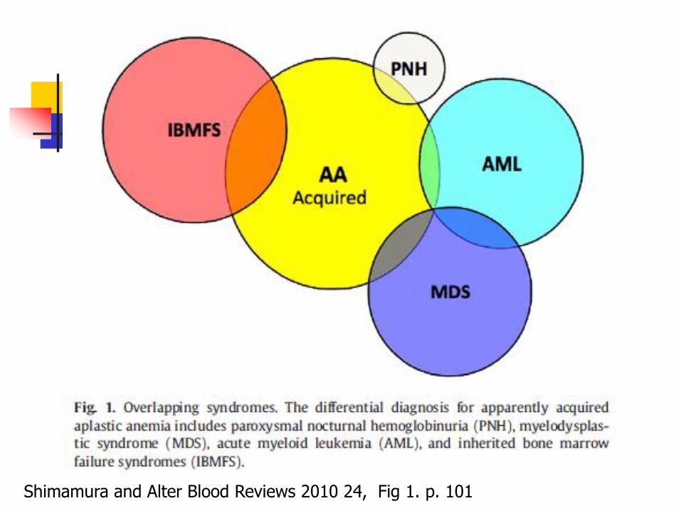
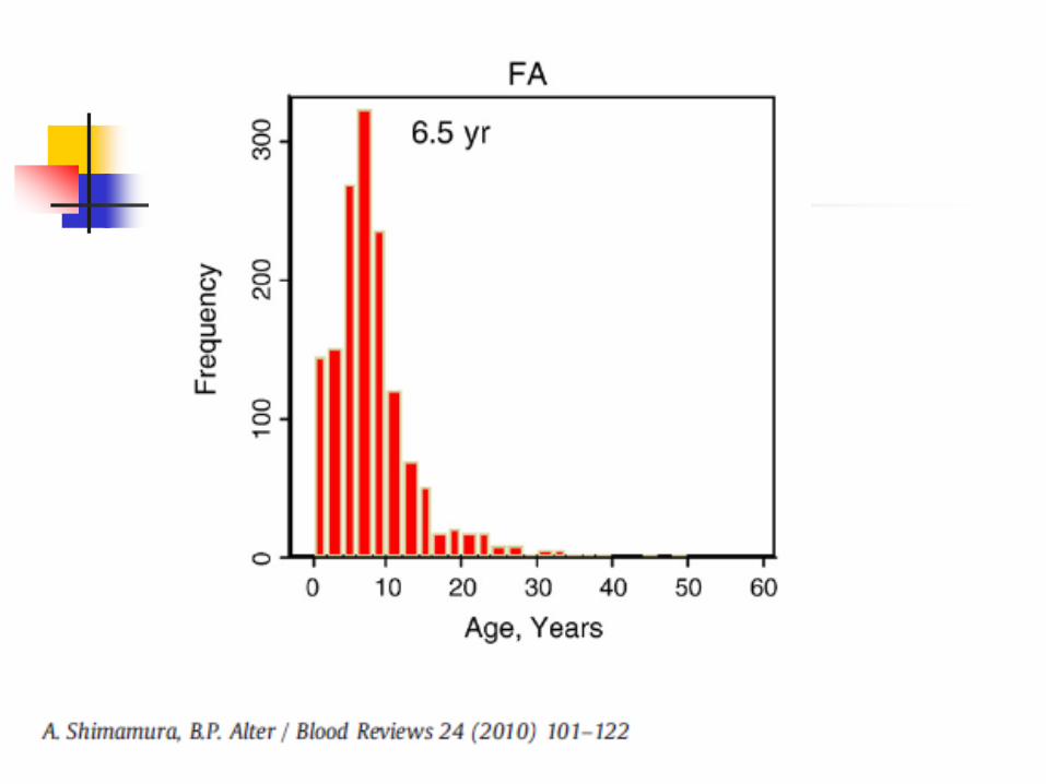
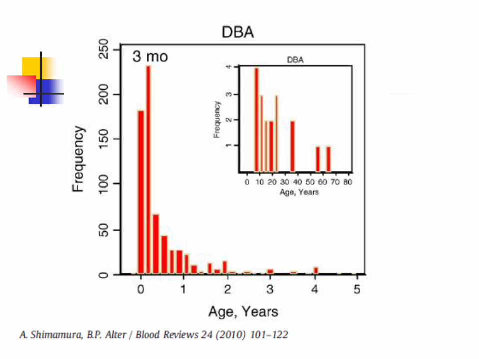
Shimamura and Alter Blood Reviews 2010 24, Fig 1. p. 101
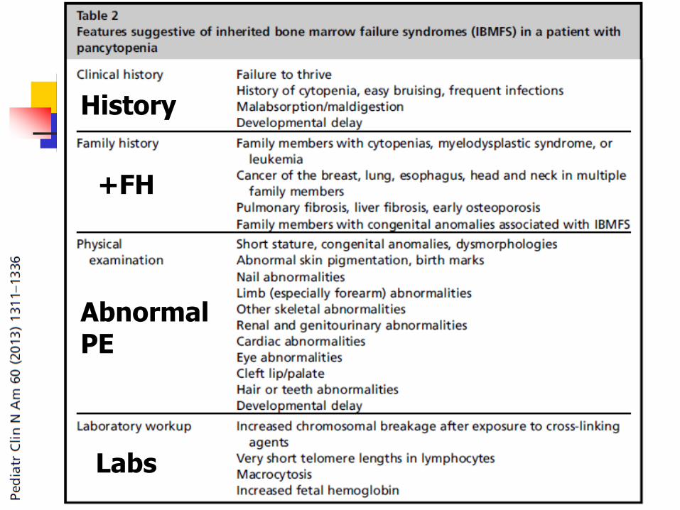
+FH
Abnormal PE
History
Labs
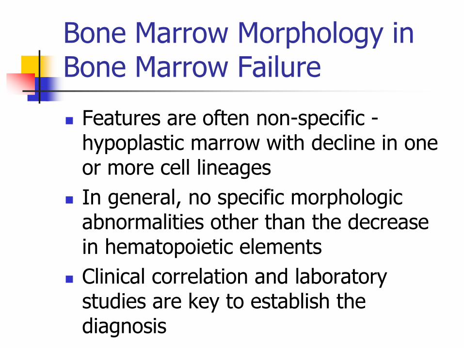
Bone Marrow Morphology in Bone Marrow Failure
Features are often non-specific - hypoplastic marrow with decline in one or more cell lineages
In general, no specific morphologic abnormalities other than the decrease in hematopoietic elements
Clinical correlation and laboratory studies are key to establish the diagnosis
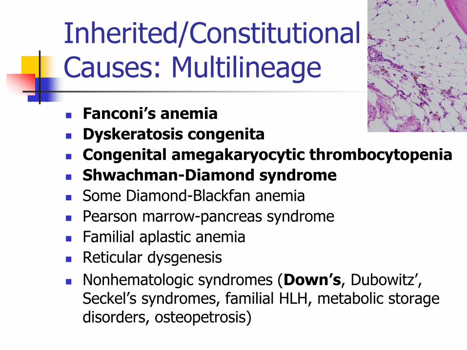
Inherited/Constitutional Causes: Multilineage
Fanconi’s anemia
Dyskeratosis congenita
Congenital amegakaryocytic thrombocytopenia
Shwachman-Diamond syndrome
Some Diamond-Blackfan anemia
Pearson marrow-pancreas syndrome
Familial aplastic anemia
Reticular dysgenesis
Nonhematologic syndromes (Down’s, Dubowitz’, Seckel’s syndromes, familial HLH, metabolic storage disorders, osteopetrosis)
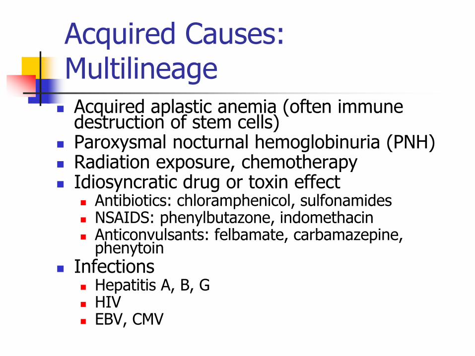
Acquired Causes: Multilineage
Acquired aplastic anemia (often immune destruction of stem cells)
Paroxysmal nocturnal hemoglobinuria (PNH) Radiation exposure, chemotherapy Idiosyncratic drug or toxin effect
Antibiotics: chloramphenicol, sulfonamides NSAIDS: phenylbutazone, indomethacin Anticonvulsants: felbamate, carbamazepine,
phenytoin
Infections Hepatitis A, B, G HIV EBV, CMV
Acquired Causes: Multilineage
Hepatitis associated Immune disorders (SLE, GVHD,
transfusion associated GVHD, eosinophilic fasciitis)
Pregnancy Bone marrow effacement
Primary/secondary neoplasm Fibrosis (neoplastic or non-neoplastic)
Nutritional deficiency (B12, folate) Myelodysplastic syndrome
Constitutional Single Lineage Disorders
Diamond-Blackfan anemia
Congenital dyserythropoietic anemia
Congenital sideroblastic anemias
Schwachman-Diamond syndrome
Severe congenital neutropenia/Kostmann syndrome
Cyclic neutropenia
Thrombocytopenia with absent radii
Acquired Causes: Single lineage – Erythroids
Viral infection (Parvovirus B19 –esp immune suppressed pts)
Transient erythroblastopenia of childhood
Neoplasm associated
Thymoma
Immune related
Iron deficiency anemia
Anemia of chronic disease
Idiosyncratic drug or toxin effects
Other/idiopathic
Acquired Causes: Single lineage – Myeloids
Drugs/ Medication related Infections
Viral: EBV, CMV, varicella, rubella, Thai hemorrhagic fever
Bacterial: meningococcus
Toxin-related
Levamisole-tainted cocaine Nutritional deficiencies
Megaloblastic anemia
Acquired Myeloid Production Problems
Infiltration of the bone marrow (metastatic cancer, myelofibrosis)
Other acquired toxic/ environmental/ immune causes
Other/idiopathic
Meg/platelet Production Problems: Congenital
Bernard Soulier syndrome
May-Hegglin anomaly
Fechtner syndrome
Sebastian syndrome
Epstein syndrome
Montreal platelet syndrome
Fanconi anemia
Wiskott-Aldrich syndrome
Thrombocytopenia with absent radii (TAR)
Congenital amegakaryocytic thrombocytopenia
Autosomal dominant & X-linked thrombocytopenia
Acquired Causes: Single lineage – Megs
Rare
Medication effect
Other/idiopathic
Acquired – Drugs Causing Marrow Suppression of Platelets
Chemotherapy
Zidovudine
Ethanol – long periods of ingestion
Interferon therapy
Large doses of estrogen, or DES
Anticonvulsants
Tranquilizers
Some antibiotics (chloramphenicol)
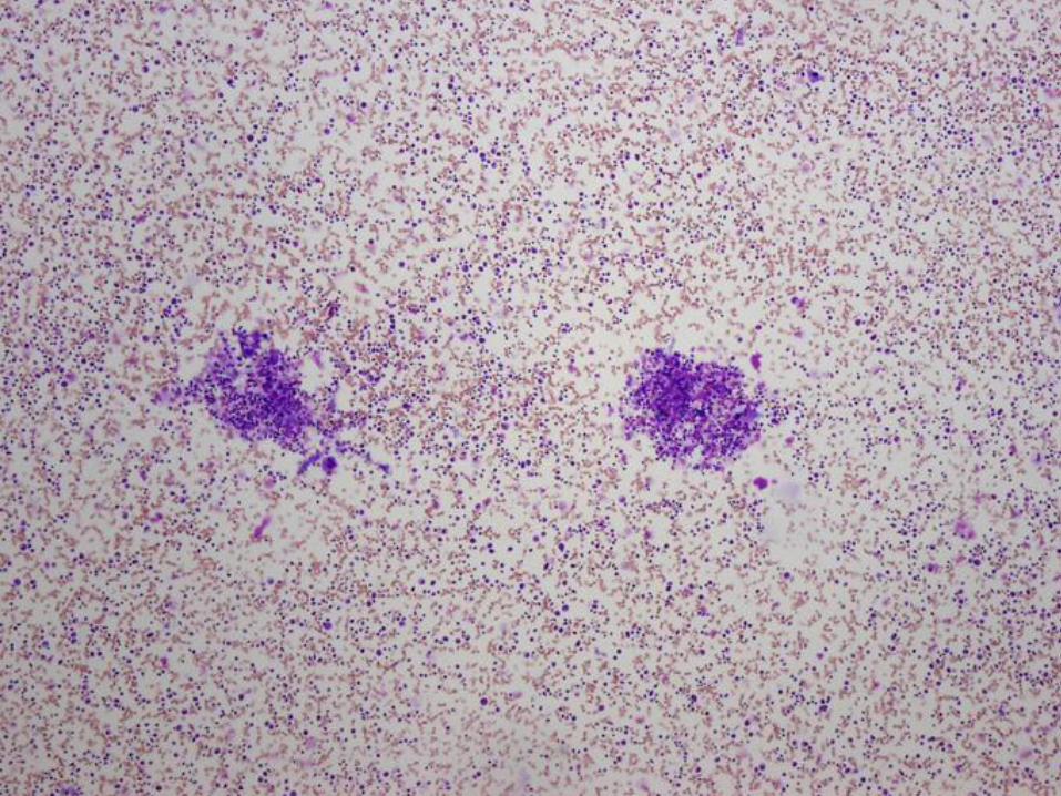
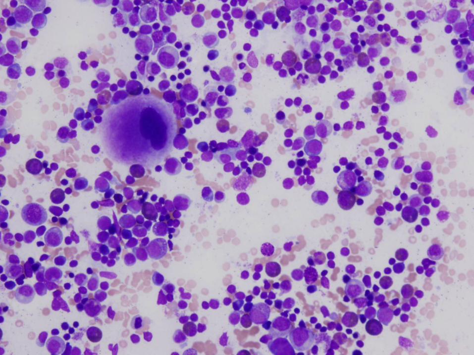
Case #1
16 year old girl with pancytopenia
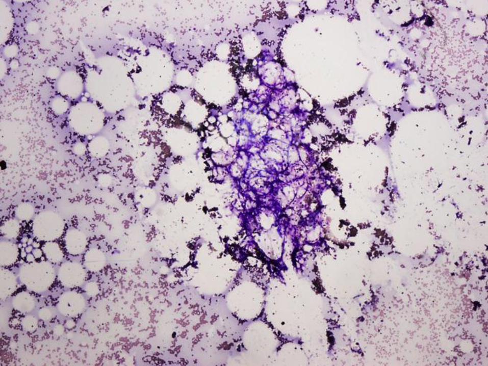
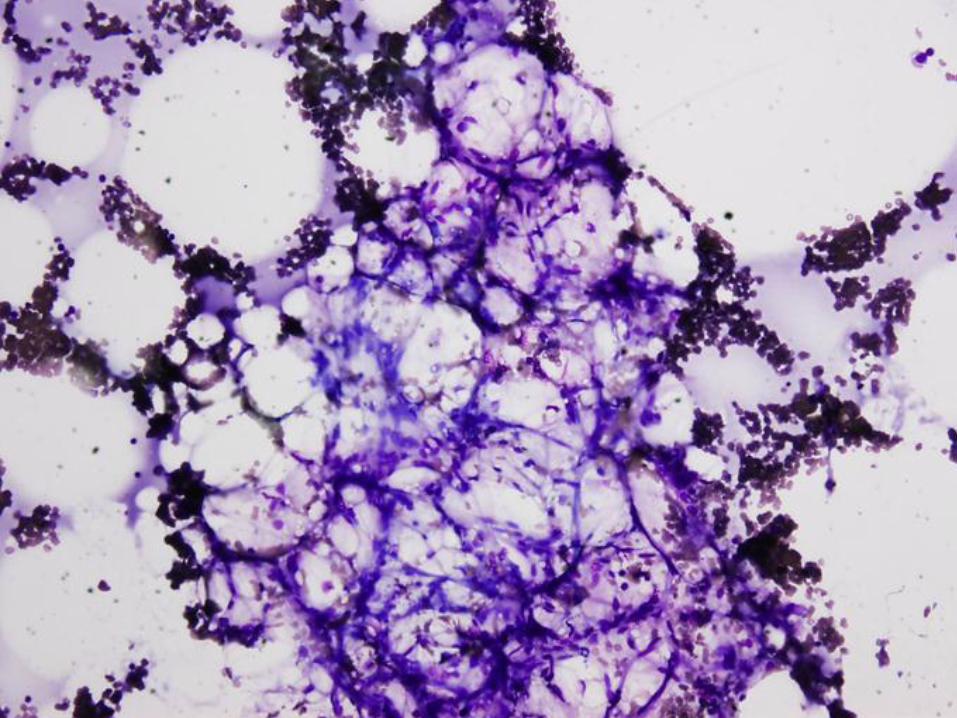

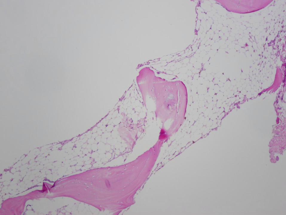
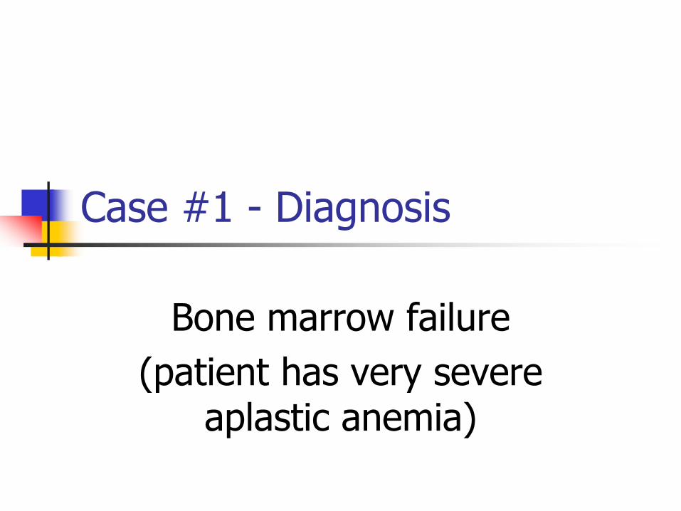
Case #1 - Diagnosis
Bone marrow failure
(patient has very severe aplastic anemia)
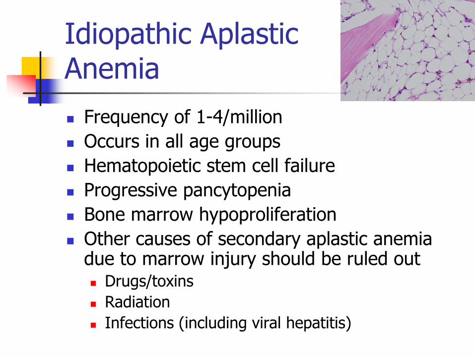
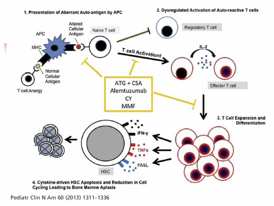
Idiopathic Aplastic Anemia
Frequency of 1-4/million
Occurs in all age groups
Hematopoietic stem cell failure
Progressive pancytopenia
Bone marrow hypoproliferation
Other causes of secondary aplastic anemia due to marrow injury should be ruled out Drugs/toxins
Radiation
Infections (including viral hepatitis)
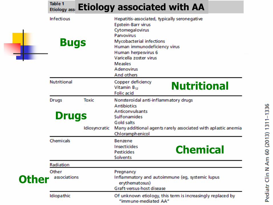
Etiology associated with AA
Bugs
Drugs
Chemical
Nutritional
Other
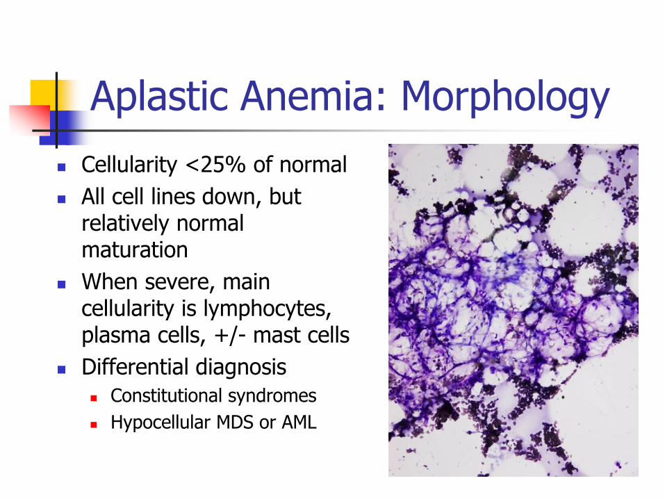
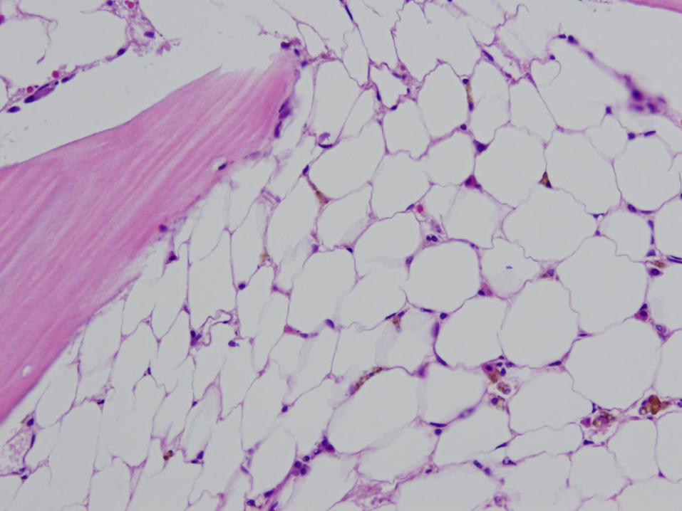
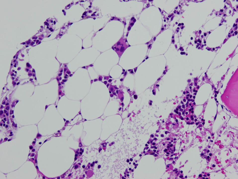
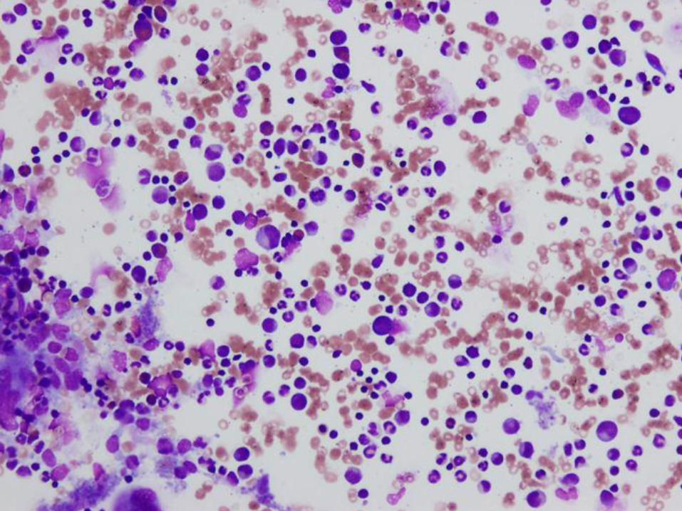
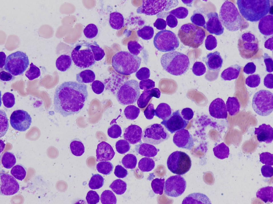
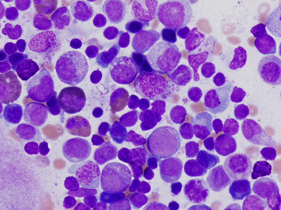
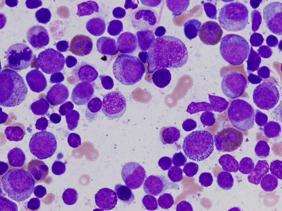
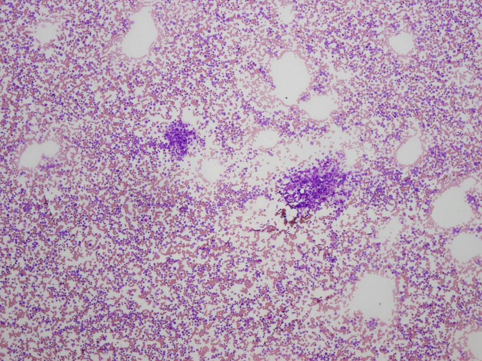
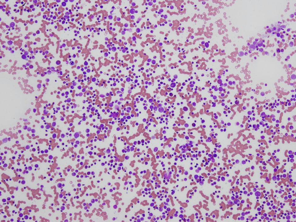
Aplastic Anemia: Morphology
Cellularity <25% of normal
All cell lines down, but relatively normal maturation
When severe, main cellularity is lymphocytes, plasma cells, +/- mast cells
Differential diagnosis
Constitutional syndromes
Hypocellular MDS or AML
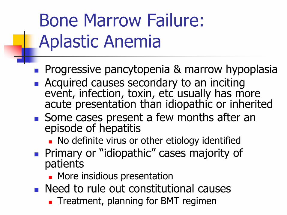
Bone Marrow Failure: Aplastic Anemia
Progressive pancytopenia & marrow hypoplasia Acquired causes secondary to an inciting
event, infection, toxin, etc usually has more acute presentation than idiopathic or inherited
Some cases present a few months after an episode of hepatitis No definite virus or other etiology identified
Primary or “idiopathic” cases majority of patients More insidious presentation
Need to rule out constitutional causes Treatment, planning for BMT regimen
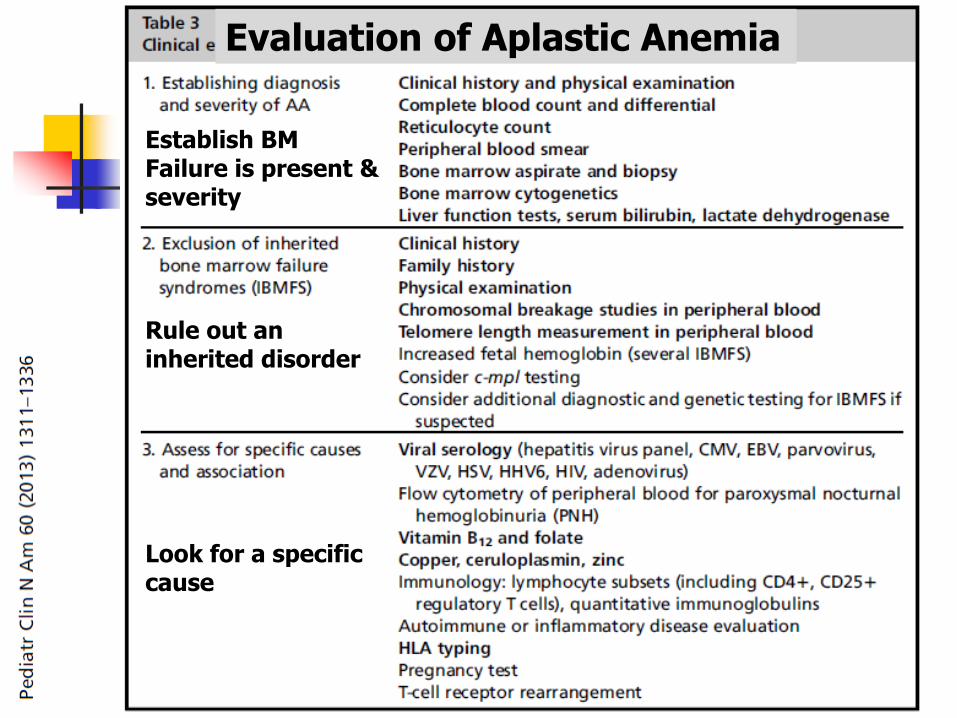
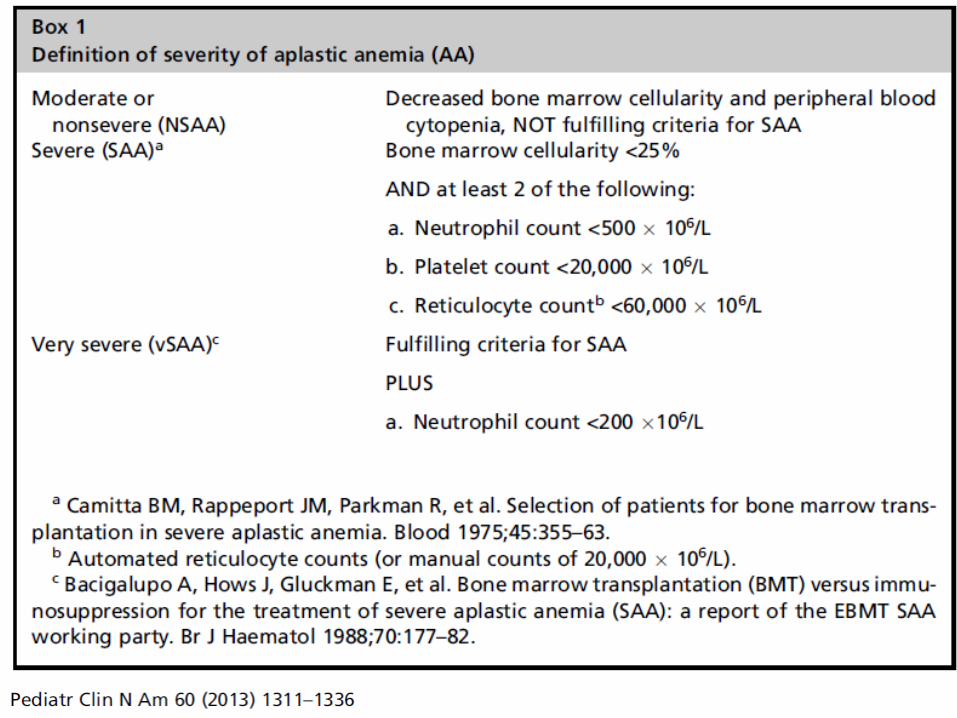
Evaluation of Aplastic Anemia
Establish BM Failure is present & severity
Rule out an inherited disorder
Look for a specific cause
Marrow Findings in Aplastic Anemia
Classified as mild, moderate, severe, or very severe based on marrow cellularity, CBC
Cellularity usually 5-10% No greater than ¼ of the age-related normal range
Decrease in all hematopoietic lineages Morphology may be normal, or may see mild
megaloblastic changes, nuclear/cytoplasmic asynchrony, dyserythropoiesis
Residual cellularity – stromal cells, macrophages, mast cells, lymphs, plasma cells Often perivascular in distribution – you’ll see them in
the particles

Inherited Marrow Failure Syndromes
Fanconi Anemia
Very rare, 1 in 350,000 – 1,000,000 births
Increased in Ashkenazi Jews
Autosomal recessive
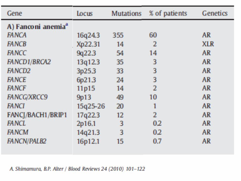
Due to mutations in FANC family of genes
DNA repair defect
Most patients present in first decade of life (median 8 years), but can vary and even present in adulthood
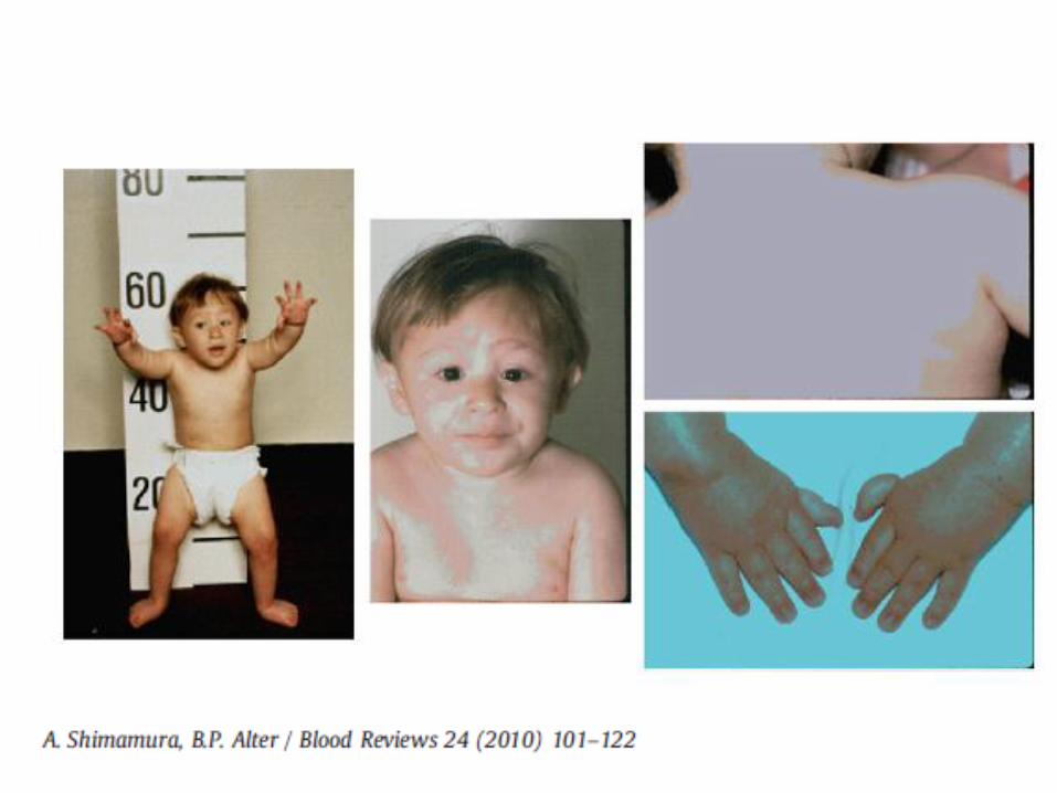
Fanconi Anemia
Characteristic physical abnormalities (not present in up to 1/3 of patients) Facial dysmorphism (hypertelorism,
microcephaly) Upper limb/hand anomalies (absent radii &
thumbs) Short stature Skin abnormalities (café au lait spots, other
pigementation abnormalities) Visceral organ defects
Gastrointestinal, renal, GU, cardiac)
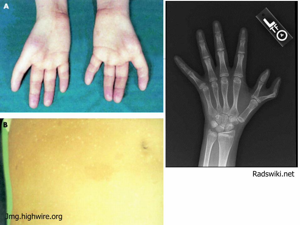
Jmg.highwire.org
Radswiki.net
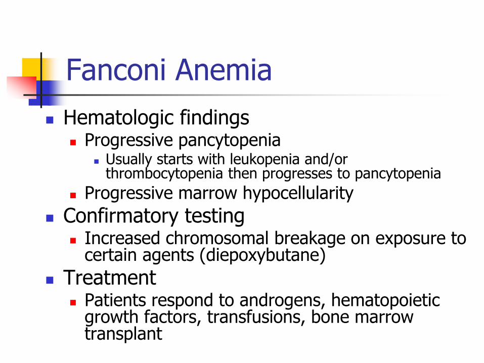
Fanconi Anemia
Hematologic findings Progressive pancytopenia
Usually starts with leukopenia and/or thrombocytopenia then progresses to pancytopenia
Progressive marrow hypocellularity
Confirmatory testing Increased chromosomal breakage on exposure to
certain agents (diepoxybutane)
Treatment Patients respond to androgens, hematopoietic
growth factors, transfusions, bone marrow transplant
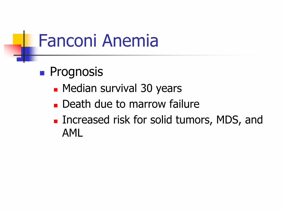
Fanconi Anemia
Prognosis
Median survival 30 years
Death due to marrow failure
Increased risk for solid tumors, MDS, and AML
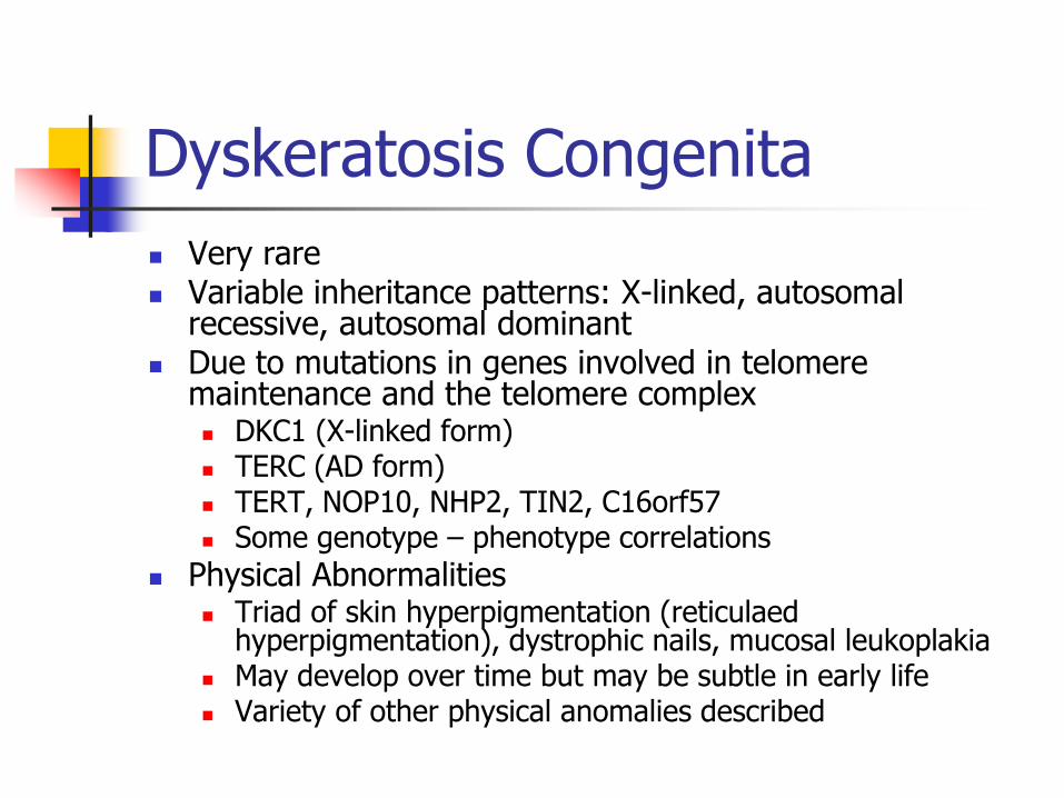
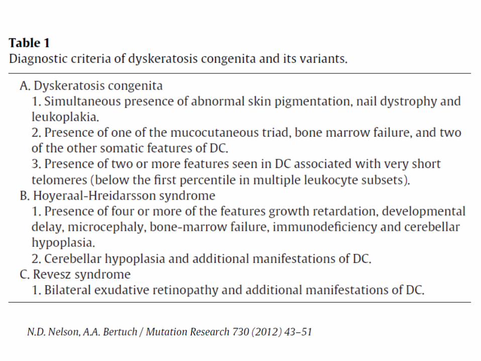
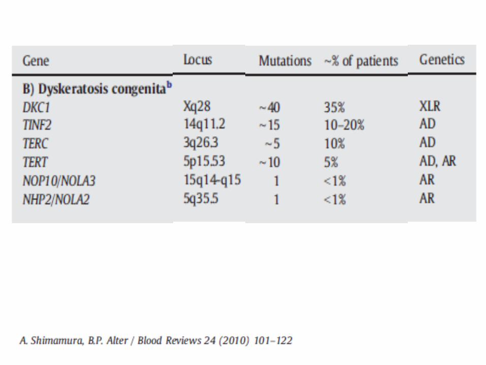
Dyskeratosis Congenita
Very rare Variable inheritance patterns: X-linked, autosomal
recessive, autosomal dominant Due to mutations in genes involved in telomere
maintenance and the telomere complex DKC1 (X-linked form) TERC (AD form) TERT, NOP10, NHP2, TIN2, C16orf57 Some genotype – phenotype correlations
Physical Abnormalities Triad of skin hyperpigmentation (reticulaed
hyperpigmentation), dystrophic nails, mucosal leukoplakia May develop over time but may be subtle in early life Variety of other physical anomalies described
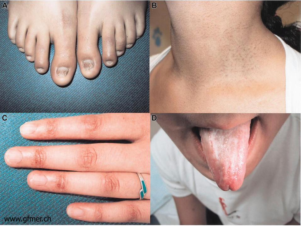
www.gfmer.ch
Physical Findings in DKC
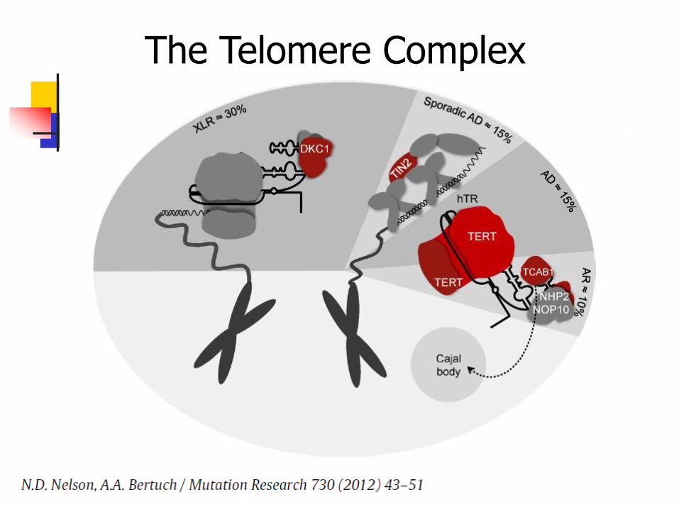
The Telomere Complex
Dyskeratosis Congenita
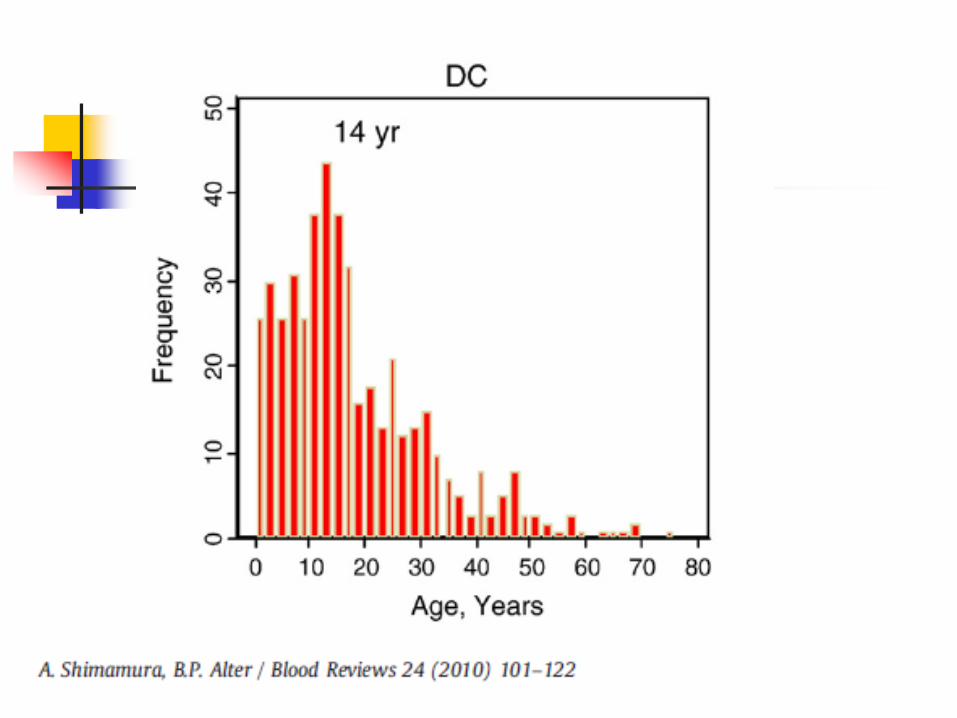
50% of patients will have bone marrow failure by the 2nd decade of life Median age is 16 years Autosomal dominant form may present in 3rd
decade
Hematologic findings Progressive pancytopenia
Starts with anemia (high MCV, incr Hgb F) and/or thromocytopenia
Progressive marrow hypoplasia
Marrow failure may be 1st indication that patient has this syndrome
Dyskeratosis Congenita
Confirmatory testing
Telomere length – these patients have extremely short telomeres
Tested by flow-FISH (combination of flow cytometry and FISH)
Prognosis
Median survival of 30 years
AD type has longer survival, but is at risk of developing solid tumors
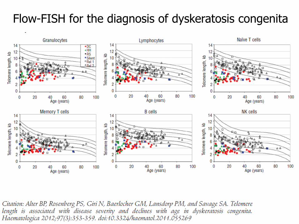
Flow-FISH for the diagnosis of dyskeratosis congenita
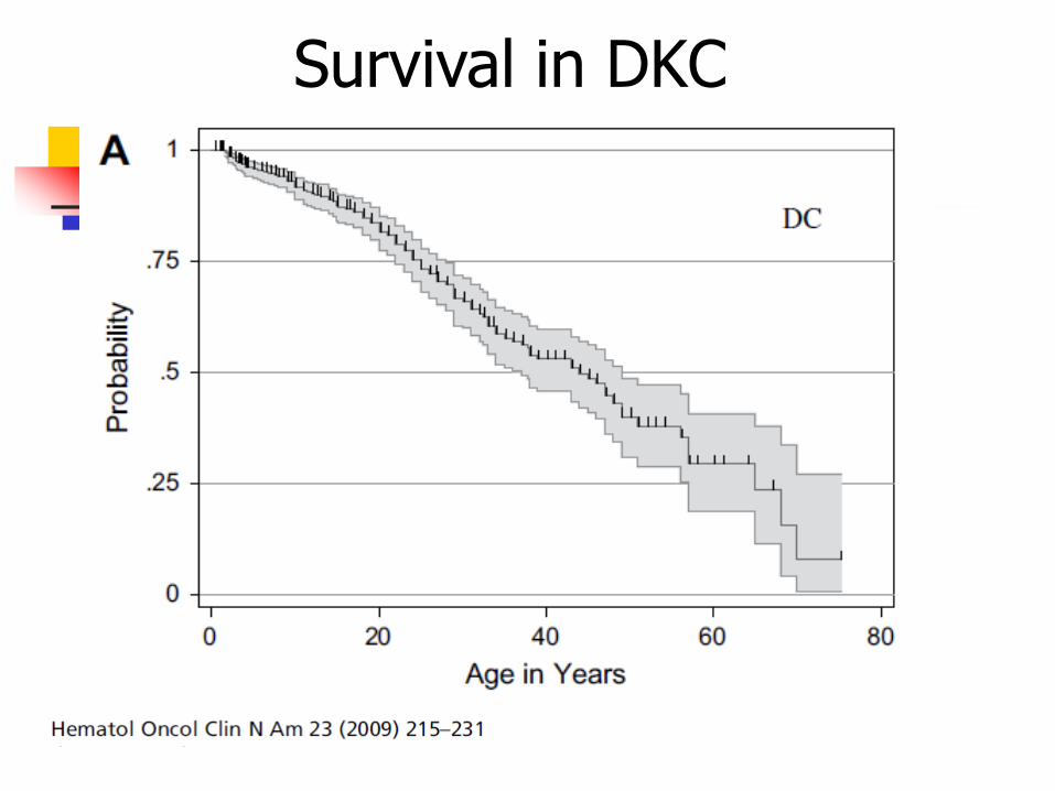
Survival in DKC
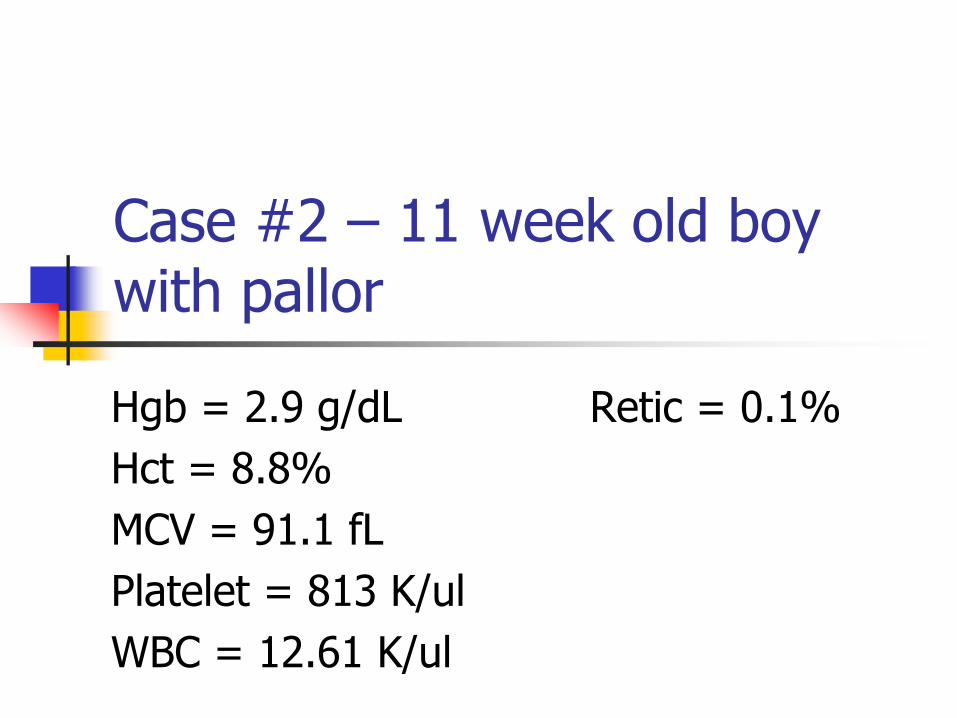
Case #2 – 11 week old boy with pallor
Hgb = 2.9 g/dL Retic = 0.1%
Hct = 8.8%
MCV = 91.1 fL
Platelet = 813 K/ul
WBC = 12.61 K/ul
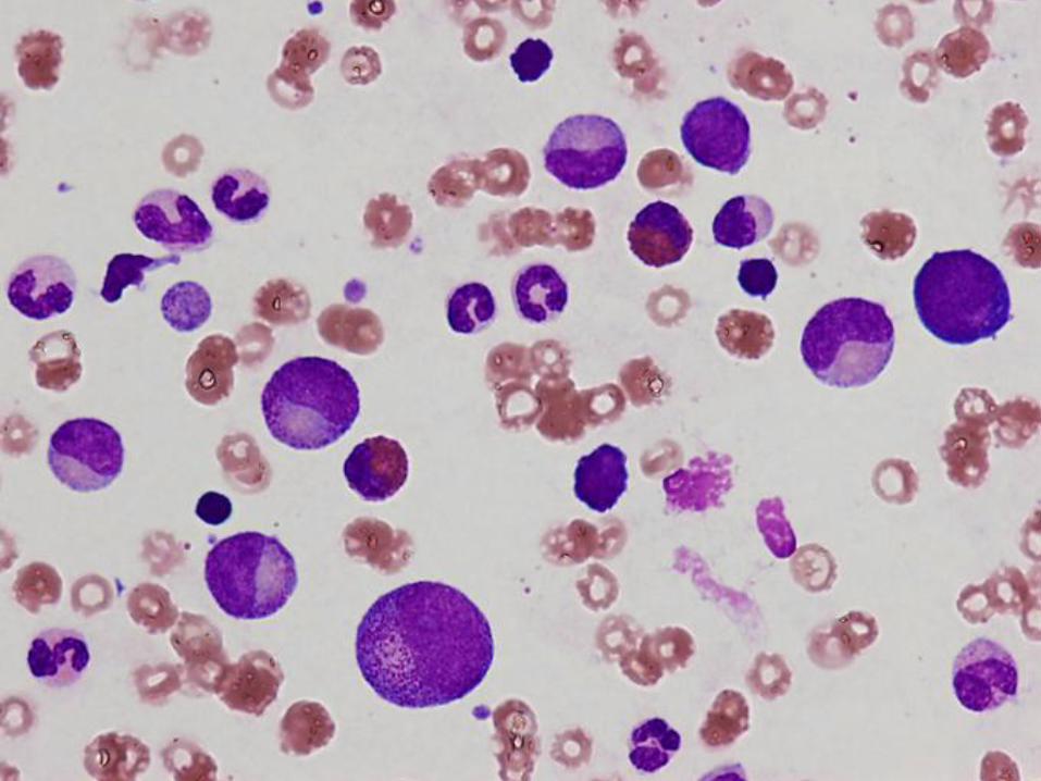
Diagnosis
Pure red cell aplasia
(Diamond Blackfan anemia)
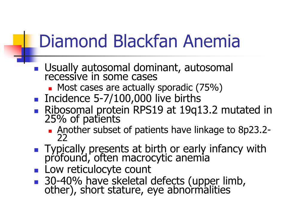
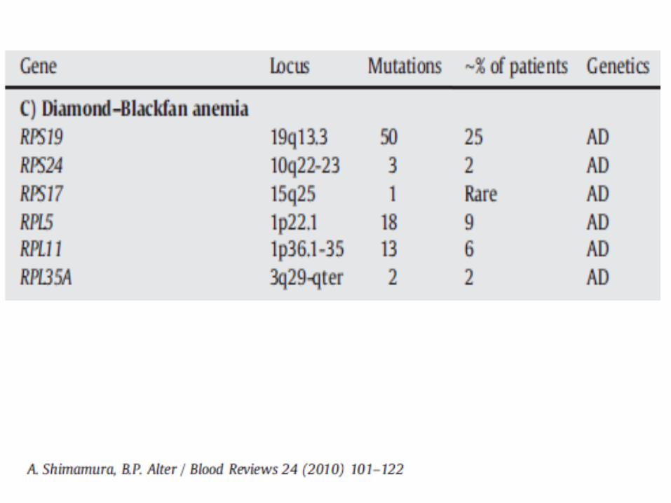
Diamond Blackfan Anemia
Usually autosomal dominant, autosomal recessive in some cases Most cases are actually sporadic (75%)
Incidence 5-7/100,000 live births Ribosomal protein RPS19 at 19q13.2 mutated in
25% of patients Another subset of patients have linkage to 8p23.2-
22 Typically presents at birth or early infancy with
profound, often macrocytic anemia Low reticulocyte count 30-40% have skeletal defects (upper limb,
other), short stature, eye abnormalities
Diamond Blackfan Anemia
Additional laboratory testing Increased red cell adenosine deaminase (ADA) &
Hgb F
Long term survivors may develop multilineage hypoplasia and additional cytopenias Neutropenia & thrombocytopenia
Small increased risk of cancer Patients often respond to early steroid therapy,
also treated with transfusions, and possibly bone marrow transplant
Median survival 40-50 years; 20-25% may spontaneously remit
Marrow Findings in Diamond Blackfan Anemia
Isolated profound erythroid hypoplasia
Only scattered unremarkable erythroblasts
May see increased hematogones
Eosinophilia may be seen
At least initially, normal myelopoiesis and megakaryocytes
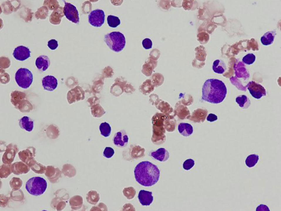
Case #3
6 week old girl with severe neutropenia
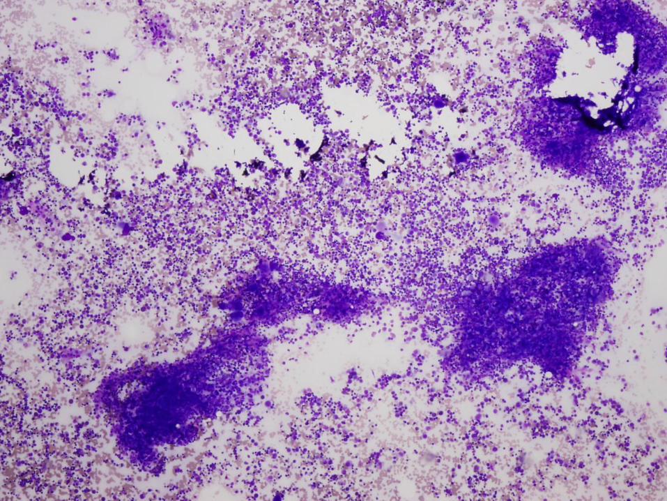
Diagnosis
Severe congenital neutropenia
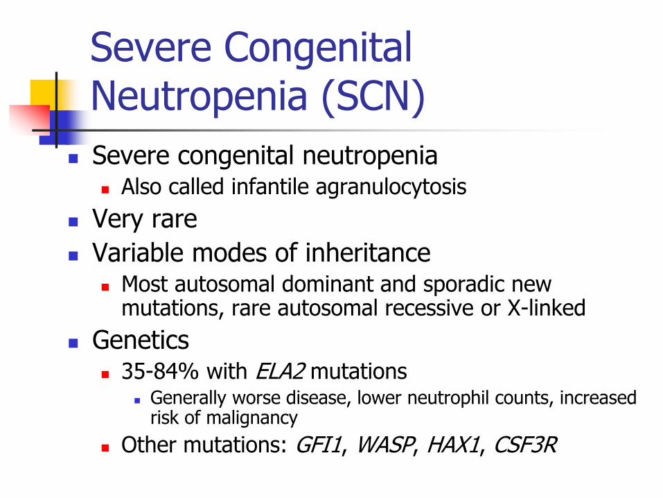
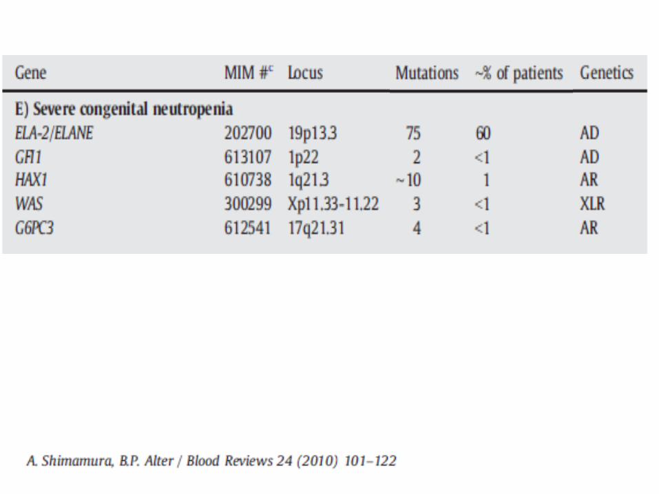
Severe Congenital Neutropenia (SCN)
Severe congenital neutropenia Also called infantile agranulocytosis
Very rare
Variable modes of inheritance Most autosomal dominant and sporadic new
mutations, rare autosomal recessive or X-linked
Genetics 35-84% with ELA2 mutations
Generally worse disease, lower neutrophil counts, increased risk of malignancy
Other mutations: GFI1, WASP, HAX1, CSF3R
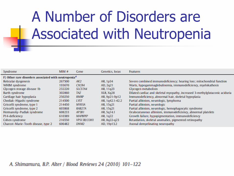
A Number of Disorders are Associated with Neutropenia
Severe Congenital Neutropenia (SCN)
Patients present with severe persistent neutropenia in infancy (ANC <200) & infections
Rest of CBC generally normal
May have peripheral monocytosis & eosinophilia
Severe infections in first month of life, die by 3 years of age unless treated with G-CSF or bone marrow transplant
Note on G-CSF therapy: risk of MDS & AML with prolonged use, increases with duration of use
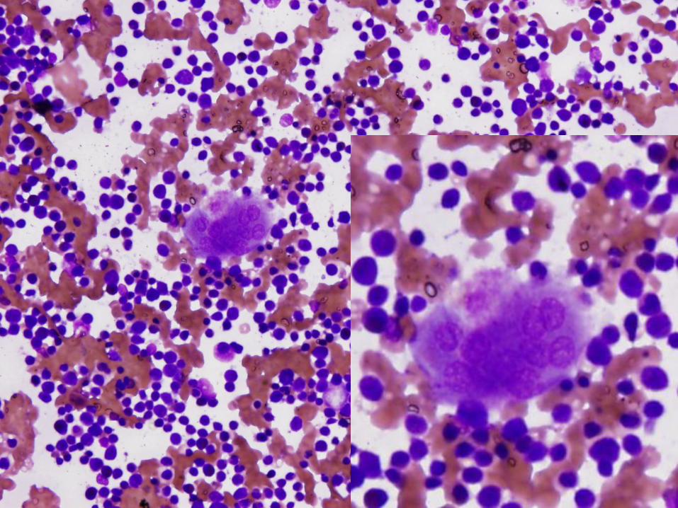
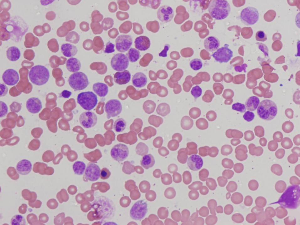
Bone Marrow Morphology in SCN
Normal to slightly decreased cellularity
Decreased myeloid precursors with maturation arrest – few precursors that mature beyond promyelocyte or myelocyte stage
May have enlarged multinucleated myeloid precursors
Promyelocytes often vacuolated
Increased monocytes, eosinophils, macrophages, plasma cells common
Schwachman-Diamond Syndrome
Very rare
Autosomal recessive
50% with short stature, metaphyseal dysostosis, exocrine pancreas insufficiency
Mutations in SBDS gene on 7q11 in 95% of patients
Presents in infancy with progressive neutropenia 40% have other cytopenias as well
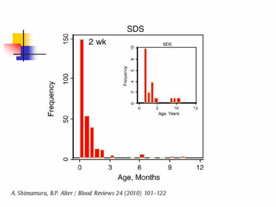
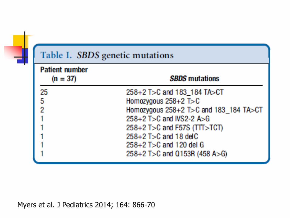
Myers et al. J Pediatrics 2014; 164: 866-70
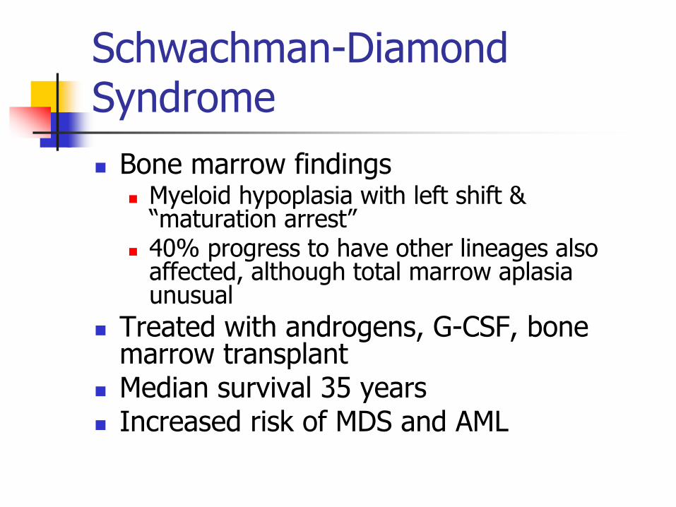
Schwachman-Diamond Syndrome
Bone marrow findings Myeloid hypoplasia with left shift &
“maturation arrest” 40% progress to have other lineages also
affected, although total marrow aplasia unusual
Treated with androgens, G-CSF, bone marrow transplant
Median survival 35 years Increased risk of MDS and AML
Case #4
5 week old boy with persistent thrombocytopenia
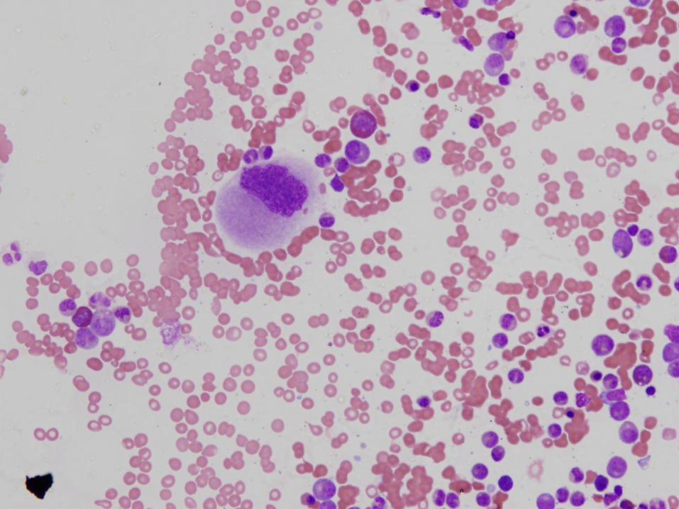
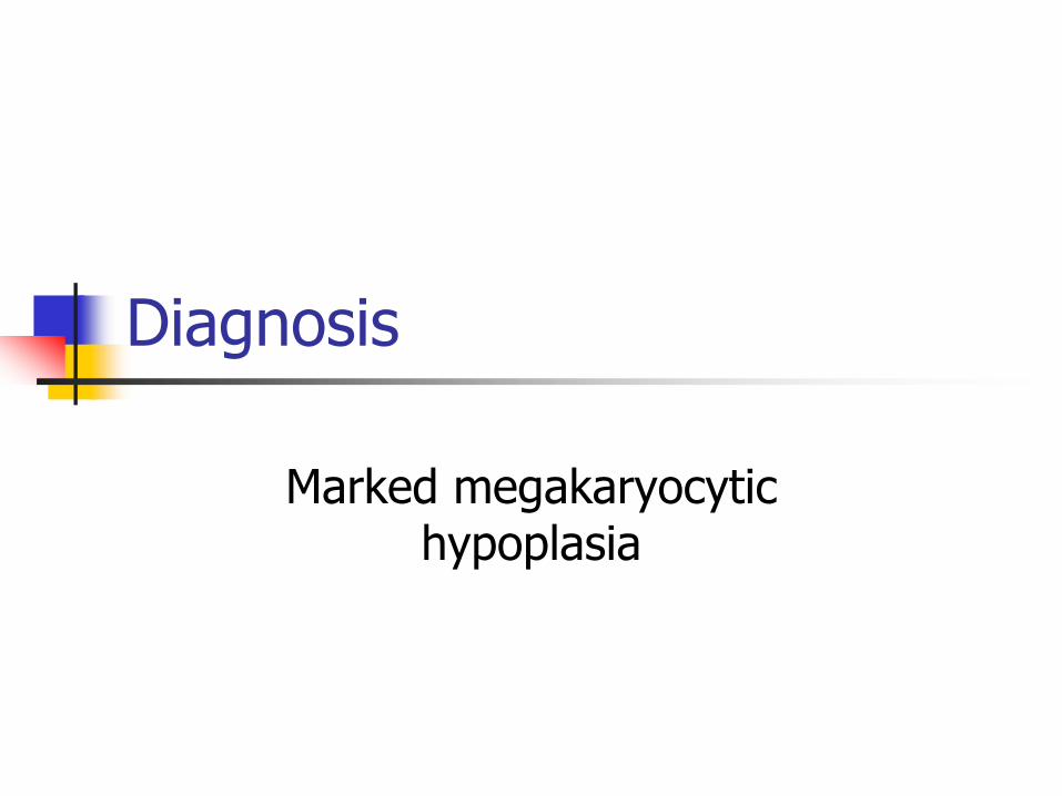
Diagnosis
Marked megakaryocytic hypoplasia
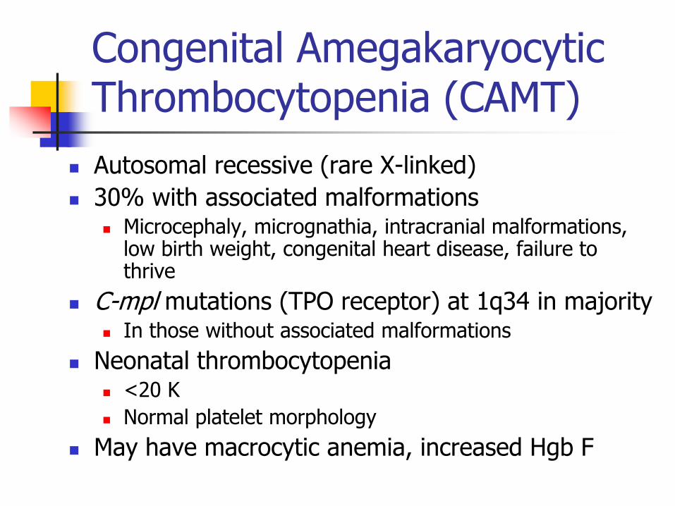
Congenital Amegakaryocytic Thrombocytopenia (CAMT)
Autosomal recessive (rare X-linked)
30% with associated malformations Microcephaly, micrognathia, intracranial malformations,
low birth weight, congenital heart disease, failure to thrive
C-mpl mutations (TPO receptor) at 1q34 in majority In those without associated malformations
Neonatal thrombocytopenia <20 K
Normal platelet morphology
May have macrocytic anemia, increased Hgb F
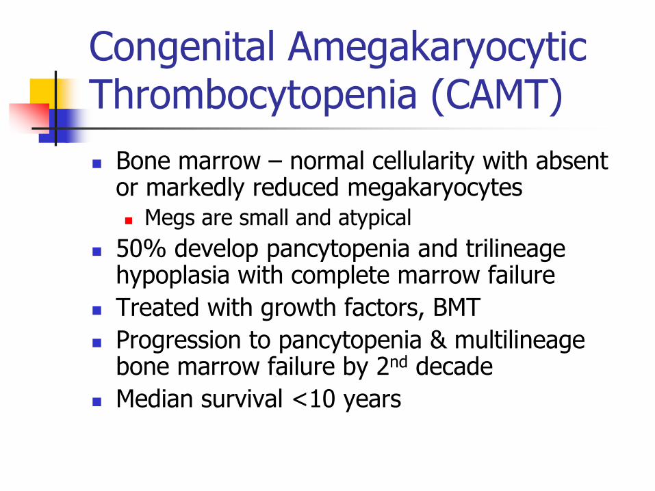
Congenital Amegakaryocytic Thrombocytopenia (CAMT)
Bone marrow – normal cellularity with absent or markedly reduced megakaryocytes Megs are small and atypical
50% develop pancytopenia and trilineage hypoplasia with complete marrow failure
Treated with growth factors, BMT
Progression to pancytopenia & multilineage bone marrow failure by 2nd decade
Median survival <10 years
Thrombocytopenia with Absent Radii (TAR)
Rare DNA repair disorder (radiation sensitive) Gene not identified as of yet, but normal c-MPL 1q21.1 appears to be the chromosomal region involved
Autosomal recessive Neonatal thrombocytopenia
10-30K range Bone abnormalities
Congenital absence or extreme hypoplasia of radial bones (thumbs are present), commonly bilateral
Absent, short, or malformed ulnae Variety of other skeletal abnormalities also
described Cardiac lesions in a subset
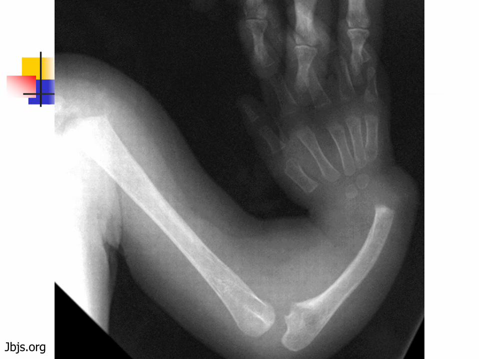
Jbjs.org
Thrombocytopenia with Absent Radii (TAR)
High incidence transient leukemoid reactions (can be >100,000)
Bone marrow with normal cellularity but absent or markedly decreased megakaryocytes in infancy If present, megs are small and immature Myeloids & erythroids are normal
Spontaneous recovery common with normalization of platelet count and more normal numbers of megakaryocytes
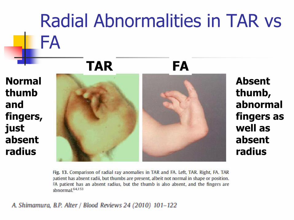
Radial Abnormalities in TAR vs FA
TAR FA Absent thumb, abnormal fingers as well as absent radius
Normal thumb and fingers, just absent radius
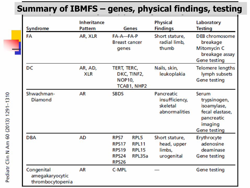
Summary of IBMFS – genes, physical findings, testing