bone marrow failure e an abnormal bone marrow
TRANSCRIPT
haematologica | 2017; 102(6) 1017
Received: October 22, 2016.
Accepted: March 20, 2017.
Pre-published: March 24, 2017.
©2017 Ferrata Storti Foundation
Material published in Haematologica is covered by copyright.All rights are reserved to the Ferrata Storti Foundation. Use ofpublished material is allowed under the following terms andconditions: https://creativecommons.org/licenses/by-nc/4.0/legalcode. Copies of published material are allowed for personal or inter-nal use. Sharing published material for non-commercial pur-poses is subject to the following conditions: https://creativecommons.org/licenses/by-nc/4.0/legalcode,sect. 3. Reproducing and sharing published material for com-mercial purposes is not allowed without permission in writingfrom the publisher.
Correspondence: [email protected] or [email protected]
Ferrata StortiFoundation
EUROPEANHEMATOLOGYASSOCIATION
Haematologica 2017Volume 102(6):1017-1027
ARTICLEBone Marrow Failure
doi:10.3324/haematol.2016.158717
Check the online version for the most updatedinformation on this article, online supplements,and information on authorship & disclosures:www.haematologica.org/content/102/6/1017
Introduction
The hematopoietic system is built upon the ordered self-renewal and differentia-tion of hematopoietic stem cells (HSC) within the bone marrow (BM). This processinvolves intrinsic and extrinsic cues including both cellular and humoral regulatorysignals generated by the HSC microenvironment, termed as “niche”. The cellularcomposition of this “niche” is heterogeneous, including endothelial cells,1
osteoblasts,2 adipocytes, and mesenchymal stem/progenitor cells (MSPC), a com-mon progenitor for many of the cell lineages comprising the HSC niche.3-5 For fatedecisions, regulatory signals from the BM microenvironment are transmitted toHSC through intercellular interactions within the proximity of the endosteal sur-face, the perivascular space, soluble factors, and the extracellular matrix.6 These cel-lular and humoral regulatory signals dictate the fates of HSC, including self-renewal,proliferation, differentiation, and apoptosis.7 In addition, there is increasing evi-dence suggesting a role of the hematopoietic microenvironment in hematopoieticdisorders, such as myeloproliferative neoplasms8,9 and myelodysplastic syndrome.10
Fanconi anemia (FA) is a complex inherited disorder caused by germline muta-tions in at least one of 16 genes including FANCA, -B, -C, -D1, -D2, -E, -F, -G, -I, -
Fanconi anemia is a complex heterogeneous genetic disorder with ahigh incidence of bone marrow failure, clonal evolution to acutemyeloid leukemia and mesenchymal-derived congenital anomalies.
Increasing evidence in Fanconi anemia and other genetic disorders pointstowards an interdependence of skeletal and hematopoietic development,yet the impact of the marrow microenvironment in the pathogenesis of thebone marrow failure in Fanconi anemia remains unclear. Here we demon-strated that mice with double knockout of both Fancc and Fancg genes haddecreased bone formation at least partially due to impaired osteoblast dif-ferentiation from mesenchymal stem/progenitor cells. Mesenchymalstem/progenitor cells from the double knockout mice showed impairedhematopoietic supportive activity. Mesenchymal stem/progenitor cells ofpatients with Fanconi anemia exhibited similar cellular deficits, includingincreased senescence, reduced proliferation, impaired osteoblast differenti-ation and defective hematopoietic stem/progenitor cell supportive activity.Collectively, these studies provide unique insights into the physiologicalsignificance of mesenchymal stem/progenitor cells in supporting the mar-row microenvironment, which is potentially of broad relevance inhematopoietic stem cell transplantation.
An abnormal bone marrow microenvironmentcontributes to hematopoietic dysfunction inFanconi anemiaYuan Zhou,1,2,3§ Yongzheng He,2,3§ Wen Xing,1,2,3 Peng Zhang,4,5 Hui Shi,1,4,5
Shi Chen,4,5 Jun Shi,1 Jie Bai,1 Steven D. Rhodes,2,3 Fengqui Zhang,1
Jin Yuan,2,3 Xianlin Yang,2,3 Xiaofan Zhu,1 Yan Li,2,3 Helmut Hanenberg,2,3,6
Mingjiang Xu,4,5 Kent A. Robertson,2,3 Weiping Yuan,1 Grzegorz Nalepa,2,3
Tao Cheng,1 D. Wade Clapp2,3 and Feng-Chun Yang4,5
§These authors contributed equally to this work.
1State Key Laboratory of Experimental Hematology, Institute of Hematology and BloodDiseases Hospital, Chinese Academy of Medical Sciences & Peking Union MedicalCollege, Tianjin, China; 2Herman B Wells Center for Pediatric Research, Indianapolis, IN,USA; 3Department of Pediatrics, Indiana University School of Medicine, Indianapolis, IN,USA; 4Sylvester Comprehensive Cancer Center, Miami, FL, USA; 5Department ofBiochemistry and Molecular Biology, University of Miami Miller School of Medicine,Miami, FL, USA and 6Department of Otorhinolaryngology and Head/Neck Surgery,Heinrich Heine University, Düsseldorf, Germany
ABSTRACT
J, -L, -M, -N, -O, –P, and –Q.11-19 Clinically, FA is a chromo-somal fragility disorder characterized by progressive BMfailure (BMF), variable developmental anomalies, and astrong propensity to develop cancer. The risk of developingBMF by 40 years of age is as high as 90%, and the cumu-lative incidence of hematologic and non-hematologicmalignancies was reported to be as high as 33% and 28%,respectively.20 In the natural course of the disease, FApatients develop progressive pancytopenia, indicating thatthe defect occurs at the level of HSC.21-23 FA patients have ahigh incidence of inherited skeletal malformations andosteoporosis,20,24 suggesting a role of FA proteins in osteoge-nesis and bone maintenance. Despite these clinical obser-vations of multiple mesenchymal defects in FA and ourincreasing awareness of the interdependence between theBM niche and hematopoiesis, relatively little attention hasbeen directed to investigating the putative associationbetween abnormal HSC function and the BM niche in FA.MSPC are a major component of the hematopoietic
niche and have been shown to serve a critical function ashematopoiesis-supporting stromal cells.25 Here, we reportthat defective MSPC are pivotal mediators in the patho-genesis of hematopoietic defects in Fancc-/-;Fancg-/- doubleknockout (DKO) mice. Our studies provide detailed cellu-lar and molecular evidence implicating mesenchymal cellsas contributory to the BMF in FA, indicating the potentialutility of MSPC/HSC co-transplantation, which mayimprove treatment of BMF in FA.
Methods
Animals and reagentsThe Fancc and Fancg double heterozygous mice used in this
study have been described previously.26-28 These mice were back-crossed into a C57BL/6J strain and were then bred to produceFancc-/-;Fancg-/- (DKO) and wild-type (WT) mice. Age- and gender-matched DKO and WT mice were used for all experiments. Allprotocols were approved by the Institutional Animal Care and UseCommittee at Indiana University School of Medicine. Chemicalswere obtained from Sigma (St. Louis, MO, USA) unless otherwiseindicated.
Isolation and expansion of mesenchymal stem/progenitor cellsMSPC from mice were generated as previously described.29
Briefly, BM mononuclear cells (BMMNC) were separated by low-density gradient centrifugation from 6- to 8-week-old, age- andgender-matched WT and DKO mice, then cultured in completemouse MesenCult medium (Stem Cell Technologies Inc,Vancouver, Canada) at 37°C in 5% CO2. MSPC between passagefive to ten were used for the following experiments. The pheno-typic analyses of MSPC were performed by evaluating the expres-sion of surface markers including CD44, CD105, CD146, CD29on a FACS Calibur flow cytometer as previously described.30 Forhuman MSPC isolation, whole BM cells from FA patients andhealthy donors were cultured in Dulbecco modified Eagle medium(DMEM)/F12 (Gibco, Carlsbad, USA), containing 10% fetalbovine serum (Hyclone, South Logan, USA), 1x Insulin transferrinselenium-A (Life Technologies, Carlsbad, USA), 10 ng/mL humanepidermal growth factor (Peprotech, Rocky Hill, NJ, USA), and 10ng/mL human platelet-derived growth factor-BB (Peprotech) at37°C in 5% CO2 and 5% O2 in a fully humidified atmosphere.MSPC at passage three to five were used for the following exper-iments.
Micro-computed tomography To evaluate trabecular microarchitecture in the distal femoral
metaphysis, fixed femora were scanned using a high-resolutiondesktop micro-computed tomography imaging system (μCT-20;Scanco Medical AG, Basserdorf, Switzerland). The region of inter-est was defined as 15% of the total femur length measured fromthe tip of the femoral condyle and extending proximally for 200slices with an increment of 9 µm, and was subsequently recon-structed, filtered (σ= 0.8 and support = 1.0), and thresholded (at22% of the possible gray scale value) for analysis, as describedelsewhere.31 Trabecular bone was contoured manually within thetrabecular compartment, excluding the cortical shell. The parame-ter of micro-architecture for bone volume fraction (BV/TV, %) wasmeasured.
Histomorphometric measurementsUpon sacrifice, the isolated bones were fixed in 10% neutral
buffered formalin for 48 h, dehydrated in graded ethanol, andembedded undecalcified in methyl methacrylate. Sagittal sections(5 µm thick) were cut from the middle of the femur. Tartrate-resis-tant acid phosphatase (TRAP) staining was performed using aleukocyte acid phosphatase kit (Sigma Diagnostics, St. Louis, MO,USA) and McNeal staining was performed using the McNeal tetra-chromat kit (Polysciences, Warrington, PA, USA), both accordingto the manufacturers’ protocols. One section per femur wasviewed at 100x magnification on a Leitz DMRXE microscope(Leica Mikroskopie und System GmbH, Wetzlar, Germany).Images were captured using a QImaging camera and QCapture-Pro software (Fryer Company Inc., Cincinnati, OH, USA). Theregion of interest for the metaphysis was defined by a rectangulararea, which begins 0.5 mm proximal to the midpoint of thegrowth plate, non-inclusive of cortical bone, and extends proxi-mally for a total area of approximately 2.8 mm2.
Bone remodeling measurementFluorochrome labeling of the bones was performed by
intraperitoneal injections of calcein (20 mg/kg, 8 days before sac-rifice) and alizarin (20 mg/kg, 4 days before sacrifice), as previ-ously described.32 Trabecular bone turnover was assessed bymeasuring the extent of single-labeled surface (sLS), double-labeled surface (dLS) and the surface of the bone (BS) betweenthe calcein and alizarin labels using Image Pro Plus version 4.1software (Media Cybernetics, Silver Spring, MD, USA). Derivedhistomorphometric parameters included: (i) mineralizing surface(MS/BS), a measure of active bone-forming surface, calculated as(dLS+sLS/2)/BS; (ii) mineral apposition rate (MAR, μm/ day), ameasure of the rate of radial expansion of new bone, calculatedas Thickness/4 day; and (iii) bone formation rate, an overallmeasure of bone formation that combines MS/BS and MAR, cal-culated as MS/BS × MAR.
Other methodsMethods for the clonogenic assay, bone mineral density quan-
tification, annexin V/propidium iodide staining, senescence assay,thymidine incorporation assay, purification of HSPC, long-termculture of HSPC on MSPC monolayers, detection of reactive oxy-gen species (ROS), osteoblast and adipocyte differentiation ofMSPC, reciprocal transplantation and co-transplantation of FABMMNC and MSPC in NS2 mice are described in detail in theOnline Supplementary Methods.
StatisticsSurvival curves were compared using the log-rank test.
Differences between two groups with equal variances wereassessed by two-tailed Student t-tests. Multiple comparisons were
Y. Zhou et al.
1018 haematologica | 2017; 102(6)
Microenvironment and Fanconi anemia
haematologica | 2017; 102(6) 1019
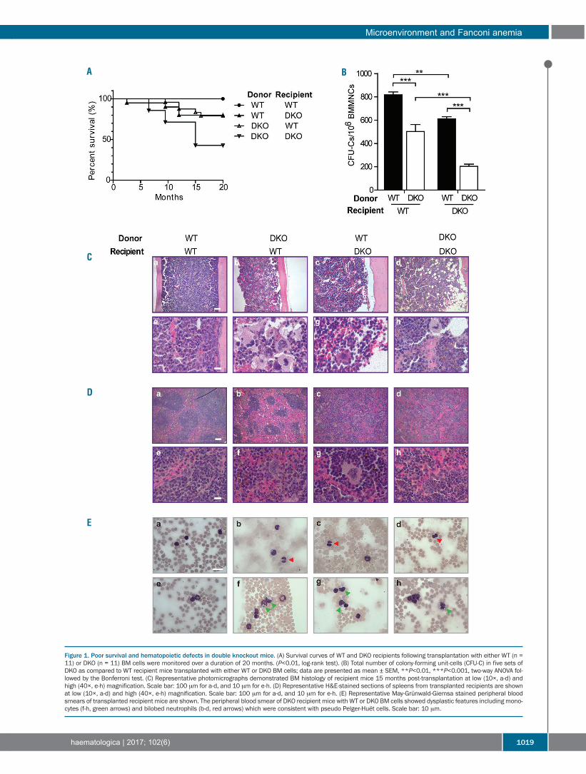
Figure 1. Poor survival and hematopoietic defects in double knockout mice. (A) Survival curves of WT and DKO recipients following transplantation with either WT (n =11) or DKO (n = 11) BM cells were monitored over a duration of 20 months. (P<0.01, log-rank test). (B) Total number of colony-forming unit-cells (CFU-C) in five sets ofDKO as compared to WT recipient mice transplanted with either WT or DKO BM cells; data are presented as mean ± SEM, **P<0.01, ***P<0.001, two-way ANOVA fol-lowed by the Bonferroni test. (C) Representative photomicrographs demonstrated BM histology of recipient mice 15 months post-transplantation at low (10×, a-d) andhigh (40×, e-h) magnification. Scale bar: 100 μm for a-d, and 10 μm for e-h. (D) Representative H&E-stained sections of spleens from transplanted recipients are shownat low (10×, a-d) and high (40×, e-h) magnification. Scale bar: 100 μm for a-d, and 10 μm for e-h. (E) Representative May-Grünwald-Giemsa stained peripheral bloodsmears of transplanted recipient mice are shown. The peripheral blood smear of DKO recipient mice with WT or DKO BM cells showed dysplastic features including mono-cytes (f-h, green arrows) and bilobed neutrophils (b-d, red arrows) which were consistent with pseudo Pelger-Huët cells. Scale bar: 10 μm.
A B
C
D
E
Y. Zhou et al.
1020 haematologica | 2017; 102(6)
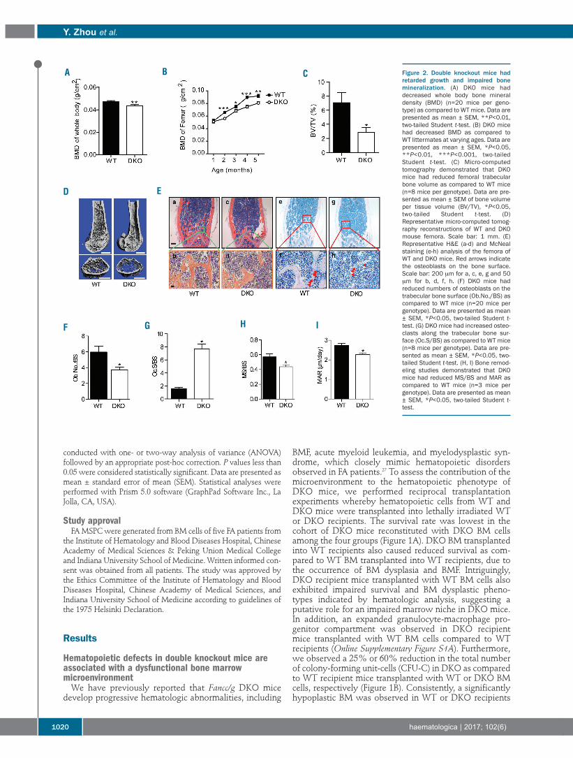
Figure 2. Double knockout mice hadretarded growth and impaired bonemineralization. (A) DKO mice haddecreased whole body bone mineraldensity (BMD) (n=20 mice per geno-type) as compared to WT mice. Data arepresented as mean ± SEM, **P<0.01,two-tailed Student t-test. (B) DKO micehad decreased BMD as compared toWT littermates at varying ages. Data arepresented as mean ± SEM, *P<0.05,**P<0.01, ***P<0.001, two-tailedStudent t-test. (C) Micro-computedtomography demonstrated that DKOmice had reduced femoral trabecularbone volume as compared to WT mice(n=8 mice per genotype). Data are pre-sented as mean ± SEM of bone volumeper tissue volume (BV/TV), *P<0.05,two-tailed Student t-test. (D)Representative micro-computed tomog-raphy reconstructions of WT and DKOmouse femora. Scale bar: 1 mm. (E)Representative H&E (a-d) and McNealstaining (e-h) analysis of the femora ofWT and DKO mice. Red arrows indicatethe osteoblasts on the bone surface.Scale bar: 200 μm for a, c, e, g and 50μm for b, d, f, h. (F) DKO mice hadreduced numbers of osteoblasts on thetrabecular bone surface (Ob.No./BS) ascompared to WT mice (n=20 mice pergenotype). Data are presented as mean± SEM, *P<0.05, two-tailed Student t-test. (G) DKO mice had increased osteo-clasts along the trabecular bone sur-face (Oc.S/BS) as compared to WT mice(n=8 mice per genotype). Data are pre-sented as mean ± SEM, *P<0.05, two-tailed Student t-test. (H, I) Bone remod-eling studies demonstrated that DKOmice had reduced MS/BS and MAR ascompared to WT mice (n=3 mice pergenotype). Data are presented as mean± SEM, *P<0.05, two-tailed Student t-test.
ED
A B C
F G H I
conducted with one- or two-way analysis of variance (ANOVA)followed by an appropriate post-hoc correction. P values less than0.05 were considered statistically significant. Data are presented asmean ± standard error of mean (SEM). Statistical analyses wereperformed with Prism 5.0 software (GraphPad Software Inc., LaJolla, CA, USA).
Study approvalFA MSPC were generated from BM cells of five FA patients from
the Institute of Hematology and Blood Diseases Hospital, ChineseAcademy of Medical Sciences & Peking Union Medical Collegeand Indiana University School of Medicine. Written informed con-sent was obtained from all patients. The study was approved bythe Ethics Committee of the Institute of Hematology and BloodDiseases Hospital, Chinese Academy of Medical Sciences, andIndiana University School of Medicine according to guidelines ofthe 1975 Helsinki Declaration.
Results
Hematopoietic defects in double knockout mice areassociated with a dysfunctional bone marrow microenvironmentWe have previously reported that Fancc/g DKO mice
develop progressive hematologic abnormalities, including
BMF, acute myeloid leukemia, and myelodysplastic syn-drome, which closely mimic hematopoietic disordersobserved in FA patients.27 To assess the contribution of themicroenvironment to the hematopoietic phenotype ofDKO mice, we performed reciprocal transplantationexperiments whereby hematopoietic cells from WT andDKO mice were transplanted into lethally irradiated WTor DKO recipients. The survival rate was lowest in thecohort of DKO mice reconstituted with DKO BM cellsamong the four groups (Figure 1A). DKO BM transplantedinto WT recipients also caused reduced survival as com-pared to WT BM transplanted into WT recipients, due tothe occurrence of BM dysplasia and BMF. Intriguingly,DKO recipient mice transplanted with WT BM cells alsoexhibited impaired survival and BM dysplastic pheno-types indicated by hematologic analysis, suggesting aputative role for an impaired marrow niche in DKO mice.In addition, an expanded granulocyte-macrophage pro-genitor compartment was observed in DKO recipientmice transplanted with WT BM cells compared to WTrecipients (Online Supplementary Figure S1A). Furthermore,we observed a 25% or 60% reduction in the total numberof colony-forming unit-cells (CFU-C) in DKO as comparedto WT recipient mice transplanted with WT or DKO BMcells, respectively (Figure 1B). Consistently, a significantlyhypoplastic BM was observed in WT or DKO recipients
transplanted with either DKO or WT BM cells, respective-ly, with the most severe BM hypoplasia occurring in DKOrecipients reconstituted with DKO BM cells (Figure 1C).The histology of the spleens of DKO recipients with eitherWT or DKO BM cells revealed disrupted architecture.Lymphoid aggregates in the white pulp (disrupted archi-tecture) of DKO recipient spleens were also smaller thanthose of WT recipients (Figure 1D). May-Grunwald-Giemsa stained peripheral blood smears prepared fromDKO recipients showed dysplastic features, includinghyposegmented (bilobed) neutrophils with fine nuclearbridging consistent with pseudo-Pelger-Huët cells (Figure1E, b-d, red arrows, Online Supplementary Figure S1B) andmonocytes (Figure 1E, f-h, green arrows), whereas blastswere rare. In addition, dysplastic megakaryocytes (multin-uclear megakaryocytes and hyposegmented megakaryo -
cytes) were observed in the BM of DKO recipients trans-planted with WT BM cells, but not in WT recipient mice(Online Supplementary Figure S1C). These data suggest thatDKO recipient mice transplanted with WT or DKO BMcells develop a myelodysplastic syndrome-like disease33and provide strong in vivo evidence that the niche plays acooperative role in the pathogenesis of impaired marrowengraftment in the DKO FA mouse model.
Impaired skeletal development and bone mass deficitsin double knockout miceSkeletal anomalies including short stature and osteope-
nia/osteoporosis are widespread among the FA popula-tion.34 We, therefore, sought to ascertain the impact ofFancc/g genetic inactivation on skeletal development. Thebody length and body weight (Online Supplementary Figure
Microenvironment and Fanconi anemia
haematologica | 2017; 102(6) 1021
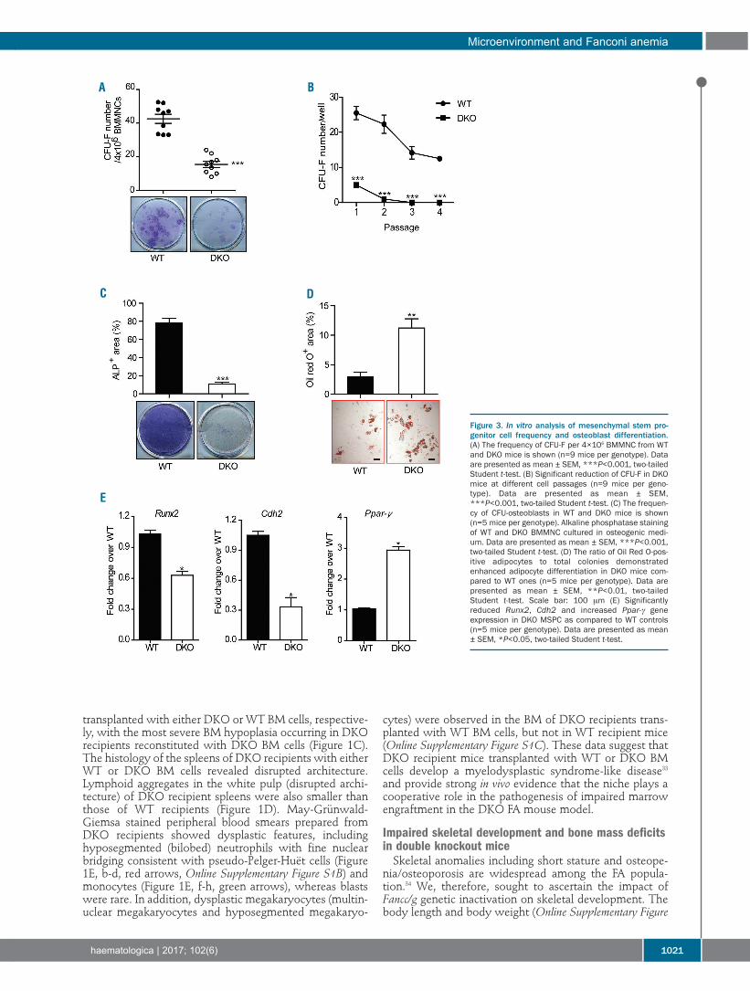
Figure 3. In vitro analysis of mesenchymal stem pro-genitor cell frequency and osteoblast differentiation.(A) The frequency of CFU-F per 4×10⁶ BMMNC from WTand DKO mice is shown (n=9 mice per genotype). Dataare presented as mean ± SEM, ***P<0.001, two-tailedStudent t-test. (B) Significant reduction of CFU-F in DKOmice at different cell passages (n=9 mice per geno-type). Data are presented as mean ± SEM,***P<0.001, two-tailed Student t-test. (C) The frequen-cy of CFU-osteoblasts in WT and DKO mice is shown(n=5 mice per genotype). Alkaline phosphatase stainingof WT and DKO BMMNC cultured in osteogenic medi-um. Data are presented as mean ± SEM, ***P<0.001,two-tailed Student t-test. (D) The ratio of Oil Red O-pos-itive adipocytes to total colonies demonstratedenhanced adipocyte differentiation in DKO mice com-pared to WT ones (n=5 mice per genotype). Data arepresented as mean ± SEM, **P<0.01, two-tailedStudent t-test. Scale bar: 100 μm (E) Significantlyreduced Runx2, Cdh2 and increased Ppar-γ geneexpression in DKO MSPC as compared to WT controls(n=5 mice per genotype). Data are presented as mean± SEM, *P<0.05, two-tailed Student t-test.
A B
C D
E
S2A,B) of DKO mice were significantly reduced as com-pared to those of age- and sex-matched WT littermates.Whole body bone mineral density, determined bypDEXA, was also reduced in DKO mice as compared toWT controls (Figure 2A), with an even more substantialreduction in femoral bone mineral density of the DKOmice versus WT controls (Figure 2B). Consistent with thedecreased bone mass in DKO mice determined by bonemineral density analysis, micro-computed tomographyanalysis of the animals at 6 months of age revealeddecreased bone volume in the mid-shaft of DKO femoracompared to WT controls (Figure 2C,D). Quantitative his-tomorphometric analysis also revealed significantlyreduced bone volume in DKO femora versus WT ones(Online Supplementary Figure S2C).Alterations in skeletal homeostasis can occur secondary
to imbalances between bone-forming osteoblast activity
and osteoclast-mediated bone resorption. To furtherassess osteoblast and osteoclast development in vivo, quan-titative histomorphometry was performed on histologicalsections from the distal femoral metaphysis stained withhematoxylin and eosin (H&E), McNeal, and the osteoclastenzyme TRAP (Figure 2E,F, Online Supplementary FigureS2D,E). Sections stained with H&E revealed a markedreduction in trabecular and cortical bone in DKO femoraas compared to WT control femora (Figure 2E, a-d).Manually counting osteoblasts in the femoral trabecularbone on McNeal-stained sections revealed that the num-ber of osteoblasts was singnificantly lower in DKO micethan in WT controls (Figure 2E, e-h, Figure 2F). In addition,a significantly increased osteoclast surface to bone surfaceratio was observed in DKO mice than in WT controls, asassessed by scoring the TRAP-positive staining osteoclastsurface in the femoral trabeculae normalized to the trabec-
Y. Zhou et al.
1022 haematologica | 2017; 102(6)
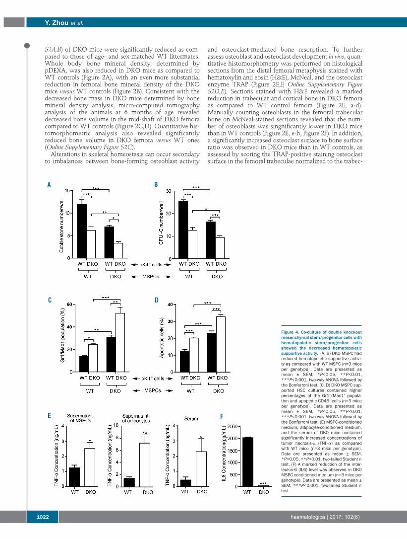
Figure 4. Co-culture of double knockoutmesenchymal stem/progenitor cells withhematopoietic stem/progenitor cellsshowed the decreased hematopoieticsupportive activity. (A, B) DKO MSPC hadreduced hematopoietic supportive activi-ty as compared with WT MSPC (n=3 miceper genotype). Data are presented asmean ± SEM, *P<0.05, **P<0.01,***P<0.001, two-way ANOVA followed bythe Bonferroni test. (C, D) DKO MSPC sup-ported HSC cultures contained higherpercentages of the Gr1+/Mac1+ popula-tion and apoptotic CD45+ cells (n=3 miceper genotype). Data are presented asmean ± SEM, *P<0.05, **P<0.01,***P<0.001, two-way ANOVA followed bythe Bonferroni test. (E) MSPC-conditionedmedium, adipocyte-conditioned medium,and the serum of DKO mice containedsignificantly increased concentrations oftumor necrosis-α (TNF-α) as comparedwith WT mice (n=3 mice per genotype).Data are presented as mean ± SEM,*P<0.05, **P<0.01, two-tailed Student t-test. (F) A marked reduction of the inter-leukin-6 (IL6) level was observed in DKOMSPC conditioned medium (n=3 mice pergenotype). Data are presented as mean ±SEM, ***P<0.001, two-tailed Student t-test.
BA
C D
E F
ular bone surface (Figure 2G, Online Supplementary FigureS2D). To study dynamic changes in bone remodeling, WTand DKO mice were injected with fluorochrome markersto label the bone surface (Online Supplementary FigureS2E).35 A 23% reduction in the mineralizing surface(MS/BS, Figure 2H, Online Supplementary Table S1), a 17%reduction in mineral apposition rate (MAR, Figure 2I,Online Supplementary Table S1), and a 36% reduction inbone formation rate (BFR)/BS were observed in DKO miceas compared to age- and sex-matched WT controls (OnlineSupplementary Figure S2F, Online Supplementary Table S1).Collectively, these data suggest that abnormal osteoblast-and osteoclast-mediated bone turnover in DKO mice leadsto pathological bone remodeling.
Fancc/g genetic ablation alters mesenchymalstem/progenitor cell fates, favoring adipogenic versus osteoblastic differentiationAs osteoblasts, the principal cells mediating bone forma-
tion were deficient in DKO mice (Figure 2F), we hypothe-sized that Fancc/g deficiency alters the proliferative and/ordifferentiative capacity of MSPC, which give rise tomature osteoblasts and their precursors. We, therefore,performed colony-forming unit-fibroblast (CFU-F) assayson BM cells of the mice to determine the frequency ofMSPC in WT versus DKO mice in vivo. DKO BM exhibiteda significant reduction in the number of CFU-F comparedto the marrow of WT littermates (Figure 3A,B).Consistently, flow cytometric analysis showed that thefrequency of CD45-CD146+Nestin+CD105+ MSPC was sig-nificantly decreased in the BM of DKO mice compared toWT controls (Online Supplementary Figure S3A).Phenotypically defined MSPC from BM of DKO and WTmice were used to conduct the following experiments(Online Supplementary Figure S3B), and a significant reduc-tion of CD146 expression was observed in DKO MSPCcompared to WT MSPC. A thymidine incorporation assaydemonstrated that DKO MSPC had significantly less pro-liferative potential compared to WT MSPC (OnlineSupplementary Figure S3C).As one of the fundamental properties of MSPC is their
capacity to differentiate into multiple lineages under spe-cific culture conditions,36 we next determined whetherFancc/g deletion altered MSPC lineage commitment byperforming osteoblast and adipogenic differentiationassays. Alkaline phosphatase activity is an indicator ofsuccessful differentiation of MSPC into osteoblasts.37Compared to WT MSPC, DKO MSPC exhibited markedlyreduced alkaline phosphatase staining following incuba-tion in osteogenic differentiation medium, indicatingimpaired osteoblast differentiation (Figure 3C). In con-trast, when MSPC were cultured in adipogenic mediumfor 14 days, a significantly increased Oil Red O-positivearea was observed in DKO cultures compared to WT con-trols (Figure 3D), suggesting that DKO MSPC had anincreased capacity of adipocyte differentiation. Theseresults indicate that Fancc/g deficiency leads to impairedMSPC proliferation (as determined by CFU-F) and lineageskewing, favoring adipocyte commitment over osteoblastdifferentiation. To delineate the molecular basis of impaired cell fate
determination in DKO MSPC, we used quantitative poly-merase chain reaction analysis to examine the expressionof critical genes governing lineage commitment of MSPC,including osteoblasts, and adipocytes in WT versus DKO
cells. Our results indicated that the expression of genescontrolling osteoblast differentiation, such as Runx2 andN-cadherin (Cdh2), were significantly reduced in DKOMSPC compared to WT controls (Figure 3E). In contrast, amarked increase in the expression of the adipogenic tran-scription factor, Ppar-γ, was observed in DKO MSPC com-pared to WT MSPC (Figure 3E). These data indicate thatFancc/g deletion alters gene expression programs govern-ing lineage commitment of MSPC, leading to deregulatedadipocyte and osteoblast lineage commitment.Osteoclasts are specialized cells derived from the mono-
cyte/macrophage hematopoietic lineage which adhere tothe bone surface, secreting acid and lytic enzymes thatdegrade the bone matrix. To determine whether geneticablation of Fancc/g alters osteoclast development, weestablished osteoclast cultures from BMMNC in the pres-ence of the osteoclast differentiating cytokines M-CSF andRANK-L. DKO BMMNC exhibited a significantlyincreased propensity to osteoclast differentiation com-pared to WT BMMNC, as quantified by TRAP staining(Online Supplementary Figure S3D). Collectively, these dataindicate that functional imbalances between osteoblastand osteoclast differentiation in the context of Fancc/gdeficiency might cooperate to alter bone remodeling invivo, contributing to short stature and osteoporosis inDKO mice as shown in Figure 2.
Double knockout mesenchymal stem/progenitor cellsexhibit defective hematopoietic supportive activity in vitroMSPC and their progeny, such as osteoblasts and
adipocytes, are widely recognized to play a critical role insupporting hematopoietic cells within the BM niche.38,39Given that DKO MSPC exhibit impaired expansion anddifferentiation, we sought to explore further the role ofFancc/g in maintaining MSPC hematopoietic supportiveactivity. We began by performing cobblestone area-form-ing cell (CAFC) assays to evaluate the hematopoietic sup-portive activity of DKO MSPC. When hematopoietic pro-genitors (LK cells) were co-cultured for 4 weeks on MSPCfeeder layers, significantly reduced CAFC numbers wereobserved when using DKO MSPC compared to WTMSPC, suggesting impaired hematopoietic supportiveactivity by DKO MSPC (Figure 4A,B). To further assesswhether Fancc/g deficiency alters the capacity of MSPC tomaintain hematopoietic cell differentiation, the percent-age of Gr1+/Mac1+ cells following 4 weeks of co-culturewas determined by flow cytometry. As shown in Figure4C, significantly increased percentages of Gr1+/Mac1+ cellswere observed in DKO MSPC-supported WT and DKOLK hematopoietic cell cultures compared to WT MSPC,indicating an enhancement of myeloid differentiation.Consistently, a significantly increased percentage ofGr1+/Mac1+ cells was observed in the peripheral blood ofDKO recipients transplanted with either WT or DKO BMcells, compared with the WT recipients (OnlineSupplementary Figure S4A). In addition, DKO MSPC-sup-ported cultures contained increased percentages of apop-totic CD45+ cells (Figure 4D). Collectively, these findingsindicate that Fancc/g-deleted MSPC increase myeloid celldifferentiation in vitro compared to WT MSPC.Secretion of trophic and paracrine factors within the BM
niche is regarded to be a central mechanism by whichMSPC function to maintain hematopoiesis. We, therefore,hypothesized that deregulated secretion of paracrine fac-
Microenvironment and Fanconi anemia
haematologica | 2017; 102(6) 1023
tors might account for the impaired hematopoietic sup-portive activity of DKO MSPC. Tumor necrosis factor-alpha (TNF-α) is an inflammatory cytokine, which hasbeen shown to preferentially induce apoptosis in FAhematopoietic cells.40,41 An enzyme-linked immunosor-bent assay showed a significantly increased level of TNF-α in DKO MSPC supernatants (Figure 4E). In addition toMSPC, adipocytes are known to be another major sourceof TNF-α production.42,43 Consistent with these data, weobserved a 4.5-fold increase in the concentration of TNF-α in conditioned media collected from DKO adipocytecultures compared to WT controls (Figure 4E). As furthervalidation, significantly increased concentrations of TNF-α were also observed in the serum of DKO mice as com-pared to that of WT mice (Figure 4E). By contrast, levels ofthe hematopoietic supportive cytokine interleukin-6 werefound to be markedly reduced in DKO MSPC conditionedmedium (Figure 4F). HSC can lose stem cell capacity anddie after exposure to ROS; DKO MSPC exposed to H2O2
produced increased ROS compared to their WT counter-part (Online Supplementary Figure S4B). This enhanced ROSproduction has also been identified along with the senes-cence and adipocyte differentiation of MSPC,44,45 which isconsistent with the characteristics of the DKO MSPC.Taken together, these data suggest that loss of Fancc/g in
MSPC alters the production of multiple cytokines andROS, which may serve to perpetuate dysfunctionalhematopoiesis in DKO mice.
Human Fanconi anemia patient-derived and doubleknockout mesenchymal stem/progenitor cells exhibitsimilar cellular phenotypesTo examine whether MSPC derived from FA patients
exhibit similar phenotypes to those observed in DKOmice, MSPC were isolated from four patients with a clini-cal diagnosis of FA (Online Supplementary Table S2) andhealthy volunteers by culturing BM cells in humanMesenCult medium and phenotypically validating the
Y. Zhou et al.
1024 haematologica | 2017; 102(6)
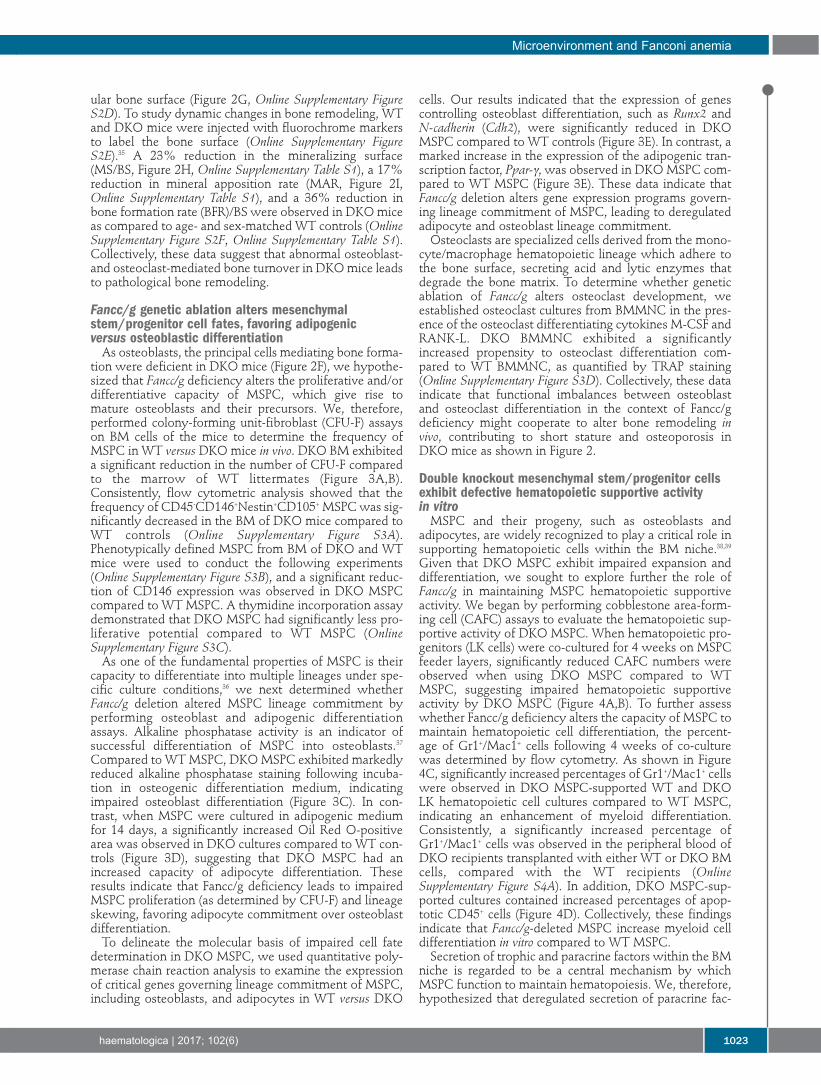
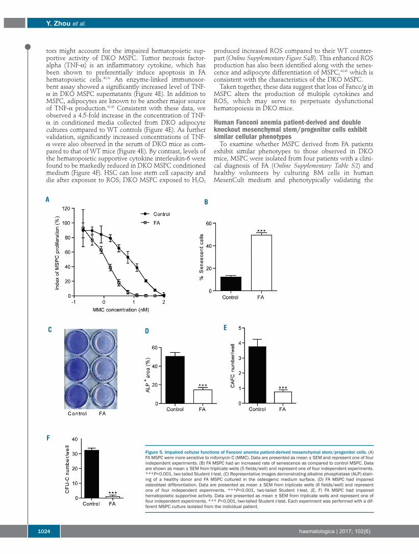
Figure 5. Impaired cellular functions of Fanconi anemia patient-derived mesenchymal stem/progenitor cells. (A)FA MSPC were more sensitive to mitomycin C (MMC). Data are presented as mean ± SEM and represent one of fourindependent experiments. (B) FA MSPC had an increased rate of senescence as compared to control MSPC. Dataare shown as mean ± SEM from triplicate wells (5 fields/well) and represent one of four independent experiments.***P<0.001, two-tailed Student t-test. (C) Representative images demonstrating alkaline phosphatase (ALP) stain-ing of a healthy donor and FA MSPC cultured in the osteogenic medium surface. (D) FA MSPC had impairedosteoblast differentiation. Data are presented as mean ± SEM from triplicate wells (6 fields/well) and representone of four independent experiments. ***P<0.001, two-tailed Student t-test. (E, F) FA MSPC had impairedhematopoietic supportive activity. Data are presented as mean ± SEM from triplicate wells and represent one offour independent experiments. *** P<0.001, two-tailed Student t-test. Each experiment was performed with a dif-ferent MSPC culture isolated from the individual patient.
A B
C D E
F
cells by flow cytometry (Online Supplementary Figure S5A).Sensitivity to mitomycin C was determined as previouslydescribed30 and was greater in FA MSPC than in healthycontrol MSPC (Figure 5A). Cellular senescence is a keypathophysiological phenomenon characterized by cellcycle arrest and upregulation of senescence-associated β-galactosidase activity. A 3-fold increase in the percentageof senescent cells was observed in FA MSPC compared tocontrol MSPC (Figure 5B, Online Supplementary Figure S5B).Like DKO MSPC, human FA MSPC exhibited markedlydefective osteoblast differentiation (Figure 5C,D) andincreased adipocyte differentiation (Online SupplementaryFigure S5C). Furthermore, osteoblast numbers were signif-icantly reduced in BM biopsy sections from FA patientscompared to those from healthy controls (OnlineSupplementary Figure S5D,E).To evaluate the hematopoietic supportive activity of
these MSPC, MSPC from FA patients were co-culturedwith cord blood CD34+ cells. After 5 weeks of co-culture,CAFC were counted, and CFU-C assays were performed.Significantly lower numbers of CAFC (Figure 5E) andCFU-C (Figure 5F) were observed in the co-cultures of FAMSPC with CD34+ cells than in those of healthy controlMSPC with CD34+ cells. These data suggest that MSPCderived from FA patients exhibit impaired HSPC support-ive activity, which is consistent with the findings in MSPCfrom DKO mice. To test whether MSPC derived from healthy donor BM,
compared to FANCG-deficient MSPC, would enhance theengraftment of human FANCG-deficient BM cells in vivo,MSPC were injected intra-tibially into sub-lethally irradi-ated NOD.Cg-Prkdcscid IL2rgtm1Wjl/Sz (NS2) recipient mice.Twenty-four hours later, BMMNC from a human FANCG-deficient patient were delivered via tail vein injection. Fourmonths following co-transplantation, human (h) CD45+cell engraftment in the BM of recipient mice was analyzedby flow cytometry. Injection of healthy MSPC dramatical-ly enhanced FANCG BMMNC engraftment (19% ofhCD45+ cells, Online Supplementary Figure S5F, right panel),while the percentage of hCD45+ cells in the mice thatreceived FANCG MSPC and FANCG BMMNC was only0.9% (Online Supplementary Figure S5F, left panel).
Discussion
MSPC act as an essential component of the BMhematopoietic microenvironment and have been provento be involved in the pathogenesis of several hematologicmalignancies.46,47 A recent translational study by Dong etal. found that Ptpn11-activating mutations in the BMMSPC and osteoprogenitors cause a juvenile myelomono-cytic leukemia-like cancer in mice through profound,detrimental effects on HSC.48 Several previous studiesindicated that MSPC from FA patients display reducedlong-term proliferation ability and spontaneous chromo-some breakages.49-51 In addition, we have previouslyreported that MSPC from the murine Fancg-/- model exhib-it impaired proliferative capacity.30 Amarachintha et al. alsoreported that MSC from Fanca-/- or Fancd2-/- mice impairedWT HSPC self-renewal and induced myeloid expansion.52Although another study showed that BM MSPC did nothave impaired function and contribute to the pathogenesisof the disease in acquired BMF such as aplastic anemia,53recent studies by Zambetti et al. demonstrated that mes-
enchymal niche-derived inflammatory signaling inducesoxidative and genotoxic stress in HSPC in Shwachman-Diamond syndrome, a rare inherited BMF syndrome.54Whether defects of BM MSPC are involved in the patho-physiology of FA deserves further in vivo investigation.FA is caused by a mutation in genes encoding proteins
required for the FA pathway. Although FA patients areclinically characterized by congenital mesenchymal anom-alies, and a uniformly progressive and fatal BMF whichbegins in infancy or childhood,20,55-58 the impact of the lossof FA genes on other stem cell compartments and the roleof the BM niche in the pathogenesis of FA-dependent BMFhave received limited attention. To date, more than ten FAgenes have been deleted or mutated in the mouse, butnone of these mouse models with single FA gene deficien-cy spontaneously develops severe hematologic abnormal-ities like FA patients.59,60 We have previously reported thatFancc/g DKO mice spontaneously develop more aggres-sive hematopoietic deficits including BMF, acute myeloidleukemia, and myelodysplastic syndrome.27 Here, usingthe DKO murine model, we provided evidence thatFanc/g-deficient MSPC within the hematopoietic microen-vironment cooperate to engender dysfunctionalhematopoiesis. These results provide new insights regard-ing the fundamental mechanisms by which the BM“niche” contributes to the pathogenesis of FA-dependentBMF. Furthermore, we demonstrated that Fancc/g-defi-cient MSPC lead to impaired osteoblast differentiationwith concomitant lineage skewing toward adipocyte com-mitment. Genetic ablation of Fancc/g also altered the pro-duction of critical inflammatory cytokines including inter-leukin-6 and TNF-α, the latter of which is known toinduce HSPC apoptosis and contribute to BMF.Collectively, this study reveals an intimate relationshipbetween FA HSPC and the BM niche in the pathogenesisof hematopoietic deficits. Our study provides strong evidence that Fancc/g defi-
ciency results in multiple skeletal pathologies, includingreduced body size and low bone mass phenotypes. Thesephenotypes in mice recapitulate the clinical features ofshort stature and osteoporosis common in FA patients.61Bone remodeling is a dynamic process controlled by thecoordinated activity of osteoblast-mediated bone forma-tion and osteoclast-mediated bone resorption. We attrib-ute the bone mass deficits in DKO mice in part to reducedosteoblast activity, as evidenced by the reduced osteoblastnumbers and impaired bone remodeling in vivo. Like HSC, MSPC have the potential to differentiate into
multiple lineages, including osteoblasts, adipocytes, andchondrocytes. The balance between osteogenesis andadipocyte formation is required for normal niche activityto maintain hematopoiesis.62-65 Our in vitro and in vivo stud-ies indicate that Fancc/g deficiency impairs the differenti-ation of MSPC into osteoblasts while favoring adipocytedifferentiation. Meanwhile, DKO mice exhibit reducedMSPC numbers and impaired self-renewal, suggesting anassociation between dysfunctional DKO MSPC andabnormal skeletal development/homeostasis in the DKOmouse model. DKO MSPC displayed dysregulated expres-sion of multiple key genes controlling osteoblast versusadipocyte lineage commitment including Runx2, Cdh2,and Ppar-γ. Cdh2 has been identified as a negative regula-tor of adipogenesis.66 Therefore, skewed lineage commit-ment of DKO MSPC away from osteoblast differentiationand toward adipocyte commitment may be associated
Microenvironment and Fanconi anemia
haematologica | 2017; 102(6) 1025
with reduced Cdh2 expression. In addition, we observed asignificant reduction of CD146 expression in DKO MSPCcompared to WT MSPC. Since CD146 has been reportedto be a marker for multilineage differentiation capacity,67the fewer CD146+ cells in DKO MSPC might be associatedwith the defective MSPC functions. Consistent withmurine data, MSPC obtained from BM biopsies of FApatients also revealed impaired proliferation andosteoblast differentiation capacity. MSPC-HSPC co-culture assays further revealed that
MSPC from DKO mice and FA patients have defectivehematopoietic supportive activity, as evidenced byreduced CAFC and CFU-C formation. Dysregulated skele-tal remodeling and impaired osteoblast differentiation inDKO mice may thus contribute to the defective BMmicroenvironment, which is unable to sustain adequateHSPC numbers. These results are consistent with findingsby Morad et al. who showed that the hematopoietic sup-portive activity of MSPC is influenced by lineage determi-nation.68It has been previously demonstrated that FA HSPC are
hypersensitive to TNF-α induced apoptosis and increasedlevels of TNF-α and other inflammatory cytokines havebeen observed in FA patients.26,69,70 Here we observedreduced levels of interleukin-6 and significantly higher lev-els of inflammatory cytokines, including TNF-α, in theDKO model. Nagajyothi et al. previously reported thatadipocytes are the major source of circulating inflammato-ry cytokines.71,72 It is also known that adipocytes negative-ly regulate hematopoietic activity and that TNF-α plays animportant role in the pathogenesis of BMF.38,73,74In addition, we observed that co-transplantation of
healthy donor MSPC enhanced the engraftment andexpansion of human BMMNC from an FANCG patient in
vivo in NS2 mice while FA MSPC failed to support FA BMcell reconstitution. Nevertheless, studies in a larger cohortof NSG mice with the co-transplantation of MSPC and FABM cells are warranted in the future.Collectively, these data suggest that the pathogenesis of
hematopoietic deficits in FA is complex and likely dependson the interplay between abnormal hematopoietic cellsand a dysfunctional BM niche. Our study indicates that FAMSPC have skewed differentiation capacity, alteredcytokine secretion and defective hematopoietic supportiveactivity, providing scientific insight into the role of FAmutations in impairing the hematopoietic niche and theirinvolvement in the pathogenesis of BMF. Clinically, amore detailed understanding of the disease might lead torethink about the niche for the development of new ther-apies for FA. While MSPC are increasingly appreciated tobe a critical part of the niche, due to the anatomical struc-ture of the niche, future studies to dissect the role of otherniche components, e.g., the endothelial niche or MSPCprogenies, with specific deletion of FA genes usingCre/loxP technology are warranted.
AcknowledgmentsThe authors thank Heather Daniel for administrative support.
FundingThis work was supported in part by the Leukemia Lymphoma
Society (LLS 6234-12 to FCY), NIH (R01CA155294-05 toDWC, F-C Y and HH), Ministry of Science and Technology ofChina (2016YFA0100600) and National Natural ScienceFoundation of China (81570113, 81270575, 81421002). SDRwas supported in part by pre-doctoral training grants from theIndiana CTSI (NCCR 5TL1RR025759-03) and Children’sTumor Foundation.
Y. Zhou et al.
1026 haematologica | 2017; 102(6)
References 1. Kiel MJ, Yilmaz OH, Iwashita T, et al. SLAM
family receptors distinguish hematopoieticstem and progenitor cells and revealendothelial niches for stem cells. Cell.2005;121(7):1109-1121.
2. Zhang J, Niu C, Ye L, et al. Identification ofthe haematopoietic stem cell niche and con-trol of the niche size. Nature.2003;425(6960):836-841.
3. Sacchetti B, Funari A, Michienzi S, et al. Self-renewing osteoprogenitors in bone marrowsinusoids can organize a hematopoieticmicroenvironment. Cell. 2007;131(2):324-336.
4. Devine SM, Hoffman R. Role of mesenchy-mal stem cells in hematopoietic stem celltransplantation. Curr Opin Hematol. 2000;7(6):358-363.
5. Dazzi F, Ramasamy R, Glennie S, Jones SP,Roberts I. The role of mesenchymal stemcells in haemopoiesis. Blood Rev.2006;20(3):161-171.
6. Schepers K, Campbell TB, Passegue E.Normal and leukemic stem cell niches:insights and therapeutic opportunities. CellStem Cell. 2015;16(3):254-267.
7. Williams DA, Cancelas JA. Leukaemia:niche retreats for stem cells. Nature.2006;444(7121):827-828.
8. Walkley CR, Olsen GH, Dworkin S, et al. A
microenvironment-induced myeloprolifera-tive syndrome caused by retinoic acid recep-tor gamma deficiency. Cell. 2007;129(6):1097-1110.
9. Kim YW, Koo BK, Jeong HW, et al. DefectiveNotch activation in microenvironment leadsto myeloproliferative disease. Blood.2008;112(12):4628-4638.
10. Raaijmakers MH, Mukherjee S, Guo S, et al.Bone progenitor dysfunction inducesmyelodysplasia and secondary leukaemia.Nature. 2010;464(7290):852-857.
11. Garaycoechea JI, Patel KJ. Why does thebone marrow fail in Fanconi anemia? Blood.2014;123(1):26-34.
12. Reid S, Renwick A, Seal S, et al. BiallelicBRCA2 mutations are associated with mul-tiple malignancies in childhood includingfamilial Wilms tumour. J Med Genet.2005;42(2):147-151.
13. Sims AE, Spiteri E, Sims RJ, 3rd, et al. FANCIis a second monoubiquitinated member ofthe Fanconi anemia pathway. Nat StructMol Biol. 2007;14(6):564-567.
14. Smogorzewska A, Matsuoka S, VinciguerraP, et al. Identification of the FANCI protein,a monoubiquitinated FANCD2 paralogrequired for DNA repair. Cell. 2007;129(2):289-301.
15. Dorsman JC, Levitus M, Rockx D, et al.Identification of the Fanconi anemia com-plementation group I gene, FANCI. Cell
Oncol. 2007;29(3):211-218.16. Wu Y, Shin-ya K, Brosh RM Jr. FANCJ heli-
case defective in Fanconia anemia and breastcancer unwinds G-quadruplex DNA todefend genomic stability. Mol Cell Biol.2008;28(12):4116-4128.
17. Vaz F, Hanenberg H, Schuster B, et al.Mutation of the RAD51C gene in a Fanconianemia-like disorder. Nat Genet. 2010;42(5):406-409.
18. Kim Y, Lach FP, Desetty R, et al. Mutationsof the SLX4 gene in Fanconi anemia. NatGenet. 2011;43(2):142-146.
19. Bogliolo M, Schuster B, Stoepker C, et al.Mutations in ERCC4, encoding the DNA-repair endonuclease XPF, cause Fanconi ane-mia. Am J Hum Genet. 2013;92(5):800-806.
20. Kutler DI, Singh B, Satagopan J, et al. A 20-year perspective on the InternationalFanconi Anemia Registry (IFAR). Blood.2003;101(4):1249-1256.
21. Ceccaldi R, Parmar K, Mouly E, et al. Bonemarrow failure in Fanconi anemia is trig-gered by an exacerbated p53/p21 DNAdamage response that impairs hematopoiet-ic stem and progenitor cells. Cell Stem Cell.2012;11(1):36-49.
22. Geiselhart A, Lier A, Walter D, Milsom MD.Disrupted signaling through the Fanconianemia pathway leads to dysfunctionalhematopoietic stem cell biology: underlyingmechanisms and potential therapeutic
strategies. Anemia. 2012;2012:265790.23. Kamimae-Lanning AN, Goloviznina NA,
Kurre P. Fetal origins of hematopoietic fail-ure in a murine model of Fanconi anemia.Blood. 2013;121(11):2008-2012.
24. Wajnrajch MP, Gertner JM, Huma Z, et al.Evaluation of growth and hormonal status inpatients referred to the International FanconiAnemia Registry. Pediatrics. 2001;107(4):744-754.
25. Prockop DJ. Marrow stromal cells as stemcells for nonhematopoietic tissues. Science.1997;276(5309):71-74.
26. Si Y, Ciccone S, Yang FC, et al. Continuousin vivo infusion of interferon-gamma (IFN-gamma) enhances engraftment of syngeneicwild-type cells in Fanca-/- and Fancg-/- mice.Blood. 2006;108(13):4283-4287.
27. Pulliam-Leath AC, Ciccone SL, Nalepa G, etal. Genetic disruption of both Fancc andFancg in mice recapitulates the hematopoi-etic manifestations of Fanconi anemia.Blood. 2010;116(16):2915-2920.
28. Yang Y, Kuang Y, Montes De Oca R, et al.Targeted disruption of the murine Fanconianemia gene, Fancg/Xrcc9. Blood.2001;98(12):3435-3440.
29. Wu X, Estwick SA, Chen S, et al.Neurofibromin plays a critical role in modu-lating osteoblast differentiation of mes-enchymal stem/progenitor cells. Hum MolGenet. 2006;15(19):2837-2845.
30. Li Y, Chen S, Yuan J, et al. Mesenchymalstem/progenitor cells promote the reconsti-tution of exogenous hematopoietic stemcells in Fancg-/- mice in vivo. Blood.2009;113(10):2342-2351.
31. Munugalavadla V, Vemula S, Sims EC, et al.The p85alpha subunit of class IA phos-phatidylinositol 3-kinase regulates theexpression of multiple genes involved inosteoclast maturation and migration. MolCell Biol. 2008;28(23):7182-7198.
32. O'Brien CA, Plotkin LI, Galli C, et al.Control of bone mass and remodeling byPTH receptor signaling in osteocytes. PLoSOne. 2008;3(8):e2942.
33. Arber DA, Orazi A, Hasserjian R, et al. The2016 revision to the World HealthOrganization classification of myeloid neo-plasms and acute leukemia. Blood.2016;127(20):2391-2405.
34. Giri N, Batista DL, Alter BP, Stratakis CA.Endocrine abnormalities in patients withFanconi anemia. J Clin Endocrinol Metab.2007;92(7):2624-2631.
35. Warden SJ, Robling AG, Sanders MS,Bliziotes MM, Turner CH. Inhibition of theserotonin (5-hydroxytryptamine) trans-porter reduces bone accrual during growth.Endocrinology. 2005;146(2):685-693.
36. Lee HS, Huang GT, Chiang H, et al.Multipotential mesenchymal stem cellsfrom femoral bone marrow near the site ofosteonecrosis. Stem Cells. 2003;21(2):190-199.
37. Sudo H, Kodama HA, Amagai Y, YamamotoS, Kasai S. In vitro differentiation and calcifi-cation in a new clonal osteogenic cell linederived from newborn mouse calvaria. J CellBiol. 1983;96(1):191-198.
38. Naveiras O, Nardi V, Wenzel PL, et al. Bone-marrow adipocytes as negative regulators ofthe haematopoietic microenvironment.Nature. 2009;460(7252):259-263.
39. Calvi LM, Adams GB, Weibrecht KW, et al.Osteoblastic cells regulate the haematopoi-etic stem cell niche. Nature. 2003;425(6960):841-846.
40. Koh PS, Hughes GC, Faulkner GR, Keeble
WW, Bagby GC. The Fanconi anemia groupC gene product modulates apoptoticresponses to tumor necrosis factor-alpha andFas ligand but does not suppress expressionof receptors of the tumor necrosis factorreceptor superfamily. Exp Hematol. 1999;27(1):1-8.
41. Dufour C, Corcione A, Svahn J, et al. TNF-alpha and IFN-gamma are overexpressed inthe bone marrow of Fanconi anemia patientsand TNF-alpha suppresses erythropoiesis invitro. Blood. 2003;102(6):2053-2059.
42. Cawthorn WP, Sethi JK. TNF-alpha andadipocyte biology. FEBS Lett. 2008;582(1):117-131.
43. Hoareau L, Bencharif K, Rondeau P, et al.Signaling pathways involved in LPS inducedTNFalpha production in human adipocytes.J Inflamm (Lond). 2010;7:1.
44. Zhang DY, Pan Y, Zhang C, et al. Wnt/beta-catenin signaling induces the aging of mes-enchymal stem cells through promoting theROS production. Mol Cell Biochem.2013;374(1-2):13-20.
45. Kanda Y, Hinata T, Kang SW, Watanabe Y.Reactive oxygen species mediate adipocytedifferentiation in mesenchymal stem cells.Life Sci. 2011;89(7-8):250-258.
46. Schepers K, Pietras EM, Reynaud D, et al.Myeloproliferative neoplasia remodels theendosteal bone marrow niche into a self-reinforcing leukemic niche. Cell Stem Cell.2013;13(3):285-299.
47. Menendez P, Catalina P, Rodriguez R, et al.Bone marrow mesenchymal stem cells frominfants with MLL-AF4+ acute leukemia har-bor and express the MLL-AF4 fusion gene. JExp Med. 2009;206(13):3131-3141.
48. Dong L, Yu WM, Zheng H, et al.Leukaemogenic effects of Ptpn11 activatingmutations in the stem cell microenviron-ment. Nature. 2016;539(7628):304-308.
49. Mantelli M, Avanzini MA, Rosti V, et al.Comprehensive characterization of mes-enchymal stromal cells from patients withFanconi anaemia. Br J Haematol. 2015;170(6):826-836.
50. Lecourt S, Vanneaux V, Leblanc T, et al. Bonemarrow microenvironment in fanconi ane-mia: a prospective functional study in acohort of fanconi anemia patients. StemCells Dev. 2010;19(2):203-208.
51. Barroca V, Mouthon MA, Lewandowski D,et al. Impaired functionality and homing ofFancg-deficient hematopoietic stem cells.Hum Mol Genet. 2012;21(1):121-135.
52. Amarachintha S, Sertorio M, Wilson A, Li X,Pang Q. Fanconi anemia mesenchymal stro-mal cells-derived glycerophospholipidsskew hematopoietic stem cell differentiationthrough toll-Like receptor signaling. StemCells. 2015;33(11):3382-3396.
53. Bueno C, Roldan M, Anguita E, et al. Bonemarrow mesenchymal stem cells frompatients with aplastic anemia maintain func-tional and immune properties and do notcontribute to the pathogenesis of the dis-ease. Haematologica. 2014;99(7):1168-1175.
54. Zambetti NA, Ping Z, Chen S, et al.Mesenchymal inflammation drives genotox-ic stress in hematopoietic stem cells and pre-dicts disease evolution in human pre-leukemia. Cell Stem Cell. 2016;19(5):613-627.
55. Kook H. Fanconi anemia: current manage-ment. Hematology. 2005;10(Suppl 1):108-110.
56. Bagby GC Jr., Segal GM, Auerbach AD, et al.Constitutive and induced expression ofhematopoietic growth factor genes by
fibroblasts from children with Fanconi ane-mia. Exp Hematol. 1993;21(11):1419-1426.
57. Auerbach AD, Buchwald M, Joenje H.Fanconi anemia. In: Vogelstein B, KinzlerKW, eds. The Genetic Bases of HumanCancer. 2002; 2nd Edition (New York:McGraw-Hill, Inc.):289-306.
58. Mathew CG. Fanconi anaemia genes andsusceptibility to cancer. Oncogene. 2006;25(43):5875-5884.
59. Parmar K, D'Andrea A, Niedernhofer LJ.Mouse models of Fanconi anemia. MutatRes. 2009;668(1-2):133-140.
60. Bakker ST, de Winter JP, te Riele H. Learningfrom a paradox: recent insights into Fanconianaemia through studying mouse models.Dis Model Mech. 2013;6(1):40-47.
61. Glanz A, Fraser FC. Spectrum of anomaliesin Fanconi anaemia. J Med Genet.1982;19(6):412-416.
62. Omatsu Y, Sugiyama T, Kohara H, et al. Theessential functions of adipo-osteogenic pro-genitors as the hematopoietic stem and pro-genitor cell niche. Immunity. 2010;33(3):387-399.
63. Mendez-Ferrer S, Michurina TV, Ferraro F, etal. Mesenchymal and haematopoietic stemcells form a unique bone marrow niche.Nature. 2010;466(7308):829-834.
64. Bianco P. Bone and the hematopoietic niche:a tale of two stem cells. Blood. 2011;117(20):5281-5288.
65. Visnjic D, Kalajzic Z, Rowe DW, et al.Hematopoiesis is severely altered in micewith an induced osteoblast deficiency.Blood. 2004;103(9):3258-3264.
66. van Oostrom AJ, van Wijk JP, Sijmonsma TP,Rabelink TJ, Castro Cabezas M. Increasedexpression of activation markers on mono-cytes and neutrophils in type 2 diabetes.Neth J Med. 2004;62(9):320-325.
67. Xu J, Wang W, Kapila Y, Lotz J, Kapila S.Multiple differentiation capacity of STRO-1+/CD146+ PDL mesenchymal progenitorcells. Stem Cells Dev. 2009;18(3):487-496.
68. Morad V, Pevsner-Fischer M, Barnees S, et al.The myelopoietic supportive capacity ofmesenchymal stromal cells is uncoupledfrom multipotency and is influenced by lin-eage determination and interference withglycosylation. Stem Cells. 2008;26(9):2275-2286.
69. Li J, Sejas DP, Zhang X, et al. TNF-alphainduces leukemic clonal evolution ex vivo inFanconi anemia group C murine stem cells. JClin Invest. 2007;117(11):3283-3295.
70. Pang Q, Keeble W, Christianson TA, FaulknerGR, Bagby GC. FANCC interacts with Hsp70to protect hematopoietic cells from IFN-gamma/TNF-alpha-mediated cytotoxicity.EMBO J. 2001;20(16):4478-4489.
71. Nagajyothi F, Desruisseaux MS, Thiruvur N,et al. Trypanosoma cruzi infection of cul-tured adipocytes results in an inflammatoryphenotype. Obesity (Silver Spring).2008;16(9):1992-1997.
72. Bastard JP, Maachi M, Lagathu C, et al.Recent advances in the relationship betweenobesity, inflammation, and insulin resist-ance. Eur Cytokine Netw. 2006;17(1):4-12.
73. Touw I, Lowenberg B. No stimulative effectof adipocytes on hematopoiesis in long-termhuman bone marrow cultures. Blood.1983;61(4):770-774.
74. Yokota T, Oritani K, Takahashi I, et al.Adiponectin, a new member of the family ofsoluble defense collagens, negatively regu-lates the growth of myelomonocytic pro-genitors and the functions of macrophages.Blood. 2000;96(5):1723-1732.
Microenvironment and Fanconi anemia
haematologica | 2017; 102(6) 1027