one of the states of bone marrow failure marion s. … of the states of bone marrow failure marion...
TRANSCRIPT

APLASTIC ANEMIA,one of the states of Bone Marrow
failure
Marion S. Sternbach, MD, FRCP(C), FACP

Aplastic Anemia - Definition
• Aplastic anemia was described by Ehrlich in 1888 in a pregnant woman.
• The term is a misnomer, since all three hematopoietic cell lines disappear gradually from the bone marrow without replacement by other cell lines.
• This defines it and differentiates it from hematopoietic replacement by : fibrosis, tumors, leukemias, granulomas, fungi, etc.

Aplastic Anemia - pathology
Bony trabeculaeMarrowSpaces All empty

Aplastic Anemia (AA) –Classification and Etiology
• A. Acquired.• B. Congenital • Incidence : Rare hematological disease
in Caucasians 3-5 / Million population/ year.
• In Orientals up to 10 - 15 / Million population / year.

Aplastic Anemia (AA) –classification and etiology continued• 1. Cytotoxic drugs, organic solvents (
benzene ), fumes, ( lindane, glue vapors), radiation.
• 2. Idiosyncratic drug reactions:• Chloramphenicol,• Gold,• Phenylbutazone, Indomethacine,• Sulfa, • Anti-epileptic drugs,• Arsenicals.

Aplastic Anemia (AA): classification and etiology continued (3)
• 3. Viral Infections: • Parvovirus B 19 – pure red cell aplasia,• Hepatitis: Non-A, non-B, non-C• HIV,• EBV• 4. Immune disorders: • Eosinophilic fasciitis,• SLE,• GVH

Aplastic Anemia (AA): classification and etiology, continued (4)
• 5. Miscellaneous:• Paroxysmal Nocturnal Hemoglobinuria
(PNH) , survival of a more adaptive stem cell population.
• Thymoma and Thymic Carcinoma –mostly pure Red Cell aplasia.’
• Pregnancy, most likely immune.

Congenital Aplastic Anemias
• 1. Fanconi Anemia: > 1850 in literature,• physical findings and chromosome
breakages.• 2. Dyskeratosis Congenita: 425 in lit.• Physical findings; telomere shortening.• 3. Diamond-Blackfan anemia 825 in lit.• Pure red cell aplasia, phys. Findings.• Adenosine deaminase deficiency.

Congenital Aplastic Anemia continued (2)
• 4. Schwachman-Diamond syndrome:• 500 in lit., phys. Findings, malabs.,
pancreatic insufficiency. Neutropenia.• 5. Severe Congenital Neutropenia: 374
in lit. ; Bone marrow – promyelocytearrest.
• 6. Amegakaryocytic Thrombocytopenia• 100 in lit. No phys. Findings.; absent
or abnormal megakaryocytes in BM.
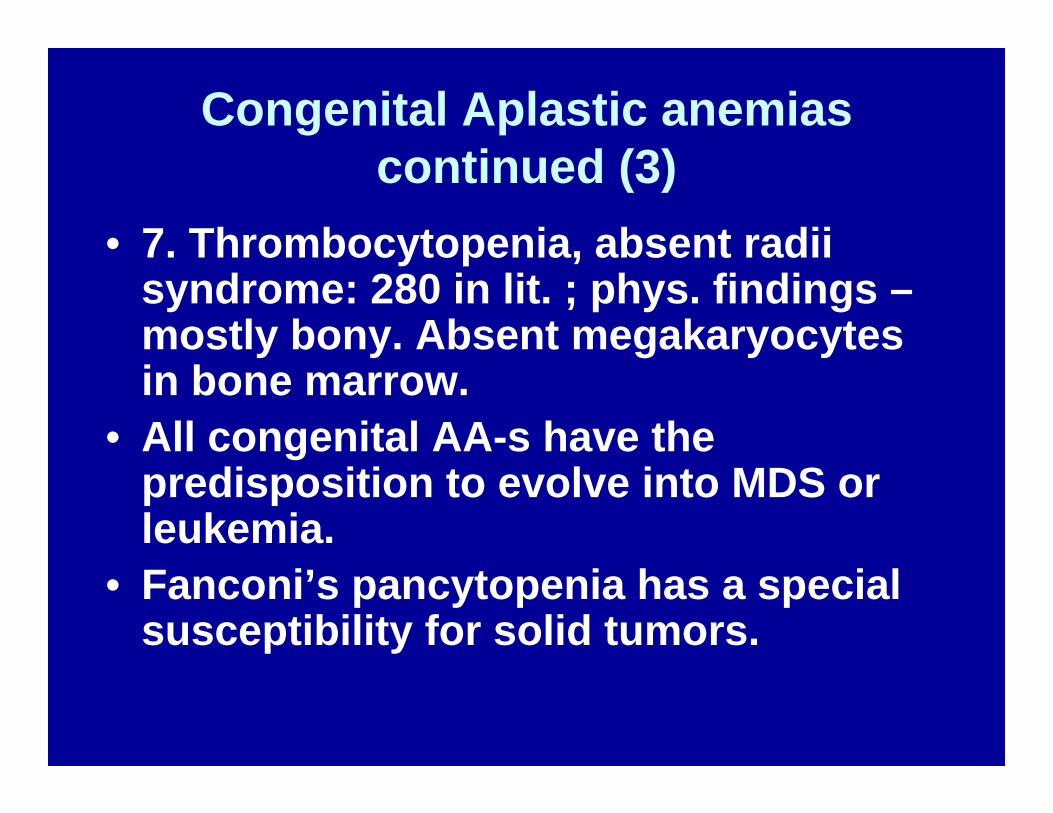
Congenital Aplastic anemiascontinued (3)
• 7. Thrombocytopenia, absent radii syndrome: 280 in lit. ; phys. findings –mostly bony. Absent megakaryocytesin bone marrow.
• All congenital AA-s have the predisposition to evolve into MDS or leukemia.
• Fanconi’s pancytopenia has a special susceptibility for solid tumors.

Aplastic Anemia : Pathogenesis of Idiopathic AA
• 1. Stem Cell anomalies:• A. Quantitative,• B. Qualitative.• 2. Stroma support anomalies,• 3. Cytokine anomalies: e.g. Interferon
Gamma, TNF Alpha, IL -6, IL – 2, lead to polyclonal T- cell expansions,
• 4. Cytotoxic – NK lymphocytes

Aplastic Anemia : Patient presentations.
• 1. Fanconi’s Pancytopenia:• During my Pediatric rotating internship in 1963, I
encountered a Moroccan family where the propositus, at 8 years was brought in with: severe anemia : Hb.-60 g/l with macrocytosis, leuco-neutropenia and thrombocytopenia, covered by purpura and ecchymoses. He also had mild renal impairment, which improved with good hydration.

Aplastic Anemia – Patient presentations (2)
Aplastic Anemias : Patient presentations (3)
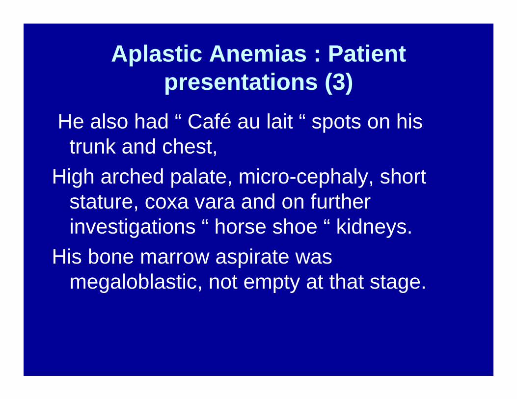
He also had “ Café au lait “ spots on his trunk and chest,
High arched palate, micro-cephaly, short stature, coxa vara and on further investigations “ horse shoe “ kidneys.
His bone marrow aspirate was megaloblastic, not empty at that stage.

Aplastic Anemia : Fanconi bone marrow
Megaloblasts
Myelo- meta-myelocytes

Aplastic Anemia : Patient presentations (4)
• Ely was found to have also an elevated Hb. F and family history revealed that there were a couple of twins, who looked very much like their brother and bruised easily.
• At age 18 months they were also pancytopenic, had dysmorphic features and were bruising a lot.
Their labs and bony anomalies were similar.

Aplastic Anemias: Patient presentations : The W. twins (5)

Aplastic anemia: patient presentations: W.twins’ hands.
• Deformed and extra thumbs.

Aplastic anemia: Patient presentations – Fanconi’s
Pancytopenia ( cont.)• These twins had also renal anomalies.• A first female cousin, 5 year old at a time had
the same dysmorphic features and pancytopenia, as well as skeletal and renal anomalies.
• In addition all of them had elevated Hb. F, hypoplastic, megaloblastic bone marrow and developed in addition FMF with renal amyloidosis, documented by renal biopsies.

Aplastic anemias: Fanconi’spancytopenia in 4 members of a
family.• Therapy:• Consisted of Oxymetholone,• Good i.v. and oral hydration for the kidneys and
FMF• Transfusions, when Hb. Dropped to below 70 g/l• Antibiotics i.v.or p.o depending on infections.• They all eventually died from renal amyloid with
renal failure.

Aplastic anemia: patient presentation – Acquired AA 1
• TV – 10 year old Caucasian boy, previously healthy, fit, hockey player.
Admitted to hospital for fatigue, listlessness, headaches, anorexia, bruising.
Exam: Pallor, purpura, ecchymoses, some shoddy cervical lymph nodes. No hep-spl. Megaly.
Hb. 72 g/l, Leuc.-1.8, Plat.-15, Retics – 20.

Aplastic anemia: acquired AA –patient 1.
• Past History: Was term baby, had all immunizations, normal mile stones, excellent student, athlete.
• Healthy 12 year old brother and healthy, unrelated parents.
• Helped his father for over 3 weeks in their house renovations, was exposed to organic solvents and paints.

Aplastic Anemia: Acquired AA Patient 1. Bone marrow Biopsy
Bone Marrow biopsy Bone Marrow aspirate

Aplastic Anemia: Acquired AAPatient 1.
• Therapy with steroids was initiated.• Within approx. six weeks, macrocytic
counts started recovering.• PNH developed with real nocturnal
brown urine.• Ham test was positive.• Patient followed for two years.• Complete recovery.

Aplastic anemia: Acquired Patient 2.
• FS, 7 year old grade 2 student, previously healthy, complained of fatigue, headaches, bruising.
• Phys. Exam. – Neg., exc. For purpura, petechiae, ecchymoses on legs.
• Severe Pancytopenia,• Empty bone marrow.• Steroids initiated. No response.
Aplastic anemia: Acquired,Patient 2.
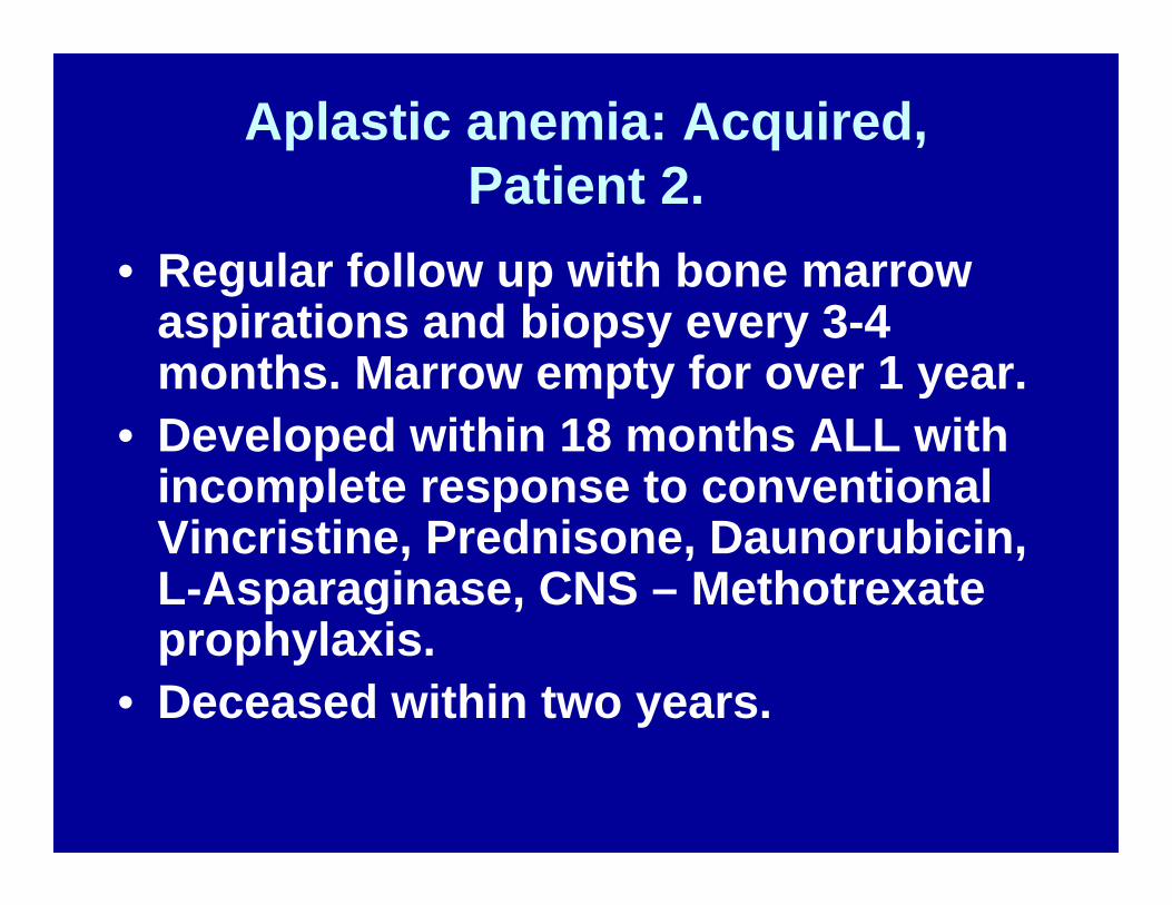
• Regular follow up with bone marrow aspirations and biopsy every 3-4 months. Marrow empty for over 1 year.
• Developed within 18 months ALL with incomplete response to conventional Vincristine, Prednisone, Daunorubicin, L-Asparaginase, CNS – Methotrexateprophylaxis.
• Deceased within two years.

Aplastic Anemia: Acquired AAPatient 3.
• 16 year old student, known at MCH for previous “ Fe. Deficiency” anemia at age 12 years. Treated with Fe++
• Now very severe pancytopenia: Hb – 68 g/l, Leucocytes – 1.0 with PMN-< 500,
• Platelets 10, Retics – 0• Exam- Pallor, bleeding, systolic ejection
murmur at all cardiac foci, no nodes and no hep-spl. Megaly. B.M empty.

Aplastic anemia: Acquired AAPatient 3.
• The year is 1978, no previous BMT done yet in Canada.
• We decide to try it in Montreal, since patient has 5 sibs.
• HLA identical, MLC compatible brother served in NATO forces in Lahr, Germany, brought home.
• Meanwhile Hb. Dropped to 32 g/l with cardiac failure and early papilledema in fundi.
• No transfusion administered.

Aplastic anemia: Acquired AA, patient 3. - BMT
• BMT expertise available at MGH ( Dr.Rybka worked in Seattle )
• Patient received conditioning with Cyclophosphamide 200 mg/kg X 4 days., followed by BMT
• Excellent, successful engraftment with minimal GVHD, treated with MTX.
• Two years later joined armed forces, four years later, fathered first baby.

Aplastic Anemia: Acquired AA post-Hepatitis B, patient 4.
14 year old student developed after episode of Hepatitis severe, fast progressing pancytopenia.Exam, apart from minimal liver enlargement and recovering LFT, unremarkable.Bone marrow – almost empty.Sister HLA identical and MLC compatible, seen and examined at Mac.

Aplastic anemia: Acquired post-Hepatitis B. Patient 4.
• Meanwhile patient administered small dose of steroids.
• 8 weeks later, one week before planned BMT, suddenly:
• Reticulocytosis, Hb. Up to 85 g/l, Leucocytes 3.4 with PMN > 500, Platelets – 78,000, Retics– 48,000
• Ham test strongly positive.• Patient followed now for 10 years with low
normal counts. PNH intermittently positive.

Aplastic Anemia: Acquired AA patient 5
• 48 year old man, president of steel company, treated in Montreal for 4 years for Wegener’s granulomatosis.
• Developed gradually ESRF, treated here with hemodialysis.
• Gradually developing Pancytopenia,• Cyclophosphamide D/C-d, Prednisone
continued.
Aplastic Anemia: Acquired AA, patient 5.
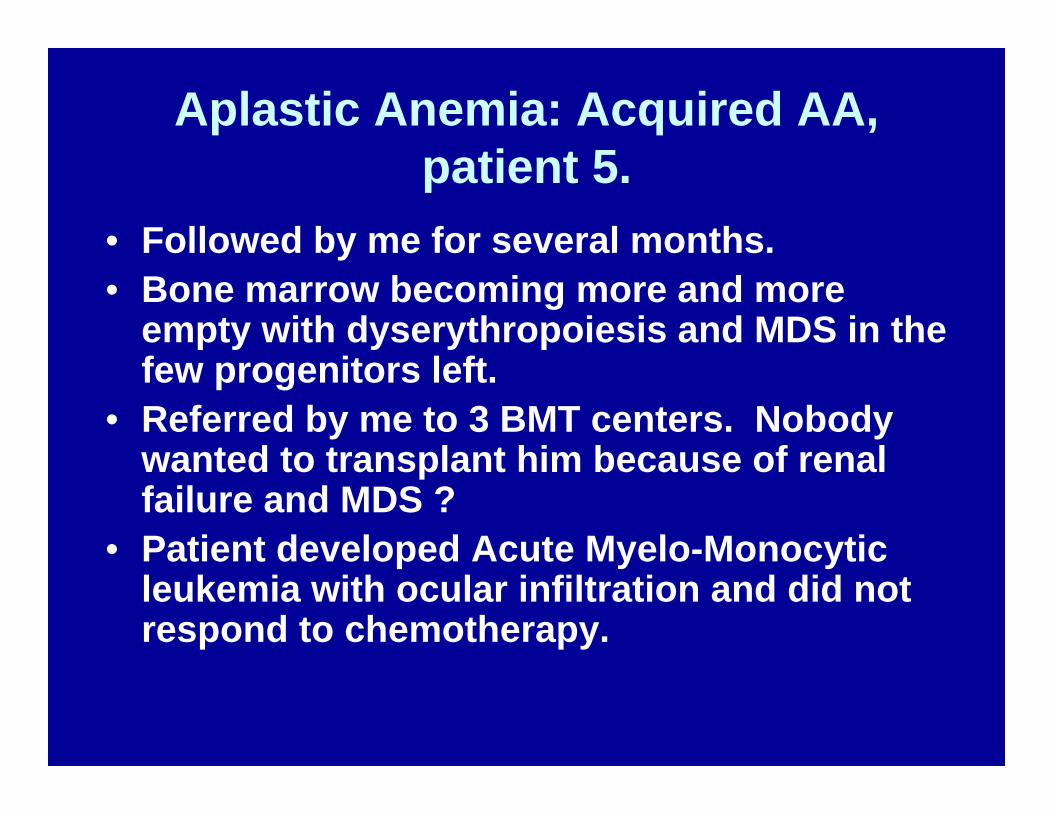
• Followed by me for several months.• Bone marrow becoming more and more
empty with dyserythropoiesis and MDS in the few progenitors left.
• Referred by me to 3 BMT centers. Nobody wanted to transplant him because of renal failure and MDS ?
• Patient developed Acute Myelo-Monocyticleukemia with ocular infiltration and did not respond to chemotherapy.

Aplastic anemia: Patient 5 evolving into MDS and AMML
Marrow aplasia, two years later AMML

Aplastic anemia – MDS – AMML, patient 5.
• Alpha Naphtol Acetate Esterase
Chloro Acetate esterase

Aplastic Anemia: Pathophysiology
• 1. Relationship of medical drug use to AA is due to deletions in the drug-metabolizing glutathione-S –transferase genes : GSTM1, GSTT1, that permit accumulation of toxic drug intermediates.
• 2. Benzene, pesticides are well documented.• 3. In Asia: unbottled water, ducks, geese,
animal fertilizer and some pesticides – are suggestive of an infectious etiology.

Aplastic anemia: Pathophysiology.
• 3. Auto-immunity – Autoantigens: • AA pts’ sera tested ag. A peptide library by genes in
fetal liver or leucemic cell lines:• Kinectin – bound to antibodies of about 40 % of AA
pts.• Diazepam binding related protein-1= essential
enzyme for oxidation of unsaturated fatty acids• Kinectin reacting cytotoxic T-cells generated in vitro,
inhibit hematopoietic colony formation. • But in vivo these cytotoxic T-cells have not been
found in AA pts.

Aplastic anemia: Pathophysiology.
• Most cases of “ Idiopathic “ AA are immune mediated diseases.
• Cellular and molecular pathways have been mapped to:
• A. Effector T-lymphocytes,• B. Target Hematopoietic stem cells and
progenitors• C. Environmental precipitants,• D. Genetic risk factors and • E. Individual characteristics of immune
response.

Aplastic anemia: Immune destruction of hematopoiesis.
•
NO
APC ,e.g.Mono-Macrophage
T-Cell
Hematop.Stem cell
Progenitor
Hematopoieticfailure
Clonal expansion
TNF-alpha
Fas Rec.
Ifn. G.
Jak1
Stat1 -3
Prot. Synth.Apoptosis,Cell cycling
AntigenT-Bet bindsTranscription F.Binds to Ifn G, inducesGene expression
RNA-se

Aplastic anemia: Immune destruction of hematopoiesis
• Evidence for this:• 1. Recovery from AA after BMT graft
rejection, due to immune suppression of conditioning regimen.
• 2. T-cells from AA pts. Inhibit CFU in normal bone marrows.
• 3. Currently good clinical response to immune suppression with ATG and CyA.
• 4. B.M. recovery of AA in pregnancy after delivery, 20% relapse risk in future pregnancy, occas. fatal.

Clinical and pathophysiol. Relationships betw.B.M failure and
autoimmune diseases.
MS, Colitis,Uveitis, DM.t1Conn. Tissue dis.
AAA
AAAA/PNH
LG
LG
LGL
MDM
MDSAA
AML

Aplastic Anemia: Clonal evolution
• 1. An abnormal expansion of suppressor T- cells may cause HSC depletion and clonal anomalies.
• 2. HSC defects may be associated with abnormalities of the microenvironment.
• 3. CD4+CD25+FOXP3+ regulatory T cells are deficient in some of AA pts.

Aplastic anemia: Clonal evolution
• Glycosyl-Phosphatidil-Inositol (GPI) anchor is an important surface glycolipid, normally positive in normal hematopoietic cells.
• Rare GPI- cells exist even in normal individuals and are predominant in PNH. (CD55 and CD59), derived from a somatic mutation in an X-linked gene called PIG-A.
• CD 52 is a GPI linked protein, the target of Alemtuzumab (Campath).

Aplastic anemia: Clonal evolution
• Following treatment with Campath, CD52 – T-cells are identified, which are also GPI- neg.
• It appears that GPI-neg. stem cells are spared from the autoimmune attack, suggesting that the “Autoantigen” may be GPI – linked.
• This may explain the PNH recovery in AA following immune suppression.

Aplastic anemia: clonal evolution.
• Bone marrow failure is a risk factor for clonalevolution: cells resistant to apoptotic cues, are adaptively selected from the stem cell pool for survival.
• 10-20 % of AA survivors will develop within a decade a clonal disease: PNH, MDS, AML ( Young, Bagby )
• “ In a stressful hematop. Micro-ecosystem a new genotype in adapted clones will result in an emergent phenotype of BM cells, more “fit” than their non adapted progenitors.”

Aplastic anemia: clonal evolution.
NormalMicroenvironm.
Abnormal Microenvironm.
Aplastic anemia
Clonal evolution
T cells – t1
IFN-G
TNF-alpha
Aplastic anemia: Therapy
• 1. Young patients who have an HLA identical and MLC compatible donor should definitely undergo BMT or HSCT.
• That is probably the only chance for complete cure, by destroying also the adaptive, resistant clone.
• Transplant should occur within less than 80 days and if possible within weeks.
• Transfusions, especially from prospective donor must be avoided.
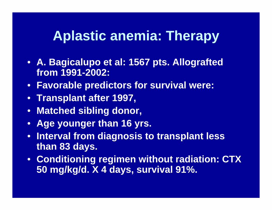
Aplastic anemia: Therapy
• A. Bagicalupo et al: 1567 pts. Allograftedfrom 1991-2002:
• Favorable predictors for survival were:• Transplant after 1997,• Matched sibling donor,• Age younger than 16 yrs.• Interval from diagnosis to transplant less
than 83 days.• Conditioning regimen without radiation: CTX
50 mg/kg/d. X 4 days, survival 91%.

Aplastic anemia: Therapy
• HSCT from PB is fraught with chronic GVHD, which is only harmful in AA.
• Cyclosporin A and Methotrexate are used to prevent and treat GVHD
• HLA identical BMT for pts. Older than 30 yrs. – conditioning regimen of CTX 300 mg./M^2, Fludarabine 30 mg./M^2 X 4 days +/- ATG – mortality 30%.

Aplastic anemia: Therapy
• Matched unrelated donor transplants (MUD):
• European Registry: 318 donor transplants from 1988-98:
• Rejection – 15%, • GVHD II-IV 48%,• 5-year survival 39%• Conditioning regimen: CTX, ATG,
minimal radiation.

Aplastic anemia: Therapy
• Immune Suppression: for patients who don’t have a donor and for older patients.
• Cyclosporin and ATG • This treatment will not prevent later
clonal evolution.• Relapses occur. Treatment may be
repeated.

Aplastic anemia: Immune suppressive therapy
• There is a real risk of progression to clonaldisorders following immune suppressive therapy:
• PNH, MDS, AML• European Registry: 10 year incidence of
malignancy in 860 pts. 19 % = 5.2 X the overall cancer risk in age matched non AA, non immune suppressed population.
• Monosomy 7 and trisomy 8. are frequent.• MDS and AML evolve sooner in pts. Treated
with G-CSF.

Aplastic anemia: Neoplasticcomplications
• Post Transplant Lymphoproliferativedisorder (PTLD) due to EBV – may be polyclonal or monoclonal.
• Polyclonal PTLD may remit spontaneously in the absence of immune suppression.
• Monoclonal PTLD has to be treated like an aggressive lymphoma.
• Congenital AA-s ( esp. Fanconi ), have a much higher incidence of solid tumors due to chromosomal breakages.

Aplastic anemia(AA): Summary.
1. AA is a rare hematological disease, characterized by Pancytopenia, empty bone marrow, absence of hepato-splenomegaly.
A. Moderate AA: marrow cell. < 30%, depression of at least 2/3 blood elem.
B. Severe AA: marrow cell. < 25 % - Retics., < 40,000, PMN- < 500, Plat. < 20,000
C. Very severe AA : PMN < 200

Aplastic Anemia (AA): Summary
• 2. AA may be congenital or acquired.• 3. Cong. AA is often associated with skeletal
and organ anomalies and have a tendency to develop neoplasias both hematological and solid tumors.
• 4. Acquired AA are sometimes due to toxic or drug induced marrow injury.
• Most of them are immune mediated.• 5. Acquired AA may evolve into clonal
disease: PNH, MDS, AML.

Aplastic Anemia (AA): Summary
• 6. AA is best treated in young patients by HLA identical BMT with only CTX conditioning and no or minimal preceding transfusions.
• 7. CR is normalization of blood counts.• PR is independence of transfusions.• 8. AA in older pts or absence of donors
should be treated with immune suppression: ATG + CyA
• 9. Clonal evolution after immune suppression is quite likely.