biventricular strain analysis at 1.5t cardiac mr imaging: preliminary results in volunteers using an...
TRANSCRIPT
POSTER PRESENTATION Open Access
Biventricular strain analysis at 1.5T cardiacMR imaging: preliminary results in volunteersusing an iterative SENSE reconstruction withL1 regularizationPeter M Smith1*, Benjamin H Freed4, Bradley D Allen1, Bruce S Spottiswoode5, Maria Carr1, Marie Wasielewski1,Karissa Campione1, Michaela Schmidt2, Mariappan S Nadar3, Michael O Zenge2, James C Carr1, Jeremy D Collins1
From 17th Annual SCMR Scientific SessionsNew Orleans, LA, USA. 16-19 January 2014
BackgroundChanges in myocardial strain have been shown to pre-cede onset of systolic dysfunction in patients with cardi-omyopathy. Traditionally performed withechocardiography, acoustic windows can limit strainevaluation. Preliminary work has shown good agreementbetween myocardial strain derived from deformationfield analysis at balanced steady-state free-precession(bSSFP) cine MRI and speckle tracking echocardiogra-phy. The application of a novel prototype iterativeSENSE reconstruction with L1 regularization (IR-SENSE) to highly accelerated segmented bSSFP cineacquisitions can maintain or improve the effective tem-poral resolution with a shorter breath-hold. The purposeof this study is to evaluate left- and right-ventricularstain analysis from bSSFP cine acquisitions using con-ventional GRAPPA and highly accelerated T-PAT with
IR-SENSE. We hypothesize that myocardial strain para-meters derived from accelerated T-PAT cine acquisi-tions with IR-SENSE are similar to those obtained usingconventional segmented bSSFP with comparable effec-tive temporal resolutions.
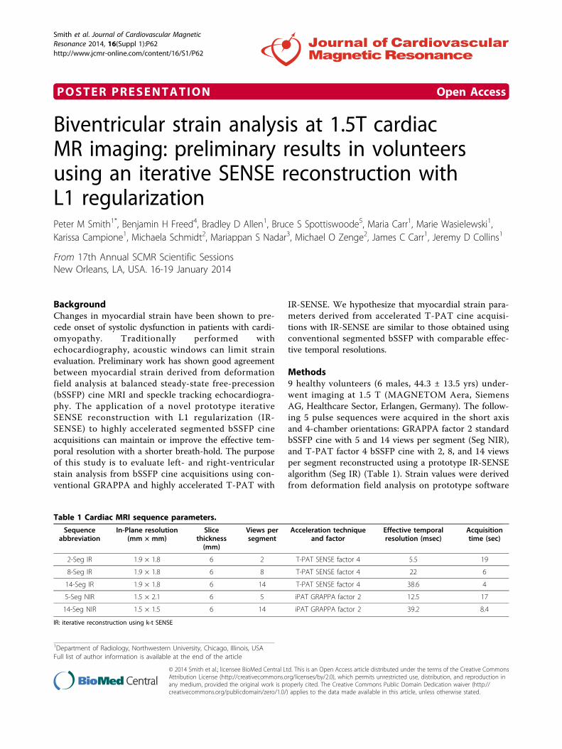
Methods9 healthy volunteers (6 males, 44.3 ± 13.5 yrs) under-went imaging at 1.5 T (MAGNETOM Aera, SiemensAG, Healthcare Sector, Erlangen, Germany). The follow-ing 5 pulse sequences were acquired in the short axisand 4-chamber orientations: GRAPPA factor 2 standardbSSFP cine with 5 and 14 views per segment (Seg NIR),and T-PAT factor 4 bSSFP cine with 2, 8, and 14 viewsper segment reconstructed using a prototype IR-SENSEalgorithm (Seg IR) (Table 1). Strain values were derivedfrom deformation field analysis on prototype software
1Department of Radiology, Northwestern University, Chicago, Illinois, USAFull list of author information is available at the end of the article
Table 1 Cardiac MRI sequence parameters.
Sequenceabbreviation
In-Plane resolution(mm × mm)
Slicethickness(mm)
Views persegment
Acceleration techniqueand factor
Effective temporalresolution (msec)
Acquisitiontime (sec)
2-Seg IR 1.9 × 1.8 6 2 T-PAT SENSE factor 4 5.5 19
8-Seg IR 1.9 × 1.8 6 8 T-PAT SENSE factor 4 22 6
14-Seg IR 1.9 × 1.8 6 14 T-PAT SENSE factor 4 38.6 4
5-Seg NIR 1.5 × 2.1 6 5 iPAT GRAPPA factor 2 12.5 17
14-Seg NIR 1.5 × 1.5 6 14 iPAT GRAPPA factor 2 39.2 8.4
IR: iterative reconstruction using k-t SENSE
Smith et al. Journal of Cardiovascular MagneticResonance 2014, 16(Suppl 1):P62http://www.jcmr-online.com/content/16/S1/P62
© 2014 Smith et al.; licensee BioMed Central Ltd. This is an Open Access article distributed under the terms of the Creative CommonsAttribution License (http://creativecommons.org/licenses/by/2.0), which permits unrestricted use, distribution, and reproduction inany medium, provided the original work is properly cited. The Creative Commons Public Domain Dedication waiver (http://creativecommons.org/publicdomain/zero/1.0/) applies to the data made available in this article, unless otherwise stated.
(Siemens Corporate Technology, Princeton, NJ). LVglobal and RV lateral wall longitudinal strains wereobtained from 4-chamber cine acquisitions. LV circum-ferential and radial strains were obtained from short-axis acquisitions for the base, mid-chamber, and apex.Average and peak strains were compared using a two-sided student’s t-test.
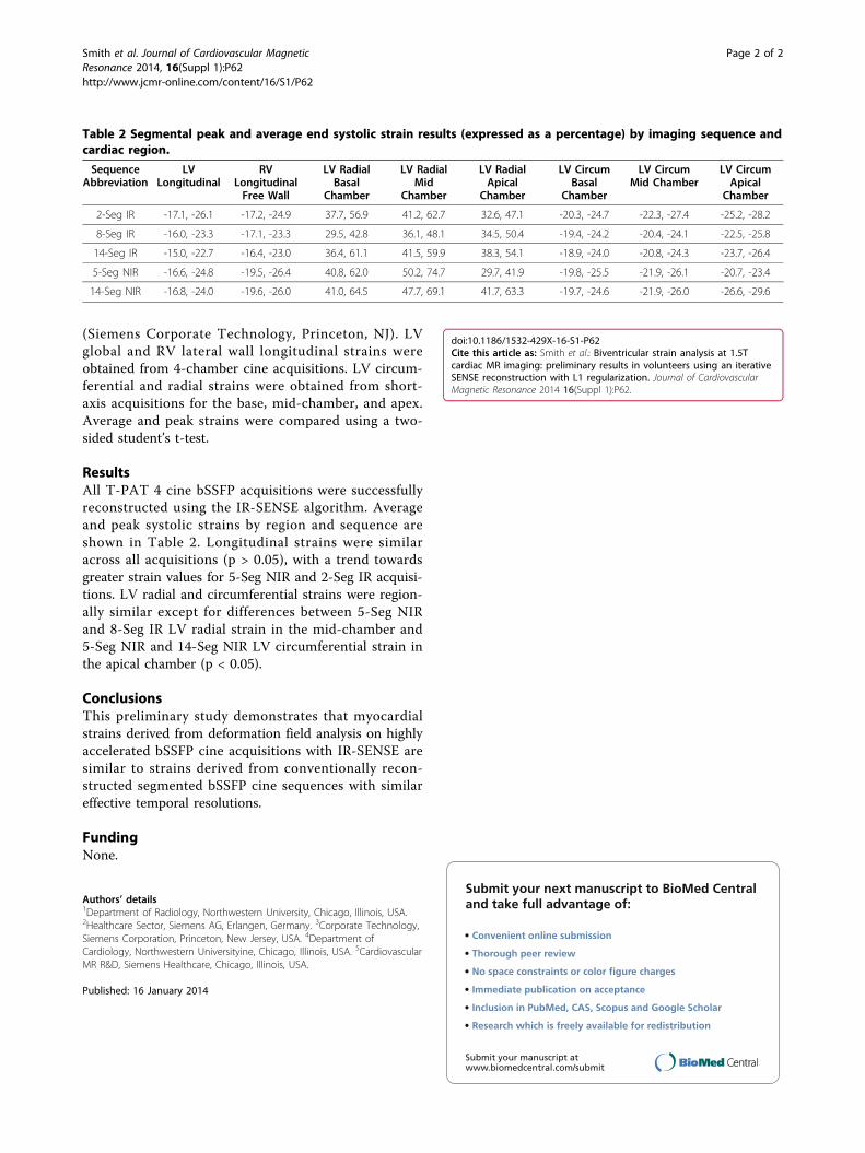
ResultsAll T-PAT 4 cine bSSFP acquisitions were successfullyreconstructed using the IR-SENSE algorithm. Averageand peak systolic strains by region and sequence areshown in Table 2. Longitudinal strains were similaracross all acquisitions (p > 0.05), with a trend towardsgreater strain values for 5-Seg NIR and 2-Seg IR acquisi-tions. LV radial and circumferential strains were region-ally similar except for differences between 5-Seg NIRand 8-Seg IR LV radial strain in the mid-chamber and5-Seg NIR and 14-Seg NIR LV circumferential strain inthe apical chamber (p < 0.05).
ConclusionsThis preliminary study demonstrates that myocardialstrains derived from deformation field analysis on highlyaccelerated bSSFP cine acquisitions with IR-SENSE aresimilar to strains derived from conventionally recon-structed segmented bSSFP cine sequences with similareffective temporal resolutions.
FundingNone.
Authors’ details1Department of Radiology, Northwestern University, Chicago, Illinois, USA.2Healthcare Sector, Siemens AG, Erlangen, Germany. 3Corporate Technology,Siemens Corporation, Princeton, New Jersey, USA. 4Department ofCardiology, Northwestern Universityine, Chicago, Illinois, USA. 5CardiovascularMR R&D, Siemens Healthcare, Chicago, Illinois, USA.
Published: 16 January 2014
doi:10.1186/1532-429X-16-S1-P62Cite this article as: Smith et al.: Biventricular strain analysis at 1.5Tcardiac MR imaging: preliminary results in volunteers using an iterativeSENSE reconstruction with L1 regularization. Journal of CardiovascularMagnetic Resonance 2014 16(Suppl 1):P62.
Submit your next manuscript to BioMed Centraland take full advantage of:
• Convenient online submission
• Thorough peer review
• No space constraints or color figure charges
• Immediate publication on acceptance
• Inclusion in PubMed, CAS, Scopus and Google Scholar
• Research which is freely available for redistribution
Submit your manuscript at www.biomedcentral.com/submit
Table 2 Segmental peak and average end systolic strain results (expressed as a percentage) by imaging sequence andcardiac region.
SequenceAbbreviation
LVLongitudinal
RVLongitudinalFree Wall
LV RadialBasal
Chamber
LV RadialMid
Chamber
LV RadialApical
Chamber
LV CircumBasal
Chamber
LV CircumMid Chamber
LV CircumApical
Chamber
2-Seg IR -17.1, -26.1 -17.2, -24.9 37.7, 56.9 41.2, 62.7 32.6, 47.1 -20.3, -24.7 -22.3, -27.4 -25.2, -28.2
8-Seg IR -16.0, -23.3 -17.1, -23.3 29.5, 42.8 36.1, 48.1 34.5, 50.4 -19.4, -24.2 -20.4, -24.1 -22.5, -25.8
14-Seg IR -15.0, -22.7 -16.4, -23.0 36.4, 61.1 41.5, 59.9 38.3, 54.1 -18.9, -24.0 -20.8, -24.3 -23.7, -26.4
5-Seg NIR -16.6, -24.8 -19.5, -26.4 40.8, 62.0 50.2, 74.7 29.7, 41.9 -19.8, -25.5 -21.9, -26.1 -20.7, -23.4
14-Seg NIR -16.8, -24.0 -19.6, -26.0 41.0, 64.5 47.7, 69.1 41.7, 63.3 -19.7, -24.6 -21.9, -26.0 -26.6, -29.6
Smith et al. Journal of Cardiovascular MagneticResonance 2014, 16(Suppl 1):P62http://www.jcmr-online.com/content/16/S1/P62
Page 2 of 2