bacterial surface protein l binds and inactivates neutrophil · bacterial surface protein l binds...
TRANSCRIPT

of July 27, 2018.This information is current as
Inactivates Neutrophil Proteins S100A8/A9Bacterial Surface Protein L Binds and
Bo Åkerström and Lars Björck
http://www.jimmunol.org/content/183/7/4583doi: 10.4049/jimmunol.0901487September 2009;
2009; 183:4583-4592; Prepublished online 14J Immunol
Referenceshttp://www.jimmunol.org/content/183/7/4583.full#ref-list-1
, 23 of which you can access for free at: cites 57 articlesThis article
average*
4 weeks from acceptance to publicationFast Publication! •
Every submission reviewed by practicing scientistsNo Triage! •
from submission to initial decisionRapid Reviews! 30 days* •
Submit online. ?The JIWhy
Subscriptionhttp://jimmunol.org/subscription
is online at: The Journal of ImmunologyInformation about subscribing to
Permissionshttp://www.aai.org/About/Publications/JI/copyright.htmlSubmit copyright permission requests at:
Email Alertshttp://jimmunol.org/alertsReceive free email-alerts when new articles cite this article. Sign up at:
Print ISSN: 0022-1767 Online ISSN: 1550-6606. Immunologists, Inc. All rights reserved.Copyright © 2009 by The American Association of1451 Rockville Pike, Suite 650, Rockville, MD 20852The American Association of Immunologists, Inc.,
is published twice each month byThe Journal of Immunology
by guest on July 27, 2018http://w
ww
.jimm
unol.org/D
ownloaded from
by guest on July 27, 2018
http://ww
w.jim
munol.org/
Dow
nloaded from

Bacterial Surface Protein L Binds and Inactivates NeutrophilProteins S100A8/A91
Bo Åkerstrom2 and Lars Bjorck2
Finegoldia magna is an anaerobic bacterial species that is part of the normal human flora on all nonsterile body surfaces, butit is also a significant opportunistic pathogen causing a wide range of infections. Some isolates of F. magna that are morefrequently associated with clinical infection express protein L, a surface protein containing multiple homologous domains(B1-B5) that bind Igs through interactions with Ig L chains. The present study shows that the N-terminal A domain of proteinL binds S100A8/A9, antibacterial proteins present in large amounts in the cytoplasm of neutrophils, but also extracellularlyin tissues during inflammation. As a result, protein L-expressing F. magna are protected against killing by S100A8/A9. Igsand S100A8/A9 were found to interact independently with protein L, demonstrating that this bacterial surface protein iscapable of manipulating both adaptive and innate immune defense mechanisms. The Journal of Immunology, 2009, 183:4583– 4592.
M any bacterial species express Ig-binding surface pro-teins, among which staphylococcal protein A (1) andstreptococcal protein G (2, 3) are the most well
known. These proteins are widely used as biomedical tools todetect and bind Abs, and they interact with the H chains of IgG.Apart from protein L, an Ig L chain-binding molecule (4), allbacterial proteins to date described that bind Ig in a nonimmunefashion interact with Ig H chains. Protein L is expressed by�10% of clinical isolates of Finegoldia magna (5), an anaer-obic Gram-positive bacterium that is part of the normal flora inthe skin, the mouth, the upper respiratory tract, the gastrointes-tinal tract, and the female genito-urinary tract. F. magna is themost significant opportunistic pathogen of the anaerobic normalbacterial flora, causing soft tissue abscesses, bone/joint infec-tions, wound infections, and vaginosis (6, 7). Protein L-express-ing F. magna strains are frequently isolated from patients withvaginosis (5), and when expressed at the surface of Streptococ-cus gordonii, protein L promotes the adhesion of these bacteriato the vaginal mucosa of mice (8). Protein L preferentially bindsIg L chains of the � type through interactions with the variabledomain, but without interfering with the Ag binding site (9 –12). The molecule contains multiple homologous Ig bindingdomains, called B repeats (13), and it activates human basophilsand mast cells by cross-linking surface-associated IgE, resulting
in the release of proinflammatory mediators (14, 15). Thesedifferent observations indicate that the presence of protein L atthe surface of F. magna enhances the potential pathogenicity ofthe bacteria.
Members of the human S100 protein family are Ca2�-bindingproteins containing helix-loop-helix motifs, so-called EF hands(16). The expression of the S100 proteins is usually regulatedby environmental and developmental factors, and is cell andtissue specific. Two members, S100A8 and S100A9, are majorcytosolic constituents (�40% of the total protein content) inneutrophils and monocytes. The apparent molecular masses onSDS-PAGE are 8 and 14 kDa, respectively, and they are alsocalled myeloid-related proteins 8 and 14, migration inhibitoryfactor-related proteins 8 and 14, or calgranulin a/b (17, 18).Their expression is up-regulated in tissue macrophages, kera-tinocytes, and epithelial cells in inflammation and cancer (19,20). S100A8 and S100A9 are secreted from activated phago-cytes by a novel tubulin-dependent mechanism (21), and theyare found in extracellular fluid and plasma during inflammation(20). The two proteins form heteromeric complexes in vivo, andat millimolar Ca levels the heterotetramer, (S100A8/A9)2, ispredominating (22, 23). The tetramer was also named calpro-tectin due to its ability to inhibit growth of various bacterial andfungal species (24). This bacteriostatic property has been as-cribed to the Zn-binding capacity of the tetramer, and it hasbeen suggested that the effect is a result of depletion of bacterialnutrient Zn ions (25) or altered structure of the protein complex(26). The observation that the S100A8/A9 complex adheres toamastigotes in skin lesions of Leishmania major-infected mice(27) indicates that S100A8/A9 could also have a function in thedefense against this parasite.
The interaction between the B repeats of protein L and Ig Lchains is well documented, but the properties of the N-terminal Adomain (Fig. 3A shows a schematic depiction of protein L) areunknown. The starting point for the present investigation was thefinding that a protein L construct containing the A domain boundS100A8/A9 with high affinity and specificity, whereas the Ig-bind-ing B repeats did not. A characterization of the interaction betweenprotein L and S100A8/A9 and its consequences for F. magna bi-ology is the theme of this work.
Department of Clinical Sciences, Division of Infection Medicine, Lund University,Lund, Sweden
Received for publication May 11, 2009. Accepted for publication July 21, 2009.
The costs of publication of this article were defrayed in part by the payment of pagecharges. This article must therefore be hereby marked advertisement in accordancewith 18 U.S.C. Section 1734 solely to indicate this fact.1 This work was supported by the Swedish Research Council (Projects 7144 and7480), Swedish Government Funds for Clinical Research (ALF), Swedish Society forMedical Research, Royal Physiographic Society (Lund), Foundations of Greta andJohan Kock, Torsten and Ragnar Soderberg, Alfred Osterlund, the Blood and DefenceNetwork, Lund University, the Crafoord Foundation, the Thelma Zoega Foundation,and Hansa Medical AB.2 Address correspondence and reprint requests to Dr. Bo Åkerstrom or Dr. LarsBjorck, Lund University, BMC, B14, Solvegatan 19, SE-22184, Lund, Sweden.E-mail addresses: [email protected] and [email protected]
Copyright © 2009 by The American Association of Immunologists, Inc. 0022-1767/09/$2.00
The Journal of Immunology
www.jimmunol.org/cgi/doi/10.4049/jimmunol.0901487
by guest on July 27, 2018http://w
ww
.jimm
unol.org/D
ownloaded from

Materials and MethodsBacteria
F. magna (strains 312, 505, 564, and 644) and group G streptococcalstrains (G42, G43, G46, and G148) are clinical isolates from the De-partment of Clinical Microbiology, Lund University Hospital. Strepto-coccus pneumoniae strains D39 and PR218 are from G. Pozzi, Univer-sity of Siena, and the Streptococcus pyogenes strains (AP1, AP4, AP6,AP12, and AP49) are from Institute of Hygiene and Epidemiology (Prague,Czech Republic). The two Staphylococcus aureus strains Cowan I andWood 46 are from T. Foster, Trinity College (Dublin, Ireland). All strainswere grown in Todd-Hewitt (TH)3 broth (Difco) at 37°C. In the case of F.magna strains, TH contained 0.5% Tween 80, and these isolates weregrown under strict anaerobic conditions.
Proteins, Abs, sequencing, and radiolabeling
Protein L (B1-B4) was expressed and purified, as described (13). Factor Xa(FXa) was purchased from ICN Pharmaceuticals. Human IgG was pur-chased from Sigma-Aldrich. Protein PAB was isolated from F. magna(strain ALB8) culture medium, as described (28). Protein A was purchasedfrom Sigma-Aldrich, and protein H and M1 protein were purified, as de-scribed (29). The synthetic peptides DNF29, corresponding to aa positions66–94 of protein L (A-C2), and MLT24, corresponding to the N-terminal24 aa of S100A8, were purchased from Innovagen. Mouse monoclonalanti-S100A8/A9 (27E10) was purchased from Abcam. Polyclonal anti-S100A8 Abs were raised in rabbits immunized with MLT24. N-terminalamino acid sequence analysis was done by 12 cycles of Edman degradation(Protein Analysis Center, Karolinska Institute) of bands separated by SDS-PAGE and transfered to polyvinylidene difluoride (PVDF) membranes(Immobilon-P; Millipore), as described (30). Proteins were labeled with125I (Amersham Biosciences; IMS.30) using the chloramin-T method (31).Protein-bound iodine was separated from free iodide by gel chromatogra-phy on a Sephadex G-25 column (PD10; Amersham Biosciences). A sp.act. of �0.1–0.2 MBq/�g proteins was obtained.
Electrophoresis, blotting, slot binding, and chromatography
SDS-PAGE was performed using 13.5% slab gels in the buffer systemdescribed (32), including 2% v/v 2-ME and using Rainbow molecular massstandards purchased from GE Healthcare. The polyacrylamide gels werestained with Coomassie brilliant blue R-250. For immunoblotting, the gelswere transferred to PVDF membranes (Immobilon-P; Millipore) and thenincubated with antisera or 125I-labeled proteins, as described (33), using 1mM CaCl2 in the incubation and washing buffers. Images of the mem-branes were developed using Fuji FLA 3000 phosphor imaging system(Fujifilm Sweden). In slot-binding experiments, proteins were applied toImmobilon-P membranes, followed by incubation with 125I-labeled proteinL or 125I-labeled S100A8/A9, and developed by phosphor imaging. Pro-teins were coupled to cyanogen bromide-activated Sepharose CL-4B (GEHealthcare) at a final density of 5–7 mg/ml gel and packed in 2-ml col-umns. Affinity chromatography was done by applying the sample, rinsingwith PBS/Ca/Zn (10 mM phosphate buffer (pH 7.4), 120 mM NaCl, 3 mMKCl, 1 mM CaCl2, and 0.1 mM ZnCl2), and eluting bound samples with 0.1M glycine-HCl (pH 2.3), immediately neutralizing with 1/10 vol 1 M Tris-HCl (pH 8.5). Gel chromatography was run manually on a 150-ml columnpacked with Sephacryl S-300 (GE Healthcare), equilibrated with 20 mMTris-HCl, 150 mM NaCl, and 0.02% NaN3 (pH 8.0) at 4°C. The columnwas eluted at a flow rate 3 ml/h, and the eluted fractions were analyzed byabsorbance at 280 nm and SDS-PAGE.
Bacterial binding assay
F. magna (four strains), human group C and G streptococci (four strains),S. pyogenes (five strains), S. aureus (two strains), and S. pneumoniae (twostrains) were grown in TH broth at 37°C. Bacteria were washed and re-suspended in PBS containing 0.02% (w/v) of NaN3 and 0.05% of Tween20 (PBSAT), and the bacterial concentration was adjusted to 2 � 109
bacteria/ml. A total of 200 �l of bacterial suspensions, undiluted or diluted,was incubated with 25 �l of 125I-labeled protein in PBSAT (�104 cpm) for30 min at room temperature. A 2.0-ml volume of PBSAT was added, andthe suspensions were centrifuged for 15 min at 1800 � g. The radioactivityof the pellets was measured in a gamma counter, and binding was ex-pressed as a percentage of the total radioactivity added. Inhibition exper-iments were performed by adding excess amounts of unlabeled protein. In
some cases, binding experiments were also performed in TH broth con-taining 0.5% Tween 80 (pH 5.5).
Bacterial growth assay
F. magna strains 312 and 505 were grown under strict anaerobic conditionsin TH broth (pH 7.5) containing 0.5% Tween 80, to A620 0.5–0.7. A totalof 4 �l of these bacterial suspensions was added to 200 �l of TH (pH 5.5or 7.5) containing 0.5% Tween 80, in UV-transparent cuvettes. At the sametime, various amounts of S100A8/A9 in 1–2 �l of PBS, or 1–2 �l of PBSalone as a control, were added to the cuvettes. These manipulations weredone in an anaerobic workstation (Electrotek). A620 was determined after24 h of incubation. In some experiments, the strains were grown at pH 5.5to A620 0.3–0.5 in the UV-transparent cuvettes, followed by the addition of5 �g of S100A8/A9. Growth curves were obtained by cultivating the bac-teria under strict anaerobic conditions and measuring A620 at different timepoints.
Construction and cloning of a protein L (A-C2) vector
A DNA fragment coding for an N-terminal FXa-site (isoleucine-glutamicacid-glycine-arginine), followed by the domains A, B1, B2, B3, B4, B5,C1, and C2 (A-C2) of protein L, and flanked by NarI- and SalI-cleavagesites, was constructed by PCR, using 10 �g of DNA from F. magna strain312 as template and 250 ng of each of the two primers, 5�-GCTCAGGCGGCGCCGATCGAGGGAAGGGCTGATGAACCTATTGATCTTG-3� and5�-AGGTCGACTTATTATTCAGCTTCTACTGGTGATAATAC-3�. ThePCR product was purified by agarose gel electrophoresis, cleaved withNarI and SalI, and ligated with the expression vector pHD389 (34), cleavedby the same enzymes. Single-positive clones of the Escherichia coli strainLB392, transformed with the ligated vector, were selected by PCR, usingthe same primer pair, and blotting with 125I-labeled human IgG after trans-fer of colonies to nitrocellulose membranes (13).
Expression and purification of protein L (A-C2)
Expression of protein L (A-C2) was induced in a 50-ml culture of trans-formed E. coli bacteria (strain LB392) by raising the temperature to 42°Cfor 3 h. The bacterial pellet was lysed on ice for 5 min by addition of 3.2mg of lysozyme (Sigma-Aldrich) in 20 ml of 0.25 M Tris-HCl (pH 8), 0.5mM EDTA added to 10 mM, and the lysate centrifuged at 10,000 � g for20 min. Protein L (A-C2) was then purified by affinity chromatography ona human IgG-Sepharose column; dialyzed against 20 mM Tris-HCl (pH8.0), 0.1 M NaCl, and 2 mM CaCl2; and cleaved by incubation with FXa(4 U/mg protein L) for 8 h at room temperature. The cleaved protein L(A-C2) was then finally purified by gel chromatography on SephacrylS-300 (GE Healthcare), dialyzed against 2 mM NH4HCO3, and freezedried. A smaller form of protein L, protein L (A70-C2), lacking the first 69aa at the N terminus, was seen in the bacterial lysate, copurified on theIgG-Sepharose column, and separated from full-length protein L on the gelchromatography step.
Neutrophil isolation and tissue extracts
Whole human blood from healthy donors was layered on Polymorph-prep(Medinor) and centrifuged at 400 � g for 35 min at 18°C. The neutrophillayer was recovered and suspended in 50 ml of Ca- and Mg-containingPBS/1.6 mM MgSO4/1.8 mM CaCl2. After centrifugation at 350 � g for 10min, erythrocytes were removed by hypotonic lysis in water (10:1) for 20 sand reconstitution with 10� PBS. The cells were then pelleted at 250 � g(5 min), counted using a hemocytometer, and resuspended in Na medium(5.6 mM glucose, 127 mM NaCl, 10.8 mM KCl, 2.4 mM KH2PO4, 1.6 mMMgSO4, 10 mM HEPES, and 1.8 mM CaCl2; pH adjusted to 7.3 withNaOH) at a concentration of 107 cells/ml. After purification, the neutro-phils were gently rotated end-over-end at room temperature. Tissue ex-tracts from fresh placenta tissue were generated, as previously reported(35), and briefly described in the paragraph below.
Purification of S100A8 and A9
Frozen, pelleted neutrophil granulocytes were thawed in 5 ml of 0.1 MTris-HCl, 1 mM CaCl2, 0.1 mM ZnCl2, and 1 mM PMSF, and centrifugedat 14,000 � g for 20 min. S100A8/A9 was purified from the supernatantby affinity chromatography on protein L (A-C2)-Sepharose, and thenremoving Igs, remaining from plasma, by affinity on protein L (B1-B4)-Sepharose. Alternatively, the proteins were purified from humanplacenta extracts (35) using sequential protein L (A-C2) and protein(B1-B4) affinity chromatography. Briefly, 200 g of a normal term pla-centa, taken within 3 h after delivery, was homogenized in 200 ml of 50mM Tris-HCl (pH 8.0), 0.25 M sucrose, 2 mM EDTA, 1 mg/L pepsta-tin, 5 mg/L antipain, and 10 mg/L leupeptin, using a Potter-Elvehjem
3 Abbreviations used in this paper: TH, Todd-Hewitt; FXa, factor Xa; PBSAT, PBScontaining 0.02% (w/v) of NaN3 and 0.05% of Tween 20; PVDF, polyvinylidenedifluoride; SPRIA, solid-phase RIA.
4584 PROTEIN L BINDS AND INACTIVATES S100 A8/A9
by guest on July 27, 2018http://w
ww
.jimm
unol.org/D
ownloaded from

apparatus with a tight-fitting teflon pestle. The homogenate was centri-fuged at 10,000 � g for 10 min. The supernatant was centrifuged at100,000 � g for 90 min and applied to the columns. RecombinantS100A8 and S100A9 were expressed separately using pET3a vectorswith S100A8- and S100A9-encoding inserts (gifts of N. Hogg, Leuko-cyte Adhesion Laboratory, Imperial Cancer Research Fund, London,U.K.) and purified, as described above. The recombinant proteins wereonly used for affinity measurements with BIAcore.
Fluorescence microscopy
For immunofluorescence on bacteria exposed to neutrophil lysate, pu-rified neutrophils were disrupted by nitrogen cavitation. The cells wereadded in a pressurized cell disruption bomb (4639; Parr Instrument) andequilibrated to 600 psi for 5 min (Na medium with Complete Miniprotease inhibitors; Roche Diagnostics; 1 tablet per 10 ml solution). Thebacteria were incubated with the dispupted neutrophils (12 � 106 perml) or only Na medium for 5 min at 37°C. Bacteria or bacteria/neutro-phil samples were washed three times and fixed in 2% PFA for 45 minat room temperature. This was followed by incubation with blockingbuffer (Na medium, 5% donkey serum, 50 mM glycine) for 10 min andtwo washes. Primary mouse Ab against S100A8/A9 was added at 1:100overnight. Following two washes, secondary goat Fab against mouseIgs (Alexa 594 conjugated; Invitrogen) was added and incubated for1 h. Finally, after two washes in Na medium, the samples were resus-pended in 50 �l of Na medium, adhered to poly(L-lysine) (m.w.150,000; Sigma-Aldrich)-coated glass coverslips for 60 min, and thenmounted using ProLong Gold antifade reagent (Invitrogen).
Electron microscopy
F. magna strains 312 and 505 were separately grown in TH broth contain-ing 0.5% Tween 80 (pH 5.5) under strict anaerobic conditions in UV-transparent cuvettes to an OD620 of 0.5–0.7. S100A8/A9, labeled withcolloidal thiocyanate gold, as previously described (36), was added to thecultures (5 �g/ml growth medium). After another 22 h of incubation, sam-ples from the cultures were adsorbed onto carbon-coated copper grids for2 min, washed briefly on two drops of water, and negatively stained on twodrops of 0.75% uranyl formate. The grids were rendered hydrophilic byglow discharge at low pressure in air. Specimens were observed in a JeolJEM 1230 electron microscope operated at 60 kV accelerating voltage.Images were recorded with a Gatan MultiScan 791 charge-coupled devicecamera.
BIAcore surface plasmon resonance interaction analysis
Protein L was diluted in 10 mM sodium acetate (pH 4; �10 �g/ml) andimmobilized via amine coupling to flow cells of a CM5 sensorchip (BIA-core). Immobilization levels were optimized to �600 response units. Aflow cell subjected to the immobilization protocol, but without any additionof protein, was used as control. For affinity measurements, the binding anddissociation phases were monitored in a BIAcore 2000 instrument. In con-trol experiments for possible mass transfer limitations, S100A8/A9 wasinjected over the protein L surface at different flow rates. No differences ininitial binding were observed at 5 �l/min or above, indicating no limita-tions to any combinations. S100 proteins were then injected at differentconcentrations (typically 6.3–100 nM) at 35 �l/min and 25°C over the flowcell in running buffer: 10 mM HEPES (pH 7.5), 150 mM NaCl, 0.005%surfactant P20, 1 mM CaCl2, and 0.1 mM ZnCl2. In some experiments, thecalcium and zinc-chloride were replaced by 3.4 mM EDTA. Between ex-periments, the surfaces were strictly regenerated with pulses of 0.1 MNaHCO3 (pH 12) and with running buffer containing 2 M NaCl, followedby an extensive wash procedure after reaching baseline. After X and Ynormalization of data, the blank curves from the control flow cell of eachinjected concentration were subtracted. The association (ka) and dissocia-tion (kd) rate constants were determined simultaneously using the equationfor 1:1 Langmuir binding in the BIA Evaluation 4.1 software (BIAcore).The binding curves were fitted locally, and the equilibration dissociationconstants (KD) were calculated from mean values of the obtained rateconstants.
Solid-phase RIA
Binding between protein L and S100 proteins was studied by solid-phaseRIA (SPRIA), essentially as described (37). Briefly, the proteins werecoated to the plastic by incubation at 1–4 �g/ml in PBS overnight at 4°C.After washing with 0.15 M NaCl, 0.05% Tween 20, and 0.1% BSA, thewells were incubated 1–3 h at room temperature with 125I-labeled proteinL or 125I-labeled S100A8/A9 (10–50 ng/ml) in 20 mM Tris-HCl (pH 8.0),0.15 M NaCl, 1 mM CaCl2, and 0.1 mM ZnCl2, with various additions, as
described in the legends to the figures. The pH dependence was studiedusing 0.1 M Na-acetate (pH 4–6.5) or 0.1 M Tris (pH 7–8.5) plus 1 mMCaCl2 and 0.1 mM ZnCl2. After washing, the wells were cut, and bound125I-labeled proteins were estimated by counting in a Wallac Wizard 1470gamma counter (PerkinElmer Life Sciences).
Molecular modeling
To construct a representative figure of protein L comprising part of the Adomain, the B1 domain, and the B2 domain (aa 66–227), aa 70–77 wereadded to the N terminus of the nuclear magnetic resonance structure of theB1 domain (aa 78–155; Protein Data Bank code 2PTL) (38). The aminoacids were added in an extended conformation and minimized. The B2domain, which has an almost identical amino acid sequence as the B1domain, was represented by the B1 domain structure truncated with the firstsix residues and was arranged with the N terminus overlapping the COOHterminus of the B1 domain. The binding site for the Ig L chain was derivedfrom the crystal structure of the protein L-Ab complex (Protein Data Bankcode 1HEZ) (12).
ResultsPurification of protein L (A-C2)
Protein L (A-C2), comprising the domains A, B1-B5, and C1-C2(see Fig. 3A), was expressed in E. coli and isolated from theperiplasma by affinity chromatography on IgG-Sepharose, as de-scribed in Materials and Methods. To obtain a homogenous Nterminus, the protein was cleaved by FXa at a site introduced im-mediately N-terminal of the A domain, and purified by gel chro-matography. The final yield was 11 mg/L E. coli culture, the N-terminal sequence was determined to ADEPIDLEKL, and theapparent Mr on SDS-PAGE was 65 kDa.
Protein L (A-C2) binds S100A8/A9
To investigate whether protein L (A-C2) has affinity for humanproteins other than Ig, human plasma, neutrophil lysate, and ex-tract from human placenta were analyzed by Western blotting us-ing protein L (A-C2) as the probe. Apart from a band of 25 kDacorresponding to Ig L chains, two additional bands �10 kDa wereclearly detected in two of the materials: the placenta extract andthe neutrophil lysate (data not shown). These materials and plasma
FIGURE 1. SDS-PAGE and Western blotting of protein L-binding mol-ecules in human plasma, neutrophils, and placenta. Human plasma, neu-trophil lysate, and placenta extract were separately subjected to affinitychromatography on protein L (B1-B4)-Sepharose (lanes 1) and protein L(A-C2)-Sepharose (lanes 2) columns. Bound proteins were eluted, sepa-rated by SDS-PAGE (13.7%), and either stained by Coomassie brilliantblue (left panel, stain) or transferred to PVDF membranes. The membraneswere either stained and protein bands cut out from the gel and sequencedby Edman degradation, or blotted against 125I-labeled protein L (B1-B4) or125I-labeled protein L (A-C2) (right panel, blot). The two bands identifiedas S100A8 and S100A9 by their amino acid sequences are denoted.
4585The Journal of Immunology
by guest on July 27, 2018http://w
ww
.jimm
unol.org/D
ownloaded from

were separately subjected to affinity chromatography on either aprotein L (B1-B4)- or a protein L (A-C2)-Sepharose column.Eluted proteins were separated by SDS-PAGE, and the bands (Fig.1, stain) were identified by N-terminal amino acid sequence anal-ysis after transfer to PVDF membranes. All three sources con-tained different Ig H chains (50–70 kDa) and Ig L chains (25 kDa),and in the neutrophil and placenta material two additional bands at8 and 14 kDa were eluted from the protein L (A-C2) column, butnot from the protein L (B1-B4) column. These bands, identified asS100A8 and S100A9, respectively, were not present in any of theeluates from plasma (Fig. 1, left panel, stain). 125I-labeled proteinL (A-C2) or protein L (B1-B4) were used as probes in Westernblot analysis of the placenta proteins eluted from the two columns(Fig. 1, right panel, blot). The probes detected Ig L chains in both
eluates, but the bands corresponding to S100A8/A9 only reactedwith radiolabeled protein L (A-C2), again demonstrating that theA-C2 fragment, but not the B1-B4 fragment, binds S100A8/A9. Itshould be noted that the Western blot experiments show that pro-tein L (A-C2) binds each monomer separately. Similar results wereobtained with the neutrophil eluates, whereas blotting of plasmaeluates with 125I-labeled protein L (A-C2) did not show any bandsin the position of S100A8/A9 (data not shown).
The binding between purified S100A8/A9 and protein L (A-C2)was then further characterized. As shown in Fig. 2A, IgG did notinhibit the binding of S100A8/A9 to protein L (A-C2), suggestingseparate binding sites. The binding was inhibited by EDTA, whichis consistent with a conformational dependence of S100A8/A9 onCa2� ions (Fig. 2B). Heparin inhibited the binding to �50% (Fig.
FIGURE 2. Characterization of the binding between S100A8/A9 and protein L. A–C, SPRIA, coating microtiter plate wells with S100A8/A9 (1 �g/ml),followed by incubation with 125I-labeled protein L (A-C2) (50 ng/ml) in the presence of a dilution series of nonlabeled IgG (E) or protein L (A-C2) (F)(A), EDTA (B), or heparin (C). D, Heparin-Sepharose affinity chromatography of 125I-labeled protein L (A-C2) (0.5 �g/ml) in the presence of 0.5 mg/mlS100A8/A9 (F) or buffer only (E). Arrows indicate sample application and elution of the bound proteins with 0.1 M glycine-HCl (pH 2.3). E, SPRIA,coating microtiter plate wells with protein L (A-C2) or protein L (B1-B4) and incubating with 125I-labeled S100A8/A9 in buffers with various pH, asdescribed in Materials and Methods. F, BIAcore surface plasmon resonance analysis of immobilized protein L (A-C2) and S100A8/A9 in the fluid phaseat 100, 50, 25, 12.5, and 6.75 �g/ml (graphs shown from top to bottom). The S100A8/A9 proteins used were purified from neutrophil lysates. A, B, C, andE, Mean values � SD of three determinations are shown.
4586 PROTEIN L BINDS AND INACTIVATES S100 A8/A9
by guest on July 27, 2018http://w
ww
.jimm
unol.org/D
ownloaded from

2C), and protein L (A-C2) bound strongly to a heparin-Sepharosecolumn in the presence, but not the absence, of S100A8/A9 (Fig.2D). A binding maximum was seen at pH 5.5–7.5 (Fig. 2E), andthe equilibrium dissociation constant (KD) of the binding be-tween protein L (A-C2) and neutrophil S100A8/A9 was deter-mined at pH 7.5 by surface plasmon resonance analysis to 2.4nM. Similar dissociation constants were obtained for S100A8and S100A9 separately, using recombinant proteins (Fig. 2F).Addition of EDTA to the buffers abolished the binding, andprotein L (B1-B4)-coupled chips did not display any binding toS100A8/A9 (data not shown).
Definition of the binding site for S100A8/A9 on protein L
The interaction of S100A8/A9 with different fragments of proteinL was examined (Fig. 3, A and B). Binding was seen to protein L(A-C2) and the N-terminally truncated protein L (A70-C2) at ap-proximately the same strength, as measured by SPRIA and West-ern blotting. A binding was also seen to the synthetic peptideDNF29 (corresponding to aa residues 66–94 of intact protein L).This interaction was weaker and could only be detected by inhi-bition of the 125I-labeled protein L binding to microtiter platescoated with S100A8/A9. No binding was seen to protein L (B1-B4) or to protein PAB from F. magna strain ALB8. The latterprotein contains a C domain with 69% amino acid sequence iden-tity to the C2 domain of protein L (28), suggesting that C domainsare not involved in the interaction with S100A8/A9. The strongbinding seen with the A70-C2 construct and the lack of bindingwith the B1-B4 construct indicate that amino acids D70-K80, lo-cated where the A domain is connected to the B1 domain (K80 isthe N terminus of B1; see Fig. 3, A and C), are essential for thebinding to S100A8/A9. The weak interaction with the DNF29 pep-tide, which contains the amino acids D70-K80, is probably due tothe absence of tertiary structure of the peptide, and the resultsdescribed above cannot exclude that amino acid residues in B1 arealso part of the binding site. However, it is clear from the com-petition experiments (Fig. 2A) that the binding sites on protein Lfor Ig L chains and S100A8/A9 are different.
Binding of S100A8/A9 to whole bacteria and isolated bacterialproteins
A strong binding of S100A8/A9 was seen to two protein L-carry-ing F. magna strains (312 and 564), whereas only backgroundbinding was detected to two strains (505 and 644) lacking proteinL (Fig. 4, A and B). All (four of four) tested group G streptococcalstrains displayed a weak binding, and the five tested strains of S.pyogenes bound S100A8/A9 at or just above background level.None of the S. aureus or the S. pneumoniae strains showed affinityfor the protein (Fig. 4A). As expected from the experiments de-scribed above, the binding of S100A8/A9 to F. magna 312 couldnot be inhibited by IgG (Fig. 4C). We also tested the binding ofradiolabeled S100A8/A9 to a number of purified bacterial Ig-bind-ing surface proteins applied in various amounts to Immobilon fil-ters (Fig. 4D). Binding was seen to protein L (A-C2) and proteinG from group G and C streptococci, whereas protein A from S.aureus and proteins H and M1 protein from S. pyogenes did notbind the radiolabeled probe. All of these initial binding experi-ments were performed at pH 7.5.
FIGURE 3. Mapping of the binding site of protein L on S100A8/A9. A,Construction of protein L fragments. The domains of protein L are desig-nated A, B, C, W, and M (13). The protein is bound to the bacterial surfacethrough the COOH-terminal membrane-spanning M domain and the cellwall-associated W domain. The function of the C domains is unknown,whereas the B repeats are responsible for Ig L chain binding. The N-terminal A domain ends with aa residue 79. Protein L (A-C2) and anN-terminally truncated fragment of protein L (A70-C2), lacking the first 69aa, were prepared as described in this work. The construct L (B1-B4) hasbeen described (13), and the peptide DNF29 was synthesized commer-cially. The binding to S100A8/A9, purified from neutrophil lysates, wasanalyzed by Western blotting, slot binding, and SPRIA. Protein L (A-C2)and protein L (A70-C2) displayed positive binding with all methods (��);protein L (B1-B4) did not bind with any method (�), and DNF29 displayedweak binding in SPRIA (�). B, Inhibition of the binding of 125I-labeledprotein L (A-C2) to microtiter plate-coated S100A8/A9 by various proteinL constructs. Microtiter plate wells were coated with S100A8/A9 (1 �g/ml), followed by incubation with 125I-labeled protein L (A-C2) (50 ng/ml)in the presence of 0.2 mg/ml protein L fragments. Control wells werecoated with buffer only. Mean values of four experiments � SD are shown.The inhibition by DNF29 was statistically significant (p � 7.7 � 10�5).The S100A8/A9 proteins used were purified from neutrophil lysates. C,Three-dimensional model of protein L (A70-B2) indicating binding sites forS100A8/A9 and Ig L chains. The model was constructed as described in
Materials and Methods and is based on the nuclear magnetic resonancestructure of the B1 domain and its Ig L chain-interacting site (38, 56), andthe structure of a protein L-Ab complex (12, 57). Amino acids (D70-K80)of the A domain involved in the binding to the S100A8/A9 complex aremarked in red color.
4587The Journal of Immunology
by guest on July 27, 2018http://w
ww
.jimm
unol.org/D
ownloaded from

S100A8/A9 released from disrupted neutophils bind to protein Lexpressing F. magna bacteria
High concentrations of S100A8/A9 are found in the cytosol ofneutrophils and monocytes, and the proteins are released from ac-tivated neutrophils by unknown tubulin-dependent mechanisms(21). As mentioned above, they are also found at high concentra-tions extracellularly in inflamed tissues. To investigate whetherS100A8/A9 released from neutrophils interact with F. magna, neu-trophils were mechanically disrupted by nitrogen cavitation. Bac-teria of strains 312 and 505 were incubated with this material, andanalysis of the bacteria with immunofluorescence (Fig. 5) revealedthat S100A8/A9 bound strongly to protein L-expressing F. magna312 bacteria (�99% of the bacteria were stained), but only weaklyto bacteria of strain 505 (�10%). These experiments were alsoperformed at pH 7.5.
Protein L confers resistance to S100A8/A9-induced bacterialkilling
S100A8/A9 has been reported to have antibacterial activity (24),but a possible effect on anaerobic bacteria has not been investi-gated previously. We hypothesized that S100A8/A9 is antibacte-rial also for F. magna, but that strains carrying protein L are pro-tected against killing by S100A8/A9. This hypothesis was tested inmicrocultures under strict anaerobic conditions, in which thegrowth of F. magna strains 312 and 505 was measured in THmedium containing 0.5% Tween 80 (pH 7.5) in the presence of
different concentrations of S100A8/A9 (added to the cultures atOD620: 0.4–0.5). At this pH, no growth inhibition was recordedfor the two strains, even at the highest concentration tested (8�g/ml). Tween 80 at this concentration did not affect the bindingto the bacteria at pH 7.5 (data not shown). In the skin, at mucosalsurfaces in the vagina, and in mouth and tissue abscesses whereprotein L-expressing strains of F. magna are commonly isolated(5, 39), the pH is acidic at �5–6 (40–43). The effect ofS100A8/A9 was therefore tested in TH broth also at pH 5.5. Inthese experiments, increasing concentrations of S100A8/A9,present from the time of inoculation, inhibited the bacterial growthin a dose-dependent manner, but 312 bacteria were significantlymore resistant. Thus, S100A8/A9 at 8 �g/ml resulted in an 80%growth inhibition of strain 505, but only 35% inhibition of strain312 (Fig. 6A). The difference between the strains was further pro-nounced when S100A8/A9 was added to bacterial cultures at ex-ponential growth phase (A620: 0.4–0.5). Under these conditions,the growth of 505 bacteria was completely inhibited, whereas nosignificant effect was seen on bacteria of strain 312 (Fig. 6B). Toinvestigate whether the growth inhibition of strain 505 at pH 5.5 isdue to a physical interaction between S100A8/A9 and the bacterialsurface, the binding of 125I-labeled S100A8/A9 was tested to both312 and 505 bacteria in TH broth (pH 5.5). The background bind-ing to the test tubes with no bacteria present was 1.7% � 0.4(mean � SEM), compared with 8.5% � 0.9 for 312 and 6.8% �0.6 for 505 bacteria. Thus, although the two strains bind
FIGURE 4. Binding of S100A8/A9 to bacteria and bacterial proteins. A, 125I-labeled S100A8/A9 was incubated with various bacterial species andstrains, as described in Materials and Methods. The binding is shown as percentage of added radiolabeled protein (mean � SEM). B, Binding of 125I-labeledS100A8/A9 to F. magna strains 312 and 505 at various concentrations starting at 2 � 1010 bacteria/ml. Each point is the mean � SEM of threedeterminations. C, The binding of 125I-labeled S100A8/A9 (�0.1 �g/ml) to F. magna strain 312 (2 � 1010 bacteria/ml) in the presence of variousconcentrations of nonlabeled S100A8/A9 or human IgG. Binding is shown as percentage of inhibition (mean � SEM) after subtracting the backgroundbinding of tubes without cells. D, Slot binding of 125I-labeled S100A8/A9 to dilutions series of purified bacterial proteins immobilized on PVDF mem-branes. S100A8/A9, purified from neutrophil lysates, was used for all experiments.
4588 PROTEIN L BINDS AND INACTIVATES S100 A8/A9
by guest on July 27, 2018http://w
ww
.jimm
unol.org/D
ownloaded from

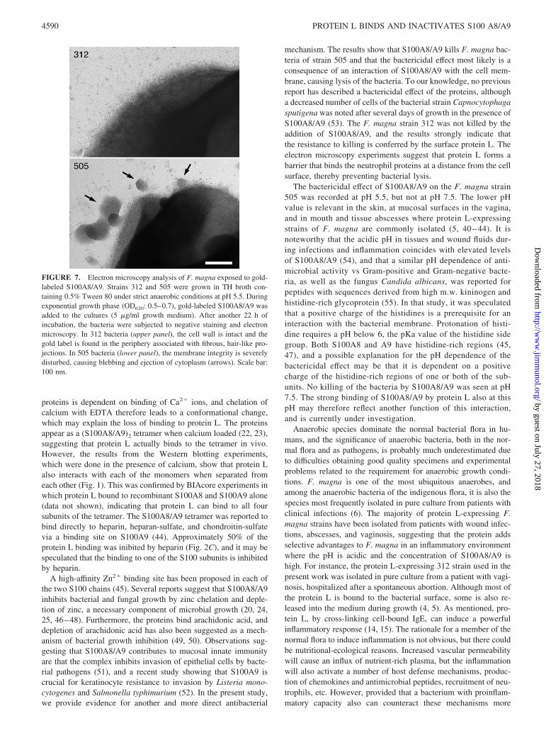
S100A8/A9 to a similar degree at pH 5.5 in TH broth (in contrastto the much higher binding to protein L-expressing 312 bacteria atpH 7.5 shown in Fig. 4B), 312 is more resistant to the antibacterialactivity of the protein complex. In an attempt to visualize thisactivity, gold-labeled S100A8/A9 (23 �g/ml) was added to cul-tures of 312 and 505 grown exponentially in TH broth (pH 5.5) for22 h. After another 18 h of incubation, when 505 in contrast to 312bacteria had stopped to grow (see Fig. 6B), the two strains wereanalyzed by electron microscopy. As shown in Fig. 7, the pictureis strikingly different. The cell wall of 312 bacteria is intact, andthe gold-labeled S100A8/A9 is associated with fibrous projectionsprotruding from the bacterial surface, and no gold-labeled materialis detected within the cell wall close to the membrane (upperpanel). Physicochemical analysis of protein L has revealed aStokes radius of 4.74 nm and a frictional ratio of 1.70, suggestingan elongated fibrous structure (9), which fits nicely with theS100A8/A9-binding surface protrusions. Contrary to the intact 312bacteria, the cell walls and membranes of 505 bacteria are disin-tegrated and gold-labeled S100A8/A9 is found at the membranes.Frequent blebs with ejected cytoplasmic material are also seen
(Fig. 7, lower panel). In summary, the results described in thisparagraph demonstrate that protein L protects F. magna from kill-ing by S100A8/A9.
DiscussionProtein L is an elongated, fibrous surface molecule of the humananerobic commensal and potential pathogen F. magna. The mol-ecule consists of multiple exposed domains, and five of these,the B1-B5 repeats, bind to human Ig L chains. In this work, weshow that the outermost domain, the A domain, has high affinityfor the neutrophil cytosol proteins S100A8 and S100A9. TheS100A8/A9 binding site on protein L was localized to a shortsegment (10 aa) most proximal to the neighboring B1 domain.The binding was dependent on an intact B1 domain, suggestingthat the side groups forming the binding site are coordinatedby the folded B1 domain. Despite the closeness to the B1 do-main, the binding of Ig and S100A8/A9 did not inhibit eachother, indicating full sterical separation of the two binding sites.The N-terminal 70 aa of the A domain did not appear to par-ticipate in the binding to S100A8/A9. This part of the moleculemay therefore have other functions, or bind to other, hithertounknown ligands of the human host.
The binding to S100A8/A9 was dependent on the presence ofdivalent cations (Fig. 2B). The conformation of both these EF hand
FIGURE 5. S100A8/A9 from disrupted neutrophils bind to protein L-expressing F. magna bacteria. F. magna 312 or F. magna 505 bacteriawere incubated with mechanically disrupted neutrophils or with bufferalone. After washing, the bacteria were stained with anti-S100A8/A9 andAlexa 594-conjugated secondary Abs. Differential interference contrast(DIC; left column) and immunofluorescence (right column) show localiza-tion of S100A8/A9 to a high degree with F. magna 312 (�99%), and to amuch lesser extent with F. magna 505 (10%). None of the strains showstaining following incubation with buffer alone. Scale bar: 10 �m.
FIGURE 6. Protein L-expressing F. magna bacteria are resistant toS100A8/A9 killing. S100A8/A9 isolated from neutrophils was used for allexperiments. Bacteria were cultivated under strict anaerobic conditions atpH 5.5, and growth was measured by reading the absorbance at 620 nm.Results are shown as mean value � SD of triplicate experiments. Thegrowth of strains 312 and 505 was statistically compared at different timepoints; �, p 0.05; ���, p 0.001, according to Student’s t test. A, F.magna strain 312 (F) expressing protein L or strain 505 (�) devoid ofprotein L was grown for 24 h at 37°C in the presence of various concen-trations of S100A8/A9. The absorbances at 24 h are relative to the meanvalue of respective strain with no addition of S100A8/A9. B, S100A8/A9was added to the cultures to a final concentration of 23 �g/ml 22 h afterinoculation. F. magna strain 312 with (f) or without addition (F) ofS100A8/A9, and strain 505 with (�) or without addition (E) ofS100A8/A9.
4589The Journal of Immunology
by guest on July 27, 2018http://w
ww
.jimm
unol.org/D
ownloaded from
proteins is dependent on binding of Ca2� ions, and chelation ofcalcium with EDTA therefore leads to a conformational change,which may explain the loss of binding to protein L. The proteinsappear as a (S100A8/A9)2 tetramer when calcium loaded (22, 23),suggesting that protein L actually binds to the tetramer in vivo.However, the results from the Western blotting experiments,which were done in the presence of calcium, show that protein Lalso interacts with each of the monomers when separated fromeach other (Fig. 1). This was confirmed by BIAcore experiments inwhich protein L bound to recombinant S100A8 and S100A9 alone(data not shown), indicating that protein L can bind to all foursubunits of the tetramer. The S100A8/A9 tetramer was reported tobind directly to heparin, heparan-sulfate, and chondroitin-sulfatevia a binding site on S100A9 (44). Approximately 50% of theprotein L binding was inibited by heparin (Fig. 2C), and it may bespeculated that the binding to one of the S100 subunits is inhibitedby heparin.
A high-affinity Zn2� binding site has been proposed in each ofthe two S100 chains (45). Several reports suggest that S100A8/A9inhibits bacterial and fungal growth by zinc chelation and deple-tion of zinc, a necessary component of microbial growth (20, 24,25, 46–48). Furthermore, the proteins bind arachidonic acid, anddepletion of arachidonic acid has also been suggested as a mech-anism of bacterial growth inhibition (49, 50). Observations sug-gesting that S100A8/A9 contributes to mucosal innate immunityare that the complex inhibits invasion of epithelial cells by bacte-rial pathogens (51), and a recent study showing that S100A9 iscrucial for keratinocyte resistance to invasion by Listeria mono-cytogenes and Salmonella typhimurium (52). In the present study,we provide evidence for another and more direct antibacterial
mechanism. The results show that S100A8/A9 kills F. magna bac-teria of strain 505 and that the bactericidal effect most likely is aconsequence of an interaction of S100A8/A9 with the cell mem-brane, causing lysis of the bacteria. To our knowledge, no previousreport has described a bactericidal effect of the proteins, althougha decreased number of cells of the bacterial strain Capnocytophagasputigena was noted after several days of growth in the presence ofS100A8/A9 (53). The F. magna strain 312 was not killed by theaddition of S100A8/A9, and the results strongly indicate thatthe resistance to killing is conferred by the surface protein L. Theelectron microscopy experiments suggest that protein L forms abarrier that binds the neutrophil proteins at a distance from the cellsurface, thereby preventing bacterial lysis.
The bactericidal effect of S100A8/A9 on the F. magna strain505 was recorded at pH 5.5, but not at pH 7.5. The lower pHvalue is relevant in the skin, at mucosal surfaces in the vagina,and in mouth and tissue abscesses where protein L-expressingstrains of F. magna are commonly isolated (5, 40 – 44). It isnoteworthy that the acidic pH in tissues and wound fluids dur-ing infections and inflammation coincides with elevated levelsof S100A8/A9 (54), and that a similar pH dependence of anti-microbial activity vs Gram-positive and Gram-negative bacte-ria, as well as the fungus Candida albicans, was reported forpeptides with sequences derived from high m.w. kininogen andhistidine-rich glycoprotein (55). In that study, it was speculatedthat a positive charge of the histidines is a prerequisite for aninteraction with the bacterial membrane. Protonation of histi-dine requires a pH below 6, the pKa value of the histidine sidegroup. Both S100A8 and A9 have histidine-rich regions (45,47), and a possible explanation for the pH dependence of thebactericidal effect may be that it is dependent on a positivecharge of the histidine-rich regions of one or both of the sub-units. No killing of the bacteria by S100A8/A9 was seen at pH7.5. The strong binding of S100A8/A9 by protein L also at thispH may therefore reflect another function of this interaction,and is currently under investigation.
Anaerobic species dominate the normal bacterial flora in hu-mans, and the significance of anaerobic bacteria, both in the nor-mal flora and as pathogens, is probably much underestimated dueto difficulties obtaining good quality specimens and experimentalproblems related to the requirement for anaerobic growth condi-tions. F. magna is one of the most ubiquitous anaerobes, andamong the anaerobic bacteria of the indigenous flora, it is also thespecies most frequently isolated in pure culture from patients withclinical infections (6). The majority of protein L-expressing F.magna strains have been isolated from patients with wound infec-tions, abscesses, and vaginosis, suggesting that the protein addsselective advantages to F. magna in an inflammatory environmentwhere the pH is acidic and the concentration of S100A8/A9 ishigh. For instance, the protein L-expressing 312 strain used in thepresent work was isolated in pure culture from a patient with vagi-nosis, hospitalized after a spontaneous abortion. Although most ofthe protein L is bound to the bacterial surface, some is also re-leased into the medium during growth (4, 5). As mentioned, pro-tein L, by cross-linking cell-bound IgE, can induce a powerfulinflammatory response (14, 15). The rationale for a member of thenormal flora to induce inflammation is not obvious, but there couldbe nutritional-ecological reasons. Increased vascular permeabilitywill cause an influx of nutrient-rich plasma, but the inflammationwill also activate a number of host defense mechanisms, produc-tion of chemokines and antimicrobial peptides, recruitment of neu-trophils, etc. However, provided that a bacterium with proinflam-matory capacity also can counteract these mechanisms more
FIGURE 7. Electron microscopy analysis of F. magna exposed to gold-labeled S100A8/A9. Strains 312 and 505 were grown in TH broth con-taining 0.5% Tween 80 under strict anaerobic conditions at pH 5.5. Duringexponential growth phase (OD620: 0.5–0.7), gold-labeled S100A8/A9 wasadded to the cultures (5 �g/ml growth medium). After another 22 h ofincubation, the bacteria were subjected to negative staining and electronmicroscopy. In 312 bacteria (upper panel), the cell wall is intact and thegold label is found in the periphery associated with fibrous, hair-like pro-jections. In 505 bacteria (lower panel), the membrane integrity is severelydisturbed, causing blebbing and ejection of cytoplasm (arrows). Scale bar:100 nm.
4590 PROTEIN L BINDS AND INACTIVATES S100 A8/A9
by guest on July 27, 2018http://w
ww
.jimm
unol.org/D
ownloaded from

efficiently than other species in the same ecological niche, this willgenerate a selective advantage.
The major finding of the present work is that a bacterial surfaceprotein specifically and independently interacts with both Igs andantibacterial proteins. To our knowledge, this is a unique propertyof protein L that emphasizes the complexity of host-microbe re-lationships and demonstrates how diffuse the borderline is betweenbacterial commensalism and pathogenicity.
AcknowledgmentsWe are grateful to the following colleagues at the Division of InfectionMedicine: Dr. Maria Allhorn for performing initial construction and ex-pression of protein L (A-C2); Monika Heidenholm, Pontus Nordenfeldt,and Dr. Hans Tapper for the fluorescence microscopy; Dr. Anders Olin forBIAcore data; Maria Baumgarten and Dr. Mattias Morgelin for electronmicroscopy; Dr. Arne Egersten, Dr. Artur Schmidtchen, and Dr. Ole Sø-rensen for stimulating discussions; and Ingbritt Gustafsson for performingmost of the experiments. Dr. Bjorn Walse (Saromics) is also gratefullyacknowledged for the molecular modeling.
DisclosuresThe authors have no financial conflict of interest.
References1. Forsgren, A., and J. Sjoquist. 1966. “Protein A” from S. aureus. I. Pseudo-im-
mune reaction with human �-globulin. J. Immunol. 97: 822–827.2. Bjorck, L., and G. Kronvall. 1984. Purification and some properties of strepto-
coccal protein G, a novel IgG-binding reagent. J. Immunol. 133: 969–974.3. Reis, K. J., E. M. Ayoub, and M. D. P. Boyle. 1984. Streptococcal Fc-receptor.
I. Isolation and partial characterization of the receptor from a group C strepto-coccus. J. Immunol. 132: 3091–3097.
4. Bjorck, L. 1988. Protein L: a novel bacterial cell wall protein with affinity for IgL chains. J. Immunol. 140: 1194–1197.
5. Kastern, W., E. Holst, E. Nielsen, U. Sjobring, and L. Bjorck. 1990. Protein L, abacterial immunoglobulin-binding protein and possible virulence determinant.Infect. Immun. 58: 1217–1222.
6. Murdoch, D. A. 1998. Gram-positive anaerobic cocci. Clin. Microbiol. Rev. 11:81–120.
7. Stephens, P., I. B. Wall, M. J. Wilson, K. E. Hill, C. E. Davies, C. M. Hill,K. G. Harding, and D. W. Thomas. 2003. Anaerobic cocci populating the deeptissues of chronic wounds impair cellular wound healing processes in vitro.Br. J. Dermatol. 148: 456–466.
8. Ricci, S., D. Medaglini, H. Marcotte, A. Olsen, G. Pozzi, and L. Bjorck. 2001.Immunoglobulin-binding domains of peptostreptococcal protein L enhance vag-inal colonization of mice by Streptococcus gordonii. Microb. Pathog. 30:229–235.
9. Åkerstrom, B., and L. Bjorck. 1989. Protein L: an immunoglobulin light chain-binding protein: characterization of binding and physicochemical properties.J. Biol. Chem. 262: 19740–19746.
10. Nilson, B. H. K., A. Solomon, L. Bjorck, and B. Åkerstrom. 1992. Protein L fromPeptostreptococcus magnus binds to the � light chain variable domain. J. Biol.Chem. 267: 2234–2239.
11. Beckingham, J. A., S. P. Bottomley, R. Hinton, B. J. Sutton, and M. G. Gore.1999. Interactions between a single immunoglobulin-binding domain of protein Lfrom Peptostreptococcus magnus and a human � light chain. Biochem. J. 240:193–199.
12. Graille, M., E. A. Stura, N. G. Housden, J. A. Beckingham, S. P. Bottomley,D. Beale, M. J. Taussig, B. J. Sutton, M. G. Gore, and J. B. Charbonnier. 2001.Complex between Peptostreptococcus magnus protein L and a human antibodyreveals structural convergence in the interaction modes of Fab binding proteins.Structure 9: 679–687.
13. Kastern, W., U. Sjobring, and L. Bjorck. 1992. Structure of peptostreptococcalprotein L and identification of a repeated immunoglobulin light chain-bindingdomain. J. Biol. Chem. 267: 12820–12825.
14. Patella, V., V. Casolaro, L. Bjorck, and G. Marone. 1990. Protein L: a bacterialIg-binding protein that activates human basophils and mast cells. J. Immunol.145: 3054–3061.
15. Genovese, A., G. Borgia, L. Bjorck, A. Petraroli, A. de Paulis, M. Piazza, andG. Marone. 2003. Immunoglobulin superantigen protein L induces IL-4 andIL-13 secretion from human Fc�RI� cells through interaction with the � lightchains of IgE. J. Immunol. 170: 1854–1861.
16. Heizmann, C. W., G. Fritz, and B. W. Schafer. 2002. S100 proteins: structure,functions and pathology. Front. Biosci. 7: 1356–1368.
17. Odink, K., N. Cerletti, J. Bruggen, R. G. Clerc, L. Tarcsay, G. Zwadio,G. Gerhards, R. Schlegel, and C. Sorg. 1987. Two calcium-binding proteins ininfiltrate macrophages of rheumatoid arthritis. Nature 330: 80–82.
18. Roth, J., T. Vogl, C. Sorg, and C. Sunderkotter. 2003. Phagocyte-specific S100proteins: a novel group of proinflammatory molecules. Trends Immunol. 24:155–158.
19. Wilkinson, M. M., A. Busuttil, C. Hayward, D. J. Brock, J. R. Dorin, andV. van Heyningen. 1987. Expression pattern of two related cystic fibrosis-asso-ciated calcium-binding proteins in normal and abnormal tissues. J. Cell Sci. 91:221–230.
20. Stritz, I., and I. Trebichavsky. 2004. Calprotectin: a pleiotropic molecule in acuteand chronic inflammation. Physiol. Res. 53: 245–253.
21. Rammes, A., J. Roth, M. Goebeler, M. Klempt, M. Hartmann, and C. Sorg. 1997.Myeloid-related protein (MRP) 8 and MRP14, calcium-binding proteins of theS100 family, are secreted by activated monocytes via a novel, tubulin-dependentpathway. J. Biol. Chem. 272: 9496–9502.
22. Strupat, K., H. Rogniaux, A. van Dorsselaer, J. Roth, and T. Vogl. 2000. Calci-um-induced non-covalently linked tetramers of MRP8 and MRP14 are confirmedby electrospray ionization-mass analysis. J. Am. Soc. Mass Spectrom. 11:780–788.
23. Leukert, N., T. Vogl, K. Strupat, R. Reichelt, C. Sorg, and J. Roth. 2006. Cal-cium-dependent tetramer formation of S100A8 and S100A9 is essential for bio-logical activity. J. Mol. Biol. 359: 961–972.
24. Steinbakk, M., C. F. Naess-Andresen, E. Lingaas, I. Dale, P. Brandtzaeg, andM. K. Fagerhol. 1990. Antimicrobial actions of calcium-binding leukocyte L1protein, calprotectin. Lancet 336: 763–765.
25. Sohnle, P. G., M. J. Hunter, B. Hahn, and W. J. Chazin. 2000. Zinc-reversibleantimicrobial activity of recombinant calprotectin (migration inhibitory factor-related proteins 8 and 14). J. Infect. Dis. 182: 1272–1275.
26. Murthy, A. R. K., R. I. Lehrer, S. S. L. Harwig, and K. T. Miyasaki. 1993. In vitrocandidastatic properties of the human neutrophil calprotectin complex. J. Immu-nol. 151: 6291–6301.
27. Goto, Y., C. Sanjoba, M. Asada, K. Saeki, T. Onodera, and Y. Matsumoto. 2008.Adhesion of MRP8/14 to amastigotes in skin lesions of Leishmania major-in-fected mice. Exp. Parasitol. 119: 80–86.
28. De Chateau, M., and L. Bjorck. 1994. Protein PAB, a mosaic albumin-bindingbacterial protein representing the first contemporary example of module shuffling.J. Biol. Chem. 269: 12147–12151.
29. Åkesson, P., K. H. Schmidt, J. Cooney, and L. Bjorck. 1994. M1 protein andprotein H: IgGFc- and albumin-binding streptococcal surface proteins encoded byadjacent genes. Biochem. J. 300: 877–886.
30. Matsudaira, P. 1987. Sequence from picomole quantities of proteins electroblot-ted onto polyvinylidene difluoride membranes. J. Biol. Chem. 262: 10035–10038.
31. Greenwood, F. C., W. M. Hunter, and J. S. Glover. 1963. The preparation of131I-labelled human growth hormone of high specific radioactivity. Biochem. J.89: 114–123.
32. Laemmli, U. K. 1970. Cleavage of structural proteins during the assembly of thehead of bacteriophage T4. Nature 227: 680–685.
33. Wester, L., M. U. Johansson, and B. Åkerstrom. 1997. Physicochemical andbiochemical characterization of human �1-microglobulin expressed in baculov-irus-infected insect cells. Protein Expression Purif. 11: 95–103.
34. Dalbøge, H., E. B. Jensen, H. Tøttrup, A. Grubb, M. Abrahamsson, I. Olafsson,and S. Carlsen. 1989. High-level expression of active human cystatin C in Esch-erichia coli. Gene 79: 325–332.
35. Berggård, T., J. J. Enghild, S. Badve, C. M. Salafia, L. Logdberg, andB. Åkerstrom. 1999. Histological distribution and biochemical properties of �1-microglobulin in human placenta. Am. J. Reprod. Immunol. 41: 52–60.
36. Baschong, W. J., M. Lucocq, and J. Roth. 1985. “Thiocyanate gold”: small (2–3nm) colloidal gold for affinity cytochemical labeling in electron microscopy. His-tochemistry 83: 409–411.
37. Elbashir, M. I., B. H. K. Nilson, P. Åkesson, L. Bjorck, and B. Åkerstrom. 1990.Antibody response in immunized rabbits measured with bacterial immunoglob-ulin-binding proteins. J. Immunol. Methods 135: 171–179.
38. Wikstrom, M., T. Drakenberg, S. Forsen, U. Sjobring, and L. Bjorck. 1994.Three-dimensional solution structure of an immunoglobulin light chain-bindingdomain of protein L: comparison with the IgG-binding domains of protein G.Biochemistry 33: 14011–14017.
39. Murphy, J. P., C. J. Duggleby, M. A. Atkinson, A. R. Trowern, T. Atkinson, andC. R. Goward. 1994. The functional units of peptostreptococcal protein L. Mol.Microbiol. 12: 911–920.
40. Rentzsch, G., and J. Wilke. 1970. Measurements of pH values in vitro and in vivoin chronic tonsillitis. Z. Laryngol. Rhinol. Otol. 49: 391–397.
41. Bryant, O. D., A. L. Rashad, J. A. Mazza, and D. Hammond. 1980. �-Lactamaseactivity in human pus. J. Infect. Dis. 142: 594–601.
42. Grinstein, S., C. J. Swallow, and O. D. Rotstein. 1991. Regulation of cytoplasmicpH in phagocytic cell function and dysfunction. Clin. Biochem. 24: 241–247.
43. Ohman, H., and A. Vahlquist. 1994. In vivo studies concerning a pH gradient inhuman stratum corneum and upper epidermis. Acta Derm. Venereol. 74:375–379.
44. Robinson, M. J., P. Tessier, R. Poulsom, and N. Hogg. 2002. The S100 familyheterodimer, MRP-8/14, binds with high affinity to heparin and heparan sulfateglycosaminoglycans on endothelial cells. J. Biol. Chem. 277: 3658–3665.
45. Clohessy, P. A., and B. E. Golden. 1996. His-X-X-X-His motif in S100 protein,calprotectin: relation to microbistatic activity. J. Leukocyte Biol. 60: 674.
46. Sohnle, P. G., C. Collins-Lech, and J. H. Wiessner. 1991. The zinc-reversibleantimicrobial activity of neutrophil lysates and abscess fluid supernatants. J. In-fect. Dis. 164: 137–142.
47. Clohessy, P. A., and B. E. Golden. 1995. Calprotectin-mediated zinc chelation asa biostatic mechanism in host defense. Scand. J. Immunol. 42: 551–556.
4591The Journal of Immunology
by guest on July 27, 2018http://w
ww
.jimm
unol.org/D
ownloaded from

48. Lusitani, D., S. E. Malawista, and R. R. Montgomery. 2003. Calprotectin, anabundant cytosolic protein from human polymorphonuclear leukocytes, inhibitsthe growth of Borrelia burgdorferi. Infect. Immun. 71: 4711–4716.
49. Klempt, M., H. Melkonyan, W. Nacken, D. Wiesmann, U. Holtkemper, andC. Sorg. 1997. The heterodimer of the Ca2�-binding proteins MRP8 and MRP14binds to arachidonic acid. FEBS Lett. 408: 81–84.
50. Siegenthaler, G., K. Roulin, D. Chatellard-Gruaz, R. Hotz, J. H. Saurat,U. Hellman, and G. Hagens. 1997. A heterocomplex formed by the calcium-binding proteins MRP8 (S100A8) and MRP14 (S100A9) binds unsaturated fattyacids with high affinity. J. Biol. Chem. 272: 9371–9377.
51. Nisapakultorn, K., K. F. Ross, and M. C. Herzberg. 2001. Calprotectin expressioninhibits bacterial binding to mucosal epithelial cells. Infect. Immun. 69:3692–3696.
52. Champaiboon, C., K. J. Sappington, B. D. Guenther, K. F. Ross, andM. C. Herzberg. 2009. Calprotectin S100A9 calcium-binding loops I and II areessential for keratinocyte resistance to bacterial invasion. J. Biol. Chem. 284:7078–7090.
53. Miyasaki, K. T., A. L. Bodeau, A. R. Murthy, and R. I. Lehrer. 1993. In vitroantimicrobial activity of the human neutrophil cytosolic S-100 protein complex,calprotectin, against Capnocytophaga sputigena. J. Dent. Res. 72: 517–523.
54. Johne, B., M. K. Fagerhol, T. Lyberg, H. Prydz, P. Brantzaeg,C. F. Naess-Andresen, and I. Dale. 1997. Functional and clinical aspects of themyelomonocyte protein calprotectin. Mol. Pathol. 50: 113–123.
55. Kacprzyk, L., V. Rydengård, M. Morgelin, M. Davoudi, M. Pasupuleti,M. Malmsten, and A. Schmidtchen. 2007. Antimicrobial activity of histidine-richpeptides is dependent on acidic conditions. Biochim. Biophys. Acta 1768:2667–2680.
56. Wikstrom, M., U. Sjobring, T. Drakenberg, S. Forsen, and L. Bjorck. 1995.Mapping of the immunoglobulin light chain-binding site of protein L. J. Mol.Biol. 250: 128–133.
57. Enokizono, J., M. Wikstrom, U. Sjobring, L. Bjorck, S. Forsen, Y. Arata,K. Kato, and I. Shimada. 2000. NMR analysis of the interaction between proteinL and Ig light chains. J. Bacteriol. 182: 1419–1422.
4592 PROTEIN L BINDS AND INACTIVATES S100 A8/A9
by guest on July 27, 2018http://w
ww
.jimm
unol.org/D
ownloaded from