neutrophil recruitment in periodontal disease
TRANSCRIPT

Neutrophil recruitment in
periodontal disease
Agnes Dahlstrand Rudin
Department of Oral Microbiology and Immunology
Institute of Odontology
Sahlgrenska Academy, University of Gothenburg
Gothenburg 2021
Cover illustration: Oil painting by Nanna Englund Mårskog
Neutrophil recruitment in periodontal disease
© Agnes Dahlstrand Rudin 2021
ISBN 978-91-8009-426-9 (PRINT)
ISBN 978-91-8009-427-6 (PDF)
Printed in Borås, Sweden 2021
Printed by Stema Specialtryck AB

ABSTRACT
Neutrophils are the first immune cells to arrive in infected or injured tissues, where they
engulf microbes and clean up cell debris. Periodontitis is one of the typical symptoms of
both neutropenia and defect neutrophil functionality, suggesting an important role for
these cells in maintenance of periodontal health. While representing a minor fraction of
the leukocytes in the periodontal lesion, neutrophils are the dominating cell type in the
periodontal pocket and gingival crevicular fluid (GCF). The overall aim of this thesis was
to characterize factors modulating neutrophil recruitment from blood to GCF in
periodontitis.
Neutrophil recruitment to the periodontal pocket is triggered by the bacterial species
colonizing this site. Although previous studies have shown that subgingival bacteria
trigger neutrophil chemotaxis, the bacterial chemoattractants responsible for this event
remained to be identified. The aims of paper I and II were to identify soluble neutrophil
chemoattractants released by the periodontitis associated bacterial species Porphyromonas
gingivalis and Fusobacterium nucleatum, and their corresponding neutrophil receptors.
Chemotactic compounds present in culture supernatants of both bacterial species where
identified as short chain fatty acids (SCFAs) specifically activating neutrophils via the
short chain fatty acid receptor 2 (FFAR2).
CD177 is a neutrophil subtype marker with unknown function, expressed by 1–100% of
circulating neutrophils depending on the donor. While CD177 has been proposed to
facilitate neutrophil transmigration, this had not yet been demonstrated in vivo. The aim
of paper III was to investigate whether CD177 expression affect neutrophil
transmigration to GCF in periodontitis. The CD177+ subtype was enriched in GCF as
compared to blood from the same donor, supporting an in vivo migration advantage of the
CD177+ subtype to this site. Periodontitis patients also exhibited higher levels of CD177+
cells in blood as compared to healthy controls, which resulted in very high proportions of
CD177+ cells in GCF. Considering this, functions differing between the subsets could
influence destructive inflammation of the periodontal tissues. As CD177 may not be the
sole factor contributing to functional differences between the subsets, further proteomic
differences between CD177+ and CD177– neutrophils were investigated in paper IV.
In conclusion, this thesis highlights SCFAs signaling via FFAR2 as factors involved in
neutrophil chemotaxis triggered by periodontitis associated bacteria. Further, the CD177+
neutrophil subtype is preferentially recruited to GCF and functions specific for this
subtype may be of importance for inducing (or suppressing) destructive inflammation in
periodontal tissues.
Keywords: Neutrophil, periodontitis, P. gingivalis, F. nucleatum, SCFA, FFAR2, CD177,
A1AT


SAMMANFATTNING PÅ SVENSKA
När neutrofiler nås av signaler om infektion eller vävnadsskada lämnar de blodbanan och
tar sig vidare ut i vävnaden för att eliminera mikroorganismer eller städa bort skadade
celler. Då parodontit är ett av de typiska symtomen vid både neutropeni och funktionella
neutrofildefekter verkar neutrofiler vara viktiga för att bibehålla ett friskt parodontium.
Medan neutrofilen endast återfinns i små mängder i parodontitlesionen är de dominerande
i gingivalvätskan från djupa tandköttsfickor hos patienter med parodontit. Denna
avhandling undersöker faktorer som påverkar rekryteringen av neutrofiler från blodbanan
till tandköttsfickan vid parodontit.
Rekrytering av neutrofiler till tandköttsfickan stimuleras av koloniserande, huvudsakligen,
anaeroba och gram-negativa bakterier. Tidigare studier har visat att dessa bakterier har
förmågan att direkt attrahera neutrofiler, men det var ännu okänt vilka specifika
kemotaktiska faktorer som utlöser cellernas rörelse. Syftet med studie I och II var att
identifiera lösliga kemoattraktanter som utsöndras av de parodontitassocierade
bakterierna, Porphyromonas gingivalis och Fusobacterium nucleatum, samt korresponderande
neutrofilreceptorer. De kemotaktiska faktorerna visade sig delvis utgöras av korta
fettsyror som aktiverar neutrofiler via den neutrofilspecifika receptorn för fria fettsyror
(FFAR2).
CD177 är en neutrofil subtypsmarkör med okänd funktion som beroende på donator
uttrycks av 1–100% av neutrofilerna i blodbanan. CD177 har föreslagits bidra till
rekrytering av neutrofiler från blod till vävnad men detta var ännu inte visat in vivo. Syftet
med studie III var att ta reda på om CD177 uttryck påverkar rekrytering av neutrofiler
från blod till tandköttsficka vid parodontit. Studien visade att CD177+ neutrofiler
ansamlades i tandköttsfickan vilket stöder teorin om att CD177 underlättar
neutrofilrekrytering till denna vävnad. Patienterna hade också generellt högre nivåer av
CD177+ celler i blodet jämfört med friska kontroller, vilket resulterade i mycket höga
nivåer av CD177+ neutrofiler i tandköttsfickan hos patienterna. Funktioner som skiljer sig
mellan subtyperna skulle därför kunna påverka utvecklingen av destruktiv inflammation i
parodontiet. Då CD177 uttryck inte nödvändigtvis är den enda faktor som bidrar till
funktionella skillnader mellan subtyperna, undersöks ytterligare skillnader på proteinnivå
mellan CD177+ and CD177– neutrofiler i studie IV.
Sammanfattningsvis lyfter denna avhandling fram korta fettsyror och FFAR2 som
bidragande faktorer till neutrofilrekrytering stimulerad av parodontitassocierade bakterier.
Den beskriver även hur den CD177+ neutrofila subtypen ackumuleras i tandköttsfickan
vid parodontit och undersöker ytterligare skillnader mellan CD177+ and CD177–
neutrofiler.


LIST OF PAPERS
This thesis is based on the following studies, referred to in the text by their Roman
numerals.
I. Porphyromonas gingivalis produce neutrophil specific
chemoattractants including short chain fatty acids
Dahlstrand Rudin A, Khamzeh A, Venkatakrishnan V, Persson T, Gabl
M, Savolainen O, Forsman H, Dahlgren C, Christenson K, Bylund J.
Frontiers in Cellular and Infection Microbiology. 2021 Jan 19.
https://doi.org/10.3389/fcimb.2020.620681
II. Short chain fatty acids released by Fusobacterium nucleatum are
neutrophil chemoattractants acting via free fatty acid receptor 2
(FFAR2)
Dahlstrand Rudin A, Khamzeh A, Venkatakrishnan V, Basic A,
Christenson K, Bylund J. Cellular Microbiology. 2021 Aug;23(8): e13348.
https://doi.org/10.1111/cmi.13348
III. The neutrophil subset defined by CD177 expression is
preferentially recruited to gingival crevicular fluid in periodontitis
Dahlstrand Rudin A, Amirbeagi F, Davidsson L, Khamzeh A, Thorbert
Mros S, Thulin P, Welin A, Björkman L, Christenson K, Bylund J. Journal
of Leukocyte Biology. 2021 Feb;109(2):349-362.
https://doi.org/10.1002/JLB.3A0520-081RR
IV. Proteomic characterization of neutrophil subsets distinguished by
membrane expression of CD177.
Dahlstrand Rudin A, Sanchez Klose FP, Komic H, Östberg A-K,
Venkatakrishnan V, Christenson K, Bylund J. In manuscript

CONTENT
INTRODUCTION ........................................................................................................ 1
THE NEUTROPHIL .................................................................................................... 3
Maturation ..................................................................................................................... 4
Life cycle ........................................................................................................................ 4
Granules......................................................................................................................... 6
Azurophil granules ................................................................................................. 7
Specific and Gelatinase granules .......................................................................... 8
Secretory vesicles .................................................................................................... 9
Killing mechanisms ...................................................................................................... 9
Phagocytosis ............................................................................................................ 9
Formation of neutrophil extracellular traps ..................................................... 11
NEUTROPHIL TRANSMIGRATION .................................................................. 13
Extravasation .............................................................................................................. 13
Chemotaxis .................................................................................................................. 16
Neutrophil chemoattractant receptors.................................................................... 17
Intermediate chemoattractants and receptors ................................................. 18
End-point chemoattractants and receptors ..................................................... 19
Tissue neutrophils ...................................................................................................... 21
PERIODONTITIS ...................................................................................................... 25
Definition, classification and management of periodontitis ............................... 26
The oral microbiome and periodontitis .................................................................. 27
Porphyromonas gingivalis ................................................................................................ 29
Fusobacterium nucleatum ................................................................................................ 30
Neutrophils in periodontitis ..................................................................................... 31
Neutrophil chemoattractants in periodontitis ....................................................... 34
Short chain fatty acids in the periodontal pocket ................................................. 36
NEUTROPHIL HETEROGENITY ....................................................................... 39
CD177 .......................................................................................................................... 41

CD177 and neutrophil transmigration ............................................................. 43
Further differences between the neutrophil subtypes defined by CD177 expression? ............................................................................................................ 45
CONCLUDING REMARKS .................................................................................... 47
ACKNOWLEDGEMENT ........................................................................................ 49
REFERENCES............................................................................................................. 51

ABBREVIATIONS
A1AT
Alpha-1-antitrypsin
AASV
ANCA associated systemic vasculitis
ACPA
Anti-citrullinated protein antibody
ANCA
Anti-neutrophil cytoplasmic autoantibody
BOP
Bleeding on probing
BPI
Bactericidal permeability-increasing protein
C5aR
C5a-receptor
CAL
Clinical attachment loss
CGD
Chronic granulomatous disease
COPD
Chronic obstructive pulmonary disease
DAMP
Danger associated molecular pattern
FFAR
Free fatty acid receptor
FPR
Formyl peptide receptor
GCF
Gingival crevicular fluid
G-CSF
Granulocyte colony stimulating factor
GPCR
G-protein coupled receptor
GPI
Glycosylphosphatidylinositol
HOCl
Hypochlorous acid
IBD
Inflammatory bowel disease
ICAM
Intercellular Adhesion Molecule
IL
Interleukin
LAD
Leukocyte adhesion deficiency
LDN
Low density neutrophil
LFA1
Lymphocyte function-associated antigen 1
LPS
Lipopolysaccharide
LTB4
Leukotriene B4
MAC Membrane attack complex
MAC-1 Macrophage-1 antigen
MPO
Myeloperoxidase
NE
Neutrophil elastase
NET
Neutrophil extracellular trap
NGAL Neutrophil gelatinase-associated lipocalin
OLFM4
Olfactomedin 4
PAD
Peptidyl-arginine deiminase
PAF
Platelet activating factor
PAMP
Pathogen associated molecular pattern
PBMC Peripheral blood mononuclear cell
PECAM1
Platelet endothelial cell adhesion molecule 1
PLA2
Phospholipase A2
PLS
Papillon-Lefèvre syndrome
PR3
Proteinase 3
PRR
Pattern recognition receptor
PSGL1
P-selectin glycoprotein ligand 1
PV
Polycythemia vera
RA
Rheumatoid arthritis
ROS
Reactive oxygen species
SCFA
Short chain fatty acid
SLE
Systemic lupus erythematosus
TLR
Toll like receptor
TNF-α
Tumor necrosis factor-α
VCAM1
Vascular Cell Adhesion Molecule 1
VLA4
Very late antigen 4

1
INTRODUCTION
Inflammation is our body´s immediate response to microbe invasion or tissue
injury. Consisting of an intricate network of inflammatory cells, tissues and
biological processes, inflammation is vital for elimination of unwanted intruders
and initiation of tissue repair. Neutrophils are the first inflammatory cells to arrive
at the infected or injured site, where they engulf microbes and cell debris or
capture intruders by release of web-like DNA structures. For these purposes,
neutrophils leave the blood stream at the vicinity of the affected tissues and
migrate further towards their prey, guided by a series of chemotactic signals. The
critical role of neutrophil recruitment from blood to tissue can be illustrated by
the fact that genetic defects disrupting this process result in recurrent severe
infections. The symptoms of such disorders typically also include destructive
inflammation of the tooth-supporting structures, i.e., periodontitis. Periodontitis
is a microbe initiated inflammatory disease leading to deepening of gingival
pockets and degradation of alveolar bone; which may eventually result in tooth
loss. Neutrophils seem to play an important role in the maintenance of
periodontal health as insufficient neutrophil numbers or defect neutrophil
functionality, as seen in patients with rare genetic defects, often lead to rapidly
progressing periodontitis. Moreover, although representing a minor fraction of
the leukocytes in the periodontal lesion, neutrophils are the dominating cell type
in the inflammatory exudate of the periodontal pocket.
This PhD thesis describes the neutrophil journey from circulation to the
periodontal pocket, with the intention of adding new insights regarding the nature
of bacteria derived chemotactic signals and neutrophil subsets operating at this
site.

2

3
THE NEUTROPHIL
Neutrophil granulocytes belong to the family of innate immune cells and the
subgroup of polymorphonuclear granulocytes (also including eosinophils and
basophils). While the term polymorphonuclear refers to the characteristic
multilobulated nuclei exhibited by these cell types (1), the name granulocyte is
derived from their cytoplasm being packed with storage granules (2). Constituting
50-70% of all white blood cells in human circulation, neutrophils have a crucial
role in our defense against invading microbes and in clearance of injury related
cell debris (3). When reached by signals of infection or injury, circulating
neutrophils swiftly leave the blood stream, enter the affected tissues and navigate
further towards the inflammatory focus where they engulf microbes or disrupted
cells (4). The neutrophil granules contain an array of toxic substances aimed for
microbe elimination (2). Thus, neutrophil recruitment needs to be tightly
regulated as excessive release of granule content may inflict tissue damage (5).
The indispensable role of neutrophils for human immune defense can be
exemplified by the fact that a marked decrease in neutrophil numbers, or altered
neutrophil functionality, typically results in severe immunodeficiency (6).
Insufficient neutrophil numbers in circulation is seen in severe congenital
neutropenia; a rare condition that is estimated to occur in 3-8.5 individuals per
million (7). This condition is caused by genetic mutations affecting neutrophil
differentiation in the bone marrow, with ELANE (encoding neutrophil elastase
(NE)) mutations being the most prevalent cause. As a result of their unusually
low neutrophil blood count, neutropenic patients suffer increased risk of
recurrent severe infections and frequently also develop periodontitis (7). Among
the most well described neutrophil functional deficiencies are Leukocyte-
adhesion deficiency (LAD) and Chronic granulomatous disease (CGD). LAD is
characterized by defect neutrophil surface adhesion molecules (β2-integrins) due
to mutations in the gene encoding for the β2-integrin subunit CD18. These
mutations had been reported in just over 300 cases between 1975 and 2017 and
is estimated to occur in 1:1 000 000 individuals (8). LAD primarily result in
inability of neutrophils to form firm adhesion to the endothelium and thereby
4
hinder their transmigration to tissues (9). CGD targets neutrophil production of
reactive oxygen species (ROS), an important bacterial killing mechanism. These
are a group of rare disorders (estimated prevalence ̴ 1:250 000) caused by
mutations in genes encoding different subunits of the ROS-producing
nikotinamid-adenin-dinukleotidfosfat complex (NADPH complex) (10, 11).
While patients who suffer from LAD develop similar symptoms as seen in
neutropenia (i.e., recurrent infections and periodontitis), CGD patients, although
being more sensitive to infections, do not typically present with periodontitis (9,
10). In addition to demonstrating the overall importance of neutrophil functions
to human immunity, studies of such specific deficiencies can provide important
clues regarding so far unknown pathologic mechanisms underlying development
of diseases with neutrophil involvement.
Maturation Similar to many other blood cells, neutrophil granulocytes are matured in, and
released from the bone marrow. Originating from the myeloid progenitor cell, the
neutrophil pass through six different developmental stages; myeloblast,
promyelocyte, myelocyte, metamyelocyte, band cell, and mature neutrophil (12,
13). After a maturation period of 14 days the terminally differentiated, non-
dividing neutrophil is released into circulation where it will have a relatively short
life. Although the longevity of neutrophils in circulation has been debated (14-
16), they are generally viewed as having a lifespan of 7-9 hours, which can be
greatly extended when exposed to inflammatory stimuli (14). Under healthy
conditions, neutrophils remain in circulation for a few hours before they are
homed to, and cleared in the bone marrow or die in the vasculature followed by
clearance in the liver by resident macrophages (4, 14).
Life cycle Neutrophils are produced at an impressive rate of about 100 billion cells per day
(17). In addition to being continuously released into circulation, a reserve of
mature cells is stored in the bone marrow, ready to be quickly released if
demanded (13). After release, circulating neutrophils can be divided into two
pools; one freely circulating intravascular pool and one marginated pool. The idea
of a marginated pool of neutrophils was launched after the discovery that up to

5
50% of radio-labelled homologous neutrophils injected into the circulation of
healthy individuals disappeared from the circulation after a short time period (18),
and that the marginated cells re-entered the circulation following injections of
adrenalin or physical exercise (19). Later studies have shown a prolonged transit
time through the lungs, liver and spleen supporting margination of neutrophils to
these organs (17, 20). Whether neutrophil accumulation in these organs have
functional implications or if the marginated pool simply consist of cells too rigid
to easily pass through narrow blood vessels is not entirely clear (4, 13, 14, 20).
Neutrophil release from the bone marrow into circulation is a regulated process
that is in balance with intravascular margination and clearance. This finely tuned
system keeps neutrophil blood levels constant at a healthy state, while cell
numbers can be rapidly increased in response to inflammatory stimuli (21). The
maintenance of homeostasis is of great importance as neutrophils, as well as being
indispensable effector cells of innate immunity, also have the capacity to cause
tissue damage (17). One factor controlling granulopoiesis is the hematopoietic
cytokine granulocyte colony stimulating factor (G-CSF). G-CSF potently
stimulates neutrophil proliferation and release (22-24) and show increased serum
concentration in response to infection (25). CXCL12 (also known as stromal
derived factor 1) is produced by bone marrow stromal cells and is, via its ligation
with the neutrophil chemokine receptor 4 (CXCR4), responsible for retaining
neutrophils in the bone marrow (17, 26). One of the mechanisms by which G-
CSF stimulate neutrophil release from the bone marrow is via inhibition of the
CXCR4-CXCL12 interaction (22). It has also been suggested that CXCR4 can be
upregulated in mature circulating neutrophils as a signal promoting their return to
the bone marrow, where they undergo apoptosis and are subsequently
phagocytosed by stromal macrophages (17, 27).
As previously mentioned, neutrophils exert their primary immune functions in
tissues after having left the circulation. The general belief is that these tissue
neutrophils subsequently die by apoptosis and are cleared by tissue resident
macrophages (4). However, studies of animal models indicate that tissue
neutrophils may also be able to return to the circulation and thereby prevent tissue
damage and facilitate resolution of inflammation (28). In 2006, this phenomenon
was visualized in vivo in zebrafish following resolution of sterile injury (29). More
recently, so-called reverse transmigration of neutrophils was reported to occur
6
after sterile hepatic injury in a mice model (30). In this study, Wang et al.
monitored (using intravital imaging) how neutrophils facilitated tissue repair via
phagocytosis of cell debris in the injured tissue and subsequently returned to the
vasculature. Briefly before returning to the bone marrow to undergo apoptosis,
the reverse transmigrated neutrophils were transiently accumulated in the lungs
where the homing receptor CXCR4 was upregulated (30). Human neutrophils are
reportedly capable of transmigrating in both directions over an endothelial
monolayer in vitro and these reversely transmigrated cells present with a phenotype
distinct from the usual transmigrated cell (CD54high and CXCR1low) (31).
Increased levels of this phenotype were also detected in blood from patients
suffering from inflammatory arthritis and atherosclerosis as compared to healthy
controls, indicating the presence of reverse transmigrated cells in these patient
groups (31). Reverse transmigration may indeed have positive effects on the
resolution of sterile inflammation but it is also speculated that the return of
activated neutrophils to circulation could cause dissemination of inflammation
leading to multiple organ failure (28, 32). Whether human neutrophils undergo
reverse migration in vivo and if it occurs following inflammation triggered by
infectious stimuli remain to be elucidated (33).
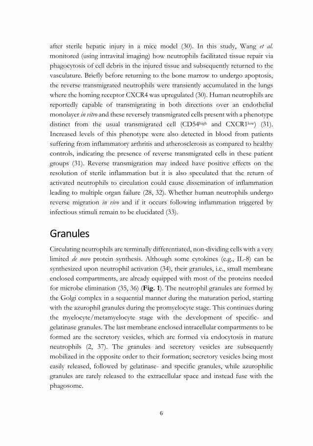
Granules Circulating neutrophils are terminally differentiated, non-dividing cells with a very
limited de novo protein synthesis. Although some cytokines (e.g., IL-8) can be
synthesized upon neutrophil activation (34), their granules, i.e., small membrane
enclosed compartments, are already equipped with most of the proteins needed
for microbe elimination (35, 36) (Fig. 1). The neutrophil granules are formed by
the Golgi complex in a sequential manner during the maturation period, starting
with the azurophil granules during the promyelocyte stage. This continues during
the myelocyte/metamyelocyte stage with the development of specific- and
gelatinase granules. The last membrane enclosed intracellular compartments to be
formed are the secretory vesicles, which are formed via endocytosis in mature
neutrophils (2, 37). The granules and secretory vesicles are subsequently
mobilized in the opposite order to their formation; secretory vesicles being most
easily released, followed by gelatinase- and specific granules, while azurophilic
granules are rarely released to the extracellular space and instead fuse with the
phagosome.
7
Sorting of proteins into the different granule types, each requiring different types
of stimuli to be mobilized, allows neutrophils to respond in an optimal way
depending on the situation. The distribution of granule proteins in the distinct
granule subtypes is a result of both proteins and granules being formed
sequentially during neutrophil maturation. Accordingly, granules store the
proteins that are synthesized at the time of granule formation, a mechanism that
is referred to as ‛targeting-by-timing’ (38). The ‛targeting-by-timing’ model
explaining the distribution of neutrophil granule proteins has recently also been
shown relevant in the context of protein glycosylation patterns, i.e., proteins that
are synthesized at a certain time point during granulopoiesis display a
characteristic set of oligosaccharides, dubbed ‛glycosylation-by-timing’ (39).
Azurophil granules
The azurophil granules are formed during the promyelocyte stage and mainly
participate in killing of microorganisms in the phagolysosome (36). These
Figure 1. Neutrophil granules and secretory vesicles. Drawing of the four distinct neutrophil granule and secretory vesicle subsets and examples of their characteristic content.
8
granules are often described as rarely undergoing exocytosis, in order to prevent
tissue damage that could otherwise be caused by their toxic content (40). Despite
this, proteins derived from azurophil granules can be found extracellularly in
inflamed tissues (41). Whether this is a result of exocytosis (42), release of
neutrophil extracellular traps (NETs), necrosis or a combination of these events
is not entirely clear.
The defining protein of azurophil granules, myeloperoxidase (MPO), contributes
to microbial killing in the phagolysosome by increasing the toxic potential of
H2O2 that is formed by the NADPH oxidase (43). After fusing of azurophil
granules with the phagosome, MPO converts H2O2 into hypochlorous acid
(HOCl). Moreover, and as discussed in more detail below, MPO participate in
most cases of neutrophil extracellular trap (NET) formation (44, 45). Other
azurophil granule proteins with microbicidal potential are the α-defensins,
bactericidal permeability-increasing protein (BPI), and serine proteases
(proteinase 3 (PR3), cathepsin G and neutrophil elastase (NE))(36). The serine
proteases are particularly interesting in the context of periodontal disease, as a
complete lack of active serine proteases result in early onset and rapidly
progressing periodontitis. This is seen in the rare autosomal recessive disorder
Papillon-Lefèvre syndrome (PLS) (46-48), that will be discussed more in the
subsequent chapter on periodontitis.
Specific- and Gelatinase granules
Specific granules and gelatinase granules are both peroxidase- (i.e., MPO) negative
but can be subdivided based on their content and formation stage (49). Specific
granules, which are formed during the myelocyte stage (37), mainly fuse with the
phagosome and contain an array of antibacterial substances including lactoferrin
(50), hCAP-18, neutrophil gelatinase-associated lipocalin (NGAL) and lysozyme
(36). In contrast to specific granules, gelatinase granules, which are formed during
the metamyelocyte stage, rarely fuse with the phagosome and are instead more
easily exocytosed (51). This is convenient as they carry matrix degrading enzymes
(e.g., the matrix metalloprotease, gelatinase) and adhesion receptors that are
required during neutrophil transmigration, in combination with less antimicrobial
substances (36). Both specific- and gelatinase granules contain cytochrome b558

9
(52), which is the membrane bound component of the oxygen radical producing
NADPH oxidase (described in more detail below).
Secretory vesicles
Secretory vesicles are the last membrane enclosed intracellular compartments to
be formed during granulopoiesis and also the most easily exocytosed upon
proinflammatory stimulation. Unlike the neutrophil granules, which are produced
by the Golgi complex (53), secretory vesicles are formed through endocytosis,
and consequently contain both membrane receptors and plasma proteins (54).
Killing mechanisms A well-functioning recruitment of neutrophils from circulation, though tissues,
and towards the inflammatory focus is inevitably a crucial step in eradication of
invading microbes. These processes will be described in more detail in the
upcoming chapter on neutrophil transmigration. Once in close contact with
invading microbes, the two primary killing mechanisms employed are
phagocytosis or NET formation.
Phagocytosis
Phagocytosis is a process during which neutrophils internalize and degrade
microbes or cell debris. This is facilitated by bacterial opsonization, i.e., coating
of bacterial surfaces with antibodies or complement opsonins, which are
recognized by neutrophils receptors (i.e., FC- or CR3 receptors respectively).
While antibodies are produced by the B-cells of adaptive immunity, complement
opsonins are generated via enzymatic cleavage of complement proteins. The
complement system consists of over 30 plasma proteins which when degraded on
bacterial surfaces generate powerful inflammatory mediators (i.e., anaphylatoxins)
(described in more detail in the chapter on neutrophil chemoattractants),
complement opsonins (e.g., C3b) and the membrane lysing ‛membrane attack
complex’ (MAC) (55).
After recognition and attachment of microbes or particles, cytoskeletal
rearrangements create a protrusion of the neutrophil plasma membrane, which
surrounds and internalizes the prey in an intracellular compartment; i.e., a

10
phagosome (56, 57). The phagosome is subsequently matured into a
phagolysosome, filled with antimicrobial substances, through fusion with
azurophil- and specific- granules. Killing of microbes in the phagolysosome is also
helped by release of ROS via activation of the membrane bound NADPH
oxidase. The NADPH oxidase is an electron transporting enzyme composed of
five different sub-units. Two of the subunits (p22phox and gp91phox, together
making up the so-called cytochrome b558) are bound to the plasma membrane and
membranes of specific and gelatinase granules (58, 59), while the complex of the
three remaining subunits (p40phox, p47phox and p67phox) can be found in the
cytosol. Following activation, the membrane bound and cytosolic compartments
of NADPH assembles creating an active enzyme complex that transfers electrons
over the membrane to O2 resulting in formation of superoxide (O2-). The
superoxide anion spontaneously dissociates into H2O2, that together with O2- are
referred to as primary ROS. The toxicity of primary ROS can be further amplified
via the previously described reaction catalyzed by MPO (from azurophil granules)
resulting in HOCl, and other secondary ROS (60).
The relative importance of the microbicidal factors released and produced in the
neutrophil phagolysosome and how these factors influence each other is still not
entirely clear (61). As previously mentioned, CGD patients who suffer from
genetic mutations in various components of the NADPH complex, display an
increased susceptibility to a variety of bacterial and fungal infections and CGD
patient neutrophils exhibit decreased bactericidal capacity in vitro (62). Thus, the
clinical presentation in CGD illustrates that ROS-production is an important
mechanism for microbe eradication by neutrophils. Despite this, today many
CGD patients survive into adulthood (63), and their neutrophils are able to kill at
least some of the pathogens that they encounter (e.g., Neisseria gonorrhoeae and
Staphylococcus aureus) (64, 65), suggesting that microbicidal mechanisms other than
ROS production can be functionally overlapping oxygen dependent killing. In
contrast to CGD, patients exhibiting complete MPO deficiency rarely show any
symptoms, with the exception of diabetic MPO deficient individuals who
reportedly suffer from systemic candidiasis (6). This could be interpreted as the
MPO dependent conversion of primary ROS into HOCl being a mechanism of
minor importance for neutrophil microbial killing. However, in vitro studies have
demonstrated that MPO deficient neutrophils exhibit decreased capability to kill
both fungi (e.g., Candida albicans) (66, 67) and several bacterial species including

11
(e.g., S aureus and Escherichia coli) (67, 68), as compared to normal neutrophils. It
has been argued that although MPO independent antimicrobial mechanisms may
be sufficient to kill invading microbes in the majority of cases, HOCl generated
via MPO could be critical in cases with extreme bacterial loads or in patients with
other immunodeficiencies (69).
In addition to oxygen dependent killing, neutrophils contain a range of
antimicrobial proteins stored in azurophil- and specific granules. These
bactericidal factors, including α-defensins and serine proteases, are released into
the phagosome upon fusion with granules. Although the α-defensins are
described as the most potent bactericidal proteins stored in neutrophil granules
(70), also neutrophil serine proteases have shown direct bactericidal activities in
vitro (71, 72). Yet, apart from the characteristic symptoms of rapidly progressing
periodontitis and palmoplantar keratosis, patients suffering from PLS (who
completely lack active neutrophil serine proteases) are relatively healthy and rarely
present with serious infections (46-48, 73). In summary, the described deficiencies
display distinct clinical presentations depending on the affected killing
mechanism. This indicates that the relative importance of the microbicidal
pathways differ depending on situation and nature of the ingested microbe.
Formation of neutrophil extracellular traps
In 2004 Brinkmann et al. reported that neutrophils were able to generate large
extracellular web-like structures, referred to as neutrophil extracellular traps
(NETs), which were able to trap and kill bacteria (74). These structures were
composed of decondensed chromatin fibers covered with various granule
proteins, including NE, cathepsin G, PR3 and MPO (75). NET formation has
been described as a mechanism aimed to neutralize bacteria, fungi, viruses and
parasites extracellularly, but it is not entirely clear if microbes are killed or simply
trapped and prevented from dissemination by the NET fibers (76, 77).
NET formation was first described as a novel form of programmed cell-death,
i.e., NETosis, during which neutrophils release chromatin fibers as a last attempt
to eradicate microbes (77, 78). However, it has since then been suggested that
neutrophils may also have the capacity to remain viable after NETosis (79). In a
majority of cases, ROS, MPO and NE are required for the processing of histones
12
and disruption of chromatin packaging during NET release (45, 77), but there is
also evidence of ROS independent NET formation triggered by S. aureus and
factors secreted by S. aureus (44, 80, 81). In line with NET formation being MPO
and NE dependent, neutrophils from patients with complete MPO or NE
deficiency (e.g., PLS patients) exhibit defect NETosis in vitro (46, 47, 82).
Despite the initial focus on the immune-protective functions of NETs, more
recent studies have highlighted NETs as a pathologic factor in a diverse set of
medical conditions (77). Isolated NETs have for instance been shown capable of
killing both human alveolar epithelial and endothelial cells in vitro (83) and tissue
damage caused by excess neutrophil infiltration and NET release have been
reported to cause acute injury in organs including kidneys, lungs and liver (84).
Overt NET release in blood can have lethal effects in sepsis and have also been
linked to deep vein thrombosis (85). In the case of thrombosis, NETs are
suggested to promote vaso-occlusion by providing a scaffold binding platelets and
red blood cells and increasing procoagulant activity (85). Moreover, NETs are
implicated in autoimmune diseases involving auto-antibodies against NET
derived proteins. One such autoimmune disease is antineutrophil cytoplasmic
autoantibody (ANCA) associated vasculitis (AASV), resulting from neutrophil
activation by autoantibodies against the NET components MPO or PR3 (86).
Another autoimmune disease, rheumatoid arthritis (RA) is associated with
autoantibodies directed against citrullinated proteins (ACPAs) and NETosis has
been correlated with the presence of ACPAs in these patients. The enzyme
catalyzing protein citrullination, peptidyl arginine deiminase (PAD), is expressed
at high levels in NETs and this has been suggested to contribute to extracellular
protein citrullination, thereby providing access to antigens in tissues (87).
Interestingly, the periodontitis associated bacterial species Porphyromonas gingivalis
has been reported as the only microbial source of PAD (88) (described in more
detail in the chapter on P. gingivalis). In summary, regulation of release and
clearance of NETs appear to be critical as there is a fine line between favorable
and harmful effects of this extracellular protection mechanism.

13
NEUTROPHIL TRANSMIGRATION
The recruitment of blood neutrophils to the site of infection or injury is a pivotal
step preceding microbial killing and tissue repair. The importance of a well-
functioning recruitment cascade can be exemplified by the fact that rare genetic
defects affecting this process results in severe immunodeficiency, as seen in the
previously described LAD (8, 9, 89).
Neutrophils circulating the blood stream are constantly surveying tissues for
indications of microbe invasion or injury. The first signals of infection are
triggered by recognition of conserved microbial molecular patterns by tissue-
resident immune cells. Such microbial patterns include lipopolysaccharide (LPS),
bacterial flagellin, lipoteichoic acid and peptidoglycan and are referred to as
pathogen associated molecular patterns (PAMPS). According to the same
principle, tissue injury is recognized via molecular patterns associated with dying
or ruptured cells (e.g., the extracellular presence of DNA, mitochondrial proteins
and ATP) referred to as damage associated molecular patterns (DAMPS) (90).
PAMPS and DAMPS activate pattern recognition receptors (PRRs; e.g., Toll-like
receptors and NOD-like receptors) expressed by tissue resident immune cells,
such as macrophages and dendritic cells. These cells respond by production and
release of proinflammatory factors initiating the recruitment and extravasation of
neutrophils. The neutrophil extravasation cascade involves five major steps;
tethering, rolling, adhesion, crawling and transmigration, which are illustrated in
Fig. 2 and will be described in detail below
Extravasation Activation of PRRs on sentinel immune cells induces the release of
proinflammatory cytokines (e.g., TNF-α, IL-1) and vasodilators (e.g., nitric oxide
(NO)) activating the local blood vessel endothelium. While NO induces vessel
dilatation and loosening of endothelial cell junctions, cytokine stimulation of
endothelial cells results in upregulation of P- and E-selectins to their luminal
surface (90). These selectins capture bypassing neutrophils via ligation to P-

14
selectin glycoprotein ligand 1 (PSGL-1), which is expressed on the neutrophil cell
membrane (35). The transient low-affinity bond between selectins and PSGL-1
allows neutrophils to roll along the vessel wall in a process referred to as tethering.
The neutrophil surface glycoprotein L-selectin is also participating in the luminal
rolling by its binding to endothelial glycans such as Sialyl LewisX (91, 92). As
cellular velocity decreases, integrins bind their endothelial ligands intercellular
adhesion molecule 1 and 2 (ICAM1, ICAM2) and vascular cell adhesion molecule
1 (VCAM1); a firm bond resulting in neutrophil adhesion to the endothelium (90,
93). ICAM1 binds to the β2-integrin lymphocyte function-associated antigen 1
(LFA-1) (consisting of the subunits CD11a and CD18), ICAM2 binds to the β2-
integrin macrophage-1 antigen (MAC-1)/complement receptor 3 (CR3;
consisting of the subunits CD11b and CD18) and VCAM1 binds to the β1-
integrin very late antigen 4 (VLA-4). As previously mentioned, LAD associated
defects in the β2-integrin subunit CD18 result in inefficient forming of firm bonds
between β2-integrins and ICAM and thereby hinder neutrophil transmigration (8,
9, 89).
While rolling along the endothelium, neutrophils come in contact with cytokines
that induce cell activation (i.e., priming). Neutrophils are circulating the blood
stream in a resting state that is characterized by low adherence and minimal
responsiveness to stimuli, this in order to prevent excessive responses that might
result in tissue damage. During priming, neutrophils undergo phenotypic and
functional changes leaving the cells in a hyperresponsive state, i.e., ready to
effectively eliminate invading microorganisms. The primed neutrophil is
characterized by augmented responses to stimulation, including increased ROS-
production, granule mobilization and adhesion, enhanced chemotaxis, and
delayed apoptosis (94, 95). Phenotypical changes following priming include
rearrangement of surface receptors (e.g., adhesion- and chemotactic receptors) via
mobilization of secretory vesicles and shedding of early adhesion molecules. In
line with this, shedding of the adhesion molecule L-selectin (CD62L) (92) and
upregulation of CR3/MAC-1 (CD11b/CD18) are routinely assessed for
determination of neutrophil activation stage (95).
After having established a firm bond to the endothelium, neutrophils start to
crawl along the vessel wall in attempt to leave the circulation as close as possible

15
Figure 2. Neutrophil transmigration and microbe elimination. Microbe stimulation of PRRs on
resident immune cells trigger release of cytokines which activate local endothelial cells. P- and
E- selectins on the activated endothelium forms low-affinity bonds with neutrophil PSGL-1,
resulting in neutrophil rolling along the vessel wall. L-selectin binds to endothelial glycans and
is subsequently shredded. As cellular velocity decreases, a firm bond is established between
neutrophil integrins and endothelial ICAMs and VCAMs. Neutrophil activation leads to
upregulation of surface receptors from secretory vesicles and degranulation of gelatinase
granules. Guided by chemotactic gradients, neutrophils transverse the endothelium, migrate
further through tissues and eliminate microbes via phagocytosis or NET-release.

16
to the site of infection or injury. Crawling is guided by chemokine gradients, i.e.,
cells move in the direction of increasing chemokine concentration. Although
neutrophils preferably transmigrate via endothelial cell-cell junctions
(paracellularly), they can also migrate directly through endothelial cells
(transcellularly) (96). Paracellular transmigration involves ICAM1, ICAM2,
VCAM1 as well as several junctional proteins including platelet endothelial cell
adhesion molecule 1 (PECAM1). PECAM1 is expressed on the cell surface of
both neutrophils and endothelial cells and its homologous binding facilitates
neutrophil transmigration over the endothelium (97). After the endothelial
crossing, neutrophils transverse the basal membrane and pericytes surrounding
the vessels. This has been attributed to the release of serine proteases (e.g., NE),
which degrade the basal membrane laminins and collagen fibers. However,
evidence also points at the existence of less dense regions of the basal membrane
which are in direct contact with gaps between pericytes, and could represent sites
of less resistance for neutrophils to emigrate (98).
Chemotaxis Once neutrophils have passed through the endothelium and basal membrane,
they need to navigate further through tissues towards invading microbes or site
of tissue damage. Cell movement towards the inflammatory focus is guided by
concentration gradients of soluble chemical signals, i.e., chemoattractants, in a
process referred to as chemotaxis (99, 100). Neutrophils express a number of
surface receptors that are able to sense chemoattractants released from host cells
(i.e., endogenous) or microbes (i.e., bacteria derived), and induce cell movement
in the direction of increasing chemoattractant concentration (101). The direction
of a chemoattractant gradient is sensed by the detection of differences in the
number of occupied chemoattractant receptors (100). When the desired direction
of cell movement has been determined, the cell develops a cellular polarity by
forming of a surface protrusion in the front (i.e., lamellipodia) while the remaining
cell becomes smooth forming a tail (i.e., uropod). The neutrophil movement is
subsequently dependent on both cellular adhesion and a combination of actin
polymerization in the leading edge and depolymerization in the trailing uropod
(100).

17
Neutrophil chemoattractant receptors Neutrophil chemotaxis is initiated by agonists binding to G-protein coupled
receptors (GPCRs), which are readily expressed on the cell surface or upregulated
from granule stores in response to priming. GPCRs, named based on their ability
to recruit heterotrimeric G-proteins, are part of a large family of receptors with
the common structural feature of seven transmembrane spanning α-helices. The
α-helices are linked together with three intra- and three extracellular loops, with
an extracellular amino-terminus and a cytoplasmic carboxyl terminus (102).
Extracellular agonist binding of these receptors is followed by conformational
changes and dissociation of the intracellular G-protein in the Gα and Gβγ
subunits (103). Further downstream signaling leads to directional migration, but
in many cases also triggers degranulation, shedding of L-selectin, and ROS release
(99, 103).
One of the earliest signaling events after GPCR activation is the cytoplasmic
release of Ca2+, directly followed by opening of Ca2+ channels in the plasma
membrane. These events can be monitored as a transient biphasic increase of the
cytoplasmic Ca2+ using intracellular Ca2+ dyes (104). As the transient increase in
intracellular Ca2+ can be regarded as a valid proxy for GPCR activation, this has
become a useful tool in studying neutrophil GPCR signaling (Paper I, II and III).
Moreover, GPCR agonists, antagonists and allosteric modulators are useful
pharmacological tools for investigation of specific receptor signaling. Receptor
agonists are ligands that bind to GPCRs and induce receptor activation, while
receptor antagonists block further activation of the receptor. Allosteric
modulators bind the receptor and modify (enhance or decrease) further responses
to receptor activation (Paper I, II).
To avoid prolonged signaling in the presence of receptor agonists, activated
GPCRs need to become non-responsive to their agonists after adequate signals
have been transmitted. This process is referred to as homologous receptor
desensitization and is subsequently followed by receptor endocytosis, i.e.,
receptors are removed from the cell surface by internalization. Homologous
receptor desensitization can be achieved by binding of β-arrestin proteins which
induces a physical separation between the receptor and G-protein, and thereby
prevent further signaling. Furthermore, β-arrestins are also suggested to be
18
involved in receptor endocytosis (105, 106). According to the classic view,
recruitment of β-arrestin is a prerequisite for termination of signaling following
GPCR activation. However, recent evidence shows that β-arrestin recruitment to
a specific receptor can be agonist dependent (107). Hence, β-arrestin recruitment
can be utilized as an indication of GPCR activation but the absence of β-arrestin
recruitment does not necessarily indicate GPCR inactivity (Paper I).
The neutrophil recruitment process is orchestrated by the simultaneous release of
several chemoattractants that are both host and microbe derived.
Chemoattractants released by host cells (e.g., IL-8, Leukotriene B4 (LTB4)) can
guide neutrophils from circulation to the general vicinity of infection or tissue
damage, and thereby have an intermediary role in the migration process.
Neutrophils subsequently need to follow gradients of chemotactic factors
originating from the direct inflammatory focus to move further towards the site
of infection or injury. These factors can be released by bacteria or damaged host
cells (e.g., formylated peptides and short chain fatty acids (SCFAs) that will be
described in more detail below), or generated on bacterial surfaces (complement
anaphylatoxins, described in more detail below) and are referred to as end-target
chemoattractants. When simultaneously encountered with multiple chemotactic
gradients, neutrophils need to prioritize end-target attractants over the
intermediate counterparts. In support for this, studies have demonstrated that
neutrophils favor microbe derived chemoattractants over endogenous ones when
they are exposed to competing gradients in vitro (108-110).
Intermediate chemoattractants and their receptors
Lipid chemoattractants, including LTB4 and platelet activating factor (PAF), are
produced and secreted rapidly following activation of inflammatory cells. The
chemotactic lipid LTB4 is a product of arachidonic acid, stemming from
enzymatic degradation of membrane phospholipids by phospholipase A2 (PLA2)
in myeloid cells (mainly macrophages) (99). LTB4 is a strong neutrophil
chemoattractant signaling via the neutrophil LTB4 receptor (111). The lipid
chemoattractant PAF, was named after the discovery of its ability to induce
aggregation and histamine release in rabbit platelets (112). PAF is a heterogenous
class of molecules which are mostly generated from lyso-PAF; an end product of
PLA2 processing of membrane phospholipids (99). The precursor lyso-PAF is

19
then converted to PAF by further enzymatic processing. Human PAF is produced
by neutrophils, monocytes, basophils, endothelial cells and platelets and is a
potent neutrophil chemoattractant signaling via the PAF-receptor (PAFR) (113,
114).
Chemokines are a group of low molecular weight (8-12 kDa) cytokines with
chemotactic activity on leukocytes. They are produced by a number of different
cell types in response to inflammatory stimuli and are classified into four different
subgroups; CXC, CC, C and CX (115). The neutrophil attracting chemokines are
predominantly of the CXC-type, including CXCL1 to CXCL3 and CXCL5 to
CXCL8, which all mediate neutrophil recruitment via the GPCRs CXCR1 and/or
CXCR2. CXCL8, also called IL-8, was the first chemokine to be discovered that
seemed to specifically attract neutrophils, and all ligands of CXCR1 and CXCR2
are thereby referred to as the CXCL8 family of chemokines (116). IL-8, which is
the prototypic CXC-chemokine, is produced by monocytes/macrophages,
endothelial- and epithelial cells upon proinflammatory stimulation. This positively
charged molecule is immobilized by glycosaminoglycans on the luminal surface
of endothelial cells, guiding neutrophils to extravasate at a site close to the
inflammatory focus, but also further through the tissue post-extravasation (117).
In addition to the members of the CXCL8 family, CXCL12 ligation of CXCR4
is, as mentioned previously, responsible for retention of neutrophils in the bone
marrow before release, as well as homing of aged neutrophils to the bone marrow
(27).
End-point chemoattractants and their receptors
While protein synthesis in eukaryotic cells is initiated with a methionine, both
prokaryotic and mitochondrial protein synthesis starts with a formylated
methionine (118, 119). Formylated peptides are released in tissues upon bacterial
invasion, or from lysed mitochondria at sites of tissue damage and thereby
constitute important patterns for neutrophils to recognize infection (i.e., PAMPs)
or injury (i.e., DAMPs). Formylated peptides of both prokaryotic and
mitochondrial origin potently stimulate neutrophil chemotaxis (120-124) via their
ligation to the formyl peptide receptor 1 (FPR1). Moreover, it is well established
that various bacterial species, including E. coli, S. aureus and Listeria monocytogenes
release formylated peptides during growth (122-125). The prototypic formylated

20
peptide fMLF (N-formyl-met-leu-phe) that was the first used synthetic FPR1
agonist is still widely used in experimental studies of this receptor (126) (paper I,
II and III). Structurally very similar to FPR1 is the FPR2 receptor, sharing 69%
of their sequence identity. Similar to FPR1, FPR2 recognizes formylated peptides
of bacterial origin, although generally longer peptides, such as Hp2-20 from
Helicobacter pylori (127, 128) and phenol soluble modulin (psm-α) peptides from
methicillin resistant S. aureus (129, 130).
The complement anaphylatoxins (C3a, C4a and C5a) are, as previously
mentioned, generated through enzymatic cleavage of complement proteins upon
activation of the complement system. The precursors of the complement
anaphylatoxins are synthesized in the liver, i.e., have an endogenous origin.
However, as the enzymatic activation of these precursors takes place on bacterial
surfaces they serve as end-point chemoattractants. Among the complement
anaphylatoxins, C5a is the most potent neutrophil chemoattractant signaling by
ligation of the G-protein coupled C5a receptor (C5aR) (131).
SCFAs have gained interest as neutrophil activating PAMPs signaling via the
recently deorphanized neutrophil GPCR, FFAR2. While SCFAs were initially
recognized as end-products of bacterial fermentation of carbohydrates in the gut
(132), they are also released during growth of amino acid fermenting oral
anaerobic bacteria (133-135) (paper I and II). As part of a larger group of free
fatty acids, SCFAs represent the subgroup exhibiting the shortest carbon core
(C2-C6) and include acetate, propionate, butyrate, iso-butyrate, valerate, iso-
valerate and caproate (136). SCFAs trigger intracellular Ca2+ signaling and
chemotaxis in human neutrophils (137-140), but by themselves do not induce
granule mobilization or assembly of NADPH oxidase (otherwise typical
neutrophil responses to GPCR activation) (141). While fairly high concentrations
of SCFAs are needed to trigger neutrophil signaling, synthetic agonists with
enhanced potency have been discovered in recent years (141). Further, the
development of FFAR2 specific synthetic antagonists and allosteric modulators
have become useful tools for characterization of this receptor (141, 142) (paper
I and II). While the previously described end-point chemoattractant receptors
(FPR1, FPR2 and C5aR) are expressed by several cell types (143) and their
agonists, in addition to neutrophils, also activate monocytes (121, 144), the
FFAR2 receptor is functionally expressed exclusively in neutrophils (141) (paper

21
I). The implications of the neutrophil specific expression of FFAR2 on neutrophil
recruitment to the periodontal pocket will be discussed more in the chapter on
neutrophil chemoattractants in periodontitis.
Tissue neutrophils Neutrophils exert many of their vital functions in tissues after having left the
vasculature and previous studies show that extravasated neutrophils collected
from different human tissues have profoundly different phenotypes as compared
to blood- or other tissue cells (51, 145-148). Knowledge about tissue neutrophils
is, however, limited due to the difficulties in sampling of such cells. Nevertheless,
techniques have been developed enabling collection of tissue neutrophils from
sites presenting with inflammation triggered by both aseptic- and microbe derived
stimuli; some of which are described below.
Extravasated neutrophils from aseptic inflammation, i.e., triggered by
endogenous stimuli, can be acquired in a controlled manner by formation of skin
blisters on healthy individuals. According to this technique, application of
negative pressure on the forearms of healthy volunteers generates exudate filled
skin blisters by separation of epidermis from the underlying tissues. Neutrophils
can then be collected directly from the blisters (149), or the blister roofs can be
removed followed by application of collection chambers filled with autologous
serum (150, 151). When autologous serum is added, neutrophil chemoattractants
such as IL-8 and C5a accumulates over time, resulting in excessive neutrophil
migration to the collection chambers (151). Although the blister model is less
artificial, the skin chamber model can be a useful technique as it yields higher
neutrophil numbers (152) (paper III and Fig. 3).
Extravasated neutrophils can also be collected by sampling of inflammatory
exudate from patients suffering from inflammatory disease, such as inflammatory
arthritis. The term inflammatory arthritis refers to a group of diseases (including
RA, juvenile idiopathic arthritis and spondyloarthritis) with the common feature
of longstanding chronic inflammation of the joints (i.e., synovitis), that is
interrupted by episodes of acute inflammation. The synovitis is in these
conditions triggered by a complement cascade activated by formation of immune
complexes in joints, that is, leukocytes are recruited by non-microbial stimuli

22
(153). The synovium is filled with a viscous fluid that is composed of plasma
dialysate and hyaluronic acid, referred to as synovial fluid. The synovial fluid is
under healthy conditions acellular but is during acute inflammatory episodes
infiltrated by leukocytes (154, 155). While the neutrophils are the most abundant
cell type found in this inflammatory exudate, both monocytes and lymphocytes
can be present in varying proportions depending on the donor (Fig 3.). Aspiration
of synovial fluid from inflamed joints is a therapeutic strategy during acute
inflammatory flares (156), and neutrophils purified from the fluid collected during
these sessions can easily be used for research purpose (145) (paper III).
While the two previously described sampling-methods provide useful information
regarding neutrophil migration towards aseptic inflammation, other methods are
required to study microbe-triggered inflammatory responses. Periodontitis is an
inflammatory disease of the tooth-supporting structures, triggered by bacterial
colonization of the gingival crevices (described in more detail in the upcoming
chapter on periodontitis). This is an attractive model for studying transmigrated
neutrophils, as the inflammatory exudate, i.e., gingival crevicular fluid (GCF)
contains abundant neutrophils and is readily accessible for collection using
minimal-invasive techniques. Sampling of GCF neutrophils can be performed by
Figure 3. In vivo transmigrated neutrophils from GCF, synovial fluid and skin chamber
exudate. GCF, synovial fluid and skin chamber exudate were sampled as described in the
chapter on tissue neutrophils, and analyzed by flow cytometry. Dot plots show distinct
neutrophil populations, gated on the basis of forward and side scatter from representative
samples of GCF, synovial fluid and skin chamber exudate. While neutrophils (red gate) are the
dominating leukocyte in all described exudates, synovial fluid may also contain monocytes
(green gate) and lymphocytes (blue gate).

23
gently washing gingival pockets with saline solution using a pipette fitted with a
gel tip (Paper III and Fig. 3 and 4).
Figure 4. GCF neutrophils.
GCF from a periodontitis
patient was cytospun and
stained with Giemsa and
May-Grünwald. The sample
was evaluated microsco-
pically at magnification x10
(A), x40 (B) and x100 (C).
Micrographs show abundant
neutrophils, epithelial cells
and bacteria. The scale bars
represent 100 µM (A), 20 µM
(B) and 10 µM (C).

24
25
PERIODONTITIS
The oral cavity exhibits a diverse set of habitats suitable for bacterial colonization
(e.g., teeth, gingival crevices and tongue) as well as providing a warm and moist
environment and accessibility to nutrients such as those ingested, salivary- and
GCF proteins. Hence, it is colonized by more than 700 bacterial species (157),
which under healthy circumstances coexist in relative harmony with the host (158,
159). However, an undisturbed accumulation of oral bacteria on tooth surfaces
along the gingival margin leads to a reversible inflammatory response in the
gingival tissues, i.e., gingivitis. Gingivitis presents with clinical signs including
erythema, bleeding, tenderness and edema, while not involving loss of tooth
supporting tissues (160, 161). If not reversed, gingivitis may progress into
periodontitis; a destructive inflammatory condition leading to loss of periodontal
ligament, deepening of gingival pockets and degradation of alveolar bone, all of
which may ultimately result in tooth loss (162).
Periodontitis is a common disease, affecting 40–50% of the population in all its
forms (163-165), while the global prevalence of severe periodontitis was reported
to be 11.2 % between 1990–2010 (166), and 9.8% in 2017 (167). Periodontitis has
a multifactorial etiology and although a substantial proportion of the
interindividual variance in disease phenotype seems to be heritable, i.e.,
attributable to genetics (168, 169), certain risk factors (including smoking and
diabetes) can influence disease development (170). Accumulation of bacterial
plaque along the gingival margin inevitably trigger gingival inflammation (171).
However, some individuals seem more prone than others to develop severe
progressive bone loss (i.e., periodontitis) in response to this inflammation (165).
On the other side of the spectrum, certain individuals can present with
longstanding gingival inflammation without signs of attachment or bone loss
(172). Despite decades of studies in search of single gene variants that predispose
for periodontitis, the factors determining the interindividual variation in
susceptibility to disease remain unclear (169). Moreover, an accumulating body of
evidence supports the epidemiological association of periodontitis with a number
of chronic systemic diseases including cardiovascular disease (173), diabetes (174),
26
chronic obstructive pulmonary disease (COPD) (175), chronic kidney disease
(176), RA (88, 177), Alzheimer disease and inflammatory bowel disease (IBD)
(178).
Definition, classification and management of periodontitis The primary characteristic of periodontitis is loss of periodontal tissue as a result
of inflammation. Periodontitis is diagnosed based on clinical findings consisting
of identification of attachment loss in combination with gingival inflammation.
Gingival inflammation can be identified by registration of bleeding on probing
(BOP), while severity of attachment loss is assessed either by measurement of
clinical attachment loss (CAL) based on probing or by measurement of bone loss
based on radiographic images. According to the most recent definition, a
periodontitis case can be defined as a patient exhibiting either interdental CAL at
≥ 2 non-adjacent teeth, or buccal or oral CAL ≥3 mm with pocketing > 3 mm at
≥ 2 teeth which cannot be a result of other causes than periodontitis (e.g.,
traumatic gingival recession, malposition of the third molar, cervical caries,
endodontic-periodontal connection or vertical root fracture) (179, 180).
The periodontitis case can be further classified in one of three different forms of
periodontitis based on pathophysiology; periodontitis, periodontitis as a
manifestation of systemic diseases or necrotizing periodontitis. The two latter
forms are differentiated from periodontitis based on the presence of systemic
diseases known to cause periodontitis (such as the previously mentioned LAD
and PLS) (181), and specific symptoms of necrotizing periodontitis, including
papilla necrosis, bleeding and pain (182). The clinical presentation in the
individual periodontitis case is characterized using a staging and grading system,
in which the stage (I-IV) largely represents severity of disease (including extent
and distribution), while the grade provides information regarding progression rate
and risk assessment for further progression and treatment outcome (179).
Although gingivitis does not always progress into periodontitis, periodontitis is
always preceded by gingivitis and treatment of this condition is therefore an
important periodontitis prevention strategy (171). Once periodontitis is
established, treatment is primarily based on controlling the accumulation of supra-

27
and subgingival biofilm, with the intention of resolving the inflammatory
response and arrest disease progression. Depending on stage of disease the
therapeutic interventions include oral hygiene instructions, professional
mechanical removal of supra- and subgingival biofilms, and in the severe cases
also surgical intervention (183). Successful periodontal treatment may result in
stable conditions with minimal or no disease progression, although stable
periodontitis patients remain at increased risk of recurrent disease, and require
continuous supportive periodontal care (183).
The oral microbiome and periodontitis The deep gingival pocket is characterized by its predominantly anaerobic
environment, as well as GCF proteins being the main accessible nutritional
source. Hence, this site is primarily colonized by gram-negative, anaerobic and
proteolytic bacterial species utilizing amino acid fermentation as their main source
of energy. During periodontitis development, the gradual transformation of the
shallow gingival crevices into deep periodontal pockets, is followed by an
outgrowth and enrichment of these anaerobic and proteolytic bacterial species
(184).
Theories about the link between the oral microbiome and periodontitis have
changed over time, originating at the end of the nineteenth century with the ‛non-
specific plaque hypothesis’, postulating that all bacteria in dental plaque in
sufficient amount was the causing agent of disease (185). In the 1970s, this was
followed by the ‛specific plaque hypothesis’ (186), focusing on specific disease-
causing bacterial species and proposing that antibiotics could have beneficial
effects in periodontitis treatment (187-189). The ‛ecological plaque hypothesis’,
which was proposed in 1990s combined concepts of the previous theories,
suggesting that periodontitis is the result of an imbalanced microflora leading to
overgrowth of disease associated oral pathogens, referred to as dysbiosis (185,
190). In 1998 Socransky et al. identified a complex consisting of the bacterial
species P. gingivalis, Treponema denticola and Tanerella forsythia as most strongly
associated with periodontitis (191). Although later culture independent studies
have expanded the list of periodontitis associated bacteria (also including Prevotella
intermedia, Fusobacterium nucleatum, Filifactor alocis and Eubacterium saphenum) the

28
association of the original red complex bacteria to periodontitis has been further
corroborated (192).
In 2012 Hajishengallis et al. proposed the “keystone pathogen hypothesis”
suggesting that specific microbial species can trigger inflammation by increasing
the quantity, not only of itself, but also of the total microbial load via manipulation
of the innate immunity. This hypothesis was based on the observation that the
periodontitis associated species P. gingivalis, despite being present in very low
amounts (i.e., < 0.01% of the total bacterial count), had the capacity to trigger
both compositional alterations and outgrowth of the total commensal oral biofilm
in a mouse model, eventually resulting in periodontal breakdown (193). The
proposed mechanisms behind the periodontitis inducing properties of P. gingivalis
include simultaneous anti- and proinflammatory actions, with the intention of
avoiding immune clearance by suppressing neutrophil and macrophage killing
capacity while maintaining the inflammatory milieu that serves the nutritional
needs of amino acid fermenting oral anaerobes (193). These, seemingly
contradictory events are achieved by interference with the complement system
via gingipain degradation of complement factors, resulting in both increased levels
of chemoattractant C5a and modulation of C5a-receptor – Toll like receptor 2
crosstalk, leading to impaired neutrophil and macrophage phagocytosis (161, 194-
196). Moreover, transient blocking of gingival epithelial IL-8 production, i.e.,
“local chemokine paralysis”, and expression of atypical LPS that exerts
antagonistic effects on TLR4 is suggested to delay neutrophil recruitment to allow
for colonization of P. gingivalis as well as the outgrowth of other oral commensals
(193, 197, 198). Although minimal abundances of P. gingivalis have been reported
to induce periodontitis in mice models, it is not entirely clear if parallels can be
drawn to human periodontal pathology. Moreover, this hypothesis fails to explain
why the 23-55% of periodontally healthy subjects who are colonized with P.
gingivalis do not develop microbial dysbiosis and periodontal breakdown (199-
201).
Historically, periodontitis has been viewed as an infectious disease and theories
describing the pathogenic mechanisms behind periodontal pathology have
primarily focused on the oral microflora as the instigating trigger in disease
initiation. In later years, more interest has been directed towards host response as
an important factor in periodontitis pathogenesis, and the disease is presently

29
referred to as a ‛chronic inflammatory disease associated with dysbiotic plaque
biofilms’ (180). In line with this, Van Dyke et al. recently proposed the
‛Inflammation-mediated polymicrobial-emergence and dysbiotic-exacerbation
model’, highlighting inflammation as the driver of disease and suggesting that
specific periodontitis associated bacteria only play a role in the late stages of
disease development (202).
Porphyromonas gingivalis As a member of Socransky’s ‛red complex’, P. gingivalis is one of the bacterial
species most highly associated with periodontitis, with up to 87% detection rate
at diseased sites (192, 199-201). This assaccharolytic, non-motile, gram-negative,
rod-shaped anaerobe acquires energy primarily by amino acid fermentation and
requires presence of heme and vitamin K for optimal growth (203, 204).
Consequently, P. gingivalis thrives in the deep periodontal pocket where oxygen
tension is low and inflow of GCF provides continuous accessibility to
proteinaceous substrates.
The main end-products stemming from the amino acid fermentation employed
by P. gingivalis are ammonia and various SCFAs (205-207). In line with this, high
concentrations (µM) of SCFAs (primarily acetate, propionate and butyrate)
released in culture supernatants of P. gingivalis were found to act as
chemoattractants specifically activating neutrophils via FFAR2 (discussed more
in the chapter on neutrophil chemoattractants in periodontitis) (paper I).
P. gingivalis utilizes cysteine proteases (i.e., proteolytic enzymes) for degradation of
proteins and peptides, a prerequisite for its ability to take up and utilize these
substrates as an energy source. Most (85%) of the proteolytic capacity of P.
gingivalis can be attributed to a group of cysteine proteases referred to as gingipains
(208), which, in addition to providing accessible nutrients, reportedly contribute
to invasion of the host immune response via degradation of various immune-
regulatory proteins (209). The gingipains in combination with other virulence
factors including capsule, fimbriae and atypical LPS, have been proposed to help
P. gingivalis to effectively avoid and subvert host defense mechanisms rather than
acting as a pro-inflammatory bacterium (198, 203).
30
As previously mentioned, periodontitis is epidemiologically associated with a
diverse set of inflammatory disorders including cardiovascular disease (173),
diabetes (174), COPD (175), chronic kidney disease (176), RA (88, 177),
Alzheimer disease and IBD (178). Low grade systemic inflammation as a result of
dissemination of oral derived microbes, such as P. gingivalis, has been proposed as
a possible pathological mechanism linking periodontitis to some of these
comorbidities (178, 210, 211). Moreover, P. gingivalis could be a direct mechanistic
link between RA and periodontitis based on its previously mentioned expression
of PAD, an enzyme known to catalyze protein citrullination. Although, the
etiology behind RA is not completely understood, studies support that
citrullinated proteins may play a key role in disease initiation (88).
Fusobacterium nucleatum Fusobacterium nucleatum is a gram-negative, anaerobic, assaccharolytic oral
commensal exhibiting a characteristic spindle shaped morphology (212). This
anaerobe is the most prevalent gram-negative species found in oral plaque from
periodontally healthy sites, with increasing presence following development of
gingivitis and periodontitis (184, 212, 213). One of the most important virulence
factors of F. nucleatum is its expression of various adhesins which bind to both
mammalian cells and other bacterial species (213). Based on the unique ability of
F. nucleatum to form aggregates with both early (e.g., Streptococcus spp.) and late
colonizers (e.g., P. gingivalis and Aggregatibacter actinomycetemcomitans), it has been
highlighted as an important bridge in the development of oral bacterial plaque
(214). Other F. nucleatum virulence factors include LPS, serine proteases.
Like most bacterial species colonizing deep periodontal pockets, F. nucleatum
utilizes amino acid fermentation as main energy source, resulting in end-products
including hydrogen sulfide, SCFAs (primarily acetate and butyrate) and ammonia
(133, 212, 215, 216). Similar to those derived from P. gingivalis, high concentrations
of SCFAs (µM-mM) released by F. nucleatum acted as FFAR2 dependent
neutrophil chemoattractants. Moreover, SCFAs constituted the primary
chemoattractants released into culture supernatants of this bacterium (paper II),
discussed more in the chapter on neutrophil chemoattractants in periodontitis.
Despite primarily being a member of the oral commensal microbiome, F.
nucleatum, is frequently isolated from extra-oral infections and is thereby regarded

31
as an opportunistic pathogen. This bacterium has been linked to systemic diseases
including atherosclerosis, RA, respiratory tract infections, gastrointestinal
disorders (213), colorectal cancer (217) and adverse pregnancy outcomes (218,
219).
Neutrophils in periodontitis Immune cells are constantly, also under healthy circumstances, recruited to the
oral tissues to eliminate microbes and thereby prevent the potentially harmful
consequences of their colonization (220). The gingival lesion in periodontitis is
dominated by adaptive immune cells (plasma cells, B cells and T helper cells),
while neutrophils constitute a relatively small proportion among the leukocytes at
this site (172, 221-223). Neutrophils are however found in large quantities on the
extraepithelial side of the gingiva (i.e., the gingival pocket and GCF) where they
form a ‘protective wall’ between microbes and host tissues (162, 220, 224). Studies
characterizing cell types in GCF have reported on neutrophils constituting 80-
98% of the leukocytes present (225-229) (Paper III). The neutrophil dominated
leukocyte profile of GCF in periodontitis indicate the presence of
chemoattractants exclusively attracting this cell type, as will be discussed below.
The critical role of neutrophils for the maintenance of oral health and for avoiding
periodontitis can be demonstrated by the fact that the previously described
genetic neutrophil deficiencies LAD and PLS as well as congenital neutropenia,
predispose to severe and rapidly progressing periodontitis (7-9, 46-48, 224).
Similar to what has previously been described for congenital neutropenia and
LAD, PLS is an extremely rare disorder with an estimated prevalence of 1-4:1
000 000 individuals (230). The rapidly progressing periodontitis seen in these
patients is clearly not directly comparable to the more common form of
periodontitis. Despite this, differences in the clinical presentation of inherited
deficiencies of distinct neutrophil functions may provide important information
regarding neutrophil involvement in periodontal pathology. The rapidly
progressing periodontitis seen in patients suffering from congenital neutropenia
suggest that a sufficient neutrophil blood count is required to avoid periodontal
destruction (7). Resembling what is seen in congenital neutropenia, the deficient
neutrophil transmigration in LAD results in insufficient neutrophil numbers at
the inflammatory focus. In line with this, LAD patients also suffer from
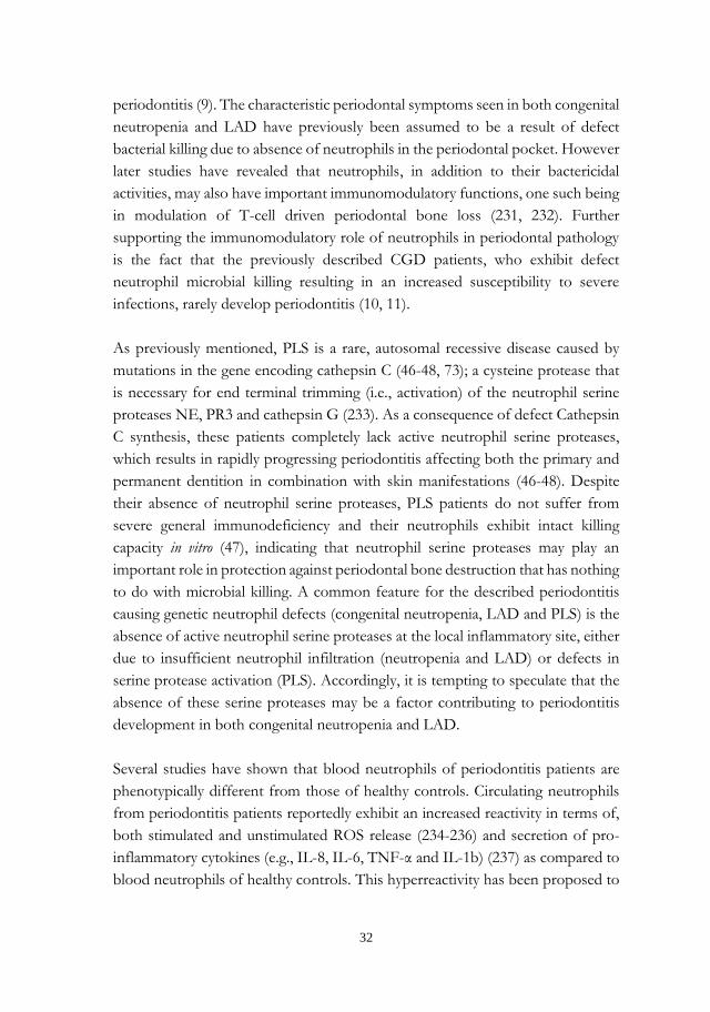
32
periodontitis (9). The characteristic periodontal symptoms seen in both congenital
neutropenia and LAD have previously been assumed to be a result of defect
bacterial killing due to absence of neutrophils in the periodontal pocket. However
later studies have revealed that neutrophils, in addition to their bactericidal
activities, may also have important immunomodulatory functions, one such being
in modulation of T-cell driven periodontal bone loss (231, 232). Further
supporting the immunomodulatory role of neutrophils in periodontal pathology
is the fact that the previously described CGD patients, who exhibit defect
neutrophil microbial killing resulting in an increased susceptibility to severe
infections, rarely develop periodontitis (10, 11).
As previously mentioned, PLS is a rare, autosomal recessive disease caused by
mutations in the gene encoding cathepsin C (46-48, 73); a cysteine protease that
is necessary for end terminal trimming (i.e., activation) of the neutrophil serine
proteases NE, PR3 and cathepsin G (233). As a consequence of defect Cathepsin
C synthesis, these patients completely lack active neutrophil serine proteases,
which results in rapidly progressing periodontitis affecting both the primary and
permanent dentition in combination with skin manifestations (46-48). Despite
their absence of neutrophil serine proteases, PLS patients do not suffer from
severe general immunodeficiency and their neutrophils exhibit intact killing
capacity in vitro (47), indicating that neutrophil serine proteases may play an
important role in protection against periodontal bone destruction that has nothing
to do with microbial killing. A common feature for the described periodontitis
causing genetic neutrophil defects (congenital neutropenia, LAD and PLS) is the
absence of active neutrophil serine proteases at the local inflammatory site, either
due to insufficient neutrophil infiltration (neutropenia and LAD) or defects in
serine protease activation (PLS). Accordingly, it is tempting to speculate that the
absence of these serine proteases may be a factor contributing to periodontitis
development in both congenital neutropenia and LAD.
Several studies have shown that blood neutrophils of periodontitis patients are
phenotypically different from those of healthy controls. Circulating neutrophils
from periodontitis patients reportedly exhibit an increased reactivity in terms of,
both stimulated and unstimulated ROS release (234-236) and secretion of pro-
inflammatory cytokines (e.g., IL-8, IL-6, TNF-α and IL-1b) (237) as compared to
blood neutrophils of healthy controls. This hyperreactivity has been proposed to

33
contribute to the tissue destruction (ROS) and increased inflammatory responses
(cytokine release) that is seen in periodontally diseased patients. Whether the
described hyperactivity of neutrophils from periodontitis patients is an innate
property or a result of priming by circulating proinflammatory factors (released
upon stimulation by oral microbes colonizing the periodontal pockets) in not
entirely clear. Similar neutrophil hyperreactivity in terms of enhanced oxidative
burst have been reported in blood cells from patients with a diverse set of medical
conditions including multiple sclerosis, acute bacteremia and AASV (238-240).
The neutrophil hyperreactivity has in these conditions been attributed to
increased levels of inflammatory cytokines in circulation (238-240). Moreover,
periodontitis patient neutrophils show decreased chemotactic capacity in
response to both fMLF and IL-8 in vitro, as compared to healthy control
neutrophils (241). This may lead to increased neutrophil transit times from blood
to the inflammatory site and therefore result in a less effective antimicrobial
defense. However, why this periodontitis associated defect in neutrophil
chemotaxis does not result in a general overt susceptibility to infection remains
to be elucidated.
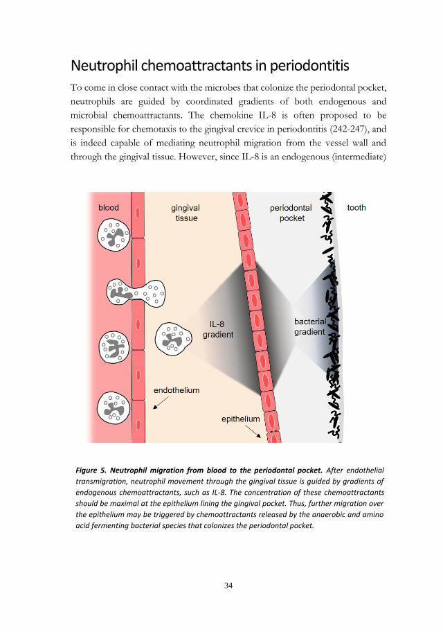
34
Neutrophil chemoattractants in periodontitis To come in close contact with the microbes that colonize the periodontal pocket,
neutrophils are guided by coordinated gradients of both endogenous and
microbial chemoattractants. The chemokine IL-8 is often proposed to be
responsible for chemotaxis to the gingival crevice in periodontitis (242-247), and
is indeed capable of mediating neutrophil migration from the vessel wall and
through the gingival tissue. However, since IL-8 is an endogenous (intermediate)
Figure 5. Neutrophil migration from blood to the periodontal pocket. After endothelial
transmigration, neutrophil movement through the gingival tissue is guided by gradients of
endogenous chemoattractants, such as IL-8. The concentration of these chemoattractants
should be maximal at the epithelium lining the gingival pocket. Thus, further migration over
the epithelium may be triggered by chemoattractants released by the anaerobic and amino
acid fermenting bacterial species that colonizes the periodontal pocket.

35
chemoattractant, produced by tissue residing cells (macrophages, endothelial cells
and epithelial cells), the concentration of this chemoattractant should be maximal
at the epithelial lining. Thus, bacteria-derived, endpoint chemoattractants should
be required to guide neutrophils over the epithelium and further towards the
microbes in the gingival pocket (Fig. 5).
Although previous studies have shown that both oral subgingival plaque
(containing multiple bacterial species) (248) and isolated oral bacterial species
(249) (paper I and II) release factors that attract neutrophils in vitro, these
chemoattractants have remained uncharacterized. The previously described end-
point chemoattractants fMLF and C5a are often mentioned as possible candidates
for the chemoattractants released by the anaerobic and proteolytic bacterial
species residing in the periodontal pocket. However, the chemoattractant
receptors activated by both fMLF and C5a (FPR1 and C5a receptor) are expressed
not only by neutrophils, but also monocytes (121, 143, 144). Moreover, their
agonists trigger chemotaxis and Ca2+ signaling not only in neutrophils, but also in
monocytes/macrophages (121, 144) (paper I). Therefore, other chemoattractants
are needed to explain the neutrophil dominated cellular migration to this
inflammatory site. In line with this, purified hydrophilic chemoattractants released
by P. gingivalis triggered chemotaxis and Ca2+ signaling (indicative of GPCR
activation) in human neutrophils, while at the same time not activating either
monocytes nor lymphocytes (paper I). This indicating activation of a GPCR
specifically expressed by neutrophils. These chemoattractants were identified
(using liquid chromatography and tandem mass spectrometric analysis) as a
mixture of SCFAs signaling via the neutrophil GPCR, FFAR2 (paper I). As
previously mentioned, the FFAR2 receptor is functionally expressed exclusively
in neutrophils (141), which explains the neutrophil specific activation capacity of
SCFAs. Moreover, SCFAs (primarily acetate and butyrate) acting via FFAR2 were
identified as the main chemoattractants released also by F. nucleatum (paper II).
As previously described, formylated peptides should theoretically be released by
all metabolically active prokaryotes. Yet, FPR1 agonists were surprisingly not
detected in the bacterial culture supernatant of P. gingivalis, at least not agonists
capable of inducing β-arrestin recruitment to a detectable extent in a cell line
overexpressing FPR1 (paper I). A possible explanation to the absence of FPR1
agonists in P. gingivalis culture supernatants could be that these bacteria may be

36
able to degrade and digest formylated peptides secreted as by-products of their
own protein synthesis. This as a result of their pronounced proteolytic and amino
acid fermenting metabolism.
Although, SCFAs released by P. gingivalis did attract neutrophils in vitro, the
chemotactic activity of these agonists is relatively modest, as compared to other
widely studied bacteria derived neutrophil chemoattractants (e.g., fMLF). This in
combination with the undetectable levels of FPR1 agonists in culture supernatants
of P. gingivalis is in line with previous theories describing this bacterium as capable
of circumventing host defense mechanisms, rather than being a potent pro-
inflammatory bacterium (198, 203). Considering this, one could speculate that a
minimal release of FPR1 agonists may constitute a virulence factor that
contributes to the overall immune avoiding profile of P. gingivalis.
Short chain fatty acids in the periodontal pocket The release of various SCFAs as end products of the amino acid fermentation
employed by oral anaerobes such as P. gingivalis (205-207) (paper I) and F.
nucleatum (215, 216, 250, 251) (paper II) is well established. SCFAs have also been
detected in GCF in varying concentrations depending on severity of periodontal
disease, ranging from undetectable in health to millimolar concentrations in
severe periodontitis (252, 253). This confirms that SCFAs are indeed produced at
high concentrations by oral bacteria also in vivo. Moreover, SCFA concentration
in GCF is positively correlated to the bacterial load of a number of oral anaerobes
including P. gingivalis and F. nucleatum (253).
Theories linking microbe released SCFAs and gingival inflammation in
periodontitis have been around for decades. Singer and Buckner first showed that
butyrate and propionate were produced by human dental plaques in
concentrations sufficient to effect human cells in vitro (inhibition of human
fibroblast proliferation) (254). The same authors later showed that pure SCFAs,
applied directly on tissues, induced gingival inflammation in vivo in beagle dogs
(255). Kashket et al. subsequently reported that application of SCFAs on human
gingival tissues in vivo induced an inflammatory response, with one of the outcome
measures being an increased neutrophil migration to GCF (256).

37
In search for the mechanisms underlying SCFA induced gingival inflammation,
the effects of SCFAs on various human cell types found in the periodontium have
been investigated in vitro (255). While, high doses of SCFAs have been described
to exert cytotoxic effects on human gingival fibroblasts (254) and epithelial cells
(257), no such effects could be seen on endothelial cells exposed to propionate
and butyrate (258). SCFAs have also been suggested to inhibit lymphocyte
proliferation and cytokine production (259). Regarding neutrophil functions, early
studies reported on decreased fMLF or casein induced neutrophil chemotaxis
following cell incubation with high concentrations (20-50 mM) of butyrate,
succinate and iso-valerate, while incubation with more moderate concentrations
of succinate (3 mM), acetate (10 mM) or butyrate (400 µM) had stimulating effects
on neutrophil migration towards zymosan activated serum (260-262). Considering
the high concentrations of SCFAs detected in GCF from periodontitis patients in
combination with the massive neutrophil infiltration to this site, it can be argued
that the inhibitory effect of SCFAs on chemotaxis seen in early in vitro studies
cannot be especially relevant in vivo. In support of this notion, later studies have
established that pure acetate, propionate and butyrate used as chemotactic stimuli
trigger directional migration in neutrophils, and that SCFA stimulated chemotaxis
is mediated via the neutrophil FFAR2-receptor (100 uM-1 mM) (137, 138).
Moreover, also a mixture of SCFAs in concentrations similar to those detected in
culture supernatants of P. gingivalis attract neutrophils in vitro (paper I).

38

39
NEUTROPHIL HETEROGENITY
Neutrophils have historically been viewed as a homogenous cell population, with
all cells carrying out the same functions and being equipped with similar granule
content and surface molecules. However, accumulating evidence supports the
simultaneous presence of phenotypically and functionally different sets of
neutrophils in the human body and numerous ways of dividing neutrophils into
subsets have been proposed in later years (263-265).
In the context of cell heterogeneity, it is important to point out that neutrophils
are plastic cells, i.e., they are able to rearrange their expression of surface
molecules in response to inflammatory signals (i.e., the priming described in the
chapter on neutrophil transmigration) or maturation. Discrepancies in surface
molecule expression between cells can thereby represent cells with different
activation or maturation status (265). Such surface molecules include the
previously mentioned CXCR4 that is expressed in less extent by neutrophils
which have recently been released from the bone marrow, but gradually increases
in expression during maturation and promotes homing of neutrophils to the bone
marrow (26). As a consequence of the circadian oscillations of neutrophil release
from the bone marrow, the expression of CXCR4 by circulating neutrophils varies
during the day (266). Other surface markers are rearranged based on cellular
priming, including shedding of L-selectin (CD62L) and upregulation of the CR3
receptor/MAC-1 (CD11b) (95).
A neutrophil subpopulation that has attracted particular attention in recent years
is referred to as ‛low density neutrophils’ (LDNs) (267, 268). This group of
neutrophils are distinguished from ‛normal density neutrophils’ based on their
localization to the peripheral blood mononuclear cell (PBMC) layer (normally
consisting of the less dense monocytes and lymphocytes) during neutrophil
purification by density gradient centrifugation. Increased numbers of LDNs in
blood have been reported from patients with a diverse set of medical conditions
including auto-immune diseases such as RA and systemic lupus erythematosus
(SLE), cancer, asthma, sepsis and pregnancy. However, it has also been reported

40
that healthy individuals exhibit a natural spectrum of neutrophil densities (267).
The LDNs have been suggested to consist of both immature cells (released from
the bone marrow upon emergency granulopoiesis) and activated mature cells,
based on increased expression of CD11b (268, 269). Whether LDNs constitute a
distinct neutrophil subset or simply represent neutrophils of varying maturation
or activation states is still a matter of debate.
Although many of the proposed neutrophil subtypes might simply represent cells
in various stages of maturation or activation, evidence also support the existence
of true (bona-fide) subsets based on the presence or absence of distinct molecular
markers (265, 270). These markers are exclusively expressed by a proportion of
neutrophils in a given individual, independent of priming or maturation. Two
such bona-fide neutrophil subset markers are olfactomedin 4 (OLFM4) (271, 272)
and CD177 (273-275).
Expression of the glycoprotein OLFM4 has been detected in a range of human
tissues including bone marrow, small intestine, stomach, colon and prostate (276).
The OLFM4 gene is expressed by all myelopoietic lineage bone marrow cells but,
interestingly, only a proportion of mature circulating neutrophils express the
protein. The interindividual variation in proportion of OLFM4 expressing blood
neutrophils have been reported to be 5-57% (271, 272). Although the precise
function of neutrophil OLFM4 is still under investigation, studies have shown
that the relative abundance of OLFM4 expressing neutrophils in circulation may
affect the immune response to severe infection, such as sepsis (277). Alder et al.
found high proportions of OLFM4 expressing neutrophils in blood to be a risk
factor for massive organ failure and death in pediatric septic shock patients (277).
In line with this, complete absence of OLFM4 had a protective function against
sepsis in a mouse model (278). Similar to OLFM4 in sepsis, the relative abundance
of CD177 expressing neutrophils in circulation may be a modifying factor in
several medical conditions that are associated with increased proportions of
CD177+ circulating neutrophils (e.g., periodontitis (paper III)), which will be
further expanded on in the subsequent chapters.
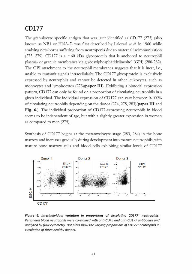
41
CD177 The granulocyte specific antigen that was later identified as CD177 (273) (also
known as NB1 or HNA-2) was first described by Lalezari et al. in 1960 while
studying new-borns suffering from neutropenia due to maternal isoimmunization
(273, 279). CD177 is a ̴ 60 kDa glycoprotein that is anchored to neutrophil
plasma- or granule membranes via glycosylphosphatidylinositol (GPI) (280-282).
The GPI attachment to the neutrophil membranes suggests that it is inert, i.e.,
unable to transmit signals intracellularly. The CD177 glycoprotein is exclusively
expressed by neutrophils and cannot be detected in other leukocytes, such as
monocytes and lymphocytes (273)(paper III). Exhibiting a bimodal expression
pattern, CD177 can only be found on a proportion of circulating neutrophils in a
given individual. The individual expression of CD177 can vary between 0-100%
of circulating neutrophils depending on the donor (274, 275, 283)(paper III and
Fig. 6.). The individual proportion of CD177-expressing neutrophils in blood
seems to be independent of age, but with a slightly greater expression in women
as compared to men (275).
Synthesis of CD177 begins at the metamyelocyte stage (283, 284) in the bone
marrow and increases gradually during development into mature neutrophils, with
mature bone marrow cells and blood cells exhibiting similar levels of CD177
Figure 6. Interindividual variation in proportions of circulating CD177+ neutrophils.
Peripheral blood neutrophils were co-stained with anti-CD45 and anti-CD177 antibodies and
analyzed by flow cytometry. Dot plots show the varying proportions of CD177+ neutrophils in
circulation of three healthy donors.

42
expression (283). Inflammatory stimulation (e.g., with fMLF or TNF-α) can
induce further upregulation of CD177 from granules to the cell surface of
CD177+ neutrophils, resulting in an increased membrane expression in this subset
(274) (paper III). However, a similar upregulation do not occur in CD177–
neutrophils (274) (paper III). Accordingly, the proportions of CD177+ and
CD177– subsets in an individual remains relatively stable over time (274, 285),
with the exception of transiently increased blood levels of CD177+ neutrophils
reported in pregnancy (286, 287) and during treatment with G-CSF (288, 289).
While a majority of donors exhibit a bimodal expression pattern (with one
CD177+ and one CD177– neutrophil subpopulation), there are individuals also
presenting with an intermediate subpopulation, referred to as a trimodal CD177
expression (275) (paper III). About 1–10% of individuals completely lack CD177
expressing neutrophils, are referred to as CD177-null and appear to be overall
healthy (273-275, 290) (paper III). CD177-null individuals are however at risk of
complications related to development of anti-CD177 antibodies if exposed to the
antigen. Such complications include transfusion related acute lung injury
following blood transfusion (291) and neonatal neutropenia affecting CD177
expressing fetuses of CD177-null mothers (279). Induction of a stop codon
(originating from a neighboring pseudogene) in the coding region of CD177 has
been identified as the genetic factor causing the complete lack of CD177
expression in these CD177-null individuals (292, 293). However, the genetics
behind the bimodal expression pattern of CD177 seen in most individuals is still
only partially understood, although studies have proposed a combination of
complex genetic and epigenetic factors (293-295).
Whereas CD177-null individuals appear to be overall healthy, increased blood
levels of CD177+ neutrophils have been associated to various medical conditions,
including severe bacterial infections (296), polycythemia vera (PV) (297) and other
myeloproliferative disorders (298), AASV (299-301), SLE (299) and periodontitis
(paper III). Considering these observations, it is tempting to speculate that the
relative proportion of CD177+ neutrophils in circulation may be a factor
influencing the susceptibility to diseases such as periodontitis, SLE, PV, and
AASV. Nonetheless, it should be kept in mind that the increased proportions of
CD177+ circulating neutrophils detected in patients with the described conditions
may as well be a consequence of unknown factors related to an ongoing
inflammation.

43
Previous studies have established that the percentage of CD177+ cells in blood
can be transiently increased by administration of G-CSF to healthy controls (288,
289). As previously mentioned (in the chapter on the neutrophil life cycle), G-
CSF regulates baseline levels of neutrophil maturation and release from the bone
marrow (22-24) and increased serum levels of this cytokine have been reported in
several inflammatory conditions (302). Increased mRNA- and protein expression
of G-CSF have also been reported in the gingival tissue of periodontitis patients,
as compared to the gingival tissue of healthy controls (303). Considering this, one
could speculate that a rise in serum G-CSF levels in response to the ongoing
inflammatory process in the periodontitis lesion may result in increased levels of
CD177+ neutrophils in circulation of this patient group. Along these lines,
Abdgawad et al. measured plasma levels of G-CSF in vasculitis patients (who also
exhibit increased proportions of CD177+ neutrophils in blood as compared to
healthy controls) but did not find increased G-CSF levels in this patient group
(300). Moreover, increased levels of G‐CSF have been reported in the serum and
synovial fluid of patients with RA (304), while this patient group did not exhibit
higher levels of CD177+ neutrophils in circulation as compared to healthy
controls (300). Thus, it is less likely that increased systemic G-CSF levels are
directly responsible for the high proportions of CD177+ neutrophils reported in
AASV and other inflammatory conditions such as periodontitis. The pattern of
high blood levels of CD177+ neutrophils in these conditions could either be due
to yet unknown external factors with the capacity to increase the CD177+ cell
proportion, or a result of innate high proportions of CD177+ neutrophils in blood
being a factor predisposing to inflammatory disease.
CD177 and neutrophil transmigration
The possible role of CD177 in neutrophil migration have attracted much
attention. This based on in vitro evidence of an increased capacity of CD177+
neutrophils to migrate over endothelial cell layers, as compared to CD177– cells
(305, 306). The migration advantage of CD177+ neutrophils has in part been
attributed to the ability of CD177 to interact with PECAM-1; an important
contributor to neutrophil transmigration that is expressed by endothelial cells,
platelets, monocytes and granulocytes (307). Furthermore, CD177 has been
shown capable of binding and presenting the neutrophil serine protease PR3 on

44
the cell surface (308, 309). The membrane presentation of PR3 has been proposed
to facilitate neutrophil migration via proteolytic degradation of extracellular
matrix- or junctional proteins (310). In contrast to the described in vitro results,
previous studies looking at neutrophil migration to both synovial fluid in RA
patients (285) and peritoneal fluid in dialysis patients suffering from peritonitis
(311) have not been able to confirm a migration advantage of CD177+ neutrophils
in vivo.
Interestingly, GCF (sampled as described in the chapter on tissue neutrophils)
from patients suffering from periodontitis (stage III-IV) exhibited higher
proportions of CD177 expressing cells as compared to blood from the same
individual, i.e., the CD177+ neutrophil subtype was enriched in GCF (paper III).
This phenomenon was not detected either in synovial fluid from inflammatory
arthritis patients or inflammatory exudate from experimental skin chambers
applied to healthy donors (paper III).
The enrichment of CD177+ neutrophils in GCF could not be attributed to general
differences in longevity between the subsets, as the CD177– subset showed similar
(in anaerobic environment) or even decreased (in aerobic environment) rate of in
vitro apoptosis as compared to the CD177+ cells (paper III). The CD177+ subtype
showed a slight migratory advantage in vitro when stimulated with IL-8 or with a
culture supernatant prepared from a subgingival bacterial sample from a
periodontitis patient. Further, the CD177+ and CD177– subtypes migrated with
equal efficiency towards fMLF (paper III). The detected migratory advantage of
CD177 expressing neutrophils in vitro was not prominent enough to be the only
factor explaining the enrichment of CD177+ cells seen in GCF. Consequently, the
accumulation of CD177+ cells in this inflammatory exudate may in also be
attributed to tissue specific properties. However, the slight migratory advantage
of CD177+ neutrophils towards the culture supernatant of the periodontitis
bacterial sample indicate that bacterial chemoattractants specific for the
periodontal pocket may influence the preferential recruitment of this subtype to
GCF in periodontitis. Considering the previously described high concentrations
of SCFAs and undetectable levels of FPR1 agonists in culture supernatants of
periodontitis associated bacteria (paper I, II), the supernatant used to stimulate
in vitro chemotaxis likely contain similar bacterial end-products. In light of these
45
observations, it is tempting to speculate that selective CD177+ neutrophil
chemotaxis can be promoted by the presence of SCFAs.
Further differences between the neutrophil subtypes defined by CD177 expression?
While studying the neutrophil subtypes defined by the presence or absence of
CD177, it is important to keep in mind that CD177 per se does not necessarily
constitute the only differentiating factor between the CD177+ and CD177–
subsets. There may be additional molecules showing a bimodal expression pattern
that completely or partially overlap CD177 expression, and thereby contribute to
functional differences between CD177+ and CD177– neutrophils. As previously
mentioned, patients suffering from periodontitis exhibit higher levels of CD177+
neutrophils in circulation, as compared to healthy donors (paper III). This, in
combination with the further enrichment of the CD177+ subtype in GCF from
these patients, resulted in very high proportions of CD177 expressing neutrophils
at this pathological site (paper III). Considering the high levels of CD177
expressing neutrophils in GCF, functions differing between the subsets could be
of importance for induction (or suppression) of destructive inflammation in the
periodontal tissues.
A previous comparison of mRNA expression between CD177+ and CD177–
neutrophils showed differential expression of a number of distinct mRNAs
including those coding for the granule proteins PR3, MPO, alpha defensin 3, BPI,
and Cathepsin G. However, subsequent immunoblotting of these proteins
showed no expression differences on protein level between the subsets (284). In
view of the minimal de novo protein synthesis employed by neutrophils after bone
marrow release, mRNA quantification is not likely to appropriately represent the
complete proteome profile of these cells and therefore comparisons performed
on the protein level are likely more informative.
Relative quantitative proteomics is a method enabling detection of proteins and
determination of their relative abundances in several samples simultaneously
(312). This method applied to samples of FACS-sorted CD177+ and CD177–
neutrophils from the same individual showed very similar expression levels of the
vast majority of detected proteins, but nonetheless, a differential expression

46
pattern of a limited number of proteins (paper IV). CD177 itself was, as expected,
the protein that primarily differentiated the subsets. Another top hit, alpha-1-
antitrypsin (A1AT), was expressed twice as much in the CD177+ neutrophil
subset as compared to the CD177– subset (paper IV). A1AT is an endogenous
neutrophil serine protease inhibitor, engaged in defending tissues from proteolytic
degradation by excessive release of proteases (313, 314). Constantly present in
circulation, A1AT diminish protease activity by complex-formation (1:1) with
neutrophil serine proteases. While 80% of the circulating A1AT is produced by
hepatocytes in the liver, it is also secreted by immune cells including monocytes,
macrophages and circulating neutrophils (315-317). Tissue A1AT levels are
significantly increased during an inflammatory response and a majority of this
increase is likely a result of local release by resident and infiltrating inflammatory
cells (314). The possible effect of an increased A1AT-expression on net protease
activity derived from CD177+ cells was further investigated. NE activity was
measured in lysates (normalized based on cell number) of FACS-sorted CD177+
and CD177– cells. In concordance with the increased A1AT expression, CD177+
neutrophils also contained lower NE activity, as compared to the CD177– cells
(paper IV). Although the difference in NE activity between the subset may in
part be due to a slightly lower (ratio 0.85) expression of the protein in the CD177+
subset, the results indicate an interesting functional difference (net NE activity)
between CD177+ and CD177– cells.
As previously described (in the chapter on periodontitis), a complete lack of active
neutrophil serine proteases (as seen in PLS patients) initiates rapidly progressing
periodontitis (46-48), indicating that the presence of neutrophil serine proteases
constitutes a critical factor for maintenance of periodontal health. Hence, factors
disturbing the protease-antiprotease balance at the local inflammatory site may
influence destructive inflammatory responses in periodontal tissues. If and how
the increased A1AT expression and decreased NE activity seen in CD177+
neutrophils affect local levels of protease activity will be further investigated in
paper IV.

47
CONCLUDING REMARKS
Paper I and II highlights the SCFA-FFAR2 interaction as a factor mediating
neutrophil recruitment from circulation and migration towards the periodontitis
associated anaerobes P. gingivalis and F. nucleatum. As SCFAs are end-products of
the amino acid fermenting metabolism that is generally employed by the anaerobic
bacterial species colonizing deep periodontal pockets, this finding could be
relevant also for other periodontitis associated bacterial species. Given the
neutrophil specific expression of FFAR2, SCFA may also contribute to the
neutrophil dominated leukocyte profile of GCF in periodontitis. This adds to our
understanding of bacteria derived factors triggering neutrophil recruitment to the
periodontal pocket.
The results of paper III show that the CD177+ neutrophil subtype is enriched in
GCF of periodontitis patients, possibly due to a combination of site specific
chemoattractants and tissue specific properties. Moreover, and similar to what
was previously reported for other inflammatory conditions (299-301),
periodontitis patients exhibited increased levels of circulating CD177+
neutrophils as compared to healthy controls. This may be a result of the systemic
presence of so far unknown inflammatory factors related to these conditions, but
could also indicate that the relative proportion of CD177+ neutrophils in
circulation is a factor influencing the susceptibility to inflammatory diseases, such
as periodontitis. Whether the increased proportions of CD177+ neutrophils in
circulation of these patients are a cause or consequence of disease remains
unresolved. To bring clarity to this issue, it would be of interest to monitor
inflammatory disease progression in individuals exhibiting varying base line levels
of CD177 expression over time.
As a result of both increased levels of CD177+ neutrophils in blood of
periodontitis patients and the further enrichment of this subtype after
transmigration to the periodontal pocket, CD177+ neutrophils typically
constituted a very large proportion of the neutrophils found in GCF of these
patients (paper III). Accordingly, functions differing between the subtypes may

48
be influencing destructive inflammation of the periodontal tissues. The
preliminary results of paper IV show that the neutrophil serine protease inhibitor
A1AT is expressed twice as much in the CD177+ neutrophil subset as compared
to the CD177– subset from the same donor. Moreover, results also indicate that
CD177+ neutrophils exhibit decreased NE activity as compared to CD177–
neutrophils. Considering that sufficient neutrophil serine protease activity seems
to be vital for periodontal health, as demonstrated by the rapid periodontal
breakdown seen in PLS, an overt inhibition of this activity may be a modulating
factor in periodontal pathology. Along these lines it is tempting to speculate that
patients suffering from periodontitis may in fact represent a ‛light version’ of what
is seen in PLS, i.e., present with a deranged protease-antiprotease balance in the
local gingival tissue resulting in less-than-optimal serine protease activity. Future
studies will have to determine whether local suboptimal protease levels can be a
factor influencing periodontal pathology.

49
ACKNOWLEDGEMENT
Thanks:
Johan, I will be forever grateful that you accepted me as a research student!
Thanks for providing such an inspiring research environment and for showing
me how fun science can be. You have given me all the freedom that I need while
always being there to give advice and discuss scientific questions. Thanks also, for
encouraging me to relax and enjoy life more.
Karin, for agreeing to be my self-selected co supervisor. You have been my rock
during the PhD years! Generously sharing your expertise in immunology, lab
techniques and always taking the time for long talks.
Anna, for bringing your enthusiasm for scientific discussion to our group
meetings and making everyone feel welcome with your warm personality. And
also, for the technical help with the layout of this book as well as previous
presentations.
Tord, for sharing your extensive knowledge in the field of periodontology and
for your valuable input on my thesis.
Sara, your help with patient recruitment, collection of patient samples and input
on my work has been invaluable. Thank you for always making time for me
despite your own busy schedule and being such a good friend.
Halla, for bearing with me during my first year at the lab and teaching me all
basic lab techniques. And also, for all our talks, play-dates, and for taking me to
my first running competition.
Gunnar, Amina, and Susanne thanks for your input on the microbiological
aspects of my work, it has been invaluable!

50
To my group members and close colleges at department of Oral Microbiology
and Immunology: Felix, Vignesh, Lisa, Anna-Karin, Sara A. You have all been
such great friends and helped me a lot during the years.
Arsham and Tishana, I would never have made this without your help with the
lab work.
To all members of the Phagocyte group: Huamei, Claes, and Lena, thanks for
all your helpful input on my work during our group meetings. Martina, André,
Simon, Marlene, Jonas M, Jonas E, Firoozeh, Mike, Amanda, and Marit, for
fun lab-work, meetings and always being there to help me when needed.
To my friends: Albin & Tove although you left dental school after only a few
months you have remained the closest of friends. Having followed you both
recently defending your PhD thesis’s have been truly inspiring! Almis, for
everything during those intense years of studies. Also, the biggest thanks for
always guiding me to the right lecture halls at Karolinska and instruments at the
student clinic. Samantha, we share a similar view of life and our discussions have
helped me in finding the right direction. Astera, for our lunches and talks about
PhD life. Cissi and Johanna, for all the (20) years of support and fun memories.
I am looking forward to many more years of friendship. Sofia, with you it was
love at first sight. Thank you for all our long lunches and for always being there
for me in both work and personal life.
Albert for giving me perspective on life and no time to (over) think, and Mattias
for moving to Gothenburg with me, and for your unconditional love and support.
My parents and siblings, for always encouraging me to follow my own path.

51
REFERENCES
1. Manley HR, Keightley MC, and Lieschke GJ. The Neutrophil Nucleus: An
Important Influence on Neutrophil Migration and Function. Front Immunol.
2018;9:2867.
2. Borregaard N, and Cowland JB. Granules of the human neutrophilic
polymorphonuclear leukocyte. Blood. 1997;89(10):3503-21.
3. Liew PX, and Kubes P. The Neutrophil's Role During Health and Disease.
Physiol Rev. 2019;99(2):1223-48.
4. Kolaczkowska E, and Kubes P. Neutrophil recruitment and function in health
and inflammation. Nat Rev Immunol. 2013;13(3):159-75.
5. Castanheira FVS, and Kubes P. Neutrophils and NETs in modulating acute and
chronic inflammation. Blood. 2019;133(20):2178-85.
6. Dinauer MC. Disorders of neutrophil function: an overview. Methods Mol Biol.
2014;1124:501-15.
7. Skokowa J, Dale DC, Touw IP, Zeidler C, and Welte K. Severe congenital
neutropenias. Nat Rev Dis Primers. 2017;3:17032.
8. Almarza Novoa E, Kasbekar S, Thrasher AJ, Kohn DB, Sevilla J, Nguyen T, et
al. Leukocyte adhesion deficiency-I: A comprehensive review of all published
cases. J Allergy Clin Immunol Pract. 2018;6(4):1418-20 e10.
9. Hanna S, and Etzioni A. Leukocyte adhesion deficiencies. Ann N Y Acad Sci.
2012;1250:50-5.
10. Roos D. Chronic Granulomatous Disease. Methods Mol Biol. 2019;1982:531-42.
11. van den Berg JM, van Koppen E, Ahlin A, Belohradsky BH, Bernatowska E,
Corbeel L, et al. Chronic granulomatous disease: the European experience. PLoS
One. 2009;4(4):e5234.
12. Bainton DF, Ullyot JL, and Farquhar MG. The development of neutrophilic
polymorphonuclear leukocytes in human bone marrow. J Exp Med.
1971;134(4):907-34.

52
13. Hidalgo A, Chilvers ER, Summers C, and Koenderman L. The Neutrophil Life
Cycle. Trends Immunol. 2019;40(7):584-97.
14. Tak T, Tesselaar K, Pillay J, Borghans JA, and Koenderman L. What's your age
again? Determination of human neutrophil half-lives revisited. J Leukoc Biol.
2013;94(4):595-601.
15. Pillay J, den Braber I, Vrisekoop N, Kwast LM, de Boer RJ, Borghans JA, et al.
In vivo labeling with 2H2O reveals a human neutrophil lifespan of 5.4 days.
Blood. 2010;116(4):625-7.
16. Lahoz-Beneytez J, Elemans M, Zhang Y, Ahmed R, Salam A, Block M, et al.
Human neutrophil kinetics: modeling of stable isotope labeling data supports
short blood neutrophil half-lives. Blood. 2016;127(26):3431-8.
17. Summers C, Rankin SM, Condliffe AM, Singh N, Peters AM, and Chilvers ER.
Neutrophil kinetics in health and disease. Trends Immunol. 2010;31(8):318-24.
18. Mauer AM, Athens JW, Ashenbrucker H, Cartwright GE, and Wintrobe mm.
leukokinetic studies. ii. a method for labeling granulocytes in vitro with
radioactive diisopropylfluorophosphate (dfp). J Clin Invest. 1960;39(9):1481-6.
19. Athens JW, Raab SO, Haab OP, Mauer AM, Ashenbrucker H, Cartwright GE,
et al. Leukokinetic studies. III. The distribution of granulocytes in the blood of
normal subjects. J Clin Invest. 1961;40(1):159-64.
20. Christoffersson G, and Phillipson M. The neutrophil: one cell on many missions
or many cells with different agendas? Cell Tissue Res. 2018;371(3):415-23.
21. Jagels MA, and Hugli TE. Neutrophil chemotactic factors promote leukocytosis.
A common mechanism for cellular recruitment from bone marrow. J Immunol.
1992;148(4):1119-28.
22. Semerad CL, Liu F, Gregory AD, Stumpf K, and Link DC. G-CSF is an essential
regulator of neutrophil trafficking from the bone marrow to the blood. Immunity.
2002;17(4):413-23.
23. Lord BI, Bronchud MH, Owens S, Chang J, Howell A, Souza L, et al. The
kinetics of human granulopoiesis following treatment with granulocyte colony-
stimulating factor in vivo. Proc Natl Acad Sci U S A. 1989;86(23):9499-503.
24. Lieschke GJ, Grail D, Hodgson G, Metcalf D, Stanley E, Cheers C, et al. Mice
lacking granulocyte colony-stimulating factor have chronic neutropenia,
granulocyte and macrophage progenitor cell deficiency, and impaired neutrophil
mobilization. Blood. 1994;84(6):1737-46.

53
25. Kawakami M, Tsutsumi H, Kumakawa T, Abe H, Hirai M, Kurosawa S, et al.
Levels of serum granulocyte colony-stimulating factor in patients with
infections. Blood. 1990;76(10):1962-4.
26. Martin C, Burdon PC, Bridger G, Gutierrez-Ramos JC, Williams TJ, and Rankin
SM. Chemokines acting via CXCR2 and CXCR4 control the release of
neutrophils from the bone marrow and their return following senescence.
Immunity. 2003;19(4):583-93.
27. De Filippo K, and Rankin SM. CXCR4, the master regulator of neutrophil
trafficking in homeostasis and disease. Eur J Clin Invest. 2018;48 Suppl 2:e12949.
28. de Oliveira S, Rosowski EE, and Huttenlocher A. Neutrophil migration in
infection and wound repair: going forward in reverse. Nat Rev Immunol.
2016;16(6):378-91.
29. Mathias JR, Perrin BJ, Liu TX, Kanki J, Look AT, and Huttenlocher A.
Resolution of inflammation by retrograde chemotaxis of neutrophils in
transgenic zebrafish. J Leukoc Biol. 2006;80(6):1281-8.
30. Wang J, Hossain M, Thanabalasuriar A, Gunzer M, Meininger C, and Kubes P.
Visualizing the function and fate of neutrophils in sterile injury and repair. Science.
2017;358(6359):111-6.
31. Buckley CD, Ross EA, McGettrick HM, Osborne CE, Haworth O, Schmutz C,
et al. Identification of a phenotypically and functionally distinct population of
long-lived neutrophils in a model of reverse endothelial migration. J Leukoc Biol.
2006;79(2):303-11.
32. Hind LE, and Huttenlocher A. Neutrophil Reverse Migration and a
Chemokinetic Resolution. Dev Cell. 2018;47(4):404-5.
33. Ji J, and Fan J. Neutrophil in Reverse Migration: Role in Sepsis. Front Immunol.
2021;12:656039.
34. Cassatella MA. The production of cytokines by polymorphonuclear neutrophils.
Immunol Today. 1995;16(1):21-6.
35. Borregaard N. Neutrophils, from marrow to microbes. Immunity. 2010;33(5):657-
70.
36. Faurschou M, and Borregaard N. Neutrophil granules and secretory vesicles in
inflammation. Microbes Infect. 2003;5(14):1317-27.
54
37. Borregaard N. Development of neutrophil granule diversity. Ann N Y Acad Sci.
1997;832:62-8.
38. Le Cabec V, Cowland JB, Calafat J, and Borregaard N. Targeting of proteins to
granule subsets is determined by timing and not by sorting: The specific granule
protein NGAL is localized to azurophil granules when expressed in HL-60 cells.
Proc Natl Acad Sci U S A. 1996;93(13):6454-7.
39. Venkatakrishnan V, Dieckmann R, Loke I, Tjondro HC, Chatterjee S, Bylund J,
et al. Glycan analysis of human neutrophil granules implicates a maturation-
dependent glycosylation machinery. J Biol Chem. 2020;295(36):12648-60.
40. Sengelov H, Kjeldsen L, and Borregaard N. Control of exocytosis in early
neutrophil activation. J Immunol. 1993;150(4):1535-43.
41. Moraes TJ, Zurawska JH, and Downey GP. Neutrophil granule contents in the
pathogenesis of lung injury. Curr Opin Hematol. 2006;13(1):21-7.
42. Potera RM, Jensen MJ, Hilkin BM, South GK, Hook JS, Gross EA, et al.
Neutrophil azurophilic granule exocytosis is primed by TNF-alpha and partially
regulated by NADPH oxidase. Innate Immun. 2016;22(8):635-46.
43. Nauseef WM. Myeloperoxidase in human neutrophil host defence. Cell Microbiol.
2014;16(8):1146-55.
44. Bjornsdottir H, Dahlstrand Rudin A, Klose FP, Elmwall J, Welin A, Stylianou
M, et al. Phenol-Soluble Modulin alpha Peptide Toxins from Aggressive
Staphylococcus aureus Induce Rapid Formation of Neutrophil Extracellular
Traps through a Reactive Oxygen Species-Independent Pathway. Front Immunol.
2017;8:257.
45. Papayannopoulos V, Metzler KD, Hakkim A, and Zychlinsky A. Neutrophil
elastase and myeloperoxidase regulate the formation of neutrophil extracellular
traps. J Cell Biol. 2010;191(3):677-91.
46. Roberts H, White P, Dias I, McKaig S, Veeramachaneni R, Thakker N, et al.
Characterization of neutrophil function in Papillon-Lefèvre syndrome. J Leukoc
Biol. 2016;100(2):433-44.
47. Sorensen OE, Clemmensen SN, Dahl SL, Ostergaard O, Heegaard NH,
Glenthoj A, et al. Papillon-Lefevre syndrome patient reveals species-dependent
requirements for neutrophil defenses. J Clin Invest. 2014;124(10):4539-48.
48. Hart TC, and Shapira L. Papillon-Lefevre syndrome. Periodontol 2000. 1994;6:88-
100.

55
49. Kjeldsen L, Bjerrum OW, Askaa J, and Borregaard N. Subcellular localization
and release of human neutrophil gelatinase, confirming the existence of separate
gelatinase-containing granules. Biochem J. 1992;287 ( Pt 2):603-10.
50. Cramer E, Pryzwansky KB, Villeval JL, Testa U, and Breton-Gorius J.
Ultrastructural localization of lactoferrin and myeloperoxidase in human
neutrophils by immunogold. Blood. 1985;65(2):423-32.
51. Sengelov H, Follin P, Kjeldsen L, Lollike K, Dahlgren C, and Borregaard N.
Mobilization of granules and secretory vesicles during in vivo exudation of
human neutrophils. J Immunol. 1995;154(8):4157-65.
52. Borregaard N, Heiple JM, Simons ER, and Clark RA. Subcellular localization of
the b-cytochrome component of the human neutrophil microbicidal oxidase:
translocation during activation. J Cell Biol. 1983;97(1):52-61.
53. Bainton DF, and Farquhar MG. Origin of granules in polymorphonuclear
leukocytes. Two types derived from opposite faces of the Golgi complex in
developing granulocytes. J Cell Biol. 1966;28(2):277-301.
54. Borregaard N, Kjeldsen L, Rygaard K, Bastholm L, Nielsen MH, Sengelov H, et
al. Stimulus-dependent secretion of plasma proteins from human neutrophils. J
Clin Invest. 1992;90(1):86-96.
55. Dunkelberger JR, and Song WC. Complement and its role in innate and adaptive
immune responses. Cell Res. 2010;20(1):34-50.
56. Lee WL, Harrison RE, and Grinstein S. Phagocytosis by neutrophils. Microbes
Infect. 2003;5(14):1299-306.
57. Nordenfelt P, and Tapper H. Phagosome dynamics during phagocytosis by
neutrophils. J Leukoc Biol. 2011;90(2):271-84.
58. Bylund J, Brown KL, Movitz C, Dahlgren C, and Karlsson A. Intracellular
generation of superoxide by the phagocyte NADPH oxidase: how, where, and
what for? Free Radic Biol Med. 2010;49(12):1834-45.
59. Kjeldsen L, Sengelov H, Lollike K, Nielsen MH, and Borregaard N. Isolation
and characterization of gelatinase granules from human neutrophils. Blood.
1994;83(6):1640-9.
60. Dahlgren C, Karlsson A, and Bylund J. Intracellular Neutrophil Oxidants: From
Laboratory Curiosity to Clinical Reality. J Immunol. 2019;202(11):3127-34.
61. Segal AW. How neutrophils kill microbes. Annu Rev Immunol. 2005;23:197-223.

56
62. Quie PG, White JG, Holmes B, and Good RA. In vitro bactericidal capacity of
human polymorphonuclear leukocytes: diminished activity in chronic
granulomatous disease of childhood. J Clin Invest. 1967;46(4):668-79.
63. Winkelstein JA, Marino MC, Johnston RB, Jr., Boyle J, Curnutte J, Gallin JI, et
al. Chronic granulomatous disease. Report on a national registry of 368 patients.
Medicine (Baltimore). 2000;79(3):155-69.
64. Rest RF, Fischer SH, Ingham ZZ, and Jones JF. Interactions of Neisseria
gonorrhoeae with human neutrophils: effects of serum and gonococcal opacity
on phagocyte killing and chemiluminescence. Infect Immun. 1982;36(2):737-44.
65. Segal AW, Geisow M, Garcia R, Harper A, and Miller R. The respiratory burst
of phagocytic cells is associated with a rise in vacuolar pH. Nature.
1981;290(5805):406-9.
66. Lehrer RI, and Cline MJ. Leukocyte myeloperoxidase deficiency and
disseminated candidiasis: the role of myeloperoxidase in resistance to Candida
infection. J Clin Invest. 1969;48(8):1478-88.
67. Decleva E, Menegazzi R, Busetto S, Patriarca P, and Dri P. Common
methodology is inadequate for studies on the microbicidal activity of
neutrophils. J Leukoc Biol. 2006;79(1):87-94.
68. Lehrer RI, Hanifin J, and Cline MJ. Defective bactericidal activity in
myeloperoxidase-deficient human neutrophils. Nature. 1969;223(5201):78-9.
69. Klebanoff SJ, Kettle AJ, Rosen H, Winterbourn CC, and Nauseef WM.
Myeloperoxidase: a front-line defender against phagocytosed microorganisms. J
Leukoc Biol. 2013;93(2):185-98.
70. Xu D, and Lu W. Defensins: A Double-Edged Sword in Host Immunity. Front
Immunol. 2020;11:764.
71. Stapels DA, Geisbrecht BV, and Rooijakkers SH. Neutrophil serine proteases in
antibacterial defense. Curr Opin Microbiol. 2015;23:42-8.
72. Standish AJ, and Weiser JN. Human neutrophils kill Streptococcus pneumoniae
via serine proteases. J Immunol. 2009;183(4):2602-9.
73. Giannetti L, Apponi R, Dello Diago AM, Jafferany M, Goldust M, and
Sadoughifar R. Papillon-Lefevre syndrome: Oral aspects and treatment. Dermatol
Ther. 2020;33(3):e13336.

57
74. Brinkmann V, Reichard U, Goosmann C, Fauler B, Uhlemann Y, Weiss DS, et
al. Neutrophil extracellular traps kill bacteria. Science. 2004;303(5663):1532-5.
75. Urban CF, Reichard U, Brinkmann V, and Zychlinsky A. Neutrophil
extracellular traps capture and kill Candida albicans yeast and hyphal forms. Cell
Microbiol. 2006;8(4):668-76.
76. Menegazzi R, Decleva E, and Dri P. Killing by neutrophil extracellular traps: fact
or folklore? Blood. 2012;119(5):1214-6.
77. Papayannopoulos V. Neutrophil extracellular traps in immunity and disease. Nat
Rev Immunol. 2018;18(2):134-47.
78. Fuchs TA, Abed U, Goosmann C, Hurwitz R, Schulze I, Wahn V, et al. Novel
cell death program leads to neutrophil extracellular traps. J Cell Biol.
2007;176(2):231-41.
79. Yousefi S, Mihalache C, Kozlowski E, Schmid I, and Simon HU. Viable
neutrophils release mitochondrial DNA to form neutrophil extracellular traps.
Cell Death Differ. 2009;16(11):1438-44.
80. Pilsczek FH, Salina D, Poon KK, Fahey C, Yipp BG, Sibley CD, et al. A novel
mechanism of rapid nuclear neutrophil extracellular trap formation in response
to Staphylococcus aureus. J Immunol. 2010;185(12):7413-25.
81. Malachowa N, Kobayashi SD, Freedman B, Dorward DW, and DeLeo FR.
Staphylococcus aureus leukotoxin GH promotes formation of neutrophil
extracellular traps. J Immunol. 2013;191(12):6022-9.
82. Metzler KD, Fuchs TA, Nauseef WM, Reumaux D, Roesler J, Schulze I, et al.
Myeloperoxidase is required for neutrophil extracellular trap formation:
implications for innate immunity. Blood. 2011;117(3):953-9.
83. Saffarzadeh M, Juenemann C, Queisser MA, Lochnit G, Barreto G, Galuska SP,
et al. Neutrophil extracellular traps directly induce epithelial and endothelial cell
death: a predominant role of histones. PLoS One. 2012;7(2):e32366.
84. Tan C, Aziz M, and Wang P. The vitals of NETs. J Leukoc Biol. 2020.
85. Martinod K, and Wagner DD. Thrombosis: tangled up in NETs. Blood.
2014;123(18):2768-76.
86. Soderberg D, and Segelmark M. Neutrophil Extracellular Traps in ANCA-
Associated Vasculitis. Front Immunol. 2016;7:256.

58
87. Lee KH, Kronbichler A, Park DD, Park Y, Moon H, Kim H, et al. Neutrophil
extracellular traps (NETs) in autoimmune diseases: A comprehensive review.
Autoimmun Rev. 2017;16(11):1160-73.
88. Potempa J, Mydel P, and Koziel J. The case for periodontitis in the pathogenesis
of rheumatoid arthritis. Nat Rev Rheumatol. 2017;13(10):606-20.
89. Schmidt S, Moser M, and Sperandio M. The molecular basis of leukocyte
recruitment and its deficiencies. Mol Immunol. 2013;55(1):49-58.
90. Zindel J, and Kubes P. DAMPs, PAMPs, and LAMPs in Immunity and Sterile
Inflammation. Annu Rev Pathol. 2020;15:493-518.
91. von Andrian UH, Chambers JD, Berg EL, Michie SA, Brown DA, Karolak D,
et al. L-selectin mediates neutrophil rolling in inflamed venules through sialyl
LewisX-dependent and -independent recognition pathways. Blood.
1993;82(1):182-91.
92. Ivetic A, Hoskins Green HL, and Hart SJ. L-selectin: A Major Regulator of
Leukocyte Adhesion, Migration and Signaling. Front Immunol. 2019;10:1068.
93. Nourshargh S, and Alon R. Leukocyte migration into inflamed tissues. Immunity.
2014;41(5):694-707.
94. Follin P, Briheim G, and Dahlgren C. Mechanisms in neutrophil priming:
characterization of the oxidative response induced by formylmethionyl-leucyl-
phenylalanine in human exudated cells. Scand J Immunol. 1991;34(3):317-22.
95. Miralda I, Uriarte SM, and McLeish KR. Multiple Phenotypic Changes Define
Neutrophil Priming. Front Cell Infect Microbiol. 2017;7:217.
96. Filippi MD. Neutrophil transendothelial migration: updates and new
perspectives. Blood. 2019;133(20):2149-58.
97. Vaporciyan AA, DeLisser HM, Yan HC, Mendiguren, II, Thom SR, Jones ML,
et al. Involvement of platelet-endothelial cell adhesion molecule-1 in neutrophil
recruitment in vivo. Science. 1993;262(5139):1580-2.
98. Wang S, Voisin MB, Larbi KY, Dangerfield J, Scheiermann C, Tran M, et al.
Venular basement membranes contain specific matrix protein low expression
regions that act as exit points for emigrating neutrophils. J Exp Med.
2006;203(6):1519-32.
99. Petri B, and Sanz MJ. Neutrophil chemotaxis. Cell Tissue Res. 2018;371(3):425-
36.

59
100. Cassimeris L, and Zigmond SH. Chemoattractant stimulation of
polymorphonuclear leucocyte locomotion. Semin Cell Biol. 1990;1(2):125-34.
101. Metzemaekers M, Gouwy M, and Proost P. Neutrophil chemoattractant
receptors in health and disease: double-edged swords. Cell Mol Immunol.
2020;17(5):433-50.
102. Gether U. Uncovering molecular mechanisms involved in activation of G
protein-coupled receptors. Endocr Rev. 2000;21(1):90-113.
103. Dahlgren C, Gabl M, Holdfeldt A, Winther M, and Forsman H. Basic
characteristics of the neutrophil receptors that recognize formylated peptides, a
danger-associated molecular pattern generated by bacteria and mitochondria.
Biochem Pharmacol. 2016;114:22-39.
104. Novak EJ, and Rabinovitch PS. Improved sensitivity in flow cytometric
intracellular ionized calcium measurement using fluo-3/Fura Red fluorescence
ratios. Cytometry. 1994;17(2):135-41.
105. Luttrell LM, and Lefkowitz RJ. The role of beta-arrestins in the termination and
transduction of G-protein-coupled receptor signals. J Cell Sci. 2002;115(Pt
3):455-65.
106. Gurevich VV, and Gurevich EV. GPCR Signaling Regulation: The Role of
GRKs and Arrestins. Front Pharmacol. 2019;10:125.
107. Gabl M, Holdfeldt A, Sundqvist M, Lomei J, Dahlgren C, and Forsman H. FPR2
signaling without beta-arrestin recruitment alters the functional repertoire of
neutrophils. Biochem Pharmacol. 2017;145:114-22.
108. Campbell JJ, Foxman EF, and Butcher EC. Chemoattractant receptor cross talk
as a regulatory mechanism in leukocyte adhesion and migration. Eur J Immunol.
1997;27(10):2571-8.
109. Shen W, Li B, Wetzel MA, Rogers TJ, Henderson EE, Su SB, et al. Down-
regulation of the chemokine receptor CCR5 by activation of chemotactic formyl
peptide receptor in human monocytes. Blood. 2000;96(8):2887-94.
110. Kim D, and Haynes CL. Neutrophil chemotaxis within a competing gradient of
chemoattractants. Anal Chem. 2012;84(14):6070-8.
111. Psychoyos S, and Uziel-Fusi S. Comparison of LTB4- and C5a-stimulated
chemotaxis of isolated human neutrophils: difference revealed by cell migration
in thick filters using the multiwell cap procedure. Agents Actions. 1989;27(3-
4):380-4.

60
112. Benveniste J, Henson PM, and Cochrane CG. Leukocyte-dependent histamine
release from rabbit platelets. The role of IgE, basophils, and a platelet-activating
factor. J Exp Med. 1972;136(6):1356-77.
113. Camussi G, Aglietta M, Coda R, Bussolino F, Piacibello W, and Tetta C. Release
of platelet-activating factor (PAF) and histamine. II. The cellular origin of
human PAF: monocytes, polymorphonuclear neutrophils and basophils.
Immunology. 1981;42(2):191-9.
114. Lad PM, Olson CV, and Grewal IS. Platelet-activating factor mediated effects
on human neutrophil function are inhibited by pertussis toxin. Biochem Biophys
Res Commun. 1985;129(3):632-8.
115. Sanz MJ, and Kubes P. Neutrophil-active chemokines in in vivo imaging of
neutrophil trafficking. Eur J Immunol. 2012;42(2):278-83.
116. Russo RC, Garcia CC, Teixeira MM, and Amaral FA. The CXCL8/IL-8
chemokine family and its receptors in inflammatory diseases. Expert Rev Clin
Immunol. 2014;10(5):593-619.
117. Handel TM, Johnson Z, Crown SE, Lau EK, and Proudfoot AE. Regulation of
protein function by glycosaminoglycans--as exemplified by chemokines. Annu
Rev Biochem. 2005;74:385-410.
118. Kozak M. Comparison of initiation of protein synthesis in procaryotes,
eucaryotes, and organelles. Microbiol Rev. 1983;47(1):1-45.
119. Laursen BS, Sorensen HP, Mortensen KK, and Sperling-Petersen HU. Initiation
of protein synthesis in bacteria. Microbiol Mol Biol Rev. 2005;69(1):101-23.
120. Carp H. Mitochondrial N-formylmethionyl proteins as chemoattractants for
neutrophils. J Exp Med. 1982;155(1):264-75.
121. Schiffmann E, Corcoran BA, and Wahl SM. N-formylmethionyl peptides as
chemoattractants for leucocytes. Proc Natl Acad Sci U S A. 1975;72(3):1059-62.
122. Marasco WA, Phan SH, Krutzsch H, Showell HJ, Feltner DE, Nairn R, et al.
Purification and identification of formyl-methionyl-leucyl-phenylalanine as the
major peptide neutrophil chemotactic factor produced by Escherichia coli. J Biol
Chem. 1984;259(9):5430-9.
123. Southgate EL, He RL, Gao JL, Murphy PM, Nanamori M, and Ye RD.
Identification of formyl peptides from Listeria monocytogenes and
Staphylococcus aureus as potent chemoattractants for mouse neutrophils. J
Immunol. 2008;181(2):1429-37.

61
124. Durr MC, Kristian SA, Otto M, Matteoli G, Margolis PS, Trias J, et al.
Neutrophil chemotaxis by pathogen-associated molecular patterns--formylated
peptides are crucial but not the sole neutrophil attractants produced by
Staphylococcus aureus. Cell Microbiol. 2006;8(2):207-17.
125. Rot A, Henderson LE, and Leonard EJ. Staphylococcus aureus-derived
chemoattractant activity for human monocytes. J Leukoc Biol. 1986;40(1):43-53.
126. Williams LT, Snyderman R, Pike MC, and Lefkowitz RJ. Specific receptor sites
for chemotactic peptides on human polymorphonuclear leukocytes. Proc Natl
Acad Sci U S A. 1977;74(3):1204-8.
127. Bylund J, Christophe T, Boulay F, Nystrom T, Karlsson A, and Dahlgren C.
Proinflammatory activity of a cecropin-like antibacterial peptide from
Helicobacter pylori. Antimicrob Agents Chemother. 2001;45(6):1700-4.
128. Betten A, Bylund J, Christophe T, Boulay F, Romero A, Hellstrand K, et al. A
proinflammatory peptide from Helicobacter pylori activates monocytes to
induce lymphocyte dysfunction and apoptosis. J Clin Invest. 2001;108(8):1221-8.
129. Kretschmer D, Gleske AK, Rautenberg M, Wang R, Koberle M, Bohn E, et al.
Human formyl peptide receptor 2 senses highly pathogenic Staphylococcus
aureus. Cell Host Microbe. 2010;7(6):463-73.
130. Wang R, Braughton KR, Kretschmer D, Bach TH, Queck SY, Li M, et al.
Identification of novel cytolytic peptides as key virulence determinants for
community-associated MRSA. Nat Med. 2007;13(12):1510-4.
131. Guo RF, and Ward PA. Role of C5a in inflammatory responses. Annu Rev
Immunol. 2005;23:821-52.
132. Alvarez-Curto E, and Milligan G. Metabolism meets immunity: The role of free
fatty acid receptors in the immune system. Biochem Pharmacol. 2016;114:3-13.
133. Takahashi N. Oral Microbiome Metabolism: From "Who Are They?" to "What
Are They Doing?". J Dent Res. 2015;94(12):1628-37.
134. Krishnan S, Alden N, and Lee K. Pathways and functions of gut microbiota
metabolism impacting host physiology. Curr Opin Biotechnol. 2015;36:137-45.
135. Bloes DA, Kretschmer D, and Peschel A. Enemy attraction: bacterial agonists
for leukocyte chemotaxis receptors. Nat Rev Microbiol. 2015;13(2):95-104.

62
136. Rodrigues HG, Takeo Sato F, Curi R, and Vinolo MAR. Fatty acids as
modulators of neutrophil recruitment, function and survival. Eur J Pharmacol.
2016;785:50-8.
137. Le Poul E, Loison C, Struyf S, Springael JY, Lannoy V, Decobecq ME, et al.
Functional characterization of human receptors for short chain fatty acids and
their role in polymorphonuclear cell activation. J Biol Chem. 2003;278(28):25481-
9.
138. Maslowski KM, Vieira AT, Ng A, Kranich J, Sierro F, Yu D, et al. Regulation of
inflammatory responses by gut microbiota and chemoattractant receptor
GPR43. Nature. 2009;461(7268):1282-6.
139. Brown AJ, Goldsworthy SM, Barnes AA, Eilert MM, Tcheang L, Daniels D, et
al. The Orphan G protein-coupled receptors GPR41 and GPR43 are activated
by propionate and other short chain carboxylic acids. J Biol Chem.
2003;278(13):11312-9.
140. Sundqvist M, Christenson K, Holdfeldt A, Gabl M, Martensson J, Bjorkman L,
et al. Similarities and differences between the responses induced in human
phagocytes through activation of the medium chain fatty acid receptor GPR84
and the short chain fatty acid receptor FFA2R. Biochim Biophys Acta Mol Cell Res.
2018;1865(5):695-708.
141. Bjorkman L, Martensson J, Winther M, Gabl M, Holdfeldt A, Uhrbom M, et al.
The Neutrophil Response Induced by an Agonist for Free Fatty Acid Receptor
2 (GPR43) Is Primed by Tumor Necrosis Factor Alpha and by Receptor
Uncoupling from the Cytoskeleton but Attenuated by Tissue Recruitment. Mol
Cell Biol. 2016;36(20):2583-95.
142. Martensson J, Holdfeldt A, Sundqvist M, Gabl M, Kenakin TP, Bjorkman L, et
al. Neutrophil priming that turns natural FFA2R agonists into potent activators
of the superoxide generating NADPH-oxidase. J Leukoc Biol. 2018;104(6):1117-
32.
143. Rabiet MJ, Huet E, and Boulay F. The N-formyl peptide receptors and the
anaphylatoxin C5a receptors: an overview. Biochimie. 2007;89(9):1089-106.
144. Marder SR, Chenoweth DE, Goldstein IM, and Perez HD. Chemotactic
responses of human peripheral blood monocytes to the complement-derived
peptides C5a and C5a des Arg. J Immunol. 1985;134(5):3325-31.
145. Bjorkman L, Christenson K, Davidsson L, Martensson J, Amirbeagi F, Welin A,
et al. Neutrophil recruitment to inflamed joints can occur without cellular
priming. J Leukoc Biol. 2018.

63
146. Rijkschroeff P, Jansen ID, van der Weijden FA, Keijser BJ, Loos BG, and Nicu
EA. Oral polymorphonuclear neutrophil characteristics in relation to oral health:
a cross-sectional, observational clinical study. Int J Oral Sci. 2016;8(3):191-8.
147. Koller B, Kappler M, Latzin P, Gaggar A, Schreiner M, Takyar S, et al. TLR
expression on neutrophils at the pulmonary site of infection: TLR1/TLR2-
mediated up-regulation of TLR5 expression in cystic fibrosis lung disease. J
Immunol. 2008;181(4):2753-63.
148. Hartl D, Krauss-Etschmann S, Koller B, Hordijk PL, Kuijpers TW, Hoffmann
F, et al. Infiltrated neutrophils acquire novel chemokine receptor expression and
chemokine responsiveness in chronic inflammatory lung diseases. J Immunol.
2008;181(11):8053-67.
149. Davidsson L, Bjorkman L, Christenson K, Alsterholm M, Movitz C, Thoren FB,
et al. A simple skin blister technique for the study of in vivo transmigration of
human leukocytes. J Immunol Methods. 2013;393(1-2):8-17.
150. Follin P, and Dahlgren C. A skin chamber technique as a human model for
studies of aseptic inflammatory reactions. Methods Mol Biol. 2007;412:333-46.
151. Follin P, Wymann MP, Dewald B, Ceska M, and Dahlgren C. Human neutrophil
migration into skin chambers is associated with production of NAP-1/IL8 and
C5a. Eur J Haematol. 1991;47(1):71-6.
152. Christenson K, Bjorkman L, Davidsson L, Karlsson A, Follin P, Dahlgren C, et
al. Collection of in vivo transmigrated neutrophils from human skin. Methods Mol
Biol. 2014;1124:39-52.
153. O'Neil LJ, and Kaplan MJ. Neutrophils in Rheumatoid Arthritis: Breaking
Immune Tolerance and Fueling Disease. Trends Mol Med. 2019;25(3):215-27.
154. Tarner IH, Harle P, Muller-Ladner U, Gay RE, and Gay S. The different stages
of synovitis: acute vs chronic, early vs late and non-erosive vs erosive. Best Pract
Res Clin Rheumatol. 2005;19(1):19-35.
155. Wright HL, Moots RJ, Bucknall RC, and Edwards SW. Neutrophil function in
inflammation and inflammatory diseases. Rheumatology (Oxford). 2010;49(9):1618-
31.
156. Courtney P, and Doherty M. Joint aspiration and injection and synovial fluid
analysis. Best Pract Res Clin Rheumatol. 2013;27(2):137-69.
157. Human Microbiome Project C. Structure, function and diversity of the healthy
human microbiome. Nature. 2012;486(7402):207-14.
64
158. Kilian M, Chapple IL, Hannig M, Marsh PD, Meuric V, Pedersen AM, et al. The
oral microbiome - an update for oral healthcare professionals. Br Dent J.
2016;221(10):657-66.
159. Lang NP, and Bartold PM. Periodontal health. J Periodontol. 2018;89 Suppl 1:S9-
S16.
160. Murakami S, Mealey BL, Mariotti A, and Chapple ILC. Dental plaque-induced
gingival conditions. J Periodontol. 2018;89 Suppl 1:S17-S27.
161. Lamont RJ, Koo H, and Hajishengallis G. The oral microbiota: dynamic
communities and host interactions. Nat Rev Microbiol. 2018;16(12):745-59.
162. Hajishengallis G. Periodontitis: from microbial immune subversion to systemic
inflammation. Nat Rev Immunol. 2015;15(1):30-44.
163. Eke PI, Dye BA, Wei L, Slade GD, Thornton-Evans GO, Borgnakke WS, et al.
Update on Prevalence of Periodontitis in Adults in the United States: NHANES
2009 to 2012. J Periodontol. 2015;86(5):611-22.
164. Petersen PE, and Ogawa H. The global burden of periodontal disease: towards
integration with chronic disease prevention and control. Periodontol 2000.
2012;60(1):15-39.
165. Hugoson A, Sjodin B, and Norderyd O. Trends over 30 years, 1973-2003, in the
prevalence and severity of periodontal disease. J Clin Periodontol. 2008;35(5):405-
14.
166. Kassebaum NJ, Bernabe E, Dahiya M, Bhandari B, Murray CJ, and Marcenes
W. Global burden of severe periodontitis in 1990-2010: a systematic review and
meta-regression. J Dent Res. 2014;93(11):1045-53.
167. Collaborators GBDOD, Bernabe E, Marcenes W, Hernandez CR, Bailey J,
Abreu LG, et al. Global, Regional, and National Levels and Trends in Burden
of Oral Conditions from 1990 to 2017: A Systematic Analysis for the Global
Burden of Disease 2017 Study. J Dent Res. 2020;99(4):362-73.
168. Nibali L, Bayliss-Chapman J, Almofareh SA, Zhou Y, Divaris K, and Vieira AR.
What Is the Heritability of Periodontitis? A Systematic Review. J Dent Res.
2019;98(6):632-41.
169. Nibali L, Di Iorio A, Tu YK, and Vieira AR. Host genetics role in the
pathogenesis of periodontal disease and caries. J Clin Periodontol. 2017;44 Suppl
18:S52-S78.

65
170. Bouchard P, Carra MC, Boillot A, Mora F, and Rangé H. Risk factors in
periodontology: a conceptual framework. J Clin Periodontol. 2017;44(2):125-31.
171. Chapple IL, Van der Weijden F, Doerfer C, Herrera D, Shapira L, Polak D, et
al. Primary prevention of periodontitis: managing gingivitis. J Clin Periodontol.
2015;42 Suppl 16:S71-6.
172. Thorbert-Mros S, Larsson L, and Berglundh T. Cellular composition of long-
standing gingivitis and periodontitis lesions. J Periodontal Res. 2015;50(4):535-43.
173. Sanz M, Marco Del Castillo A, Jepsen S, Gonzalez-Juanatey JR, D'Aiuto F,
Bouchard P, et al. Periodontitis and cardiovascular diseases: Consensus report.
J Clin Periodontol. 2020;47(3):268-88.
174. Sanz M, Ceriello A, Buysschaert M, Chapple I, Demmer RT, Graziani F, et al.
Scientific evidence on the links between periodontal diseases and diabetes:
Consensus report and guidelines of the joint workshop on periodontal diseases
and diabetes by the International diabetes Federation and the European
Federation of Periodontology. Diabetes Res Clin Pract. 2018;137:231-41.
175. Hobbins S, Chapple IL, Sapey E, and Stockley RA. Is periodontitis a
comorbidity of COPD or can associations be explained by shared risk
factors/behaviors? Int J Chron Obstruct Pulmon Dis. 2017;12:1339-49.
176. Deschamps-Lenhardt S, Martin-Cabezas R, Hannedouche T, and Huck O.
Association between periodontitis and chronic kidney disease: Systematic review
and meta-analysis. Oral Dis. 2019;25(2):385-402.
177. Lopez-Oliva I, de Pablo P, Dietrich T, and Chapple I. Gums and joints: is there
a connection? Part one: epidemiological and clinical links. Br Dent J.
2019;227(7):605-9.
178. Hajishengallis G, and Chavakis T. Local and systemic mechanisms linking
periodontal disease and inflammatory comorbidities. Nat Rev Immunol. 2021.
179. Tonetti MS, Greenwell H, and Kornman KS. Staging and grading of
periodontitis: Framework and proposal of a new classification and case
definition. J Clin Periodontol. 2018;45 Suppl 20:S149-S61.
180. Papapanou PN, Sanz M, Buduneli N, Dietrich T, Feres M, Fine DH, et al.
Periodontitis: Consensus report of workgroup 2 of the 2017 World Workshop
on the Classification of Periodontal and Peri-Implant Diseases and Conditions.
J Clin Periodontol. 2018;45 Suppl 20:S162-S70.
66
181. Albandar JM, Susin C, and Hughes FJ. Manifestations of systemic diseases and
conditions that affect the periodontal attachment apparatus: Case definitions and
diagnostic considerations. J Periodontol. 2018;89 Suppl 1:S183-S203.
182. Papapanou PN, Sanz M, Buduneli N, Dietrich T, Feres M, Fine DH, et al.
Periodontitis: Consensus report of workgroup 2 of the 2017 World Workshop
on the Classification of Periodontal and Peri-Implant Diseases and Conditions.
J Periodontol. 2018;89 Suppl 1:S173-S82.
183. Sanz M, Herrera D, Kebschull M, Chapple I, Jepsen S, Beglundh T, et al.
Treatment of stage I-III periodontitis-The EFP S3 level clinical practice
guideline. J Clin Periodontol. 2020;47 Suppl 22:4-60.
184. Moore WE, and Moore LV. The bacteria of periodontal diseases. Periodontol
2000. 1994;5:66-77.
185. Rosier BT, De Jager M, Zaura E, and Krom BP. Historical and contemporary
hypotheses on the development of oral diseases: are we there yet? Front Cell Infect
Microbiol. 2014;4:92.
186. Loesche WJ. Clinical and microbiological aspects of chemotherapeutic agents
used according to the specific plaque hypothesis. J Dent Res. 1979;58(12):2404-
12.
187. Loesche WJ. The therapeutic use of antimicrobial agents in patients with
periodontal disease. Scand J Infect Dis Suppl. 1985;46:106-14.
188. Socransky SS, and Haffajee AD. Effect of Therapy on Periodontal Infections. J
Periodontol. 1993;64 Suppl 8S:754-9.
189. Ciancio SG, Slots J, Reynolds HS, Zambon JJ, and McKenna JD. The effect of
short-term administration of minocycline HCl on gingival inflammation and
subgingival microflora. J Periodontol. 1982;53(9):557-61.
190. Marsh PD. Microbial ecology of dental plaque and its significance in health and
disease. Adv Dent Res. 1994;8(2):263-71.
191. Socransky SS, Haffajee AD, Cugini MA, Smith C, and Kent RL, Jr. Microbial
complexes in subgingival plaque. J Clin Periodontol. 1998;25(2):134-44.
192. Feres M, Retamal-Valdes B, Goncalves C, Cristina Figueiredo L, and Teles F.
Did Omics change periodontal therapy? Periodontol 2000. 2021;85(1):182-209.
193. Hajishengallis G, Liang S, Payne MA, Hashim A, Jotwani R, Eskan MA, et al.
Low-abundance biofilm species orchestrates inflammatory periodontal disease

67
through the commensal microbiota and complement. Cell Host Microbe.
2011;10(5):497-506.
194. Hajishengallis G, Darveau RP, and Curtis MA. The keystone-pathogen
hypothesis. Nat Rev Microbiol. 2012;10(10):717-25.
195. Maekawa T, Krauss JL, Abe T, Jotwani R, Triantafilou M, Triantafilou K, et al.
Porphyromonas gingivalis manipulates complement and TLR signaling to
uncouple bacterial clearance from inflammation and promote dysbiosis. Cell Host
Microbe. 2014;15(6):768-78.
196. Wang M, Krauss JL, Domon H, Hosur KB, Liang S, Magotti P, et al. Microbial
hijacking of complement-toll-like receptor crosstalk. Sci Signal. 2010;3(109):ra11.
197. Darveau RP, Belton CM, Reife RA, and Lamont RJ. Local chemokine paralysis,
a novel pathogenic mechanism for Porphyromonas gingivalis. Infect Immun.
1998;66(4):1660-5.
198. Hajishengallis G, and Diaz PI. Porphyromonas gingivalis: Immune subversion
activities and role in periodontal dysbiosis. Curr Oral Health Rep. 2020;7(1):12-21.
199. Griffen AL, Becker MR, Lyons SR, Moeschberger ML, and Leys EJ. Prevalence
of Porphyromonas gingivalis and periodontal health status. J Clin Microbiol.
1998;36(11):3239-42.
200. Yang HW, Huang YF, and Chou MY. Occurrence of Porphyromonas gingivalis
and Tannerella forsythensis in periodontally diseased and healthy subjects. J
Periodontol. 2004;75(8):1077-83.
201. Amano A, Kuboniwa M, Nakagawa I, Akiyama S, Morisaki I, and Hamada S.
Prevalence of specific genotypes of Porphyromonas gingivalis fimA and
periodontal health status. J Dent Res. 2000;79(9):1664-8.
202. Van Dyke TE, Bartold PM, and Reynolds EC. The Nexus Between Periodontal
Inflammation and Dysbiosis. Front Immunol. 2020;11:511.
203. Bostanci N, and Belibasakis GN. Porphyromonas gingivalis: an invasive and
evasive opportunistic oral pathogen. FEMS Microbiol Lett. 2012;333(1):1-9.
204. How KY, Song KP, and Chan KG. Porphyromonas gingivalis: An Overview of
Periodontopathic Pathogen below the Gum Line. Front Microbiol. 2016;7:53.
205. Takahashi N, Sato T, and Yamada T. Metabolic pathways for cytotoxic end
product formation from glutamate- and aspartate-containing peptides by
Porphyromonas gingivalis. J Bacteriol. 2000;182(17):4704-10.

68
206. Sato M, Yoshida Y, Nagano K, Hasegawa Y, Takebe J, and Yoshimura F. Three
CoA Transferases Involved in the Production of Short Chain Fatty Acids in
Porphyromonas gingivalis. Front Microbiol. 2016;7:1146.
207. Diaz PI, and Rogers AH. The effect of oxygen on the growth and physiology of
Porphyromonas gingivalis. Oral Microbiol Immunol. 2004;19(2):88-94.
208. Potempa J, Pike R, and Travis J. Titration and mapping of the active site of
cysteine proteinases from Porphyromonas gingivalis (gingipains) using peptidyl
chloromethanes. Biol Chem. 1997;378(3-4):223-30.
209. Hocevar K, Potempa J, and Turk B. Host cell-surface proteins as substrates of
gingipains, the main proteases of Porphyromonas gingivalis. Biol Chem.
2018;399(12):1353-61.
210. Bui FQ, Almeida-da-Silva CLC, Huynh B, Trinh A, Liu J, Woodward J, et al.
Association between periodontal pathogens and systemic disease. Biomed J.
2019;42(1):27-35.
211. Schenkein HA, Papapanou PN, Genco R, and Sanz M. Mechanisms underlying
the association between periodontitis and atherosclerotic disease. Periodontol
2000. 2020;83(1):90-106.
212. Bolstad AI, Jensen HB, and Bakken V. Taxonomy, biology, and periodontal
aspects of Fusobacterium nucleatum. Clin Microbiol Rev. 1996;9(1):55-71.
213. Han YW. Fusobacterium nucleatum: a commensal-turned pathogen. Curr Opin
Microbiol. 2015;23:141-7.
214. Kolenbrander PE, Andersen RN, Blehert DS, Egland PG, Foster JS, and Palmer
RJ, Jr. Communication among oral bacteria. Microbiol Mol Biol Rev.
2002;66(3):486-505, table of contents.
215. Loesche WJ, and Gibbons RJ. Amino acid fermentation by Fusobacterium
nucleatum. Arch Oral Biol. 1968;13(2):191-202.
216. Bartold PM, Gully NJ, Zilm PS, and Rogers AH. Identification of components
in Fusobacterium nucleatum chemostat-culture supernatants that are potent
inhibitors of human gingival fibroblast proliferation. J Periodontal Res.
1991;26(4):314-22.
217. Brennan CA, and Garrett WS. Fusobacterium nucleatum - symbiont,
opportunist and oncobacterium. Nat Rev Microbiol. 2019;17(3):156-66.

69
218. Vander Haar EL, So J, Gyamfi-Bannerman C, and Han YW. Fusobacterium
nucleatum and adverse pregnancy outcomes: Epidemiological and mechanistic
evidence. Anaerobe. 2018;50:55-9.
219. Gonzales-Marin C, Spratt DA, Millar MR, Simmonds M, Kempley ST, and
Allaker RP. Levels of periodontal pathogens in neonatal gastric aspirates and
possible maternal sites of origin. Mol Oral Microbiol. 2011;26(5):277-90.
220. Nicu EA, and Loos BG. Polymorphonuclear neutrophils in periodontitis and
their possible modulation as a therapeutic approach. Periodontol 2000.
2016;71(1):140-63.
221. Berglundh T, Zitzmann NU, and Donati M. Are peri-implantitis lesions
different from periodontitis lesions? J Clin Periodontol. 2011;38 Suppl 11:188-202.
222. Carcuac O, and Berglundh T. Composition of human peri-implantitis and
periodontitis lesions. J Dent Res. 2014;93(11):1083-8.
223. Berglundh T, and Donati M. Aspects of adaptive host response in periodontitis.
J Clin Periodontol. 2005;32 Suppl 6:87-107.
224. Silva LM, Brenchley L, and Moutsopoulos NM. Primary immunodeficiencies
reveal the essential role of tissue neutrophils in periodontitis. Immunol Rev.
2019;287(1):226-35.
225. Attstrom R. Presence of leukocytes in crevices of healthy and chronically
inflamed gingivae. J Periodontal Res. 1970;5(1):42-7.
226. Skapski H, and Lehner T. A crevicular washing method for investigating
immune components of crevicular fluid in man. J Periodontal Res. 1976;11(1):19-
24.
227. Wilton JM, Renggli HH, and Lehner T. The isolation and identification of
mononuclear cells from the gingival crevice in man. J Periodontal Res.
1976;11(5):262-8.
228. Kowolik MJ, and Raeburn JA. Functional integrity of gingival crevicular
neutrophil polymorphonuclear leucocytes as demonstrated by nitroblue
tetrazolium reduction. J Periodontal Res. 1980;15(5):483-91.
229. Delima AJ, and Van Dyke TE. Origin and function of the cellular components
in gingival crevice fluid. Periodontol 2000. 2003;31:55-76.
230. Gorlin RJ, Sedano H, and Anderson VE. The Syndrome of Palmar-Plantar
Hyperkeratosis and Premature Periodontal Destruction of the Teeth. A Clinical
70
and Genetic Analysis of the Papillon-Lef'evre Syndrome. J Pediatr. 1964;65:895-
908.
231. Moutsopoulos NM, Konkel J, Sarmadi M, Eskan MA, Wild T, Dutzan N, et al.
Defective neutrophil recruitment in leukocyte adhesion deficiency type I disease
causes local IL-17-driven inflammatory bone loss. Sci Transl Med.
2014;6(229):229ra40.
232. Zenobia C, and Hajishengallis G. Basic biology and role of interleukin-17 in
immunity and inflammation. Periodontol 2000. 2015;69(1):142-59.
233. Kettritz R. Neutral serine proteases of neutrophils. Immunol Rev.
2016;273(1):232-48.
234. Matthews JB, Wright HJ, Roberts A, Ling-Mountford N, Cooper PR, and
Chapple IL. Neutrophil hyper-responsiveness in periodontitis. J Dent Res.
2007;86(8):718-22.
235. Matthews JB, Wright HJ, Roberts A, Cooper PR, and Chapple IL. Hyperactivity
and reactivity of peripheral blood neutrophils in chronic periodontitis. Clin Exp
Immunol. 2007;147(2):255-64.
236. Fredriksson MI, Gustafsson AK, Bergstrom KG, and Asman BE.
Constitutionally hyperreactive neutrophils in periodontitis. J Periodontol.
2003;74(2):219-24.
237. Ling MR, Chapple IL, and Matthews JB. Peripheral blood neutrophil cytokine
hyper-reactivity in chronic periodontitis. Innate Immun. 2015;21(7):714-25.
238. Naegele M, Tillack K, Reinhardt S, Schippling S, Martin R, and Sospedra M.
Neutrophils in multiple sclerosis are characterized by a primed phenotype. J
Neuroimmunol. 2012;242(1-2):60-71.
239. Bass DA, Olbrantz P, Szejda P, Seeds MC, and McCall CE. Subpopulations of
neutrophils with increased oxidative product formation in blood of patients with
infection. J Immunol. 1986;136(3):860-6.
240. Harper L, Cockwell P, Adu D, and Savage CO. Neutrophil priming and
apoptosis in anti-neutrophil cytoplasmic autoantibody-associated vasculitis.
Kidney Int. 2001;59(5):1729-38.
241. Roberts HM, Ling MR, Insall R, Kalna G, Spengler J, Grant MM, et al. Impaired
neutrophil directional chemotactic accuracy in chronic periodontitis patients. J
Clin Periodontol. 2015;42(1):1-11.

71
242. Finoti LS, Nepomuceno R, Pigossi SC, Corbi SC, Secolin R, and Scarel-
Caminaga RM. Association between interleukin-8 levels and chronic periodontal
disease: A PRISMA-compliant systematic review and meta-analysis. Medicine
(Baltimore). 2017;96(22):e6932.
243. Stadler AF, Angst PD, Arce RM, Gomes SC, Oppermann RV, and Susin C.
Gingival crevicular fluid levels of cytokines/chemokines in chronic
periodontitis: a meta-analysis. J Clin Periodontol. 2016;43(9):727-45.
244. Sahingur SE, and Yeudall WA. Chemokine function in periodontal disease and
oral cavity cancer. Front Immunol. 2015;6:214.
245. Yucel-Lindberg T, and Bage T. Inflammatory mediators in the pathogenesis of
periodontitis. Expert Rev Mol Med. 2013;15:e7.
246. Tonetti MS, Imboden MA, and Lang NP. Neutrophil migration into the gingival
sulcus is associated with transepithelial gradients of interleukin-8 and ICAM-1. J
Periodontol. 1998;69(10):1139-47.
247. Garlet GP, Martins W, Jr., Ferreira BR, Milanezi CM, and Silva JS. Patterns of
chemokines and chemokine receptors expression in different forms of human
periodontal disease. J Periodontal Res. 2003;38(2):210-7.
248. Lindhe J, and Hellden L. Neutrophil chemotactic activity elaborated by dental
plaque. J Periodontal Res. 1972;7(4):297-303.
249. Van Dyke TE, Bartholomew E, Genco RJ, Slots J, and Levine MJ. Inhibition of
neutrophil chemotaxis by soluble bacterial products. J Periodontol.
1982;53(8):502-8.
250. Rogers AH, Zilm PS, Gully NJ, Pfennig AL, and Marsh PD. Aspects of the
growth and metabolism of Fusobacterium nucleatum ATCC 10953 in
continuous culture. Oral Microbiol Immunol. 1991;6(4):250-5.
251. Barker HA, Kahn JM, and Hedrick L. Pathway of lysine degradation in
Fusobacterium nucleatum. J Bacteriol. 1982;152(1):201-7.
252. Niederman R, Buyle-Bodin Y, Lu BY, Naleway C, Robinson P, and Kent R. The
relationship of gingival crevicular fluid short chain carboxylic acid concentration
to gingival inflammation. J Clin Periodontol. 1996;23(8):743-9.
253. Niederman R, Buyle-Bodin Y, Lu BY, Robinson P, and Naleway C. Short-chain
carboxylic acid concentration in human gingival crevicular fluid. J Dent Res.
1997;76(1):575-9.

72
254. Singer RE, and Buckner BA. Butyrate and propionate: important components
of toxic dental plaque extracts. Infect Immun. 1981;32(2):458-63.
255. Niederman R, Zhang J, and Kashket S. Short-chain carboxylic-acid-stimulated,
PMN-mediated gingival inflammation. Crit Rev Oral Biol Med. 1997;8(3):269-90.
256. Kashket S, Zhang J, and Niederman R. Gingival inflammation induced by food
and short-chain carboxylic acids. J Dent Res. 1998;77(2):412-7.
257. Zhang J, and Kashket S. Cytotoxic effects of short-chain carboxylic acids on
human gingival epithelial cells. Oral Microbiol Immunol. 1997;12(6):345-9.
258. Tse CS, and Williams DM. Inhibition of human endothelial cell proliferation in
vitro in response to n-butyrate and propionate. J Periodontal Res. 1992;27(5):506-
10.
259. Kurita-Ochiai T, Fukushima K, and Ochiai K. Volatile fatty acids, metabolic by-
products of periodontopathic bacteria, inhibit lymphocyte proliferation and
cytokine production. J Dent Res. 1995;74(7):1367-73.
260. Botta G.A. EC, Costa A., Tonetti M., van Steenbergen T.J.M., de Graaff J.
Influence of volatile fatty acids on human granulocyte chemotaxis. FEMS
Microbiol Lett. 1985;27(1):69-72.
261. Rotstein OD, Pruett TL, Fiegel VD, Nelson RD, and Simmons RL. Succinic
acid, a metabolic by-product of Bacteroides species, inhibits polymorphonuclear
leukocyte function. Infect Immun. 1985;48(2):402-8.
262. Stringer RE, Hart CA, and Edwards SW. Sodium butyrate delays neutrophil
apoptosis: role of protein biosynthesis in neutrophil survival. Br J Haematol.
1996;92(1):169-75.
263. Grieshaber-Bouyer R, and Nigrovic PA. Neutrophil Heterogeneity as
Therapeutic Opportunity in Immune-Mediated Disease. Front Immunol.
2019;10:346.
264. Silvestre-Roig C, Fridlender ZG, Glogauer M, and Scapini P. Neutrophil
Diversity in Health and Disease. Trends Immunol. 2019;40(7):565-83.
265. Deniset JF, and Kubes P. Neutrophil heterogeneity: Bona fide subsets or
polarization states? J Leukoc Biol. 2018;103(5):829-38.
266. Aroca-Crevillen A, Adrover JM, and Hidalgo A. Circadian Features of
Neutrophil Biology. Front Immunol. 2020;11:576.
73
267. Hassani M, Hellebrekers P, Chen N, van Aalst C, Bongers S, Hietbrink F, et al.
On the origin of low-density neutrophils. J Leukoc Biol. 2020;107(5):809-18.
268. McKenna E, Mhaonaigh AU, Wubben R, Dwivedi A, Hurley T, Kelly LA, et al.
Neutrophils: Need for Standardized Nomenclature. Front Immunol.
2021;12:602963.
269. Liu Y, Hu Y, Gu F, Liang J, Zeng Y, Hong X, et al. Phenotypic and clinical
characterization of low density neutrophils in patients with advanced lung
adenocarcinoma. Oncotarget. 2017;8(53):90969-78.
270. Beyrau M, Bodkin JV, and Nourshargh S. Neutrophil heterogeneity in health
and disease: a revitalized avenue in inflammation and immunity. Open Biol.
2012;2(11):120134.
271. Welin A, Amirbeagi F, Christenson K, Bjorkman L, Bjornsdottir H, Forsman H,
et al. The Human Neutrophil Subsets Defined by the Presence or Absence of
OLFM4 Both Transmigrate into Tissue In Vivo and Give Rise to Distinct NETs
In Vitro. Plos One. 2013;8(7).
272. Clemmensen SN, Bohr CT, Rorvig S, Glenthoj A, Mora-Jensen H, Cramer EP,
et al. Olfactomedin 4 defines a subset of human neutrophils. J Leukoc Biol.
2012;91(3):495-500.
273. Lalezari P, Murphy GB, and Allen FH, Jr. NB1, a new neutrophil-specific
antigen involved in the pathogenesis of neonatal neutropenia. J Clin Invest.
1971;50(5):1108-15.
274. Goldschmeding R, van Dalen CM, Faber N, Calafat J, Huizinga TW, van der
Schoot CE, et al. Further characterization of the NB 1 antigen as a variably
expressed 56-62 kD GPI-linked glycoprotein of plasma membranes and specific
granules of neutrophils. Br J Haematol. 1992;81(3):336-45.
275. Matsuo K, Lin A, Procter JL, Clement L, and Stroncek D. Variations in the
expression of granulocyte antigen NB1. Transfusion. 2000;40(6):654-62.
276. Zhang J, Liu WL, Tang DC, Chen L, Wang M, Pack SD, et al. Identification and
characterization of a novel member of olfactomedin-related protein family,
hGC-1, expressed during myeloid lineage development. Gene. 2002;283(1-2):83-
93.
277. Alder MN, Opoka AM, Lahni P, Hildeman DA, and Wong HR. Olfactomedin-
4 Is a Candidate Marker for a Pathogenic Neutrophil Subset in Septic Shock.
Crit Care Med. 2017;45(4):e426-e32.

74
278. Stark JE, Opoka AM, Mallela J, Devarajan P, Ma Q, Levinsky NC, et al. Juvenile
OLFM4-null mice are protected from sepsis. Am J Physiol Renal Physiol.
2020;318(3):F809-F16.
279. Lalezari P, Nussbaum M, Gelman S, and Spaet TH. Neonatal neutropenia due
to maternal isoimmunization. Blood. 1960;15:236-43.
280. Skubitz KM, Stroncek DF, and Sun B. Neutrophil-specific antigen NB1 is
anchored via a glycosyl-phosphatidylinositol linkage. J Leukoc Biol.
1991;49(2):163-71.
281. Stroncek DF, Skubitz KM, and McCullough JJ. Biochemical characterization of
the neutrophil-specific antigen NB1. Blood. 1990;75(3):744-55.
282. Rorvig S, Ostergaard O, Heegaard NH, and Borregaard N. Proteome profiling
of human neutrophil granule subsets, secretory vesicles, and cell membrane:
correlation with transcriptome profiling of neutrophil precursors. J Leukoc Biol.
2013;94(4):711-21.
283. Meyerson HJ, Osei E, Schweitzer K, Blidaru G, Edinger A, and Balog A. CD177
expression on neutrophils: in search of a clonal assay for myeloid neoplasia by
flow cytometry. Am J Clin Pathol. 2013;140(5):658-69.
284. Hu N, Mora-Jensen H, Theilgaard-Monch K, Doornbos-van der Meer B,
Huitema MG, Stegeman CA, et al. Differential expression of granulopoiesis
related genes in neutrophil subsets distinguished by membrane expression of
CD177. PLoS One. 2014;9(6):e99671.
285. Halbwachs-Mecarelli L, Bessou G, Lesavre P, Lopez S, and Witko-Sarsat V.
Bimodal distribution of proteinase 3 (PR3) surface expression reflects a
constitutive heterogeneity in the polymorphonuclear neutrophil pool. FEBS
Lett. 1995;374(1):29-33.
286. Caruccio L, Bettinotti M, Matsuo K, Sharon V, and Stroncek D. Expression of
human neutrophil antigen-2a (NB1) is increased in pregnancy. Transfusion.
2003;43(3):357-63.
287. Taniguchi K, Nagata H, Katsuki T, Nakashima C, Onodera R, Hiraoka A, et al.
Significance of human neutrophil antigen-2a (NB1) expression and neutrophil
number in pregnancy. Transfusion. 2004;44(4):581-5.
288. Stroncek DF, Jaszcz W, Herr GP, Clay ME, and McCullough J. Expression of
neutrophil antigens after 10 days of granulocyte-colony-stimulating factor.
Transfusion. 1998;38(7):663-8.

75
289. Volkmann J, Schmitz J, Nordlohne J, Dong L, Helmke A, Sen P, et al. Kidney
injury enhances renal G-CSF expression and modulates granulopoiesis and
human neutrophil CD177 in vivo. Clin Exp Immunol. 2020;199(1):97-108.
290. Taniguchi K, Kobayashi M, Harada H, Hiraoka A, Tanihiro M, Takata N, et al.
Human neutrophil antigen-2a expression on neutrophils from healthy adults in
western Japan. Transfusion. 2002;42(5):651-7.
291. Bux J, Becker F, Seeger W, Kilpatrick D, Chapman J, and Waters A.
Transfusion-related acute lung injury due to HLA-A2-specific antibodies in
recipient and NB1-specific antibodies in donor blood. Br J Haematol.
1996;93(3):707-13.
292. Li Y, Mair DC, Schuller RM, Li L, and Wu J. Genetic mechanism of human
neutrophil antigen 2 deficiency and expression variations. PLoS Genet.
2015;11(5):e1005255.
293. Wu Z, Liang R, Ohnesorg T, Cho V, Lam W, Abhayaratna WP, et al.
Heterogeneity of Human Neutrophil CD177 Expression Results from
CD177P1 Pseudogene Conversion. PLoS Genet. 2016;12(5):e1006067.
294. Eulenberg-Gustavus C, Bahring S, Maass PG, Luft FC, and Kettritz R. Gene
silencing and a novel monoallelic expression pattern in distinct CD177
neutrophil subsets. J Exp Med. 2017;214(7):2089-101.
295. Wu J, Li Y, Schuller RM, Li L, Litmeyer AS, Bein G, et al. The nonconservative
CD177 single-nucleotide polymorphism c.1291G>A is a genetic determinant for
human neutrophil antigen-2 atypical/low expression and deficiency. Transfusion.
2019;59(5):1836-42.
296. Gohring K, Wolff J, Doppl W, Schmidt KL, Fenchel K, Pralle H, et al.
Neutrophil CD177 (NB1 gp, HNA-2a) expression is increased in severe bacterial
infections and polycythaemia vera. Br J Haematol. 2004;126(2):252-4.
297. Stroncek DF, Caruccio L, and Bettinotti M. CD177: A member of the Ly-6 gene
superfamily involved with neutrophil proliferation and polycythemia vera. J
Transl Med. 2004;2(1):8.
298. Stroncek DF. Neutrophil-specific antigen HNA-2a, NB1 glycoprotein, and
CD177. Curr Opin Hematol. 2007;14(6):688-93.
299. Hu N, Westra J, Huitema MG, Bijl M, Brouwer E, Stegeman CA, et al.
Coexpression of CD177 and membrane proteinase 3 on neutrophils in
antineutrophil cytoplasmic autoantibody-associated systemic vasculitis: anti-

76
proteinase 3-mediated neutrophil activation is independent of the role of
CD177-expressing neutrophils. Arthritis Rheum. 2009;60(5):1548-57.
300. Abdgawad M, Gunnarsson L, Bengtsson AA, Geborek P, Nilsson L, Segelmark
M, et al. Elevated neutrophil membrane expression of proteinase 3 is dependent
upon CD177 expression. Clin Exp Immunol. 2010;161(1):89-97.
301. Witko-Sarsat V, Lesavre P, Lopez S, Bessou G, Hieblot C, Prum B, et al. A large
subset of neutrophils expressing membrane proteinase 3 is a risk factor for
vasculitis and rheumatoid arthritis. J Am Soc Nephrol. 1999;10(6):1224-33.
302. Hareng L, and Hartung T. Induction and regulation of endogenous granulocyte
colony-stimulating factor formation. Biol Chem. 2002;383(10):1501-17.
303. Zhang Z, Yuan W, Deng J, Wang D, Zhang T, Peng L, et al. Granulocyte colony
stimulating factor (G-CSF) regulates neutrophils infiltration and periodontal
tissue destruction in an experimental periodontitis. Mol Immunol. 2019;117:110-
21.
304. Nakamura H, Ueki Y, Sakito S, Matsumoto K, Yano M, Miyake S, et al. High
serum and synovial fluid granulocyte colony stimulating factor (G-CSF)
concentrations in patients with rheumatoid arthritis. Clin Exp Rheumatol.
2000;18(6):713-8.
305. Sachs UJ, Andrei-Selmer CL, Maniar A, Weiss T, Paddock C, Orlova VV, et al.
The neutrophil-specific antigen CD177 is a counter-receptor for platelet
endothelial cell adhesion molecule-1 (CD31). J Biol Chem. 2007;282(32):23603-
12.
306. Bayat B, Werth S, Sachs UJ, Newman DK, Newman PJ, and Santoso S.
Neutrophil transmigration mediated by the neutrophil-specific antigen CD177
is influenced by the endothelial S536N dimorphism of platelet endothelial cell
adhesion molecule-1. J Immunol. 2010;184(7):3889-96.
307. Newman PJ, Berndt MC, Gorski J, White GC, 2nd, Lyman S, Paddock C, et al.
PECAM-1 (CD31) cloning and relation to adhesion molecules of the
immunoglobulin gene superfamily. Science. 1990;247(4947):1219-22.
308. Bauer S, Abdgawad M, Gunnarsson L, Segelmark M, Tapper H, and Hellmark
T. Proteinase 3 and CD177 are expressed on the plasma membrane of the same
subset of neutrophils. J Leukoc Biol. 2007;81(2):458-64.
309. von Vietinghoff S, Tunnemann G, Eulenberg C, Wellner M, Cristina Cardoso
M, Luft FC, et al. NB1 mediates surface expression of the ANCA antigen
proteinase 3 on human neutrophils. Blood. 2007;109(10):4487-93.

77
310. Kuckleburg CJ, Tilkens SB, Santoso S, and Newman PJ. Proteinase 3 contributes
to transendothelial migration of NB1-positive neutrophils. J Immunol.
2012;188(5):2419-26.
311. Wang L, Ge S, Agustian A, Hiss M, Haller H, and von Vietinghoff S. Surface
receptor CD177/NB1 does not confer a recruitment advantage to neutrophilic
granulocytes during human peritonitis. Eur J Haematol. 2013;90(5):436-7.
312. Thompson A, Schafer J, Kuhn K, Kienle S, Schwarz J, Schmidt G, et al. Tandem
mass tags: a novel quantification strategy for comparative analysis of complex
protein mixtures by MS/MS. Anal Chem. 2003;75(8):1895-904.
313. Ohlsson K, and Olsson I. Neutral proteases of human granulocytes. III.
Interaction between human granulocyte elastase and plasma protease inhibitors.
Scand J Clin Lab Invest. 1974;34(4):349-55.
314. Janciauskiene SM, Bals R, Koczulla R, Vogelmeier C, Köhnlein T, and Welte T.
The discovery of α1-antitrypsin and its role in health and disease. Respir Med.
2011;105(8):1129-39.
315. Perlmutter DH, Cole FS, Kilbridge P, Rossing TH, and Colten HR. Expression
of the alpha 1-proteinase inhibitor gene in human monocytes and macrophages.
Proc Natl Acad Sci U S A. 1985;82(3):795-9.
316. Clemmensen SN, Jacobsen LC, Rorvig S, Askaa B, Christenson K, Iversen M,
et al. Alpha-1-antitrypsin is produced by human neutrophil granulocytes and
their precursors and liberated during granule exocytosis. Eur J Haematol.
2011;86(6):517-30.
317. de Serres F, and Blanco I. Role of alpha-1 antitrypsin in human health and
disease. J Intern Med. 2014;276(4):311-35.