assessment of serum levels of calcium and phosphorous in … · 2016-08-10 · affects on calcium...
TRANSCRIPT
Received on: 03-10-2013 Accepted on: 20-10-2013 Published on: 15-11-2013
Elhashimi E. Hassan *
Department of Clinical Chemistry. Faculty of Medical Laboratory Sciences. Alzaiem Alazhari University. Khartoum, Sudan Email: [email protected]
QR Code for Mobile users
Assessment of Serum Levels of Calcium and Phosphorous in Sudanese Patients with
Hypothyroidism AlaEldin S. Ashmaik1, Haala M. Gabra2, Amna O M. Elzein 3, Nassr Eldin M A.Shrif3,
Elhashimi E. Hassan*3 1. Department of Clinical Chemistry. Faculty of Medical Laboratory Sciences. National
Ribat University Khartoum, Sudan 2. Department of clinical chemistry, Fedail Hospital, Khartoum, Sudan 3. Department of Clinical Chemistry. Faculty of Medical Laboratory Sciences. Alzaiem
Alazhari University. Khartoum, Sudan
Abstract Objective: Thyroid hormones play an important role in homeostasis of Calcium and Phosphorous levels by their direct action on bone turnover. The importance of this study was that the thyroid hormones inter in calcium & phosphorous metabolism and it frequently disturbed in thyroid dysfunction. It is very important to monitor changes in the levels of serum calcium & phosphorous in thyroid disorder. The objective of the present study is to assess the levels of serum calcium and phosphorus in Sudanese patients with hypothyroidism in comparison with apparently healthy controls. Materials & Methods: A clinical-based descriptive study was conducted during the period from February 2012 to June 2013. Fifty patients with Hypothyroidism (25 males and 25 females) were selected as a test group compared to fifty (25 males and 25 females) apparently healthy control from the Khartoum Teaching Hospital in Khartoum state, Sudan. Blood specimens were collected from both groups and serum levels of Calcium, Phosphrous, Thyroid stimulating hormone, Thyroxin and Tri-Iodothyronine were estimated. Results : In this study the results shows a significant decrease in the mean of serum levels of Calcium in the test group compared with the control group (p >0.0001) and a significant increase in the mean of serum levels of phosphorus in the test group compared with the control group (p >0.0001). Conclusion: The serum calcium & phosphorous levels are significantly altered in hypothyroid disorders. It is important to check the levels of these minerals in hypothyroidisms patients. Treatment of the primary cause and if necessary supplementation of minerals should be done in order to prevent further bone complications. Keywords: Calcium, Phosphrous, Hypothyroidism, Thyroid stimulating hormone, Thyroxin and Tri-Iodothyronine.
Cite this article as:
AlaEldin S. Ashmaik, Haala M. Gabra, Amna O M. Elzein, Nassr Eldin M A.Shrif, Elhashimi E. Hassan. Assessment of Serum Levels of Calcium and Phosphorous in Sudanese Patients with Hypothyroidism. Asian Journal of Biomedical and Pharmaceutical Sciences; 03 (25); 2013; 21-26.
Elhashimi E. Hassan et al.: Asian Journal of Biomedical and Pharmaceutical Sciences; 3(25) 2013, 21-26.
© Asian Journal of Biomedical and Pharmaceutical Sciences, all rights reserved. Volume 3, Issue 25, 2013. 22
1. INTRODUCTIONThyroid disease is a pathological state that adversely affects on calcium and phosphorus metabolism .Abnormalities of calcium and phosphorus homeostasis are common, and collectively are called disorders of mineral metabolism. Thyroid disorders are important cause for secondary osteoporosis. Serum calcium and phosphorous levels can be fairly used as index of bone resorption. Thyroid hormones exert its effects on osteoblasts via nuclear receptors to stimulate osteoclastic bone resorption [1] [2]. Thyroid hormone is essential for normal growth and maturation of the skeleton [3].Thyroid gland produces T3 & T4 these hormones play a critical role in cell differentiation during development and help to maintain thermogenic, mineral, metabolic homeostasis in the adult. Hypothyroidism: subnormal activity of thyroid gland that leads mental and physical slowing because of decrease is the basal metabolic rate [4]. Hyperthyroidism: excess activity of thyroid gland that leads mental and physical slowing because of increases the basal metabolic rate .The mean annual incidence rate of hypothyroidism is up to 4 per 1000 women, 1 per 1000 men, 1 in 4000 in borns.[4] The prevalence of overt hypothyroidism increase with age [4]. Deficiency of thyroid hormone in early life leads to both delay in the development of bone and stippled appearance of epiphysial centers of ossification, this result possible dwarfism. In hypothyroidism there is a depressed turnover due to impaired mobilization of calcium into the bone than leads to decrease the blood calcium level
[3]. In hyperthyroidism there is poor mobilization of calcium than leads to increases the blood calcium level. In hypothyroidism increased production of thyroid calcitonin [3]. Can promote the tubular reabsorption of phosphate and also favors the tubular excretion of calcium [3]. In hyperthyroidism decreased production of thyroid calcitonin [6]. Can promote the tubular excretion of phosphate and also favors the tubular absorption of calcium [3]. Previous studies done on serum calcium and phosphorus levels in thyroid disorders have conflicting results. Some studies have reported normal levels [5],
[6].while others have reported decreased serum calcium and phosphorous levels in hypothyroidism [7].
Study carried out at India 2010 by Shivaleela MB et al reported that both the mean serum calcium & phosphorous levels were significantly lower in Hypothyroidism patients (p<0.01) compared to controls.[8] Study carried out at India 2011 by B. Suneel et al reported that in hypothyroid patients the serum levels of Calcium were significantly decreased (p<0.001) and
phosphorous levels were significantly increased (p<0.001) compared to controls [9].
Study carried out at Switzerland in 2012 by (Christoph Schwarza et al ) reported that phosphate levels were higher in patients with elevated TSH than in patients with normal TSH (p <0.01). Serum calcium levels were significantly lower in patients with high TSH than with normal TSH (p <0.01). Serum calcium & phosphate correlated significantly with TSH (p <0.05) [10]. In Sudan there is no published data concerning serum calcium and serum phosphorus in patients with hypothyroidism. Therefore the present study was conducted to find out the status of minerals calcium and phosphorus in hypothyroid Sudanese patients. 2. MATERIALS & METHODS: A clinical-based descriptive study was conducted during the period from February to June 2013. Fifty patients with Hypothyroidism (25 males and 25 females) were selected as a test group from the Khartoum Teaching Hospital in Khartoum state, Sudan. The test group was compared with a control group which included 50 apparently healthy volunteers (25 males and 25 females). Blood specimens were collected from both groups and serum levels of Ca, PO4, TSH, T3 andT4 were estimated. Age and gender of the test group were matched with the control group. Serum Calcium was measured by cresolphthalein complexone (CPC) methods by using automated analyzer Hitachi 902. Serum phosphorus was measured by monoreagent phosphomolybdate methods by using automated analyzer Hitachi 902. The thyroid estimating hormone (TSH), tri-iodothyronine (T3) and thyroxine (T4) measured by Electrochemiluminescent methods by using automated analyzer (Elecsys 2010). 3. STATISTICAL ANALYSIS: Statistical Package for Social Science (SPSS version 17) computer software was used for data analysis. Independent T-test and Persons’correlation were used (significance levels was set at P < 0.05). 4. RESULTS: In this study the test group was composed of 25 males (50%) and 25 females (50%), where as the control group was composed of 25 males (50%) and 25 females (50%). Table (1) shows an insignificant difference between the means of age in test group and control group, (33.26 ± 13.18 years) versus (32.00± 15.76 years) (p= 0.666), significant difference between the means of calcium in test group and control group. (9.07±.48mg/dl) versus (7.97±.62mg/dl) (p >0.0001), significant difference between the means of phosphorus in test group and control group (3.53±0.427mg/dl) versus (4.68±1.29mg/dl) (p >

Elhashimi E. Hassan et al.: Asian Journal of Biomedical and Pharmaceutical Sciences; 3(25) 2013, 21-26.
© Asian Journal of Biomedical and Pharmaceutical Sciences, all rights reserved. Volume 3, Issue 25, 2013. 23
0.0001), significant difference between the means of (TSH) in test group and control group (1.79±1.00 µIU/ml) versus (26.02±34.74µIU/ml) (p >
0.0001),significant difference between the means of (T3) in test group and control group (1.17± 0.28 ng/ml) versus (0.68±0.32 ng/ml) (p >0.0001) and significant difference between the means of (T4) in test group and control group. (7.64±1.60µg/dl) versus (4.34±2.16 µg/dl) (p >0.0001).
Variable Mean±SD P value
Age (Years)
Study Group (n=50) Range
32.00± 15.76 (15 - 60)
= 0.666 Control Group
(n=50) Range
33.26 ± 13.18 (15 - 60)
S.Ca++ (mg/dl)
Study Group (n=50) Range
7.97± .62 (5.70 – 9.20)
< 0.0001 Control Group
(n=50) Range
9.07± .48 (8.10 – 10.40)
S.Po4-2(mg/dl)
Study Group (n=50) Range
4.68± 1.29 (3.00 – 9.60)
< 0.0001 Control Group
(n=50) Range
3.53± .427 (2.80 – 4.70)
S.TSH(µlU/ml)
Study Group (n=50) Range
26.02± 34.74 (0.10 - 100)
< 0.0001 Control Group
(n=50) Range
1.79± 1.00 (0.26 – 4.23)
S.T3(ng/ml)
Study Group (n=50) Range
0.68 ± .32 (0.10 – 1.40)
< 0.0001 Control Group
(n=50) Range
1.17± .28 (0.80 – 2.00)
S.T4(µg/dl)
Study Group (n=50) Range
4.34± 2.16 (0.50 – 9.60)
< 0.0001 Control Group
(n=50) Range
7.64 ± 1.60 (5.20 – 11.80)
Table (1): Comparison of means of age and serum levels of
calcium, phosphorus, TSH, T3 and T4 of the test group and the
control group.
Table (2) shows an insignificant difference between
the means of S.Calcium (mg/dl) and S.Phosphorus
(mg/dl) in male and female in test group (8.10 ±
0.428mg/dl) versus (7.83± 0.757mg/dl) (P = 0.138) ;
(4.74± 1.13mg/dl) versus (4.61± 1.4mg/dl) (P=0.725)
respectively.
Variable Male N=25
Female N=25
P value
Ca++ (mg/dl)
8.10 ± 0.428 (7.30 – 9.20)
7.83± 0.757 (5.70 – 9.20)
= 0.138
Po4-2 (mg/dl)
4.74± 1.13 (3.00 – 8.40)
4.61± 1.46 (3.10 – 9.60)
= 0.725
The table shows the mean ± SD (mini - max) and probability (P). T-test was used for comparison. P value ≤ 0.05 was considered significant.
Table (2): Comparison of means of S. Calcium (mg/dl) & S. Phosphorus (mg/dl) between male and female in the test group.
Figure (1) shows a significant correlation between S.Calcium (mg/dl) and the S.TSH (µlU/ml) of the test group (r=0.282, P = 0.0480).
Fig. (4-1): Ascatter plot shows the relationship between S.TSH (µlU/ml) & S.Ca (mg/dl) of the test group (r=-0.282, P= 0.0480). Figure (2) shows a significant correlation between S.Calcium (mg/dl) and S.T3 (ng/ml) of the test group (r=0.329, P = 0.020).
Fig. (4-2): Ascatter plot shows the relationship between S.T3
(ng/ml) & S. Ca (mg/dl) of the test group (r=0.329, P= 0.020)
Elhashimi E. Hassan et al.: Asian Journal of Biomedical and Pharmaceutical Sciences; 3(25) 2013, 21-26.
© Asian Journal of Biomedical and Pharmaceutical Sciences, all rights reserved. Volume 3, Issue 25, 2013. 24
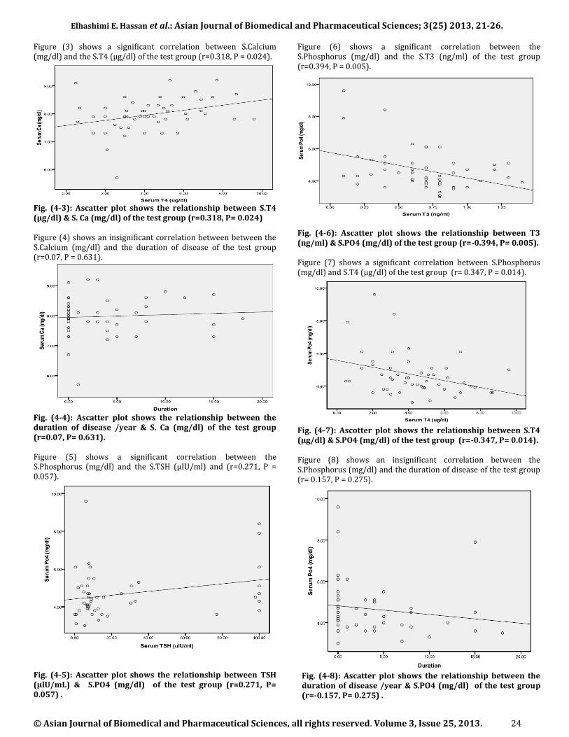
Figure (3) shows a significant correlation between S.Calcium (mg/dl) and the S.T4 (µg/dl) of the test group (r=0.318, P = 0.024).
Fig. (4-3): Ascatter plot shows the relationship between S.T4 (µg/dl) & S. Ca (mg/dl) of the test group (r=0.318, P= 0.024)
Figure (4) shows an insignificant correlation between between the S.Calcium (mg/dl) and the duration of disease of the test group (r=0.07, P = 0.631).
Fig. (4-4): Ascatter plot shows the relationship between the duration of disease /year & S. Ca (mg/dl) of the test group (r=0.07, P= 0.631). Figure (5) shows a significant correlation between the S.Phosphorus (mg/dl) and the S.TSH (µIU/ml) and (r=0.271, P = 0.057).
Fig. (4-5): Ascatter plot shows the relationship between TSH (µlU/mL) & S.PO4 (mg/dl) of the test group (r=0.271, P= 0.057) .
Figure (6) shows a significant correlation between the S.Phosphorus (mg/dl) and the S.T3 (ng/ml) of the test group (r=0.394, P = 0.005).
Fig. (4-6): Ascatter plot shows the relationship between T3 (ng/ml) & S.PO4 (mg/dl) of the test group (r=-0.394, P= 0.005). Figure (7) shows a significant correlation between S.Phosphorus (mg/dl) and S.T4 (µg/dl) of the test group (r= 0.347, P = 0.014).
Fig. (4-7): Ascotter plot shows the relationship between S.T4 (µg/dl) & S.PO4 (mg/dl) of the test group (r=-0.347, P= 0.014).
Figure (8) shows an insignificant correlation between the S.Phosphorus (mg/dl) and the duration of disease of the test group (r= 0.157, P = 0.275).
Fig. (4-8): Ascatter plot shows the relationship between the duration of disease /year & S.PO4 (mg/dl) of the test group (r=-0.157, P= 0.275) .
Elhashimi E. Hassan et al.: Asian Journal of Biomedical and Pharmaceutical Sciences; 3(25) 2013, 21-26.
© Asian Journal of Biomedical and Pharmaceutical Sciences, all rights reserved. Volume 3, Issue 25, 2013. 25
5. DISCUSSION:
Thyroid disorders are important cause for secondary osteoporosis. Serum calcium and phosphorous levels can be fairly used as index of bone resorption [1,] [2]. The present study undertaken to assessed the levels of serum calcium (S.Ca) and phosphorus (S.PO4-2) in Sudanese patients with hypothyroidism. In this study patients with hypothyroidism have a significant decrease in the mean of S.Ca++ compared with the control subjects (p< 0.0001). This agrees with a study done by Shivaleela MB, et al [8], who reported that there was a significant decrease of the mean of the levels of S.Ca in patients with hypothyroidism compared to control. This is mainly due to the low levels of Parathyroid hormone and low levels of calcitonin in hypothyroidism. Patients with hypothyroidism in the present study have a significant increase in the mean of S.PO4-2 compared with the control subjects (p< 0.0001). This agrees with a study done by B. Suneel, et al [9] who reported that there was a significant decrease of the mean of S.PO4-2 in patients with hypothyroidism compared to control. This is mainly due to calcitonin which is regulates the over tubular reabsorption of PO4-2 from kidney. PO4-2 levels are raised due to compensatory effect of calcitonin and parathrmone which favour tubular excretion (by inhibiting tubular reabsorption). In current study there was a significant correlation between S.Ca++ and serum TSH, T3 and T4 in hypothyroid patients. This was further confirmed by the study done by Christoph Schwarza, et al [10] who reported that there was a significant correlation between S.Ca++ and serum TSH, T3 and T4 in hypothyroid patients. Also the study found that there was a significant correlation between S.PO4-2 and serum TSH, T3 and T4 in hypothyroid patients. This was further confirmed by the study done by Christoph Schwarza, et al [10] who reported that there was a significant correlation between S.PO4-2 and serum TSH, T3 and T4 in hypothyroid patients. This study reveal that, there was no differences in the mean levels of S.Ca++ & S.PO4
-2 in male compared with female & that the S.Ca++ & S.PO4-2 can’t affected by the duration of the disease in hypothyroid patients. 6. CONCLUSION: This study concluded that in Sudanese patients with hypothyroid the levels of S.Ca++ was decreased compared to control group with a positive correlation between the S.Ca++ & serum TSH, T3 and T4, where as the levels of S.PO4
-2 was increased compared to control group with a positive correlation between the S.PO4
-2 & serum TSH, T3 and T4 while S.Ca++ & S.PO4-2 not affected by gender and duration of the disease.
7. RECOMMENDATIONS: This study recommended that patients with hypothyroid disorder need to be regularly checked levels of serum calcium & phosphorus, calcium supplementation in form of dietary components and medications is important in patients with hypothyroidism to prevent further bone complications. 8. REFERENCES [1] Rizzoli R, Poser J, Burgi U. Nuclear thyroid hormone receptors in cultured bone cells. Metabolism .1986;35 ( 1) 71-74. [2] Sato k, Han DC, Fujii Y, Tsushima T, Shizume K. Thyroid hormone stimulates alkaline phosphatase activity in cultured rat osteoblastic cells through 3,5,3’-triiodo-L-thyronine nuclear receptors. Endocrinology.1987;120 (5) 1873-1881. [3] Shlomo Melmed, Kenneth S. Polonsky, P. Reed Larsen and Henry M. Kronenberg. William’s text book of endocrinology.Calcium and phosphorus metabolism in hypothyroidism. 12 th ed .2011;10-11. [4] Simsek G. Andican G, Karkoc Y et al, Biol. Trac.Elem.Res Ca,Mg & Zn status in experimental hypothyroidism.1997; 60 (3) 205-213. [5] Beqic KS, Wagner B, Raber w, Schneider B, Hamwi A, Waldhausl W, Vierhapper H.Serum calcium in thyroid disease. Wiener klinische Wochenschrift. 2001;113(1-2) 65-68. [6] Sabuncu T, Aksoy N , Arikan E, Ugur B , Tasan E, Hatemi H. Early changes in parameters of bone and mineral metabolism during therapy for hyperthyroidism and hypothyroidism. Endocrine Research . 2001;27(1-2) 201-213. [7] Gammage MD and Logan SD. Effects of thyroid dysfunction on serum calcium in the rat .Clinical Science. 1986; 71(3) 271-276. [8] Shivaleela MB, Poornima RTand jayaprakash murthy DS. Serum calcium and phosphorus levels in thyroid dysfunction. Indian journal of fundamental and applied life sciences.2012; 2(2):179-183. [9] B. Suneel , D.R.Nagendra , R.R.Aparna, D.Balakrishna, J.N.Naidu. Mineral Status in Thyroid Disorder (Hypo & Hyper). International journal of applied biology and pharmaceutical technology.2011; 2(4): 423-429. [10] Christoph Schwarza, Alexander Benedikt Leichtle, Spiros A, Georg Martin F, Heins Z,Aristmenis K,Gregor L. Thyroid function and serum electrolytes . Swiss Medical Weekly.2012; 13669 - 142. [11] Ringer S.A further contribution regarding the influence of different constituents of blood on contraction of the heart.J Physilo 1883; 4-29. [12] Michael L.Bishop, Edward P Fody, Larry Schoeff. Clinical chemistry principle,procedure,correlations.5th ed.2005;331-342. [13] Shiber JR, Mattu A. Serum phosphate abnormalities in the emergency department. J Emerg Med. 2002; 23:359-400. [14] Venturi, S Donati, FM Venturi, A Venturi. Environmental iodine deficiency. Thyroid : official journal of the American Thyroid Association . 2000;10 (8): 727-9. [15] Berbel P, Navarro D, Ausó E, Varea E, Rodríguez AE, Ballesta JJ, Salinas M, Flores E. Role of late maternal thyroid hormones in cerebral cortex development: an experimental model for human prematurity. Cereb Cortex. 2010; 20 (6): 1462–75. [16] Stephen Nussey and Saffron Whitehead. The thyroid gland in Endocrinology: An Integrated Approach Published by BIOS Scientific Publishers Ltd. 2001. [17] Ekholm R, Bjorkman U.Glutathione peroxidase degrades intracellular hydrogen peroxide and thereby inhibits intracellular protein iodination in thyroid epithelium. Endocrinology. 1997;138 (7): 2871–2878. [18] Bianco AC, Salvatore D, Gereben B, Berry MJ, Larsen PR. Biochemistry, cellular and molecular biology, and physiological roles of the iodothyronine selenodeiodinases. Endocr Rev.
Elhashimi E. Hassan et al.: Asian Journal of Biomedical and Pharmaceutical Sciences; 3(25) 2013, 21-26.
© Asian Journal of Biomedical and Pharmaceutical Sciences, all rights reserved. Volume 3, Issue 25, 2013. 26
2002;23 (1): 38–89. [19] Jansen J, Friesema EC, Milici C, Visser TJ. "Thyroid hormone transporters in health and disease. Thyroid. 2005;15 (8): 757–68. [20] Kester MH, Martinez de Mena R, Obregon MJ, Marinkovic D, Howatson A, Visser TJ, Hume R, Morreale de Escobar G. Iodothyronine levels in the human developing brain: major regulatory roles of iodothyronine deiodinases in different areas. J Clin Endocrinol Metab.2004; 89 (7): 3117–3128. [21] Roshni PR, RajanVK,Meenuvijayan and Remya Reghu . Evaluation of patient with thyroid disorder. journal of research in pharmacy and chemistry. 2013; 3(2) 244-249. [22] Patrick L . Iodine: deficiency and therapeutic considerations. Altern Med Rev. 2008; 13 (2): 116–27. [23] Nicholson WK, Robinson KA, Smallridge RC, Ladenson PW, Powe NR. Prevalence of postpartum thyroid dysfunction: a quantitative review. Thyroid. 2006;16(6):573–582. [24]. Carl A. burtis, Edward R. Ashwood, David E. bruns. Disorder of bone In : loren Wilson.ed. tietz fundementals of clinical chemistry. Sixth edition.2008;712-17. [25] Lambers TT, Bindels RJ, Hoenderop JG. Coordinated control of renal Ca2+ handling. Kidney Int. 2006;69:650. [26] Poncin S,Colin IM and Decallonne B. A protective effect against autoimmune thyroid destruction .The American Journal of Pathology. 2010;177(1):219–228. [27] V.Torlak, T. Zemunik, D. Modun, V. Capkun, V. Pesutic-Pisac, A. arkotić, M. Pavela-Vrancic and A. Stanicic .induced changes in rat thyroid gland function. Braz J Med Biol Res. 2007; 40(8) :1087-1094. [28] Braverman, MD, Lewis E., and Robert D. Utiger, MD.Werner and Ingbar's The Thyroid: A Fundamental and Clinical Text. 9th ed. 2005; 293(24):3107-3112. [29] Mary J Shomon .Living Well With Graves' Disease and Hyperthyroidism: What Your Doctor Doesn't Tell You. 2005; 2 (3) 120-170. [30] Yamamoto M, Shibuya N, Chen LC, Ogata E.Seasonal recurrence of transient hypothyroidism in a patient with autoimmune thyroiditis . Endocrinol.1988; 35 (1): 135–42.