organic phosphorous and calcium source induce the
TRANSCRIPT

NANO EXPRESS Open Access
Organic Phosphorous and Calcium SourceInduce the Synthesis of Yolk-ShellStructured Microspheres of CalciumPhosphate with High-Specific Surface Area:Application in HEL AdsorptionXianshuo Cao1†, Guizhen Wang1†, Kai Wang2, Lan Guo1, Yang Cao1, Xianying Cao1* and Yong Yang1*
Abstract
Yolk-shell-structured calcium phosphate microspheres have a great potential for medical applications due to theirexcellent physicochemical properties and biocompatibility. However, developing a yolk-shell-structured calciumphosphate with high adsorption capability remains a challenge. Herein, a porous yolk-shell-structured microsphere(ATP-CG) of calcium phosphate with high-specific surface area [SBET = 143 m2 g−1, which is approximately threetimes as high as that of ATP-CL microspheres synthesized by replacing calcium source with calcium L-lactatepentahydrate (CL)] was successfully synthesized by using adenosine 5'-triphosphate disodium salt (ATP) as thephosphorous source and calcium gluconate monohydrate (CG) as calcium source through a self-templatingapproache. The influences of molar ratio of Ca to P (Ca/P), hydrothermal temperature, and time on the morphologyof ATP-CG microspheres were also investigated. It is found that the organic calcium source and organicphosphorous source play a vital role in the formation of yolk-shell structure. Furthermore, a batch of adsorptionexperiments were investigated to illuminate the adsorption mechanism of two kinds of yolk-shell-structuredmicrospheres synthesized with different calcium sources. The results show that the adsorption capacity of ATP-CGmicrospheres (332 ± 36 mg/g) is about twice higher than that of ATP-CL microspheres (176 ± 33 mg/g). Moreover,the higher-specific surface area caused by the calcium source and unique surface chemical properties for ATP-CGmicrospheres play an important role in the improvement of HEL adsorption capability. The study indicates that theas-prepared yolk-shell-structured microsphere is promising for application in drug delivery fields and provides aneffective approach for improving drug adsorption capability.
Keywords: Yolk-shell, Calcium phosphate, Adsorption, Specific surface area
© The Author(s). 2020 Open Access This article is licensed under a Creative Commons Attribution 4.0 International License,which permits use, sharing, adaptation, distribution and reproduction in any medium or format, as long as you giveappropriate credit to the original author(s) and the source, provide a link to the Creative Commons licence, and indicate ifchanges were made. The images or other third party material in this article are included in the article's Creative Commonslicence, unless indicated otherwise in a credit line to the material. If material is not included in the article's Creative Commonslicence and your intended use is not permitted by statutory regulation or exceeds the permitted use, you will need to obtainpermission directly from the copyright holder. To view a copy of this licence, visit http://creativecommons.org/licenses/by/4.0/.
* Correspondence: [email protected]; [email protected]†Xianshuo Cao and Guizhen Wang contributed equally to this study.1College of Life Science and Pharmacy, School of Materials Science andEngineering; State Key Laboratory of Marine Resource Utilization in SouthChina Sea, College of Food Science and Engineering, Analytical and TestingCentre, Hainan University, Haikou 570228, People’s Republic of ChinaFull list of author information is available at the end of the article
Cao et al. Nanoscale Research Letters (2020) 15:69 https://doi.org/10.1186/s11671-020-03298-w

IntroductionCalcium phosphate has gained considerable attentionduring the past few years due to its excellent biocom-patibility [1], high-loading capacity, and delivery effi-ciency. Calcium phosphate-related biomaterials havebeen widely used in various biomedical fields, such astissue engineering [2], bone repair [3], and drug delivery[4]. In order to extend the application range and im-prove the performance of calcium phosphate-based ma-terials, various calcium phosphate materials withvarieties of morphologies and microstructures includingcarbonated hydroxyapatite (HAp) microspheres [5],HAp microtubes [6], hollow HAp microspheres [7],mesoporous yolk@cage-shell nanospheres of amorphouscalcium phosphate (ACP) [8] have been reported.Among various morphologies, yolk-shell-structured
microspheres have attracted more and more attentions,as they are not only frontier materials science but alsoshow unique morphological features. In yolk-shell-structured microspheres, the void space between yolkcore and shell can serve as a storage reservoir for variouscargoes and the porous-structured shell can provide adiffusion pathway for guest molecules, which makesthem have great potential for diverse applications in-cluding catalysis [9], lithium-ion batteries [10], photoca-talyst [11], and biomedicine [12]. Traditionally,sacrificing template methods are the primary ones toprepare yolk-shell-structured microspheres [13, 14].These template strategies have achieved great success inadjusting the structure and properties. However, theseapproaches present some disadvantages. For instance, te-dious processing steps and surfactant or structure-directing reagents, which may be hazardous to humanbeing’s health. Currently, the self-templating methodshave been widely used in the research of yolk-shell-structured microspheres [15, 16]. Unlike the traditionaltemplating approaches, the templates employed in self-templating approaches are not only the templates forforming the voids, but also the precursor of yolk-shell-structured microspheres. Thus, self-templatingmethods are convenient approaches to prepare yolk-shell-structured microspheres. However, the introduc-tion of self-templating approaches to the synthesis ofyolk-shell-structured calcium phosphate microspheresremains an interesting challenge.Furthermore, calcium phosphate materials have been
utilized to carry different types of cargoes such as pro-teins [17], DNA [18], and siRNA [19]. However, poordrug adsorption capability of calcium phosphate needsto be solved urgently. Generally, the approaches of drugmolecules immobilizing over the surface of carrier de-pend on the surface properties containing surface poten-tial [20], hydrophobicity/hydrophilicity [21], hydrogenbond [22], and specific surface area [23]. So, improving
surface properties and specific surface area is a valid ap-proach for ehancing drug adsorption capability ofcarrier.Herein, we prepared a kind of porous yolk-shell-
structured microspheres of calcium phosphate by usingadenosine 5'-triphosphate disodium salt (ATP) as thephosphorous source and calcium gluconate monohy-drate (CG) as the calcium source through self-templating approach. Without any addition of templat-ing agent, the as-prepared yolk-shell-structured calciumphosphate microspheres display a particularly high-specific surface area. Furthermore, the hen egg lysozyme(HEL) adsorption behavior of ATP-CG microsphereswas investigated in comparison with the ATP-CL micro-spheres prepared by replacing calcium source with cal-cium L-lactate pentahydrate (CL). The results reveal thatthe difference of specific surface area caused by the cal-cium source and surface chemical properties play a vitalrole in the improvement of HEL adsorption capability.
MethodsMaterialsAdenosine 5'-triphosphate disodium salt (ATP) was ob-tained from Macklin Biochemical Co., Ltd (Shanghai,China). Calcium gluconate monohydrate (CG) and cal-cium (L)-lactate pentahydrate (CL) were acquired fromSangon Biotech Co., Ltd (Shanghai, China). The hen egglysozyme (HEL, ~ 70000 U/mg) was purchased fromSigma-Aldrich (Taufkirchen, Germany).
Synthesis and Characterization of ATP-CG and ATP-CLYolk-Shell-Structured MicrospheresThe ATP-CG yolk-shell-structured calcium phosphate mi-crospheres were prepared as follows: In brief, 0.9 g of CGwas dissolved in 20 mL of ultrapure water to form solutionC at 60 °C and 0.11 g of ATP was dissolved in 5 mL of ul-trapure water to form solution P. Then, solution C wascooled down to room temperature and mixed with solutionP under vigorous stirring and the pH of the solution wasadjusted by 2 M NaOH solution to 5. The final volume ofthe solution was 30 mL with the extra addition of ultrapurewater and the molar ratio of Ca to P (Ca/P) was 3.3. Thefinal solution was transferred into a microwave digestionsystem for microwave hydrothermal reaction and treated at120 °C for 15 min. The resulting precipitates were collectedby centrifugation (4500 rpm, 10 min), rinsed with ultrapurewater and lyophilized for 48 h. The ATP-CL microsphereswere prepared according to literature procedures [24].The crystalline phase of the microspheres was charac-
terized by X-ray diffraction (XRD, Cu Kα source, λ =0.154). The morphology of microspheres was observedby scanning electron microscopy (SEM), transmissionelectron microscopy (TEM), and high-resolution TEM(HRTEM). The compositions of microspheres were
Cao et al. Nanoscale Research Letters (2020) 15:69 Page 2 of 11

studied by Fourier transform infrared spectrophotometer(FTIR). The specific surface area of microspheres wasdetermined by Brunauer-Emmett-Teller (BET). Thermo-gravimetryanalysis (TGA) was employed to study thethermal properties of samples at a heating rate of 10 °C/min in nitrogen atmosphere.
HEL Adsorption and CharacterizationThe HEL adsorption experiments of two kinds of micro-spheres were conducted as follows: the certain amountsyolk-shell microspheres (ATP-CG, Ca/P = 3.3, 120 °C,15 min, and ATP-CL, Ca/P = 2.5, 120 °C, 30 min) weredispersed in the water with constant ultrasonic treat-ment for 10 min to form 1.5 mg/mL of microspheressuspension. Then, 0.5 mL of aqueous solutions that con-tain various concentrations of HEL were immediatelyadded into 1 mL above suspension and the final concen-trations of drug were 1–7.5 mg/mL. Each solution wasshaken (200 rpm) at 37 °C for 6 h. Later, the solutionswere centrifuged and the amounts of HEL in the super-natants were measured by UV-vis spectrophotometer at280 nm. The zeta potentials and compositions of micro-spheres before and after drug loading were characterizedby zeta potential analyzer, FTIR spectrometer, and ther-mogravimetric analyzer (TGA, heating rate 10 °C min−1,nitrogen atmosphere).
Adsorption IsothermIn order to investigate adsorption behavior, Dubinin-Radushkevic isotherm (D-R) model was conducted inour study. The D-R model is based on the theory ofmicropore filling, which is used to describe the non-
Fig. 1 SEM images of ATP-CG microspheres prepared by microwave hydrothermal method at 120 °C
Fig. 2 FTIR spectra of ATP-CG microspheres synthesized with variousCa/P at 120 °C for 15 min
Cao et al. Nanoscale Research Letters (2020) 15:69 Page 3 of 11

Fig. 3 SEM and TEM images of ATP-CG microspheres synthesized with various of Ca/P. a, b Ca/P = 0.8. c, d Ca/P = 1.67. e, f Ca/P = 2.5. g, h Ca/P= 3.3. i HRTEM, j EDS-mapping, k EDS spectra, l XPS spectra of ATP-CG microspheres with Ca/P = 3.3
Fig. 4 SEM and TEM images of ATP-CG microspheres synthesized with Ca/P = 3.3 under different experimental conditions. a, b T = 120 °C, t = 5min. c, d T = 120 °C, t = 15 min. e–h T = 120 °C, t = 60 min. i–l T = 160 °C, t = 15 min
Cao et al. Nanoscale Research Letters (2020) 15:69 Page 4 of 11
ideal sorption on a heterogeneous surface as well as dis-tinguish the sorption mechanism (physical sorption orchemical sorption). The model is expressed by the fol-lowing equation: where Qeq is the adsorption capacity ofadsorbent at equilibrium (mg/g), Ceq is the adsorbateconcentration in the aqueous phase at equilibrium (mL/L). Qm is the maximum adsorption capacity. R is gasconstant, 8.314 J/(mol ∙ k). T is absolute temperature. Erepresents the mean free energy for estimating the typeof adsorption. If the E value is below 8 kJ/mol, the ad-sorption type can be explained by physical adsorption,between 8 and 16 kJ/mol, the adsorption type belongs toion exchange and greater than 16 kJ/mol, the adsorptiontype can be described by chemical adsorption.
Qeq ¼ Qm exp −KDR ε2� � ð1Þ
ε ¼ RTlnð1þ 1Ceq
Þ ð2Þ
E ¼ 1ffiffiffiffiffiffiffiffiffiKDR
p ð3Þ
Statistical Analysis of Drug AdsorptionData were presented as mean ± standard deviation (SD)value. Significant differences (p < 0.05) were statisticallycalculated among different groups using the one-wayANOVA. All the experiments were carried out in tripli-cate and data was analyzed by using the DPS software.
Results and DiscussionMorphology and Chemical Characterization ofMicrospheresATP-CG Yolk-Shell-Structured MicrospheresSEM images in Fig. 1 show the morphologies of varioussamples obtained under different reaction conditions. Att = 5 min or 15 min, all products are composed of uni-form microspheres. However, when the hydrothermaltime is further increased to 30 min, nanosheets self-
assembled microspheres were formed (as shown in Fig.1 f, i, l). Meanwhile, the effect of Ca/P on the morph-ology of products is also observed at t = 30 min. As theincrease of Ca/P, the nanosheets self-assembled micro-spheres were gradually formed (as shown in Fig. 1 f, i, l).The formation of nanosheets self-assembled micro-spheres could be explained by the following reasons.Firstly, under the microwave hydrothermal process, ATPmolecules could hydrolyze to form adenosine-basedmolecules including adenosine diphosphate (ADP), ad-enosine monophosphate (AMP) and adenosine, and sim-ultaneously release phosphate ions (PO4
3−). Meanwhile,CG molecules could hydrolyze to form gluconate andcalcium ions (Ca2+). Then, phosphate ions would reactwith calcium ions to form primary ACP nuclei [25].Then, the initial ACP nuclei grow and assemble to formACP microspheres. Therefore, when hydrothermal timeis further extended, the ATP and CG molecules in solu-tion are further hydrolyzed and release more PO4
3− andCa2+ ions, which causes the formation of nanosheetsself-assembled microspheres through improving thesupersaturation of system and the nucleation rate. Inaddition, by increasing the Ca/P, the local high concen-tration of Ca2+ also accelerates the morphology trans-formation of products in the same way as above. Theabove analysis indicates that the hydrothermal time andCa/P have an important influence on the morphology ofproducts.Next, the FTIR spectra of microspheres synthesized
with various Ca/P at 120 °C for 15 min are investigated(Fig. 2). The peaks at 1620 cm−1, 1383 cm−1, and 912cm−1 attributed to the characteristic peaks of the C=O,C–O of CG and P–O groups of ATP [26], respectively,implying that unhydrolyzed CG and ATP molecules ortheir derivatives are absorbed on the surface of the mi-crospheres. The faint characteristic peak of the PO4
3−
from HAp is located at 1035 cm−1 [27] and the absorp-tion peaks at 1122 cm−1 and 567 cm−1 are assigned toPO4
3− ions of ACP [28], indicating that the products arecomposed of ACP and HAp. The FTIR results suggest
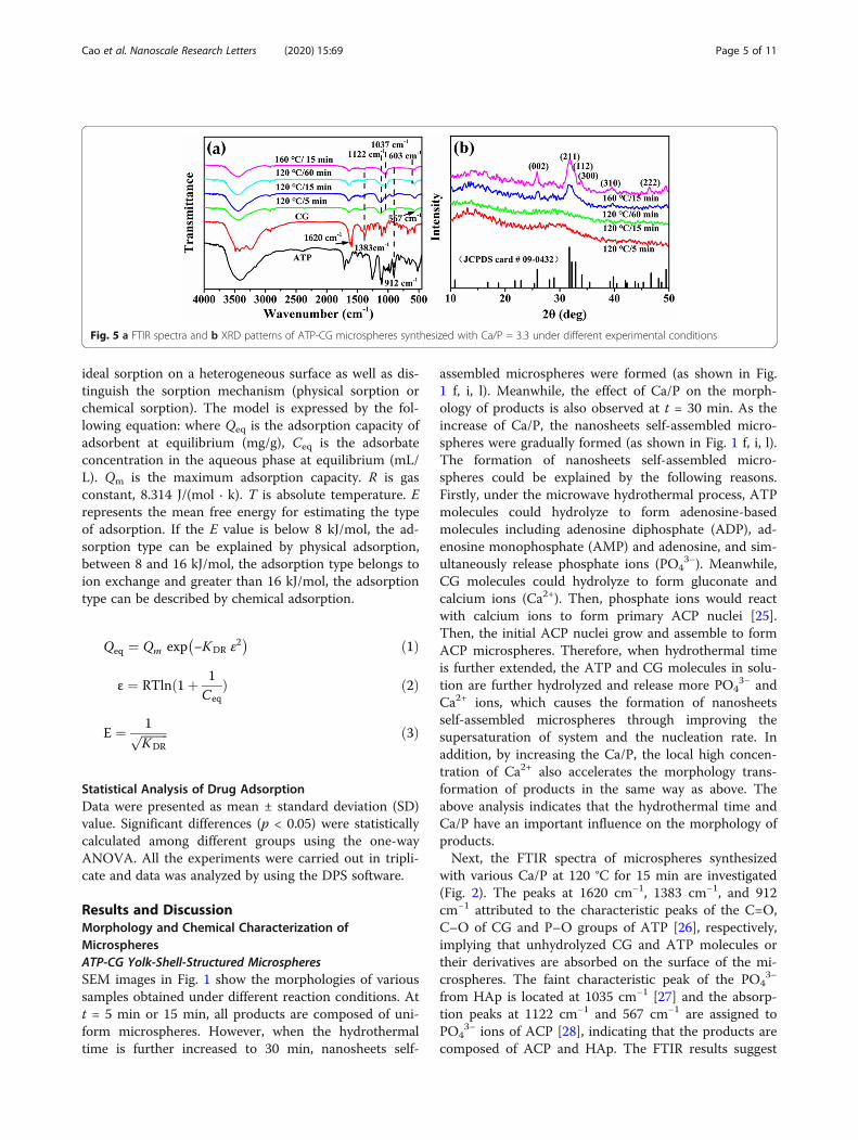
Fig. 5 a FTIR spectra and b XRD patterns of ATP-CG microspheres synthesized with Ca/P = 3.3 under different experimental conditions
Cao et al. Nanoscale Research Letters (2020) 15:69 Page 5 of 11
the calcium phosphate is succesfully prepared by usingATP as phosphorous source and CG as calcium source.Furthermore, the SEM and TEM images of samples
synthesized with various Ca/P through microwavehydrothermal method at 120 °C for 15 min are displayedin Fig. 3. When the Ca/P is 0.8 or 1.67, the samples con-sist of porous microspheres (Fig. 3b, d). When the Ca/Pis 2.5, the morphology of products starts transforming toyolk-shell-structured microspheres (Fig. 3f). As the Ca/Pfurther increases to 3.3, the products are entirely com-posed of yolk-shell-structured microspheres (Fig. 3h).Beyond that, some of broken spheres and the exposedcores of the yolk-shell microspheres (insert in Fig. 3g)are observed after mechanical fracturing, providing evi-dence of a hollow structure between yolk and shell.
Based on the above observation, we tentatively proposethe formation mechanism of yolk-shell-structured mi-crospheres synthesized with various Ca/P. When the Ca/P is lower, the porous ACP microspheres are formedfirst, which is attributed to the inhibition effect of ATPand CG molecules or their derivatives adsorbed on thesurface on the microspheres. Then, as the Ca/P is fur-ther increased, the metastable ACP will further grow,which is drived by the high supersaturation of system. Fi-nally, the crystalline HAps are formed on the externalsurface, which is confirmed by the high-resolution TEM(HRTEM) image of microspheres in Fig. 3i (the interpla-nar distance of 0.308 nm can be indexed to (210) ofHAp). As a result, the hollow structures between yolkand shell are generated due to the difference in volume
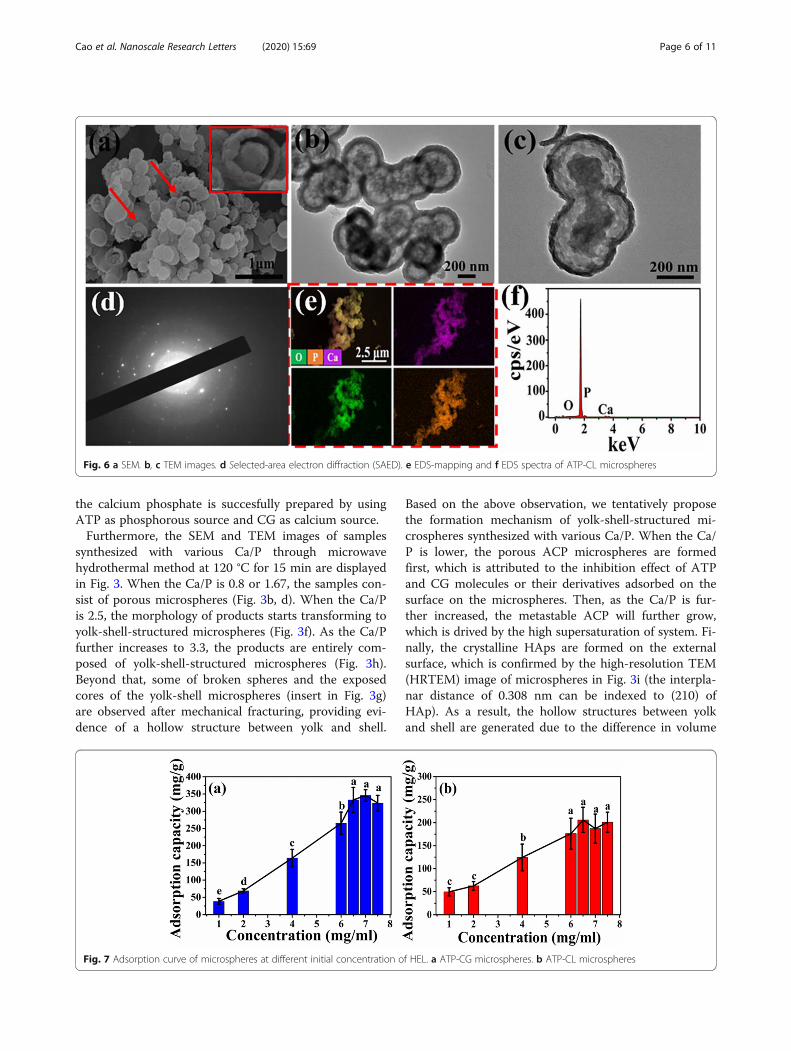
Fig. 6 a SEM. b, c TEM images. d Selected-area electron diffraction (SAED). e EDS-mapping and f EDS spectra of ATP-CL microspheres
Fig. 7 Adsorption curve of microspheres at different initial concentration of HEL. a ATP-CG microspheres. b ATP-CL microspheres
Cao et al. Nanoscale Research Letters (2020) 15:69 Page 6 of 11

or density between HAp and ACP [24]. The correspond-ing EDS mapping indicates that the Ca, P, and O ele-ments are uniformly distributed throughout themicrospheres. The EDS spectra in Fig. 3k and XPSspectrum in Fig. 3l reveal that the chemical elements ofmicrospheres mainly include Ca, P, and O, which is con-sistent with the result of FTIR (Fig. 2).The impact of microwave-assisted hydrothermal time
and temperature on the morphology of microspheressynthesized with Ca/P = 3.3 is further investigated. Asshown in Fig. 4a-b, when the hydrothermal time is 5min, the samples are composed of porous microspheres.As discussed above, when t = 15 min, the product is alsocomposed of yolk-shell-structured microspheres (Fig.
4c-d). When hydrothermal time is extended to 60 minor temperature increased to 160 oC, sheets or rodsboundles are observed (Fig. 4e-l). The morphology trans-formation from porous to yolk-shell to sheet or rod isattributed to the further growth of ACP with the con-tinuous hydrolysis of ATP and CG molecules in solu-tion. Moreover, the hydrolysis of ATP and CGmolecules or their derivatives adsorbed on the surfaceon the ACP microspheres also accelerates the growth ofACP. An interesting phenomenon emerged at 60 min or160 oC, these sheets or rods are also developed fromACP nanoparticles (as shown in red boxes), which isconfirmed by the DTA analysis in Fig. S1. An exother-mic peak at 650 °C is observed in the DTA curves [29,
Fig. 8 FT-IR spectra and TGA curves of microspheres before and after HEL adsorption. a FTIR spectra and c TGA curves of ATP-CG microspheres,b FTIR spectra and d TGA curves of ATP-CL microspheres
Fig. 9 a Adsorption isotherms model of HEL on ATP-CG microspheres. b Adsorption isotherms model of HEL on ATP-CL microspheres
Cao et al. Nanoscale Research Letters (2020) 15:69 Page 7 of 11

30], which is attributed to the ACP crystallization. Theexothermic peak gradually becomes weak with the in-crease of hydrothermal time or temperature, implyingthat the transformation of ACP in the products towardcrystilline calcium phosphate.The chemical constitution and structure of samples
synthesized with Ca/P = 3.3 under different hydrother-mal time or temperature are investigated by the FTIRand XRD. As shown in Fig. 5a, the characteristic peaksof PO4
3− ions of HAp are located at 1037 cm−1 and 603cm−1 [27]. The peak at 1122 cm−1 is assigned to charac-teristic peak of PO4
3− ions from ACP. The absorptionpeaks at 1620 cm−1 and 1383 cm−1 are attributed tocharacteristic peak of C=O and C–O groups from CG,respectively. The absorption peak at 912 cm−1 refers tothe asymmetric P–O stretching vibration of ATP. By in-creasing hydrothermal time or temperature, the intensityof characteristic peaks of CG and ATP is gradually de-creased, indicating the ATP and CG molecules or theirderivatives adsorbed on the surface of microspheres arefurther hydrolyzed. Meanwhile, the intensity of charac-teristic peak of PO4
3− ions in HAp presents gradually in-creased trend with the decrease of intensity of ACPcharacteristic peak, illuminating the transformation ofcrystalline phase of products toward to HAp phase.Figure 5b shows XRD patterns of different samples. A
characteristic hump of amorphous phase at around 2θ =30° of microspheres synthesized at 5 or 15 min is ob-served. However, when either hydrothermal time is ex-tended to 60 min or temperature increased to 160 °C,the crystalline phase of microspheres thoroughly
transforms to HAp, which could be indexed as thestandard data (JDCPS no. 09-0432). The improvement inthe relative intensity of (211), (300), and (002) latticeplanes could further explain the increase in the crystal-linity of products. Thus, the XRD and FTIR results fur-ther confirm the crystalline phase transformation ofproducts with the increase in the hydrothermaltemperature or time.
ATP-CL Yolk-Shell-Structured MicrospheresIn ordrer to compare drug adsorption behavior, theother yolk-shell-structured microspheres were preparedby using CL as organic calcium source through micro-wave hydrothermal method [24]. In terms of morph-ology, the samples still consist of yolk-shell-structuredmicrospheres, which is verified by the broken spheres(insert in Fig. 6a) and TEM images (Fig. 6b, c). The re-sult demonstrates that the change in calcium sourcehave no significant effect on morphology of products. Inaddition, the selected-area electron diffraction (SAED)shows the discrete SAED spots (Fig. 6d), demonstratingthat well-crystallized microspheres are obtained. Inaddition, the EDS-mapping exhibits the even distributionof Ca, P and O elements in microspheres (Fig. 6e). Thecorresponding EDS spectra also comfirms the presenceof Ca, P, and O elemnets in microspheres (Fig. 6f), indi-cating that the as-prepared microspheres are calciumphosphate.
HEL Adsorption and Adsorption Mechanism of MicrospheresAs shown in Fig. 7, the adsorption capacity of two kindsof microspheres increases with the increasing initial con-centration of HEL. When the initial concentration ofHEL increases to 6.5 mg/mL, the adsorption capacity ofthe ATP-CG microspheres reaches a plateau and themaximum adsorption capacity of microspheres is about332 ± 36 mg/g (Fig. 7a), which is about twice higherthan that of ATP-CL microspheres (176 ± 33 mg/g, 6mg/mL, Fig. 7b).The HEL adsorption result is further supported by the
FTIR spectra and TG curves. As shown in Fig. 8a, b, theabsorption peaks at 1134 cm−1 (1139 cm−1) and 563cm−1 (568 cm−1) assigned to the characteristic peakPO4
3− ions of ACP and 1039 cm−1 (1040 cm−1) assignedto characteristic peak of PO4
3− ions of HAp are observedin the HEL-adsorbed microspheres, which indicates thatthe introduction of HEL in microspheres does not causeany significant change in the structure of microspheres.The adsorption peaks at 1542 cm−1 and 1545 cm−1 at-tributed to amide group of HEL are observed in HEL-adsorbed microspheres, confirming that HEL is success-fully adsorbed on the microspheres. Meanwhile, the ad-sorption bands at 2966, 2962, 2935, and 2927 cm−1
originated from –CH3 and –CH2 groups of HEL are also
Table 1 Parameters of adsorption isotherms for microspheres
ATP-CG ATP-CL
D-R model Qm (mg/g) 381 192
R2 97 89
E (kJ/mol) 0.687 0.855
Fig. 10 Zeta potentials of HEL and microspheres before and afterHEL adsorption
Cao et al. Nanoscale Research Letters (2020) 15:69 Page 8 of 11

detected in HEL-adsorbed microspheres, which furtherverifies the presence of HEL on the microspheres. TheTGA curves display that the weight loss of ATP-CG mi-crospheres before and after HEL adsorption is 11.3% and36.7%, respectively (Fig. 8c). Therefore, the HEL adsorp-tion capacity of ATP-CG microspheres is approximately340 mg/g. However, a weight loss of 21.1% of ATP-CL isobtained before HEL adsorption and 37% appears on theHEL-adsorbed microspheres (Fig. 8d). So, the HEL ad-sorption capacity is 189 mg/g for ATP-CL microspheres.The TGA results are closed to the result from the Fig. 7.To investigate the cause of adsorption capacity differ-
ence between two kinds of microspheres, the equilib-rium adsorption data of microspheres is further analyzedaccording to D-R isotherm model. The fitting curves areshown in Fig. 9 and fitting parameters are listed in Table1, respectively. From the fitting results, the correlationcoefficient from ATP-CG is higher than ATP-CL, sug-gesting D-R model is suitable for describing the drug ad-sorption behavior of ATP-CG microspheres. Since the Evalue is below 8 kJ/mol, the adsorption of HEL ontoATP-CG microspheres is physical sorption. The max-imum capacity (Qm) of ATP-CG microspheres for HELcould reach as high as near 381 mg/g, which is close toresult from Fig. 7a.Since the adsorption of HEL on the ATP-CG micro-
spheres is physical sorption, the surface potential of mi-crospheres is investigated. As shown in Fig. 10a, the zetapotential value of ATP-CG, ATP-CL microspheres andHEL in ultrapure water is – 17 mV, − 22 mV, and 20mV, respectively. After HEL adsorption, the zeta poten-tial value of ATP-CG and ATP-CL microspheres
changes to 2.7 mV and 1.5 mV, respectively, indicatingthat the adsorption of HEL molecules onto the surfaceof microspheres through the attractive electrostaticforce. However, the attractive electrostatic force is notthe major cause of adsorption capacity difference be-tween two kinds of microspheres, because there is nosiginificant difference in zeta potential values (− 17 mVand – 22 mV) between microspheres.Therefore, in order to further illuminate the reason
causing absorption capacity difference between micro-spheres, the specific surface area of microspheres is in-vestigated. As shown in Fig. 11a, the BET specificsurface area (SBET) of ATP-CG microspheres is 143 m2
g−1, which is approximately three times as high as theATP-CL microspheres (55 m2 g−1, Table 2). So, the spei-cific surface area can contribute to the absorption cap-acity difference between microspheres. Such high-specific surface area of ATP-CG microspheres is mainlyattributed to the low crystallinity [31]. From Fig. 11b,ATP-CG microspheres exhibit lower crystallinity thanthat of ATP-CL microspheres. Moreover, the differenceof crystallinity between ATP-CG and ATP-CL is chieflydue to the different synthesis conditions. Generally, theproduct crystallinity increases with the hydrolysis extentof reactants under certain pressure and temperature.Herein, the acidity of gluconic acid (pKa = 3.39) ishigher than L-Lactic acid (pKa = 3.86), which wouldcause a slower hydrolysis rate and ultimately present alower crystallinity. As a result, ATP-CG microsphereswith a higher-specific surface area are obtained by cha-ging calcium source.
ConclusionsThe ATP-CG yolk-shell microspheres have been de-signed by using ATP as organic phosphorous source andCG as organic calcium source through a microwave-assisted hydrothermal method. The microspheres displaya high-specific surface area and high adsorption capabil-ity. The influences of Ca/P, hydrothermal temperature,and time on the morphology and structure of
Fig. 11 a Nitrogen adsorption-desorption isotherms. b XRD patterns of microspheres
Table 2 Textural properties of microspheres
ATP-CL ATP-CG
SBET (m2 g−1) 55 143
Pore size (nm) 16 8
Pore volume (cm3 g−1) 0.2 0.3
Cao et al. Nanoscale Research Letters (2020) 15:69 Page 9 of 11

microspheres were also investigated. The study indicatesthat organic phosphrous source and organic calciumsource have a significant effect on the formation of yolk-shell-structured microspheres. Moreover, the hydrother-mal conditions including Ca/P, hydrothermal, andtemperature are responsible for the formation of yolk-shell microspheres. Furthermore, we find that the spe-cific surface area and surface chemical properties suchas surface potential are two key factors that affect ad-sorption capacity of microspheres by comparing theHEL adsorption behavior of two kinds of microspheressynthesized with different calcium source.
Supplementary informationSupplementary information accompanies this paper at https://doi.org/10.1186/s11671-020-03298-w.
Additional file 1: Figure S1. DTA curves of ATP-CG microspheressynthesized with Ca/P = 3.3 under different experimental conditions.
AbbreviationsBET: Brunauer-Emmet-Teller measurements; FTIR: Fourier transform infraredspectroscopy; TEM: Transmission electron microscopy; XRD: X-ray diffraction;TGA: Thermogravimetry analysis; HRTEM: High-resolution TEM;SAED: Selected-area electron diffraction
AcknowledgementsWe acknowledged Gengping Wan and Huifeng Zhang from HainanUniversity who gave us many suggestions for measurement.
Authors’ ContributionsXianying Cao designed the study. Xianshuo Cao and Guizhen Wangconducted experiments and analysis, and prepared the manuscript. YongYang and Guizhen Wang revised the manuscript. Yang Cao, Kai Wang, andLan Guo provided advice for the experimental design. All authors read andapproved the final manuscript.
FundingThis research was funded by the foundation of National Key R&D Program ofChina, grant number 2017YFC1103800. Hainan Province Natural ScienceFoundation of China, grant number 819QN231.
Availability of Data and MaterialsAll data supporting the conclusions of this article are included within thearticle.
Competing InterestsAll authors declare that they have no competing interests.
Author details1College of Life Science and Pharmacy, School of Materials Science andEngineering; State Key Laboratory of Marine Resource Utilization in SouthChina Sea, College of Food Science and Engineering, Analytical and TestingCentre, Hainan University, Haikou 570228, People’s Republic of China.2Department of Biochemistry and Molecular Biology, Hainan Medical College,Haikou 571199, People’s Republic of China.
Received: 18 December 2019 Accepted: 13 March 2020
References1. Dorozhkin SV (2009) Calcium orthophosphates in nature, biology and
medicine [J]. Materials 2(2):399–4982. Xu S, Liu J, Zhang L et al (2017) Effects of HAp and TCP in constructing
tissue engineering scaffolds for bone repair [J]. J. Mat. Chem. B 5(30):6110–6118
3. Subramaniam S, Fang YH, Sivasubramanian S et al (2016) Hydroxyapatite-calcium sulfate-hyaluronic acid composite encapsulated with collagenase asbone substitute for alveolar bone regeneration [J]. Biomaterials 74:99–108
4. Govindan B, Swarna LB, Nagamony P et al (2017) Designed synthesis ofnanostructured magnetic hydroxyapatite based drug nanocarrier for anti-cancer drug delivery toward the treatment of human epidermoidcarcinoma [J]. Nanomaterials 7(6):138
5. Guo YJ, Wang YY, Chen T et al (2013) Hollow carbonated hydroxyapatitemicrospheres with mesoporous structure: hydrothermal fabrication anddrug delivery property [J]. Mater. Sci. Eng. C-Mater. Biol. Appl. 33(6):3166–3172
6. Zhang YG, Zhu YJ, Chen F et al (2017) Ultralong hydroxyapatite microtubes:solvothermal synthesis and application in drug loading and sustained drugrelease [J]. Crystengcomm 19(14):1965–1973
7. Zhang M, Ni S, Zhang X et al (2019) Dexamethasone-loaded hollowhydroxyapatite microsphere promotes odontogenic differentiation ofhuman dental pulp cells in vitro [J]. Odontology 4:1–9
8. Huang S, Li C, Xiao Q (2017) Yolk@cage-shell hollow mesoporousmonodispersion nanospheres of amorphous calcium phosphate for drugdelivery with high loading capacity [J]. Nanoscale Res. Lett 12(1):275
9. Yang T, Wang Y, Wei W et al (2019) Synthesis of octahedral Pt-Ni-Ir yolk-shell nanoparticles and their catalysis in oxygen reduction and methanoloxidization under both acidic and alkaline conditions [J]. Nanoscale 11(48):23206–23216
10. Choi JH, Park GD, Jung DS et al (2019) Pitch-derived carbon coated SnO2-CoO yolk-shell microspheres with excellent long-term cycling and rateperformances as anode materials for lithium-ion batteries [J]. Chem. Eng. J.369:726–735
11. Chiu YH, Naghadeh SB, Lindley SA et al (2019) Yolk-shell nanostructures asan emerging photocatalyst paradigm for solar hydrogen generation [J].Nano Energy 62:289–298
12. Gu D, An P, He X et al (2020) A novel versatile yolk-shell nanosystem basedon NIR-elevated drug release and GSH depletion-enhanced Fenton-likereaction for synergistic cancer therapy [J]. Colloid Surf. B-Biointerfaces 189:110810
13. Chen Q, Wei W, Tang J et al (2019) Dopamine-assisted preparation ofFe3O4@MnO2 yolk@shell microspheres for improved pseudocapacitiveperformance [J]. Electrochim. Acta 317:628–637
14. Qi X, Yan Z, Liu Y et al (2018) Ni and Co doped yolk-shell type Fe2O3 hollowmicrospheres as anode materials for lithium-ion batteries [J]. Mater. Chem.Phys. 211:452–461
15. Wang L, Jiao X, Liu P et al (2018) Self-template synthesis of yolk-shelledNiCo2O4 spheres for enhanced hybrid supercapacitors [J]. Appl. Surf. Sci.427:174–181
16. Li J, Li X, Chen X et al (2019) In situ construction of yolk-shell zinc ferritewith carbon and nitrogen co-doping for highly efficient solar lightharvesting and improved catalytic performance [J]. J. Colloid Interface Sci.554:91–102
17. Huang B, Wu Z, Ding S et al (2018) Localization and promotion ofrecombinant human bone morphogenetic protein-2 bioactivity onextracellular matrix mimetic chondroitin sulfate-functionalized calciumphosphate cement scaffolds [J]. Acta Biomater. 71:184–199
18. Shubhra QTH, Oyane A, Araki H et al (2017) Calcium phosphatenanoparticles prepared from infusion fluids for stem cell transfection:process optimization and cytotoxicity analysis [J]. Biomater. Sci. 5(5):972–981
19. Bakan F, Kara G, Cokol Cakmak M et al (2017) Synthesis and characterizationof amino acid-functionalized calcium phosphate nanoparticles for siRNAdelivery [J]. Colloid Surf. B-Biointerfaces 158:175–181
20. Tian B, Liu S, Wu S et al (2017) pH-responsive poly (acrylic acid)-gatedmesoporous silica and its application in oral colon targeted drug deliveryfor doxorubicin [J]. Colloid Surf. B-Biointerfaces 154:287–296
21. Zhang L, Chan JM, Gu FX et al (2008) Self-assembled lipid-polymer hybridnanoparticles: a robust drug delivery platform [J]. ACS nano 2(8):1696–1702
22. Thuan Van T, Duyen Thi Cam N, Le HTN et al (2019) A hollow mesoporouscarbon from metal-organic framework for robust adsorbability of ibuprofendrug in water [J]. R. Soc. Open Sci 6(5):190058
23. Wang L, Zhu H, Shi Y et al (2018) Novel catalytic micromotor of porouszeolitic imidazolate framework-67 for precise drug delivery [J]. Nanoscale10(24):11384–11391
24. Ding GJ, Zhu YJ, Qi C et al (2015) Yolk-shell porous microspheres of calciumphosphate prepared by using calcium L-lactate and adenosine 5′-
Cao et al. Nanoscale Research Letters (2020) 15:69 Page 10 of 11

triphosphate disodium salt: application in protein/drug delivery [J]. Chem.-Eur. J. 21(27):9868–9876
25. Chen F, Huang P, Qi C et al (2014) Multifunctional biodegradablemesoporous microspheres of Eu3+-doped amorphous calcium phosphate:microwave-assisted preparation, pH-sensitive drug release and bioimaging[J]. J. Mat. Chem. B 2(41):7132–7140
26. Takeuchi H, Murata H, Harada I (1988) Interaction of adenosine 5′-triphosphate with Mg2+: vibrational study of coordination sites by use of18O-labeled triphosphates [J]. J. Am. Chem. Soc. 110(2):392–397
27. Haque S, Rehman I, Darr JA (2007) Synthesis and characterization of graftednanohydroxyapatites using functionalized surface agents [J]. Langmuir23(12):6671–6676
28. Somrani S, Rey C, Jemal M (2003) Thermal evolution of amorphoustricalcium phosphate [J]. J. Mat. Chem. 13(4):888–892
29. Uskokovic V, Markovic S, Veselinovic L et al (2018) Insights into the kineticsof thermally induced crystallization of amorphous calcium phosphate [J].Phys. Chem. Chem. Phys. 20(46):29221–29235
30. Combes C, Rey C (2010) Amorphous calcium phosphates: synthesis,properties and uses in biomaterials [J]. Acta Biomater. 6(9):3362–3378
31. Wei W, Zhang X, Cui J et al (2011) Interaction between low molecularweight organic acids and hydroxyapatite with different degrees ofcrystallinity [J]. Colloid Surf. A-Physicochem. Eng. Asp. 392(1):67–75
Publisher’s NoteSpringer Nature remains neutral with regard to jurisdictional claims inpublished maps and institutional affiliations.
Cao et al. Nanoscale Research Letters (2020) 15:69 Page 11 of 11