renal board review - monster headphones · kdigo guidelines 2016 1. phosphorus-restricted diet. 2....
TRANSCRIPT
Renal Board Review
Brenda Shinar, MD
Question 1.
• Answer: A; Add chlorthalidone

Over 115/75 mm Hg, for every increase in systolic BP by 20 mm Hg or every increase in diastolic blood pressure by 10 mm Hg,
the risk of cardiovascular death doubles!

Treatment of Hypertension• Make the diagnosis
– Measure correctly– Ambulatory monitoring/Home
monitoring– End organ damage/CV risks
• Look for modifiable risk factors– Obesity, sedentary lifestyle, alcohol
use, drugs, dietary factors
• Think about secondary causes– RAS, OSA, endocrine, coarctation,
renal disease
• Pick your medication(s)– First line for AA, Caucasian,
underlying conditions
• Treat to goal– Add synergistic agents, avoid side
effects
Resistant hypertension:Blood pressure that is uncontrolled
on 3 medications from different classes, at optimal dose, one of
which is a diuretic OR controlled on 4 or more medications
• On lisinopril, nifedipine, atenolol• ADD thiazide (GFR > 30) or loop
diuretic (GFR <30)

Question 2.
• Answer: D; Lisinopril

Treating Stage 1 hypertension in African Americans
• Thiazide diuretics and calcium channel blockers (amlodipine and diltiazem) are first line treatment for blood pressure management even in the setting of diabetes for black patients!
• Black patients with CKD can start with ACEI as first line!
Beta blockers are never first line antihypertensive drugs!


Question 3.
• Answer: D; Continue clinical observation
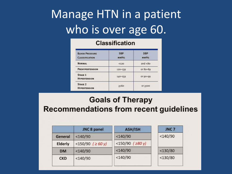
Manage HTN in a patient who is over age 60.

Question 4.
• Answer: B: Plasma aldosterone-plasma renin activity ratio
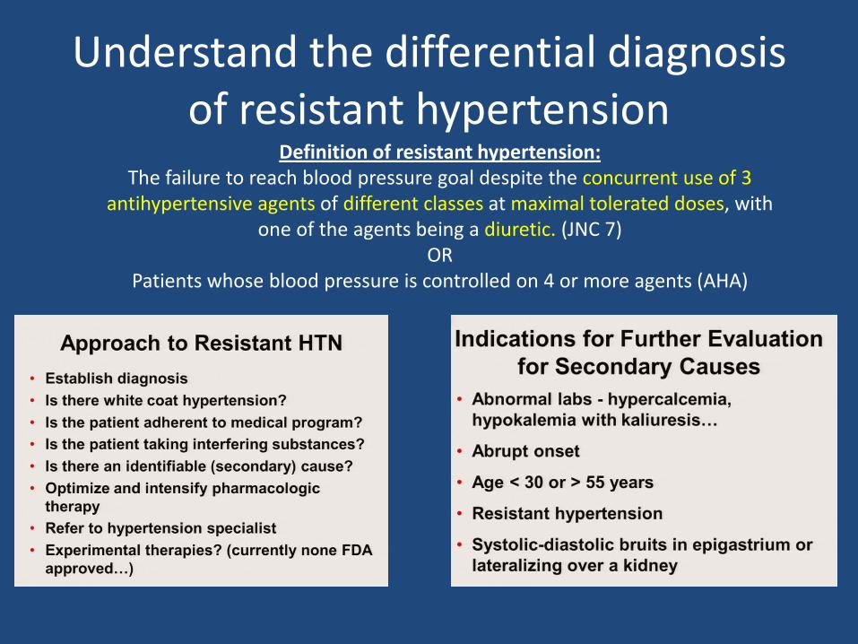
Understand the differential diagnosis of resistant hypertension
Definition of resistant hypertension:The failure to reach blood pressure goal despite the concurrent use of 3
antihypertensive agents of different classes at maximal tolerated doses, with one of the agents being a diuretic. (JNC 7)
OR Patients whose blood pressure is controlled on 4 or more agents (AHA)

Primary Hyperaldosteronism:60%+ of patients have NORMAL K level
• ALDOSTERONE HIGH• RENIN LOW• AR RATIO >25 is
suggestive BUT NOT diagnostic
• Metabolic alkalosis and hypokalemia MAY OR MAY NOT be present
• 8 AM draw• OFF spironolactone or
eplerenone for 6 weeks• Possibly off ACEI
• NO CONFIRMATION test needed:
– Spontaneous hypokalemia– Undetectable renin level– Aldosterone >30 ng/dL
Question 5.
• Answer: D; Sevelamer


Treat hyperphosphatemia in a patient with CKD
KDIGO guidelines 20161. Phosphorus-restricted diet2. Phosphorus binders
– Calcium-containing P04 binders:• Calcium carbonate (Tums)• Calcium acetate (PhosLo)
• Appropriate for all hypo-calcemic patients and normocalcemic patients without
vascular calcification or adynamic bone disease
– Noncalcium-containing P04 binders:– Sevelamer (Renagel, Renvela)
– Lanthanum (FosRenal)– Appropriate for hypercalcemic patients and
patients with vascular calcification, adynamic bone disease, or patients on 1,25-
OH vitamin D supplements


Question 6.
• Answer: C: Measure urine chloride level

Evaluate a Patient withHypokalemic Metabolic Alkalosis
URINE CHLORIDE < 15 (LOW)=Chloride Responsive (90%)
• Low effective circulating volume• Volume depleted (orthostatic,
hypotensive)• NORMAL increase in renin,
angiotensin, aldosterone• Urine chloride LOW <15• Cannot replace K until volume is
replaced (GIVE sodium CHLORIDE)
URINE CHLORIDE >15 (HIGH) =Chloride Unresponsive (10%)
• Hypertensive• Hypervolemic• ABNORMAL increase in aldosterone
(Primary hyperaldo) or renin (Secondary hyperaldo)
• Urine chloride HIGH >15


Question 7.
• Answer: E, Estimated GFR of < 15mL/min

Know how to appropriately dosing medications in acute kidney injury
Estimation of Creatinine Clearance1. Crockcoft-gault2. MDRD3. CKD-EPI
All equations require steady-state, stable serum creatinine to be valid.

Question 8.
• Answer: B: Hypokalemic distal (type 1) renal tubular acidosis

Diagnose hypokalemic distal (type 1) renal tubular acidosis
Type 1 RTA (distal)
Type 2 RTA(proximal)
Type 4 RTA(distal)
Chloride ↑ ↑ ↑
Bicarbonate ↓ ↓ ↓
Potassium ↓ NL ↑
Urine pH High ( > 5.5) Low (<5.5) except with bicarb load
Low (<5.5)
Etiologies Chronic hepatitisAmphotericin BTolueneLithiumSjogren’s; SLE
Multiple MyelomaMetal poisoning
Acetazolamide
Diabetes mellitusSickle cellSpironolactone
Associations Nephrolithiasisdue to
hypercalcuria

Question 9.
• Answer: B: Chlorthalidone

Manage hypercalciuria in a patient with nephrolithiasis
• Hypercalciuria:– >250 mg/24 hours
WOMEN– >300 mg/24 hours MEN– > 200 mg/liter
• WORSENED by:– High sodium diet– High animal protein diet– Loop diuretics
• DO NOT advise a calcium restricted diet, as this increases GI absorption of oxalate, increasing oxaluria
• DO ADVISE:– Thiazide diuretic– Fluids > 2 liters/day– Low purine diet– Low sodium diet

Question 10.
• Answer: C; Oral sodium bicarbonate

Treating metabolic acidosis in CKDProblem List:
• 58 year-old woman• HTN, CKD stage 3b d/t analgesics• Amlodipine• Low serum bicarb, pH 7.36 , AG of 12• Mild asymptomatic NAGMA
Non-AG metabolic acidosis:• Renal (RTAs) – impaired acid excreation• GI (diarrhea) – loss of bicarb• Medications: Lithium, TPN
Our patients history suggests Type IV RTA1. H/o analgesic nephropathy2. Normal AG MA3. Hyperkalemia
Treatment goal: • Serum bicarb goal 23-29 which
reduces risk of CKD progression• Bicarbonate administration will help
correct the acidosis.
• Dose: 0.5 to 1mEq/kg/day
Question 11.
• Answer: A; AL amyloidosis
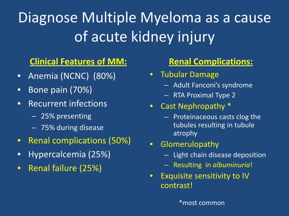
Diagnose Multiple Myeloma as a cause of acute kidney injury
Clinical Features of MM:• Anemia (NCNC) (80%)• Bone pain (70%)• Recurrent infections
– 25% presenting– 75% during disease
• Renal complications (50%)• Hypercalcemia (25%)• Renal failure (25%)
Renal Complications:• Tubular Damage
– Adult Fanconi’s syndrome– RTA Proximal Type 2
• Cast Nephropathy *– Proteinaceous casts clog the
tubules resulting in tubule atrophy
• Glomerulopathy– Light chain disease deposition– Resulting in albuminuria!
• Exquisite sensitivity to IV contrast!
*most common

Amyloidosis: In an apple-green nutshellPrimary AL Amyloidosis• Monoclonal protein
related to plasma cell dyscrasia
• SPEP/IFE, UPEP/IFE, free light chains
• Bone marrow positive for plasma cell proliferation
• Monoclonal proteins infiltrate tissues; heart, kidney, GI tract, pleura, nerves (peripheral and autonomic)
• Kidney involvement may result in nephrotic proteinuria (albumin)
Secondary AA Amyloidosis• Polyclonal protein
related to inflammatory state; rheumatologic or infectious
• Juvenile RA, anklyosingspondylitis, psoriatic arthritis, IBD
• Familial Mediterranean Fever (60% of cases in Turkey)
• NEGATIVE SPEP/IFE etcfor monoclonal proteins
• Kidney disease may result in nephroticproteinurea (albumin)
Hereditary ATTR Amyloidosis• Mutation in the
transthyretin gene making abnormal proteins that infiltrate organs
• Cardiac and neurologic involvement more common than renal involvement
• NEGATIVE SPEP/IFE etc for monoclonal proteins
• Rare to have kidney involvement

Question 12.
• Answer: A: Interstitial nephritis

Diagnose AcuteInterstitial Nephritis
Clinical Presentation:• Fever, rash, eosinophilia,
and elevated creatinine (10%)
• UA: WBC, WBC casts• NEGATIVE CULTURE (sterile
pyuria)• Eosinophiluria (sensitive but
not specific)• < 2 gm/ 24 hr proteinuria
Causes: Drugs, Infections, Illnesses
– Antibiotics (B-lactam, cephalosporins, quinolones, Bactrim, rifampin)
– NSAIDS * (nephroticproteinuria) with minimal change disease on biopsy
– Thiazides– Proton Pump Inhibitors– Phenytoin– Allopurinol– 5-aminosalicylates

Question 13.
• Answer: D; Continued current therapy

Diagnose and manage infection-related glomerulonephritis
• 24 y/o man IVDU• H/o previous staph endocarditis
with prolonged antibiotics• Recent dx of MRSA endocarditis on
IV vanco• Worsening renal function
Laboratory StudiesCreatinine 2.8Active UA with protein, RBCs and RBC castsLow C3, nL C4Negative cryoglobulinsRenal U/S with Doppler: normal kidneys
DDx:• Acute interstitial nephritis• Drug induced nephrotoxicity• Infection related GN• Septic emboli
Clinical picture and labs suggest infection related GN.
Treatment is to continue treating the infection.

Manage Post-Infectious Glomerulonephritis (PIGN)
• MANY bacteria, viruses and parasites can cause PIGN
• Most common nephritogenicstrains of strep and staph
• Rapid onset of hypertension, oliguria, erythrocyte casts, and edema, LOW C3
Pathophysiology:• Immune complex-mediated
disease• Complexes deposit in
glomerulous and active complement, recruiting inflammatory cells and causing proliferative GN
• Common in non-strep infections (antigen is the infection)
Why isn’t Vancomycin the culprit?Drug induced tubular toxicity occurs 7 -10 days after Abxinitiation and there is usually no cells in urine sediment.

Question 14.
• Answer: B; Ethylene glycol poisoning

AGMA Toxic alcohol NAGMA
Methanol Isopropyl Alcohol
Ethylene Glycol
Sources: rubbing alcohol/hand sanitizerEffects: hemorrhagic gastritis, fruity breathLabs: Osmolar gap ONLY (no anion gap), ketones
Sources: windshield wiper fluid, moonshine, perfumesEffects: formic acid causes vision changes (blindness), “snowfield vision” (toxic to mitochondrial of retina and optic nerve), basal ganglia effects and pancreatitisLabs: Anion gap + Osmolar gap
Sources: antifreezeEffects: oxalic acid causes cardiac and neuro effectsLabs: Anion gap + Osmolar gap, calcium oxalate crystals deposit in renal tubules, elevated creatinine, false lactic acid elevation from blood gas analyzer (serum lactate is normal)
Diagnose ethylene glycol poisoning

Ethylene Glycol PoisoningWork up:
• Check blood gas and electrolytes• If +AGMA, check ketones, lactic
acid and calculate osmolar gap
Treatment:• Fomepizole (blocks alcohol
dehydrogenase and autometabolizes)
• Ethanol administration is complicated by inebriation, erratic metabolism and hypoglycemia
• HD removes alcohol and toxic metabolites
• Dialyze if high levels of toxic alcohols, severe AGMA, electrolyte disturbances
Osmolar gap = measured – calculated osmolarity
Calculated osmolarity= (Na x 2) + (BUN/2.8) + (Glucose/18) = 287, Measured= 314 - 287 = 27 gap.
>10 suggest the presence of an osmotically active substance>20 is usually due to toxic alcohol ingestion, >50 for sure!
Question 15.
• Answer: A: Cryoglobulinemia associated with Hepatitis C

Diagnose Hepatitis C virus-associated membranoproliferative glomerulonephritis due to
cryoglobulins• Occurs in up to
20% of patients with chronic Hepatitis C
• Presents as membranoproliferative gnitisor mixed cryoglobulinemia(skin , kidney, and nerve involvement)
• 1/3 relapsing dz with progression to ESRD
• Low complement (C4)• + Rheumatoid factor• TREAT Hep C virus
EXTRA-HEPATIC MANIFESTATIONS OF HEPATITIS C INFECTION:
1. Membranoproliferative GNitis2. Mixed Essential Cryoglobulinemia3. Lichen Planus4. Porphyria Cutanea Tarda

Question 16.
• Answer: C; Hemoglobinuria

Evaluation of Red UrineHemoglobinuria due to Intravascular hemolysis:
• Microangiopathic hemolytic anemia
• Transfusion reactions• Mechanical shear from
valves• Infections
– (Clostridial sepsis, Malaria)• Paroxysmal Nocturnal
Hemoglobinuria (PNH)• Hypotonic fluid infusions• Exposure to compounds
with high oxidant potential (copper poisoning, Wilson’s disease)
Question 17.
• Answer:B; HIV antibody test

Diagnose Focal Segmental Glomerulosclerosis: the most common renal cause of nephrotic syndrome in US (36-80%)
Primary Disease: Podocyte damage similar to minimal change dz
Secondary Disease Associations:• Morbid obesity• Heroin use• HIV• Vesicoureteral reflux
Risk factors for progression to ESRD:• Black race• >2 gm/24 hr proteinuria• Low GFR• BMI>27• Hypertension
Clinical Manifestations:• Acute onset of nephrotic syndrome
with hematuria, renal failure and hypertension in primary disease;
• Asymptomatic subnephrotic to nephrotic proteinuria in secondary disease
Management:• Immunosuppression with steroids
or calcineurin inhibitors in primary disease
• ACEI +/- ARB in primary and secondary disease
• BP goal < 125/75 mm Hg

Question 18.
• Answer: B: Membranous nephropathy

Diagnose Membranous Nephropathy: the second most common renal cause of nephrotic syndrome
Primary disease: • Immune complexes of IgG react with
antigens in the outer aspect of GBM• Antibody to PLA2
Secondary disease associations:• Malignancy• Hep B and C• NSAIDS
Risk factors for progression to ESRD:• Male gender• Age>50• Persistent proteinuria > 4g/24 h over 6
months• Declining GFR
Clinical manifestations:• Nephrotic syndrome with
preserved GFR• Renal vein thrombosis• Hypercoagulability
Management:• ACE and/or ARB• BP goal < 125/75 mm Hg• Persistent proteinuria > 4g/24 hr• Cyclophosphamide, corticosteroids,
or calcineurin inhibitor with possible need for rituxan

Question 19.
• Answer: B; Hyperglycemia

Patient has hypertonic hyponatremia!Correction factor: 1.6mEq/L decrease in the serum sodium level per
every 100mg/dL of plasma glucose over 100mg/dL. Treatment: fluids and treat hyperglycemia.

Algorithm for hypotonic hyponatremia

Manage asymptomatic hypotonic hyponatremia

Question 20.
• Answer: B; Substitute labetalol for lisinopril

Manage HTN in a woman of childbearing age
Normal physiology in pregnancy
• Blood pressure lowers• Plasma volume increases• GFR increases• Renal pelvis, calices, and
ureters dilate• Increased risk for pyelo• Hyperventilation causes
resp alkalosis with increased renal bicarb excretion Pearls in Pregnancy “20 weeks”:
ACEI, ARB, and Renin-Inhibitors are CONTRAINDICATED in pregnancy!!