adenovirus infections in immunocompetent and ... · adenovirus infections in immunocompetent and...
TRANSCRIPT
Adenovirus Infections in Immunocompetent andImmunocompromised Patients
Thomas Lion
Children’s Cancer Research Institute and Medical University Vienna, Vienna, Austria
SUMMARY . . . . . . . . . . . . . . . . . . . . . . . . . . . . . . . . . . . . . . . . . . . . . . . . . . . . . . . . . . . . . . . . . . . . . . . . . . . . . . . . . . . . . . . . . . . . . . . . . . . . . . . . . . . . . . . . . . . . . . . . . . . . . . . . . . . . . . . . . . . . . . . . . .441INTRODUCTION . . . . . . . . . . . . . . . . . . . . . . . . . . . . . . . . . . . . . . . . . . . . . . . . . . . . . . . . . . . . . . . . . . . . . . . . . . . . . . . . . . . . . . . . . . . . . . . . . . . . . . . . . . . . . . . . . . . . . . . . . . . . . . . . . . . . . . . . . . . .441NOMENCLATURE AND TYPING OF HUMAN ADENOVIRUSES . . . . . . . . . . . . . . . . . . . . . . . . . . . . . . . . . . . . . . . . . . . . . . . . . . . . . . . . . . . . . . . . . . . . . . . . . . . . . . . . . . . . . . . . . . . .442
General Description, Structural Proteins, and Genomic Structure . . . . . . . . . . . . . . . . . . . . . . . . . . . . . . . . . . . . . . . . . . . . . . . . . . . . . . . . . . . . . . . . . . . . . . . . . . . . . . . . . . . . . . . .442Phylogeny and Human Adenoviral Species . . . . . . . . . . . . . . . . . . . . . . . . . . . . . . . . . . . . . . . . . . . . . . . . . . . . . . . . . . . . . . . . . . . . . . . . . . . . . . . . . . . . . . . . . . . . . . . . . . . . . . . . . . . . . .442(Sero)typing of Human Adenoviruses . . . . . . . . . . . . . . . . . . . . . . . . . . . . . . . . . . . . . . . . . . . . . . . . . . . . . . . . . . . . . . . . . . . . . . . . . . . . . . . . . . . . . . . . . . . . . . . . . . . . . . . . . . . . . . . . . . . .442Present Controversies in HAdV Typing . . . . . . . . . . . . . . . . . . . . . . . . . . . . . . . . . . . . . . . . . . . . . . . . . . . . . . . . . . . . . . . . . . . . . . . . . . . . . . . . . . . . . . . . . . . . . . . . . . . . . . . . . . . . . . . . . . .443Current Status of HAdV Types and Evolution of Human Adenoviruses. . . . . . . . . . . . . . . . . . . . . . . . . . . . . . . . . . . . . . . . . . . . . . . . . . . . . . . . . . . . . . . . . . . . . . . . . . . . . . . . . . .443
PATHOGENESIS AND IMMUNITY . . . . . . . . . . . . . . . . . . . . . . . . . . . . . . . . . . . . . . . . . . . . . . . . . . . . . . . . . . . . . . . . . . . . . . . . . . . . . . . . . . . . . . . . . . . . . . . . . . . . . . . . . . . . . . . . . . . . . . . . . .444Prevalence of HAdV Species and Types . . . . . . . . . . . . . . . . . . . . . . . . . . . . . . . . . . . . . . . . . . . . . . . . . . . . . . . . . . . . . . . . . . . . . . . . . . . . . . . . . . . . . . . . . . . . . . . . . . . . . . . . . . . . . . . . . .444Transmission . . . . . . . . . . . . . . . . . . . . . . . . . . . . . . . . . . . . . . . . . . . . . . . . . . . . . . . . . . . . . . . . . . . . . . . . . . . . . . . . . . . . . . . . . . . . . . . . . . . . . . . . . . . . . . . . . . . . . . . . . . . . . . . . . . . . . . . . . . . . .444Tissue Tropism. . . . . . . . . . . . . . . . . . . . . . . . . . . . . . . . . . . . . . . . . . . . . . . . . . . . . . . . . . . . . . . . . . . . . . . . . . . . . . . . . . . . . . . . . . . . . . . . . . . . . . . . . . . . . . . . . . . . . . . . . . . . . . . . . . . . . . . . . . . .444Primary Infection and Persistence . . . . . . . . . . . . . . . . . . . . . . . . . . . . . . . . . . . . . . . . . . . . . . . . . . . . . . . . . . . . . . . . . . . . . . . . . . . . . . . . . . . . . . . . . . . . . . . . . . . . . . . . . . . . . . . . . . . . . . . .444Immune Responses to Adenoviral Infection. . . . . . . . . . . . . . . . . . . . . . . . . . . . . . . . . . . . . . . . . . . . . . . . . . . . . . . . . . . . . . . . . . . . . . . . . . . . . . . . . . . . . . . . . . . . . . . . . . . . . . . . . . . . . .445De Novo Infection and Viral Reactivation in Transplant Recipients . . . . . . . . . . . . . . . . . . . . . . . . . . . . . . . . . . . . . . . . . . . . . . . . . . . . . . . . . . . . . . . . . . . . . . . . . . . . . . . . . . . . . . .445
RISK FACTORS, INCIDENCE, AND CLINICAL MANIFESTATIONS OF INVASIVE HAdV INFECTION IN IMMUNOCOMPROMISED PATIENTS . . . . . . . . . . . . . .445Risk Factors . . . . . . . . . . . . . . . . . . . . . . . . . . . . . . . . . . . . . . . . . . . . . . . . . . . . . . . . . . . . . . . . . . . . . . . . . . . . . . . . . . . . . . . . . . . . . . . . . . . . . . . . . . . . . . . . . . . . . . . . . . . . . . . . . . . . . . . . . . . . . . .445Incidence of HAdV Infections in Immunocompromised Adult and Pediatric Patients . . . . . . . . . . . . . . . . . . . . . . . . . . . . . . . . . . . . . . . . . . . . . . . . . . . . . . . . . . . . . . . . . . .446Definitions of Adenoviral Infection and Disease . . . . . . . . . . . . . . . . . . . . . . . . . . . . . . . . . . . . . . . . . . . . . . . . . . . . . . . . . . . . . . . . . . . . . . . . . . . . . . . . . . . . . . . . . . . . . . . . . . . . . . . . . .446Clinical Presentations and Outcomes of HAdV Infections in Solid Organ and Allogeneic Stem Cell Transplant Recipients . . . . . . . . . . . . . . . . . . . . . . . . . . . . . .446Predictive Value of Viremia and HAdV Proliferation in the Gastrointestinal Tract . . . . . . . . . . . . . . . . . . . . . . . . . . . . . . . . . . . . . . . . . . . . . . . . . . . . . . . . . . . . . . . . . . . . . . . . .447
DIAGNOSIS AND MONITORING . . . . . . . . . . . . . . . . . . . . . . . . . . . . . . . . . . . . . . . . . . . . . . . . . . . . . . . . . . . . . . . . . . . . . . . . . . . . . . . . . . . . . . . . . . . . . . . . . . . . . . . . . . . . . . . . . . . . . . . . . . .447Diagnostic Screening. . . . . . . . . . . . . . . . . . . . . . . . . . . . . . . . . . . . . . . . . . . . . . . . . . . . . . . . . . . . . . . . . . . . . . . . . . . . . . . . . . . . . . . . . . . . . . . . . . . . . . . . . . . . . . . . . . . . . . . . . . . . . . . . . . . . .447Relevance of HAdV Detection and Quantification at Specific Sites . . . . . . . . . . . . . . . . . . . . . . . . . . . . . . . . . . . . . . . . . . . . . . . . . . . . . . . . . . . . . . . . . . . . . . . . . . . . . . . . . . . . . . .448Adenovirus Typing . . . . . . . . . . . . . . . . . . . . . . . . . . . . . . . . . . . . . . . . . . . . . . . . . . . . . . . . . . . . . . . . . . . . . . . . . . . . . . . . . . . . . . . . . . . . . . . . . . . . . . . . . . . . . . . . . . . . . . . . . . . . . . . . . . . . . . .449Diagnostic Recommendations as a Basis for Preemptive Treatment . . . . . . . . . . . . . . . . . . . . . . . . . . . . . . . . . . . . . . . . . . . . . . . . . . . . . . . . . . . . . . . . . . . . . . . . . . . . . . . . . . . . .449
CURRENT TREATMENT MODALITIES . . . . . . . . . . . . . . . . . . . . . . . . . . . . . . . . . . . . . . . . . . . . . . . . . . . . . . . . . . . . . . . . . . . . . . . . . . . . . . . . . . . . . . . . . . . . . . . . . . . . . . . . . . . . . . . . . . . . . . .452Antiviral Drugs. . . . . . . . . . . . . . . . . . . . . . . . . . . . . . . . . . . . . . . . . . . . . . . . . . . . . . . . . . . . . . . . . . . . . . . . . . . . . . . . . . . . . . . . . . . . . . . . . . . . . . . . . . . . . . . . . . . . . . . . . . . . . . . . . . . . . . . . . . . .452Immunotherapy . . . . . . . . . . . . . . . . . . . . . . . . . . . . . . . . . . . . . . . . . . . . . . . . . . . . . . . . . . . . . . . . . . . . . . . . . . . . . . . . . . . . . . . . . . . . . . . . . . . . . . . . . . . . . . . . . . . . . . . . . . . . . . . . . . . . . . . . . .452
SUMMARY AND PERSPECTIVES . . . . . . . . . . . . . . . . . . . . . . . . . . . . . . . . . . . . . . . . . . . . . . . . . . . . . . . . . . . . . . . . . . . . . . . . . . . . . . . . . . . . . . . . . . . . . . . . . . . . . . . . . . . . . . . . . . . . . . . . . . . .453ACKNOWLEDGMENT . . . . . . . . . . . . . . . . . . . . . . . . . . . . . . . . . . . . . . . . . . . . . . . . . . . . . . . . . . . . . . . . . . . . . . . . . . . . . . . . . . . . . . . . . . . . . . . . . . . . . . . . . . . . . . . . . . . . . . . . . . . . . . . . . . . . . . .454REFERENCES . . . . . . . . . . . . . . . . . . . . . . . . . . . . . . . . . . . . . . . . . . . . . . . . . . . . . . . . . . . . . . . . . . . . . . . . . . . . . . . . . . . . . . . . . . . . . . . . . . . . . . . . . . . . . . . . . . . . . . . . . . . . . . . . . . . . . . . . . . . . . . . .454AUTHOR BIO . . . . . . . . . . . . . . . . . . . . . . . . . . . . . . . . . . . . . . . . . . . . . . . . . . . . . . . . . . . . . . . . . . . . . . . . . . . . . . . . . . . . . . . . . . . . . . . . . . . . . . . . . . . . . . . . . . . . . . . . . . . . . . . . . . . . . . . . . . . . . . . .462
SUMMARY
Human adenoviruses (HAdVs) are an important cause of infec-tions in both immunocompetent and immunocompromised in-dividuals, and they continue to provide clinical challenges per-taining to diagnostics and treatment. The growing number ofHAdV types identified by genomic analysis, as well as the im-proved understanding of the sites of viral persistence and reacti-vation, requires continuous adaptions of diagnostic approaches tofacilitate timely detection and monitoring of HAdV infections. Inview of the clinical relevance of life-threatening HAdV diseases inthe immunocompromised setting, there is an urgent need forhighly effective treatment modalities lacking major side effects.The present review summarizes the recent progress in the under-standing and management of HAdV infections.
INTRODUCTION
Since their first isolation from adenoid tissue over 60 years ago(1), human adenoviruses (HAdVs) (adénos, gland) have pro-
vided continuous challenges in a variety of clinical settings. In
addition to their well-established role as infectious agents, adeno-viral genomes were also shown to contain potent oncogenes, andthe ability of certain types of the virus to induce tumor growth hasbeen demonstrated in different mammalian animal models (2–4).Despite a number of studies addressing the possible role of HAdVsin human malignant disease, their putative oncogenicity in hu-mans has remained enigmatic (5–7). Investigations of adenovirusbiology have led to Nobel Prize-winning discoveries in mRNAsplicing and to important progress in the understanding of anti-gen presentation to T cells (8). Moreover, the ability of adenovi-ruses to infect many cell types facilitated their exploitation as vec-tors for gene delivery to generate new tools for innovativetreatments of important diseases, such as cancer and cardiovascu-
Address correspondence to [email protected].
Copyright © 2014, American Society for Microbiology. All Rights Reserved.
doi:10.1128/CMR.00116-13
July 2014 Volume 27 Number 3 Clinical Microbiology Reviews p. 441– 462 cmr.asm.org 441
on May 22, 2020 by guest
http://cmr.asm
.org/D
ownloaded from
lar disorders (9–13). Hence, adenoviruses are highly versatile or-ganisms with a broad spectrum of clinical roles and applications.
HAdVs were initially isolated mainly from military recruitswith acute febrile respiratory disease and were subsequently asso-ciated with a number of clinical manifestations, including gastro-enteritis, hepatitis, keratoconjunctivitis, meningoencephalitis,cystitis, upper and lower respiratory tract infections, and myocar-ditis, but also with noninflammatory conditions, such as obesity(14–21). HAdV infections are readily transmittable and, in someinstances, highly contagious. Although the clinical courses areusually mild and self-limiting, infections may cause local out-breaks with severe courses, occasionally leading to a lethal out-come even in immunocompetent individuals (22–26). However,adenoviruses play a particularly important role in patients withstrongly impaired immune responses, in whom viral disease isassociated with high morbidity and mortality, and infections inthis setting are an important focus of the present review. Progressin molecular detection methods has rendered the detection, typ-ing, and monitoring of adenoviral infections readily applicable inthe clinical routine, and the tools required for risk assessment andtimely diagnosis of invasive infection are available. However, ef-fective treatment of HAdV-related diseases in immunocompro-mised patients still poses great challenges.
NOMENCLATURE AND TYPING OF HUMAN ADENOVIRUSES
General Description, Structural Proteins, and GenomicStructure
HAdVs are nonenveloped viruses with a diameter of 70 to 100 nm.The external protein shell of the virus is icosahedral (eikosí, twen-ty; hédra, seat), with 20 triangular faces, 30 edges, and 12 vertices,and this symmetry is made up in large parts by the major virusprotein, hexon. The viral capsid is composed of 252 capsomeres(capsa, box; méros, portion), including 240 hexons and 12 pen-tons. Five penton base proteins form individual capsomeres at the12 vertices, where each capsomere supports a trimeric fiber pro-tein of variable size projecting from the vertex of the capsid. Thefiber protein contains three structural domains: the tail, the shaft,and the knob. The tail domain is the binding site for the pentonbase. The shaft domain shows various lengths between HAdVtypes, resulting in different flexibilities of the fiber and differencesin the interaction with host cell integrins. The fiber knob domain,located at the distal, C-terminal end of the protein, binds the virusto the primary host cell receptor (27–30). Most adenoviruses bindto the classical adenovirus receptor CAR (coxsackie adenovirusreceptor), but some use the membrane cofactor CD46 and/or thecell adhesion protein desmoglein 2 (DSG2) as an attachment re-ceptor (27–29).
Minor capsid proteins IIIa, VI, VIII, and IX confer stability tothe hexon shell and the entire virion and play roles in events oc-curring after internalization, such as endosome penetration, tran-scriptional activation, and nuclear reorganization (protein IX)(31, 32). Additional proteins in the core of the capsid (proteins V,VII, and X and terminal protein) interact with the viral DNA, e.g.,by facilitating transportation into the nucleus of the infected cell(protein V) (30, 33).
HAdVs are double-stranded, linear DNA viruses displaying ge-nome sizes ranging from 34 to more than 37 kb and carrying some40 genes (33, 34). All HAdVs share a similar organization of thegenome, which is divided into early, intermediate, and late regions
corresponding to the infectious cycle of the virus and reflecting thetranscription patterns. The early region of the genome includesfour transcript families, termed E1 to E4, which are required forviral replication. The E3 transcription unit, which is highly diver-gent between HAdV species, also encodes proteins modulating thehost immune response. The intermediate genes are represented bytwo transcripts, termed IX and IVa2, and the late region containsfive transcript families, referred to as L1 to L5, which are involvedin the production of mature virions. Moreover, the genomes dis-play inverted terminal repeat (ITR) regions at the 3= and 5= endsthat contain conserved sequence motifs and serve as origins ofviral replication. Furthermore, depending on the HAdV type, thegenomes display one or two noncoding virus-associated (VA)RNA genes involved in translational regulation, and one of them(VA RNAI) can act as a microRNA (miRNA) (30, 35).
Phylogeny and Human Adenoviral Species
Together with other mammalian adenoviruses, HAdVs are classi-fied into the genus Mastadenovirus (mastós, breast) and are furtherparsed into seven species, termed A to G, with further subdivisionof species B into subspecies B1 and B2 (33, 36–40). Species desig-nation depends on several of the following characteristics: phylo-genetic distance (�5 to 15%, based primarily on distance matrixanalysis of the DNA polymerase amino acid sequence), genomeorganization (characteristically in the E3 region), nucleotide com-position (G�C%), oncogenicity in rodents, host range, cross-neutralization, ability to recombine, number of VA RNA genes,and hemagglutination (33). More than 30 simian adenoviruses(SAdVs) display sequence identitites to their human counterpartsto such an extent that they have also been included in the taxon-omy of human adenoviruses, within species B, C, E, and G (33).Previously, HAdVs were identified, characterized, and classifiedby serum neutralization (SN) and hemagglutination inhibition(HI) assays, but more recently, genomic and bioinformatic anal-yses of the entire viral genome have superseded serological meth-ods for the typing of novel viruses (36–42). The viruses belongingto individual HAdV species display high similarity to each other atthe nucleotide level and do not commonly recombine with mem-bers of other species. The grouping into different species reflects,in part, the general cell tropism of the viruses and the resultingdiseases and symptoms. Examples of common associations of in-dividual HAdV species with infections at specific locations includegastroenteritis (HAdV-F and -G), pneumonia (HAdV-B, -C, and -E),hepatitis (HAdV-C), meningoencephalitis (HAdV-A, -B, and -D),cystitis (HAdV-B), and keratoconjunctivitis (HAdV-B and -D), butother HAdV species may also occur at the indicated sites of infection(30, 36, 43).
(Sero)typing of Human Adenoviruses
HAdV subtyping below the species level by SN and HI assays led tothe identification of 51 serotypes (Table 1). The hypervariableloops (L1 and L2) of the hexon protein form the SN epitope andare the main determinants of serologic reactivity, while the fiberprotein is responsible for HI typing and is a major determinant oftropism. The combination of the SN and HI tests facilitates morecomplete virus identification than that with either method alone(30).
The first HAdV identified on the basis of genetic analysis wasalso classified as a novel species (HAdV-G) and received the chro-nological number 52 (36). Because all subsequently identified
Lion
442 cmr.asm.org Clinical Microbiology Reviews
on May 22, 2020 by guest
http://cmr.asm
.org/D
ownloaded from
novel HAdVs were detected and characterized using computa-tional analyses of genomic data (Table 1), it has been agreed toreplace the term “serotype” by “type,” and criteria for the assign-ment of new types have been established (41, 42).
Present Controversies in HAdV Typing
There seems to be broad agreement that DNA sequence informa-tion should be exploited for HAdV typing, but differing views onthe required extent of sequencing and the future role of serologicalmethods have been presented. Some researchers take the stancethat type definition should be based on the sequence of the majorcapsid protein, hexon, as the primary identifier, and that onlystrains carrying novel hexon genes should be considered “candi-date new types,” because the hexon contains the major neutraliz-ing epitope frequently targeted in molecular diagnosis (42). Thedesignations of natural or engineered intertypic recombinantHAdVs should include the identity of the hexon gene (H) and alsothat of the fiber gene (F). These designations should replace thoseresulting from SN and HI assays, because published data supporta strong correlation between identities established by sequenceanalyses of hexon and fiber and those determined by SN and HIassays, respectively (42). The extent of sequences required for con-sideration of a new candidate type is under evaluation but shouldnot exceed the complete hexon and fiber knob or complete fibergene sequence. The data should be integrated with the establishedimmunotyping scheme, and serology should continue to be usedin order to characterize the antigenic phenotypes. Some authorshave expressed concern that the designation of new types by se-quential numbers according to the order in which novel genomesequences are reported will likely lead to a large increase in theirnumber (42).
In contrast, a proposal published by members of the adenovirusresearch community states that HAdVs should be identified, char-acterized, typed, and consecutively numbered on the basis of com-plete genome sequence analyses rather than by serological meth-ods (41). Due to the power of novel genetic and bioinformatictools, a paradigm shift in recognizing and naming HAdVs isneeded. Recombination is an accepted feature of HAdV evolution,and recombinants will be classified as novel types provided thatthere are sufficient genomic, biological, or pathogenic differencesfrom related types.
Serology can suggest possible recombinant viruses as intertypicstrains by revealing conflicting results between SN and HI assaysthat are indicative of two different HAdV types. However, thesemethods cannot completely characterize the newly identified vi-
rus. Serologic determinants represent less than 5 to 6% of the totalviral genome and thus can scarcely be regarded as adequate for fullcharacterization of new HAdVs in the era of genomics (30). Re-cent data suggest that serologic and genomic analyses do not al-ways correlate, and serotyping alone can provide misleading re-sults (44, 45). Nevertheless, SN and HI testing should continue tobe used as an additional criterion in order to comply with thecurrent definitions of the International Committee on Taxonomyof Viruses (ICTV) (41). Recently, the full-genome sequencing ofevery prototype HAdV strain was completed, including a typingalgorithm that includes the sequences supporting serological fea-tures (8). It is therefore conceivable that it will be possible toimpute serologic test results by computing genomic sequencedata, thus possibly obviating and replacing SN and HI assays.
Current Status of HAdV Types and Evolution of HumanAdenoviruses
To date, 67 HAdV types have been published, and additional typesare already in the pipeline. As outlined in Table 1, HAdV types 1 to51 were characterized by serotyping, while the remaining types,identified since 2007, were detected and described by genomic andbioinformatic analyses (36, 46).
Homologous recombination (HR) and mutation are importantevolutionary processes driving genetic variation within HAdV ge-nomes (34). They are favored by the immune pressure of the hostand by environmental bottlenecks. In HAdV species B, mutationsseem to play a more important role, whereas among the largestHAdV species, species D, homologous recombination is the pre-dominant mechanism contributing to genomic diversity (34). HRof tumorigenic adenoviruses in vitro was already documented inthe 1970s (47, 48) and was shown to occur predominantly be-tween HAdV types belonging to the same species, within regionsof high sequence homology (30). The recent availability of whole-genome sequencing and bioinformatics has permitted the de-scription of recombination events within genomes of HAdV spe-cies A, B, and D, particularly within the penton base, hexon, andfiber genes (37, 49–51). The requirements for recombinationevents appear to include coinfection of individual cells with atleast two different adenoviruses displaying very similar nucleotidesequences at the recombination hot spots in the genome, as well aslong-term viral persistence in the host (52, 53). The emergence ofnew HAdV-D types in patients with AIDS indicates a role of mul-tiple persisting viruses under impaired immune surveillance (34,54). HAdV-D genomes seem to recombine more frequently thanother human adenoviral species, and several of the currently morethan 40 HAdV-D types apparently emerged via recombinationbetween hexon and fiber coding regions (34). The majority ofnovel HAdV types identified by genomic analysis belong to speciesD, and they were shown to include sequences derived from mul-tiple other types from the same species. For example, HAdV-D53resulted from recombination in the penton, hexon, and fiber re-gions of HAdV-D22, -D37, and -D8, respectively. Similarly,HAdV-D67 was identified as a recombinant between HAdV-D9,-D25, -D26, -D33, and -D46 (46, 55). Recent data provide evi-dence for the occurrence of recombination between differentHAdV species, and even between HAdVs and SAdVs (56, 57).Computational analysis of HAdV-E4, the only representative ofspecies E, indicated that this virus is of zoonotic origin and evolvedthrough two interspecies recombination events with lateral partialgene transfer. HAdV-E4 contains 97% of a SAdV-E26-like ge-
TABLE 1 Current spectrum of published human adenovirusesa
Species Types (serotypes/genotypes)
A 12, 18, 31, 61B 3, 7, 11, 14, 16, 21, 34, 35, 50, 55, 66C 1, 2, 5, 6, 57D 8–10, 13, 15, 17, 19, 20, 22–30, 32, 33, 36–39,
42–49, 51, 53, 54, 56, 58-60, 63-67E 4F 40, 41G 52a The HAdV species (A to G) and types (1 to 67) belonging to individual species areindicated. While types 1 to 51 were identified by serotyping, all subsequently identifiedtypes, indicated in italics (types 52 to 67), were identified by genomic sequencing andcomputational analysis.
Adenovirus Infections
July 2014 Volume 27 Number 3 cmr.asm.org 443
on May 22, 2020 by guest
http://cmr.asm
.org/D
ownloaded from
nome chassis with a hexon containing the L1 and L2 regions froma HAdV-B16-like virus, which may provide compatibility with thenew host (57). Adaptation of the virus to the new host could alsobe related to the acquirement of an NF-1 binding site motif, whichis required for efficient viral replication, in a further recombina-tion event.
Molecular evolution of HAdVs by homologous recombinationcan result in new viruses displaying different tissue tropisms andincreased virulence. An improved knowledge of homologous re-combination might facilitate the prediction of potential emergingHAdV types. In addition to their role in the evolution of novelHAdV types, it is important to understand the recombinationmechanisms if adenoviral vectors are to be used in human pa-tients, who might coincidentally be infected with a wild-type vi-rus. Moreover, the occurrence of viral recombinants with lateralDNA and epitope transfers between HAdVs and SAdVs must beborne in mind when chimpanzee adenoviruses are considered asvectors for gene delivery in human patients to exploit the lack ofimmunoreactivity to these viruses.
PATHOGENESIS AND IMMUNITY
Prevalence of HAdV Species and Types
Most HAdV species appear to circulate globally, but predominanttypes differ between countries or geographic regions, and theychange over time (58–60). Transmission of new strains acrosscontinents may occur and lead to replacement of hitherto domi-nant HAdV types (61). The adenoviruses most commonly re-ported to be associated with human disease worldwide are HAdV-C1, -C2, -C5, -B3, -B7, -B21, -E4, and -F41 (20, 62–66). Inimmunocompromised patients in the transplant setting, some ofthe most commonly reported adenovirus types include HAdV-C1, -C2, -C5, -A12, -A31, -B3, -B11, -B16, -B34, and -B35, with astrong predominance of species C in most instances (67–70). Forexample, in the transplant unit of St. Anna Children’s Hospital,Vienna, Austria, HAdV species C accounts for about 80% of alladenoviral infections observed, and similar numbers were alsoreported from other transplant centers in different geographicregions (43, 69, 71–73). Sequential or concomitant coinfectionswith different adenoviruses from the same or different species arequite commonly observed in both the immunocompetent andimmunocompromised patient settings (74–76) and may thus playa role in the generation of recombinant HAdV types.
Transmission
Infections in the immunocompetent host are typically caused byexposure to infected individuals via inhalation of aerosolizeddroplets or direct conjunctival inoculation, but transmission mayalso occur by fecal-oral spread, including contact with recre-ational freshwater or tap water, infected tissue, airflow filters, orenvironmental surfaces (77–81). The stability of the virus at lowpH is a matter of debate, but HAdVs are resistant to gastric andbiliary secretions and can therefore be detected at high levels infeces (82). Moreover, HAdVs can retain their infectious propertieseven after several weeks in moisture-free environments, and be-cause they are nonenveloped viruses, they are resistant to manydisinfectants. Treatment of surfaces with alcohol solutions (85 to95%) for at least 2 min or with sodium hypochlorite for 10 min iseffective at inactivating the virus (83). Efficient decontaminationof surfaces is of paramount importance, particularly in transplant
and intensive care units, to prevent this mode of transmission inimmunosuppressed patients (84). Although exogenous infectionby nosocomial or community acquisition in the inpatient settingis a rather rare cause of HAdV-related diseases, outbreaks of in-fections on hematology or transplant wards as well as in eye clin-ics, resulting in closures, have been documented (19, 85, 86, 87).
Tissue Tropism
The general affinity of HAdV species for individual tissues is out-lined above, but in particular, members of the largest species, spe-cies D, show great variability in their tropisms, with growth intissues ranging from ocular to gastrointestinal (GI) and respira-tory tissues (37, 88). The basis of tissue tropism is still not wellestablished. Adenoviral keratoconjunctivitis, which is a majorcause of ocular morbidity, is most commonly caused by represen-tatives of species D, including types 8, 19, and 37, but also byHAdV-E4, -C5, -B3, -B7, -B11, and -B14 (20). Gastrointestinalmanifestations are mainly associated with HAdV-F40 and -F41,but HAdV-G52 and different members of species D, includingsome of the most recently identified types (types 65 and 67), havealso been observed (20, 36, 43, 46, 89). Respiratory tract involve-ment has been associated mainly with HAdV-B3, -B7, -B16, -B21,and -E4 and various members of species C (43, 90). These exam-ples indicate that certain adenoviruses have strong tropisms forspecific tissues, but the same clinical manifestations can be causedby other HAdV types and species, thus requiring diagnosticscreening methods with broad specificity.
Primary Infection and Persistence
Following HAdV transmission, the incubation period ranges from2 days to 2 weeks, depending on the viral type and mechanism ofacquisition, and the spectrum of clinical manifestations is broad(20). The majority of HAdV infections occur at a young age, andepidemics have been documented for both healthy children andadults in closed or crowded settings, including particularly mili-tary recruits (91–93). Vaccination programs for U.S. militarytrainees, covering the most commonly occurring HAdV types(types 4 and 7), were discontinued many years ago and were re-cently resumed with a newly available FDA-approved live oralvaccine against these two HAdV types. The vaccine comes as twotablets to be taken at the same time and is compatible with con-comitant performance of other vaccinations. It is recommendedby the Department of Defense for enlisted soldiers entering basictraining but may also be encouraged for other military personnelat high risk for adenovirus infection. The vaccine is reported toprevent illness caused by these two virus types, with an efficacy of99.3% (95% confidence interval [CI], 96.0 to 99.9%; P � 0.001),and the virus isolation rates fell dramatically after reinitiation ofthe vaccination program (94, 95). Updates on the vaccine areavailable at the website of the Centers for Disease Control andPrevention (www.cdc.gov/vaccines). Most HAdV epidemics inimmunocompetent individuals are observed in winter and earlyspring, but infections in immunocompromised patients occurthroughout the year (96, 97). Epidemiological data indicate thatthe majority of primary HAdV infections occur during the first 5years of life, due to the lack of humoral immunity. In children,HAdV infections account for up to 15% of upper respiratory tractand about 5% of lower respiratory tract inflammatory diseases(98). In immunocompetent individuals, the infections are mostly
Lion
444 cmr.asm.org Clinical Microbiology Reviews
on May 22, 2020 by guest
http://cmr.asm
.org/D
ownloaded from
mild and self-limiting, but severe and even fatal courses have beenreported (26, 99, 100).
Owing to their genetic heterogeneity, HAdVs diplay broad tis-sue tropism and can infect several cell types. Not surprisingly,therefore, currently available evidence indicates that they can per-sist in a latent state in a variety of susceptible cells following pri-mary infection. Latency is characterized by expression of viral pro-teins by the host cell without replication of a complete virus. Alatent form of adenovirus infection was shown to persist in ton-sillar lymphocytes in nearly 80% of children investigated, and thenumber of adenoviral genomes per lymphoid cell apparently de-clines with age (101–103). Moreover, latent HAdV infections weredescribed to occur in intestinal T lymphocytes and in lung epithe-lial cells, where they seem to play a role in the pathogenesis ofobstructive airway disease (104). Other sites of HAdV persistencehave also been suggested, but the experimental evidence is limited(105–107).
Previous observations indicated that the central nervous systemis apparently a sanctuary for adenoviral persistence (5), and recentfindings revealed that the entire GI tract is a common location ofHAdV persistence in children (unpublished data). Evasion fromimmune surveillance is a prerequisite for the establishment of per-sistent infections in permissive cells and tissues. Immune escape ofadenoviruses can be mediated by different mechanisms. Specificviral proteins can block responses to anti-inflammatory and cyto-lytic cytokines, intrinsic cellular apoptosis, and innate and adap-tive cellular immune responses (108, 109). Moreover, the viralprotein E3 can downregulate major histocompatibility complex(MHC) class I molecules, thereby affecting antigen presentationand reducing T-cell attack of the infected cells (110–112).
Immune Responses to Adenoviral Infection
Similar to other viruses, HAdV is controlled by innate and adap-tive immune responses (113). Rapid secretion of antiviral cyto-kines, such as gamma interferon (IFN-�), tumor necrosis factor(TNF), interleukin-1 (IL-1), IL-2, and macrophage inflammatoryprotein, is triggered by HAdV and targets different steps in theviral life cycle, thereby limiting the amplification and spread of thevirus. Moreover, innate effector cells, particularly natural killercells, which can destroy virus-infected cells in a nonspecific fash-ion, are recruited and activated (104, 113). The effect of infection-induced cytokines is counteracted by viral products, such as theHAdV-encoded E1B 55-kDa protein. This protein mediates tran-scriptional repression of IFN-inducible genes, thereby facilitatingviral replication (114).
In addition to providing the first line of defense, the innateimmune system supports proliferation and differentiation of theadaptive immune response mediated by T and B cells. The gener-ation of HAdV-specific T cells facilitates lysis of infected cells by aperforin-dependent mechanism (115). Although the large num-ber of existing HAdV types implies that the expression of antigensthat represent potential T-cell targets can be expected to be highlypolymorphic, T cells raised against HAdV, including the CD4 andCD8 subsets, were shown to display cross-reactivity with differentadenoviral species (116). These observations indicate that such Tcells recognize conserved sequences of amino acid residues from astructural protein of HAdV (117). Indeed, one of the most impor-tant, immunodominant T-cell targets is the adenoviral hexon pro-tein, which contains generic antigenic components common to alladenoviral species (118). Hence, exposure to adenoviruses during
childhood and the ensuing generation of cross-reactive cytotoxicT cells are believed to lead to broad HAdV immunity in adults(119–121). Healthy individuals usually carry HAdV-specific Tcells, which can be identified by various methods, such as gammainterferon secretion assays, cytokine flow cytometry, or detectionof MHC class I multimers (118, 122, 123). The absence of HAdV-specific T cells has a negative impact on the course of HAdV in-fections, and conversely, reconstitution of the HAdV-specific T-cell response correlates with viral clearance (122, 124). The findingthat many CD4- or CD8-restricted hexon epitopes are sharedamong different HAdV species and types suggests that T cells withsuch specificities can be protective against most, if not all, humanadenonoviruses, and this fact can be exploited for vaccine-basedor adoptive T-cell transfer immunotherapy for treating infectionsby these viruses, as outlined below.
De Novo Infection and Viral Reactivation in TransplantRecipients
In the allogeneic transplantation setting, adenoviral complica-tions can arise from de novo infection or reactivation of persistentendogenous HAdV. Exogenous infection can occur by virus trans-mission from the donor via the graft or from the environment(125, 126). The occurrence of outbreaks on transplant wards dem-onstrates the role of environmental sources in HAdV spread (86;unpublished observations). However, endogenous reactivation ofpersistent HAdV appears to be the predominant cause of HAdV-associated disease in severely immunocompromised patients.This notion is supported by the absence of a seasonal pattern ofinfections in this setting and the finding that the HAdV straindetected prior to allogeneic hematopoietic stem cell transplanta-tion (allo-HSCT) is generally identical to the strain isolated duringthe posttransplant period (126, 127). The presence of high neu-tralizing antibody titers against specific HAdV types before allo-HSCT permitted prediction of reactivation and viral diseasecaused by the same HAdV type (126). Detection of HAdV DNA infeces or nasopharyngeal aspirates of the recipient prior to HSCThas been reported as a risk factor for viral dissemination afterHSCT (69, 128). Although HAdV reactivation in the immuno-compromised setting could conceivably occur at different sites,observations in pediatric allo-HSCT recipients made over a periodof more than 15 years suggest that viral proliferation precedinginvasive infection almost invariably occurs in the GI tract (69, 71).The monitoring of viral loads in serial stool samples during theposttransplant period therefore permits timely assessment of im-pending disseminated disease (69), as outlined below.
RISK FACTORS, INCIDENCE, AND CLINICALMANIFESTATIONS OF INVASIVE HAdV INFECTION INIMMUNOCOMPROMISED PATIENTS
Risk Factors
Major factors conferring a high risk of invasive HAdV infectionand disseminated disease include allogeneic stem cell (or organ)transplantation and any severe immunosuppression with a lack ofcellular antiadenoviral activity. More specifically, the most prom-inent risk factors include allogeneic transplantations with in vivoand/or ex vivo T-cell depletion, grafts from unrelated donors orcord blood, treatment with the anti-CD52 antibody alemtuzumab(Campath) or anti-thymocyte globulin (ATG), and the presenceof graft-versus-host disease (GvHD) grades III and IV associated
Adenovirus Infections
July 2014 Volume 27 Number 3 cmr.asm.org 445
on May 22, 2020 by guest
http://cmr.asm
.org/D
ownloaded from
with the use of immunosuppressive agents (129–136). Addition-ally, severe lymphopenia, with CD3� cell counts of �300 per �lperipheral blood (PB), and an absence of HAdV-specific T cellsplay an important role in the development of viral disease (122,124, 137–141). In contrast to the case with allo-HSCT recipients, adonor-positive and recipient-negative HAdV serostatus appearsto be a risk factor for a severe course of infection in patients un-dergoing solid organ transplantation (43, 142, 143).
Incidence of HAdV Infections in ImmunocompromisedAdult and Pediatric Patients
In individuals with various congenital immunodeficiencies, par-ticularly severe combined immunodeficiency (SCID) syndrome,severe and recurrent pulmonary HAdV infections, and even lethaldisseminated disease, are not uncommon (20, 144), with reportedfatality rates reaching up to 55% (43). In contrast, life-threateningdisease currently appears to be relatively rare in acquired immu-nodeficiency associated with HIV infection (145, 146), whereHAdV infections are mostly associated with acute diarrhea only(147). Other clinical manifestations in this setting have becomerather exceptional (148, 149), which is attributable to the avail-ability of highly effective antiretroviral treatment strategies (150).Before the era of effective antiretroviral therapy, a number of au-thors reported severe and fatal cases in patients with HIV/AIDS,associated with pneumonia, hepatitis, nephritis, meningoenceph-alitis, and disseminated disease (151, 152).
For patients undergoing chemotherapy for malignant diseases,respiratory infections caused by HAdV have been documentedduring phases of neutropenia, and lethal HAdV disease has beenreported for children receiving chemotherapy for acute lympho-blastic leukemia and adults treated with alemtuzumab (153, 154).In solid organ transplant (SOT) recipients, HAdV infections canbe asymptomatic, but prolonged and severe courses affectingmorbidity, graft loss, and mortality may occur (155, 156). Infec-tions can be acquired de novo or via reactivation of latent virusfrom the recipient or the transplanted organ (157). The occur-rence of adenoviremia has been reported to be less than 10% ofadult patients after kidney, heart, or liver transplantation; al-though the symptoms of HAdV infection are usually mild, andinvasive infection does not correlate with organ rejection (157),severe and even fatal courses have also been described (158, 159).Studies in patients undergoing lung transplantation showed thatpulmonary infection with HAdV can correlate with significantlyelevated rates of rejection, bronchiolitis obliterans, and mortality(160–162). In line with the epidemiology of HAdV infections,detection of this virus appears to be more common in pediatricSOT recipients (155, 163), with reported rates of HAdV infectionranging from 3.5% to 38% after liver transplantation (164, 165),from 7% to 50% after lung and heart transplantation (166–168),and from 4% to 57% after intestinal or multivisceral transplanta-tion (169, 170). In children after small bowel transplantation, bi-opsy specimens often revealed the presence of HAdV, but theoccurrence of virus-related disease seemed to correlate primarilywith the intensity of immunosuppressive treatment (170).
In the autologous HSCT setting, HAdV infections seem to be arare event (171), while allogeneic HSCT represents a major riskfactor. In allo-HSCT recipients, young age was shown to confer anelevated risk of HAdV infection (127), and life-threatening diseasewas invariably associated with adenoviremia (or, more precisely,HAdV DNAemia, because detection is generally based on PCR-
based analysis) (71, 172). The incidences of HAdV DNAemia re-ported for pediatric allo-HSCT recipients range from 6% to 42%(127, 173). In the adult allo-HSCT setting, the incidences ofHAdV DNAemia are apparently lower, ranging from 3% to 15%(174, 175).
Definitions of Adenoviral Infection and Disease
In the past, different groups have proposed definitions of localizedand disseminated adenovirus infection as well as probable andproven/definite adenovirus disease based on various technical ap-proaches to virus detection (69, 176–178). Owing to the fact thathighly sensitive techniques, based primarily on PCR, have becomethe gold standard for the detection and monitoring of HAdV in-fections, the European Conference on Infections in Leukemia(ECIL) recently recommended the following definitions (143): (i)local infection—positive HAdV PCR, virus isolation, or antigendetection in biopsy material or fluids other than peripheral blood;(ii) systemic (invasive) infection—positive HAdV PCR (viremia/DNAemia), virus isolation, or antigen detection in peripheralblood; (iii) probable disease—HAdV infection plus correspond-ing symptoms and signs without histological confirmation; and(iv) proven disease—HAdV infection plus corresponding symp-toms related to the infection, with histological confirmation ofHAdV infection in the appropriate location.
Moreover, in previous studies, intestinal adenovirus disease wasdefined as reproducible detection of HAdV in stool specimens atlevels detectable and quantifiable by real-time PCR, together withenteritis and in the absence of other infections or GvHD, whilethe mere presence of HAdV in stool was regarded as virus shed-ding only. Disseminated HAdV disease was defined as diseasewith multiple-organ involvement (e.g., hepatitis, encephalitis,and retinitis) in the presence of two or more HAdV-positivePCR assays for peripheral blood and other sites tested (e.g.,cerebrospinal fluid, bronchoalveolar lavage [BAL] fluid, respi-ratory secretions, or urine), in the absence of other identifiablecauses. HAdV-associated death was defined as multiple-organfailure in the presence of increasing or persisting adenoviralloads in peripheral blood, in association with AdV detectionfrom multiple other sites (69, 71).
Clinical Presentations and Outcomes of HAdV Infections inSolid Organ and Allogeneic Stem Cell Transplant Recipients
The most common occurrence of HAdV disease is observed be-tween 2 and 3 months posttransplantation, and the first symp-toms include fever, enteritis, elevated liver enzymes, and second-ary pancytopenia (82). In SOT recipients, the transplanted organis often the primary site of HAdV-related disease. Clinical mani-festations reported for patients receiving lung, liver, kidney, orsmall bowel transplants include pneumonia, hepatitis, nephritis,hemorrhagic cystitis, enteritis, and disseminated disease (97). Themanifestations tend to be more severe in pediatric transplant pop-ulations, with reported mortality rates occasionally exceeding50% (179), and surveillance of HAdV loads in peripheral bloodmay be instrumental for identifying patients requiring antiviraltreatment to prevent fatal disease (159, 164, 180). In adult patientsundergoing SOT, the incidence of viremia is lower, and the pres-ence of HAdV is often transient and self-limited, with asymptom-atic clinical courses. Routine surveillance of HAdV by PCR there-fore does not seem to be indicated for adult organ transplantrecipients (157, 181).
Lion
446 cmr.asm.org Clinical Microbiology Reviews
on May 22, 2020 by guest
http://cmr.asm
.org/D
ownloaded from
Data on HAdV infections in the hematopoietic stem cell trans-plantation setting are far more abundant. In allo-HSCT recipi-ents, the spectrum of HAdV-associated diseases can range frommild gastroenteric or respiratory symptoms to severe manifesta-tions, including hemorrhagic enteritis or cystitis, pneumonia,hepatitis, nephritis, encephalitis, myocarditis, and, occasionally,concomitant involvement of several organs, which may lead to alethal outcome by multiorgan failure (69, 182, 183). Postmorteminvestigation of the affected organs, including the liver in partic-ular, reveals massive replication of the virus, with lysis of the in-fected cells and release of viral particles into peripheral blood(184), underlining the diagnostic relevance of viremia. In a studyperformed at our institution in the pediatric allo-HSCT setting,transplant-related mortality associated with HAdV reached 6% ofthe entire patient cohort investigated (69, 71), but fatal diseaseattributable to HAdV infection has been reported in up to 50% ofpatients with DNAemia (69, 133). The majority of transplant-related deaths attributable to HAdV infection occur within thefirst 100 days posttransplantation (69).
It is important to emphasize that the occurrence and overallmortality of HAdV infections are apparently lower in adult pa-tients undergoing allo-HSCT (�1%) but can also be very high inthe presence of HAdV DNAemia (133, 175, 185). A fatal outcomeis particularly frequent in cases of DNAemia associated with dis-seminated disease, with reported lethality rates reaching up to 60to 80% for both children and adults (69, 71, 131, 186).
Previous observations indicate that the clinical courses of inva-sive HAdV infection in children undergoing allo-HSCT can befulminant, with a fatal outcome within a few days after onset of thefirst clinical symptoms of viral disease (69). Timely start of treat-ment is important for successful control of HAdV infections inimmunocompromised patients, but an immediate availability ofeffective therapeutic strategies is still limited. Rapid and reliablediagnosis of impending HAdV disease is therefore of paramountimportance.
Predictive Value of Viremia and HAdV Proliferation in theGastrointestinal Tract
Since spread of the virus into peripheral blood is a characteristicsign of disseminated HAdV disease and viral load values corre-spond to the severity of organ pathology (187), quantitative mon-itoring in plasma or serum has become an essential screening toolafter allo-HSCT. Additionally, there is growing evidence thatHAdV detection and surveillance of virus proliferation kinetics instool provide early information on impending invasive infectionand HAdV disease (69, 188, 189). Viral persistence in the GI tractand shedding of HAdV into feces are common findings which, inprevious experience, occur in more than one-third of pediatricpatients after allo-HSCT and may not necessarily be associatedwith clinical symptoms of intestinal infection (69). Detection andquantitative surveillance of HAdV in serial stool samples revealedtwo distinct patterns indicating the risk of invasive infection. Pa-tients displaying very slow or absent proliferation kinetics in serialanalyses, with maximum HAdV loads below 106 virus copies pergram of stool, apparently have a very low risk of adenoviremia. Incontrast, patients showing rapid proliferation, exceeding thethreshold of 106 virus copies per gram and sometimes revealingextremely high viral loads of �1011 copies per gram, have a veryhigh risk of experiencing viremia and disseminated disease. In astudy performed at our center, viremia occurred in more than
70% of pediatric patients with these findings, whereas none of theindividuals with maximum viral loads below the indicated thresh-old experienced invasive HAdV infection (69). These observationswere recently confirmed by other groups (188, 189) and are ex-pected to have important implications for future diagnostic andtreatment strategies.
DIAGNOSIS AND MONITORING
Diagnostic Screening
Conventional approaches to HAdV detection in affected samples,such as peripheral blood, stool, urine, BAL fluid, nasopharyngealaspirates, or swabs, include primarily immunofluorescence stain-ing for antigen detection and viral culture (43, 97, 190, 191). How-ever, due to the limited sensitivity and, in case of viral culture,rather long time to readout, these methods have largely been sup-planted in routine clinical diagnostics by molecular screening ap-proaches generally relying on PCR-based techniques (71, 97, 192).Owing to the superior sensitivity and specificity of molecular tests,facilitating equally effective detection of all HAdV types in anydiagnostic material, PCR assays have become a standard screeningtool (143).
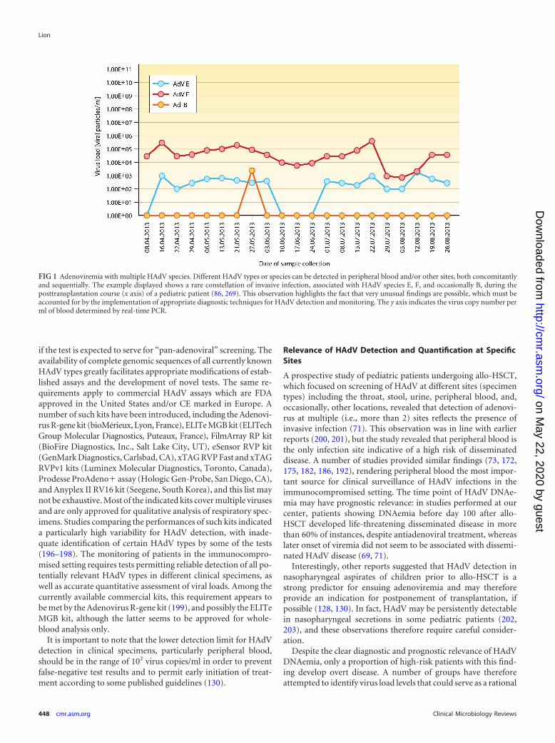
Despite the predominance of certain HAdV species in specificclinical settings, including immunocompromised patients, em-ployment of broad-spectrum HAdV screening assays is necessaryin order to permit reliable detection, even of rarely occurringHAdV species and types, with adequate sensitivity (Fig. 1). Severalgroups have established such “pan-adenoviral” assays based onPCR, exploiting the sequence information available at the respec-tive time points (73, 192–195). HAdV screening assays target con-served regions within the HAdV genome, most commonly withinthe hexon gene, but the inclusion of additional target regions, e.g.,within the fiber gene, may be required to ensure reliable detectionof all known types with comparable sensitivities (194). Due to thefact that the spectrum of newly identified HAdV types has beenexpanding based on the implementation of genomic analyses, es-tablished assays need to be updated in order to facilitate reliablecoverage of the entire range of human adenoviruses. Since newlyidentified HAdV types generally result from recombination eventswithin the same or different human-specific species of the virus(34), the target regions of established PCR assays are preserved inmost instances, thus permitting equally sensitive detection of thenew recombinants. Nevertheless, this issue requires careful atten-tion, as exemplified by the HAdV screening assay established atour center in 2005, based on the sequence information accessibleat that time (194). The test was originally demonstrated to coverall 51 known HAdV (sero)types with comparable detection limits.Alignment with genomic sequences of all newly published HAdVtypes revealed that the current primer-probe combinations of thisreal-time PCR assay can be expected to reliably cover nearly allhitherto identified HAdV types, with two exceptions. The se-quence of HAdV-A61 revealed a few mismatches in the targetregion of the downstream primer, possibly affecting the sensitivityof detection. This finding required the addition of an appropri-ately modified primer to the reaction mix, with subsequent con-firmation of this adaption in vitro. The second exception wasHAdV-G52, which displays the greatest similarity to a simian ad-enovirus (SAdV-1) and is not reliably covered by the assay. Thisexample highlights the need to control and adequately adapt es-tablished diagnostic assays based on newly identified HAdV types
Adenovirus Infections
July 2014 Volume 27 Number 3 cmr.asm.org 447
on May 22, 2020 by guest
http://cmr.asm
.org/D
ownloaded from
if the test is expected to serve for “pan-adenoviral” screening. Theavailability of complete genomic sequences of all currently knownHAdV types greatly facilitates appropriate modifications of estab-lished assays and the development of novel tests. The same re-quirements apply to commercial HAdV assays which are FDAapproved in the United States and/or CE marked in Europe. Anumber of such kits have been introduced, including the Adenovi-rus R-gene kit (bioMérieux, Lyon, France), ELITe MGB kit (ELITechGroup Molecular Diagnostics, Puteaux, France), FilmArray RP kit(BioFire Diagnostics, Inc., Salt Lake City, UT), eSensor RVP kit(GenMark Diagnostics, Carlsbad, CA), xTAG RVP Fast and xTAGRVPv1 kits (Luminex Molecular Diagnostics, Toronto, Canada),Prodesse ProAdeno� assay (Hologic Gen-Probe, San Diego, CA),and Anyplex II RV16 kit (Seegene, South Korea), and this list maynot be exhaustive. Most of the indicated kits cover multiple virusesand are only approved for qualitative analysis of respiratory spec-imens. Studies comparing the performances of such kits indicateda particularly high variability for HAdV detection, with inade-quate identification of certain HAdV types by some of the tests(196–198). The monitoring of patients in the immunocompro-mised setting requires tests permitting reliable detection of all po-tentially relevant HAdV types in different clinical specimens, aswell as accurate quantitative assessment of viral loads. Among thecurrently available commercial kits, this requirement appears tobe met by the Adenovirus R-gene kit (199), and possibly the ELITeMGB kit, although the latter seems to be approved for whole-blood analysis only.
It is important to note that the lower detection limit for HAdVdetection in clinical specimens, particularly peripheral blood,should be in the range of 102 virus copies/ml in order to preventfalse-negative test results and to permit early initiation of treat-ment according to some published guidelines (130).
Relevance of HAdV Detection and Quantification at SpecificSites
A prospective study of pediatric patients undergoing allo-HSCT,which focused on screening of HAdV at different sites (specimentypes) including the throat, stool, urine, peripheral blood, and,occasionally, other locations, revealed that detection of adenovi-rus at multiple (i.e., more than 2) sites reflects the presence ofinvasive infection (71). This observation was in line with earlierreports (200, 201), but the study revealed that peripheral blood isthe only infection site indicative of a high risk of disseminateddisease. A number of studies provided similar findings (73, 172,175, 182, 186, 192), rendering peripheral blood the most impor-tant source for clinical surveillance of HAdV infections in theimmunocompromised setting. The time point of HAdV DNAe-mia may have prognostic relevance: in studies performed at ourcenter, patients showing DNAemia before day 100 after allo-HSCT developed life-threatening disseminated disease in morethan 60% of instances, despite antiadenoviral treatment, whereaslater onset of viremia did not seem to be associated with dissemi-nated HAdV disease (69, 71).
Interestingly, other reports suggested that HAdV detection innasopharyngeal aspirates of children prior to allo-HSCT is astrong predictor for ensuing adenoviremia and may thereforeprovide an indication for postponement of transplantation, ifpossible (128, 130). In fact, HAdV may be persistently detectablein nasopharyngeal secretions in some pediatric patients (202,203), and these observations therefore require careful consider-ation.
Despite the clear diagnostic and prognostic relevance of HAdVDNAemia, only a proportion of high-risk patients with this find-ing develop overt disease. A number of groups have thereforeattempted to identify virus load levels that could serve as a rational
FIG 1 Adenoviremia with multiple HAdV species. Different HAdV types or species can be detected in peripheral blood and/or other sites, both concomitantlyand sequentially. The example displayed shows a rare constellation of invasive infection, associated with HAdV species E, F, and occasionally B, during theposttransplantation course (x axis) of a pediatric patient (86, 269). This observation highlights the fact that very unusual findings are possible, which must beaccounted for by the implementation of appropriate diagnostic techniques for HAdV detection and monitoring. The y axis indicates the virus copy number perml of blood determined by real-time PCR.
Lion
448 cmr.asm.org Clinical Microbiology Reviews
on May 22, 2020 by guest
http://cmr.asm
.org/D
ownloaded from
basis for the start of preemptive antiviral treatment. However, thethresholds suggested by different authors are highly divergent,ranging from 102 (in individuals with high risk) to �106 copies/ml(130, 135, 172, 175, 186, 204, 205), and it is difficult therefore todraw generally applicable conclusions. Moreover, the measuredabsolute values are, at least to some extent, dependent on theindividual technique used, and in the absence of appropriate in-terlaboratory standardization, the values cannot readily be ad-opted by or exchanged between centers. Other authors haveshown that rapidly rising viral loads are detectable in peripheralblood prior to the onset of clinical symptoms of HAdV disease,suggesting that the monitoring of viral titer kinetics may be a morereadily applicable parameter (71, 180). Moreover, in addition tofacilitating prediction of HAdV-related disease, surveillance of HAdVtiter kinetics in peripheral blood is also instrumental for assessment ofthe response to therapy (69, 130, 206). In previous studies, a decreasein viral load of at least 1 log within 2 to 3 weeks of antiviral treatmentwas regarded as a minimum requirement for an adequate response(69, 71), but there are no generally accepted guidelines for diagnosticdefinitions of response at this time.
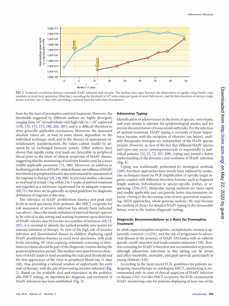
The relevance of HAdV proliferation kinetics and peak loadlevels in stool specimens from pediatric allo-HSCT recipients forrisk assessment of invasive infection has already been indicated(see above). Since the timely initiation of antiviral therapy appearsto be critical in this setting and starting treatment upon detectionof HAdV viremia may be too late in a number of instances (69, 71,207), it is essential to identify the earliest possible time points forrational initiation of therapy. In view of the high risk of invasiveinfection and disseminated disease in children displaying rapidHAdV proliferation kinetics in serial stool specimens, with peaklevels exceeding 106 virus copies/g, systematic screening of intes-tinal excretions should be part of the diagnostic routine during theposttransplantation period. The median time span between detec-tion of HAdV loads in stool exceeding the indicated threshold andthe first appearance of the virus in peripheral blood was 11 days(69), thus providing a rational window of opportunity for earlystart of therapy, with the aim of preventing invasive infection (Fig.2). Based on the available data and experience in the pediatricallo-HSCT setting, an algorithm for diagnosis and treatment ofHAdV infections has been established (Fig. 3).
Adenovirus Typing
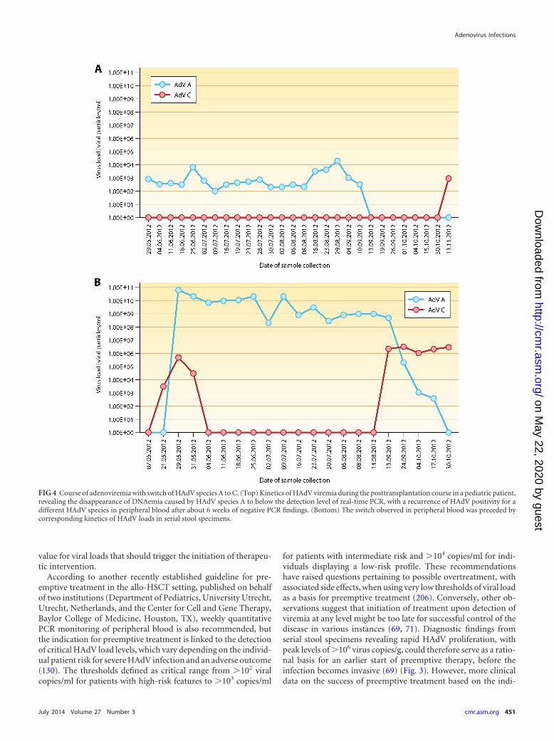
Identification of adenoviruses at the levels of species, (sero)types,and even strains is relevant for epidemiological studies and forprecise documentation of nosocomial outbreaks. For the selectionof optimal treatment, HAdV typing is currently of lesser impor-tance because, with the exception of ribavirin (see below), avail-able therapeutic strategies are independent of the HAdV speciespresent. However, in view of the fact that different HAdV speciesand types may occur contemporaneously or sequentially in indi-vidual patients (52, 53, 72, 107, 208), typing may permit a betterunderstanding of the dynamics and evolution of HAdV infection(Fig. 4).
Typing was traditionally performed by serological methods(209), but these approaches have mostly been replaced by molec-ular techniques based on PCR amplification of specific target re-gions coupled with different detection formats, such as fragmentlength analysis, hybridization to species-specific probes, or se-quencing (210–215). Molecular typing methods are more rapidand readily applicable and can provide better discriminatory ca-pacity. Owing to the decreasing costs of next-generation sequenc-ing (NGS) approaches, whole-genome analysis (36) may becomethe method of choice for detailed HAdV typing in the foreseeablefuture, even in the routine diagnostic setting.
Diagnostic Recommendations as a Basis for PreemptiveTreatment
In adult organ transplant recipients, asymptomatic viremia is ap-parently common (�22%), and the risk of progression to adeno-viral disease in the presence of HAdV DNAemia with or withoutspecific cutoff values for viral loads remains unknown (156). Rou-tine screening for HAdV is therefore not recommended at present,although adenovirus infections in this setting can be severeand affect morbidity, mortality, and graft survival, particularly inyoung children (156).
According to the most recent ECIL guidelines for patients un-dergoing chemotherapy or autologous HSCT, monitoring is rec-ommended only in cases of clinical suspicion of HAdV infectionor disease (143). For allo-HSCT recipients, the ECIL recommendsHAdV monitoring only for patients displaying at least one of the
FIG 2 Temporal correlation between intestinal HAdV infection and viremia. The median time span between the observation of rapidly rising HAdV copynumbers in serial stool specimens (blue line), exceeding the threshold of 106 virus copies per gram of stool (left arrow), and the first detection of viremia (rightarrow; red line) was 11 days (69), providing a rational basis for early start of treatment.
Adenovirus Infections
July 2014 Volume 27 Number 3 cmr.asm.org 449
on May 22, 2020 by guest
http://cmr.asm
.org/D
ownloaded from
risk factors for HAdV disease (see above); in these instances,quantitative PCR monitoring of peripheral blood should be per-formed at weekly or shorter intervals until adequate immune re-constitution is established (143). Although the fact that molecularmonitoring of viral loads in serial stool specimens can facilitateearly detection of impending invasive HAdV infection is acknowl-edged by the ECIL, it has not been included in the current diag-nostic guidelines because the level of available evidence was notdeemed sufficient. Hence, detection of viremia (DNAemia) in thepresence of at least one risk factor is presently the indication forinitiation of preemptive antiviral treatment according to the ECILcriteria.
Somewhat older guidelines for preventing infectious complica-tions in HSCT recipients, published on behalf of the Center forInternational Blood and Marrow Transplant Research (CIBMTR)and a consortium of several other societies, recommend weeklymonitoring of peripheral blood for active HAdV infections byPCR during the first 6 months posttransplantation or the durationof severe immunosuppression/lymphopenia only for patients dis-playing the highest risk for adenoviral disease (216). This subset ofpatients includes individuals with refractory GvHD, recipients ofT-cell-depleted stem cell grafts, haploidentical transplants, or um-bilical cord transplants, and patients treated with anti-T-cell anti-bodies. However, these guidelines do not provide any critical
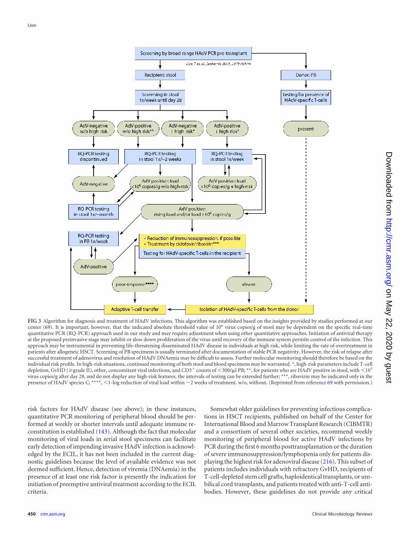
FIG 3 Algorithm for diagnosis and treatment of HAdV infections. This algorithm was established based on the insights provided by studies performed at ourcenter (69). It is important, however, that the indicated absolute threshold value of 106 virus copies/g of stool may be dependent on the specific real-timequantitative PCR (RQ-PCR) approach used in our study and may require adjustment when using other quantitative approaches. Initiation of antiviral therapyat the proposed preinvasive stage may inhibit or slow down proliferation of the virus until recovery of the immune system permits control of the infection. Thisapproach may be instrumental in preventing life-threatening disseminated HAdV disease in individuals at high risk, while limiting the rate of overtreatment inpatients after allogeneic HSCT. Screening of PB specimens is usually terminated after documentation of stable PCR negativity. However, the risk of relapse aftersuccessful treatment of adenovirus and resolution of HAdV DNAemia may be difficult to assess. Further molecular monitoring should therefore be based on theindividual risk profile. In high-risk situations, continued monitoring of both stool and blood specimens may be warranted. *, high-risk parameters include T-celldepletion, GvHD (�grade II), other, concomitant viral infections, and CD3� counts of �300/�l PB; **, for patients who are HAdV positive in stool, with �103
virus copies/g after day 28, and do not display any high-risk features, the intervals of testing can be extended further; ***, ribavirin may be indicated only in thepresence of HAdV species C; ****, �1-log reduction of viral load within �2 weeks of treatment. w/o, without. (Reprinted from reference 69 with permission.)
Lion
450 cmr.asm.org Clinical Microbiology Reviews
on May 22, 2020 by guest
http://cmr.asm
.org/D
ownloaded from
value for viral loads that should trigger the initiation of therapeu-tic intervention.
According to another recently established guideline for pre-emptive treatment in the allo-HSCT setting, published on behalfof two institutions (Department of Pediatrics, University Utrecht,Utrecht, Netherlands, and the Center for Cell and Gene Therapy,Baylor College of Medicine, Houston, TX), weekly quantitativePCR monitoring of peripheral blood is also recommended, butthe indication for preemptive treatment is linked to the detectionof critical HAdV load levels, which vary depending on the individ-ual patient risk for severe HAdV infection and an adverse outcome(130). The thresholds defined as critical range from �102 viralcopies/ml for patients with high-risk features to �103 copies/ml
for patients with intermediate risk and �104 copies/ml for indi-viduals displaying a low-risk profile. These recommendationshave raised questions pertaining to possible overtreatment, withassociated side effects, when using very low thresholds of viral loadas a basis for preemptive treatment (206). Conversely, other ob-servations suggest that initiation of treatment upon detection ofviremia at any level might be too late for successful control of thedisease in various instances (69, 71). Diagnostic findings fromserial stool specimens revealing rapid HAdV proliferation, withpeak levels of �106 virus copies/g, could therefore serve as a ratio-nal basis for an earlier start of preemptive therapy, before theinfection becomes invasive (69) (Fig. 3). However, more clinicaldata on the success of preemptive treatment based on the indi-
FIG 4 Course of adenoviremia with switch of HAdV species A to C. (Top) Kinetics of HAdV viremia during the posttransplantation course in a pediatric patient,revealing the disappearance of DNAemia caused by HAdV species A to below the detection level of real-time PCR, with a recurrence of HAdV positivity for adifferent HAdV species in peripheral blood after about 6 weeks of negative PCR findings. (Bottom) The switch observed in peripheral blood was preceded bycorresponding kinetics of HAdV loads in serial stool specimens.
Adenovirus Infections
July 2014 Volume 27 Number 3 cmr.asm.org 451
on May 22, 2020 by guest
http://cmr.asm
.org/D
ownloaded from
cated recommendations are needed to provide unequivocal sup-port for the currently available diagnostic guidelines.
CURRENT TREATMENT MODALITIES
Present recommendations for treatment of HAdV infections fo-cus on immunocompromised patients, particularly allogeneictransplant recipients, who apparently carry the greatest risk ofsevere and life-threatening clinical courses. The approaches pur-sued may include prophylaxis, preemptive treatment based onvirus detection prior to onset of clinical symptoms, sometimeslinked to specific thresholds of viral load, or therapeutic (symp-tomatic) treatment in the presence of virus-related disease. Atpresent, there is little evidence for a beneficial effect of HAdVprophylaxis, and the ECIL does not recommend prophylactic an-tiviral therapy with currently available virustatic drugs (143). ForSOT recipients, the treatment indication for mild or asymptom-atic HAdV infection is not clear, since prospective studies haveshown that adenoviremia may be present without any clinicalsymptoms and may clear spontaneously (157). Some authorstherefore recommend antiviral treatment only for symptomaticpatients (20). In contrast, for patients undergoing allo-HSCT,preemptive treatment is strongly advocated by all major guide-lines in order to inhibit or slow down viral replication, with theaim to prevent overt disease until immune reconstitution fromthe allograft permits clearance of the infection (130, 143, 216). Theprincipal options for preemptive treatment include (i) the taper-ing of immunosuppressive therapy, which should be performedwhenever possible; (ii) use of antiviral drugs; and (iii) immuno-therapy in case of failure of the previous lines of treatment.
Antiviral Drugs
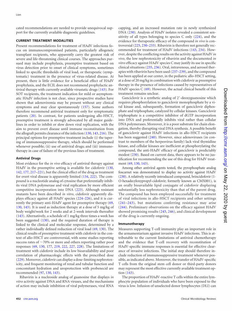
Most evidence for the in vivo efficacy of antiviral therapy againstHAdV in the preemptive setting is available for cidofovir (130,142, 177, 217–221), but the clinical effect of the drug as treatmentfor overt viral disease is apparently limited (134, 222). The com-pound is a nucleotide analog of cytosine that preferentially inhib-its viral DNA polymerase and viral replication by more efficientcompetitive incorporation into DNA (223). Although resistantmutants have been described in vitro, cidofovir apparently dis-plays efficacy against all HAdV species (224–226), and it is cur-rently the primary anti-HAdV agent for preemptive therapy (69,130, 143). It is used as induction therapy at a dose of 5 mg/kg ofbody weight/week for 2 weeks and at 2-week intervals thereafter(143). Alternatively, a schedule of 1 mg/kg three times a week hasbeen suggested (130), and the required duration of therapy islinked to the clinical and molecular response, determined by arather individually defined reduction of viral load (69, 130). Theclinical results of preemptive treatment with cidofovir in the con-text of allo-HSCT are controversial, with some studies reportingsuccess rates of �70% or more and others reporting rather poorresponses (69, 130, 177, 219, 222, 227, 228). The limitations oftreatment with cidofovir include its low bioavailability and poorcorrelation of pharmacologic effects with the prescribed dose(229). Moreover, cidofovir can display a dose-limiting nephrotox-icity, and frequent monitoring of renal and tubular function andconcomitant hydration and uroprotection with probenecid arerecommended (97, 130, 143).
Ribavirin is a nucleoside analog of guanosine that displays invitro activity against DNA and RNA viruses, and the mechanismsof action may include inhibition of viral polymerases, viral RNA
capping, and an increased mutation rate in newly synthesizedDNA (230). Analysis of HAdV isolates revealed a consistent sen-sitivity of all types belonging to species C only (224), and theevidence for therapeutic efficacy of the compound in vivo is con-troversial (225, 230–233). Ribavirin is therefore not generally rec-ommended for treatment of HAdV infections (143, 234). How-ever, despite the conflicting results on the activity against HAdV invivo, the low nephrotoxicity of ribavirin and the documented invitro efficacy against HAdV species C may justify its use in specificclinical situations (235, 236). Oral, intravenous, and aerosol ther-apies with ribavirin have been used (237–239), and the compoundhas been applied at our center, in the pediatric allo-HSCT setting,at a dose of 20 mg/kg in combination with cidofovir as preemptivetherapy in the presence of infections caused by representatives ofHAdV species C (69). However, the actual clinical benefit of thistreatment remains unclear.
Ganciclovir is a synthetic analog of 2=-deoxyguanosine whichrequires phosphorylation to ganciclovir monophosphate by a vi-ral kinase and, subsequently, formation of ganciclovir diphos-phate and triphosphate, catalyzed by cellular kinases. Ganciclovirtriphosphate is a competitive inhibitor of dGTP incorporationinto DNA and preferentially inhibits viral rather than cellularDNA polymerases. Moreover, it is a poor substrate for chain elon-gation, thereby disrupting viral DNA synthesis. A possible benefitof ganciclovir against HAdV infections in allo-HSCT recipientshas been suggested (240). However, since adenoviruses (in con-trast to members of the herpesvirus family) lack viral thymidinekinase, and cellular kinases are inefficient at phosphorylating thecompound, the anti-HAdV efficacy of ganciclovir is predictablymodest (230). Based on current data, there appears to be no jus-tification for recommending the use of this drug for HAdV treat-ment (69, 130, 143).
Among other antiviral agents tested, the pyrophosphate analogfoscarnet was demonstrated to display no activity against HAdV(230). A relatively recently introduced compound, brincidofovir (1-O-hexadecyloxypropyl-cidofovir; formerly known as CMX001), isan orally bioavailable lipid conjugate of cidofovir displayingsubstantially less nephrotoxicity than that of the parent drug.The compound has been employed successfully for treatmentof viral infections in allo-HSCT recipients and other settings(241–243), but mutations conferring resistance may arise(244). Preliminary observations on the efficacy against HAdVshowed promising results (245, 246), and clinical developmentof the drug is currently ongoing.
Immunotherapy
Measures supporting T-cell immunity play an important role inthe armamentarium against invasive HAdV infections. This is at-tributable to the current limitations of antiviral chemotherapyand the evidence that T-cell recovery with reconstitution ofHAdV-specific immune responses is essential for effective clear-ance of invasive infections. The initial step should therefore in-clude reduction of immunosuppressive treatment whenever pos-sible, as indicated above. Moreover, the transfer of HAdV-specificT cells from the original stem cell donor or third-party donorsmay represent the most effective currently available treatment op-tion (143).
The proportion of HAdV-reactive T cells within the entire lym-phocyte population of individuals who have been exposed to thevirus is low. Infusion of unselected donor lymphocytes (DLI) can
Lion
452 cmr.asm.org Clinical Microbiology Reviews
on May 22, 2020 by guest
http://cmr.asm
.org/D
ownloaded from
still provide antiviral immunity (247), but the potentially highfrequency of alloreactive T cells and the ensuing side effects aremajor impediments to this approach (130). The isolation ofHAdV-specific T cells from peripheral blood of the original stemcell donor has therefore become the method of choice for treat-ment of HAdV infections in allo-HSCT recipients not respondingto antiviral chemotherapy, and different approaches to the gener-ation of such cells have been established. Regardless of the HAdVspecies the donor has been exposed to, the HAdV-reactive T cellsare expected to be cross-reactive with all HAdV types, because thehexon, the main constituent of the viral capsid, is the immuno-dominant T-cell target, containing several epitopes that are con-served among adenoviruses (116, 248). One of the early successfulattempts of adoptive T-cell transfer was based on the isolation ofdonor-derived mononuclear cells, their stimulation ex vivo withHAdV antigen, and magnetic separation of reactive T cells secret-ing IFN-�, which included both CD4� and CD8� T cells. The cellswere infused without further in vitro expansion, and the resultsindicated that the efficacy of this treatment does not depend onthe dose of infused cells, because even very small numbers ofHAdV-specific donor-derived T cells expanded easily in vivo inthe presence of the constant antigen challenge mediated by theviral infection (207). The most critical parameter for the success oftreatment was appropriate timing, i.e., early T-cell transfer upondetection of viremia (207). These observations indicate that rapidavailability of T cells for adoptive transfer, based on methods re-quiring only short or no in vitro expansion, is essential. Based onthis notion, a number of different approaches to selection ofHAdV-specific T cells have been introduced. The considerablevariety of approaches includes, for example, different types ofMHC multimers facilitating clinical-grade enrichment of HAdV-specific or multivirus-specific T cells displaying low or absent al-loreactivity (249, 250). The MHC multimer technology requiresknowledge of immunodominant human leukocyte antigen(HLA)-restricted peptide epitopes and facilitates the isolation ofantigen-specific CD8� T cells (MHC class I multimers) or CD4�
T cells (MHC class II multimers) of high purity (251). Short-termin vitro expansion under good manufacturing practice (GMP)conditions can render adoptive T-cell transfer available in lessthan 2 weeks (123, 252–254). In addition to a variety of methodsbased on the isolation of HAdV-specific T cells from the originalstem cell donors, several approaches exploiting third-party do-nors are emerging (141, 255–263). Allogeneic third-party donorsare a particularly important alternative option for cord blood re-cipients, for patients receiving allografts from HAdV-seronegativedonors, and for solid organ transplantations from cadavericdonors, where donor blood is not available. Healthy seroposi-tive individuals have been exploited to generate partially HLA-matched virus-specific T cells (VSTs) for adoptive immunother-apy (251). However, clinical implementation of this approachrequires the availability of a large pool of HLA-typed healthy do-nors, and the use of incompletely HLA-matched T cells bears therisk of complications resulting from alloreactive side effects (264).Despite the existing concerns, this approach appears to be feasible,and current clinical results are encouraging (265). Another re-cently presented methodology is the generation of VST cell linesfrom healthy donors with common HLA polymorphisms (266).The employment of banked third-party-derived VSTs was dem-onstrated to represent an additional safe and readily applicablestrategy for rapidly available treatment of severe viral infections in
allo-HSCT recipients (266), thus further expanding the spectrumof clinical options for effective immunotherapy in immunocom-promised patients. Although adoptive transfer of HAdV-specificT cells is currently one of the most promising treatment ap-proaches for high-risk patient populations, it still needs to be re-garded as experimental and should only be performed in the con-text of clinical trials in specialized centers.
SUMMARY AND PERSPECTIVES
Despite the considerable progress in diagnosis and treatment ofadenoviral infections over the past years, a number of relevantissues remain to be solved. The steadily growing spectrum ofHAdV types emanating from genomic analyses is a challenge formolecular screening assays. Although newly identified types of thevirus generally result from recombination events between knownHAdV representatives, and the sequences targeted by PCR testsmay be preserved, it is necessary to test and occasionally adapt theassays to ensure adequate coverage of existing HAdV types. Com-plete HAdV genome sequences are now available in public data-bases, thereby permitting at least in silico control of the primersand probes used to ensure coverage of any newly identified HAdVtype by the assay used. However, in view of the clinical importanceof adenoviral infections, the availability of commercial, regularlyupdated diagnostic tests (199, 267) is highly desirable to facilitatestandardized diagnostics under stringent quality control condi-tions. Commercial assays should permit pan-HAdV screening,quantification of viral loads, and, ideally, typing at least to thespecies level in order to provide a basis for more specifically tar-geted treatment approaches (251, 268). It is conceivable that easieraccess to next-generation sequencing facilities and decreasingcosts of analysis will enable detailed HAdV typing, even in theroutine clinical setting, in the near future.
Current recommendations for adenovirus screening and mon-itoring as a basis for preemptive treatment in patients at high riskfor HAdV disease are still relatively diverse (69, 130, 143), andfurther studies are needed to provide reliable data permitting theestablishment of standardized approaches. Optimized diagnosticswill greatly affect the rational and timely initiation of antiviraltreatment, which was shown to be a prerequisite for successfultherapy. Despite current recommendations for preemptive ad-ministration of antiviral drugs, unequivocal evidence for a bene-ficial effect on mortality is still missing, and appropriate prospec-tive studies are needed. The introduction of novel antiviral agentscan be expected to further improve the efficacy of treatment and toreduce the toxicity of some commonly prescribed virustatic drugs.In this regard, the orally bioavailable and less nephrotoxic lipidester of cidofovir, brincidofovir, could provide an important im-provement of current antiviral therapies. Preliminary data fromongoing studies are promising, and an international therapy trialwith brincidofovir in transplant recipients displaying intestinalHAdV infection, who have an elevated risk of viremia and dissem-inated disease (69, 188, 189), is currently in preparation. Thisstudy is expected to provide important evidence for the efficacy ofearly preemptive virustatic treatment. Another interesting novelantiviral compound is ganciclovir triphosphate, which does notrequire further phosphorylation by viral kinases and can thereforebe expected to display therapeutic activity against HAdV.
In addition to the favorable properties of new antiviral drugs,advances in antiviral immunotherapy with adenovirus-specific Tcells offer great potential for further improvement in the preven-
Adenovirus Infections
July 2014 Volume 27 Number 3 cmr.asm.org 453
on May 22, 2020 by guest
http://cmr.asm
.org/D
ownloaded from
tion or treatment of HAdV infections. Anti-HAdV T cells gener-ated by different approaches are cross-reactive with all types of thevirus and can therefore provide broad antiadenoviral immunity. Itis not entirely clear, however, whether an equivalent quality ofimmune response to any HAdV type can be expected. It is con-ceivable that the generation of T cells targeted to individual HAdVtypes and mediating specific interactions with type-restrictedepitopes (268), in addition to the broadly shared hexon epitopes,may elicit more effective immune responses (251). This notionmay warrant careful attention. If the safety, efficacy, and feasibilityof HAdV-specific or multivirus-specific T-cell transfer can befirmly established, it is reasonable to envision that this treatmentwill be employed successfully not only in the settings of preemp-tive and symptomatic therapy but also as prophylaxis in high-riskpatients to prevent severe viral diseases. The establishment of al-logeneic T-cell donor registries of HLA-typed healthy donorstested for the presence of virus-specific T cells could serve as arapidly available source for adoptive immunotherapy in immuno-compromised patients lacking a suitable T-cell donor. Such T-celldonor registries might provide readily available off-the-shelfproducts facilitating rapid initiation of immunotherapy in pa-tients carrying a high risk of life-threatening viral infections.Broad availability of banked third-party virus-specific T cellsand/or virus-specific T-cell lines could mark the beginning of anew era in combatting the threats of viral infections in immuno-compromised patients in the foreseeable future.
ACKNOWLEDGMENT
I have no conflict of interest relevant in the context of the present paper.
REFERENCES1. Rowe JP, Huebner RJ, Gilmore LK, Parrott RH, Ward TG. 1953.
Isolation of a cytopathogenic agent from human adenoids undergoingspontaneous degeneration in tissue culture. Proc. Soc. Exp. Biol. Med.84:570 –573.
2. Trentin JJ, Yabe Y, Taylor G. 1962. The quest for human cancer viruses.Science 137:835– 841. http://dx.doi.org/10.1126/science.137.3533.835.
3. Hohlweg U, Dorn A, Hosel M, Webb D, Buettner R, Doerfler W. 2004.Tumorigenesis by adenovirus type 12 in newborn Syrian hamsters. Curr.Top. Microbiol. Immunol. 273:215–244. http://dx.doi.org/10.1007/978-3-662-05599-1_7.
4. Javier RT. 1994. Adenovirus type 9 E4 open reading frame 1 encodes atransforming protein required for the production of mammary tumorsin rats. J. Virol. 68:3917–3924.
5. Kosulin K, Haberler C, Hainfellner JA, Amann G, Lang S, Lion T.2007. Investigation of adenovirus occurrence in pediatric tumor entities.J. Virol. 81:7629 –7635. http://dx.doi.org/10.1128/JVI.00355-07.
6. Kosulin K, Hoffmann F, Clauditz TS, Wilczak W, Dobner T. 2013.Presence of adenovirus species C in infiltrating lymphocytes of humansarcoma. PLoS One 8:e63646. http://dx.doi.org/10.1371/journal.pone.0063646.
7. Sanchez-Prieto R, de Alava E, Palomino T, Guinea J, Fernandez V,Cebrian SLLM, Cabello P, Martin P, San Roman C, Bornstein R,Pardo J, Martinez A, Diaz-Espada F, Barrios Y, Ramon y Cajal S. 1999.An association between viral genes and human oncogenic alterations: theadenovirus E1A induces the Ewing tumor fusion transcript EWS-FLI1.Nat. Med. 5:1076 –1079. http://dx.doi.org/10.1038/12516.
8. Greber UF, Arnberg N, Wadell G, Benko M, Kremer EJ. 2013. Adeno-viruses—from pathogens to therapeutics: a report on the 10th Interna-tional Adenovirus Meeting. Cell. Microbiol. 15:16 –23. http://dx.doi.org/10.1111/cmi.12031.
9. Dicks MD, Spencer AJ, Edwards NJ, Wadell G, Bojang K, Gilbert SC,Hill AV, Cottingham MG. 2012. A novel chimpanzee adenovirus vectorwith low human seroprevalence: improved systems for vector derivationand comparative immunogenicity. PLoS One 7:e40385. http://dx.doi.org/10.1371/journal.pone.0040385.
10. Miura Y, Yamasaki S, Davydova J, Brown E, Aoki K, Vickers S,Yamamoto M. 2013. Infectivity-selective oncolytic adenovirus devel-oped by high-throughput screening of adenovirus-formatted library.Mol. Ther. 21:139 –148. http://dx.doi.org/10.1038/mt.2012.205.
11. Yamasaki S, Miura Y, Davydova J, Vickers SM, Yamamoto M. 2013.Intravenous genetic mesothelin vaccine based on human adenovirus 40inhibits growth and metastasis of pancreatic cancer. Int. J. Cancer 133:88 –97. http://dx.doi.org/10.1002/ijc.27983.
12. Diaconu I, Cerullo V, Hirvinen ML, Escutenaire S, Ugolini M, Pe-sonen SK, Bramante S, Parviainen S, Kanerva A, Loskog AS, Eliopou-los AG, Pesonen S, Hemminki A. 2012. Immune response is an impor-tant aspect of the antitumor effect produced by a CD40L-encodingoncolytic adenovirus. Cancer Res. 72:2327–2338. http://dx.doi.org/10.1158/0008-5472.CAN-11-2975.
13. Shang Q, Wang H, Song Y, Wei L, Lavebratt C, Zhang F, Gu H. 2014.Serological data analyses show that adenovirus 36 infection is associatedwith obesity: a meta-analysis involving 5739 subjects. Obesity 22:895–900. http://dx.doi.org/10.1002/oby.20533.
14. Shauer A, Gotsman I, Keren A, Zwas DR, Hellman Y, Durst R, AdmonD. 2013. Acute viral myocarditis: current concepts in diagnosis and treat-ment. Isr. Med. Assoc. J. 15:180 –185.
15. de Ory F, Avellon A, Echevarria JE, Sanchez-Seco MP, Trallero G,Cabrerizo M, Casas I, Pozo F, Fedele G, Vicente D, Pena MJ, MorenoA, Niubo J, Rabella N, Rubio G, Perez-Ruiz M, Rodriguez-Iglesias M,Gimeno C, Eiros JM, Melon S, Blasco M, Lopez-Miragaya I, Varela E,Martinez-Sapina A, Rodriguez G, Marcos MA, Gegundez MI, Cilla G,Gabilondo I, Navarro JM, Torres J, Aznar C, Castellanos A, GuisasolaME, Negredo AI, Tenorio A, Vazquez-Moron S. 2013. Viral infectionsof the central nervous system in Spain: a prospective study. J. Med. Virol.85:554 –562. http://dx.doi.org/10.1002/jmv.23470.
16. Chhabra P, Payne DC, Szilagyi PG, Edwards KM, Staat MA, ShirleySH, Wikswo M, Nix WA, Lu X, Parashar UD, Vinje J. 2013. Etiologyof viral gastroenteritis in children �5 years of age in the United States,2008 –2009. J. Infect. Dis. 208:790 – 800. http://dx.doi.org/10.1093/infdis/jit254.
17. Rocholl C, Gerber K, Daly J, Pavia AT, Byington CL. 2004. Adenoviralinfections in children: the impact of rapid diagnosis. Pediatrics 113:e51–e56. http://dx.doi.org/10.1542/peds.113.1.e51.
18. Mameli C, Zuccotti GV. 2013. The impact of viral infections in childrenwith community-acquired pneumonia. Curr. Infect. Dis. Rep. 15:197–202. http://dx.doi.org/10.1007/s11908-013-0339-z.
19. Centers for Disease Control and Prevention. 2013. Adenovirus-associated epidemic keratoconjunctivitis outbreaks—four states, 2008 –2010. MMWR Morb. Mortal. Wkly. Rep. 62:637– 641.
20. Lynch JP, 3rd, Fishbein M, Echavarria M. 2011. Adenovirus. Semin.Respir. Crit. Care Med. 32:494 –511. http://dx.doi.org/10.1055/s-0031-1283287.
21. Esposito S, Preti V, Consolo S, Nazzari E, Principi N. 2012. Adenovi-rus 36 infection and obesity. J. Clin. Virol. 55:95–100. http://dx.doi.org/10.1016/j.jcv.2012.06.009.
22. Berciaud S, Rayne F, Kassab S, Jubert C, Faure-Della Corte M, SalinF, Wodrich H, Lafon ME, Typadeno Study Members. 2012. Adenovi-rus infections in Bordeaux University Hospital 2008 –2010: clinical andvirological features. J. Clin. Virol. 54:302–307. http://dx.doi.org/10.1016/j.jcv.2012.04.009.
23. Chen SP, Huang YC, Chiu CH, Wong KS, Huang YL, Huang CG, TsaoKC, Lin TY. 2013. Clinical features of radiologically confirmed pneu-monia due to adenovirus in children. J. Clin. Virol. 56:7–12. http://dx.doi.org/10.1016/j.jcv.2012.08.021.
24. Alharbi S, Van Caeseele P, Consunji-Araneta R, Zoubeidi T, Fanella S,Souid AK, Alsuwaidi AR. 2012. Epidemiology of severe pediatric adenovi-rus lower respiratory tract infections in Manitoba, Canada, 1991–2005.BMC Infect. Dis. 12:55. http://dx.doi.org/10.1186/1471-2334-12-55.
25. Lewis PF, Schmidt MA, Lu X, Erdman DD, Campbell M, Thomas A,Cieslak PR, Grenz LD, Tsaknardis L, Gleaves C, Kendall B, Gilbert D.2009. A community-based outbreak of severe respiratory illness causedby human adenovirus serotype 14. J. Infect. Dis. 199:1427–1434. http://dx.doi.org/10.1086/598521.
26. Savon C, Acosta B, Valdes O, Goyenechea A, Gonzalez G, Pinon A,Mas P, Rosario D, Capo V, Kouri V, Martinez PA, Marchena JJ,Gonzalez G, Rodriguez H, Guzman MG. 2008. A myocarditis outbreakwith fatal cases associated with adenovirus subgenera C among children
Lion
454 cmr.asm.org Clinical Microbiology Reviews
on May 22, 2020 by guest
http://cmr.asm
.org/D
ownloaded from
from Havana City in 2005. J. Clin. Virol. 43:152–157. http://dx.doi.org/10.1016/j.jcv.2008.05.012.
27. Nilsson EC, Storm RJ, Bauer J, Johansson SM, Lookene A, AngstromJ, Hedenstrom M, Eriksson TL, Frangsmyr L, Rinaldi S, Willison HJ,Pedrosa Domellof F, Stehle T, Arnberg N. 2011. The GD1a glycan is acellular receptor for adenoviruses causing epidemic keratoconjunctivitis.Nat. Med. 17:105–109. http://dx.doi.org/10.1038/nm.2267.
28. Trinh HV, Lesage G, Chennamparampil V, Vollenweider B, Burck-hardt CJ, Schauer S, Havenga M, Greber UF, Hemmi S. 2012. Aviditybinding of human adenovirus serotypes 3 and 7 to the membrane cofac-tor CD46 triggers infection. J. Virol. 86:1623–1637. http://dx.doi.org/10.1128/JVI.06181-11.
29. Wang H, Li ZY, Liu Y, Persson J, Beyer I, Moller T, Koyuncu D,Drescher MR, Strauss R, Zhang XB, Wahl JK, 3rd, Urban N, DrescherC, Hemminki A, Fender P, Lieber A. 2011. Desmoglein 2 is a receptorfor adenovirus serotypes 3, 7, 11 and 14. Nat. Med. 17:96 –104. http://dx.doi.org/10.1038/nm.2270.
30. Robinson CM, Seto D, Jones MS, Dyer DW, Chodosh J. 2011. Mo-lecular evolution of human species D adenoviruses. Infect. Genet. Evol.11:1208 –1217. http://dx.doi.org/10.1016/j.meegid.2011.04.031.
31. Smith JG, Wiethoff CM, Stewart PL, Nemerow GR. 2010. Adenovirus.Curr. Top. Microbiol. Immunol. 343:195–224. http://dx.doi.org/10.1007/82_2010_16.
32. Wiethoff CM, Wodrich H, Gerace L, Nemerow GR. 2005. Adeno-virus protein VI mediates membrane disruption following capsid dis-assembly. J. Virol. 79:1992–2000. http://dx.doi.org/10.1128/JVI.79.4.1992-2000.2005.
33. Harrach B, Benkö M, Both GW, Brown M, Davison AJ, Echavarria M,Hess M, Jones MS, Kajon A, Lehmkuhl HD, Mautner V, Mittal SK,Wadell G. 2012. Adenoviridae—ninth report of the International Com-mittee on Taxonomy of Viruses, p 125–141. In King AMQ, Adams MJ,Carstens EB, Lefkowitz EJ (ed), Virus taxonomy. Elsevier, Oxford,United Kingdom.
34. Robinson CM, Singh G, Lee JY, Dehghan S, Rajaiya J, Liu EB, YousufMA, Betensky RA, Jones MS, Dyer DW, Seto D, Chodosh J. 2013.Molecular evolution of human adenoviruses. Sci. Rep. 3:1812. http://dx.doi.org/10.1038/srep01812.
35. Kamel W, Segerman B, Oberg D, Punga T, Akusjarvi G. 2013. Theadenovirus VA RNA-derived miRNAs are not essential for lytic virusgrowth in tissue culture cells. Nucleic Acids Res. 41:4802– 4812. http://dx.doi.org/10.1093/nar/gkt172.
36. Jones MS, 2nd, Harrach B, Ganac RD, Gozum MM, Dela Cruz WP,Riedel B, Pan C, Delwart EL, Schnurr DP. 2007. New adenovirusspecies found in a patient presenting with gastroenteritis. J. Virol. 81:5978 –5984. http://dx.doi.org/10.1128/JVI.02650-06.
37. Robinson CM, Singh G, Henquell C, Walsh MP, Peigue-Lafeuille H,Seto D, Jones MS, Dyer DW, Chodosh J. 2011. Computational analysisand identification of an emergent human adenovirus pathogen impli-cated in a respiratory fatality. Virology 409:141–147. http://dx.doi.org/10.1016/j.virol.2010.10.020.
38. Walsh MP, Seto J, Liu EB, Dehghan S, Hudson NR, Lukashev AN,Ivanova O, Chodosh J, Dyer DW, Jones MS, Seto D. 2011. Compu-tational analysis of two species C human adenoviruses provides evidenceof a novel virus. J. Clin. Microbiol. 49:3482–3490. http://dx.doi.org/10.1128/JCM.00156-11.
39. Liu EB, Ferreyra L, Fischer SL, Pavan JV, Nates SV, Hudson NR,Tirado D, Dyer DW, Chodosh J, Seto D, Jones MS. 2011. Geneticanalysis of a novel human adenovirus with a serologically unique hexonand a recombinant fiber gene. PLoS One 6:e24491. http://dx.doi.org/10.1371/journal.pone.0024491.
40. Seto J, Walsh MP, Mahadevan P, Purkayastha A, Clark JM, Tibbetts C,Seto D. 2009. Genomic and bioinformatics analyses of HAdV-14p, refer-ence strain of a re-emerging respiratory pathogen and analysis of B1/B2.Virus Res. 143:94–105. http://dx.doi.org/10.1016/j.virusres.2009.03.011.
41. Seto D, Chodosh J, Brister JR, Jones MS, Members of the AdenovirusResearch Community. 2011. Using the whole-genome sequence to char-acterize and name human adenoviruses. J. Virol. 85:5701–5702. http://dx.doi.org/10.1128/JVI.00354-11.
42. Aoki K, Benko M, Davison AJ, Echavarria M, Erdman DD, Harrach B,Kajon AE, Schnurr D, Wadell G, Members of the Adenovirus ResearchCommunity. 2011. Toward an integrated human adenovirus designa-tion system that utilizes molecular and serological data and serves both
clinical and fundamental virology. J. Virol. 85:5703–5704. http://dx.doi.org/10.1128/JVI.00491-11.
43. Echavarria M. 2008. Adenoviruses in immunocompromised hosts. Clin.Microbiol. Rev. 21:704 –715. http://dx.doi.org/10.1128/CMR.00052-07.
44. Liu EB, Wadford DA, Seto J, Vu M, Hudson NR, Thrasher L, TorresS, Dyer DW, Chodosh J, Seto D, Jones MS. 2012. Computational andserologic analysis of novel and known viruses in species human adeno-virus D in which serology and genomics do not correlate. PLoS One7:e33212. http://dx.doi.org/10.1371/journal.pone.0033212.
45. Walsh MP, Seto J, Jones MS, Chodosh J, Xu W, Seto D. 2010.Computational analysis identifies human adenovirus type 55 as a re-emergent acute respiratory disease pathogen. J. Clin. Microbiol. 48:991–993. http://dx.doi.org/10.1128/JCM.01694-09.
46. Matsushima Y, Shimizu H, Kano A, Nakajima E, Ishimaru Y, Dey SK,Watanabe Y, Adachi F, Mitani K, Fujimoto T, Phan TG, Ushijima H.2013. Genome sequence of a novel virus of the species human adenovirusD associated with acute gastroenteritis. Genome Announc. 1:e00068 –12.http://dx.doi.org/10.1128/genomeA.00068-12.
47. Takemori N. 1972. Genetic studies with tumorigenic adenoviruses. 3.Recombination in adenovirus type 12. Virology 47:157–167.
48. Williams J, Grodzicker T, Sharp P, Sambrook J. 1975. Adenovirusrecombination: physical mapping of crossover events. Cell 4:113–119.http://dx.doi.org/10.1016/0092-8674(75)90117-8.
49. Walsh MP, Chintakuntlawar A, Robinson CM, Madisch I, Harrach B,Hudson NR, Schnurr D, Heim A, Chodosh J, Seto D, Jones MS. 2009.Evidence of molecular evolution driven by recombination events influ-encing tropism in a novel human adenovirus that causes epidemic kera-toconjunctivitis. PLoS One 4:e5635. http://dx.doi.org/10.1371/journal.pone.0005635.
50. Lukashev AN, Ivanova OE, Eremeeva TP, Iggo RD. 2008. Evidence offrequent recombination among human adenoviruses. J. Gen. Virol. 89:380 –388. http://dx.doi.org/10.1099/vir.0.83057-0.
51. Robinson CM, Rajaiya J, Walsh MP, Seto D, Dyer DW, Jones MS,Chodosh J. 2009. Computational analysis of human adenovirus type 22provides evidence for recombination among species D human adenovi-ruses in the penton base gene. J. Virol. 83:8980 – 8985. http://dx.doi.org/10.1128/JVI.00786-09.
52. Vora GJ, Lin B, Gratwick K, Meador C, Hansen C, Tibbetts C, StengerDA, Irvine M, Seto D, Purkayastha A, Freed NE, Gibson MG, RussellK, Metzgar D. 2006. Co-infections of adenovirus species in previouslyvaccinated patients. Emerg. Infect. Dis. 12:921–930. http://dx.doi.org/10.3201/eid1206.050245.
53. McCarthy T, Lebeck MG, Capuano AW, Schnurr DP, Gray GC. 2009.Molecular typing of clinical adenovirus specimens by an algorithmwhich permits detection of adenovirus coinfections and intermediateadenovirus strains. J. Clin. Virol. 46:80 – 84. http://dx.doi.org/10.1016/j.jcv.2009.06.008.
54. Curlin ME, Huang ML, Lu X, Celum CL, Sanchez J, Selke S, BaetenJM, Zuckerman RA, Erdman DD, Corey L. 2010. Frequent detection ofhuman adenovirus from the lower gastrointestinal tract in men who havesex with men. PLoS One 5:e11321. http://dx.doi.org/10.1371/journal.pone.0011321.
55. Kaneko H, Aoki K, Ishida S, Ohno S, Kitaichi N, Ishiko H, FujimotoT, Ikeda Y, Nakamura M, Gonzalez G, Koyanagi KO, Watanabe H,Suzutani T. 2011. Recombination analysis of intermediate human ade-novirus type 53 in Japan by complete genome sequence. J. Gen. Virol.92:1251–1259. http://dx.doi.org/10.1099/vir.0.030361-0.
56. Wevers D, Metzger S, Babweteera F, Bieberbach M, Boesch C, Cam-eron K, Couacy-Hymann E, Cranfield M, Gray M, Harris LA, Head J,Jeffery K, Knauf S, Lankester F, Leendertz SA, Lonsdorf E, Mugisha L,Nitsche A, Reed P, Robbins M, Travis DA, Zommers Z, Leendertz FH,Ehlers B. 2011. Novel adenoviruses in wild primates: a high level ofgenetic diversity and evidence of zoonotic transmissions. J. Virol. 85:10774 –10784. http://dx.doi.org/10.1128/JVI.00810-11.
57. Dehghan S, Seto J, Liu EB, Walsh MP, Dyer DW, Chodosh J, Seto D.2013. Computational analysis of four human adenovirus type 4 genomesreveals molecular evolution through two interspecies recombination events.Virology 443:197–207. http://dx.doi.org/10.1016/j.virol.2013.05.014.
58. Lin KH, Lin YC, Chen HL, Ke GM, Chiang CJ, Hwang KP, Chu PY,Lin JH, Liu DP, Chen HY. 2004. A two decade survey of respiratoryadenovirus in Taiwan: the reemergence of adenovirus types 7 and 4. J.Med. Virol. 73:274 –279. http://dx.doi.org/10.1002/jmv.20087.
59. Ishiko H, Shimada Y, Konno T, Hayashi A, Ohguchi T, Tagawa Y,
Adenovirus Infections
July 2014 Volume 27 Number 3 cmr.asm.org 455
on May 22, 2020 by guest
http://cmr.asm
.org/D
ownloaded from
Aoki K, Ohno S, Yamazaki S. 2008. Novel human adenovirus causingnosocomial epidemic keratoconjunctivitis. J. Clin. Microbiol. 46:2002–2008. http://dx.doi.org/10.1128/JCM.01835-07.
60. Ampuero JS, Ocana V, Gomez J, Gamero ME, Garcia J, Halsey ES,Laguna-Torres VA. 2012. Adenovirus respiratory tract infections in Peru.PLoS One 7:e46898. http://dx.doi.org/10.1371/journal.pone.0046898.
61. Kajon AE, Lu X, Erdman DD, Louie J, Schnurr D, George KS,Koopmans MP, Allibhai T, Metzgar D. 2010. Molecular epidemiologyand brief history of emerging adenovirus 14-associated respiratory dis-ease in the United States. J. Infect. Dis. 202:93–103. http://dx.doi.org/10.1086/653083.
62. Qurei L, Seto D, Salah Z, Azzeh M. 2012. A molecular epidemiologysurvey of respiratory adenoviruses circulating in children residing inSouthern Palestine. PLoS One 7:e42732. http://dx.doi.org/10.1371/journal.pone.0042732.
63. Guo L, Gonzalez R, Zhou H, Wu C, Vernet G, Wang Z, Wang J. 2012.Detection of three human adenovirus species in adults with acute respi-ratory infection in China. Eur. J. Clin. Microbiol. Infect. Dis. 31:1051–1058. http://dx.doi.org/10.1007/s10096-011-1406-8.
64. Yliharsila M, Harju E, Arppe R, Hattara L, Holsa J, Saviranta P,Soukka T, Waris M. 2013. Genotyping of clinically relevant humanadenoviruses by array-in-well hybridization assay. Clin. Microbiol. In-fect. 19:551–557. http://dx.doi.org/10.1111/j.1469-0691.2012.03926.x.
65. Tabain I, Ljubin-Sternak S, Cepin-Bogovic J, Markovinovic L, Kn-ezovic I, Mlinaric-Galinovic G. 2012. Adenovirus respiratory infectionsin hospitalized children: clinical findings in relation to species and sero-types. Pediatr. Infect. Dis. J. 31:680 – 684. http://dx.doi.org/10.1097/INF.0b013e318256605e.
66. Barrero PR, Valinotto LE, Tittarelli E, Mistchenko AS. 2012. Moleculartyping of adenoviruses in pediatric respiratory infections in Buenos Ai-res, Argentina (1999 –2010). J. Clin. Virol. 53:145–150. http://dx.doi.org/10.1016/j.jcv.2011.11.001.
67. Madisch I, Wolfel R, Harste G, Pommer H, Heim A. 2006. Molecularidentification of adenovirus sequences: a rapid scheme for early typing ofhuman adenoviruses in diagnostic samples of immunocompetent andimmunodeficient patients. J. Med. Virol. 78:1210 –1217. http://dx.doi.org/10.1002/jmv.20683.
68. Leen AM, Rooney CM. 2005. Adenovirus as an emerging pathogen inimmunocompromised patients. Br. J. Haematol. 128:135–144. http://dx.doi.org/10.1111/j.1365-2141.2004.05218.x.
69. Lion T, Kosulin K, Landlinger C, Rauch M, Preuner S, Jugovic D,Potschger U, Lawitschka A, Peters C, Fritsch G, Matthes-Martin S.2010. Monitoring of adenovirus load in stool by real-time PCR permitsearly detection of impending invasive infection in patients after alloge-neic stem cell transplantation. Leukemia 24:706 –714. http://dx.doi.org/10.1038/leu.2010.4.
70. Al Qurashi YM, Guiver M, Cooper RJ. 2011. Sequence typing ofadenovirus from samples from hematological stem cell transplant recip-ients. J. Med. Virol. 83:1951–1958. http://dx.doi.org/10.1002/jmv.22204.
71. Lion T, Baumgartinger R, Watzinger F, Matthes-Martin S, Suda M,Preuner S, Futterknecht B, Lawitschka A, Peters C, Potschger U,Gadner H. 2003. Molecular monitoring of adenovirus in peripheralblood after allogeneic bone marrow transplantation permits early diag-nosis of disseminated disease. Blood 102:1114 –1120. http://dx.doi.org/10.1182/blood-2002-07-2152.
72. Kroes AC, de Klerk EP, Lankester AC, Malipaard C, de Brouwer CS,Claas EC, Jol-van der Zijde EC, van Tol MJ. 2007. Sequential emer-gence of multiple adenovirus serotypes after pediatric stem cell trans-plantation. J. Clin. Virol. 38:341–347. http://dx.doi.org/10.1016/j.jcv.2007.01.001.
73. Echavarria M, Forman M, van Tol MJ, Vossen JM, Charache P, KroesAC. 2001. Prediction of severe disseminated adenovirus infection byserum PCR. Lancet 358:384 –385. http://dx.doi.org/10.1016/S0140-6736(01)05580-5.
74. Kwon HJ, Rhie YJ, Seo WH, Jang GY, Choi BM, Lee JH, Lee CK, KimYK. 2013. Clinical manifestations of respiratory adenoviral infectionamong hospitalized children in Korea. Pediatr. Int. 55:450 – 454. http://dx.doi.org/10.1111/ped.12108.
75. Takayama R, Hatakeyama N, Suzuki N, Yamamoto M, Hayashi T,Ikeda Y, Ikeda H, Nagano H, Ishida T, Tsutsumi H. 2007. Quantifi-cation of adenovirus species B and C viremia by real-time PCR in adultsand children undergoing stem cell transplantation. J. Med. Virol. 79:278 –284. http://dx.doi.org/10.1002/jmv.20796.
76. Gray GC, McCarthy T, Lebeck MG, Schnurr DP, Russell KL, KajonAE, Landry ML, Leland DS, Storch GA, Ginocchio CC, Robinson CC,Demmler GJ, Saubolle MA, Kehl SC, Selvarangan R, Miller MB,Chappell JD, Zerr DM, Kiska DL, Halstead DC, Capuano AW, Set-terquist SF, Chorazy ML, Dawson JD, Erdman DD. 2007. Genotypeprevalence and risk factors for severe clinical adenovirus infection,United States 2004 –2006. Clin. Infect. Dis. 45:1120 –1131. http://dx.doi.org/10.1086/522188.
77. Lessa FC, Gould PL, Pascoe N, Erdman DD, Lu X, Bunning ML,Marconi VC, Lott L, Widdowson MA, Anderson LJ, Srinivasan A.2009. Health care transmission of a newly emergent adenovirus serotypein health care personnel at a military hospital in Texas, 2007. J. Infect.Dis. 200:1759 –1765. http://dx.doi.org/10.1086/647987.
78. Russell KL, Broderick MP, Franklin SE, Blyn LB, Freed NE, Moradi E,Ecker DJ, Kammerer PE, Osuna MA, Kajon AE, Morn CB, Ryan MA.2006. Transmission dynamics and prospective environmental samplingof adenovirus in a military recruit setting. J. Infect. Dis. 194:877– 885.http://dx.doi.org/10.1086/507426.
79. Zhu Z, Zhang Y, Xu S, Yu P, Tian X, Wang L, Liu Z, Tang L, Mao N,Ji Y, Li C, Yang Z, Wang S, Wang J, Li D, Xu W. 2009. Outbreak ofacute respiratory disease in China caused by B2 species of adenovirustype 11. J. Clin. Microbiol. 47:697–703. http://dx.doi.org/10.1128/JCM.01769-08.
80. Wan GH, Huang CG, Huang YC, Huang JP, Yang SL, Lin TY, TsaoKC. 2012. Surveillance of airborne adenovirus and Mycoplasma pneu-moniae in a hospital pediatric department. PLoS One 7:e33974. http://dx.doi.org/10.1371/journal.pone.0033974.
81. Soller JA, Bartrand T, Ashbolt NJ, Ravenscroft J, Wade TJ. 2010.Estimating the primary etiologic agents in recreational freshwaters im-pacted by human sources of faecal contamination. Water Res. 44:4736 –4747. http://dx.doi.org/10.1016/j.watres.2010.07.064.
82. Matthes-Martin S, Boztug H, Lion T. 2013. Diagnosis and treatment ofadenovirus infection in immunocompromised patients. Expert Rev. AntiInfect. Ther. 11:1017–1028. http://dx.doi.org/10.1586/14787210.2013.836964.
83. Rutala WA, Peacock JE, Gergen MF, Sobsey MD, Weber DJ. 2006.Efficacy of hospital germicides against adenovirus 8, a common cause ofepidemic keratoconjunctivitis in health care facilities. Antimicrob.Agents Chemother. 50:1419 –1424. http://dx.doi.org/10.1128/AAC.50.4.1419-1424.2006.
84. Calkavur S, Olukman O, Ozturk AT, Kilic FK, Gulfidan G, Devrim I,Malatyali R, Oruc Y, Atlihan F. 2012. Epidemic adenoviral keratocon-junctivitis possibly related to ophthalmological procedures in a neonatalintensive care unit: lessons from an outbreak. Ophthalmic Epidemiol.19:371–379. http://dx.doi.org/10.3109/09286586.2012.718402.
85. Leruez-Ville M, Chardin-Ouachee M, Neven B, Picard C, Le GuincheI, Fischer A, Rouzioux C, Blanche S. 2006. Description of an adenovirusA31 outbreak in a paediatric haematology unit. Bone Marrow Trans-plant. 38:23–28. http://dx.doi.org/10.1038/sj.bmt.1705389.
86. Mattner F, Sykora KW, Meissner B, Heim A. 2008. An adenovirus typeF41 outbreak in a pediatric bone marrow transplant unit: analysis ofclinical impact and preventive strategies. Pediatr. Infect. Dis. J. 27:419 –424. http://dx.doi.org/10.1097/INF.0b013e3181658c46.
87. Jalal H, Bibby DF, Tang JW, Bennett J, Kyriakou C, Peggs K, CubittD, Brink NS, Ward KN, Tedder RS. 2005. First reported outbreak ofdiarrhea due to adenovirus infection in a hematology unit for adults.J. Clin. Microbiol. 43:2575–2580. http://dx.doi.org/10.1128/JCM.43.6.2575-2580.2005.
88. Henquell C, Boeuf B, Mirand A, Bacher C, Traore O, Dechelotte P,Labbe A, Bailly JL, Peigue-Lafeuille H. 2009. Fatal adenovirus infectionin a neonate and transmission to health-care workers. J. Clin. Virol.45:345–348. http://dx.doi.org/10.1016/j.jcv.2009.04.019.
89. Matsushima Y, Shimizu H, Kano A, Nakajima E, Ishimaru Y, Dey SK,Watanabe Y, Adachi F, Suzuki K, Mitani K, Fujimoto T, Phan TG,Ushijima H. 2012. Novel human adenovirus strain, Bangladesh. Emerg.Infect. Dis. 18:846 – 848. http://dx.doi.org/10.3201/eid1805.111584.
90. Metzgar D, Gibbins C, Hudson NR, Jones MS. 2010. Evaluation of mul-tiplex type-specific real-time PCR assays using the LightCycler and jointbiological agent identification and diagnostic system platforms for detectionand quantitation of adult human respiratory adenoviruses. J. Clin. Micro-biol. 48:1397–1403. http://dx.doi.org/10.1128/JCM.01600-09.
91. Moura PO, Roberto AF, Hein N, Baldacci E, Vieira SE, Ejzenberg B,Perrini P, Stewien KE, Durigon EL, Mehnert DU, Harsi CM. 2007.
Lion
456 cmr.asm.org Clinical Microbiology Reviews
on May 22, 2020 by guest
http://cmr.asm
.org/D
ownloaded from
Molecular epidemiology of human adenovirus isolated from childrenhospitalized with acute respiratory infection in Sao Paulo, Brazil. J. Med.Virol. 79:174 –181. http://dx.doi.org/10.1002/jmv.20778.
92. Kajon AE, Moseley JM, Metzgar D, Huong HS, Wadleigh A, Ryan MA,Russell KL. 2007. Molecular epidemiology of adenovirus type 4 infec-tions in US military recruits in the postvaccination era (1997–2003). J.Infect. Dis. 196:67–75. http://dx.doi.org/10.1086/518442.
93. Chang SY, Lee CN, Lin PH, Huang HH, Chang LY, Ko W, Chang SF,Lee PI, Huang LM, Kao CL. 2008. A community-derived outbreak ofadenovirus type 3 in children in Taiwan between 2004 and 2005. J. Med.Virol. 80:102–112. http://dx.doi.org/10.1002/jmv.21045.
94. Hoke CH, Jr, Snyder CE, Jr. 2013. History of the restoration of adeno-virus type 4 and type 7 vaccine, live oral (adenovirus vaccine) in thecontext of the Department of Defense acquisition system. Vaccine 31:1623–1632. http://dx.doi.org/10.1016/j.vaccine.2012.12.029.
95. Kuschner RA, Russell KL, Abuja M, Bauer KM, Faix DJ, Hait H,Henrick J, Jacobs M, Liss A, Lynch JA, Liu Q, Lyons AG, Malik M,Moon JE, Stubbs J, Sun W, Tang D, Towle AC, Walsh DS, WilkersonD, Adenovirus Vaccine Efficacy Trial Consortium. 2013. A phase 3,randomized, double-blind, placebo-controlled study of the safety andefficacy of the live, oral adenovirus type 4 and type 7 vaccine, in U.S.military recruits. Vaccine 31:2963–2971. http://dx.doi.org/10.1016/j.vaccine.2013.04.035.
96. Mitchell LS, Taylor B, Reimels W, Barrett FF, Devincenzo JP. 2000.Adenovirus 7a: a community-acquired outbreak in a children’s hospital.Pediatr. Infect. Dis. J. 19:996 –1000. http://dx.doi.org/10.1097/00006454-200010000-00011.
97. Ison MG. 2006. Adenovirus infections in transplant recipients. Clin.Infect. Dis. 43:331–339. http://dx.doi.org/10.1086/505498.
98. Hong JY, Lee HJ, Piedra PA, Choi EH, Park KH, Koh YY, Kim WS.2001. Lower respiratory tract infections due to adenovirus in hospital-ized Korean children: epidemiology, clinical features, and prognosis.Clin. Infect. Dis. 32:1423–1429. http://dx.doi.org/10.1086/320146.
99. Carr MJ, Kajon AE, Lu X, Dunford L, O’Reilly P, Holder P, De GascunCF, Coughlan S, Connell J, Erdman DD, Hall WW. 2011. Deaths associ-ated with human adenovirus-14p1 infections, Europe, 2009–2010. Emerg.Infect. Dis. 17:1402–1408. http://dx.doi.org/10.3201/1708.101760.
100. Siminovich M, Murtagh P. 2011. Acute lower respiratory tract infections byadenovirus in children: histopathologic findings in 18 fatal cases. Pediatr.Dev. Pathol. 14:214–217. http://dx.doi.org/10.2350/10-05-0838-OA.1.
101. Garnett CT, Erdman D, Xu W, Gooding LR. 2002. Prevalence andquantitation of species C adenovirus DNA in human mucosallymphocytes. J. Virol. 76:10608 –10616. http://dx.doi.org/10.1128/JVI.76.21.10608-10616.2002.
102. Roy S, Calcedo R, Medina-Jaszek A, Keough M, Peng H, Wilson JM.2011. Adenoviruses in lymphocytes of the human gastro-intestinal tract.PLoS One 6:e24859. http://dx.doi.org/10.1371/journal.pone.0024859.
103. Garnett CT, Talekar G, Mahr JA, Huang W, Zhang Y, Ornelles DA,Gooding LR. 2009. Latent species C adenoviruses in human tonsil tis-sues. J. Virol. 83:2417–2428. http://dx.doi.org/10.1128/JVI.02392-08.
104. Hogg JC. 2001. Role of latent viral infections in chronic obstructivepulmonary disease and asthma. Am. J. Respir. Crit. Care Med. 164:S71–S75. http://dx.doi.org/10.1164/ajrccm.164.supplement_2.2106063.
105. Guarner J, de Leon-Bojorge B, Lopez-Corella E, Ferebee-Harris T,Gooding L, Garnett CT, Shieh WJ, Dawson J, Erdman D, Zaki SR.2003. Intestinal intussusception associated with adenovirus infection inMexican children. Am. J. Clin. Pathol. 120:845– 850. http://dx.doi.org/10.1309/LBRNGF9MJW2MHT97.
106. Leung AY, Chan M, Cheng VC, Yuen KY, Kwong YL. 2005. Quanti-fication of adenovirus in the lower respiratory tract of patients withoutclinical adenovirus-related respiratory disease. Clin. Infect. Dis. 40:1541–1544. http://dx.doi.org/10.1086/429627.
107. Zheng X, Lu X, Erdman DD, Anderson EJ, Guzman-Cottrill JA,Kletzel M, Katz BZ. 2008. Identification of adenoviruses in specimensfrom high-risk pediatric stem cell transplant recipients and controls. J.Clin. Microbiol. 46:317–320. http://dx.doi.org/10.1128/JCM.01585-07.
108. Schneider-Brachert W, Tchikov V, Merkel O, Jakob M, Hallas C, KruseML, Groitl P, Lehn A, Hildt E, Held-Feindt J, Dobner T, Kabelitz D,Kronke M, Schutze S. 2006. Inhibition of TNF receptor 1 internalization byadenovirus 14.7K as a novel immune escape mechanism. J. Clin. Invest.116:2901–2913. http://dx.doi.org/10.1172/JCI23771.
109. Wold WS, Doronin K, Toth K, Kuppuswamy M, Lichtenstein DL,Tollefson AE. 1999. Immune responses to adenoviruses: viral evasion
mechanisms and their implications for the clinic. Curr. Opin. Immunol.11:380 –386. http://dx.doi.org/10.1016/S0952-7915(99)80064-8.
110. Lichtenstein DL, Toth K, Doronin K, Tollefson AE, Wold WS. 2004.Functions and mechanisms of action of the adenovirus E3 proteins. Int. Rev.Immunol. 23:75–111. http://dx.doi.org/10.1080/08830180490265556.
111. Sester M, Koebernick K, Owen D, Ao M, Bromberg Y, May E, StockE, Andrews L, Groh V, Spies T, Steinle A, Menz B, Burgert HG. 2010.Conserved amino acids within the adenovirus 2 E3/19K protein differ-entially affect downregulation of MHC class I and MICA/B proteins. J.Immunol. 184:255–267. http://dx.doi.org/10.4049/jimmunol.0902343.
112. Hansen TH, Bouvier M. 2009. MHC class I antigen presentation: learn-ing from viral evasion strategies. Nat. Rev. Immunol. 9:503–513. http://dx.doi.org/10.1038/nri2575.
113. Guidotti LG, Chisari FV. 2001. Noncytolytic control of viral infectionsby the innate and adaptive immune response. Annu. Rev. Immunol.19:65–91. http://dx.doi.org/10.1146/annurev.immunol.19.1.65.
114. Chahal JS, Gallagher C, DeHart CJ, Flint SJ. 2013. The repressiondomain of the E1B 55-kilodalton protein participates in countering in-terferon-induced inhibition of adenovirus replication. J. Virol. 87:4432–4444. http://dx.doi.org/10.1128/JVI.03387-12.
115. Heemskerk B, van Vreeswijk T, Veltrop-Duits LA, Sombroek CC,Franken K, Verhoosel RM, Hiemstra PS, van Leeuwen D, Ressing ME,Toes RE, van Tol MJ, Schilham MW. 2006. Adenovirus-specific CD4�T cell clones recognizing endogenous antigen inhibit viral replication invitro through cognate interaction. J. Immunol. 177:8851– 8859. http://dx.doi.org/10.4049/jimmunol.177.12.8851.
116. Leen AM, Christin A, Khalil M, Weiss H, Gee AP, Brenner MK,Heslop HE, Rooney CM, Bollard CM. 2008. Identification of hexon-specific CD4 and CD8 T-cell epitopes for vaccine and immunotherapy. J.Virol. 82:546 –554. http://dx.doi.org/10.1128/JVI.01689-07.
117. Veltrop-Duits LA, Heemskerk B, Sombroek CC, van Vreeswijk T,Gubbels S, Toes RE, Melief CJ, Franken KL, Havenga M, van Tol MJ,Schilham MW. 2006. Human CD4� T cells stimulated by conservedadenovirus 5 hexon peptides recognize cells infected with different spe-cies of human adenovirus. Eur. J. Immunol. 36:2410 –2423. http://dx.doi.org/10.1002/eji.200535786.
118. Serangeli C, Bicanic O, Scheible MH, Wernet D, Lang P, RammenseeHG, Stevanovic S, Handgretinger R, Feuchtinger T. 2010. Ex vivodetection of adenovirus specific CD4� T-cell responses to HLA-DR-epitopes of the hexon protein show a contracted specificity ofT(HELPER) cells following stem cell transplantation. Virology 397:277–284. http://dx.doi.org/10.1016/j.virol.2009.10.049.
119. Hutnick NA, Carnathan D, Demers K, Makedonas G, Ertl HC, BettsMR. 2010. Adenovirus-specific human T cells are pervasive, polyfunc-tional, and cross-reactive. Vaccine 28:1932–1941. http://dx.doi.org/10.1016/j.vaccine.2009.10.091.
120. Leen AM, Sili U, Vanin EF, Jewell AM, Xie W, Vignali D, Piedra PA,Brenner MK, Rooney CM. 2004. Conserved CTL epitopes on the ade-novirus hexon protein expand subgroup cross-reactive and subgroup-specific CD8� T cells. Blood 104:2432–2440. http://dx.doi.org/10.1182/blood-2004-02-0646.
121. Heemskerk B, Veltrop-Duits LA, van Vreeswijk T, ten Dam MM,Heidt S, Toes RE, van Tol MJ, Schilham MW. 2003. Extensive cross-reactivity of CD4� adenovirus-specific T cells: implications for immu-notherapy and gene therapy. J. Virol. 77:6562– 6566. http://dx.doi.org/10.1128/JVI.77.11.6562-6566.2003.
122. Feuchtinger T, Lucke J, Hamprecht K, Richard C, Handgretinger R,Schumm M, Greil J, Bock T, Niethammer D, Lang P. 2005. Detectionof adenovirus-specific T cells in children with adenovirus infection afterallogeneic stem cell transplantation. Br. J. Haematol. 128:503–509. http://dx.doi.org/10.1111/j.1365-2141.2004.05331.x.
123. Geyeregger R, Freimuller C, Stevanovic S, Stemberger J, Mester G,Dmytrus J, Lion T, Rammensee HG, Fischer G, Eiz-Vesper B,Lawitschka A, Matthes S, Fritsch G. 2013. Short-term in-vitro expan-sion improves monitoring and allows affordable generation of virus-specific T-cells against several viruses for a broad clinical application.PLoS One 8:e59592. http://dx.doi.org/10.1371/journal.pone.0059592.
124. Zandvliet ML, Falkenburg JH, van Liempt E, Veltrop-Duits LA,Lankester AC, Kalpoe JS, Kester MG, van der Steen DM, van TolMJ, Willemze R, Guchelaar HJ, Schilham MW, Meij P. 2010.Combined CD8� and CD4� adenovirus hexon-specific T cells asso-ciated with viral clearance after stem cell transplantation as treatment
Adenovirus Infections
July 2014 Volume 27 Number 3 cmr.asm.org 457
on May 22, 2020 by guest
http://cmr.asm
.org/D
ownloaded from
for adenovirus infection. Haematologicaae 95:1943–1951. http://dx.doi.org/10.3324/haematol.2010.022947.
125. Runde V, Ross S, Trenschel R, Lagemann E, Basu O, Renzing-KohlerK, Schaefer UW, Roggendorf M, Holler E. 2001. Adenoviral infectionafter allogeneic stem cell transplantation (SCT): report on 130 patientsfrom a single SCT unit involved in a prospective multi center surveillancestudy. Bone Marrow Transplant. 28:51–57. http://dx.doi.org/10.1038/sj.bmt.1703083.
126. Veltrop-Duits LA, van Vreeswijk T, Heemskerk B, Thijssen JC, ElSeady R, Jol-van der Zijde EM, Claas EC, Lankester AC, van Tol MJ,Schilham MW. 2011. High titers of pre-existing adenovirus serotype-specific neutralizing antibodies in the host predict viral reactivation afterallogeneic stem cell transplantation in children. Clin. Infect. Dis. 52:1405–1413. http://dx.doi.org/10.1093/cid/cir231.
127. van Tol MJ, Kroes AC, Schinkel J, Dinkelaar W, Claas EC, Jol-van derZijde CM, Vossen JM. 2005. Adenovirus infection in paediatric stem celltransplant recipients: increased risk in young children with a delayedimmune recovery. Bone Marrow Transplant. 36:39 –50. http://dx.doi.org/10.1038/sj.bmt.1705003.
128. de Pagter AP, Haveman LM, Schuurman R, Schutten M, Bierings M,Boelens JJ. 2009. Adenovirus DNA positivity in nasopharyngeal aspiratepreceding hematopoietic stem cell transplantation: a very strong riskfactor for adenovirus DNAemia in pediatric patients. Clin. Infect. Dis.49:1536 –1539. http://dx.doi.org/10.1086/644739.
129. Kampmann B, Cubitt D, Walls T, Naik P, Depala M, SamarasingheS, Robson D, Hassan A, Rao K, Gaspar H, Davies G, Jones A, CaleC, Gilmour K, Real M, Foo M, Bennett-Rees N, Hewitt A, AmroliaP, Veys P. 2005. Improved outcome for children with disseminatedadenoviral infection following allogeneic stem cell transplantation.Br. J. Haematol. 130:595– 603. http://dx.doi.org/10.1111/j.1365-2141.2005.05649.x.
130. Lindemans CA, Leen AM, Boelens JJ. 2010. How I treat adenovirus inhematopoietic stem cell transplant recipients. Blood 116:5476 –5485.http://dx.doi.org/10.1182/blood-2010-04-259291.
131. La Rosa AM, Champlin RE, Mirza N, Gajewski J, Giralt S, Rolston KV,Raad I, Jacobson K, Kontoyiannis D, Elting L, Whimbey E. 2001.Adenovirus infections in adult recipients of blood and marrow trans-plants. Clin. Infect. Dis. 32:871– 876. http://dx.doi.org/10.1086/319352.
132. Myers GD, Krance RA, Weiss H, Kuehnle I, Demmler G, Heslop HE,Bollard CM. 2005. Adenovirus infection rates in pediatric recipients ofalternate donor allogeneic bone marrow transplants receiving either an-tithymocyte globulin (ATG) or alemtuzumab (Campath). Bone MarrowTransplant. 36:1001–1008. http://dx.doi.org/10.1038/sj.bmt.1705164.
133. Taniguchi K, Yoshihara S, Tamaki H, Fujimoto T, Ikegame K, KaidaK, Nakata J, Inoue T, Kato R, Fujioka T, Okada M, Soma T, Ogawa H.2012. Incidence and treatment strategy for disseminated adenovirus dis-ease after haploidentical stem cell transplantation. Ann. Hematol. 91:1305–1312. http://dx.doi.org/10.1007/s00277-012-1440-3.
134. Robin M, Marque-Juillet S, Scieux C, Peffault de Latour R, Ferry C,Rocha V, Molina JM, Bergeron A, Devergie A, Gluckman E, Ribaud P,Socie G. 2007. Disseminated adenovirus infections after allogeneic he-matopoietic stem cell transplantation: incidence, risk factors andoutcome. Haematologicaae 92:1254 –1257. http://dx.doi.org/10.3324/haematol.11279.
135. Lee YJ, Chung D, Xiao K, Papadopoulos EB, Barker JN, Small TN,Giralt SA, Zheng J, Jakubowski AA, Papanicolaou GA. 2013. Adeno-virus viremia and disease: comparison of T cell-depleted and conven-tional hematopoietic stem cell transplantation recipients from a singleinstitution. Biol. Blood Marrow Transplant. 19:387–392. http://dx.doi.org/10.1016/j.bbmt.2012.10.014.
136. Hiwarkar P, Gaspar HB, Gilmour K, Jagani M, Chiesa R, Bennett-ReesN, Breuer J, Rao K, Cale C, Goulden N, Davies G, Amrolia P, Veys P,Qasim W. 2013. Impact of viral reactivations in the era of pre-emptiveantiviral drug therapy following allogeneic haematopoietic SCT in pae-diatric recipients. Bone Marrow Transplant. 48:803– 808. http://dx.doi.org/10.1038/bmt.2012.221.
137. Feuchtinger T, Lang P, Handgretinger R. 2007. Adenovirus infectionafter allogeneic stem cell transplantation. Leuk. Lymphoma 48:244 –255.http://dx.doi.org/10.1080/10428190600881157.
138. Myers GD, Bollard CM, Wu MF, Weiss H, Rooney CM, Heslop HE,Leen AM. 2007. Reconstitution of adenovirus-specific cell-mediated im-munity in pediatric patients after hematopoietic stem cell transplanta-
tion. Bone Marrow Transplant. 39:677– 686. http://dx.doi.org/10.1038/sj.bmt.1705645.
139. Heemskerk B, Lankester AC, van Vreeswijk T, Beersma MF, Claas EC,Veltrop-Duits LA, Kroes AC, Vossen JM, Schilham MW, van Tol MJ.2005. Immune reconstitution and clearance of human adenovirusviremia in pediatric stem-cell recipients. J. Infect. Dis. 191:520 –530. http://dx.doi.org/10.1086/427513.
140. Guerin-El Khourouj V, Dalle JH, Pedron B, Yakouben K, BensoussanD, Cordeiro DJ, Peltier L, Ouachee-Chardin M, Baruchel A, SterkersG. 2011. Quantitative and qualitative CD4 T cell immune responsesrelated to adenovirus DNAemia in hematopoietic stem cell transplanta-tion. Biol. Blood Marrow Transplant. 17:476 – 485. http://dx.doi.org/10.1016/j.bbmt.2010.09.010.
141. Zandvliet ML, van Liempt E, Jedema I, Kruithof S, Kester MG, GuchelaarHJ, Falkenburg JH, Meij P. 2011. Simultaneous isolation of CD8(�) andCD4(�) T cells specific for multiple viruses for broad antiviral immunereconstitution after allogeneic stem cell transplantation. J. Immunother. 34:307–319. http://dx.doi.org/10.1097/CJI.0b013e318213cb90.
142. Anderson EJ, Guzman-Cottrill JA, Kletzel M, Thormann K, SullivanC, Zheng X, Katz BZ. 2008. High-risk adenovirus-infected pediatricallogeneic hematopoietic progenitor cell transplant recipients and pre-emptive cidofovir therapy. Pediatr. Transplant. 12:219 –227. http://dx.doi.org/10.1111/j.1399-3046.2007.00851.x.
143. Matthes-Martin S, Feuchtinger T, Shaw PJ, Engelhard D, Hirsch HH,Cordonnier C, Ljungman P, Fourth European Conference on Infec-tions in Leukemia. 2012. European guidelines for diagnosis and treat-ment of adenovirus infection in leukemia and stem cell transplantation:summary of ECIL-4 (2011). Transpl. Infect. Dis. 14:555–563. http://dx.doi.org/10.1111/tid.12022.
144. Al-Herz W, Moussa MA. 2012. Survival and predictors of death amongprimary immunodeficient patients: a registry-based study. J. Clin. Im-munol. 32:467– 473. http://dx.doi.org/10.1007/s10875-011-9636-1.
145. Nebbia G, Chawla A, Schutten M, Atkinson C, Raza M, Johnson M,Geretti A. 2005. Adenovirus viraemia and dissemination unresponsiveto antiviral therapy in advanced HIV-1 infection. AIDS 19:1339 –1340.http://dx.doi.org/10.1097/01.aids.0000180115.26561.27.
146. Adeyemi OA, Yeldandi AV, Ison MG. 2008. Fatal adenovirus pneumo-nia in a person with AIDS and Burkitt lymphoma: a case report andreview of the literature. AIDS Read. 18:196 –198, 201–202, 206 –207.
147. Thomas PD, Pollok RC, Gazzard BG. 1999. Enteric viral infections as acause of diarrhoea in the acquired immunodeficiency syndrome. HIVMed. 1:19 –24. http://dx.doi.org/10.1046/j.1468-1293.1999.00004.x.
148. Dikov D, Chatelet FP, Dimitrakov J. 2005. Pathologic features of ne-crotizing adenoviral prostatitis in an AIDS patient. Int. J. Surg. Pathol.13:227–231. http://dx.doi.org/10.1177/106689690501300218.
149. Vincentelli C, Schniederjan MJ, Brat DJ. 2010. 35-year-old HIV-positive woman with basal forebrain mass. Brain Pathol. 20:265–268.http://dx.doi.org/10.1111/j.1750-3639.2009.00347.x.
150. Huppmann AR, Orenstein JM. 2010. Opportunistic disorders of thegastrointestinal tract in the age of highly active antiretroviral therapy.Hum. Pathol. 41:1777–1787. http://dx.doi.org/10.1016/j.humpath.2010.06.007.
151. Khoo SH, Bailey AS, de Jong JC, Mandal BK. 1995. Adenovirusinfections in human immunodeficiency virus-positive patients: clinicalfeatures and molecular epidemiology. J. Infect. Dis. 172:629 – 637. http://dx.doi.org/10.1093/infdis/172.3.629.
152. Schnurr D, Bollen A, Crawford-Miksza L, Dondero ME, Yagi S. 1995.Adenovirus mixture isolated from the brain of an AIDS patient withencephalitis. J. Med. Virol. 47:168 –171. http://dx.doi.org/10.1002/jmv.1890470210.
153. Steiner I, Aebi C, Ridolfi Luthy A, Wagner B, Leibundgut K. 2008.Fatal adenovirus hepatitis during maintenance therapy for childhoodacute lymphoblastic leukemia. Pediatr. Blood Cancer 50:647– 649. http://dx.doi.org/10.1002/pbc.21120.
154. Roch N, Salameire D, Gressin R, Morand P, Epaulard O, Pavese P,Brion JP, Stahl JP. 2008. Fatal adenoviral and enteroviral infections andan Epstein-Barr virus positive large B-cell lymphoma after alemtuzumabtreatment in a patient with refractory Sezary syndrome. Scand. J. Infect.Dis. 40:343–346. http://dx.doi.org/10.1080/00365540701684817.
155. Hoffman JA. 2009. Adenovirus infections in solid organ transplant re-cipients. Curr. Opin. Organ Transplant. 14:625– 633. http://dx.doi.org/10.1097/MOT.0b013e3283324e1b.
Lion
458 cmr.asm.org Clinical Microbiology Reviews
on May 22, 2020 by guest
http://cmr.asm
.org/D
ownloaded from
156. Florescu DF, Hoffman JA, AST Infectious Diseases Community ofPractice. 2013. Adenovirus in solid organ transplantation. Am. J. Trans-plant. 13(Suppl 4):206 –211. http://dx.doi.org/10.1111/ajt.12112.
157. Humar A, Kumar D, Mazzulli T, Razonable RR, Moussa G, Paya CV,Covington E, Alecock E, Pescovitz MD, PV16000 Study Group. 2005.A surveillance study of adenovirus infection in adult solid organ trans-plant recipients. Am. J. Transplant. 5:2555–2559. http://dx.doi.org/10.1111/j.1600-6143.2005.01033.x.
158. Florescu MC, Miles CD, Florescu DF. 2013. What do we know aboutadenovirus in renal transplantation? Nephrol. Dial. Transplant. 28:2003–2010. http://dx.doi.org/10.1093/ndt/gft036.
159. Watcharananan SP, Avery R, Ingsathit A, Malathum K, ChantratitaW, Mavichak V, Chalermsanyakorn P, Jirasiritham S, Sumethkul V.2011. Adenovirus disease after kidney transplantation: course of infec-tion and outcome in relation to blood viral load and immune recovery.Am. J. Transplant. 11:1308 –1314. http://dx.doi.org/10.1111/j.1600-6143.2011.03479.x.
160. Clark NM, Lynch JP, 3rd, Sayah D, Belperio JA, Fishbein MC, WeigtSS. 2013. DNA viral infections complicating lung transplantation. Se-min. Respir. Crit. Care Med. 34:380 – 404. http://dx.doi.org/10.1055/s-0033-1348473.
161. Vu DL, Bridevaux PO, Aubert JD, Soccal PM, Kaiser L. 2011. Respi-ratory viruses in lung transplant recipients: a critical review and pooledanalysis of clinical studies. Am. J. Transplant. 11:1071–1078. http://dx.doi.org/10.1111/j.1600-6143.2011.03490.x.
162. Bridges ND, Spray TL, Collins MH, Bowles NE, Towbin JA. 1998.Adenovirus infection in the lung results in graft failure after lung trans-plantation. J. Thorac. Cardiovasc. Surg. 116:617– 623. http://dx.doi.org/10.1016/S0022-5223(98)70168-0.
163. Moulik M, Breinholt JP, Dreyer WJ, Kearney DL, Price JF, Clunie SK,Moffett BS, Kim JJ, Rossano JW, Jefferies JL, Bowles KR, O’BrianSmith E, Bowles NE, Denfield SW, Towbin JA. 2010. Viral endomyo-cardial infection is an independent predictor and potentially treatablerisk factor for graft loss and coronary vasculopathy in pediatric cardiactransplant recipients. J. Am. Coll. Cardiol. 56:582–592. http://dx.doi.org/10.1016/j.jacc.2010.02.060.
164. Engelmann G, Heim A, Greil J, Schmitt CP, Flechtenmacher C,Daum E, Kusters U, Schmidt J, Meyburg J, Schnitzler P. 2009.Adenovirus infection and treatment with cidofovir in children afterliver transplantation. Pediatr. Transplant. 13:421– 428. http://dx.doi.org/10.1111/j.1399-3046.2008.01014.x.
165. de Mezerville MH, Tellier R, Richardson S, Hebert D, Doyle J, AllenU. 2006. Adenoviral infections in pediatric transplant recipients: a hos-pital-based study. Pediatr. Infect. Dis. J. 25:815– 818. http://dx.doi.org/10.1097/01.inf.0000233542.48267.fd.
166. Liu M, Worley S, Arrigain S, Aurora P, Ballmann M, Boyer D, ConradC, Eichler I, Elidemir O, Goldfarb S, Mallory GB, Mogayzel PJ,Parakininkas D, Visner G, Sweet S, Faro A, Michaels M, Danziger-Isakov LA. 2009. Respiratory viral infections within one year after pedi-atric lung transplant. Transpl. Infect. Dis. 11:304 –312. http://dx.doi.org/10.1111/j.1399-3062.2009.00397.x.
167. Liu M, Mallory GB, Schecter MG, Worley S, Arrigain S, Robertson J,Elidemir O, Danziger-Isakov LA. 2010. Long-term impact of respira-tory viral infection after pediatric lung transplantation. Pediatr. Trans-plant. 14:431– 436. http://dx.doi.org/10.1111/j.1399-3046.2010.01296.x.
168. Shirali GS, Ni J, Chinnock RE, Johnston JK, Rosenthal GL, Bowles NE,Towbin JA. 2001. Association of viral genome with graft loss in childrenafter cardiac transplantation. N. Engl. J. Med. 344:1498 –1503. http://dx.doi.org/10.1056/NEJM200105173442002.
169. Pinchoff RJ, Kaufman SS, Magid MS, Erdman DD, Gondolesi GE, Men-delson MH, Tane K, Jenkins SG, Fishbein TM, Herold BC. 2003. Adeno-virus infection in pediatric small bowel transplantation recipients. Trans-plantation 76:183–189. http://dx.doi.org/10.1097/01.TP.0000072808.93060.0F.
170. Florescu DF, Islam MK, Mercer DF, Grant W, Langnas AN, Freifeld AG,Sudan D, Basappa R, Dimaio D, Kalil AC. 2010. Adenovirus infections inpediatric small bowel transplant recipients. Transplantation 90:198–204.http://dx.doi.org/10.1097/00007890-201007272-00380, http://dx.doi.org/10.1097/TP.0b013e3181e0de97.
171. Abinun M, Flood TJ, Cant AJ, Veys P, Gennery AR, Foster HE,Friswell M, Baildam E, Davidson J, Southwood TR, Livermore P,Wedderburn LR. 2009. Autologous T cell depleted haematopoietic stemcell transplantation in children with severe juvenile idiopathic arthritis in
the UK (2000 –2007). Mol. Immunol. 47:46 –51. http://dx.doi.org/10.1016/j.molimm.2008.12.029.
172. Erard V, Huang ML, Ferrenberg J, Nguy L, Stevens-Ayers TL, Hack-man RC, Corey L, Boeckh M. 2007. Quantitative real-time polymerasechain reaction for detection of adenovirus after T cell-replete hemato-poietic cell transplantation: viral load as a marker for invasive disease.Clin. Infect. Dis. 45:958 –965. http://dx.doi.org/10.1086/521851.
173. Walls T, Hawrami K, Ushiro-Lumb I, Shingadia D, Saha V, ShankarAG. 2005. Adenovirus infection after pediatric bone marrow transplan-tation: is treatment always necessary? Clin. Infect. Dis. 40:1244 –1249.http://dx.doi.org/10.1086/429235.
174. Chakrabarti S, Mautner V, Osman H, Collingham KE, Fegan CD,Klapper PE, Moss PA, Milligan DW. 2002. Adenovirus infectionsfollowing allogeneic stem cell transplantation: incidence and out-come in relation to graft manipulation, immunosuppression, andimmune recovery. Blood 100:1619 –1627. http://dx.doi.org/10.1182/blood-2002-02-0377.
175. Gustafson I, Lindblom A, Yun Z, Omar H, Engstrom L, Lewensohn-Fuchs I, Ljungman P, Broliden K. 2008. Quantification of adenovirusDNA in unrelated donor hematopoietic stem cell transplant recipients. J.Clin. Virol. 43:79 – 85. http://dx.doi.org/10.1016/j.jcv.2008.04.014.
176. Flomenberg P, Babbitt J, Drobyski WR, Ash RC, Carrigan DR, Sed-mak GV, McAuliffe T, Camitta B, Horowitz MM, Bunin N. 1994.Increasing incidence of adenovirus disease in bone marrow transplantrecipients. J. Infect. Dis. 169:775–781. http://dx.doi.org/10.1016/j.jacc.2010.02.060.
177. Ljungman P, Ribaud P, Eyrich M, Matthes-Martin S, Einsele H,Bleakley M, Machaczka M, Bierings M, Bosi A, Gratecos N, Cordon-nier C, Infectious Diseases Working Party of the European Group forBlood and Marrow Transplantation. 2003. Cidofovir for adenovirusinfections after allogeneic hematopoietic stem cell transplantation: a sur-vey by the Infectious Diseases Working Party of the European Group forBlood and Marrow Transplantation. Bone Marrow Transplant. 31:481–486. http://dx.doi.org/10.1038/sj.bmt.1703798.
178. Suparno C, Milligan DW, Moss PA, Mautner V. 2004. Adenovirusinfections in stem cell transplant recipients: recent developments in un-derstanding of pathogenesis, diagnosis and management. Leuk. Lym-phoma 45:873– 885. http://dx.doi.org/10.1080/10428190310001628176.
179. Kojaoghlanian T, Flomenberg P, Horwitz MS. 2003. The impact ofadenovirus infection on the immunocompromised host. Rev. Med. Vi-rol. 13:155–171. http://dx.doi.org/10.1002/rmv.386.
180. Seidemann K, Heim A, Pfister ED, Koditz H, Beilken A, Sander A,Melter M, Sykora KW, Sasse M, Wessel A. 2004. Monitoring of ade-novirus infection in pediatric transplant recipients by quantitative PCR:report of six cases and review of the literature. Am. J. Transplant. 4:2102–2108. http://dx.doi.org/10.1111/j.1600-6143.2004.00631.x.
181. Humar A, Doucette K, Kumar D, Pang XL, Lien D, Jackson K,Preiksaitis J. 2006. Assessment of adenovirus infection in adult lungtransplant recipients using molecular surveillance. J. Heart Lung Trans-plant. 25:1441–1446. http://dx.doi.org/10.1016/j.healun.2006.09.015.
182. Kalpoe JS, van der Heiden PL, Barge RM, Houtzager S, Lankester AC,van Tol MJ, Kroes AC. 2007. Assessment of disseminated adenovirusinfections using quantitative plasma PCR in adult allogeneic stem celltransplant recipients receiving reduced intensity or myeloablative condi-tioning. Eur. J. Haematol. 78:314 –321. http://dx.doi.org/10.1111/j.1600-0609.2007.00821.x.
183. Omar H, Yun Z, Lewensohn-Fuchs I, Perez-Bercoff L, Orvell C,Engstrom L, Vuong GK, Ljungman P. 2010. Poor outcome of adeno-virus infections in adult hematopoietic stem cell transplant patients withsustained adenovirus viremia. Transpl. Infect. Dis. 12:465– 469. http://dx.doi.org/10.1111/j.1399-3062.2010.00528.x.
184. Forstmeyer D, Henke-Gendo C, Brocker V, Wildner O, Heim A. 2008.Quantitative temporal and spatial distribution of adenovirus type 2 cor-relates with disease manifestations and organ failure during dissemi-nated infection. J. Med. Virol. 80:294 –297. http://dx.doi.org/10.1002/jmv.21071.
185. Avivi I, Chakrabarti S, Milligan DW, Waldmann H, Hale G, OsmanH, Ward KN, Fegan CD, Yong K, Goldstone AH, Linch DC,Mackinnon S. 2004. Incidence and outcome of adenovirus disease intransplant recipients after reduced-intensity conditioning with alem-tuzumab. Biol. Blood Marrow Transplant. 10:186 –194. http://dx.doi.org/10.1016/j.bbmt.2003.11.001.
186. Ganzenmueller T, Buchholz S, Harste G, Dammann E, Trenschel R,
Adenovirus Infections
July 2014 Volume 27 Number 3 cmr.asm.org 459
on May 22, 2020 by guest
http://cmr.asm
.org/D
ownloaded from
Heim A. 2011. High lethality of human adenovirus disease in adult alloge-neic stem cell transplant recipients with high adenoviral blood load. J. Clin.Virol. 52:55–59. http://dx.doi.org/10.1016/j.jcv.2011.06.005.
187. Heim A. 2011. Advances in the management of disseminated adenovirusdisease in stem cell transplant recipients: impact of adenovirus load(DNAemia) testing. Expert Rev. Anti Infect. Ther. 9:943–945. http://dx.doi.org/10.1586/eri.11.113.
188. Jeulin H, Salmon A, Bordigoni P, Venard V. 2011. Diagnostic value ofquantitative PCR for adenovirus detection in stool samples as comparedwith antigen detection and cell culture in haematopoietic stem cell trans-plant recipients. Clin. Microbiol. Infect. 17:1674 –1680. http://dx.doi.org/10.1111/j.1469-0691.2011.03488.x.
189. Legoff J, Feghoul L, Mercier-Delarue S, Dalle JH, Scieux C, Cherot J,de Fontbrune FS, Baruchel A, Socie G, Simon F. 2013. Broad-rangePCR-electrospray ionization mass spectrometry for detection and typingof adenovirus and other opportunistic viruses in stem cell transplantpatients. J. Clin. Microbiol. 51:4186 – 4192. http://dx.doi.org/10.1128/JCM.01978-13.
190. Kim YJ, Boeckh M, Englund JA. 2007. Community respiratory virusinfections in immunocompromised patients: hematopoietic stem celland solid organ transplant recipients, and individuals with human im-munodeficiency virus infection. Semin. Respir. Crit. Care Med. 28:222–242. http://dx.doi.org/10.1055/s-2007-976494.
191. Lee J, Choi EH, Lee HJ. 2010. Comprehensive serotyping and epidemi-ology of human adenovirus isolated from the respiratory tract of Koreanchildren over 17 consecutive years (1991–2007). J. Med. Virol. 82:624 –631. http://dx.doi.org/10.1002/jmv.21701.
192. Lankester AC, van Tol MJ, Claas EC, Vossen JM, Kroes AC. 2002.Quantification of adenovirus DNA in plasma for management of infec-tion in stem cell graft recipients. Clin. Infect. Dis. 34:864 – 867. http://dx.doi.org/10.1086/339073.
193. Huang ML, Nguy L, Ferrenberg J, Boeckh M, Cent A, Corey L. 2008.Development of multiplexed real-time quantitative polymerase chainreaction assay for detecting human adenoviruses. Diagn. Microbiol. In-fect. Dis. 62:263–271. http://dx.doi.org/10.1016/j.diagmicrobio.2008.06.009.
194. Ebner K, Suda M, Watzinger F, Lion T. 2005. Molecular detection andquantitative analysis of the entire spectrum of human adenoviruses by atwo-reaction real-time PCR assay. J. Clin. Microbiol. 43:3049 –3053.http://dx.doi.org/10.1128/JCM.43.7.3049-3053.2005.
195. Heim A, Ebnet C, Harste G, Pring-Akerblom P. 2003. Rapid andquantitative detection of human adenovirus DNA by real-time PCR. J.Med. Virol. 70:228 –239. http://dx.doi.org/10.1002/jmv.10382.
196. Popowitch EB, O’Neill SS, Miller MB. 2013. Comparison of the BiofireFilmArray RP, Genmark eSensor RVP, Luminex xTAG RVPv1, and Lu-minex xTAG RVP Fast multiplex assays for detection of respiratory vi-ruses. J. Clin. Microbiol. 51:1528 –1533. http://dx.doi.org/10.1128/JCM.03368-12.
197. Kim HK, Oh SH, Yun KA, Sung H, Kim MN. 2013. Comparison ofAnyplex II RV16 with the xTAG respiratory viral panel and Seeplex RV15for detection of respiratory viruses. J. Clin. Microbiol. 51:1137–1141.http://dx.doi.org/10.1128/JCM.02958-12.
198. Couturier MR, Barney T, Alger G, Hymas WC, Stevenson JB, HillyardD, Daly JA. 2013. Evaluation of the FilmArray(R) respiratory panel forclinical use in a large children’s hospital. J. Clin. Lab. Anal. 27:148 –154.http://dx.doi.org/10.1002/jcla.21576.
199. Jeulin H, Salmon A, Bordigoni P, Venard V. 2010. Comparison ofin-house real-time quantitative PCR to the Adenovirus R-Gene kit fordetermination of adenovirus load in clinical samples. J. Clin. Microbiol.48:3132–3137. http://dx.doi.org/10.1128/JCM.00976-10.
200. Legrand F, Berrebi D, Houhou N, Freymuth F, Faye A, Duval M,Mougenot JF, Peuchmaur M, Vilmer E. 2001. Early diagnosis of ade-novirus infection and treatment with cidofovir after bone marrow trans-plantation in children. Bone Marrow Transplant. 27:621– 626. http://dx.doi.org/10.1038/sj.bmt.1702820.
201. Baldwin A, Kingman H, Darville M, Foot AB, Grier D, Cornish JM,Goulden N, Oakhill A, Pamphilon DH, Steward CG, Marks DI. 2000.Outcome and clinical course of 100 patients with adenovirus infectionfollowing bone marrow transplantation. Bone Marrow Transplant. 26:1333–1338. http://dx.doi.org/10.1038/sj.bmt.1702716.
202. Kalu SU, Loeffelholz M, Beck E, Patel JA, Revai K, Fan J, HenricksonKJ, Chonmaitree T. 2010. Persistence of adenovirus nucleic acids in
nasopharyngeal secretions: a diagnostic conundrum. Pediatr. Infect. Dis.J. 29:746 –750. http://dx.doi.org/10.1097/INF.0b013e3181d743c8.
203. Jaggi P, Kajon AE, Mejias A, Ramilo O, Leber A. 2013. Humanadenovirus infection in Kawasaki disease: a confounding bystander?Clin. Infect. Dis. 56:58 – 64. http://dx.doi.org/10.1093/cid/cis807.
204. Teramura T, Naya M, Yoshihara T, Kanoh G, Morimoto A, ImashukuS. 2004. Adenoviral infection in hematopoietic stem cell transplantation:early diagnosis with quantitative detection of the viral genome in serumand urine. Bone Marrow Transplant. 33:87–92. http://dx.doi.org/10.1038/sj.bmt.1704320.
205. Claas EC, Schilham MW, de Brouwer CS, Hubacek P, Echavarria M,Lankester AC, van Tol MJ, Kroes AC. 2005. Internally controlledreal-time PCR monitoring of adenovirus DNA load in serum or plasmaof transplant recipients. J. Clin. Microbiol. 43:1738 –1744. http://dx.doi.org/10.1128/JCM.43.4.1738-1744.2005.
206. Ganzenmueller T, Heim A. 2012. Adenoviral load diagnostics by quan-titative polymerase chain reaction: techniques and application. Rev.Med. Virol. 22:194 –208. http://dx.doi.org/10.1002/rmv.724.
207. Feuchtinger T, Matthes-Martin S, Richard C, Lion T, Fuhrer M,Hamprecht K, Handgretinger R, Peters C, Schuster FR, Beck R,Schumm M, Lotfi R, Jahn G, Lang P. 2006. Safe adoptive transfer ofvirus-specific T-cell immunity for the treatment of systemic adenovirusinfection after allogeneic stem cell transplantation. Br. J. Haematol. 134:64 –76. http://dx.doi.org/10.1111/j.1365-2141.2006.06108.x.
208. Echavarria M, Maldonado D, Elbert G, Videla C, Rappaport R, Car-ballal G. 2006. Use of PCR to demonstrate presence of adenovirus spe-cies B, C, or F as well as coinfection with two adenovirus species inchildren with flu-like symptoms. J. Clin. Microbiol. 44:625– 627. http://dx.doi.org/10.1128/JCM.44.2.625-627.2006.
209. De Jong JC, Wermenbol AG, Verweij-Uijterwaal MW, Slaterus KW,Wertheim-Van Dillen P, Van Doornum GJ, Khoo SH, Hierholzer JC.1999. Adenoviruses from human immunodeficiency virus-infected indi-viduals, including two strains that represent new candidate serotypesAd50 and Ad51 of species B1 and D, respectively. J. Clin. Microbiol.37:3940 –3945.
210. Xu W, McDonough MC, Erdman DD. 2000. Species-specific identifi-cation of human adenoviruses by a multiplex PCR assay. J. Clin. Micro-biol. 38:4114 – 4120.
211. Vabret A, Gouarin S, Joannes M, Barranger C, Petitjean J, Corbet S,Brouard J, Lafay F, Duhamel JF, Guillois B, Freymuth F. 2004.Development of a PCR- and hybridization-based assay (PCR AdenovirusConsensus) for the detection and the species identification of adenovi-ruses in respiratory specimens. J. Clin. Virol. 31:116 –122. http://dx.doi.org/10.1016/j.jcv.2004.04.003.
212. Casas I, Avellon A, Mosquera M, Jabado O, Echevarria JE, CamposRH, Rewers M, Perez-Brena P, Lipkin WI, Palacios G. 2005. Molecularidentification of adenoviruses in clinical samples by analyzing a partialhexon genomic region. J. Clin. Microbiol. 43:6176 – 6182. http://dx.doi.org/10.1128/JCM.43.12.6176-6182.2005.
213. Sarantis H, Johnson G, Brown M, Petric M, Tellier R. 2004. Compre-hensive detection and serotyping of human adenoviruses by PCR andsequencing. J. Clin. Microbiol. 42:3963–3969. http://dx.doi.org/10.1128/JCM.42.9.3963-3969.2004.
214. Ebner K, Rauch M, Preuner S, Lion T. 2006. Typing of human adeno-viruses in specimens from immunosuppressed patients by PCR-fragment length analysis and real-time quantitative PCR. J. Clin. Micro-biol. 44:2808 –2815. http://dx.doi.org/10.1128/JCM.00048-06.
215. Lu X, Erdman DD. 2006. Molecular typing of human adenoviruses byPCR and sequencing of a partial region of the hexon gene. Arch. Virol.151:1587–1602. http://dx.doi.org/10.1007/s00705-005-0722-7.
216. Tomblyn M, Chiller T, Einsele H, Gress R, Sepkowitz K, Storek J,Wingard JR, Young JA, Boeckh MJ. 2009. Guidelines for preventinginfectious complications among hematopoietic cell transplant recipi-ents: a global perspective. Preface. Bone Marrow Transplant. 44:453–455. http://dx.doi.org/10.1038/bmt.2009.254.
217. Hoffman JA, Shah AJ, Ross LA, Kapoor N. 2001. Adenoviral infectionsand a prospective trial of cidofovir in pediatric hematopoietic stem celltransplantation. Biol. Blood Marrow Transplant. 7:388 –394. http://dx.doi.org/10.1053/bbmt.2001.v7.pm11529489.
218. Leruez-Ville M, Minard V, Lacaille F, Buzyn A, Abachin E, Blanche S,Freymuth F, Rouzioux C. 2004. Real-time blood plasma polymerasechain reaction for management of disseminated adenovirus infection.Clin. Infect. Dis. 38:45–52. http://dx.doi.org/10.1086/380450.
Lion
460 cmr.asm.org Clinical Microbiology Reviews
on May 22, 2020 by guest
http://cmr.asm
.org/D
ownloaded from
219. Yusuf U, Hale GA, Carr J, Gu Z, Benaim E, Woodard P, Kasow KA,Horwitz EM, Leung W, Srivastava DK, Handgretinger R, Hayden RT.2006. Cidofovir for the treatment of adenoviral infection in pediatrichematopoietic stem cell transplant patients. Transplantation 81:1398 –1404. http://dx.doi.org/10.1097/01.tp.0000209195.95115.8e.
220. Neofytos D, Ojha A, Mookerjee B, Wagner J, Filicko J, Ferber A,Dessain S, Grosso D, Brunner J, Flomenberg N, Flomenberg P. 2007.Treatment of adenovirus disease in stem cell transplant recipients withcidofovir. Biol. Blood Marrow Transplant. 13:74 – 81. http://dx.doi.org/10.1016/j.bbmt.2006.08.040.
221. Doan ML, Mallory GB, Kaplan SL, Dishop MK, Schecter MG, McKenzieED, Heinle JS, Elidemir O. 2007. Treatment of adenovirus pneumonia withcidofovir in pediatric lung transplant recipients. J. Heart Lung Transplant.26:883–889. http://dx.doi.org/10.1016/j.healun.2007.06.009.
222. Symeonidis N, Jakubowski A, Pierre-Louis S, Jaffe D, Pamer E, Sep-kowitz K, O’Reilly RJ, Papanicolaou GA. 2007. Invasive adenoviralinfections in T-cell-depleted allogeneic hematopoietic stem cell trans-plantation: high mortality in the era of cidofovir. Transpl. Infect. Dis.9:108 –113. http://dx.doi.org/10.1111/j.1399-3062.2006.00184.x.
223. Lenaerts L, Naesens L. 2006. Antiviral therapy for adenovirus infections.Antiviral Res. 71:172–180. http://dx.doi.org/10.1016/j.antiviral.2006.04.007.
224. Morfin F, Dupuis-Girod S, Mundweiler S, Falcon D, Carrington D,Sedlacek P, Bierings M, Cetkovsky P, Kroes AC, van Tol MJ, Thou-venot D. 2005. In vitro susceptibility of adenovirus to antiviral drugs isspecies-dependent. Antivir. Ther. 10:225–229.
225. Morfin F, Dupuis-Girod S, Frobert E, Mundweiler S, Carrington D,Sedlacek P, Bierings M, Cetkovsky P, Kroes AC, van Tol MJ, ThouvenotD. 2009. Differential susceptibility of adenovirus clinical isolates to cidofovirand ribavirin is not related to species alone. Antivir. Ther. 14:55–61.
226. Kinchington PR, Araullo-Cruz T, Vergnes JP, Yates K, Gordon YJ.2002. Sequence changes in the human adenovirus type 5 DNA poly-merase associated with resistance to the broad spectrum antiviralcidofovir. Antiviral Res. 56:73– 84. http://dx.doi.org/10.1016/S0166-3542(02)00098-0.
227. Nagafuji K, Aoki K, Henzan H, Kato K, Miyamoto T, Eto T, NagatoshiY, Ohba T, Obama K, Gondo H, Harada M. 2004. Cidofovir fortreating adenoviral hemorrhagic cystitis in hematopoietic stem celltransplant recipients. Bone Marrow Transplant. 34:909 –914. http://dx.doi.org/10.1038/sj.bmt.1704682.
228. Bhadri VA, Lee-Horn L, Shaw PJ. 2009. Safety and tolerability of cido-fovir in high-risk pediatric patients. Transpl. Infect. Dis. 11:373–379.http://dx.doi.org/10.1111/j.1399-3062.2009.00391.x.
229. Cundy KC. 1999. Clinical pharmacokinetics of the antiviral nucleotideanalogues cidofovir and adefovir. Clin. Pharmacokinet. 36:127–143.http://dx.doi.org/10.2165/00003088-199936020-00004.
230. Naesens L, Lenaerts L, Andrei G, Snoeck R, Van Beers D, Holy A,Balzarini J, De Clercq E. 2005. Antiadenovirus activities of severalclasses of nucleoside and nucleotide analogues. Antimicrob. Agents Che-mother. 49:1010 –1016. http://dx.doi.org/10.1128/AAC.49.3.1010-1016.2005.
231. Abe S, Miyamura K, Oba T, Terakura S, Kasai M, Kitaori K, Sasaki T,Kodera Y. 2003. Oral ribavirin for severe adenovirus infection afterallogeneic marrow transplantation. Bone Marrow Transplant. 32:1107–1108. http://dx.doi.org/10.1038/sj.bmt.1704276.
232. Lankester AC, Heemskerk B, Claas EC, Schilham MW, Beersma MF,Bredius RG, van Tol MJ, Kroes AC. 2004. Effect of ribavirin on theplasma viral DNA load in patients with disseminating adenovirus infec-tion. Clin. Infect. Dis. 38:1521–1525. http://dx.doi.org/10.1086/420817.
233. Homma M, Inoue Y, Hasegawa Y, Kojima H, Kohda Y. 2008. Bloodribavirin concentration in high-dose ribavirin for adenovirus-inducedhaemorrhagic cystitis—a case report. J. Clin. Pharm. Ther. 33:75–78.http://dx.doi.org/10.1111/j.1365-2710.2008.00874.x.
234. Kinchington PR, Romanowski EG, Jerold Gordon Y. 2005. Prospectsfor adenovirus antivirals. J. Antimicrob. Chemother. 55:424 – 429. http://dx.doi.org/10.1093/jac/dki057.
235. Riner A, Chan-Tack KM, Murray JS. 2009. Intravenous ribavirin—review of the FDA’s Emergency Investigational New Drug Database(1997–2008) and literature review. Postgrad. Med. 121:139 –146. http://dx.doi.org/10.3810/pgm.2009.05.2014, http://dx.doi.org/10.3810/pgm.2009.03.1986.
236. Breuer S, Rauch M, Matthes-Martin S, Lion T. 2012. Molecular diag-nosis and management of viral infections in hematopoietic stem cell
transplant recipients. Mol. Diagn. Ther. 16:63–77. http://dx.doi.org/10.1007/BF03256431.
237. Gavin PJ, Katz BZ. 2002. Intravenous ribavirin treatment for severeadenovirus disease in immunocompromised children. Pediatrics 110:e9.http://dx.doi.org/10.1542/peds.110.1.e9.
238. Darr S, Madisch I, Heim A. 2008. Antiviral activity of cidofovir andribavirin against the new human adenovirus subtype 14a that is associ-ated with severe pneumonia. Clin. Infect. Dis. 47:731–732. http://dx.doi.org/10.1086/590970.
239. Shah DP, Ghantoji SS, Mulanovich VE, Ariza-Heredia EJ, ChemalyRF. 2012. Management of respiratory viral infections in hematopoieticcell transplant recipients. Am. J. Blood Res. 2:203–218.
240. Bruno B, Gooley T, Hackman RC, Davis C, Corey L, Boeckh M. 2003.Adenovirus infection in hematopoietic stem cell transplantation: effectof ganciclovir and impact on survival. Biol. Blood Marrow Transplant.9:341–352. http://dx.doi.org/10.1016/S1083-8791(03)00102-2.
241. Marty FM, Winston DJ, Rowley SD, Vance E, Papanicolaou GA,Mullane KM, Brundage TM, Robertson AT, Godkin S, Mommeja-Marin H, Boeckh M, CMX001-201 Clinical Study Group. 2013.CMX001 to prevent cytomegalovirus disease in hematopoietic-celltransplantation. N. Engl. J. Med. 369:1227–1236. http://dx.doi.org/10.1056/NEJMoa1303688.
242. Bonnafous P, Bogaert S, Godet AN, Agut H. 2013. HDP-CDV as analternative for treatment of human herpesvirus-6 infections. J. Clin. Vi-rol. 56:175–176. http://dx.doi.org/10.1016/j.jcv.2012.11.006.
243. De Clercq E. 11 March 2013. A cutting-edge view on the current state ofantiviral drug development. Med. Res. Rev. http://dx.doi.org/10.1002/med.21281.
244. James SH, Price NB, Hartline CB, Lanier ER, Prichard MN. 2013.Selection and recombinant phenotyping of a novel CMX001 and cidofo-vir resistance mutation in human cytomegalovirus. Antimicrob. AgentsChemother. 57:3321–3325. http://dx.doi.org/10.1128/AAC.00062-13.
245. Paolino K, Sande J, Perez E, Loechelt B, Jantausch B, Painter W,Anderson M, Tippin T, Lanier ER, Fry T, DeBiasi RL. 2011. Eradica-tion of disseminated adenovirus infection in a pediatric hematopoieticstem cell transplantation recipient using the novel antiviral agentCMX001. J. Clin. Virol. 50:167–170. http://dx.doi.org/10.1016/j.jcv.2010.10.016.
246. Florescu DF, Pergam SA, Neely MN, Qiu F, Johnston C, Way S, SandeJ, Lewinsohn DA, Guzman-Cottrill JA, Graham ML, Papanicolaou G,Kurtzberg J, Rigdon J, Painter W, Mommeja-Marin H, Lanier R,Anderson M, van der Horst C. 2012. Safety and efficacy of CMX001 assalvage therapy for severe adenovirus infections in immunocompro-mised patients. Biol. Blood Marrow Transplant. 18:731–738. http://dx.doi.org/10.1016/j.bbmt.2011.09.007.
247. Chakrabarti S, Collingham KE, Fegan CD, Pillay D, Milligan DW.2000. Adenovirus infections following haematopoietic cell transplanta-tion: is there a role for adoptive immunotherapy? Bone Marrow Trans-plant. 26:305–307. http://dx.doi.org/10.1038/sj.bmt.1702508.
248. Tang J, Olive M, Pulmanausahakul R, Schnell M, Flomenberg N,Eisenlohr L, Flomenberg P. 2006. Human CD8� cytotoxic T cell re-sponses to adenovirus capsid proteins. Virology 350:312–322. http://dx.doi.org/10.1016/j.virol.2006.01.024.
249. Tischer S, Kaireit T, Figueiredo C, Hiller O, Maecker-Kolhoff B,Geyeregger R, Immenschuh S, Blasczyk R, Eiz-Vesper B. 2012. Estab-lishment of the reversible peptide-major histocompatibility complex(pMHC) class I histamer technology: tool for visualization and selectionof functionally active antigen-specific CD8(�) T lymphocytes. Int. Im-munol. 24:561–572. http://dx.doi.org/10.1093/intimm/dxs059.
250. Schmitt A, Tonn T, Busch DH, Grigoleit GU, Einsele H, Odendahl M,Germeroth L, Ringhoffer M, Ringhoffer S, Wiesneth M, Greiner J,Michel D, Mertens T, Rojewski M, Marx M, von Harsdorf S, DohnerH, Seifried E, Bunjes D, Schmitt M. 2011. Adoptive transfer and selec-tive reconstitution of streptamer-selected cytomegalovirus-specificCD8� T cells leads to virus clearance in patients after allogeneic periph-eral blood stem cell transplantation. Transfusion 51:591–599. http://dx.doi.org/10.1111/j.1537-2995.2010.02940.x.
251. Eiz-Vesper B, Maecker-Kolhoff B, Blasczyk R. 2012. Adoptive T-cellimmunotherapy from third-party donors: characterization of donorsand set up of a T-cell donor registry. Front. Immunol. 3:410. http://dx.doi.org/10.3389/fimmu.2012.00410.
252. Chakupurakal G, Onion D, Bonney S, Cobbold M, Mautner V, MossP. 2013. HLA-peptide multimer selection of adenovirus-specific T cells
Adenovirus Infections
July 2014 Volume 27 Number 3 cmr.asm.org 461
on May 22, 2020 by guest
http://cmr.asm
.org/D
ownloaded from
for adoptive T-cell therapy. J. Immunother. 36:423– 431. http://dx.doi.org/10.1097/CJI.0b013e3182a8029e.
253. Uhlin M, Gertow J, Uzunel M, Okas M, Berglund S, Watz E, Brune M,Ljungman P, Maeurer M, Mattsson J. 2012. Rapid salvage treatmentwith virus-specific T cells for therapy-resistant disease. Clin. Infect. Dis.55:1064 –1073. http://dx.doi.org/10.1093/cid/cis625.
254. Gerdemann U, Vera JF, Rooney CM, Leen AM. 2011. Generation ofmultivirus-specific T cells to prevent/treat viral infections after allogeneichematopoietic stem cell transplant. J. Vis. Exp. 2011:2736. http://dx.doi.org/10.3791/2736.
255. Comoli P, Schilham MW, Basso S, van Vreeswijk T, Bernardo ME,Maccario R, van Tol MJ, Locatelli F, Veltrop-Duits LA. 2008. T-celllines specific for peptides of adenovirus hexon protein and devoid ofalloreactivity against recipient cells can be obtained from HLA-haploidentical donors. J. Immunother. 31:529 –536. http://dx.doi.org/10.1097/CJI.0b013e31817b9c6b.
256. Feuchtinger T, Richard C, Joachim S, Scheible MH, Schumm M,Hamprecht K, Martin D, Jahn G, Handgretinger R, Lang P. 2008.Clinical grade generation of hexon-specific T cells for adoptive T-celltransfer as a treatment of adenovirus infection after allogeneic stem celltransplantation. J. Immunother. 31:199 –206. http://dx.doi.org/10.1097/CJI.0b013e31815ef862.
257. Leen AM, Christin A, Myers GD, Liu H, Cruz CR, Hanley PJ,Kennedy-Nasser AA, Leung KS, Gee AP, Krance RA, Brenner MK,Heslop HE, Rooney CM, Bollard CM. 2009. Cytotoxic T lymphocytetherapy with donor T cells prevents and treats adenovirus and Ep-stein-Barr virus infections after haploidentical and matched unre-lated stem cell transplantation. Blood 114:4283– 4292. http://dx.doi.org/10.1182/blood-2009-07-232454.
258. Khanna N, Stuehler C, Conrad B, Lurati S, Krappmann S, Einsele H,Berges C, Topp MS. 2011. Generation of a multipathogen-specific T-cellproduct for adoptive immunotherapy based on activation-dependentexpression of CD154. Blood 118:1121–1131. http://dx.doi.org/10.1182/blood-2010-12-322610.
259. Sili U, Leen AM, Vera JF, Gee AP, Huls H, Heslop HE, Bollard CM,Rooney CM. 2012. Production of good manufacturing practice-gradecytotoxic T lymphocytes specific for Epstein-Barr virus, cytomegalovirusand adenovirus to prevent or treat viral infections post-allogeneic hema-topoietic stem cell transplant. Cytotherapy 14:7–11. http://dx.doi.org/10.3109/14653249.2011.636963.
260. Gerdemann U, Keirnan JM, Katari UL, Yanagisawa R, Christin AS,Huye LE, Perna SK, Ennamuri S, Gottschalk S, Brenner MK, HeslopHE, Rooney CM, Leen AM. 2012. Rapidly generated multivirus-specificcytotoxic T lymphocytes for the prophylaxis and treatment of viral infec-tions. Mol. Ther. 20:1622–1632. http://dx.doi.org/10.1038/mt.2012.130.
261. Lugthart G, Albon SJ, Ricciardelli I, Kester MG, Meij P, Lankester AC,Amrolia PJ. 2012. Simultaneous generation of multivirus-specific andregulatory T cells for adoptive immunotherapy. J. Immunother. 35:42–53. http://dx.doi.org/10.1097/CJI.0b013e31823569e2.
262. Haveman LM, Bierings M, Klein MR, Beekman JM, de Jager W, KuisW, Albani S, Prakken BJ. 2013. Selection of perforin expressing CD4�adenovirus-specific T-cells with artificial antigen presenting cells. Clin.Immunol. 146:228 –239. http://dx.doi.org/10.1016/j.clim.2013.01.002.
263. Qasim W, Gilmour K, Zhan H, Derniame S, McNicol AM, Ip W,Hiwarkar P, Veys P, Gaspar HB. 2013. Interferon-gamma capture T celltherapy for persistent adenoviraemia following allogeneic haematopoi-etic stem cell transplantation. Br. J. Haematol. 161:449 – 452. http://dx.doi.org/10.1111/bjh.12251.
264. Qasim W, Derniame S, Gilmour K, Chiesa R, Weber M, Adams S, RaoK, Amrolia P, Goulden N, Veys P, Gaspar H. 2011. Third-partyvirus-specific T cells eradicate adenoviraemia but trigger bystander graft-versus-host disease. Br. J. Haematol. 154:150 –153. http://dx.doi.org/10.1111/j.1365-2141.2011.08579.x.
265. Sukdolak C, Tischer S, Dieks D, Figueiredo C, Goudeva L, Heuft HG,Verboom M, Immenschuh S, Heim A, Borchers S, Mischak-Weissinger E, Blasczyk R, Maecker-Kolhoff B, Eiz-Vesper B. 2013.CMV-, EBV- and ADV-specific T cell immunity: screening and monitor-ing of potential third-party donors to improve post-transplantation out-come. Biol. Blood Marrow Transplant. 19:1480 –1492. http://dx.doi.org/10.1016/j.bbmt.2013.07.015.
266. Leen AM, Bollard CM, Mendizabal AM, Shpall EJ, Szabolcs P, AntinJH, Kapoor N, Pai SY, Rowley SD, Kebriaei P, Dey BR, Grilley BJ, GeeAP, Brenner MK, Rooney CM, Heslop HE. 2013. Multicenter study ofbanked third-party virus-specific T cells to treat severe viral infectionsafter hematopoietic stem cell transplantation. Blood 121:5113–5123.http://dx.doi.org/10.1182/blood-2013-02-486324.
267. Pillet S, Lardeux M, Dina J, Grattard F, Verhoeven P, Le Goff J, VabretA, Pozzetto B. 2013. Comparative evaluation of six commercializedmultiplex PCR kits for the diagnosis of respiratory infections. PLoS One8:e72174. http://dx.doi.org/10.1371/journal.pone.0072174.
268. Imahashi N, Nishida T, Ito Y, Kawada J, Nakazawa Y, Toji S, SuzukiS, Terakura S, Kato T, Murata M, Naoe T. 2013. Identification of anovel HLA-A*24:02-restricted adenovirus serotype 11-specific CD8(�)T-cell epitope for adoptive immunotherapy. Mol. Immunol. 56:399 –405. http://dx.doi.org/10.1016/j.molimm.2013.05.232.
269. Slatter MA, Read S, Taylor CE, Crooks BN, Abinun M, Flood TJ,Cant AJ, Wright C, Gennery AR. 2005. Adenovirus type F subtype 41causing disseminated disease following bone marrow transplantationfor immunodeficiency. J. Clin. Microbiol. 43:1462–1464. http://dx.doi.org/10.1128/JCM.43.3.1462-1464.2005.
Thomas Lion received his M.D. in 1984, fromthe University of Vienna, Vienna, Austria, andhis Ph.D. in 1995, from the Charles University,Prague, Czech Republic. He was a postdoctoralresearch fellow at the University of Chicago andwas appointed as a professor at the University ofVienna in 1997. He is a certified specialist inlaboratory medicine (1993), pediatrics (with afocus on hemato-oncology; 1996), and humangenetics (1997). Professor Lion is Head of theDivision for Molecular Microbiology at theChildren’s Cancer Research Institute (CCRI), Medical Director of the diag-nostic center LabDia Labordiagnostik, Vienna, Austria, and Chairman of theScientific Board of the Austrian Society of Pediatrics. He has published nu-merous papers on infectious diseases in the immunocompromised host andhas served as a section editor and editorial board member for various jour-nals. He has received 16 national and international research awards andowns different patents on molecular diagnostic techniques.
Lion
462 cmr.asm.org Clinical Microbiology Reviews
on May 22, 2020 by guest
http://cmr.asm
.org/D
ownloaded from