a review of rttqa audit - uk dosimetry audit network
TRANSCRIPT

A review of RTTQA audit
Karen Venables

CHART 8 hours on the machine

Chart cord

CHART QA: Phantoms(1st use of
anatomical phantoms)
• Designed for treatments in Bronchus and Head
and Neck (2D only)
• Outlines sent to centre in advance of visit
• Phantom set up by centre staff
• Dose delivered and measured promptly (using
semi-flex-0.125cc- ion chamber) by visiting staff

CHART QA; some results of
phantom measurements
• Dose delivered to the prescription point within 4% of 1.5Gy
• Variation of dose across volume: 5%
• Variation of dose to critical structures: very dependent on planning method……….
• Dose to spinal cord lower in non-UK centres where only 2 fields (opposed pair) were used instead of 3 fields in UK
• Correction for lung: quite good! (most centres using stored data with bulk correction)

START QA Visit• Measured dose to prescription point: average =0.985
• Cobalt60
• Incorrect normalisation point
Figure -1 Breast phantom showing
measurement points
0.85
0.9
0.95
1
1.05
1.1
1 2 3 4 5 6 7 8 9 10 11 12 13 14 15 16 17 18 19 20 21 22 23 24 25 26 27 28 29 30 31 32 33 34 35 36
Centre number
Measu
red
/Exp
ecte
d D
ose

START Reference Point Chest wall
phantom
0.9
0.92
0.94
0.96
0.98
1
1.02
1.04
1.06
1 3 5 7 9 11 13 15 17 20 22 24 26 28 31 33 36
Centre
Me
asu
red
/Ex
pe
cte
d D
os
e
Mean measured/expected dose 0.98
Tolerance 4%
Some centres implemented a ‘lack of scatter correction’

Machine Issues found during audit
visits - START
• Wedge
• Monitor ion chamber varying during the day
• Flatness at non zero gantry angles
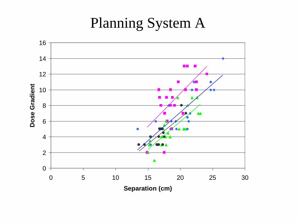
Planning System A
0
2
4
6
8
10
12
14
16
0 5 10 15 20 25 30
Separation (cm)
Do
se G
rad
ien
t

Junction Phantom
• Overdoses of over
20% were found with
some techniques in the
junction between SCF
and tangential fields

3D phantom (K Venables
Liz Miles)
7inf
3.5inf
Central
3.5sup
7sup
Medial Reference
LungApex
Posterior Field Edge
(50%)
Results: 14 planning systemsMean dose 0.987 (SD 0.013)All relative measurements within 5% of calculated; largest discrepancies at edge of fieldSmall number of depts still not using lung correction

Pencil Beam II, n=7
Sup 7 Med
Sup 7 Lung
Sup 7 Ref
Sup 35 Med
Sup 35 Apex
Sup 35 Lung
Sup 35 Ref
CA Med
CA Apex
CA Lung
CA Ref
Inf 35 Med
Inf 35 Lung
Inf 35 Ref
Inf 7 Lung
Mea
n m
easu
red
/ cal
cula
ted
rela
tive
dose
1.04
1.02
1.00
.98
.96
.94
.92
Collapsed Cone, n=2
Sup 7 Med
Sup 7 Lung
Sup 7 Ref
Sup 35 Med
Sup 35 Apex
Sup 35 Lung
Sup 35 Ref
CA Med
CA Apex
CA Lung
CA Ref
Inf 35 Med
Inf 35 Lung
Inf 35 Ref
Inf 7 Lung
Mea
n m
easu
red
/ cal
cula
ted
rela
tive
dose
1.04
1.02
1.00
.98
.96
.94
.92
Pencil Beam I, n=11
Sup 7 Med
Sup 7 Lung
Sup 7 Ref
Sup 35 Med
Sup 35 Apex
Sup 35 Lung
Sup 35 Ref
CA Med
CA Apex
CA Lung
CA Ref
Inf 35 Med
Inf 35 Lung
Inf 35 Ref
Inf 7 Lung
Mea
n m
easu
red
/ cal
cula
ted
rela
tive
dose
1.04
1.02
1.00
.98
.96
.94
.92
Beam Model Systems, n=2
S7 Med
S7 Lung
S7 Ref
S35 Med
S35 Apex
S35 Lung
S35 Ref
CA Med
CA Apex
CA Lung
CA Ref
Inf 35 Med
Inf 35 Lung
Inf 35 Ref
Inf 7 Lung
Mea
n m
easu
red
/ cal
cula
ted
rela
tive
dose
1.04
1.02
1.00
.98
.96
.94
.92
Stored Beam Systems, n=12
Sup 7 Med
Sup 7 Lung
Sup 7 Ref
Sup 35 Med
Sup 35 Apex
Sup 35 Lung
Sup 35 Ref
CA Med
CA Apex
CA Lung
CA Ref
Inf 35 Med
Inf 35 Lung
Inf 35 Ref
Inf 7 Lung
Mea
n m
easu
red
/ cal
cula
ted
rela
tive
dose
1.04
1.02
1.00
.98
.96
.94
.92
a
.b
.
c
.
d.
e
.
Figure 3-11 Accuracy of algorithms in the 3D breast phantom
3D phantom

PROSTATE PHANTOM for RT01
Moore AR, Warrington AP, Aird EG,
Bidmead AM, Dearnaley DP
Small ion chamber for
immediate dose measured
at selected 3D points
Independent Dose check
with Alanine-from NPL
Constructed from water/WEP
“Silver” prostate used for
localisation
Measuring points located in 3
planes
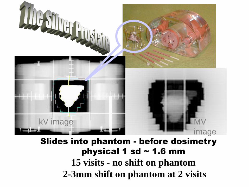
Slides into phantom - before dosimetry
physical 1 sd ~ 1.6 mm
15 visits - no shift on phantom
2-3mm shift on phantom at 2 visits
kV image MV
image

All measured data
differences from TPS
All relative data
10.0
8.0
6.0
4.0
2.0
0.0
-2.0
-4.0
-6.0
-8.0
-10.0
-12.0
-14.0
-16.0
-18.0
-20.0
-22.0
-24.0
-26.0
80
60
40
20
0
Std. Dev = 4.01
Mean = -1.8
N = 204.00
%
Number
of data
Mean of all points
-1.8 %SD 4.0%
Mean of ref pt: -1.0%
(with Alanine: -0.5%)
Prescription point
within 2%

Some more RT01 results
• Rectal dose generally OK
• ……but plan data used critically to determine new Rectal Volume constraints ( ref: Dose-volume constraints to reduce rectal side effects from prostate radiotherapy: evidence from MRC RT01 Trial ISRCTN 47772397.Gulliford SL, Foo K, Morgan RC, Aird EG, Bidmead AM, Critchley H, Evans PM, Gianolini S, Mayles WP, Moore AR, Sánchez-Nieto B, Partridge M, Sydes MR, Webb S, Dearnaley DP.

0.7 0.5 0.3 0.5Gy
3x3 or 4x4
Depending on
MLC
Parsport
Clark et al R+O 93(2009)102-109
PARSPORT TRIAL TPS tests

Parsport
• 5-6 hours of machine measurement
• CIRS head and neck phantom
– Conventional plan
– IMRT plan
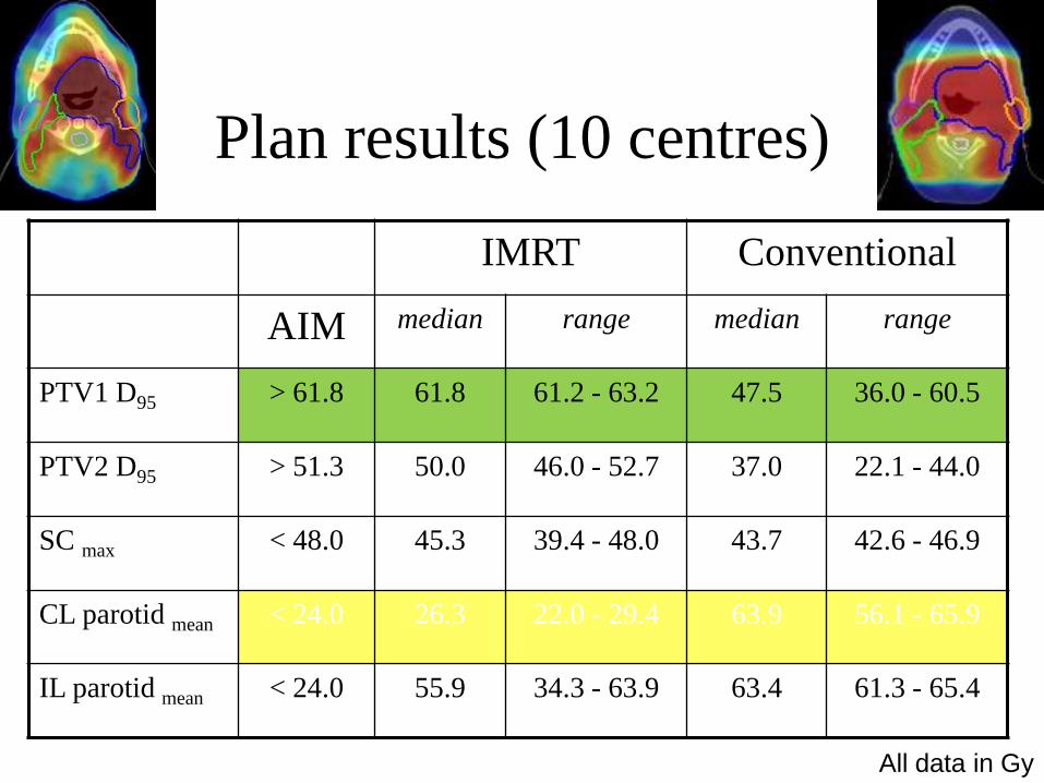
Plan results (10 centres)
IMRT Conventional
AIM median range median range
PTV1 D95 > 61.8 61.8 61.2 - 63.2 47.5 36.0 - 60.5
PTV2 D95 > 51.3 50.0 46.0 - 52.7 37.0 22.1 - 44.0
SC max < 48.0 45.3 39.4 - 48.0 43.7 42.6 - 46.9
CL parotid mean < 24.0 26.3 22.0 - 29.4 63.9 56.1 - 65.9
IL parotid mean < 24.0 55.9 34.3 - 63.9 63.4 61.3 - 65.4
All data in Gy

Catharine Clark1,2,3,4, M Hussein1,4, Y Tsang3,5, R Thomas2, C
Gouldstone2, G Bass2,
D Maughan2, J Snaith2, S Bolton6,7, D Wilkinson3,5, L Ciurlionis3,5,
R Nutbrown2, K Venables3,5, A Nisbet1,4
1Royal Surrey County Hospital, 2National Physical Laboratory, 3Radiotherapy
Trials QA (RTTQA), 4University of Surrey, 5Mount Vernon Hospital, 6Christie
Hospital, 7Institute of Physics and Engineering in Medicine

PTW Octavius II phantom with various detectors
Alanine
PTW Semiflex
ion chambers
Gafchromic
film
PTW 729
2D array

3DTPS test plan
OAR
PTVs
Tsang et al. Br J Radiol. 2013 Feb;86(1022)
1st Coronal
Plane, through
multiple PTVs
2nd Coronal
Plane,
through PTVs
and OAR
Sagittal
Plane,
through PTVs
and OAR
0
10
20
30
40
50
60
70
-15 -13 -11 -9 -7 -5 -3 -1 1 3 5 7 9 11 13 15
Fre
qu
ency
Point dose percentage difference
Mean 0.1%
SD 2.6%
0
10
20
30
40
50
60
-15 -13 -11 -9 -7 -5 -3 -1 1 3 5 7 9 11 13 15
Fre
qu
ency
Point dose percentage difference
Mean 0.2%
SD 2.0%
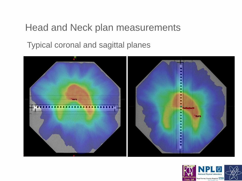
Head and Neck plan measurements
Typical coronal and sagittal planes

Point dose differences in clinical plans
0
5
10
15
20
25
30
35
-15 -13 -11 -9 -7 -5 -3 -1 1 3 5 7 9 11 13 15
Fre
qu
ency
Point dose percentage difference
Mean 0.2%
SD 2.0%
0
5
10
15
20
25
30
-15 -13 -11 -9 -7 -5 -3 -1 1 3 5 7 9 11 13 15F
req
uen
cy
Point dose percentage difference
Mean -0.2%
SD 1.6%

Gamma analysis results
mean pass rate
Clinical 3DTPS
2%/2mm 93.6% 91.5%
3%/2mm 97.9% 96.3%
3%/3mm 99.3% 98.3%
4%/4mm 99.9% 99.7%
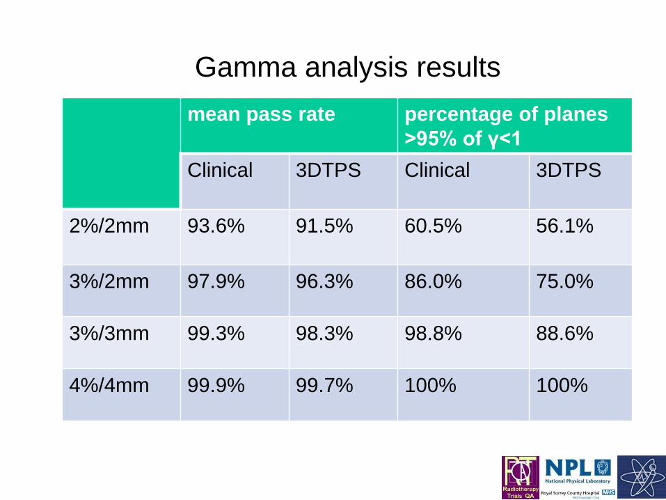
mean pass rate percentage of planes
>95% of γ<1
Clinical 3DTPS Clinical 3DTPS
2%/2mm 93.6% 91.5% 60.5% 56.1%
3%/2mm 97.9% 96.3% 86.0% 75.0%
3%/3mm 99.3% 98.3% 98.8% 88.6%
4%/4mm 99.9% 99.7% 100% 100%
Gamma analysis results

Gamma analysis results at 2%/2mm
mean pass
rate
percentage of
planes >95% of
γ<1
Breast 99.8% 100%
Prostate and
Nodes
94.9% 73.1%
Head and
Neck
93.4% 55.4%
3DTPS 91.5% 56.1%

Rotational Audit Issues identified
• Lack of couch modelling
• Minimum leaf gap too small
• High modulation / high MUs
• Non-continuously variable dose rate
• Lack of information as to what some TPS/Linac
combinations are capable of achieving
• Lasers and barometers

Conclusions
• A national dosimetry audit of rotational radiotherapy has been undertaken
• More than 93% of analysed planes achieved more than 95% pass rates for
gamma parameters of 3%/3mm
• For many systems 3%/2mm were better criteria
• The majority of centres achieved accurate implementation of TPS modelling
and delivery for VMAT and Tomotherapy
• Evaluation of the standards which others starting a VMAT program should
be able to achieve

Conclusion
• The implementation of QA in radiotherapy has become vitally important in recent years. Often, as has been demonstrated here, a clinical trial has led the way to the general benefit of all patients receiving radiotherapy. By pursuing QA in the first year of the clinical trial, the standard of treatment was set and any later uncertainties when analysing the results were avoided. Wariness at each centre visited was replaced by active co-operation and satisfaction with the high standards that could be achieved and maintained. In addition, these visits gave an opportunity for mutual exchange of ideas.
Aird et al R+O 36(1995)235-245

Acknowledgements
• All staff who have worked on clinical trials
particularly those who have provided slides
and information
• Funding bodies
• NPL and IPEM for collaberative work