introduction to dosimetry - canadian nuclear safety commission · february 2012 info - 0827,...
TRANSCRIPT
Introduction to DosimetryINFO-0827
February 2012
Introduction to Dosimetry
© Minister of Public Works and Government Services Canada (PWGSC) 2011 PWGSC catalogue number CC172-79/2011-PDF ISBN 978-1-100-19735-7
Published by the Canadian Nuclear Safety Commission (CNSC) CNSC catalogue number: INFO-0827
Extracts from this document may be reproduced for individual use without permission provided the source is fully acknowledged. However, reproduction in whole or in part for purposes of resale or redistribution requires prior written permission from the Canadian Nuclear Safety Commission.
Également publié en français sous le titre de : Introduction à la dosimétrie
Document availabilityThis document can be viewed on the CNSC Web site at nuclearsafety.gc.ca
Canadian Nuclear Safety Commission 280 Slater Street P.O. Box 1046, Station B Ottawa, Ontario K1P 5S9 CANADA Tel.: 613-995-5894 or 1-800-668-5284 (in Canada only) Facsimile: 613-995-5086 Email: [email protected] Web site: nuclearsafety.gc.ca
TABLE OF CONTENTS
1.0 OVERVIEW 1
2.0 INTRODUCTION 2
3.0 WHAT IS DOSIMETRY? 3
4.0 FUNDAMENTAL CONCEPTS 5
4.1 Structure of the atom 5
4.2 Radioactive decay 5
4.3 Types of radiation 5
5.0 DOSE CONCEPTS 8
5.1 Absorbed dose 8
5.2 Equivalent dose 8
5.3 Effective dose 9
5.4 About dose limits 9
6.0 DOSE REPORTING AND RECORD KEEPING 11
7.0 THE CNSC’S ROLE IN DOSIMETRY 12
8.0 EXTERNAL DOSIMETRY 13
8.1 About dosimeters 13
8.2 Dosimetry for photon and beta radiation 15
8.3 Dosimetry for neutron radiation 20
8.4 Measurement uncertainty in external dosimetry 22
8.5 Methods to monitor workers for external radiation exposure 22
9.0 INTERNAL DOSIMETRY 24
9.1 In-vivo bioassay (direct measurement of radioactivity in the body) 24
9.2 In-vitro bioassay (measurement of radioactivity in substances excreted by the body) 26
9.3 Measurement of radon decay products in workplace air 28
9.4 How internal radiation doses are calculated 30
9.5 Measurement uncertainty in internal dosimetry 30
9.6 Methods to monitor workers for internal radiation exposure 31
10.0 LICENSED DOSIMETRY SERVICES 33
10.1 How the CNSC issues dosimetry service licences 33
10.2 How the CNSC monitors licensed dosimetry services 33
SUMMARY 34
GLOSSARY 35
REFERENCES 38

February 2012 INFO - 0827, Introduction to Dosimetry
1
1.0 OVERVIEW
This document defines dosimetry, introduces the fundamentals of dosimetry techniques and practices, and explains the importance of choosing appropriate dosimetry methods. It addresses how these techniques are used to demonstrate compliance with the Radiation Protection Regulations.
Different types of ionizing radiation are described, and radioactive decay is explained. The concepts of absorbed, equivalent and effective dose are introduced and discussed as they relate to the dose limits in the Radiation Protection Regulations.
Different methods (both direct and indirect) used to ascertain doses are provided, and the various dosimetry techniques used to measure doses – depending on whether the nuclear substance or radiation source is inside or outside of the body – are presented.
The mandatory use of a licensed dosimetry service, which is required when effective doses are equal to or higher than 5 milliSieverts (mSv) per year, is also described. The document explains how regulatory requirements govern licensed dosimetry services in order to ensure that doses are appropriately measured and monitored.
This document also outlines how doses recorded by a licensed dosimetry service for a given worker must be submitted to Health Canada’s National Dose Registry (NDR), and how the Canadian Nuclear Safety Commission (CNSC) uses the NDR to fulfill its regulatory obligations. Finally, the CNSC’s role in the regulation of dosimetry is explained.
February 2012 INFO - 0827, Introduction to Dosimetry
2
2.0 INTRODUCTION
Radiation is energy that is transmitted in the form of waves or particles. There are two types of radiation: ionizing and non-ionizing. The type of radiation discussed in this document is ionizing radiation.
People are constantly exposed to low levels of ionizing radiation that are naturally present in the environment. Other sources of exposure include some medical treatments and other activities that involve the use of radioactive materials. Research has shown that exposure to ionizing radiation above certain levels can cause adverse health effects, including cancer and hereditary effects (effects that can be passed on to offspring). Therefore, exposure to ionizing radiation is monitored and controlled.
The Canadian Nuclear Safety Commission (CNSC) has the mandate to regulate the use of nuclear energy and materials, in order to protect the health, safety and security of Canadians and the environment. In carrying out this mandate, the CNSC is responsible for protecting the public and workers from exposure to ionizing radiation. This document focuses on ionizing radiation and the resulting doses it may cause.
A key way in which the CNSC fulfills its mandate is through its Radiation Protection Regulations [1], which set radiation dose limits based on sound science. In addition to ensuring that dose limits are respected, the CNSC requires radiation doses to be kept as low as reasonably achievable (ALARA), with social and economic factors taken into account.
As Canada’s nuclear regulator, the CNSC licenses various activities that involve nuclear energy and radioactive sources, and it monitors radiation doses that result from these licensed activities. The CNSC evaluates methods for measuring and calculating radiation doses, to ensure the methods are sufficiently accurate and precise relative to the risk of the exposures being measured. The measurement and calculation of radiation doses is called dosimetry, whose methods can be classified in three general categories:
• Direct monitoring: Direct monitoring, also called personal dosimetry, is used primarily (but not exclusively) to determine doses to individuals who are exposed to radiation related to their work activities. In Canada, if effective doses of radiation could exceed 5 mSv per year, personal dosimetry must be performed by a licensed dosimetry service.
• Indirect monitoring: Indirect monitoring methods use measured dose rates in air, measured concentrations of nuclear substances in air along with other information to estimate doses.
• Indirect monitoring: Indirect monitoring using environmental pathways analysis, along with other information to estimate doses.
Personal dosimetry techniques vary and depend partly on whether the source of radiation is outside the body (external) or taken into the body (internal). Personal dosimeters are used to measure external radiation exposures. For internal exposures, two principal dosimetry techniques are involved: the measurement of the presence of nuclear substances in the body, or the measurement of nuclear substances excreted by the body.
In Canada, radiation doses to individuals rarely exceed regulatory limits, partly because CNSC licensees must have radiation protection programs to keep doses ALARA. The CNSC reviews and approves required radiation protection programs as part of its licensing process. It also verifies licensee compliance with radiation protection requirements after a licence has been issued.
February 2012 INFO - 0827, Introduction to Dosimetry
3
3.0 WHAT IS DOSIMETRY?
Dosimetry is the act of measuring or estimating radiation doses and assigning those doses to individuals. The Radiation Protection Regulations [1] require licensees to control doses to workers and to the public and to ascertain these doses. Licensees must use a licensed dosimetry service to measure doses when there is a reasonable probability that the effective annual dose to a nuclear energy worker (NEW) will exceed 5 mSv.
There are two types of radiation exposure:
• external exposure, which occurs when the radiation source or nuclear substance is outside of the body.
• internal exposure, which occurs when the radiation is emitted by nuclear substances inside the body.
Three methods are commonly used to determine radiation doses to humans: personal dosimetry; indirect monitoring using measured dose rates or airborne concentrations of nuclear substances; and indirect monitoring using environmental pathways analysis.
1. Personal dosimetry
Personal dosimetry is used primarily to ascertain doses to workers in the nuclear industry. External exposures are typically monitored by the use of small radiation detectors called dosimeters, which are worn on the person. Internal exposures are typically monitored by measuring the presence of nuclear substances in the body, or by measuring nuclear substances excreted by the body. In Canada, dosimetry service providers are licensed by the CNSC.
2. Indirect monitoring using measured dose rates or airborne concentrations of nuclear substances
This monitoring method is used when a person occupies an area with a known concentration of airborne radioactivity, or a known radioactive field, for a known period of time. This knowledge can be used in conjunction with other information to estimate the person’s radiation dose during the particular occupancy.
This approach is often used where an airborne radioactive substance, such as radon progeny, is the source of the exposure and personal monitoring cannot be carried out. In such instances, the concentration in air of radon progeny (either in a specific area or in close proximity to each worker) is measured by air sampling or another method, and the time spent in the areas by a person or persons is recorded. Measurements such as concentrations of airborne radioactivity, recorded period of occupancy and air inhalation rates can then be used to estimate a person’s dose. This method is often called workplace monitoring.
3. Indirect monitoring using environmental pathways analysis
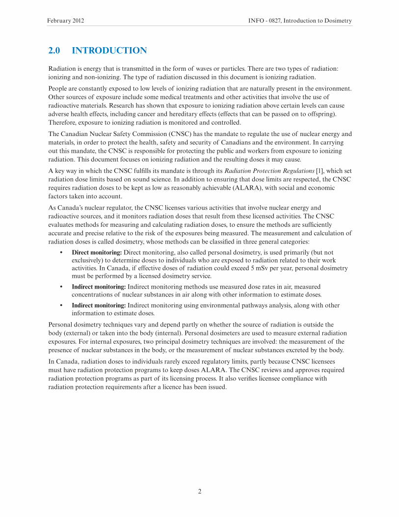
A second indirect monitoring method is based on what is called environmental pathways analysis. In this method, population exposures are modelled by measuring the amount of nuclear substances released to the environment from a source (such as an exhaust stack), or by measuring the presence of nuclear substances in the environment occupied by individuals.
Environmental pathways analysis is the most common approach for estimating public doses. In this method, doses are calculated using relevant site-specific data such as the amount of nuclear substances in air, water or locally grown produce. While these dose calculations are only estimates, they are quite realistic, since they are based on measured concentrations in air, water and food. Conservative data – regarding the activities and dietary habits of people living in the vicinity of CNSC-licensed facilities – is also incorporated. Figure 1 gives a graphical presentation of environmental pathways analysis.

February 2012 INFO - 0827, Introduction to Dosimetry
4
FIGURE 1. Environmental pathways analysis.
February 2012 INFO - 0827, Introduction to Dosimetry
5
4.0 FUNDAMENTAL CONCEPTS
To understand dosimetry, one must also understand other related concepts such as atomic structure, radioactive decay and types of radiation. These concepts are presented in sections 4.1, 4.2 and 4.3, respectively.
4.1 Structure of the atom
An atom is made up of a central nucleus that contains two types of subatomic particles: protons and neutrons. Protons have a positive electric charge, and neutrons have no charge. Therefore, the nucleus has an overall positive charge.
A third type of subatomic particle called an electron surrounds the atom’s nucleus. Electrons are negatively charged and have a much smaller mass than both neutrons and protons.
Each element of the periodic table represents an atom with a unique number of protons. The number of protons in an atom is called the atomic number. The total number of protons and neutrons in the nucleus is called the atomic mass number.
Atoms with the same number of protons (that have the same atomic number and that are therefore of the same element), but a different number of neutrons (and therefore different atomic mass numbers) are called isotopes. As an example, hydrogen has the following three isotopes:
• hydrogen (abbreviated as H-1), which has one proton
• deuterium (H-2), which has one proton and one neutron
• tritium (H-3), which has one proton and two neutrons
4.2 Radioactive decay
Isotopes can be divided into two categories: stable isotopes and radioactive isotopes. In this document, radioactive isotopes will also be referred to as “radioactive nuclear substances” or “nuclear substances”.
In a stable isotope, the number of protons and neutrons in the nucleus is balanced in a stable configuration.
A radioactive isotope is unstable because of an unbalanced number of protons and neutrons in the nucleus (often when the ratio of neutrons to protons is too low). The nucleus of a radioactive isotope spontaneously disintegrates through a process known as radioactive decay, during which it emits excess energy in order to become stable. This energy is called radiation.
The result of the radioactive decay process is the creation of a more stable nucleus. However, the newly formed nucleus may still be radioactive (although more stable than the previous nucleus). The process will continue until a stable nucleus is created, at which point radioactive decay will stop. The time it takes for a nuclear substance to decay to one half of its original value is referred to as the “half-life”. Each nuclear substance has a unique half-life.
The quantity of radiation emitted by an isotope of an element is called the activity, and the unit used to measure this activity is the becquerel (Bq). One becquerel is equal to the decay of one atom per second.
4.3 Types of radiation
Radiation is energy that is transmitted in the form of waves or particles. Radiation is generally classified as ionizing radiation or non-ionizing radiation.
Ionizing radiation has sufficient energy to remove an electron from an atom. It includes the radiation that comes from both natural and man-made sources.
Non-ionizing radiation has less energy than ionizing radiation and cannot remove an electron from an atom. Examples of non-ionizing radiation include radio waves and microwaves.
February 2012 INFO - 0827, Introduction to Dosimetry
6
Background radiation is the radiation constantly present in the environment. It is emitted by natural and artificial sources.
This document focuses on ionizing radiation and the resulting doses that it may cause. Four main types of ionizing radiation are discussed:
• alpha
• beta
• photon (X-rays and gamma rays)
• neutron
Alpha and beta radiation may be emitted while a nucleus undergoes radioactive decay. Alpha and beta particles are often also accompanied by the release of additional energy, in the form of photon radiation.
Neutron radiation can be produced from nuclear fission, which occurs only for certain nuclear substances with a high atomic number, such as uranium and plutonium. Except for several fission fragments with very short half-lives, and californium-252, which undergoes spontaneous fission, there are no other radioisotopes that emit neutrons. Other neutron sources depend on nuclear reactions for the emission of neutrons.
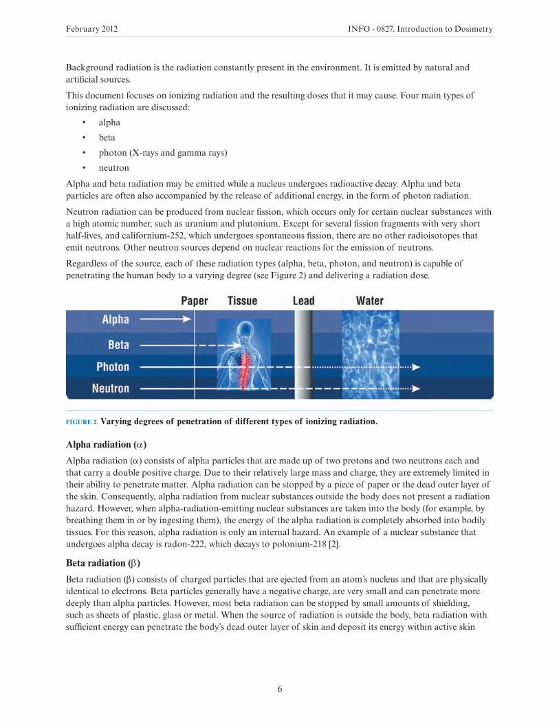
Regardless of the source, each of these radiation types (alpha, beta, photon, and neutron) is capable of penetrating the human body to a varying degree (see Figure 2) and delivering a radiation dose.
FIGURE 2. Varying degrees of penetration of different types of ionizing radiation.
Alpha radiation (α)
Alpha radiation (α) consists of alpha particles that are made up of two protons and two neutrons each and that carry a double positive charge. Due to their relatively large mass and charge, they are extremely limited in their ability to penetrate matter. Alpha radiation can be stopped by a piece of paper or the dead outer layer of the skin. Consequently, alpha radiation from nuclear substances outside the body does not present a radiation hazard. However, when alpha-radiation-emitting nuclear substances are taken into the body (for example, by breathing them in or by ingesting them), the energy of the alpha radiation is completely absorbed into bodily tissues. For this reason, alpha radiation is only an internal hazard. An example of a nuclear substance that undergoes alpha decay is radon-222, which decays to polonium-218 [2].
Beta radiation (β)
Beta radiation (β) consists of charged particles that are ejected from an atom’s nucleus and that are physically identical to electrons. Beta particles generally have a negative charge, are very small and can penetrate more deeply than alpha particles. However, most beta radiation can be stopped by small amounts of shielding, such as sheets of plastic, glass or metal. When the source of radiation is outside the body, beta radiation with sufficient energy can penetrate the body’s dead outer layer of skin and deposit its energy within active skin
February 2012 INFO - 0827, Introduction to Dosimetry
7
cells. However, beta radiation is very limited in its ability to penetrate to deeper tissues and organs in the body. Beta-radiation-emitting nuclear substances can also be hazardous if taken into the body. An example of a nuclear substance that undergoes beta emission is tritium (hydrogen-3), which decays to helium-3 [2].
Photon radiation (gamma [γ] and X-ray)
Photon radiation is electromagnetic radiation. Two types of photon radiation are of interest for the purpose of dosimetry: gamma (γ) and X-ray.
Gamma radiation consists of photons that originate from within the nucleus, and X-ray radiation consists of photons1 that originate from outside the nucleus.
While “gamma radiation” is a familiar term for electromagnetic radiation, this document uses the term “photon” to denote electromagnetic radiation – so as to include X-rays generated from nuclear substances. Photon radiation can penetrate very deeply and sometimes can only be reduced in intensity by materials that are quite dense, such as lead or steel. In general, photon radiation can travel much greater distances than alpha or beta radiation, and it can penetrate bodily tissues and organs when the radiation source is outside the body. Photon radiation can also be hazardous if photon-emitting nuclear substances are taken into the body. An example of a nuclear substance that undergoes photon emission is cobalt-60, which decays to nickel-60 [2].
Neutron radiation (n)
Apart from cosmic radiation, spontaneous fission is the only natural source of neutrons (n). A common source of neutrons is the nuclear reactor, in which the splitting of a uranium or plutonium nucleus is accompanied by the emission of neutrons. The neutrons emitted from one fission event can strike the nucleus of an adjacent atom and cause another fission event, inducing a chain reaction. The production of nuclear power is based upon this principle.
All other sources of neutrons depend on reactions where a nucleus is bombarded with a certain type of radiation (such as photon radiation or alpha radiation), and where the resulting effect on the nucleus is the emission of a neutron. Neutrons are able to penetrate tissues and organs of the human body when the radiation source is outside the body. Neutrons can also be hazardous if neutron-emitting nuclear substances are deposited inside the body. Neutron radiation is best shielded or absorbed by materials that contain hydrogen atoms, such as paraffin wax and plastics. This is because neutrons and hydrogen atoms have similar atomic weights and readily undergo collisions between each other.
1 A photon is an elementary particle that is the basic quanta of light and all other forms of electromagnetic radiation.
February 2012 INFO - 0827, Introduction to Dosimetry
8
5.0 DOSE CONCEPTS
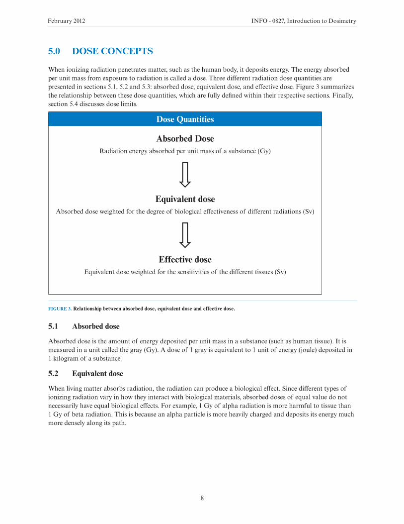
When ionizing radiation penetrates matter, such as the human body, it deposits energy. The energy absorbed per unit mass from exposure to radiation is called a dose. Three different radiation dose quantities are presented in sections 5.1, 5.2 and 5.3: absorbed dose, equivalent dose, and effective dose. Figure 3 summarizes the relationship between these dose quantities, which are fully defined within their respective sections. Finally, section 5.4 discusses dose limits.
Dose Quantities
Absorbed DoseRadiation energy absorbed per unit mass of a substance (Gy)
Equivalent doseAbsorbed dose weighted for the degree of biological effectiveness of different radiations (Sv)
Effective doseEquivalent dose weighted for the sensitivities of the different tissues (Sv)
FIGURE 3. Relationship between absorbed dose, equivalent dose and effective dose.
5.1 Absorbed dose
Absorbed dose is the amount of energy deposited per unit mass in a substance (such as human tissue). It is measured in a unit called the gray (Gy). A dose of 1 gray is equivalent to 1 unit of energy (joule) deposited in 1 kilogram of a substance.
5.2 Equivalent dose
When living matter absorbs radiation, the radiation can produce a biological effect. Since different types of ionizing radiation vary in how they interact with biological materials, absorbed doses of equal value do not necessarily have equal biological effects. For example, 1 Gy of alpha radiation is more harmful to tissue than 1 Gy of beta radiation. This is because an alpha particle is more heavily charged and deposits its energy much more densely along its path.
February 2012 INFO - 0827, Introduction to Dosimetry
9
A radiation weighting factor (wR) is used to equate different types of radiation with different levels of biological effectiveness. These weighting factors are provided in schedule 2 of the Radiation Protection Regulations [1]. In this schedule, absorbed doses are multiplied by their respective radiation weighting factors in order to obtain equivalent doses. The concept of equivalent dose allows different types of ionizing radiation to be considered equally with respect to their potential to cause harm. Equivalent dose is expressed in a unit called the sievert (Sv). It can therefore be stated that an equivalent dose of 1 Sv of alpha radiation will, on average, have the same biological effectiveness as an equivalent dose as 1 Sv of beta radiation.
5.3 Effective dose
Different tissues and organs may vary in how they respond biologically to a given type of radiation. For example, a given equivalent dose (per sievert) has a higher risk of inducing fatal cancer in the lung than in the thyroid gland. Effects can be different both in type and magnitude and must be considered when assessing radiation exposure’s overall detriment to human health. This is taken into account by multiplying the equivalent dose to an organ or tissue by its respective weighting factor (wT).
Organ and tissue weighting factors are provided in schedule 1 of the Radiation Protection Regulations [1]. Weighted doses can be calculated for each organ or tissue using their respective weighting factors, and then added together to provide a total effective dose to the body as a whole. Organ and tissue weighting factors consider the relative susceptibility of a body part to cancer, death and hereditary effects. Effective dose is also expressed using the sievert (Sv).
5.4 About dose limits
Dose limits prescribed by the Radiation Protection Regulations [1] refer specifically to equivalent and effective dose. Limits are in place to minimize the risk of adverse health effects caused by radiation exposure. These effects are classified as one of two types: stochastic or deterministic:
• A stochastic effect is an adverse health effect that occurs with a probability that is proportional to the radiation dose received (the higher the radiation dose, the more likely the effect is to occur). The severity of a stochastic effect does not depend on the dose. Examples of stochastic effects include cancers and hereditary effects.
• A deterministic effect is an adverse health effect caused by exposure to radiation and whose severity increases with doses above a certain threshold (the level of harm increases as the dose increases above that threshold). Examples of deterministic effects include burns to the skin and cataracts of the eye
Limits on effective doses
Limits on effective doses that are prescribed by the Radiation Protection Regulations (see Table 1) [1] reduce the probability of stochastic effects. These restrictions are also sufficient to limit deterministic effects in almost all tissues and organs.
February 2012 INFO - 0827, Introduction to Dosimetry
10
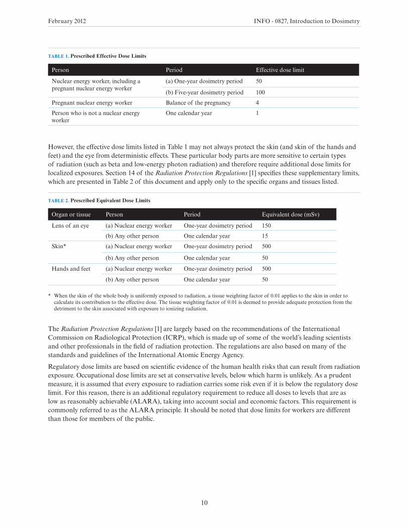
TABLE 1. Prescribed Effective Dose Limits
Person Period Effective dose limit
Nuclear energy worker, including a pregnant nuclear energy worker
(a) One-year dosimetry period 50
(b) Five-year dosimetry period 100
Pregnant nuclear energy worker Balance of the pregnancy 4
Person who is not a nuclear energy worker
One calendar year 1
However, the effective dose limits listed in Table 1 may not always protect the skin (and skin of the hands and feet) and the eye from deterministic effects. These particular body parts are more sensitive to certain types of radiation (such as beta and low-energy photon radiation) and therefore require additional dose limits for localized exposures. Section 14 of the Radiation Protection Regulations [1] specifies these supplementary limits, which are presented in Table 2 of this document and apply only to the specific organs and tissues listed.
TABLE 2. Prescribed Equivalent Dose Limits
Organ or tissue Person Period Equivalent dose (mSv)
Lens of an eye (a) Nuclear energy worker One-year dosimetry period 150
(b) Any other person One calendar year 15
Skin* (a) Nuclear energy worker One-year dosimetry period 500
(b) Any other person One calendar year 50
Hands and feet (a) Nuclear energy worker One-year dosimetry period 500
(b) Any other person One calendar year 50
* When the skin of the whole body is uniformly exposed to radiation, a tissue weighting factor of 0.01 applies to the skin in order to calculate its contribution to the effective dose. The tissue weighting factor of 0.01 is deemed to provide adequate protection from the detriment to the skin associated with exposure to ionizing radiation.
The Radiation Protection Regulations [1] are largely based on the recommendations of the International Commission on Radiological Protection (ICRP), which is made up of some of the world’s leading scientists and other professionals in the field of radiation protection. The regulations are also based on many of the standards and guidelines of the International Atomic Energy Agency.
Regulatory dose limits are based on scientific evidence of the human health risks that can result from radiation exposure. Occupational dose limits are set at conservative levels, below which harm is unlikely. As a prudent measure, it is assumed that every exposure to radiation carries some risk even if it is below the regulatory dose limit. For this reason, there is an additional regulatory requirement to reduce all doses to levels that are as low as reasonably achievable (ALARA), taking into account social and economic factors. This requirement is commonly referred to as the ALARA principle. It should be noted that dose limits for workers are different than those for members of the public.
February 2012 INFO - 0827, Introduction to Dosimetry
11
6.0 DOSE REPORTING AND RECORD KEEPING
The CNSC requires all licensees to ascertain and record doses assigned to anyone who performs duties associated with licensed activities or who is present at the site of licensed activities. Licensees must make dose records available and report them to the CNSC.
Where dose measurements are recorded by a licensed dosimetry service, records for nuclear energy workers (NEWs) must also be submitted to the National Dose Registry (NDR), along with specific personal information identified in the Radiation Protection Regulations [1]. A dose to an individual that has been submitted to the NDR is commonly referred to as a “dose of record”.
The NDR is a database, owned and operated by Health Canada, which tracks the lifetime dose history of registered individuals. Health Canada provides the CNSC with access to the NDR and advises the CNSC of any records indicating that a dose limit has been exceeded. Prompt identification of such records allows the CNSC to act immediately to ensure that licensees have taken appropriate actions.
Access to the NDR allows the CNSC to gain information on trends of dose data for facilities or groups of facilities; detailed dose information for individuals and licensees; and necessary information for health studies, including epidemiological studies.
The CNSC has a memorandum of understanding with Health Canada, which sets out Health Canada’s responsibilities to operate the registry, to maintain the information required under the Radiation Protection Regulations [1], and to make this information available to the CNSC electronically and via written request.
February 2012 INFO - 0827, Introduction to Dosimetry
12
7.0 THE CNSC’S ROLE IN DOSIMETRY
The CNSC’s role in assessing licensee dosimetry programs varies based on potential doses to workers and the public from a given licensed activity:
• Where effective doses to NEWs may exceed 5 mSv per one-year dosimetry period: If effective doses to NEWs are expected to exceed 5 mSv in a one-year dosimetry period, the Radiation Protection Regulations [1] require licensees to use licensed dosimetry services to measure and monitor radiation doses. This requirement ensures that doses are monitored with sufficient accuracy and precision.
• Where effective doses to NEWs are not expected to exceed 5 mSv per one-year dosimetry period: If effective doses are not expected to exceed 5 mSv per one-year dosimetry period, licensees must ascertain doses to their employees, but are not required to use licensed dosimetry services. In such cases, licensees may choose to use licensed dosimetry services or to determine doses using other acceptable techniques based on the level of risk in question. When considering an application (for a facility or activity) that proposes to measure doses by means other than via a licensed dosimetry service, the CNSC evaluates the applicant’s suggested measurement method in consideration of the relative potential risk.2
In any case, the CNSC evaluates the measurement method used to determine doses to workers and the public – both when deciding whether to grant a licence and during its ongoing evaluations of licensee compliance. The CNSC also ensures that doses received by the workers and the general public are ascertained, recorded and reported as per section 27 of the Nuclear Safety and Control Act. The CNSC inspects and monitors compliance with these requirements to ensure that radiation doses to people in Canada are recorded appropriately.
The remainder of this document focuses, in part, on the direct dosimetry method of estimating personal doses. It will detail the dosimetry methods used to measure radiation doses to individuals from external and internal exposure, and will outline the CNSC’s licensing process for dosimetry services.
2 Even though they are not required to do so, many licensees use licensed dosimetry services to measure and monitor radiation doses to individuals who are likely to receive annual effective doses that are far lower than 5 mSv.
February 2012 INFO - 0827, Introduction to Dosimetry
13
8.0 EXTERNAL DOSIMETRY
External dosimetry is the measurement of dose when the radiation source is outside of (or external to) the body. Therefore, in terms of dose to humans, external dosimetry is concerned with radiation that can penetrate the skin: beta, photon, and neutron radiation. Since photons and beta interact through electronic forces (interactions between charged particles) and neutrons interact through nuclear forces, their detection methods and dosimetry are substantially different.
The fundamental basis of external dosimetry is the determination of the absorbed energy in matter and, more specifically, human tissue.
Section 8.1 outlines the general characteristics of dosimeters, different dosimeter types and factors to consider when selecting a dosimeter. Section 8.2 presents three kinds of dosimeters for measuring beta and photon radiation, and section 8.3 discusses neutron dosimeters. Section 8.4 explains why external dose values calculated using a dosimeter may be uncertain and how measurement uncertainty is taken into consideration. Finally, methods to monitor workers for external radiation exposure are presented in section 8.5.
8.1 About dosimeters
A dosimeter is a small radiation detection device worn by an individual, used to measure doses from ionizing radiation.
General characteristics
Dosimeters are classified into two general categories, passive and active:
• A passive dosimeter produces a radiation-induced signal, which is stored in the device. The dosimeter is then processed and the output is analyzed.
• An active dosimeter produces a radiation-induced signal and displays a direct reading of the detected dose or dose rate in real time.
Only passive dosimeters are licensed for use by dosimetry services in Canada. Passive dosimeters are provided to users, who wear them for a defined period of time (this time period is called the exchange or wear period). The dosimeters are then returned to a dosimetry service, which processes and analyzes them and sends the dose results to users and the NDR. A dosimetry service may have several options for the length of an exchange period; for example, monthly, quarterly or semi-annually. A dosimetry service user (for example, a CNSC licensee) may choose an appropriate length of time for the exchange period.
Dosimeters used to estimate effective doses are typically worn between the waist and the neck, on the front of the torso, facing the radioactive source. Dosimeters worn on the torso are often called whole-body dosimeters. Dosimeters may also be worn on the extremities or near the eye to measure equivalent dose to these tissues.
A personal dosimeter offers the following principal advantages:
• Since it is worn on the person, it gives the best of estimate of the radiation field that the person is in.
• It measures dose with no need for interpretation by the user.
The personal dosimeters in use today are not absolute instruments, but reference instruments.3 Reference is made possible through traceable calibrations of dosimeters that are irradiated to a known amount of radiation. When a reference dosimeter is calibrated, a calibration factor can be determined; this factor relates the exposure quantity to the reported dose. Validity of the calibration is demonstrated by maintaining
3 An absolute instrument measures a quantity in absolute units by means of simple physical measurements on the device. Conversely, a reference instrument provides relative measurements that are compared to a defined quantity (the “reference measurement”).
February 2012 INFO - 0827, Introduction to Dosimetry
14
traceability of the source used to calibrate the dosimeter. The traceability is achieved by comparison of the source with a “primary standard” at a reference calibration centre, such as the National Research Council of Canada.
Choosing a dosimeter
There are many types of dosimeters, and each type has limitations. Many factors influence the quality of a dosimeter’s results. Some key considerations when choosing a dosimeter include:
• Energy dependence and angle dependence: A dosimeter’s response4 will vary depending on the energy of the radiation and the angle(s) between the source and the dosimeter’s detector.
• Radiation type to be detected: Dosimeters vary in their abilities to detect different kinds of radiation (alpha, beta, photon or neutron).
• Fading: A dosimeter’s signal can be lost or fade over time. This can be caused by external factors such as temperature, light and humidity.
• The ability to be re-read: Certain types of dosimeters lose their signals upon processing. Others retain their signals and can therefore be processed more than once.
• Minimum measurable dose or limit of detection (the lowest dose that can be measured with a certain specified confidence level): Some dosimeters are more sensitive and can detect a lower quantity of radiation than others.
• Ruggedness and ease of wear: Dosimeters differ in their ability to withstand severe environmental conditions, and some are smaller, lighter and more portable than others.
While all dosimeters have advantages and disadvantages, an ideal instrument would have the following qualities:
• low energy dependence and angle dependence
• the ability to detect several radiation types
• high resistance to fading (stability when exposed to high temperatures and humidity levels)
• a linear response to dose (response does not change with increasing dose)
• a low minimum measurable dose
No single dosimeter will have every one of these characteristics. Therefore, a dosimeter user must understand the environment where the instrument will be used, as well as the shortcomings of different dosimeter types, in order to select a dosimeter that is most appropriate to the intended use.
Dosimeter type testing
The CNSC requires dosimetry services that measure and monitor radiation doses from external exposures to undergo extensive type testing of their dosimetry systems [3]. Type testing involves calibrating a dosimetry system under a series of irradiation and storage conditions, which include some of the aforementioned considerations when choosing a dosimeter. Tests evaluate the system’s performance characteristics and quantify any sources of error (see section 8.4 of this document for discussion of measurement uncertainty in external dosimetry).
4 A dosimeter’s response is the measured or evaluated value divided by the conventionally true value.
February 2012 INFO - 0827, Introduction to Dosimetry
15
8.2 Dosimetry for photon and beta radiation
Photon radiation has greater penetrating power than alpha and beta radiation. Alpha radiation cannot penetrate the outermost dead layer of human skin, so it poses no external human health hazard.
Beta and photon radiation are hazardous to the skin and the eye, as they can deposit energy in the sensitive cells of these tissues. Beta radiation does not pose a significant risk to organs under the skin, since it typically5 cannot penetrate this deeply. Therefore, while both photon and beta radiation can contribute to an equivalent dose to the skin and eye, photons are the main contributor to the external component of the effective dose.
The penetrating ability or probability of interaction of radiation is related to the radiation’s energy. Each different nuclear substance emits a specific energy or energy range when it undergoes radioactive decay; therefore, only some beta particles and photons present an external risk to the human body.
For example, tritium (H-3) is a nuclear substance that emits only beta radiation with an average energy of 6 kiloelectronvolts [2]. This level of energy is too low to penetrate any more deeply than the dead layer of human skin. Therefore, external beta radiation from tritium is not a hazard; tritium presents solely an internal radiation hazard.
As discussed in section 8.1, dosimeters vary in the lowest amount of energy they are able to measure. The types of dosimeters described in this document, for example, respond minimally or not at all to many sources of low-energy beta radiation. A user must therefore be aware of the radiation fields to which he or she will be exposed, so as to choose a suitable dosimeter. Dosimetry services must also clearly explain the operating conditions under which they will be using particular dosimeters and justify an appropriate instrument selection.
A typical dosimeter consists of a detector inserted in a holder. Various dosimeters are configured differently; in general, the detector contains the sensitive element(s) and the holder contains the filter(s). In a dosimeter that measures photon and beta radiation, it is mainly the filter/holder that permits the instrument to differentiate between the equivalent dose to the skin or eye and the effective dose. One part of the holder may have an open window (no filter or a very thin filter) to measure equivalent dose to skin, and the other part of the detector may have a thicker filter that allows for measurement of effective dose. The thicker filter or filters shield the low-energy photons and beta radiation and allow only the more penetrating radiation to deposit energy. Some dosimeters have several filters of different thickness and composition that allow them to discriminate among different energy levels.
There are many types of dosimeters for measuring beta and photon radiation. These include film dosimeters, thermoluminescent dosimeters (TLDs), optically stimulated luminescence dosimeters (OSLDs), and direct reading dosimeters (DRDs). The following text discusses TLDs, OSLDs and DRDs in further detail [4].
5 Some very high-energy beta emitters can travel through skin to reach deeper organs and tissues. However, their contribution to dose to those tissues and organs is limited. Therefore, beta emitters located outside the body are usually considered hazardous only to skin.
February 2012 INFO - 0827, Introduction to Dosimetry
16
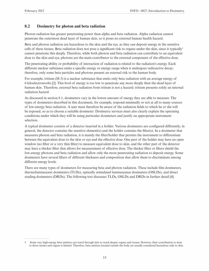
Thermoluminescent dosimeters
Since the 1950s, there has been extensive research on thermoluminescence6 and its application in dosimetry. This research, which was only widely applied beginning in the 1970s, guided the development of the TLD – which is now the most commonly used type of dosimeter in Canada and around the world.
The following basic overview explains how a TLD works:
• When ionizing radiation passes through the detector (chip), its atoms release some of their electrons.
• The electrons eventually become trapped in impurities (also called doping centres) within the dosimeter, where they remain in their excited state (a state in which their energy is elevated relative to the ground state).
• The chip is then heated in a TLD reader7 (consisting mainly of a heater, a photomultiplier tube8 and a recorder), and the trapped electrons return to the ground state and emit photons of visible light. The amount of light emitted relative to the temperature is called the glow curve. This curve is analyzed to determine the dose.
There are many types of TLDs on the market. These include lithium fluoride, calcium sulphate and lithium borate dosimeters, each of which has advantages and disadvantages.
Figure 4 displays a typical TLD and its detector.
FIGURE 4. A whole-body thermoluminescent dosimeter badge (left), and the detector found inside (right).
6 Thermoluminescence is a form of light emission exhibited by certain materials. This phenomenon occurs when previously absorbed energy from radiation is re-emitted as light, when the material is heated.
7 The TLD system consists of a dosimeter and a reader, both of which play a key role in determining dose and dose accuracy.8 A photomultiplier tube is an electronic device that amplifies weak light pulses into a large electrical signal.

February 2012 INFO - 0827, Introduction to Dosimetry
17
Optically stimulated luminescence dosimeters
OSLDs offer advantages that include the ability to be re-read and a high sensitivity (low minimum measurable dose), and they have become popular because of these favourable properties. OSLDs operate much like TLDs; their major difference is that luminescence is produced by a light beam, rather than by heat. Currently, TLDs and OSLDs were the only types of passive dosimeters licensed for use in Canada under the Radiation Protection Regulations [1].
Figure 5 displays an example of an OSLD.
FIGURE 5. A whole-body optically stimulated luminescence dosimeter.
February 2012 INFO - 0827, Introduction to Dosimetry
18
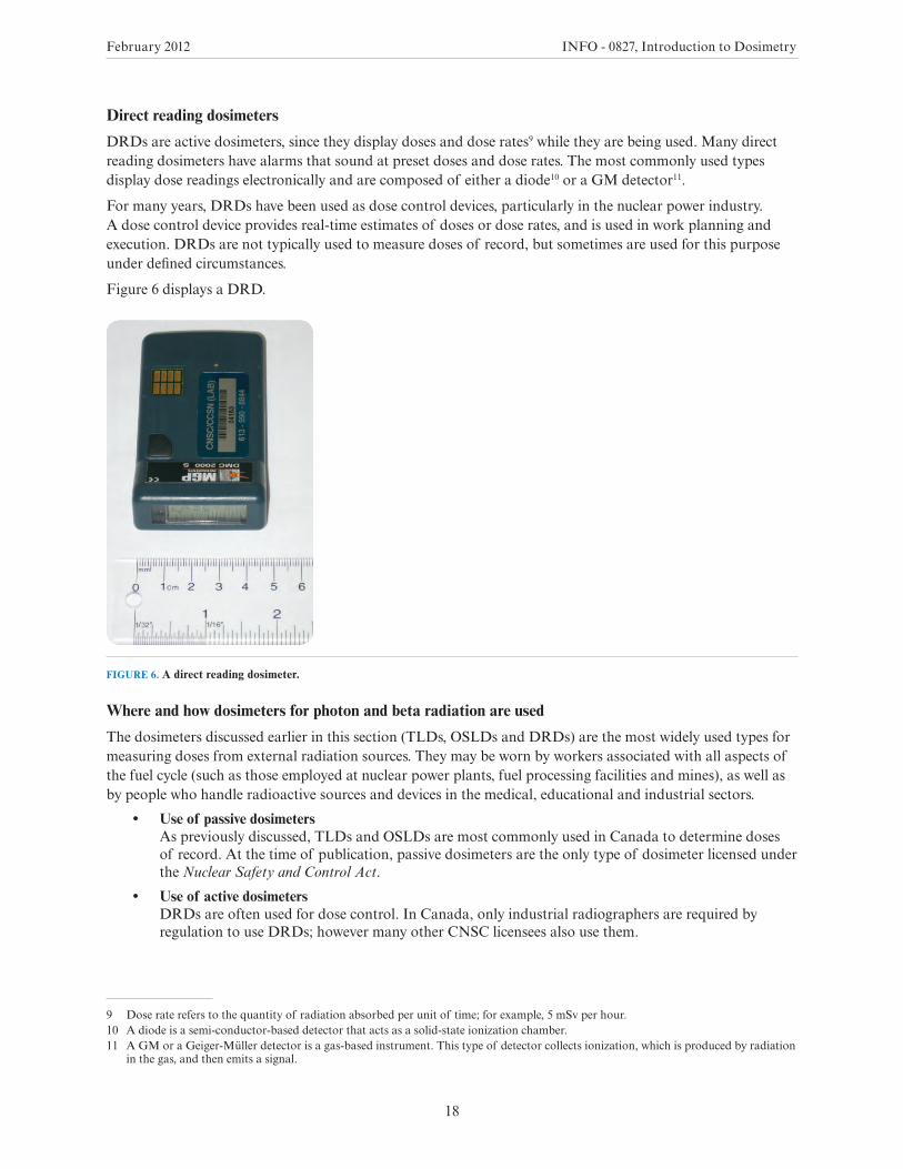
Direct reading dosimeters
DRDs are active dosimeters, since they display doses and dose rates9 while they are being used. Many direct reading dosimeters have alarms that sound at preset doses and dose rates. The most commonly used types display dose readings electronically and are composed of either a diode10 or a GM detector11.
For many years, DRDs have been used as dose control devices, particularly in the nuclear power industry. A dose control device provides real-time estimates of doses or dose rates, and is used in work planning and execution. DRDs are not typically used to measure doses of record, but sometimes are used for this purpose under defined circumstances.
Figure 6 displays a DRD.
FIGURE 6. A direct reading dosimeter.
Where and how dosimeters for photon and beta radiation are used
The dosimeters discussed earlier in this section (TLDs, OSLDs and DRDs) are the most widely used types for measuring doses from external radiation sources. They may be worn by workers associated with all aspects of the fuel cycle (such as those employed at nuclear power plants, fuel processing facilities and mines), as well as by people who handle radioactive sources and devices in the medical, educational and industrial sectors.
• Use of passive dosimeters As previously discussed, TLDs and OSLDs are most commonly used in Canada to determine doses of record. At the time of publication, passive dosimeters are the only type of dosimeter licensed under the Nuclear Safety and Control Act.
• Use of active dosimeters DRDs are often used for dose control. In Canada, only industrial radiographers are required by regulation to use DRDs; however many other CNSC licensees also use them.
9 Dose rate refers to the quantity of radiation absorbed per unit of time; for example, 5 mSv per hour.10 A diode is a semi-conductor-based detector that acts as a solid-state ionization chamber.11 A GM or a Geiger-Müller detector is a gas-based instrument. This type of detector collects ionization, which is produced by radiation
in the gas, and then emits a signal.
February 2012 INFO - 0827, Introduction to Dosimetry
19
It should be noted that industrial radiographers are a special case and are required to use both passive and active dosimeters. An industrial radiographer must wear a whole-body dosimeter provided by a licensed dosimetry service (such as a TLD or OSLD) as well as a DRD.
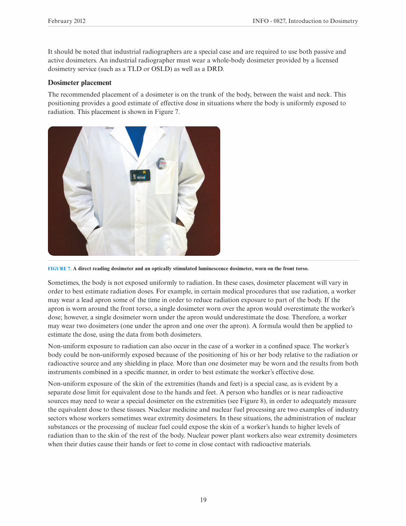
Dosimeter placement
The recommended placement of a dosimeter is on the trunk of the body, between the waist and neck. This positioning provides a good estimate of effective dose in situations where the body is uniformly exposed to radiation. This placement is shown in Figure 7.
FIGURE 7. A direct reading dosimeter and an optically stimulated luminescence dosimeter, worn on the front torso.
Sometimes, the body is not exposed uniformly to radiation. In these cases, dosimeter placement will vary in order to best estimate radiation doses. For example, in certain medical procedures that use radiation, a worker may wear a lead apron some of the time in order to reduce radiation exposure to part of the body. If the apron is worn around the front torso, a single dosimeter worn over the apron would overestimate the worker’s dose; however, a single dosimeter worn under the apron would underestimate the dose. Therefore, a worker may wear two dosimeters (one under the apron and one over the apron). A formula would then be applied to estimate the dose, using the data from both dosimeters.
Non-uniform exposure to radiation can also occur in the case of a worker in a confined space. The worker’s body could be non-uniformly exposed because of the positioning of his or her body relative to the radiation or radioactive source and any shielding in place. More than one dosimeter may be worn and the results from both instruments combined in a specific manner, in order to best estimate the worker’s effective dose.
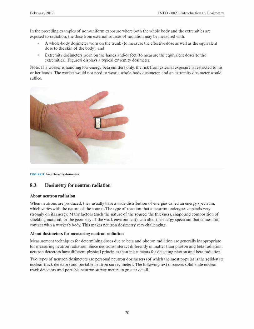
Non-uniform exposure of the skin of the extremities (hands and feet) is a special case, as is evident by a separate dose limit for equivalent dose to the hands and feet. A person who handles or is near radioactive sources may need to wear a special dosimeter on the extremities (see Figure 8), in order to adequately measure the equivalent dose to these tissues. Nuclear medicine and nuclear fuel processing are two examples of industry sectors whose workers sometimes wear extremity dosimeters. In these situations, the administration of nuclear substances or the processing of nuclear fuel could expose the skin of a worker’s hands to higher levels of radiation than to the skin of the rest of the body. Nuclear power plant workers also wear extremity dosimeters when their duties cause their hands or feet to come in close contact with radioactive materials.
February 2012 INFO - 0827, Introduction to Dosimetry
20
In the preceding examples of non-uniform exposure where both the whole body and the extremities are exposed to radiation, the dose from external sources of radiation may be measured with:
• A whole-body dosimeter worn on the trunk (to measure the effective dose as well as the equivalent dose to the skin of the body); and
• Extremity dosimeters worn on the hands and/or feet (to measure the equivalent doses to the extremities). Figure 8 displays a typical extremity dosimeter.
Note: If a worker is handling low-energy beta emitters only, the risk from external exposure is restricted to his or her hands. The worker would not need to wear a whole-body dosimeter, and an extremity dosimeter would suffice.
FIGURE 8. An extremity dosimeter.
8.3 Dosimetry for neutron radiation
About neutron radiation
When neutrons are produced, they usually have a wide distribution of energies called an energy spectrum, which varies with the nature of the source. The type of reaction that a neutron undergoes depends very strongly on its energy. Many factors (such the nature of the source; the thickness, shape and composition of shielding material; or the geometry of the work environment), can alter the energy spectrum that comes into contact with a worker’s body. This makes neutron dosimetry very challenging.
About dosimeters for measuring neutron radiation
Measurement techniques for determining doses due to beta and photon radiation are generally inappropriate for measuring neutron radiation. Since neutrons interact differently in matter than photon and beta radiation, neutron detectors have different physical principles than instruments for detecting photon and beta radiation.
Two types of neutron dosimeters are personal neutron dosimeters (of which the most popular is the solid-state nuclear track detector) and portable neutron survey meters. The following text discusses solid-state nuclear track detectors and portable neutron survey meters in greater detail.
February 2012 INFO - 0827, Introduction to Dosimetry
21
Solid-state nuclear track detectors
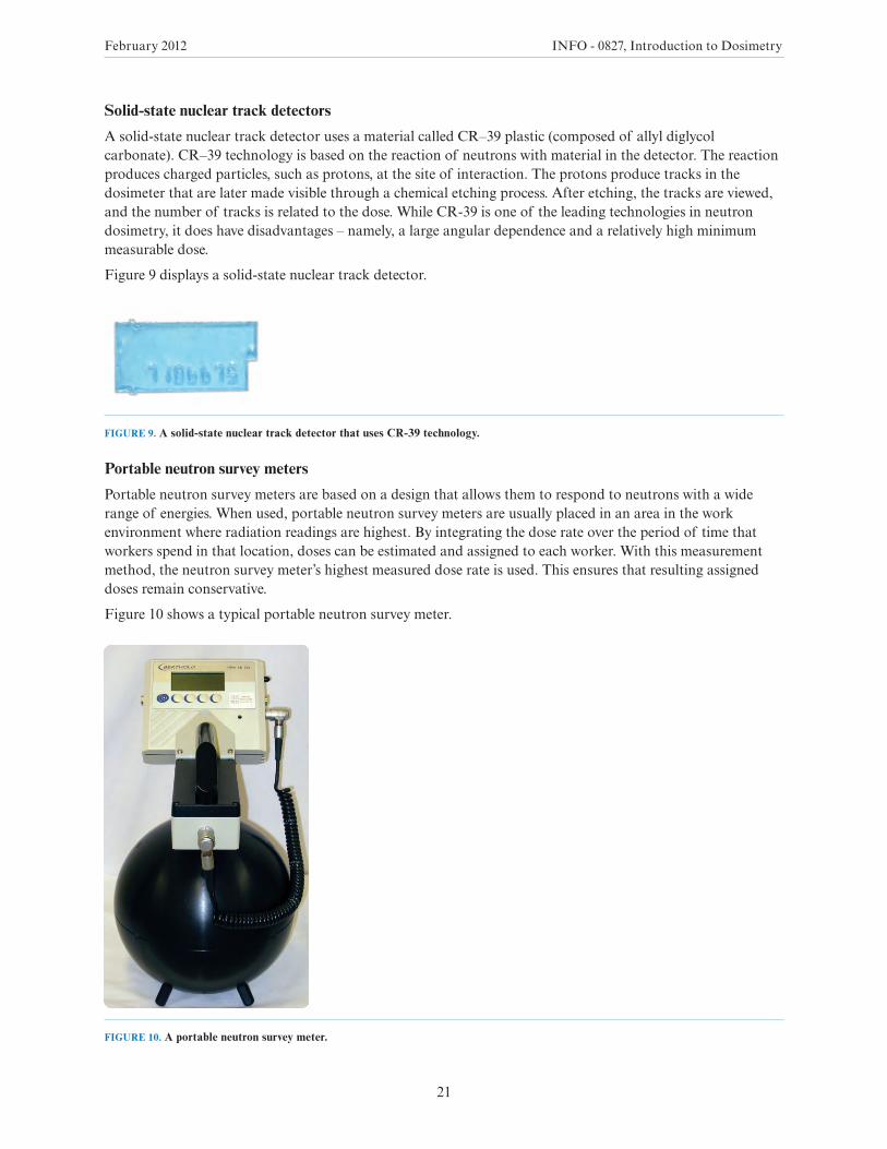
A solid-state nuclear track detector uses a material called CR–39 plastic (composed of allyl diglycol carbonate). CR–39 technology is based on the reaction of neutrons with material in the detector. The reaction produces charged particles, such as protons, at the site of interaction. The protons produce tracks in the dosimeter that are later made visible through a chemical etching process. After etching, the tracks are viewed, and the number of tracks is related to the dose. While CR-39 is one of the leading technologies in neutron dosimetry, it does have disadvantages – namely, a large angular dependence and a relatively high minimum measurable dose.
Figure 9 displays a solid-state nuclear track detector.
FIGURE 9. A solid-state nuclear track detector that uses CR-39 technology.
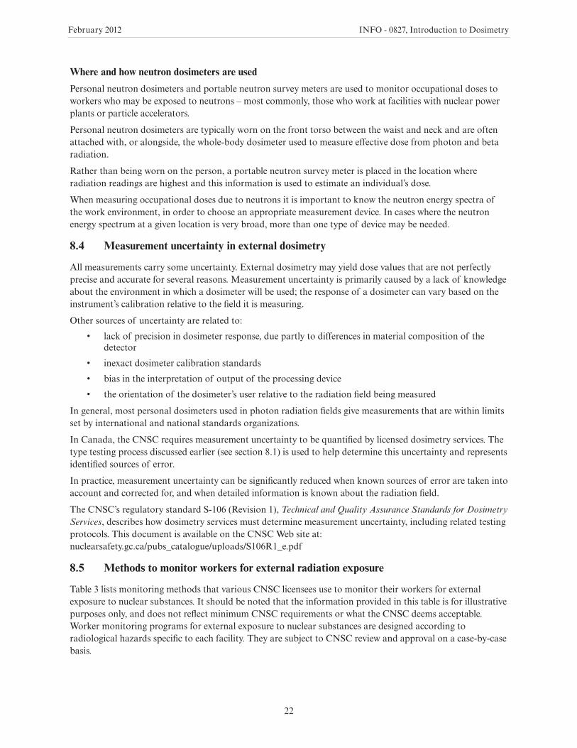
Portable neutron survey meters
Portable neutron survey meters are based on a design that allows them to respond to neutrons with a wide range of energies. When used, portable neutron survey meters are usually placed in an area in the work environment where radiation readings are highest. By integrating the dose rate over the period of time that workers spend in that location, doses can be estimated and assigned to each worker. With this measurement method, the neutron survey meter’s highest measured dose rate is used. This ensures that resulting assigned doses remain conservative.
Figure 10 shows a typical portable neutron survey meter.
FIGURE 10. A portable neutron survey meter.
February 2012 INFO - 0827, Introduction to Dosimetry
22
Where and how neutron dosimeters are used
Personal neutron dosimeters and portable neutron survey meters are used to monitor occupational doses to workers who may be exposed to neutrons – most commonly, those who work at facilities with nuclear power plants or particle accelerators.
Personal neutron dosimeters are typically worn on the front torso between the waist and neck and are often attached with, or alongside, the whole-body dosimeter used to measure effective dose from photon and beta radiation.
Rather than being worn on the person, a portable neutron survey meter is placed in the location where radiation readings are highest and this information is used to estimate an individual’s dose.
When measuring occupational doses due to neutrons it is important to know the neutron energy spectra of the work environment, in order to choose an appropriate measurement device. In cases where the neutron energy spectrum at a given location is very broad, more than one type of device may be needed.
8.4 Measurement uncertainty in external dosimetry
All measurements carry some uncertainty. External dosimetry may yield dose values that are not perfectly precise and accurate for several reasons. Measurement uncertainty is primarily caused by a lack of knowledge about the environment in which a dosimeter will be used; the response of a dosimeter can vary based on the instrument’s calibration relative to the field it is measuring.
Other sources of uncertainty are related to:
• lack of precision in dosimeter response, due partly to differences in material composition of the detector
• inexact dosimeter calibration standards
• bias in the interpretation of output of the processing device
• the orientation of the dosimeter’s user relative to the radiation field being measured
In general, most personal dosimeters used in photon radiation fields give measurements that are within limits set by international and national standards organizations.
In Canada, the CNSC requires measurement uncertainty to be quantified by licensed dosimetry services. The type testing process discussed earlier (see section 8.1) is used to help determine this uncertainty and represents identified sources of error.
In practice, measurement uncertainty can be significantly reduced when known sources of error are taken into account and corrected for, and when detailed information is known about the radiation field.
The CNSC’s regulatory standard S-106 (Revision 1), Technical and Quality Assurance Standards for Dosimetry Services, describes how dosimetry services must determine measurement uncertainty, including related testing protocols. This document is available on the CNSC Web site at: nuclearsafety.gc.ca/pubs_catalogue/uploads/S106R1_e.pdf
8.5 Methods to monitor workers for external radiation exposure
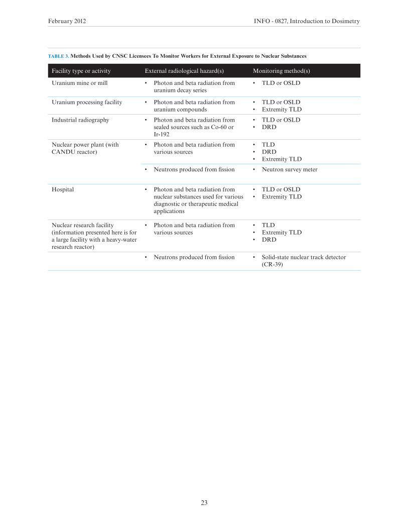
Table 3 lists monitoring methods that various CNSC licensees use to monitor their workers for external exposure to nuclear substances. It should be noted that the information provided in this table is for illustrative purposes only, and does not reflect minimum CNSC requirements or what the CNSC deems acceptable. Worker monitoring programs for external exposure to nuclear substances are designed according to radiological hazards specific to each facility. They are subject to CNSC review and approval on a case-by-case basis.
February 2012 INFO - 0827, Introduction to Dosimetry
23
TABLE 3. Methods Used by CNSC Licensees To Monitor Workers for External Exposure to Nuclear Substances
Facility type or activity External radiological hazard(s) Monitoring method(s)
Uranium mine or mill • Photon and beta radiation from uranium decay series
• TLD or OSLD
Uranium processing facility • Photon and beta radiation from uranium compounds
• TLD or OSLD• Extremity TLD
Industrial radiography • Photon and beta radiation from sealed sources such as Co-60 or Ir-192
• TLD or OSLD• DRD
Nuclear power plant (with CANDU reactor)
• Photon and beta radiation from various sources
• TLD• DRD• Extremity TLD
• Neutrons produced from fission • Neutron survey meter
Hospital • Photon and beta radiation from nuclear substances used for various diagnostic or therapeutic medical applications
• TLD or OSLD• Extremity TLD
Nuclear research facility (information presented here is for a large facility with a heavy-water research reactor)
• Photon and beta radiation from various sources
• TLD• Extremity TLD• DRD
• Neutrons produced from fission • Solid-state nuclear track detector (CR-39)
February 2012 INFO - 0827, Introduction to Dosimetry
24
9.0 INTERNAL DOSIMETRY
Internal dosimetry is the measurement of doses due to nuclear substances that have entered the body by way of ingestion, inhalation or other means.
Internal dosimetry involves two steps:
1. The level of radiation inside a person’s body is estimated using one of three methods:
• in-vivo bioassay (direct measurement of radioactivity in the body)
• in-vitro bioassay (measurement of radioactivity in a person’s urine or feces)
• measurement of radioactivity in workplace air
2. The resulting internal radiation dose is calculated.
Sections 9.1 to 9.3 outline the three methods of measuring internal radiation. Section 9.4 explains the method of calculation for internal radiation doses, and section 9.5 explains why calculated internal dose values may be uncertain and how measurement uncertainty is accounted for. Finally, methods to monitor workers for internal radiation exposure are presented in section 9.6.
9.1 In-vivo bioassay (direct measurement of radioactivity in the body)
In-vivo bioassay involves the measurement of nuclear substances within the body. This measurement method is performed using external instruments that detect the radiation emitted by these substances.
In order to detect radiation emitted by nuclear substances within the body, these substances must emit radiation with sufficient range to escape from the body. In-vivo bioassay is therefore appropriate for measuring gamma radiation – since this type of radiation typically has sufficient range to be detected outside of the body, even when it originated inside the person.
In-vivo bioassay measures gamma radiation using a detector positioned near the person. The most common types of detectors used in this manner are whole-body counters, lung counters, and thyroid counters. These devices are calibrated to identify and determine the amount of gamma-emitting nuclear substances within the body. The following text discusses these three types of detectors.
Whole-body counters
A whole-body counter is a device used to measure radioactivity within the human body. This instrument is intended to detect the presence of nuclear substances in a person’s body, and identify the type and amount of nuclear substances detected.
Whole-body counters are used at every Canadian nuclear power plant (NPP), at Atomic Energy of Canada Limited’s Chalk River Laboratories and Whiteshell Laboratories, and at Health Canada’s Radiation Protection Bureau.
NPPs have “self-serve” whole-body counters, which allow workers to be counted without the need for assistance from a technician. The devices are typically placed at a convenient, easily accessible location on the NPP site, so workers can be counted as they leave the plant. A self-serve whole-body counter is activated by workers themselves as they swipe their identity cards in a reader. This allows worker information (for example, name, employee number, and date and time of the count) to be stored in a database.
A self-serve whole-body counter offers the advantage of rapid worker monitoring. Workers simply stand in the device, which contains some lead shielding to reduce background radiation, while they are counted for a short period of time; for example, 90 seconds. If no activity is detected, the results are recorded and no dose is assigned; this is the case for most workers counted. However, if the counter does detect activity on the worker, it prompts the user to conduct a longer count (for example, five minutes) and the facility’s radiation protection staff are notified for potential follow-up.
February 2012 INFO - 0827, Introduction to Dosimetry
25
Monitoring frequency depends on the potential for exposure, the nature of the work performed and the associated radiological hazard. As the potential for nuclear substances to be taken into the body increases, monitoring is carried out more often. For example, a nuclear power facility’s workers who handles nuclear fuel and may be exposed to fission and activation products (such as cobalt-60) are monitored monthly, or more frequently, depending on the task performed. Other workers whose tasks are less likely to result in exposure to fission and activation products may be subject to whole-body counting less often, such as quarterly or annually. CNSC licensees select appropriate monitoring frequencies (with the CNSC’s approval) that will ensure their whole-body counting activities can detect doses of 0.1 mSv per year.
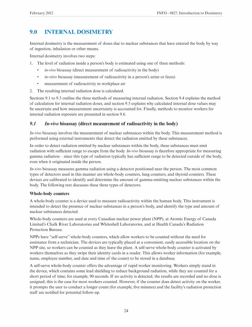
Lung counters
A lung counter is a device used to monitor the inhalation of airborne contaminants, such as uranium oxides or insoluble transuranics like plutonium.
Many alpha-emitting radioactive airborne contaminants that are deposited primarily in the respiratory tract emit gamma radiation of relatively low energy. Measurements taken with a lung counter typically require longer counting times than measurements taken with a whole-body counter. This is due to the difficulty in distinguishing the airborne contaminants’ gamma radiation from background radiation, for two reasons: first, the gamma radiation’s energy is significantly reduced by tissue overlying the lungs; and second, a significant proportion of background radiation also consists of low-energy gamma radiation [5]. A lung counter is typically used over a period of about 30 minutes to detect doses of a few millisieverts.
Figure 11 displays a lung counter.
FIGURE 11. A typical lung counter. The device shown is used by Health Canada at its Radiation Protection Bureau in Ottawa.
Two CNSC licensees use lung counters. One of the counters is located at the AECL Chalk River Laboratories, and the other is a custom mobile system owned by Cameco. The latter is set up in a trailer at the Port Hope Conversion Facility and serves this facility as well as the Blind River Refinery.
February 2012 INFO - 0827, Introduction to Dosimetry
26
Thyroid counters
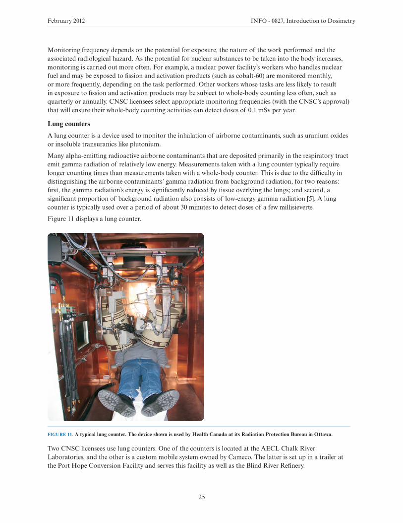
A thyroid counter consists of an appropriately calibrated detector (see Figure 12) that is placed in front of the thyroid gland, which is located in the neck.
Thyroid counters are used in workplaces where radioactive iodine presents an internal hazard. When taken into the body, radioactive iodine deposits principally in the thyroid gland [6].
Thyroid counters are used by many CNSC licensees, most of which are medical and academic institutions.
Figure 12 displays a typical thyroid counter.
FIGURE 12. A thyroid counter.
9.2 In-vitro bioassay (measurement of radioactivity in substances excreted by the body)
In-vitro bioassay is used to determine the presence of nuclear substances or to estimate their amount in urine, feces or other biological materials removed from the body. Shorter-range radiations, namely alpha (α) and beta (β), are generally less penetrating than photon radiation (gamma radiation and X-rays), and therefore cannot be detected from outside the body. They can be detected, however, in material excreted from the body. The purpose of in-vitro bioassay is to determine the quantity of nuclear substances excreted from the body, in order to estimate the quantity present within the body.
February 2012 INFO - 0827, Introduction to Dosimetry
27
The most common type of in-vitro bioassay carried out by CNSC licensees is the measurement of tritium in urine. Another in-vitro bioassay method involves the analysis of urine and feces for the presence of other nuclear substances. The following text discusses these two in-vitro bioassay methods.
Measurement of tritium in urine
Tritium emits low-energy beta radiation. Due to this radiation’s short range of travel (up to 0.006 mm) in tissue [7], it cannot reach the skin’s outer surface from within the body. Therefore, beta radiation emitted by tritium atoms inside the body cannot be detected from outside the body and requires in-vitro bioassay.
Internal exposure to tritium at CNSC-licensed facilities originates almost exclusively from tritiated water. When tritiated water is taken into a worker’s body, it is retained by the body, and then distributed and excreted following the same pattern as regular water.
All nuclear power plants in Canada test workers’ urine to measure for the presence of tritium. This type of analysis is also performed at the AECL Chalk River Laboratory and at other facilities whose workers handle tritium (for example, the tritium light source manufacturers SRBT Inc. and Shield Source Inc.).
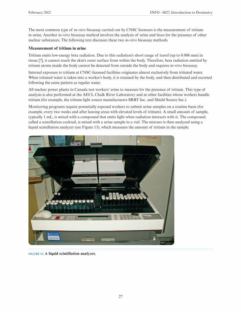
Monitoring programs require potentially exposed workers to submit urine samples on a routine basis (for example, every two weeks and after leaving areas with elevated levels of tritium). A small amount of sample, typically 1 mL, is mixed with a compound that emits light when radiation interacts with it. The compound, called a scintillation cocktail, is mixed with a urine sample in a vial. The mixture is then analyzed using a liquid scintillation analyzer (see Figure 13), which measures the amount of tritium in the sample.
FIGURE 13. A liquid scintillation analyzer.
February 2012 INFO - 0827, Introduction to Dosimetry
28
Analysis of urine and feces for the presence of nuclear substances other than tritium
Workers at many nuclear facilities (such as nuclear power plants) can be exposed to nuclear substances other than tritium, which also cannot be detected using in-vivo bioassay methods. Therefore, urine samples submitted as part of a tritium monitoring program are also screened for the presence of other nuclear substances, via in-vitro bioassay.
If nuclear substances are detected, further monitoring, such as the analysis of urine collected over a 24-hour period or analysis of feces, is performed. These further tests aim to accurately quantify the amount of nuclear substances excreted. Some nuclear substances are typically excreted in urine in amounts that would not normally be detected using such methods. Among these nuclear substances are transuranic elements (also known as “transuranics”), which are elements heavier than uranium and include americium and plutonium. Therefore, these substances are monitored using fecal analysis.
Many transuranics emit alpha radiation and very low levels of photon radiation. In operating nuclear reactors, gamma-emitting nuclear substances are also formed as a result of neutron irradiation of uranium fuel. These gamma-emitting substances, which include zirconium-95 and cerium-144, can be used to indicate a potential intake of transuranic nuclear substances if they are detected in a urine sample or a whole-body count [8]. Hence, monitoring for intakes of transuranics by workers at operating reactors is done by screening for indicator nuclear substances (such as zirconium-95 and cerium-144) in both urine samples and in whole-body counts. If one of these indicator nuclear substances is detected, further monitoring is carried out; for example, by measuring the ratio of the indicator nuclear substance to alpha emitters in the workplace, and using this ratio to estimate the amount of transuranics taken into the body.
If this follow-up monitoring suggests a dose greater than a pre-defined trigger level (for example, an investigation level defined in the licensee’s radiation protection program), further follow-up monitoring is carried out, in order to more accurately determine the amount of transuranics in the body and the resultant dose. Follow-up monitoring includes removing the worker from the duties or environment that involved the potential radiological exposure, and increasing the frequency and type of bioassay sampling. Further bioassay samples could be analyzed with a more sensitive method that could better quantify the level of transuranic nuclear substances. One such method is to first perform a radiochemical separation of transuranics in the urine or feces sample, before analyzing the sample to determine the amount of nuclear substances present.
9.3 Measurement of radon decay products in workplace air
Sometimes, neither in-vivo bioassay nor in-vitro bioassay can be used to monitor workers for the intake of nuclear substances. This is the case with short-lived nuclear substances, such as radon decay products12.
Since radon decay products have such short half-lives, they decay quickly and before their activity could be measured via bioassay methods. Radiation from radon decay products must therefore be measured in workplace air. An instrument which is used to estimate individual exposures to radon progeny is called a personal alpha dosimeter (PAD). One example of a workplace where a PAD is required is a uranium mine, where workers may inhale radon decay products and uranium ore dust.
12 Radon gas (radon-222) has a half-life of 3.8 days [2]. As an inert gas, it is not readily absorbed into the respiratory tract when inhaled. However, as it decays, radon gas forms four other nuclear substances (polonium-218, lead-214, bismuth-214, and polonium-214) called radon decay products, each with a half-life of under 30 minutes. Of these four radon decay products, polonium-218 and polonium-214 emit alpha particles and are responsible for the bulk of the dose to the respiratory tract resulting from inhaled radon. They can be retained in the respiratory tract and further decay there (causing damage to the body) before the body’s lung-clearing mechanism removes them.
February 2012 INFO - 0827, Introduction to Dosimetry
29
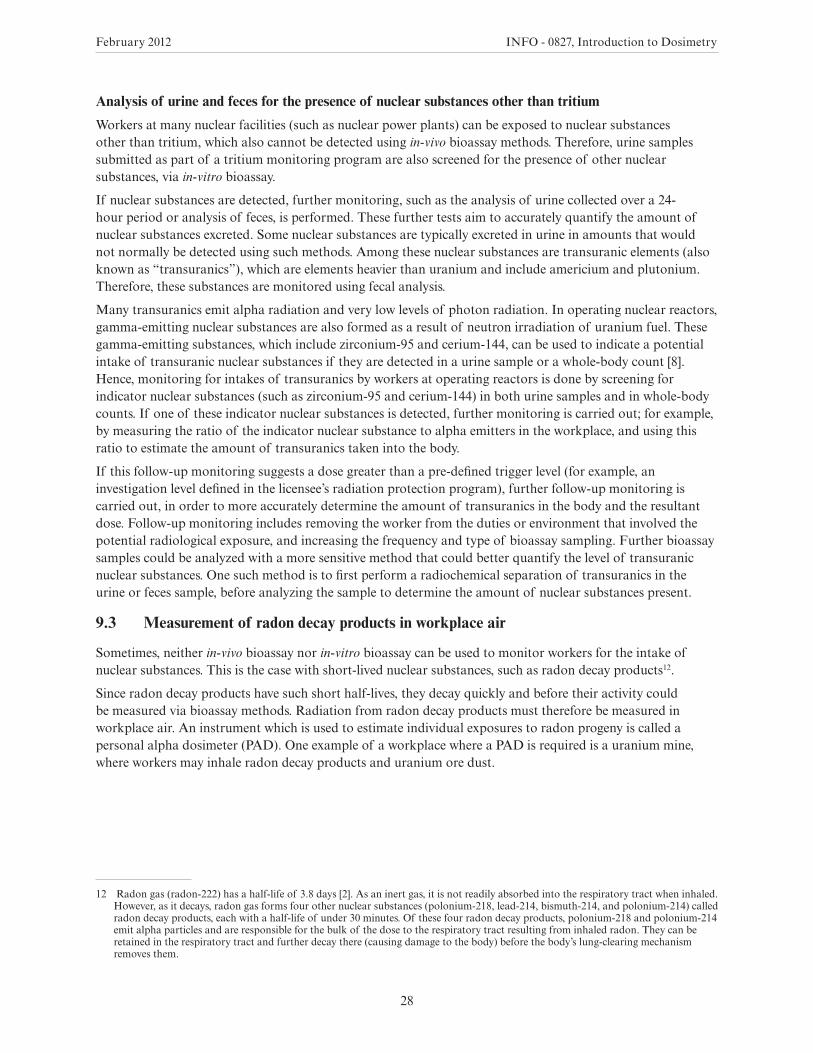
A PAD consists of a battery-operated air pump and a filter. The following basic overview presents how the device works.
• Air is drawn through the dosimeter, which is worn by a worker on his or her belt.
• The filter captures radon decay products. As the products decay, they emit alpha particles, some of which interact with a cellulose film placed a few centimetres from the filter. These interactions cause microscopic damage (tracks) to the film.
• The cellulose film is etched and the number of tracks is counted using a microscope.
• A calibration factor is used to convert the number of tracks into an air concentration of radon decay products.
• The worker’s exposure is calculated by taking the concentration of radon decay products in air and the worker’s exposure time into account (accurate records of time spent by workers in the workplace are necessary).
Figure 14 displays a typical PAD used to measure radon decay products in workplace air.
FIGURE 14: A personal alpha dosimeter.
About exposure results determined by personal alpha dosimeters
Exposure results for workers monitored by PADs are reported to the National Dose Registry. These results are expressed in a unit of measure called the working level month (WLM). The following text explains the meaning of this unit.
The health risk from radon is related to the concentration of the alpha-particle energy released by its decay products in air. Instead of determining the concentration of each radon decay product in air, it is simpler to measure the concentration of alpha-particle energy released by all radon decay products in air. This quantity is called the potential alpha energy concentration (PAEC), which is the amount of alpha-particle energy per unit volume of air that can be released from the radon decay products.
Historically, the PAEC unit was known as the working level (WL), and this unit is still used today. A WL is defined as the PAEC that results from the presence of 3.7 Bq of each radon decay product (namely, polonium-218, lead-214, bismuth-214 and polonium-214) per litre of air. Exposure to radon decay products is determined by the PAEC multiplied by the amount of time a person is exposed to radon decay products.
February 2012 INFO - 0827, Introduction to Dosimetry
30
Exposure to radon decay products is often expressed in working level months (WLMs), with a working month being defined as 170 hours. If a person is exposed to 1 WL for a full working month of 170 hours, his or her exposure would equal 1 WLM.
The WLM value is related to dose on the basis of epidemiological studies. Currently, the ICRP’s recommendation is that 1 WLM equals a dose of 5 mSv. This value is also identified in the Radiation Protection Regulations [1].
9.4 How internal radiation doses are calculated
Recall that dose is the amount of radiation energy absorbed per mass unit of tissue, weighted by the radiation type’s biological effectiveness and by the susceptibility of a tissue to developing cancer or a heritable disease from radiation exposure (see section 5.3 of this document). The dose to a worker caused by internal radiation exposure is calculated using the following two steps:
1. Determine which organs and tissues nuclear substances are deposited into, after the substances have been taken into the body.
2. Estimate how much radiation energy is absorbed by the organs and tissues.
Step 1 is performed using biokinetic models that are specific to different nuclear substances, and step 2 is completed using dosimetric models.
A biokinetic model calculates the amount of nuclear substances that deposits in organs and tissues after they are taken into the body. It relates the quantity of nuclear substances taken into the body to the quantity retained in organs and tissues, and to the rate at which the substances are excreted from the body.
A dosimetric model calculates how much radiation energy is absorbed by the organs and tissues as a result of the nuclear substances that are in the body.
With the use of biokinetic and dosimetric models, it is possible to ascertain internal radiation doses using data from both in-vivo and in-vitro bioassay methods, as well from air monitoring. The ICRP recommends and regularly updates the models and their formulae, which are available as part of computer software packages designed to calculate doses from intakes of nuclear substances.
Doses ascertained using the method just discussed are generally less than a few mSv annually at CNSC-licensed facilities. For example:
• For all Canadian nuclear power plants, where internal exposures are almost exclusively due to tritium, reported average yearly tritium doses varied from 0.04 mSv to 0.19 mSv in 2008 [9].
• In Canada’s operating uranium mines, radon decay products are the principal contributors to dose from internal radiation exposure. In the year 2008, the average annual dose from radon decay products at these facilities varied from 0.08 mSv to 0.63 mSv [9].
9.5 Measurement uncertainty in internal dosimetry
In internal dosimetry, several factors contribute to uncertainty in calculated doses. These consist of a combination of factors that arise during three stages of the dose estimate:
• when individual monitoring measurements are taken
• when the amount of nuclear substances taken into the body (often referred to as the intake) is estimated
• when the mathematical models are used to calculate the dose based on the intake
Uncertainties related to individual monitoring measurements include those related to the calibration of the measurement instrument used. These uncertainties arise because of the statistical nature of radioactive decay. Nuclear substances remaining in the body from previous intakes may also need to be accounted for during the measurement stage.
February 2012 INFO - 0827, Introduction to Dosimetry
31
When the amount of nuclear substances taken into the body is estimated, sources of uncertainty include:
• lack of knowledge of the exact time of an intake or of the duration of an intake that took place over time
• variation among individuals’ metabolisms
• uncertainty or variability in the characteristics of the material to which a worker may have been exposed (particularly the size of dust particles); absorption characteristics of the material in the respiratory tract and in the gut; and the composition of the mixture of nuclear substances to which a worker may have been exposed
When calculating internal dose based on intake, uncertainties can arise from the models used. Those that are based on human studies are the most reliable. These include the models for nuclear substances such as caesium, strontium and iodine. For many other nuclear substances, there can be either very little or no information available from human studies. In that case, models are based on other sources of information, such as the results from studies in animals and chemical analogy: some substances are known to be chemically similar to others and therefore behave in a similar manner in the body. For example, metabolic studies of carbon and hydrogen in the diet can be analogs for tritium and carbon-14.
To account for these uncertainties, mathematical models used for calculating doses are generally conservative; that is, they tend to overestimate the dose. Also, licensed dosimetry services are required to take part in independent tests, which verify the accuracy of the measurement part of the process.
9.6 Methods to monitor workers for internal radiation exposure
Table 4 lists methods that various CNSC licensees use to monitor their workers for internal exposure to nuclear substances. It should be noted that the information provided in this table is for illustrative purposes only, and does not reflect minimum CNSC requirements or what the CNSC deems acceptable. Worker monitoring programs for internal exposure to nuclear substances are designed according to radiological hazards specific to each facility. They are subject to CNSC review and approval on a case-by-case basis.
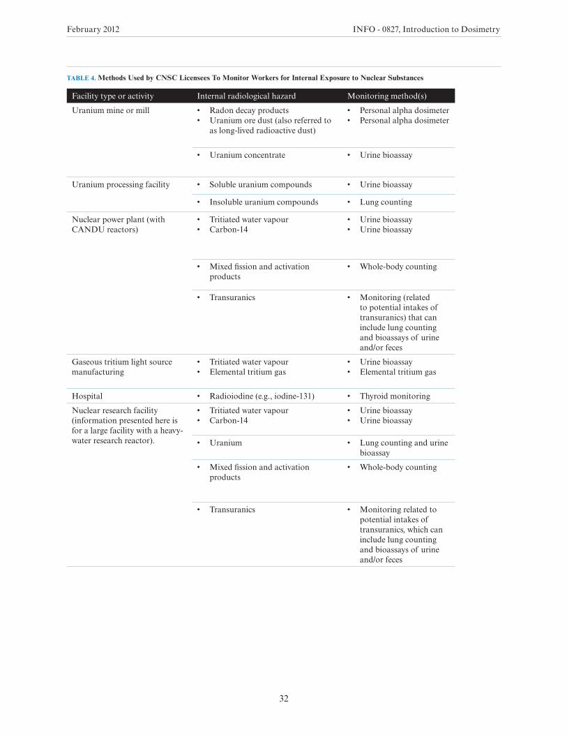
February 2012 INFO - 0827, Introduction to Dosimetry
32
TABLE 4. Methods Used by CNSC Licensees To Monitor Workers for Internal Exposure to Nuclear Substances
Facility type or activity Internal radiological hazard Monitoring method(s)
Uranium mine or mill • Radon decay products• Uranium ore dust (also referred to
as long-lived radioactive dust)
• Personal alpha dosimeter• Personal alpha dosimeter
• Uranium concentrate • Urine bioassay
Uranium processing facility • Soluble uranium compounds • Urine bioassay
• Insoluble uranium compounds • Lung counting
Nuclear power plant (with CANDU reactors)
• Tritiated water vapour• Carbon-14
• Urine bioassay• Urine bioassay
• Mixed fission and activation products
• Whole-body counting
• Transuranics • Monitoring (related to potential intakes of transuranics) that can include lung counting and bioassays of urine and/or feces
Gaseous tritium light source manufacturing
• Tritiated water vapour• Elemental tritium gas
• Urine bioassay• Elemental tritium gas
Hospital • Radioiodine (e.g., iodine-131) • Thyroid monitoring
Nuclear research facility (information presented here is for a large facility with a heavy-water research reactor).
• Tritiated water vapour• Carbon-14
• Urine bioassay• Urine bioassay
• Uranium • Lung counting and urine bioassay
• Mixed fission and activation products
• Whole-body counting
• Transuranics • Monitoring related to potential intakes of transuranics, which can include lung counting and bioassays of urine and/or feces
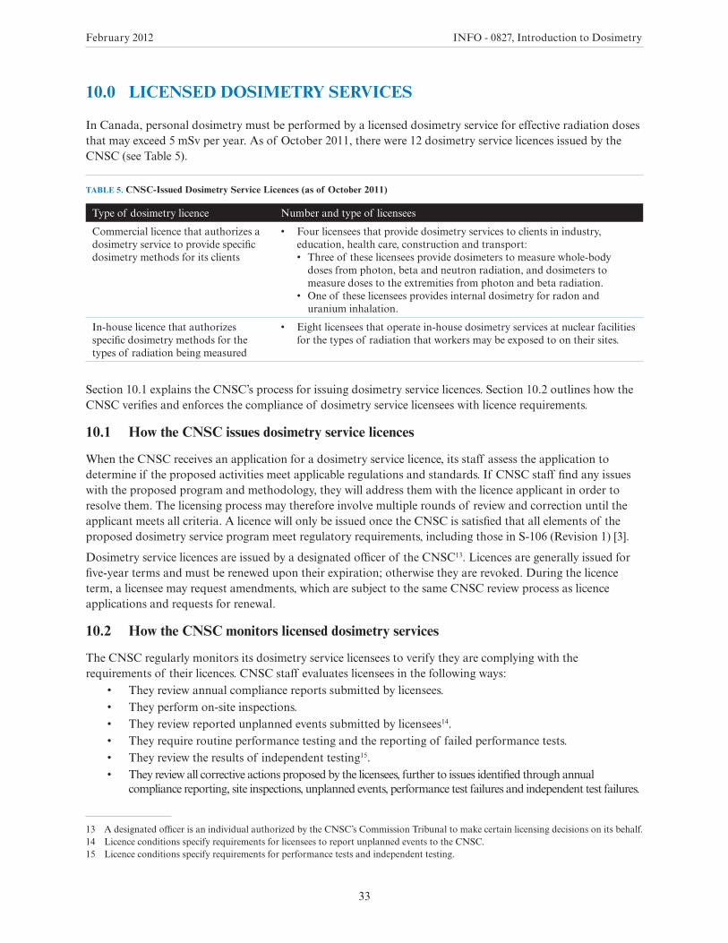
February 2012 INFO - 0827, Introduction to Dosimetry
33
10.0 LICENSED DOSIMETRY SERVICES
In Canada, personal dosimetry must be performed by a licensed dosimetry service for effective radiation doses that may exceed 5 mSv per year. As of October 2011, there were 12 dosimetry service licences issued by the CNSC (see Table 5).
TABLE 5. CNSC-Issued Dosimetry Service Licences (as of October 2011)
Type of dosimetry licence Number and type of licensees
Commercial licence that authorizes a dosimetry service to provide specific dosimetry methods for its clients
• Four licensees that provide dosimetry services to clients in industry, education, health care, construction and transport:• Threeoftheselicenseesprovidedosimeterstomeasurewhole-body
doses from photon, beta and neutron radiation, and dosimeters to measure doses to the extremities from photon and beta radiation.
• Oneoftheselicenseesprovidesinternaldosimetryforradonanduranium inhalation.
In-house licence that authorizes specific dosimetry methods for the types of radiation being measured
• Eight licensees that operate in-house dosimetry services at nuclear facilities for the types of radiation that workers may be exposed to on their sites.
Section 10.1 explains the CNSC’s process for issuing dosimetry service licences. Section 10.2 outlines how the CNSC verifies and enforces the compliance of dosimetry service licensees with licence requirements.
10.1 How the CNSC issues dosimetry service licences
When the CNSC receives an application for a dosimetry service licence, its staff assess the application to determine if the proposed activities meet applicable regulations and standards. If CNSC staff find any issues with the proposed program and methodology, they will address them with the licence applicant in order to resolve them. The licensing process may therefore involve multiple rounds of review and correction until the applicant meets all criteria. A licence will only be issued once the CNSC is satisfied that all elements of the proposed dosimetry service program meet regulatory requirements, including those in S-106 (Revision 1) [3].
Dosimetry service licences are issued by a designated officer of the CNSC13. Licences are generally issued for five-year terms and must be renewed upon their expiration; otherwise they are revoked. During the licence term, a licensee may request amendments, which are subject to the same CNSC review process as licence applications and requests for renewal.
10.2 How the CNSC monitors licensed dosimetry services
The CNSC regularly monitors its dosimetry service licensees to verify they are complying with the requirements of their licences. CNSC staff evaluates licensees in the following ways:
• They review annual compliance reports submitted by licensees.• They perform on-site inspections.• They review reported unplanned events submitted by licensees14.• They require routine performance testing and the reporting of failed performance tests.• They review the results of independent testing15.• They review all corrective actions proposed by the licensees, further to issues identified through annual
compliance reporting, site inspections, unplanned events, performance test failures and independent test failures.
13 A designated officer is an individual authorized by the CNSC’s Commission Tribunal to make certain licensing decisions on its behalf.14 Licence conditions specify requirements for licensees to report unplanned events to the CNSC.15 Licence conditions specify requirements for performance tests and independent testing.
February 2012 INFO - 0827, Introduction to Dosimetry
34
SUMMARY
The CNSC’s mandate is to regulate the use of nuclear energy and materials to protect the health, safety and security and the environment.
To meet this objective, the CNSC sets regulatory limits on radiation exposure to workers and the public on the basis of the ICRP’s recommendations. These limits are outlined in the Radiation Protection Regulations. The CNSC also publishes guidance documents to help licensees implement a radiation protection program and other measures that keep the amount of exposure to ionizing radiation as low as reasonably achievable (ALARA).
The purpose of these regulatory limits and guidance documents is to prevent health effects and to establish an acceptable level of safety for workers and members of the public. Setting adequate protective exposure limits and providing appropriate guidance requires an assessment of the risks, which depend on the magnitude of the radiological exposures.
Exposure to radiation is measured in a quantity called dose. Licensees are required to ascertain the level of doses that persons receive due to exposure to radiation from licensed activities. The CNSC evaluates dose assessment methods to ensure they are sufficiently accurate and precise with respect to the risks being measured.
When effective doses of radiation may exceed 5 mSv per year, a licensed dosimetry service is required to perform personal dosimetry. Licensed dosimetry services are subject to extensive requirements, which are in place to demonstrate that personal doses are accurate and reliable. This is done through technical and quality assurance requirements that include, for example, various prescribed testing programs, the results of which are subject to review by the CNSC.
Canadian requirements for measuring radiation doses are based on sound, proven science. This assures people receive adequate protection from the potential harmful effects of radiation exposure, and that any radiation doses they receive will remain below limits set out in the Radiation Protection Regulations.
February 2012 INFO - 0827, Introduction to Dosimetry
35
GLOSSARY
ALARA (as low as reasonably achievable) A radiation protection principle that states doses should be kept as low as reasonably achievable (ALARA), with social and economic factors taken into account. In addition to respecting regulatory dose limits, CNSC licensees are required to keep doses ALARA.
alpha radiation Radiation that consists of alpha particles that are made up of two protons and two neutrons each. Alpha particles carry a positive charge; due to their charge and relatively large mass, they are extremely limited in their ability to penetrate matter.
atom A unit of matter made up of three particles (called subatomic particles): protons, neutrons, and electrons. The protons and the neutrons are in the atom’s centre (nucleus), and the electrons surround the nucleus.
background radiation The radiation constantly present in the environment, emitted by natural and artificial sources.
beta radiation Radiation that consists of charged particles, which are ejected from the nucleus of an atom and that have a negative charge. Beta particles are physically identical to electrons.
becquerel (Bq) The unit of measure for the quantity of radioactive material. One Bq is equal to the decay of one atom per second.
calibration factor A factor that is used in the calibration of a reference dosimeter and that relates the exposure quantity to the reported dose.
decay process The process by which an unstable nucleus emits excess energy to achieve greater stability.
dose of record A dose to an individual that has been measured by a licensed dosimetry service and is submitted to the National Dose Registry.
dosimeter A radiation detection device worn by an individual and that is capable of measuring dose from ionizing radiation. A dosimeter can be passive (stores a signal and is later processed and analyzed) or active (displays a direct reading of the detected dose or dose rate in real time).
electrical interaction A force of repulsion acting between electric charges of like sign (charge), or a force of attraction acting between electric charges of unlike sign (charge).
electron A subatomic particle that surrounds the nucleus of an atom and that has a negative electric charge.
electronvolt (eV) A unit of energy employed in radiation physics. It is equal to the energy that an electron gains when it passes through a potential difference of 1 volt. 1eV= 1.6 x 10⁻¹⁹ joules.
excited state A state of energy of an electron when its energy level is higher than in the ground state.
external dosimetry The measurement of doses resulting from radiation sources outside the body.
fading The loss of a dosimeter’s signal over time, caused by external factors such as temperature and humidity.
February 2012 INFO - 0827, Introduction to Dosimetry
36
gamma radiation Electromagnetic radiation that consists of photons originating from within an atom’s nucleus. Also called photon radiation.
gray (Gy) The unit of measurement used to express absorbed dose. One grey is defined as the absorption of 1 joule of ionizing radiation by 1 kilogram of matter
ground state The lowest-energy and most stable state of an electron.
half-life The period of time for the number of atoms in a substance undergoing radioactive decay to decrease by one half of its original value.
in-vitro bioassay An internal dosimetry method used to determine the presence of nuclear substances or to estimate their amount in urine, feces or other biological materials removed from the body.
in-vivo bioassay An internal dosimetry method that involves the measurement of nuclear substances within the body. This measurement method is performed using external instruments that detect the radiation emitted by these substances.
internal dosimetry The measurement of doses due to nuclear substances that have entered the body by way of ingestion, inhalation or other means.
ionizing radiation Radiation with sufficient energy to remove an electron from an atom. It includes the radiation that comes from both natural and man-made sources.
isotopes Variants of atoms of a particular chemical element, which have differing numbers of neutrons but the same number of protons.
National Dose Registry (NDR) A database that is owned and operated by Health Canada and that tracks the lifetime dose history of registered individuals.
neutron A subatomic particle present in the nucleus of an atom. It has no electric charge.
non-ionizing radiation Radiation that has a lower energy level than ionizing radiation and that cannot remove an electron from an atom. Examples of non-ionizing radiation include radio waves and microwaves.
nuclear energy worker (NEW) A person who is required, in the course of his or her business or occupation in connection with a nuclear substance or nuclear facility, to perform duties in such circumstances that there is a reasonable probability of receiving a dose of radiation that is greater than the prescribed limit for the general public.
nucleus The very dense region at the centre of an atom that consists of protons and neutrons.
photon An elementary particle that is the basic quanta of light and all other forms of electromagnetic radiation.
proton A subatomic particle present in the nucleus of an atom and that has a positive electric charge.
radioactive decay The process by which the nucleus of a radioactive isotope spontaneously disintegrates, during which the nucleus emits excess energy (radiation) in order to become stable.
February 2012 INFO - 0827, Introduction to Dosimetry
37
radon decay products The nuclear substances (polonium-218, lead-214, bismuth-214, and polonium-214) emitted when radon gas undergoes radioactive decay. Polonium-218 and polonium-214 emit alpha particles and are responsible for the bulk of the dose to the respiratory tract resulting from inhaled radon. All four radon decay products have half-lives of less than 30 minutes.
response In terms of measurement, the measured or evaluated value divided by the conventionally true value.
sievert (Sv) The unit used to express equivalent and effective dose. 1 Sv=1 J/kg.
transuranic elements Chemical elements that are heavier than uranium. Transuranic elements include americium and plutonium.
working level (WL) The potential alpha energy concentration that results from the presence of 3.7 Bq of each radon decay product.
working level month (WLM) A unit of measure to express the level of exposure to radon decay products. One working month equals 170 hours. Therefore, 1 working level month is equal to the exposure of a person exposed to 1 working level for 170 hours.
X-ray radiation Electromagnetic radiation that consists of photons originating from outside an atom’s nucleus.
February 2012 INFO - 0827, Introduction to Dosimetry
38
REFERENCES
1. Canadian Nuclear Safety Commission, Radiation Protection Regulations, May 2000.
2. International Commission on Radiological Protection, Radionuclide Transformations, Vols. 11–13, Publication 38, 1983.
3. Canadian Nuclear Safety Commission, Technical and Quality Assurance Requirements for Dosimetry Services, S-106/Rev. 1, June 2006.
4. Poston, J., Sr., “External Dosimetry and Personnel Monitoring”, Health Physics, 88(4): 289–296; 2005.
5. International Atomic Energy Agency, Assessment of Occupational Exposure Due to Intakes of Radionuclides, Safety Guide RS-G-1.2, 1999.
6. International Commission on Radiological Protection, Age-Dependent Doses to Members of the Public from Intakes of Radionuclides: Part 1, Publication 56, Vol. 20 No. 2, Pergamon Press, 1989.
7. Advisory Group on Ionizing Radiation, Review of Risks from Tritium, Health Protection Agency, Radiation, Chemical and Environmental Hazards, 2007.
8. Thind, K. S., “Monitoring Methods and Dose Assessment for Internal Exposures Involving Mixed Fission and Activation Products Containing Actinides”, Health Physics, 80(1): 47–53; January 2001.
9. Canadian Nuclear Safety Commission, Occupational Dose Data for Major Canadian Nuclear Facilities 2001–07, INFO-0775, June 2009.