a highly sensitive enzyme immunoassay of anti-insulin antibodies in human serum
TRANSCRIPT
Journal of Clinical Laboratory Analysis 1 :170-174 (1987)
A Highly Sensitive Enzyme lmmunoassay of Anti-Insulin Antibodies in Human Serum Takeyuki Kohno,’ Eiji Ishikawa,’ Satoru Sugiyama,2
Syuji Nakamura,2 and Yoshimasa Kanemaru3 Department of Biochemistry’ and First Department of Internal Medicine,
Medical College of Miyazaki, and Heiwadai Clinic, Miyazaki, Japan
A highly sensitive enzyme immunoassay who had been treated with insulin for 0.6- of anti-insulin antibodies in human serum 24 months. The detection limit of anti-in- is described. Serum samples were sub- sulin IgG in human serum was 1,000 to jected to successive processes of the in- 3,000-fold less than that obtained by the cubation with insulin, the dextran-charcoal previously reported enzyme immunoas- treatment to remove free insulin, the pre- say, in which an insulin-coated polystyrene cipitation of insulin anti-insulin antibodies ball was incubated with diluted serum and by polyethylene glycol, the acid treatment subsequently with (antihuman IgG ?-chain) of the precipitates to inactivate anti-insulin Fab’-horseradish peroxidase conjugate. antibodies, and the measurement of insu- The present enzyme immunoassay may be lin by sandwich enzyme immunoassay useful for the measurement of antibodies technique. By this enzyme immunoassay, for not only insulin but also other antigens anti-insulin antibodies were demonstrated that are not precipitated by polyethylene in most of serum samples from patients glycol.
Key words: Insulin, peroxidase, Fab’, IgG, diabetes rnellitus
INTRODUCTION
In the most widely used enzyme immunoassay of antibod- ies in serum, an antigen-coated solid phase is incubated with diluted serum containing antibodies and subsequently re- acted with anti-immunoglobulin-enzyme conjugate. The de- tection limit of antibodies in serum in this type of assay is seriously limited by the nonspecific binding of nonspecific immunoglobulins in antiserum to the solid phase (1). In the previous study, we considerably reduced the nonspecific binding of nonspecific IgG by a combination of various means and improved the detection limit of anti-insulin IgG in guinea pig serum (1). However, the detection limit of an- tibodies in serum was not sufficiently low.
This paper describes a highly sensitive enzyme immu- noassay of anti-insulin antibodies in human serum, in which insulin bound to anti-insulin antibodies was precipitated by polyethylene glycol, dissociated by inactivation of anti-in- sulin antibodies, and finally measured by a sensitive sand- wich enzyme immunoassay.
MATERIALS AND METHODS
Buffer
The regularly used buffer was 10 mmol/l sodium phos- phate buffer, pH 7.0, containing 0.1 mol/l NaCl and 1.0 g/l bovine serum albumin (fraction V, Armour Pharmaceutical Co., Kankakee, IL) (buffer A). 0 1987 Alan R. Liss , Inc.
Enzymes
Horseradish peroxidase (EC 1.11.1.7) (grade I, lyophi- lized RZ = 3.0) and pepsin from porcine gastric mucosa (EC 3.4.23.1) were obtained from Boehringer Mannheim GmbH, Mannheim, Federal Republic of Germany.
Antigen and Antibodies
Insulin (Actrapid MC) was obtained from Novo Industri A/S, Copenhagen, Denmark. The amount of insulin was calculated from the absorbance at 280 nm by taking the ex- tinction coefficient and molecular weight to be 0.9 g-I.1- cm-l and 5,778, respectively (1).
Guinea pig anti-insulin serum, rabbit (antihuman IgG y- chain) serum and rabbit (antihuman IgM p-chain) serum were obtained from Medical and Biological Laboratories Co., Ltd., Nagoya, Japan.
IgG was prepared by fractionation of serum with Na2S04 followed by passage through a column of diethylaminoethyl- cellulose (Whatman Chemical Separation, Ltd., Kent, UK) (2). F(ab’)z was prepared by digestion of IgG with pepsin,
Address reprint requests to E. Ishikawa, Department of Biochemistry, Medical College of Miyazaki, Kiyotake. Miyazaki 889-16, Japan.

Enzyme lmmunoassay of Anti-Insulin Antibodies 171
and Fab' was prepared by reduction of F(ab')* with 2-mer- captoethylamine (2). The amounts of IgG and its fragments were calculated from the absorbance at 280 nm (2).
Assay of Peroxidase Activity
The activity of peroxidase was assayed by fluorimetry at 30°C using 3-(4-hydroxyphenyl)propionic acid (Aldrich Chemical Co., Inc., Milwaukee, WI) as a substrate (3). The fluorescence intensity was measured relative to 1 mg/l qui- nine in 0.05 mol/l H2S04 using 320 nm for excitation and 405 nm for emission in a Shimadzu spectrofluorophotome- ter (RF-510, Shimadzu Seisakusyo Ltd., Kyoto, Japan).
Preparation of Protein-Sepharose 4 6
IgG (10 mg) and the mercaptosuccinylated insulin (4) (1.6 mg) were coupled to CNBr-activated Sepharose 4B (1 g) and activated thiol Sepharose 4B (1 g) (Pharmacia Fine Chemi- cals AB, Uppsala, Sweden), respectively, according to the instructions of Pharmacia.
Preparation of Fab'-Peroxidase Conjugates
Anti-insulin Fab', (antihuman IgG y-chain) Fab' and (an- tihuman IgM p-chain) Fab' were conjugated to horseradish peroxidase using N-succinimidyl-6-maleimidohexanoate (Dojindo Laboratories, Kumamoto, Japan) (5) . Anti-insulin Fab'-peroxidase conjugate was affinity-purified by elution from insulin-Sepharose 4B at pH 2.5 and purified by gel fil- tration on a column (1.5 X 100 cm) of Ultrogel AcA 44 (LKB, Stockholm, Sweden) using buffer A(4). The amount of the conjugate was calculated from peroxidase activity (2).
Preparation of IgG-Coated Polystyrene Balls
Polystyrene balls (3.2 mm in diameter, Precision Plastic Ball Co., Chicago, IL) were coated with IgG (0.1 g/l) by physical absorption (6).
Preparation of the Dextran-Charcoal
The dextran-charcoal was prepared by the method of Dixon (7) with the following substitutions. Bovine serum al- bumin (fraction V, Armour Pharmaceutical Co., Kankakee, IL) and Norit A (Nakarai Chemicals, Ltd., Kyoto, Japan) were substituted for human serum albumin and Norit NK (Norit GSX), respectively. The dextran-charcoal suspension contained 60 mg of charcoal in dry weight per ml.
Serum Samples
Serum samples were obtained from normal subjects (23 men and 20 women, aged 33-67 years) with no previous ex- posure to exogenous insulin and patients (28 women and 17 men, aged 22-77 years) who had been treated for 0.2-24 months with porcine insulin (6--28 U/day, Insulin Novo Act-
rapid MC and Monotard MC from Novo Industri A/S, Co- penhagen, Denmark and Insulin Velosulin Nordisk and In- sulatard Nordisk from Nordisk Gentofte A/S, Gentofte, Denmark) and bovine insulin plus porcine insulin (6-24 U/ day, Insulin Novo Rapitard and Lente MC from Novo In- dustri A/S, Copenhagen, Denmark).
Present Enzyme lmmunoassay
A 0.11 ml aliquot of human serum samples was incubated with insulin (16 pU) in 0.44 ml of buffer A at 37°C for 3 h. After incubation, 0.055 ml of the dextran-charcoal suspen- sion was added to the reaction mixture followed by inter- mittent stirring for 5 min. Thc mixture was centrifuged at 1,500 X g for 15 min and the supernatant was centrifuged in the same way. The supernatant (0.55 ml) was mixed with 0.45 ml of buffer A and 1 ml of 250 g/l polyethylene glycol (#6,000, Nakarai Chemicals, Ltd., Kyoto, Japan) and cen- trifuged at 1,500 X g for 15 min. The precipitates were mixed with 1.0 ml of 125 g/l polyethylene glycol followed by centrifugation as described above. This was done twice. The precipitates were suspended in 1.0 ml of ice-cold ace- tone followed by centrifugation as described above. The precipitates were dried at room temperature, suspended in 0.07 ml of water, and mixed with 0.01 ml of 0.8 mol/l HCI. The mixture was allowed to stand at room temperature for 1 h. After neutralization with 0.02 ml of I molh Tris-HCI buffer, pH 8.0, 0.05 ml of 30 mmol/l sodium phosphate buffer, pH 7.0, containing 1.2 mol/l NaCl, 3 g/l NaN3 and 3 g/l bovine serum albumin was added.
The amount of insulin in the final mixture was measured by sandwich enzyme immunoassay technique, in which an anti-insulin IgG-coated polystyrene ball was incubated with the final mixture (0.15 ml) and then with 5 ng of affinity-pu- rified anti-insulin Fab'-peroxidase conjugate as described previously (3).
Preparation of Insulin-Coated Polystyrene Balls
Polystyrene balls (3.2 mm in diameter, Precision Plastic Ball Co., Chicago, IL), which had been coated with nonspe- citic rabbit IgG (2 mg/ml) by physical adsorption (6), were treated with glutaraldehyde and reacted with insulin (1).
Treatment of Human Serum With the Dextran-Charcoal at pH 6.0
A 0.1 ml aliquot of human serum samples was mixed with 0.125 ml of 32 mmolil HC1 to adjust pH to 6.0 and then with 0.05 ml of the dextran-charcoal suspension. The mixture was intermittently stirred for 5 min and neutralized by addition of 0.125 ml of 8 mmol/l NaOH. The neutralized mixture was centrifuged at 1,500 x g for 10 min, and the superna- tant was centrifuged in the same way to completely remove the dextran-charcoal.

172 Kohno et al.
._ 2. > u ._ c
4
v c 3 0 m
L
L 0
a ._ v)
c a, c c - a, u c a, U v) a, * 0 3
U -
Insulin Added (d l l tube)
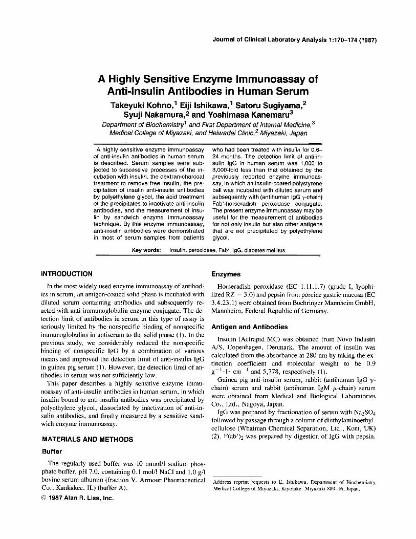
Fig. 1. Effect of the dextran-charcoal treatment in the present enzyme immunoassay. Normal serum was subjected to the present enzyme immu- noassay by incubation with various amounts of insulin with and without the dextran-charcoal treatment. Circles and squares indicate the results obtained with and without the dextran-charcoal treatment, respectively.
Previous Enzyme lmmunoassay
An insulin-coated polystyrenc ball was incubated with 0.15 ml of diluted serum samples and then with (antihuman IgG y-chain) or (antihuman IgM p-chain) Fab'-peroxidase con- jugate as describcd previously (1). Serum samples were treatcd with the dextran-charcoal at pH 6.0 as described above and diluted 7.500-fold with buffer A.
Affinity-Purification of Rabbit (Antihuman IgG y-Chain) IgG
(Antihuman IgG y-chain) IgG (35 mg) in 35 ml ofO. 1 mol/ 1 sodium phosphate buffer, pH 7.0, containing 1 g/l NaN3 was applied to a column (3.5 X 20 mm) of nonspecific hu- man IgG-Sepharose 4B at a flow rate of 1 ml/h using the same buffer. The specific IgG was elutcd with 0.1 mol/l gly- cine-HC1 buffer, pH 2.5 at a flow rate of 60 ml/h, and the eluate was immediately neutralized by addition of 2 mol/l Tris-HCI buffer, pH 8.0. The amount of the affinity-purified IgG was 4.1 mg.
Identification of the Class of Anti-Insulin Antibodies in Human Serum
Human scrum (3 pl) containing anti-insulin antibodies was mixed with 0.2 ml of buffer A containing 1 g/l NaN3 and then passed through a column (3.5 X 40 mm) of affinity-pu- rified rabbit (antihuman IgG y-chain) IgG-Scpharose 4B at a
flow rate of 1 ml/h using thc same buffer. The effluent (1.0 ml) was diluted with nonspecific human serum to various extents and subjected to the present enzyme immunoassay for anti-insulin antibodies.
Purification of Anti-Insulin IgG from Human Serum
Human serum (3 ml) containing anti-insulin antibodies was diluted 2-fold with 20 mmol/l sodium phosphate buffer, pH 7.0, and applied to a column (1.5 X 1.7 mm) of insulin- Sepharose 4B using the same buffer at a flow rate of 1 ml/h. The anti-insulin IgG was eluted with 1.3 mmol/l HCI, pH 2.5 at a flow rate of 60 ml/h, and the eluate was immediately neutralized by addition of 1 mol/l sodium phosphate buffer, pH 7.0. The amount of the affinity-purified IgG was 1.4 pg.
Determination of the Amount of Human IgG by Sandwich Enzyme lmmunoassay Technique
An (antihuman IgG y-chain) IgG-coated polystyrene ball was incubated with human IgG in 0.15 ml of buffer A at 37°C for 5 h and at 4°C overnight, and then with 100 ng of (antihuman IgG y-chain) Fab'-peroxidase conjugate in 0.15 ml of buffer A at 20°C for 4 h as described previously (8).
RESULTS AND DISCUSSION
Effect of the Dextran-Charcoal Treatment in the Present Enzyme lmmunoassay
In the present enzyme immunoassay, a 0.11 ml aliquot of human serum samples was incubated with 16 pU of insulin and subjected to successive processes of the dextran-char- coal treatment at neutral pH to remove free insulin, the ad- dition of polyethylene glycol to precipitate insulin anti- insulin antibodies, the acid treatment of the precipitates to inactivate anti-insulin antibodies, and finally the measure- ment of insulin by sandwich enzyme immunoassay tech- nique using affinity-purified anti-insulin Fab'-peroxidase conjugate. When the amount of insulin incubated with nor- mal scrum samples was increased from 1.6 pU to 160 pU, there was little increase in bound peroxidase activity, whcreas tremendous increases were observed without the dextran-charcoal treatment (Fig. I ) . This indicated that a wide variation of scrum insulin level did not interfere with the present enzyme immunoassay.
Effect of the Dextran-Charcoal Treatment in the Previous Enzyme lmmunoassay
In the previous enzyme immunoassay, an insulin-coated polystyrene ball was incubated with a diluted serum sample, which had been treated with the dextran-charcoal at pH 6.0 to remove insulin present in the serum sample, and subse- quently with (antihuman IgG y-chain) Fab'-peroxidasc con- jugate. The dctection limit of anti-insulin IgG in human serum was 2- to 3-fold less than that obtained without the
Enzyme lmmunoassay of Anti-Insulin Antibodies 173
dextran-charcoal treatment. The dextran-charcoal treatment at pH 2.5-5.5 of normal serum caused a higher bound pcr- oxidase activity, that is, a higher background, which may have been due to an increased nonspecific binding of par- tially denatured nonspecific IgG to the solid phase.
100
l0L
** 0 . .
0 0
a a 0
0
0 . a
(month 1 Normal
Period of Treatment with Insu l i n
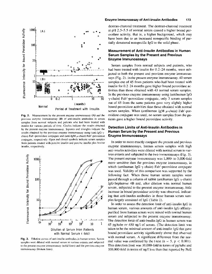
Fig. 2. Measurement by the present enzyme immunoassay (A) and the previous enzyme immunoassay (B) of anti-insulin antibodies in serum samples from normal subjects and patients who had been treated with insulin for various periods of time. Circles indicate the results obtained by the present enzyme immunoassay. Squares and triangles indicate the results obtained by the previous enzyme immunoassay using (anti-IgG y- chain) Fab'-peroxidase conjugate and (anti-IgM p-chain) Fab'-peroxidase conjugate, respectively. Open and closed symbols indicate serum samples from patients treated with porcine insulin and porcine insulin plus bovine insulin, respectively.
Dilution of Serum from Patients with Normal Serum (- fo ld)
Fig. 3. Dilution curves of anti-insulin antibodies in human serum. Serum samples were diluted with normal serum to various extents and subjected to the present enzyme immunoassay (solid lines) and the previous enzyme immunoassay (broken lines).
Measurement of Anti-Insulin Antibodies in Human Serum Samples by the Present and Previous Enzyme lmmunoassays
Serum samples from normal subjects and patients, who had been treated with insulin for 0.2-24 months, were sub- jected to both the present and previous enzyme immunoas- says (Fig. 2). In the present enzyme immunoassay, 40 serum samples out of 45 from patients who had been treated with insulin for 0.2-24 months gave higher bound peroxidase ac- tivities than those obtained with 43 normal serum samples. In the previous enzyme immunoassay using (antihuman IgG y-chain) Fab'-peroxidase conjugate, only 3 serum samples out of 45 from the same patients gave very slightly higher bound peroxidase activities than those obtained with normal serum samples. When (antihuman IgM p-chain) Fab'-per- oxidase conjugate was used, no serum samples from the pa- tients gave a higher bound peroxidase activity.
Detection Limits of Anti-Insulin Antibodies in Human Serum by the Present and Previous Enzyme lmmunoassays
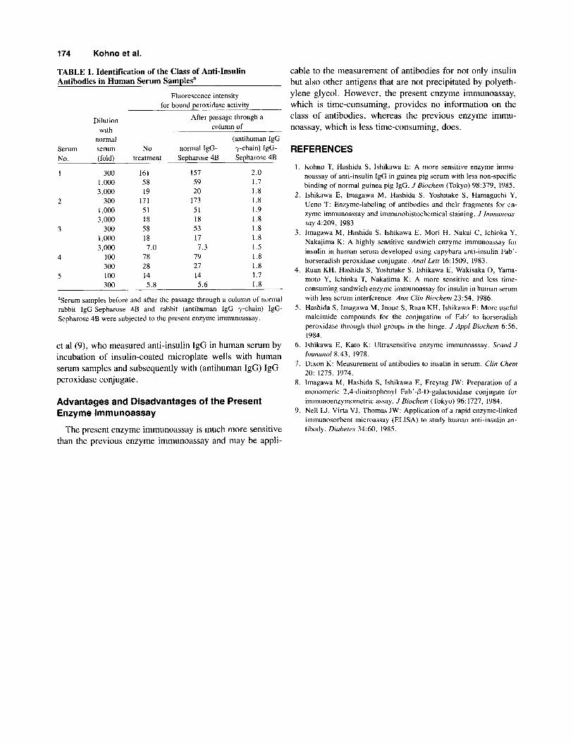
In order to more exactly compare the present and previous enzyme immunoassays, human serum samples with high anti-insulin activities were diluted with normal serum to var- ious extents and subjected to the two immunoassays (Fig. 3 ) . The present enzyme immunoassay was 1,000- to 3,000-fold more sensitive than the previous enzyme immunoassay, in which (antihuman IgG y-chain) Fab'-peroxidase conjugate was used. Validity of this comparison was supported by the following fact. When those human serum samples were passed through a column of rabbit (antihuman IgG y-chain) IgG-Sepharose 4B and, after dilution w-icn normal human serum, subjected to the present enzyme immunoassay, little increase in bound peroxidase activity was observed, indicat- ing that anti-insulin antibodies in those human serum sam- ples largely consisted of IgG (Table 1).
In order to assess the detection limit of anti-insulin IgG in human serum, various amounts of anti-insulin IgG affinity- purified from human serum were mixed with normal human serum and subjected to the present enzyme immunoassay. The detection limit of anti-insulin IgG in human serum was 45 pg/tube or 450 ng/l of serum. (The detection limit was taken to be the minimal amount of anti-insulin IgG that gave bound peroxidase activity significantly above that observed with normal serum. A significant difference from the nor- mal value was confirmed by the t-test (n = 5, p < 0.001). This detection limit was 10,000-fold in terms of pg/tube and 100,000-fold in terms of ng/l less than that reported by Nell
174 Kohno et al.
TABLE 1. Identification of the Class of Anti-Insulin Antibodies in Human Serum Samplesa
cable to the measurement of antibodies for not only insulin but also other antigens that are not precipitated by polyeth-
Serum No.
Dilution with
normal serum (fold)
300 1 ,OOo 3 ,m
300 1 ,OOo 3,000
300 1 ,OOo 3 ,m
100 300 100 300
Fluorescence intensity for bound peroxidase activity
ylene glycol. However, the present enzyme immunoassay, which is time-consuming, provides no information on the class of antibodies, whereas the previous enzyme immu- noassay, which is less time-consuming, does.
After passage through a column of
(antihuman IgG NO normal IgG- y-chain) IgG- REFERENCES
treatment
161 58 19
171 51 18 58 18
78 28 14
7.0
5.8
Sepharose 4B
IS7 59 20
173 51 18 53 17
79 27 14
7.3
5.6
Sepharose 4B
2.0 1.7 1.8 I .8 I .9 1.8 1.8 I .8 1.5 1.8 1.8 1.7 1.8
I .
2 .
3.
4.
'Serum samples before and after the passage through a column of normal rabbit IgG-Sepharose 4B and rabbit (antihuman IgG y-chain) IgG- Sepharose 4B were subjected to the present enzyme immunoassay.
et al (9), who measured anti-insulin IgG in human serum by incubation of insulin-coated microplate wells with human serum samples and subsequently with (antihuman IgG) IgG- peroxidase conjugate.
Advantages and Disadvantages of the Present Enzyme lmmunoassay
The present enzyme immunoassay is much more sensitive than the previous enzyme immunoassay and may be appli-
Kohno T, Hashida S, Ishikawa E: A more sensitive enzyme immu- noassay of anti-insulin IgG in guinea pig serum with less non-specific binding of normal guinea pig IgG. J Biochem (Tokyo) 98:379, 1985. Ishikawa E, Imagawa M, Hashida S. Yoshitake S, Hamaguchi Y, Ueno T: Enzyme-labeling of antibodies and their fragments for en- zyme immunoassay and immunohistochemical staining. J Immunoas- say 4:209, 1983. lmagawa M, Hashida S, Ishikawa E, Mori H, Nakai C, Ichioka Y, Nakajima K: A highly sensitive sandwich enzyme immunoassay for insulin in human serum developed using capybara anti-insulin Fdb'- horseradish peroxidase conjugate. Anal Lett 16: 1509, 1983. Ruan KH, Hashida S, Yoshitake S, Ishikawa E, Wakisaka 0, Yama- moto Y, Ichioka T, Nakajima K: A more sensitive and less time- consuming sandwich enzyme immunoassay for insulin in human serum with less serum interference. Ann Cfin Biochem 23:54, 1986.
5. Hashida S , Imagawa M, Inoue S, Ruan KH, Ishikawa E: More useful maleimide compounds for the conjugation of Fab' to horseradish peroxidase through thiol groups in the hinge. J Appl Biochrm 6 5 6 , 1984.
6. Ishikawa E, Kato K: Ultrasensitive enzyme immunoassay. Srand J Immunol8:43, 1978.
7. Dixon K: Measurement of antibodies to insulin in serum. Clin Chem 20: 1275, 1974.
8. lmagawa M, Hashida S, Ishikawa E, Freytag JW: Preparation of a monomeric 2,4-dinitrophenyl Fab'-P-D-galactosidase conjugate for immunoenzymometric assay. J Biochem (Tokyo) 96: 1727, 1984.
9. Nell U, Virta VJ, Thomas JW: Application of a rapid enzyme-linked immunosorbent microassay (ELISA) to study human anti-insulin an- tibody. Diabetes 34:60, 1985.