47 diffusely enlarged uterus on magnetic resonance imaging
TRANSCRIPT

47 Diffusely Enlarged Uterus on Magnetic Resonance Imaging

CLINICAL IMAGAGINGAN ATLAS OF DIFFERENTIAL DAIGNOSIS
EISENBERG
DR. Muhammad Bin Zulfiqar PGR-FCPS III SIMS/SHL

• Fig GU 47-1 Delayed uterine involution. Sagittal T2-weighted MR image 10 days after vaginal delivery shows that the uterus is still enlarged. The myometrium has high signal intensity with prominent vessels (arrowheads). Low-signal-intensity fluid is seen in the endometrial cavity (arrows), a finding indicative of blood products.73

• Fig GU 47-2 Postpregnancy endometritis. Sagittal contrast T1-weighted MR image in a woman who experienced septic shock 3 weeks after caesarean section shows intense enhancement of the uterus with particularly prominent cervical enhancement (arrowheads). The cervix does not usually enhance so intensely unless it is significantly inflamed. The fluid seen in the endometrial cavity was found to be pus related to infection by methicillin-resistant Staphylococcus aureus.73

• Fig GU 47-3 Retained placenta. Sagittal contrast T1-weighted MR image shows marked enhancement of the lesion protruding into the uterine cavity (arrow). The nonenhancing area in the lesion corresponds to a clot. Arrowhead = blood in the uterine cavity.73
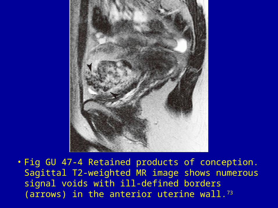
• Fig GU 47-4 Retained products of conception. Sagittal T2-weighted MR image shows numerous signal voids with ill-defined borders (arrows) in the anterior uterine wall.73

• Fig GU 47-5 Estroprogesterone. Sagittal T2-weighted MR image shows a globular uterine corpus (arrowheads), a myometrium with increased signal intensity (higher than that of a normal uterus in a reproductive-age woman), and an atrophic endometrium.73
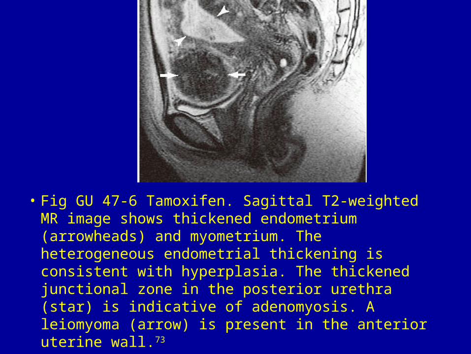
• Fig GU 47-6 Tamoxifen. Sagittal T2-weighted MR image shows thickened endometrium (arrowheads) and myometrium. The heterogeneous endometrial thickening is consistent with hyperplasia. The thickened junctional zone in the posterior urethra (star) is indicative of adenomyosis. A leiomyoma (arrow) is present in the anterior uterine wall.73

• Fig GU 47-7 Granulosa cell tumor. Sagittal contrast T1-weighted MR image shows marked enhancement of the endometrium (arrowheads), which is indicative of cystic hyperplasia. The ovarian wall and septa also demonstrate significant enhancement. After surgery, the estradiol level returned to normal.73

• Fig GU 47-8 Pelvic congestion syndrome. Sagittal T2-weighted MR image shows a thickened myometrium with numerous dilated signal voids (arrows).73

• Fig GU 47-9 Adenomyosis. Sagittal T2-weighted MR image shows indistinct zonal anatomy. Widening of the junctional zone is clearly seen in the region around the distorted endometrium (arrowheads). The myometrium has decreased signal with tiny spots of high signal intensity (arrows).73
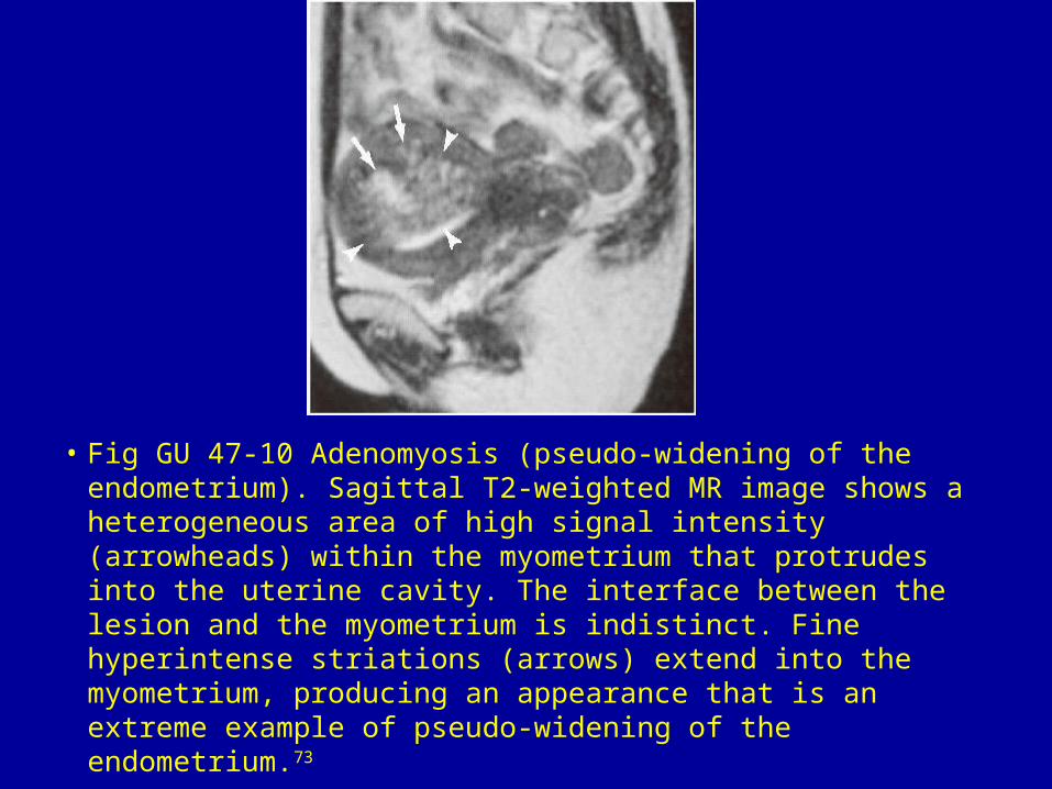
• Fig GU 47-10 Adenomyosis (pseudo-widening of the endometrium). Sagittal T2-weighted MR image shows a heterogeneous area of high signal intensity (arrowheads) within the myometrium that protrudes into the uterine cavity. The interface between the lesion and the myometrium is indistinct. Fine hyperintense striations (arrows) extend into the myometrium, producing an appearance that is an extreme example of pseudo-widening of the endometrium.73

• Fig GU 47-11 Multiple leiomyomas. Sagittal T2-weighted MR image shows a diffusely enlarged uterus containing several fatty lesions with clear margins and distinctive low signal intensity.73

• Fig GU 47-12 Diffuse leiomyomatosis. Sagittal T2-weighted MR image demonstrates a prominently enlarged uterus with innumerable leiomyomas that appear to blend with one another. The endometrium (arrows) is markedly elongated and distorted by the multiple submucosal nodules.73

• Fig GU 47-13 Leiomyosarcoma. Sagittal T2-weighted MR image shows an enlarged, irregularly shaped uterus. The tumor occupies the endometrial cavity (arrows). Note the proiminent nodules (m) in the anterior wall.73

• Fig GU 47-14 Endometrial stromal sarcoma. Sagittal T2-weighted image shows a huge tumor that replaces the endometrial cavity (arrowheads) and infiltrates the myometrium. Bands of low signal intensity (arrows) are seen in the infiltrated myometrium. These bands corresponded to preserved bundles of myometrium at histologic examination of this low-grade tumor.73

• Fig GU 47-15 Intrauterine device. The IUD (arrow) appears as a band of low signal intensity in the endometrium of a globular uterus.73

