1 the pio operon is essential for phototrophic fe(ii)...
TRANSCRIPT
1
The pio operon is essential for phototrophic Fe(II) oxidation 1
in Rhodopseudomonas palustris TIE-1 2
3 4
Yongqin Jiao1 and Dianne K. Newman1,2,3* 5
6
1Division of Geological and Planetary Sciences, 2Division of Biology, 3Howard Hughes 7
Medical Institute, California Institute of Technology, Pasadena, California, United States 8
of America 9
10
* To whom correspondence should be addressed. Email: [email protected] 11
Mailing address: California Institute of Technology, Division of Geological and 12
Planetary Sciences, Mail Stop 100-23, Pasadena, CA 91125. Phone: (626) 395-6790. 13
Fax: (626) 683-0621 14
15
16
17
Running title: Phototrophic Fe(II) oxidation by R. palustris TIE-1 18
Key words: iron oxidation, anoxygenic photosynthesis, c-type cytochrome, 19
Rhodopseudomonas palustris 20
ACCEPTED
Copyright © 2006, American Society for Microbiology and/or the Listed Authors/Institutions. All Rights Reserved.J. Bacteriol. doi:10.1128/JB.00776-06 JB Accepts, published online ahead of print on 22 December 2006
on May 28, 2018 by guest
http://jb.asm.org/
Dow
nloaded from
2
Abstract 1 2
Phototrophic Fe(II) oxidizing bacteria couple the oxidation of ferrous iron [Fe(II)] to 3
reductive CO2 fixation using light energy, but until recently, little has been understood 4
about the molecular basis for this process. Using Rhodopseudomonas palustris TIE-1 as 5
a model organism, here we report the discovery of a 3-gene operon, designated as the pio 6
operon (for phototrophic iron oxidation) that is necessary for phototrophic Fe(II) 7
oxidation. The first gene in the operon, pioA, encodes a c-type cytochrome that is 8
upregulated under Fe(II)-grown conditions. PioA contains a signal sequence and shares 9
homology with MtrA, a decaheme cytochrome c from Shewanella oneidensis MR-1. The 10
second gene, pioB, encodes a putative outer membrane beta-barrel protein. PioB is a 11
homologue of MtrB from S. oneidensis MR-1. The third gene, pioC, encodes a putative 12
high potential iron sulfur protein (HiPIP) with a Tat signal sequence, and is similar to the 13
putative Fe(II) oxidoreductase (Iro) from Acidithiobacillus ferrooxidans. Like PioA, 14
PioB and PioC appear to be secreted proteins. Deletion of the pio operon results in loss 15
of Fe(II) oxidation activity and growth on Fe(II). Complementation studies confirm that 16
the phenotype of this mutant is due to loss of the pio genes. Deletion of pioA alone 17
results in loss of almost all Fe(II) oxidation activity, however, deletion of either pioB or 18
pioC alone results in only a partial loss of Fe(II) oxidation activity. Together, these 19
results suggest that proteins encoded by the pio operon are essential and specific for 20
phototrophic Fe(II) oxidation in R. palustris TIE-1. 21
ACCEPTED
on May 28, 2018 by guest
http://jb.asm.org/
Dow
nloaded from
3
Introduction 1
2
One of the distinguishing features of microbial metabolism is its diversity: over 3
billions of years of Earth history, microbes have evolved an impressive array of strategies 4
to obtain energy for growth. The process of photosynthesis, for example, goes well 5
beyond the ability to split water and produce oxygen. Different groups of 6
microorganisms carry out “anoxygenic” photosynthesis, using substrates such as 7
molecular hydrogen (H2), various sulfur species, small organic molecules, or ferrous iron 8
[Fe(II)] as an exogenous electron donor to drive reductive CO2 fixation (6, 8, 12, 17). If 9
we seek to understand the origins of the remarkable metabolic diversity that characterizes 10
modern life on Earth, it is important to know how different types of metabolisms operate 11
at the molecular level. This is necessary both to be able to compare the components of 12
different metabolisms to each other, and to inform our search for biosignatures unique to 13
these metabolisms in the rock record. 14
As a step towards this general goal, we have chosen to focus on the process of 15
phototrophic Fe(II) oxidation, which can be described by the following equation: 16
4Fe2+ + CO2 + 11H2O + hν = [CH2O] + 4Fe(OH)3 + 8H+ 17
This type of photosynthesis is interesting in the context of metabolic evolution for several 18
reasons. First, phototrophic Fe(II)-oxidation is phylogenetically widespread, appearing in 19
purple and green bacteria (10, 14, 21, 22, 65, 66); phylogenic comparisons of genes from 20
different photosynthetic organisms suggest that anoxygenic photosynthesis is more 21
ancient than oxygenic photosynthesis (57, 72). Second, iron has an intermediate redox 22
potential (∆E0’= -0.11 V) (28) compared to other substrates used as electron donors in 23
ACCEPTED
on May 28, 2018 by guest
http://jb.asm.org/
Dow
nloaded from
4
photosynthesis (e.g. H2 (∆E0’= -0.41 V) or H2O (∆E0
’= 0.82 V)) (28, 44). It has been 1
suggested, therefore, that Fe(II)-based photosynthesis may represent a transition form of 2
metabolism from anoxygenic to oxygenic photosynthesis (57). Third, Fe(II) is thought to 3
have been the most widespread source of reducing power in the late Archean and early 4
Proterozoic (3.8-1.6 billion years ago (Ga)) with an estimated concentration of about 0.1 5
– 1 mM in seawater (69); atmospheric oxygen seems to have appeared in significant 6
amounts only after 2.4 Ga (15, 26, 30, 60). 7
8
Banded Iron Formations (BIFs), are an ancient class of iron ore deposits that may 9
record the story of the evolution of photosynthesis. Because the use of Fe(II) results in 10
the production of ferric iron [Fe(III)] minerals, it has been suggested that Fe(II)-based 11
phototrophy might have been responsible for catalyzing BIF deposition early in Earth 12
history (14, 34, 71). Later occurrences of BIFs (e.g at 1.8 Ga), however, are believed to 13
have resulted from Fe(II) oxidation catalyzed by molecular oxygen produced by 14
cyanobacteria. Episodic deposition of BIFs throughout the Precambrian thus may reflect 15
a transition from anoxygenic to oxygenic photosynthesis. How did ancient phototrophs 16
evolve from using Fe(II) as an electron donor to using H2O? 17
18
To address this question, we must understand the molecular machinery of 19
phototrophic Fe(II) oxidation. Discovered in the early 1990’s by Widdel and co-workers 20
(71), phototrophic Fe(II) oxidizing bacteria such as Thiodictyon, Rhodobacter, and 21
Chlorobium species have been isolated from a wide variety of environments, including 22
both freshwater and marine settings (14, 21, 29, 66, 71). However, very little is 23
ACCEPTED
on May 28, 2018 by guest
http://jb.asm.org/
Dow
nloaded from
5
understood at the molecular level about the mechanism of Fe(II) oxidation in any of these 1
organisms. In the companion paper to this article, we report the discovery of a c-type 2
cytochrome and a putative pyrroloquinoline quinone containing enzyme from an Fe(II) 3
oxidizing strain--Rhodobacter SW2--that stimulates Fe(II) oxidation activity in its close 4
relative, Rhodobacter capsulatus SB1003 (11). Because our ability to explore the 5
mechanistic basis of Fe(II) oxidation in SW2 itself is limited due to the impracticality of 6
direct mutational analysis (11), we established a genetic system in a different Fe(II)-7
oxidizing phototroph, Rhodopseudomonas palustris TIE-1 (28). In this report, we 8
describe the identification of the pio operon, a 3-gene operon essential for phototrophic 9
growth on Fe(II) by R. palustris TIE-1. 10
11
ACCEPTED
on May 28, 2018 by guest
http://jb.asm.org/
Dow
nloaded from
6
Materials and Methods 1
2
Bacterial strains and plasmids. Bacterial strains and plasmids used in this study are 3
listed in Table 1. R. palustris CGA010, derived from parent strain CGA009 after a frame 4
shift in the hupV gene was repaired, was kindly provided by F. Rey and C. S. Harwood 5
(University of Washington). 6
7
Media and culture conditions For aerobic growth R. palustris strains were grown in YP 8
medium (0.3% yeast extract and 0.3% Bacto Peptone (Difco)) with shaking at 30˚C. For 9
anaerobic growth R. palustris strains were grown without shaking at 30˚C in FEM, a 10
defined basal medium for phototrophic Fe(II)-oxidizing bacteria (12). For 11
photoheterotrophic growth FEM was supplemented with 10 mM acetate. For 12
photoautotrophic growth electron donors were used such as thiosulfate (10 mM), 13
hydrogen (80% atmosphere), and soluble Fe(II). FEM Medium containing soluble Fe(II) 14
was prepared as previously described and the final Fe(II) concentration is in the range of 15
4-6 mM (28). Cultures were incubated 20 to 30 cm from a 34 W tungsten, incandescent 16
light source at 30°C. All phototrophic cultures, except those grown on hydrogen, were 17
grown in an atmosphere consisting of 80% N2 and 20% CO2. Escherichia coli strains 18
were cultured in lysogeny broth (LB) at 37°C. E. coli WM3064 was supplemented with 19
300 µM diaminopimelic acid (DAP). Kanamycin and gentamicin were used at 100 and 20
200 µg/ml for R. palustris and 50 and 20 µg/ml for E. coli, respectively. 21
22
ACCEPTED
on May 28, 2018 by guest
http://jb.asm.org/
Dow
nloaded from
7
Cell suspension assay All cell suspension assays were conducted at room temperature in 1
an anaerobic chamber containing an atmosphere of 80% N2, 15% CO2 and 5% H2 (12, 2
28). Fe(II) cultures used for this assay contained 10 mM nitrilotriacetic acid (NTA) to 3
prevent ferric iron precipitation. NTA alone does not support phototrophic growth of R. 4
palustris (data not shown). Cells were pre-grown in the medium indicated until mid-5
exponential phase to an OD 660 nm of ~ 0.3 measured by 96-well plate reader (Synergy 6
HT, Bio-Tek, Winooski, VT) with a volume of 200 µl. Cells were harvested by 7
centrifugation (10,000 x g for 15 min) and washed in the same volume of HEPES buffer 8
(50 mM N-2-hydroxyethylpiperazine-N`-2-ethanesulfonic acid with 20 mM NaCl, 9
pH 7.0). To start the assay, cells were resuspended in HEPES buffer containing 20 mM 10
NaHCO3 and either 400 µM or 1 mM (as indicated in Fig. 1) of FeCl2. Cells were 11
concentrated approximately 3 times compared to the original growth culture and 100 µl 12
of the cell suspension was aliquoted into a 96 well plate. The OD reading is about 0.7 13
measured by the 96-well plate reader. The plates were incubated at room temperature in 14
the glove box under a 40 W tungsten light with light intensity of about 3000 lux. Over 15
time 100 µl ferrozine solution (1 g of ferrozine plus 500 g of ammonia acetate in 1 L of 16
ddH2O) was added to the wells to monitor Fe(II) levels (64). The rate of Fe(II) oxidation 17
was calculated based on the linear portion of the curves generated. 18
19
Extract preparation, SDS_PAGE analysis and heme staining R. palustris TIE-1 was 20
grown on either H2, thiosulfate or Fe(II) plus NTA until mid-exponential phase and cells 21
were harvested by centrifugation at 10, 000 g for 15 min. Cell pellets were resuspended 22
and washed 3 times in the same volume of HEPES buffer and resuspended in the same 23
ACCEPTED
on May 28, 2018 by guest
http://jb.asm.org/
Dow
nloaded from
8
buffer containing Protease inhibitor cocktail (Roche) and 50 µM DNase (Roche) and 1
incubated at 4 °C for 30 min. Cells were disrupted by French press (3 passes at 18,000 2
psi) and the cell lysate was clarified by centrifugation at 10, 000 g for 15 min at 4 °C. 3
The resulting supernatant was centrifuged at 200,000 g for 120 min at 4°C. The 4
supernatant was defined as the soluble fraction and the pellet, which was resuspended in 5
HEPES buffer, was defined as the membrane fraction. Protein concentrations were 6
determined by the Bradford assay (7). SDS-PAGE was preformed by standard 7
procedures according to Laemmli (38). Soluble and membrane fractions were incubated 8
in loading buffer containing dithiothreitol at room temperature for 10 minutes without 9
heating, and separated on a 12% Tris/HCl pre-cast gel (Bio-Rad). Coomassie staining 10
was performed using BioRad standard staining protocol as described by the 11
manufacturer. Gels stained for heme-containing proteins were performed according to 12
the in-gel peroxidase activity assay as described (16). 13
14
RT-PCR R. palustris TIE-1 was grown photoautotrophically on Fe(II) plus NTA until 15
exponential phase. Total RNA was extracted as described previously (56). Briefly, cells 16
were harvested and resuspended in 1 ml TE buffer (10 mM Tris/HCl, 1 mM EDTA, pH 17
8.0). Cells were disrupted using a Mini-BeadBeater-8 (BioSpec Products, Bartlesville) in 18
2 ml screw-capped tubes containing approximately 1 ml 0.1 mm zirconia/silica beads 19
(BioSpec) for 1 min periods, with cooling on ice after each period with a total of 4 min. 20
RNA extraction was then carried out using Qiagen RNA extraction kit. DNase digestion 21
was performed on the Mini-column with the Qiagen RNase-free DNase set. The RNA 22
was eluted from the column, and a second DNase treatment was performed with Roche 23
ACCEPTED
on May 28, 2018 by guest
http://jb.asm.org/
Dow
nloaded from
9
RNase-Free DNase. The RNA was finally resuspended in 40 µl nuclease free water. 1
cDNA was synthesized using BioRad iScript cDNA Synthesis Kit. Control PCR using 2
RNA as template in the absence of reverse transcriptase confirmed that the isolated RNA 3
was free of contaminating genomic DNA. Primers used for all RT-PCR reactions are 4
listed in Table 2. To test if pioABC are cotranscribed, primers RT-pioA-L1 and RT-5
pioB-R1 were used to detect the presence of transcript pioAB and primers RT-pioB-L1 6
and RT-pioC-R1 were used to detect transcript pioBC. To test the transcription of pio 7
genes in the mutant background ∆pioA, ∆pioB or ∆pioC, RT-pioAL and RT-pioAR was 8
used to detect pioA, RT-pioBL and RT-pioBR for pioB and RT-pioCL and RT-pioCR for 9
pioC. 10
11
Cloning, DNA manipulations, and mutant construction. Standard protocols were used 12
for DNA cloning and transformation (28). Plasmids were purified on QIAprep spin 13
columns (Qiagen Inc., Chatsworth, Calif.). R. palustris TIE-1 chromosomal DNA was 14
isolated using DNeasy kit (Qiagen). DNA was extracted from agarose gels using the 15
Qiaquick gel extraction kit (Qiagen), and plasmid DNA was purified with the Qiaprep 16
spin miniprep kit (Qiagen). DNA was sequenced at the Laragen DNA sequencing center 17
(http://www.laragen.com/services.htm) by standard automated-sequencing technology. 18
19
Construction of deletion mutant All primer sequences used in construction of the 20
mutants are listed in Table 2. For construction of pio operon deletion mutant TIE-3, a 1 21
kb DNA fragment upstream of pioA was produced by PCR with primers pioA1 and 22
pioA1p using TIE-1 genomic DNA as template. Similarly, a 1 kb PCR fragment 23
ACCEPTED
on May 28, 2018 by guest
http://jb.asm.org/
Dow
nloaded from
10
downstream of pioC was generated with primers pioC2 and pioC2p. The PCR products 1
were used as templates for another round of fusion PCR with primers pioA1 and pioC2. 2
The resulting 2 kb fusion PCR product was gel purified and restriction digested using 3
restriction enzyme Spe I, and cloned into the suicide vector pJQ200sk (59) to generate 4
pYQABC. pYQABC was mobilized into TIE-1 by conjugation from E. coli S17-1 (13). 5
Selection of single recombinants on PM plates containing 400 µg/ml of gentamicin 6
followed by selection of double recombinants on PM sucrose (10%) plates were 7
conducted as described (13). Individual gene deletion mutant ∆pioA and ∆pioB and 8
∆pioC were made in a similar manner via suicide plasmids pYQA, pYQB and pYQC, 9
respectively. Primers used for generating pYQA are pioA1, pioA1p, pioA2 and pioA2p, 10
for pYQB are pioB1, pioB1p, pioB2 and pioB2p, for pYQC are pioC1, pioC1p, pioC2 11
and pioC2p. PCR was used to verify that the expected deletion had occurred. 12
13
Generation of complementing plasmids. The pioABC operon and the individual pio 14
genes were amplified from genomic DNA of TIE-1 using the FailSafe PCR kit 15
(Epicentre, WI). The PCR products were designed to have EcoR I and Hind III 16
restriction sites, and were ligated in trans into vector pBBRMCS-2 (35, 36) digested with 17
the same enzymes. The resulting plasmids were conjugated into R. palustris strains 18
indicated (28). The pioABC operon was amplified with primers pioA-start and pioC-end 19
(pYQ01), pioA gene with primers cyc-start and cytc-end (pYQ02), pioB gene with 20
primers pioB-start and pioB-end (pYQ03), pioC gene with primers pioC-start and pioC-21
end (pYQ04). 22
ACCEPTED
on May 28, 2018 by guest
http://jb.asm.org/
Dow
nloaded from
11
Results 1
2
Identification of an Fe(II)-oxidation specific c-type cytochrome 3
With the goal of identifying proteins that are expressed when TIE-1 grows on 4
Fe(II), we compared the Fe(II)-oxidation activity of cell suspensions that had been pre-5
grown photoautotrophically on different electron donors including H2, thiosulfate and 6
Fe(II). Cells were collected and resuspended in buffer containing Fe(II), and Fe(II) 7
oxidation was followed by the ferrozine assay. When 1 mM initial Fe(II) was provided, 8
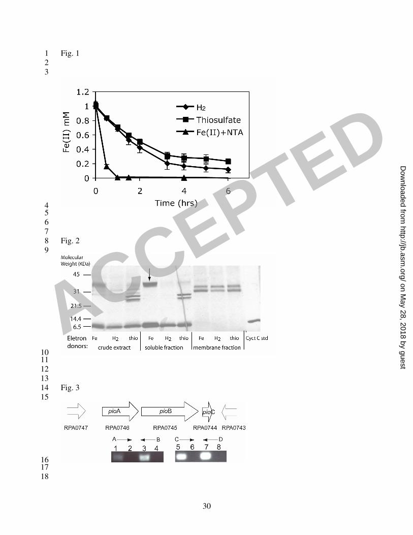
approximately 0.8 mM of Fe(II) was oxidized within the first half hour with Fe(II)-grown 9
cells, whereas only 0.2 mM of Fe(II) was oxidized with H2- or thiosulfate-grown cells 10
(Fig. 1). Compared to the H2- or thiosulfate-grown cells, Fe(II)-grown cells showed a 4-5 11
fold higher rate of Fe(II) oxidation activity, suggesting that specific proteins were 12
induced under Fe(II)-grown conditions. Given these results, we assayed for differential 13
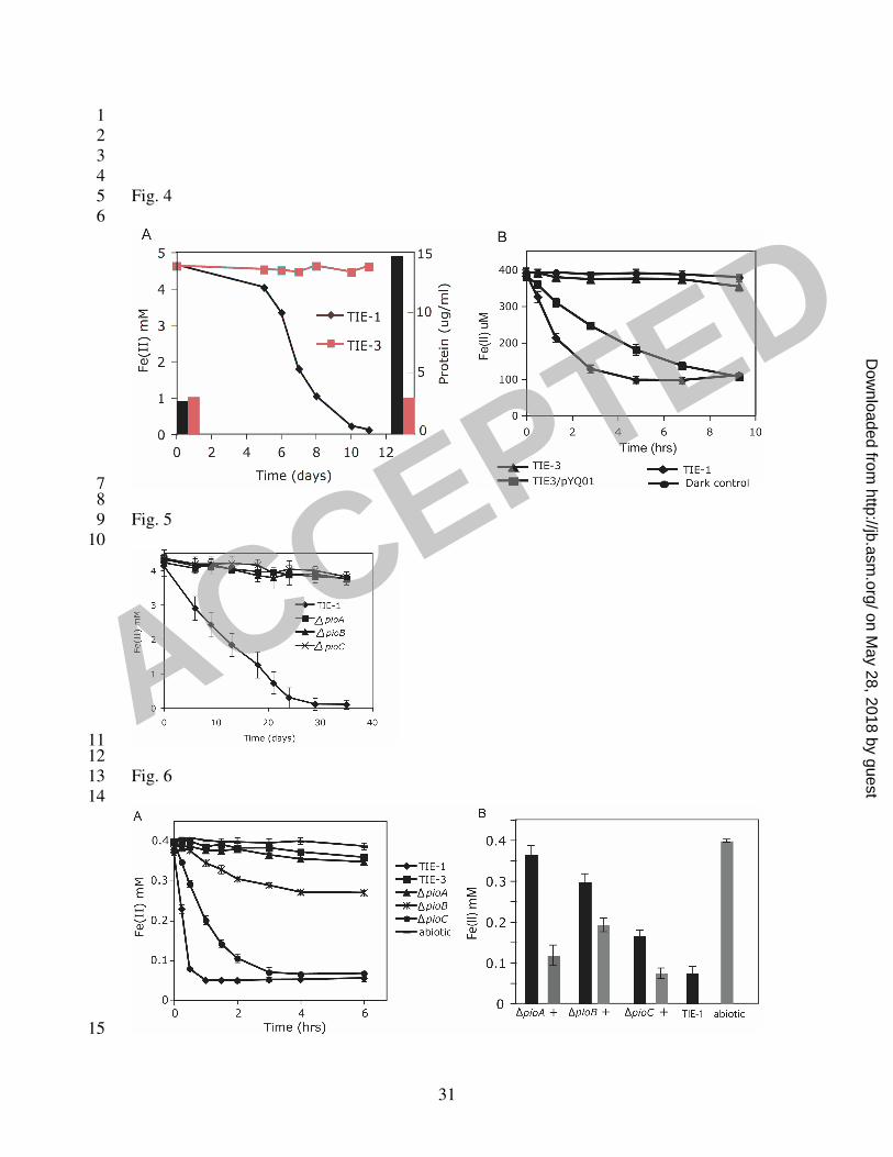
protein expression with cells grown on Fe(II) compared to other electron donors. Crude 14
cell extracts from cells grown on H2, thiosulfate or Fe(II) were separated by SDS-PAGE. 15
Although no significant differences were detected visually by coomassie staining (data 16
not shown), a difference in expression of c-type cytochromes was observed by heme 17
staining. Accordingly, we characterized the expression profile of c-type cytochromes 18
from soluble and membrane fractions of cells grown on Fe(II), H2 and thiosulfate (Fig. 19
2). A unique c-type cytochrome (~40 kDa) appeared in significant quantity in the soluble 20
fraction only when cells were grown on Fe(II). Protein identification by mass 21
spectrometry indicated that peptide fragments of this protein match those of a putative 22
deca-heme c-type cytochrome from Rhodopseudomonas palustris CGA009 (encoded by 23
ACCEPTED
on May 28, 2018 by guest
http://jb.asm.org/
Dow
nloaded from
12
gene RPA0746) (39). 1
2
Identification and sequence analysis of the pio genes 3
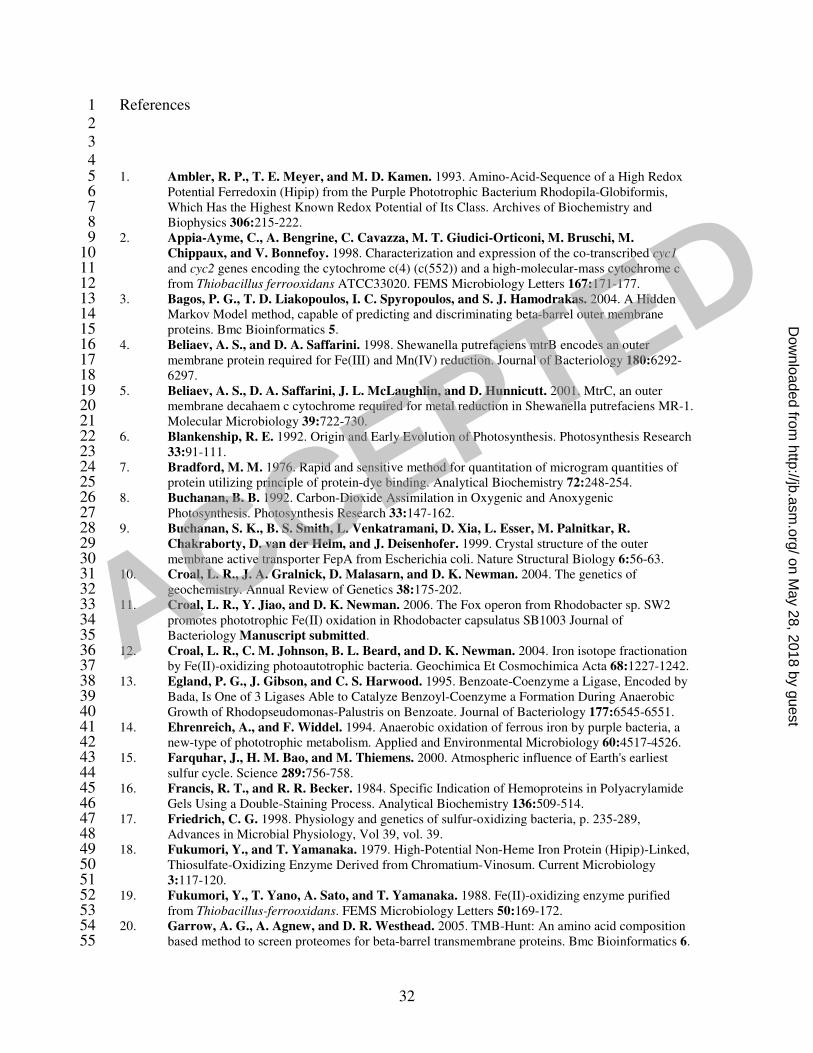
By designing primers based on the CGA009 genome, we were able to sequence a 5.7 kb 4
region from TIE-1 that includes the deca-heme c-type cytochrome open reading frame 5
(ORF) as well as two downstream ORFs (Fig. 3). We designate these genes pioA, pioB 6
and pioC, where pio stands for phototrophic iron oxidation. The DNA sequence of pioA, 7
pioB and pioC was deposited in the Genbank database under the accession numbers 8
EF119739 , EF119740 and EF119741, respectively. The deduced protein sequences of 9
pioA, pioB and pioC are about 98%, 97% and 100% identical to those of RPA0746, 10
RPA0745 and RPA0744, respectively, indicating high sequence similarity between TIE-1 11
and CGA009 over this region, consistent with the highly conserved sequences previously 12
identified between these two strains (28). To test the hypothesis that genes pioABC form 13
an operon, we carried out RT-PCR experiments using primers designed to amplify the 14
intergenic regions. RT-PCR products were obtained for both intergenic regions in the 15
cluster (Fig. 3). No product was obtained in controls to which reverse transcriptase or 16
template was omitted. These results show that pioABC are co-transcribed. An intergenic 17
region of about 700 bp is present upstream of pioA, proceeded by an ORF encoding a 18
protein homologous to a subunit of the putative sulfate ABC transporter CysA from E. 19
coli K-12 (27). The ORF downstream of pioC transcribes in the opposite direction 20
relative to the pio operon. Because of the presence of the large intergenic region 21
upstream of the pio operon, as well as the opposite direction of transcription for the 22
downstream ORF, it seems likely that the pio operon functions independently of the 23
ACCEPTED
on May 28, 2018 by guest
http://jb.asm.org/
Dow
nloaded from
13
adjacent genes. 1
The deduced amino acid sequence of pioA consists of 540 amino acids with a 2
putative signal sequence characteristic of secreted proteins through the Sec pathway; a 3
cleavage site is predicted between residue 40 and 41, according to LipoP 4
(http://www.cbs.dtu.dk/services/LipoP/) and SignalP 5
(http://www.cbs.dtu.dk/services/SignalP/). Lack of hydrophobic regions within PioA, 6
with the exception of the signal sequence, as well as the observation that PioA is in the 7
soluble fraction (Fig. 2), suggest that PioA is likely to be a periplasmic protein. PioA 8
contains 10 putative heme-binding sites (CXXCH) characteristic of c-type cytochromes. 9
Comparison of PioA to sequences in the NCBI database 10
(http://www.ncbi.nlm.nih.gov/blast/) reveals that it is similar to several deca-heme c-type 11
cytochromes in Shewanella, Vibrio and Geobacter species (4, 40-43, 48, 51-54). In 12
particular, it has 40% identity and 55% similarity over 285 amino acids to MtrA from 13
Shewanella oneidensis MR-1, which is involved in metal (e.g. Fe(III) and Mn(IV)) 14
reduction (5, 49, 50, 58), and this similarity is mostly due to the highly conserved nature 15
of the heme-binding sites that are present close to the C-terminal end of PioA. However, 16
approximately 270 amino acids close to the N-terminus of PioA have no homolog in the 17
database. No significant similarity was found when comparing PioA to other proteins in 18
the database. 19
20
The second ORF, pioB, is 99 nucleotides downstream of pioA. pioB encodes a 21
protein of 810 amino acids and contains a putative signal peptide with a predicted 22
cleavage site between residue 25 and 26 based on the LipoP program, suggesting it is also 23
ACCEPTED
on May 28, 2018 by guest
http://jb.asm.org/
Dow
nloaded from
14
secreted through the Sec pathway. It has a putative porin motif close to the C-terminus 1
according to InterProScan (http://www.ebi.ac.uk/InterProScan/) and is predicted to be an 2
outer membrane β-barrel protein according to Transmembrane Barrel Hunt (20) and 3
PRED-TMBB programs (3). Comparison of PioB to sequences in the databases reveals 4
similarities to several outer membrane proteins from Shewanella and Geobacter species. 5
In particular, it has 21% identity and 38% similarity over 536 amino acids close to the c-6
terminus of an outer membrane protein MtrB from Shewanella oneidensis MR-1, which 7
is involved in metal (e.g. Fe(III) and Mn(IV)) reduction (4, 51). However, approximately 8
120 amino acids at the N-terminus show no homology to anything in the database. 9
According to the secondary structure predicted by PRED-TMBB (3), both PioB and 10
MtrB are outer membrane porins with 28 transmembrane beta-strands, the largest number 11
of beta-strands among all known outer membrane porins (9, 33, 62). Similar to other 12
outer membrane porins, PioB and MtrB are predicted to have long loops protruding into 13
the extracellular space and short turns on the periplasmic side, except that PioB has 14
longer extracellular loops compared to MtrB, consistent with the sequence length 15
difference between the two proteins. The conserved regions between PioB and MtrB 16
mainly occur in the transmembrane regions, consistent with the idea that these regions are 17
generally more conserved than the loop regions among outer membrane porins (62). 18
The third ORF, pioC, is 140 nucleotides downstream of pioB. pioC encodes a 19
putative high potential iron-sulfur protein (HiPIP) that contains an iron-sulfur binding 20
site. The deduced amino acid sequence of pioC consists of 94 amino acids with a 21
predicted twin-arginine translocation (Tat) signal sequence at the N-terminus, suggesting 22
export into the periplasm through the Tat protein translocation pathway. A signal 23
ACCEPTED
on May 28, 2018 by guest
http://jb.asm.org/
Dow
nloaded from
15
sequence cleavage site was predicted between residues 37 and 38 based on the SignalP 1
program. Because there is no transmembrane region other than the signal peptide 2
predicted by HMMTOP (http://www.enzim.hu/hmmtop/html/submit.html), we predict 3
PioC resides in the periplasm. Comparison of PioC to sequences in the database reveals 4
similarities to HiPIPs from several bacteria, with most of the similarity occurring over 5
approximately 50 amino acid residues close to the C-terminus spanning the iron-sulfur 6
cluster binding site. PioC is 47% identical and 52% similar over 48 amino acids to a 7
HiPIP from Rhodopila globiformis (1), is 32% identical to a hypothetical protein encoded 8
by gene RPA3566 from R. palustris CGA009, and is 44% identical and 53% similar over 9
51 amino acids to a HiPIP from Acidithiobacillus ferrooxidans, a putative iron 10
oxidoreductase known as the “Iro” protein (19, 37). 11
12
pioABC are specifically required for phototrophic Fe(II) oxidation 13
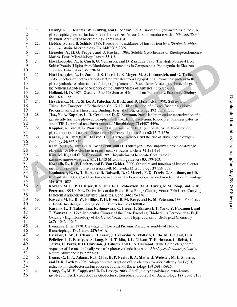
To determine whether the pio operon is necessary for growth on Fe(II), we 14
constructed a mutant (TIE-3) in which all 3 genes in the pio operon were deleted from the 15
chromosome by homologous recombination. We tested the ability of mutant TIE-3 to 16
grow on different substrates. When Fe(II) was provided as the electron donor for 17
photoautotrophic growth, very little Fe(II) was oxidized by strain TIE-3 in a period of 2 18
weeks (Fig. 4A). In contrast, wild type strain TIE-1 oxidized Fe(II) to completion within 19
this time period. End point measurements of total protein content in the cultures 20
indicated that TIE-3 did not grow over the course of incubation in contrast to TIE-1 (Fig. 21
4A). To determine if TIE-3 was specifically defective for growth on Fe(II), we tested 22
growth on substrates other than Fe(II). Photoautotrophic growth of TIE-3 on H2 or 23
ACCEPTED
on May 28, 2018 by guest
http://jb.asm.org/
Dow
nloaded from
16
thiosulfate and photoheterotrophic growth on acetate were tested by measuring cell OD. 1
TIE-3 grew on these substrates as well as TIE-1 (Table 3). These results indicate that the 2
pioABC operon is essential and specific for growth on Fe(II). 3
To further characterize the pioABC operon deletion mutant, with respect to its 4
Fe(II) oxidation phenotype, we performed a cell suspension assay. In this assay, H2 5
grown cells of wide type and mutant TIE-3 were washed then incubated with Fe(II) under 6
light in the anaerobic chamber. Fe(II) oxidation activity was followed using ferrozine 7
assay. The Fe(II) oxidation activity we observed was light dependent (Fig. 4B). Over a 8
period of several hours for the equivalent density of H2 grown cells, 400 µM of Fe(II) 9
was oxidized to completion by TIE-1, but very little Fe(II) was oxidized by TIE-3. This 10
indicates that the pio operon is responsible for almost all the Fe(II) oxidation activity in 11
H2-grown TIE-1. Considering the initial rate of Fe(II) oxidation, the activity of TIE-3 12
could be restored to about 50% of the wide type level by complementation with the entire 13
pio operon (Fig. 4B); the total amount of Fe(II) that was oxidized over a period of 9 hours 14
was the same between wide type TIE-1 and the complemented strain. The vector alone 15
did not affect Fe(II) oxidation by TIE-3 or TIE-1 (data not shown). However, 16
complementation with each individual gene (pioA, pioB, or pioC), did not restore any 17
Fe(II) oxidation activity (data not shown). This suggests that more than one gene in the 18
pio operon is necessary for this activity. 19
Because the pio operon is so highly conserved between strain CGA009 and TIE-20
1, we checked whether the pio operon also confers Fe(II) oxidation to CGA009. Deletion 21
of the genes corresponding to pioABC (i.e., RPA0746, RPA0745 and RPA0744) in 22
CGA009 resulted in a large defect in Fe(II) oxidation activity (data not shown), similar to 23
ACCEPTED
on May 28, 2018 by guest
http://jb.asm.org/
Dow
nloaded from

17
that observed in TIE-1. Strain CGA009 shows a similar amount of Fe(II) oxidation 1
activity in the cell suspension assay as H2-grown TIE-1. However, it does not show 2
measurable growth over the same time period as TIE-1, therefore, we chose to work with 3
strain TIE-1 for further analysis. 4
To access the relative importance of the individual pio genes for Fe(II) oxidation, 5
we constructed three individual deletion mutants, ∆pioA, ∆pioB, and ∆pioC. We 6
confirmed that the mutations were nonpolar by RT-PCR (data not shown) using primers 7
listed in Table 2. Neither growth nor Fe(II) oxidation occurred for any of these mutants 8
during growth assay on Fe(II) (Fig. 5); growth of these mutants on other substrates such 9
as H2, thiosulfate or acetate was unaffected (data not shown). In contrast, ∆pioA lost 10
almost all Fe(II) oxidation activity in the cell suspension assay with H2-grown cells, 11
similar to TIE-3, whereas ∆pioB and ∆pioC only partially lost Fe(II) oxidation activity, 12
exhibiting approximately 10% and 40% of the initial rate of Fe(II) oxidation of wild type 13
level (Fig. 6A). The partial defect in Fe(II) oxidation by ∆pioC may be explained by 14
functional substitution of other small soluble electron carriers in the cell (e.g. the other 15
HiPIP encoded by the homolog of RPA3566). Complementation by the respective wild-16
type copies of the genes restored Fe(II) oxidation activity to different extents in the 17
mutants. In comparing the total amount of Fe(II) oxidized after 12 hours, 18
complementation of ∆pioA, ∆pioB and ∆pioC resulted in 85, 60 and 99% of that 19
achieved by TIE-1 in the same amount of time (Fig. 6B). The reason for the relatively 20
low extent of complementation for ∆pioB compared to TIE-1 is not clear. Perhaps it is 21
caused by different levels of expression of pioB when expressed on a vector driven by a 22
non-native promoter versus when expressed from the endogenous promoter. Together, 23
ACCEPTED
on May 28, 2018 by guest
http://jb.asm.org/
Dow
nloaded from

18
these results indicate that all 3 Pio proteins are required for full Fe(II) oxidation activity 1
in R. palustris TIE-1.2
ACCEPTED
on May 28, 2018 by guest
http://jb.asm.org/
Dow
nloaded from
19
Discussion 1
2
Iron is thought to have been an important substrate for microbial metabolism on 3
the early Earth, including ancient types of photosynthesis. Although the molecular basis 4
of Fe(II) oxidation by acidophilic bacteria has been studied for decades (67, 68, 74, 75, 5
77), it is only very recently that Fe(II) oxidation has been examined in anoxygenic 6
phototrophs (11, 28). Because photoautotrophic Fe(II) oxidation is likely to have been 7
one of the most ancient forms of microbial Fe(II) oxidation (12), understanding the 8
molecular basis of this metabolism is not only relevant for understanding the evolution of 9
photosynthesis, but for understanding the evolution of other Fe(II) oxidizing systems. 10
C-type cytochromes with a wide range of redox potentials are involved in Fe(II) 11
oxidation by A. ferrooxidans (2, 68, 76, 77) and Rhodobacter sp. SW2 (11) as well as 12
dissimilatory Fe(III) reduction by Shewanella and Geobacter species (4, 40, 42, 43, 73). 13
Consistent with this, we found a c-type cytochrome to be upregulated when R. palustris 14
TIE-1 was grown photoautotrophically on Fe(II). By reverse genetic analysis, we 15
identified a 3-gene operon (the pio operon) that seems likely to encode the phototrophic 16
Fe(II) oxidoreductase complex. Detailed biochemical studies are needed to confirm this 17
and understand the mechanism of electron transfer from Fe(II), however, based on the 18
results of this study, we can suggest potential functions for the Pio proteins. 19
The first gene in the pio operon encodes PioA, a putative deca-heme c-type 20
cytochrome. Because the ∆pioA mutant lost almost all its Fe(II) oxidation activity, 21
similar to the pio operon deletion mutant TIE-3, this suggests that PioA plays an essential 22
role during Fe(II) oxidation. We postulate that it receives electrons directly from Fe(II), 23
ACCEPTED
on May 28, 2018 by guest
http://jb.asm.org/
Dow
nloaded from
20
serving as the Fe(II) oxidoreductase. This function would be analogous to that of c-type 1
cytochromes in S. oneidensis and in A. ferrooxidans (2, 54) that serve as direct electron 2
donors to Fe(III) and direct electron acceptors from Fe(II), respectively. Although 3
confirmation of protein localization is necessary, sequence analyses suggest that PioA is 4
a soluble protein that resides in the periplasm. 5
The second gene in the operon encodes PioB, a putative outer membrane beta barrel 6
protein with no obvious redox active prosthetic groups. While not as severe as the ∆pioA 7
phenotype, deletion of pioB caused a large defect in Fe(II) oxidation, suggesting that 8
PioB also plays an important role in this process. We suggest that it functions as an iron 9
transporter, given its similarity to other known outer membrane porins (55, 62) and its 10
lack of redox-cofactor binding motifs. However, at this stage, neither the transport 11
direction nor the substrate (e.g. an Fe(II) or Fe(III) complex) of PioB is known. The 12
closest relative of PioB is MtrB from S. oneidensis MR-1, which is involved in 13
dissimilatory Fe(III) reduction (4, 48, 51, 52, 54). It has been suggested that MtrB helps 14
localize the Fe(III) reductase complex in S. oneidensis MR-1 to the outside of the cell 15
(51). By analogy, it is also possible that PioB may assist in the localization of other 16
proteins involved in Fe(II) oxidation that remain to be identified. 17
The third gene in the operon encodes PioC, a putative HiPIP. Given that PioC is 18
required for growth on Fe(II), we suggest that it functions as an electron carrier from 19
PioA to the photosynthetic reaction center. Based on the redox potential of a HiPIP 20
(0.345 V) measured from Rhodopseudomonas marina (23, 47), the calculated iron couple 21
Fe(OH)3/Fe2+ (-1.1V) (28) and the measured reaction center (0.4 ~ 0.5 V) in purple 22
bacteria (57), a HiPIP is a reasonable candidate for this function because its redox 23
ACCEPTED
on May 28, 2018 by guest
http://jb.asm.org/
Dow
nloaded from
21
potential falls between that of the iron couple and the reaction center (RC). Spectroscopic 1
and kinetic experiments have shown that HiPIPs can mediate electron transfer to the RC 2
directly or via a RC-bound cytochrome in various purple bacteria (24, 25, 45, 46, 61). In 3
this way, HiPIPs can functionally substitute for cytochrome c2, a common electron 4
carrier in the periplasm of purple bacteria that shuttles electrons between the cytochrome 5
bc1 complex and the RC during cyclic electron flow (47). In the case of R. palustris 6
CGA009, genome annotation predicts the presence of cytochrome c2 (encoded by gene 7
RPA1535) , along with another HiPIP (encoded by gene RPA3566). The fact that ∆pioC 8
does not have a phototrophic growth defect on H2 suggests that PioC has a function 9
specific for Fe(II) phototrophy. Interestingly, a HiPIP has been demonstrated to serve as 10
the electron acceptor for a thiosulfate:tetrathionate oxidoreductase during phototrophic 11
growth of Chromatium vinosum on thiosulfate (18). PioC is also homologous to a HiPIP 12
(Iro) found in A. ferrooxidans, an acidophilic Fe(II) oxidizing bacterium that couples 13
Fe(II) oxidation to the reduction of oxygen at low pH. Because of its high redox 14
potential, in vitro ability to oxidize Fe(II) and donate electrons to cytochrome c-552, as 15
well as its stability under acidic conditions, Iro was proposed to catalyze Fe(II) oxidation 16
in A. ferrooxidans (19, 37); whether this applies in vivo has been disputed, however (76, 17
77). Nevertheless, the finding that a HiPIP is involved in Fe(II) oxidation in both R. 18
palustris and A. ferroxidans suggests some evolutionary relationship between the two 19
Fe(II) oxidation systems. 20
21
In summary, the pio operon appears to encode proteins that are responsible for Fe(II) 22
oxidation in R. palustris TIE-1. Determining their localization will be important for 23
ACCEPTED
on May 28, 2018 by guest
http://jb.asm.org/
Dow
nloaded from
22
gaining insight into how this organism traffics in iron. Although much is understood 1
about Fe(III) acquisition for assimilatory purposes when Fe(II) is limiting (70), R. 2
palustris presents an opportunity to understand the opposite problem: how does a cell 3
dispose of Fe(III) when it is growing on Fe(II)? Interestingly, in phototrophic Fe(II) 4
oxidizing bacteria, the Fe(III) mineral product appears to be deposited exclusively outside 5
the cell (28, 29); this make sense because precipitation of ferric minerals inside the cell 6
could be fatal given the highly insoluble nature of Fe(III) at neutral pH. If our 7
predictions are correct, and the Fe(II) oxidoreductase complex resides in the periplasm, 8
how then does the cell avoid this problem? Are there specific ligands that keep Fe(III) 9
soluble? Or are there protein complexes that bind and transport Fe(III) out of the cell so 10
efficiently that internal ferric mineral precipitation is precluded? We hope that future 11
biochemical studies of the Pio proteins and their associated partners will address these 12
questions.13
ACCEPTED
on May 28, 2018 by guest
http://jb.asm.org/
Dow
nloaded from

23
Acknowledgements 1
We give special thanks to C. Romano and S. Potter for their help in sequencing 2
pioA and pioB. We thank D. Lies and L. Dietrich for guidance throughout this study, D. 3
Lies, N. Caizza and C. Romano for comments on the manuscript, and all the Newman lab 4
members for helpful discussions. This work was supported by grants from the Packard 5
Foundation and Howard Hughes Medical Institute to D.K.N. 6
ACCEPTED
on May 28, 2018 by guest
http://jb.asm.org/
Dow
nloaded from
24
Figure legends 1
2
Fig. 1 Fe(II) oxidation activity of R. palustris TIE-1 tested by a cell suspension assay 3
with cells pre-grown phototrophically with Fe(II), H2 or thiosulfate as the electron donor. 4
Approximately 5x10⁹ cells/ml were used in the cell suspension assay. Compared to the 5
H2- or thiosulfate-grown cells, Fe(II)-grown cells showed a 4-5 fold higher rate of Fe(II) 6
oxidation activity, suggesting that specific proteins were induced under Fe(II)-grown 7
conditions. 8
9
Fig. 2 Heme staining of crude cell extract, soluble and membrane proteins of TIE-1 10
grown on Fe(II), H2, and thiosulfate, separated by SDS-PAGE. A c-type cytochrome 11
(~40 kDa) indicated by the black arrow is highly expressed in the soluble fraction of 12
Fe(II)-grown cells. Approximately 100 mg of protein was loaded per lane. The dark 13
diagonal line in the “soluble fraction thio” lane is a tear in the gel. 14
15
Fig. 3 Organization of the pio genes on the R. palustris TIE-1 chromosome. Arrows 16
indicate the direction of transcription. The gene numbers corresponding to these genes in 17
R. palustris CGA009 are given. The small black arrows A, B, C and D indicate the 18
locations of primers used for RT-PCR experiments. PCR products were obtained for 19
both of the regions between the pio genes, indicating they constitute an operon: RT 20
reactions (lane 1, and 5), control with no reverse transcriptase added to cDNA (lane 2 and 21
6), TIE-1 genomic DNA control (lane 3 and 7), and no template control (lane 4 and 8). 22
23
ACCEPTED
on May 28, 2018 by guest
http://jb.asm.org/
Dow
nloaded from
25
Fig. 4 (A) Defect in growth and phototrophic Fe(II) oxidation in the pio operon deletion 1
mutant TIE-3. Data are representative of triplicate cultures. Whereas TIE-1 oxidized 2
Fe(II) to completion in a period of 2 weeks, very little Fe(II) was oxidized by TIE-3. End 3
point measurements of total protein content in these cultures revealed that TIE-3 did not 4
grow during the course of incubation, in contrast to TIE-1. (B) TIE-3 is defective in 5
Fe(II) oxidation activity measured by the cell suspension assay compared to TIE-1. 6
Complementation with the pio operon on a plasmid (pYQ01) restored TIE-3’s Fe(II) 7
oxidation activity to about 50% of that of TIE-1, whereas a vector control (pBBRMCS-2) 8
had no effect (data not shown). 9
10
Fig. 5 Growth on Fe(II) by individual pio deletion mutants (∆pioA, ∆pioB, and ∆pioC) 11
when Fe(II) is provided as the sole electron donor. Data is representative of triplicate 12
cultures. Whereas the wild type (TIE-1) oxidized Fe(II) to completion in a period of 3 13
weeks, very little Fe(II) was oxidized by each mutant. No growth occurred for any of 14
these mutants based on measurement of protein content (data not shown). 15
16
Fig. 6 (A) Fe(II) oxidation activity by individual pio deletion mutants (∆pioA, ∆pioB, 17
and ∆pioC) in the cell suspension assay. ∆pioA lost nearly all Fe(II) oxidation activity, 18
similar to the pio operon deletion mutant TIE-3. ∆pioB and ∆pioC mutant showed 19
approximately 10% and 40% of the activity compared to TIE-1 (as measured by 20
calculating the rate of Fe(II) oxidation for the linear portion of the curve). Data represent 21
the mean ± standard deviations of 3 independent cultures. (B) Complementation by the 22
respective wild-type copies of the pio genes restored Fe(II) oxidation activity to different 23
ACCEPTED
on May 28, 2018 by guest
http://jb.asm.org/
Dow
nloaded from

26
extents in the mutants. In comparing the total amount of Fe(II) oxidized after 12 hours, 1
complementation of ∆pioA, ∆pioB and ∆pioC resulted in 85, 60 and 99% of that 2
achieved by TIE-1 in the same amount of time. 3
ACCEPTED
on May 28, 2018 by guest
http://jb.asm.org/
Dow
nloaded from
27
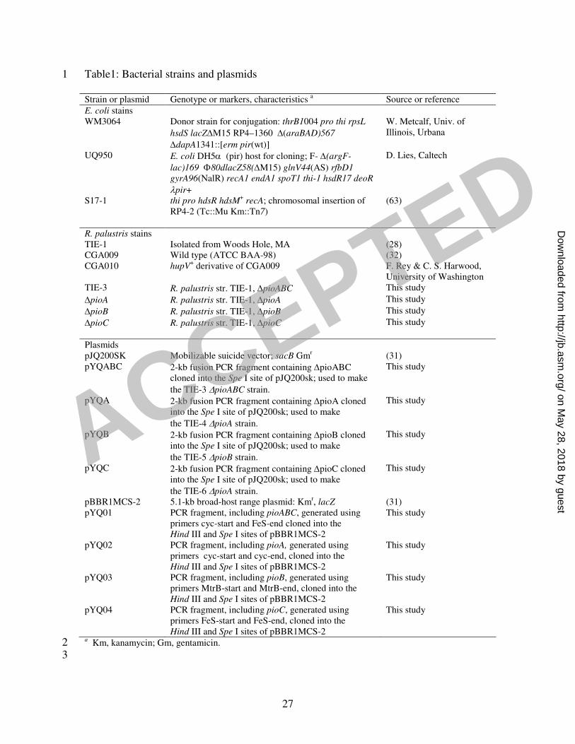
Table1: Bacterial strains and plasmids 1
Strain or plasmid Genotype or markers, characteristics a Source or reference
E. coli stains WM3064 Donor strain for conjugation: thrB1004 pro thi rpsL
hsdS lacZ∆M15 RP4–1360 ∆(araBAD)567
∆dapA1341::[erm pir(wt)]
W. Metcalf, Univ. of Illinois, Urbana
UQ950 E. coli DH5α (pir) host for cloning; F- ∆(argF-
lac)169 Φ80dlacZ58(∆M15) glnV44(AS) rfbD1
gyrA96(NalR) recA1 endA1 spoT1 thi-1 hsdR17 deoR
λpir+
D. Lies, Caltech
S17-1 thi pro hdsR hdsM+ recA; chromosomal insertion of
RP4-2 (Tc::Mu Km::Tn7) (63)
R. palustris stains TIE-1 Isolated from Woods Hole, MA (28) CGA009 Wild type (ATCC BAA-98) (32) CGA010 hupV
+ derivative of CGA009 F. Rey & C. S. Harwood, University of Washington
TIE-3 R. palustris str. TIE-1, ∆pioABC This study
∆pioA R. palustris str. TIE-1, ∆pioA This study
∆pioB R. palustris str. TIE-1, ∆pioB This study
∆pioC R. palustris str. TIE-1, ∆pioC This study
Plasmids pJQ200SK Mobilizable suicide vector; sacB Gmr (31) pYQABC 2-kb fusion PCR fragment containing ∆pioABC
cloned into the Spe I site of pJQ200sk; used to make
the TIE-3 ∆pioABC strain.
This study
pYQA 2-kb fusion PCR fragment containing ∆pioA cloned into the Spe I site of pJQ200sk; used to make
the TIE-4 ∆pioA strain.
This study
pYQB 2-kb fusion PCR fragment containing ∆pioB cloned into the Spe I site of pJQ200sk; used to make
the TIE-5 ∆pioB strain.
This study
pYQC 2-kb fusion PCR fragment containing ∆pioC cloned into the Spe I site of pJQ200sk; used to make
the TIE-6 ∆pioA strain.
This study
pBBR1MCS-2 5.1-kb broad-host range plasmid: Kmr, lacZ (31) pYQ01 PCR fragment, including pioABC, generated using
primers cyc-start and FeS-end cloned into the Hind III and Spe I sites of pBBR1MCS-2
This study
pYQ02 PCR fragment, including pioA, generated using primers cyc-start and cyc-end, cloned into the Hind III and Spe I sites of pBBR1MCS-2
This study
pYQ03 PCR fragment, including pioB, generated using primers MtrB-start and MtrB-end, cloned into the Hind III and Spe I sites of pBBR1MCS-2
This study
pYQ04 PCR fragment, including pioC, generated using primers FeS-start and FeS-end, cloned into the Hind III and Spe I sites of pBBR1MCS-2
This study
a Km, kanamycin; Gm, gentamicin. 2
3
ACCEPTED
on May 28, 2018 by guest
http://jb.asm.org/
Dow
nloaded from
28
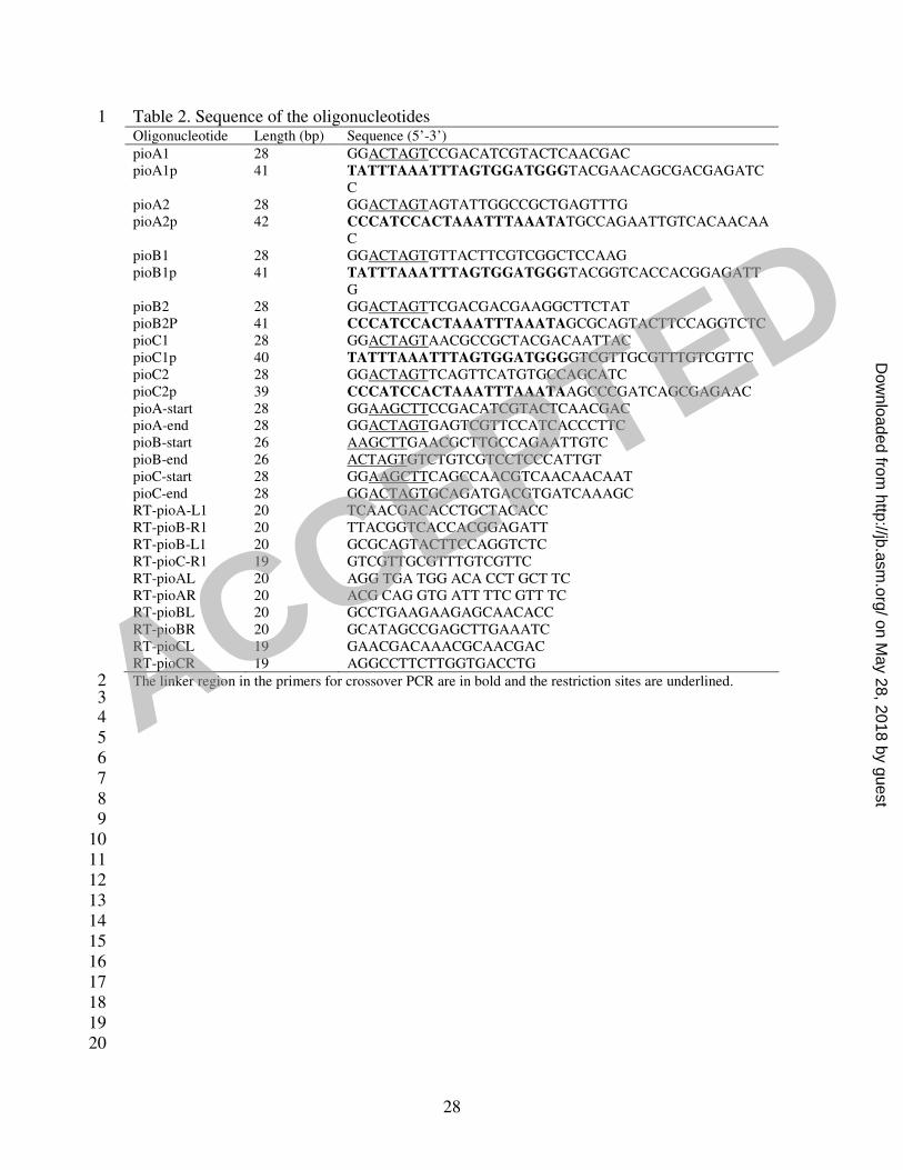
Table 2. Sequence of the oligonucleotides 1 Oligonucleotide Length (bp) Sequence (5’-3’)
pioA1 28 GGACTAGTCCGACATCGTACTCAACGAC pioA1p 41 TATTTAAATTTAGTGGATGGGTACGAACAGCGACGAGATC
C
pioA2 28 GGACTAGTAGTATTGGCCGCTGAGTTTG pioA2p 42 CCCATCCACTAAATTTAAATATGCCAGAATTGTCACAACAA
C pioB1 28 GGACTAGTGTTACTTCGTCGGCTCCAAG pioB1p 41 TATTTAAATTTAGTGGATGGGTACGGTCACCACGGAGATT
G pioB2 28 GGACTAGTTCGACGACGAAGGCTTCTAT pioB2P 41 CCCATCCACTAAATTTAAATAGCGCAGTACTTCCAGGTCTC pioC1 28 GGACTAGTAACGCCGCTACGACAATTAC pioC1p 40 TATTTAAATTTAGTGGATGGGGTCGTTGCGTTTGTCGTTC pioC2 28 GGACTAGTTCAGTTCATGTGCCAGCATC pioC2p 39 CCCATCCACTAAATTTAAATAAGCCCGATCAGCGAGAAC pioA-start 28 GGAAGCTTCCGACATCGTACTCAACGAC pioA-end 28 GGACTAGTGAGTCGTTCCATCACCCTTC pioB-start 26 AAGCTTGAACGCTTGCCAGAATTGTC pioB-end 26 ACTAGTGTCTGTCGTCCTCCCATTGT pioC-start 28 GGAAGCTTCAGCCAACGTCAACAACAAT pioC-end 28 GGACTAGTGCAGATGACGTGATCAAAGC RT-pioA-L1 20 TCAACGACACCTGCTACACC RT-pioB-R1 20 TTACGGTCACCACGGAGATT RT-pioB-L1 20 GCGCAGTACTTCCAGGTCTC RT-pioC-R1 19 GTCGTTGCGTTTGTCGTTC RT-pioAL 20 AGG TGA TGG ACA CCT GCT TC RT-pioAR 20 ACG CAG GTG ATT TTC GTT TC RT-pioBL 20 GCCTGAAGAAGAGCAACACC RT-pioBR 20 GCATAGCCGAGCTTGAAATC RT-pioCL 19 GAACGACAAACGCAACGAC RT-pioCR 19 AGGCCTTCTTGGTGACCTG
The linker region in the primers for crossover PCR are in bold and the restriction sites are underlined. 2 3 4 5 6 7 8 9 10 11 12 13 14 15 16 17 18 19 20
ACCEPTED
on May 28, 2018 by guest
http://jb.asm.org/
Dow
nloaded from

29
Table 3. Comparison of doubling time of R. palustris TIE-1 and TIE-3 grown on different 1 substrates 2
Substrates TIE-1 (hours) TIE-3 (hours)
Fe(II) 80±10 -
H2 40±5 36±7 Thiosulfate 55±7 57±6
Acetate 8±2 8±3
“-” means no growth observed.3
ACCEPTED
on May 28, 2018 by guest
http://jb.asm.org/
Dow
nloaded from
30
Fig. 1 1 2 3
4 5 6 7 Fig. 2 8 9
10 11 12 13 Fig. 3 14 15
16 17 18
ACCEPTED
on May 28, 2018 by guest
http://jb.asm.org/
Dow
nloaded from
31
1 2 3 4 Fig. 4 5 6
7 8 Fig. 5 9 10
11 12 Fig. 6 13 14
15
ACCEPTED
on May 28, 2018 by guest
http://jb.asm.org/
Dow
nloaded from
32
References 1 2 3 4 1. Ambler, R. P., T. E. Meyer, and M. D. Kamen. 1993. Amino-Acid-Sequence of a High Redox 5
Potential Ferredoxin (Hipip) from the Purple Phototrophic Bacterium Rhodopila-Globiformis, 6 Which Has the Highest Known Redox Potential of Its Class. Archives of Biochemistry and 7 Biophysics 306:215-222. 8
2. Appia-Ayme, C., A. Bengrine, C. Cavazza, M. T. Giudici-Orticoni, M. Bruschi, M. 9 Chippaux, and V. Bonnefoy. 1998. Characterization and expression of the co-transcribed cyc1 10 and cyc2 genes encoding the cytochrome c(4) (c(552)) and a high-molecular-mass cytochrome c 11 from Thiobacillus ferrooxidans ATCC33020. FEMS Microbiology Letters 167:171-177. 12
3. Bagos, P. G., T. D. Liakopoulos, I. C. Spyropoulos, and S. J. Hamodrakas. 2004. A Hidden 13 Markov Model method, capable of predicting and discriminating beta-barrel outer membrane 14 proteins. Bmc Bioinformatics 5. 15
4. Beliaev, A. S., and D. A. Saffarini. 1998. Shewanella putrefaciens mtrB encodes an outer 16 membrane protein required for Fe(III) and Mn(IV) reduction. Journal of Bacteriology 180:6292-17 6297. 18
5. Beliaev, A. S., D. A. Saffarini, J. L. McLaughlin, and D. Hunnicutt. 2001. MtrC, an outer 19 membrane decahaem c cytochrome required for metal reduction in Shewanella putrefaciens MR-1. 20 Molecular Microbiology 39:722-730. 21
6. Blankenship, R. E. 1992. Origin and Early Evolution of Photosynthesis. Photosynthesis Research 22 33:91-111. 23
7. Bradford, M. M. 1976. Rapid and sensitive method for quantitation of microgram quantities of 24 protein utilizing principle of protein-dye binding. Analytical Biochemistry 72:248-254. 25
8. Buchanan, B. B. 1992. Carbon-Dioxide Assimilation in Oxygenic and Anoxygenic 26 Photosynthesis. Photosynthesis Research 33:147-162. 27
9. Buchanan, S. K., B. S. Smith, L. Venkatramani, D. Xia, L. Esser, M. Palnitkar, R. 28 Chakraborty, D. van der Helm, and J. Deisenhofer. 1999. Crystal structure of the outer 29 membrane active transporter FepA from Escherichia coli. Nature Structural Biology 6:56-63. 30
10. Croal, L. R., J. A. Gralnick, D. Malasarn, and D. K. Newman. 2004. The genetics of 31 geochemistry. Annual Review of Genetics 38:175-202. 32
11. Croal, L. R., Y. Jiao, and D. K. Newman. 2006. The Fox operon from Rhodobacter sp. SW2 33 promotes phototrophic Fe(II) oxidation in Rhodobacter capsulatus SB1003 Journal of 34 Bacteriology Manuscript submitted. 35
12. Croal, L. R., C. M. Johnson, B. L. Beard, and D. K. Newman. 2004. Iron isotope fractionation 36 by Fe(II)-oxidizing photoautotrophic bacteria. Geochimica Et Cosmochimica Acta 68:1227-1242. 37
13. Egland, P. G., J. Gibson, and C. S. Harwood. 1995. Benzoate-Coenzyme a Ligase, Encoded by 38 Bada, Is One of 3 Ligases Able to Catalyze Benzoyl-Coenzyme a Formation During Anaerobic 39 Growth of Rhodopseudomonas-Palustris on Benzoate. Journal of Bacteriology 177:6545-6551. 40
14. Ehrenreich, A., and F. Widdel. 1994. Anaerobic oxidation of ferrous iron by purple bacteria, a 41 new-type of phototrophic metabolism. Applied and Environmental Microbiology 60:4517-4526. 42
15. Farquhar, J., H. M. Bao, and M. Thiemens. 2000. Atmospheric influence of Earth's earliest 43 sulfur cycle. Science 289:756-758. 44
16. Francis, R. T., and R. R. Becker. 1984. Specific Indication of Hemoproteins in Polyacrylamide 45 Gels Using a Double-Staining Process. Analytical Biochemistry 136:509-514. 46
17. Friedrich, C. G. 1998. Physiology and genetics of sulfur-oxidizing bacteria, p. 235-289, 47 Advances in Microbial Physiology, Vol 39, vol. 39. 48
18. Fukumori, Y., and T. Yamanaka. 1979. High-Potential Non-Heme Iron Protein (Hipip)-Linked, 49 Thiosulfate-Oxidizing Enzyme Derived from Chromatium-Vinosum. Current Microbiology 50 3:117-120. 51
19. Fukumori, Y., T. Yano, A. Sato, and T. Yamanaka. 1988. Fe(II)-oxidizing enzyme purified 52 from Thiobacillus-ferrooxidans. FEMS Microbiology Letters 50:169-172. 53
20. Garrow, A. G., A. Agnew, and D. R. Westhead. 2005. TMB-Hunt: An amino acid composition 54 based method to screen proteomes for beta-barrel transmembrane proteins. Bmc Bioinformatics 6. 55
ACCEPTED
on May 28, 2018 by guest
http://jb.asm.org/
Dow
nloaded from
33
21. Heising, S., L. Richter, W. Ludwig, and B. Schink. 1999. Chlorobium ferrooxidans sp nov., a 1 phototrophic green sulfur bacterium that oxidizes ferrous iron in coculture with a "Geospirillum" 2 sp strain. Archives of Microbiology 172:116-124. 3
22. Heising, S., and B. Schink. 1998. Phototrophic oxidation of ferrous iron by a Rhodomicrobium 4 vannielii strain. Microbiology-Uk 144:2263-2269. 5
23. Henseler, A., H. G. Truper, and U. Fischer. 1986. Soluble Cytochromes of Rhodopseudomonas-6 Marina. Fems Microbiology Letters 33:1-8. 7
24. Hochkoeppler, A., S. Ciurli, G. Venturoli, and D. Zannoni. 1995. The High-Potential Iron-8 Sulfur Protein (Hipip) from Rhodoferax Fermentans Is Competent in Photosynthetic Electron-9 Transfer. Febs Letters 357:70-74. 10
25. Hochkoeppler, A., D. Zannoni, S. Ciurli, T. E. Meyer, M. A. Cusanovich, and G. Tollin. 11 1996. Kinetics of photo-induced electron transfer from high-potential iron-sulfur protein to the 12 photosynthetic reaction center of the purple phototroph Rhodoferax fermentans. Proceedings of 13 the National Academy of Sciences of the United States of America 93:6998-7002. 14
26. Holland, H. D. 1973. Oceans - Possible Source of Iron in Iron-Formations. Economic Geology 15 68:1169-1172. 16
27. Hryniewicz, M., A. Sirko, A. Palucha, A. Bock, and D. Hulanicka. 1990. Sulfate and 17 Thiosulfate Transport in Escherichia-Coli K-12 - Identification of a Gene Encoding a Novel 18 Protein Involved in Thiosulfate Binding. Journal of Bacteriology 172:3358-3366. 19
28. Jiao, Y., A. Kappler, L. R. Croal, and D. K. Newman. 2005. Isolation and characterization of a 20 genetically tractable photo autotrophic Fe(II)-oxidizing bacterium, Rhodopseudomonas palustris 21 strain TIE-1. Applied and Environmental Microbiology 71:4487-4496. 22
29. Kappler, A., and D. K. Newman. 2004. Formation of Fe(III)-minerals by Fe(II)-oxidizing 23 photoautotrophic bacteria. Geochimica Et Cosmochimica Acta 68:1217-1226. 24
30. Karhu, J. A., and H. D. Holland. 1996. Carbon isotopes and the rise of atmospheric oxygen. 25 Geology 24:867-870. 26
31. Keen, N. T., S. Tamaki, D. Kobayashi, and D. Trollinger. 1988. Improved broad-host-range 27 plasmids for DNA cloning in gram-negative Bacteria. Gene 70:191-197. 28
32. Kim, M. K., and C. S. Harwood. 1991. Regulation of benzoate-CoA ligase in 29 Rhodopseudomonas-palustris. FEMS Microbiology Letters 83:199-203. 30
33. Koebnik, R., K. P. Locher, and P. Van Gelder. 2000. Structure and function of bacterial outer 31 membrane proteins: barrels in a nutshell. Molecular Microbiology 37:239-253. 32
34. Konhauser, K. O., T. Hamade, R. Raiswell, R. C. Morris, F. G. Ferris, G. Southam, and D. 33 E. Canfield. 2002. Could bacteria have formed the Precambrian banded iron formations? Geology 34 30:1079-1082. 35
35. Kovach, M. E., P. H. Elzer, D. S. Hill, G. T. Robertson, M. A. Farris, R. M. Roop, and K. M. 36 Peterson. 1995. 4 New Derivatives of the Broad-Host-Range Cloning Vector Pbbr1mcs, Carrying 37 Different Antibiotic-Resistance Cassettes. Gene 166:175-176. 38
36. Kovach, M. E., R. W. Phillips, P. H. Elzer, R. M. Roop, and K. M. Peterson. 1994. Pbbr1mcs - 39 a Broad-Host-Range Cloning Vector. Biotechniques 16:800-&. 40
37. Kusano, T., T. Takeshima, K. Sugawara, C. Inoue, T. Shiratori, T. Yano, Y. Fukumori, and 41 T. Yamanaka. 1992. Molecular-Cloning of the Gene Encoding Thiobacillus-Ferrooxidans Fe(Ii) 42 Oxidase - High Homology of the Gene-Product with Hipip. Journal of Biological Chemistry 43 267:11242-11247. 44
38. Laemmli, U. K. 1970. Cleavage of Structural Proteins During Assembly of Head of 45 Bacteriophage-T4. Nature 227:680-&. 46
39. Larimer, F. W., P. Chain, L. Hauser, J. Lamerdin, S. Malfatti, L. Do, M. L. Land, D. A. 47 Pelletier, J. T. Beatty, A. S. Lang, F. R. Tabita, J. L. Gibson, T. E. Hanson, C. Bobst, J. 48 Torres, C. Peres, F. H. Harrison, J. Gibson, and C. S. Harwood. 2004. Complete genome 49 sequence of the metabolically versatile photosynthetic bacterium Rhodopseudomonas palustris. 50 Nature Biotechnology 22:55-61. 51
40. Leang, C., L. A. Adams, K. J. Chin, K. P. Nevin, B. A. Methe, J. Webster, M. L. Sharma, 52 and D. R. Lovley. 2005. Adaptation to disruption of the electron transfer pathway for Fe(III) 53 reduction in Geobacter sulfurreducens. Journal of Bacteriology 187:5918-5926. 54
41. Leang, C., M. V. Coppi, and D. R. Lovley. 2003. OmcB, a c-type polyheme cytochrome, 55 involved in Fe(III) reduction in Geobacter sulfurreducens. Journal of Bacteriology 185:2096-2103. 56
ACCEPTED
on May 28, 2018 by guest
http://jb.asm.org/
Dow
nloaded from