yersinia pestis subverts the dermal neutrophil response in - mbio
TRANSCRIPT
Yersinia pestis Subverts the Dermal Neutrophil Response in a MouseModel of Bubonic Plague
Jeffrey G. Shannon,a Aaron M. Hasenkrug,a David W. Dorward,b Vinod Nair,b Aaron B. Carmody,b B. Joseph Hinnebuscha
Laboratory of Zoonotic Pathogens,a Research Technologies Branch,b Rocky Mountain Laboratories, National Institute of Allergy and Infectious Diseases, National Institutesof Health, Hamilton, Montana, USA
ABSTRACT The majority of human Yersinia pestis infections result from introduction of bacteria into the skin by the bite of aninfected flea. Once in the dermis, Y. pestis can evade the host’s innate immune response and subsequently disseminate to thedraining lymph node (dLN). There, the pathogen replicates to large numbers, causing the pathognomonic bubo of bubonicplague. In this study, several cytometric and microscopic techniques were used to characterize the early host response to intra-dermal (i.d.) Y. pestis infection. Mice were infected i.d. with fully virulent or attenuated strains of dsRed-expressing Y. pestis,and tissues were analyzed by flow cytometry. By 4 h postinfection, there were large numbers of neutrophils in the infected der-mis and the majority of cell-associated bacteria were associated with neutrophils. We observed a significant effect of the viru-lence plasmid (pCD1) on bacterial survival and neutrophil activation in the dermis. Intravital microscopy of i.d. Y. pestis infec-tion revealed dynamic interactions between recruited neutrophils and bacteria. In contrast, very few bacteria interacted withdendritic cells (DCs), indicating that this cell type may not play a major role early in Y. pestis infection. Experiments using neu-trophil depletion and a CCR7 knockout mouse suggest that dissemination of Y. pestis from the dermis to the dLN is not depen-dent on neutrophils or DCs. Taken together, the results of this study show a very rapid, robust neutrophil response to Y. pestis inthe dermis and that the virulence plasmid pCD1 is important for the evasion of this response.
IMPORTANCE Yersinia pestis remains a public health concern today because of sporadic plague outbreaks that occur throughoutthe world and the potential for its illegitimate use as a bioterrorism weapon. Since bubonic plague pathogenesis is initiated bythe introduction of Y. pestis into the skin, we sought to characterize the response of the host’s innate immune cells to bacteriaearly after intradermal infection. We found that neutrophils, innate immune cells that engulf and destroy microbes, are rapidlyrecruited to the injection site, irrespective of strain virulence, indicating that Y. pestis is unable to subvert neutrophil recruit-ment to the site of infection. However, we saw a decreased activation of neutrophils that were associated with Y. pestis strainsharboring the pCD1 plasmid, which is essential for virulence. These findings indicate a role for pCD1-encoded factors in sup-pressing the activation/stimulation of these cells in vivo.
Received 11 March 2013 Accepted 26 July 2013 Published 27 August 2013
Citation Shannon JG, Hasenkrug AM, Dorward DW, Nair V, Carmody AB, Hinnebusch BJ. 2013. Yersinia pestis subverts the dermal neutrophil response in a mouse model ofbubonic plague. mBio 4(5):e00170-13. doi:10.1128/mBio.00170-13.
Editor Olaf Schneewind, The University of Chicago
Copyright © 2013 Shannon et al. This is an open-access article distributed under the terms of the Creative Commons Attribution-Noncommercial-ShareAlike 3.0 Unportedlicense, which permits unrestricted noncommercial use, distribution, and reproduction in any medium, provided the original author and source are credited.
Address correspondence to Jeffrey G. Shannon, [email protected].
Yersinia pestis is a Gram-negative bacterial pathogen that cancause three distinct forms of plague: pneumonic, septicemic,
and bubonic. Bubonic plague is the most common form in hu-mans and results from the introduction of Y. pestis into the skin bythe bite of an infected flea. Once in the dermis, Y. pestis can sur-vive, replicate, and eventually migrate to the regional draininglymph node (dLN). There, the pathogen can replicate to very highnumbers, resulting in massive swelling of the lymph node, termeda bubo. The cellular architecture of this bubo can subsequentlybreak down, resulting in systemic hematogenous spread of thebacteria.
The progression of Y. pestis infection can be divided into twodistinct phases. The initial bubonic phase, consisting of the first 2to 3 days postinfection, is characterized by a minimal inflamma-tory response in the dLN despite the presence of large numbers ofreplicating bacteria (1, 2). If the infection is allowed to spread
beyond the regional draining lymphoid tissue, the proinflamma-tory or hyperinflammatory phase of infection ensues. This sec-ondary septic phase is characterized by a massive upregulation ofmyriad inflammatory cytokines that quickly leads to multiorgansystem failure and the death of the infected individual (1, 2).
Current understanding of the interactions between Y. pestisand mammalian host cells in vivo is limited to just a few publishedstudies. Virulent Y. pestis associates with polymorphonuclear leu-kocytes (neutrophils) and macrophages (M�) after subcutaneousinfection (3). It is thought that bacteria are quickly killed afterphagocytosis by neutrophils, whereas Y. pestis bacteria taken up byM� are able to survive, replicate, and disseminate (4). This hy-pothesis is supported by a number of in vitro studies; however, theroles of these cell types in the innate response to Y. pestis in vivoremain unclear.
Virulent strains of Y. pestis possess a 70-kb virulence plasmid
RESEARCH ARTICLE
September/October 2013 Volume 4 Issue 5 e00170-13 ® mbio.asm.org 1
Dow
nloa
ded
from
http
s://j
ourn
als.
asm
.org
/jour
nal/m
bio
on 1
7 D
ecem
ber
2021
by
200.
199.
202.
66.
(pCD1) that encodes a type III secretion apparatus and severaltype III secreted effectors. These effectors dramatically affect avariety of cell signaling pathways in the mammalian host, result-ing in the inhibition of both innate and adaptive immune re-sponses (5). The pCD1 plasmid is required for the immune sup-pression observed early after Y. pestis infection (1). Additionally,the Y. pestis genome contains a pathogenicity island termed thepigmentation locus (pgm). This locus contains genes involved in avariety of functions necessary for flea colonization and transmis-sion, iron acquisition, and intracellular survival (6–8).
The goals of this study were to characterize the very early hostcell response to intradermal (i.d.) Y. pestis and to evaluate theeffects of pCD1 and pgm on this response. To this end, we usedflow cytometry and intravital microscopy to study a mouse i.d.model of bubonic plague. Additionally, we examined the effects ofneutrophil and dendritic cell (DC) migration on the kinetics ofY. pestis dissemination. Our results indicate a prominent role forneutrophils in the very early response to plague bacilli in the der-mis and show that the virulence plasmid pCD1 is important forevasion of this rapid response.
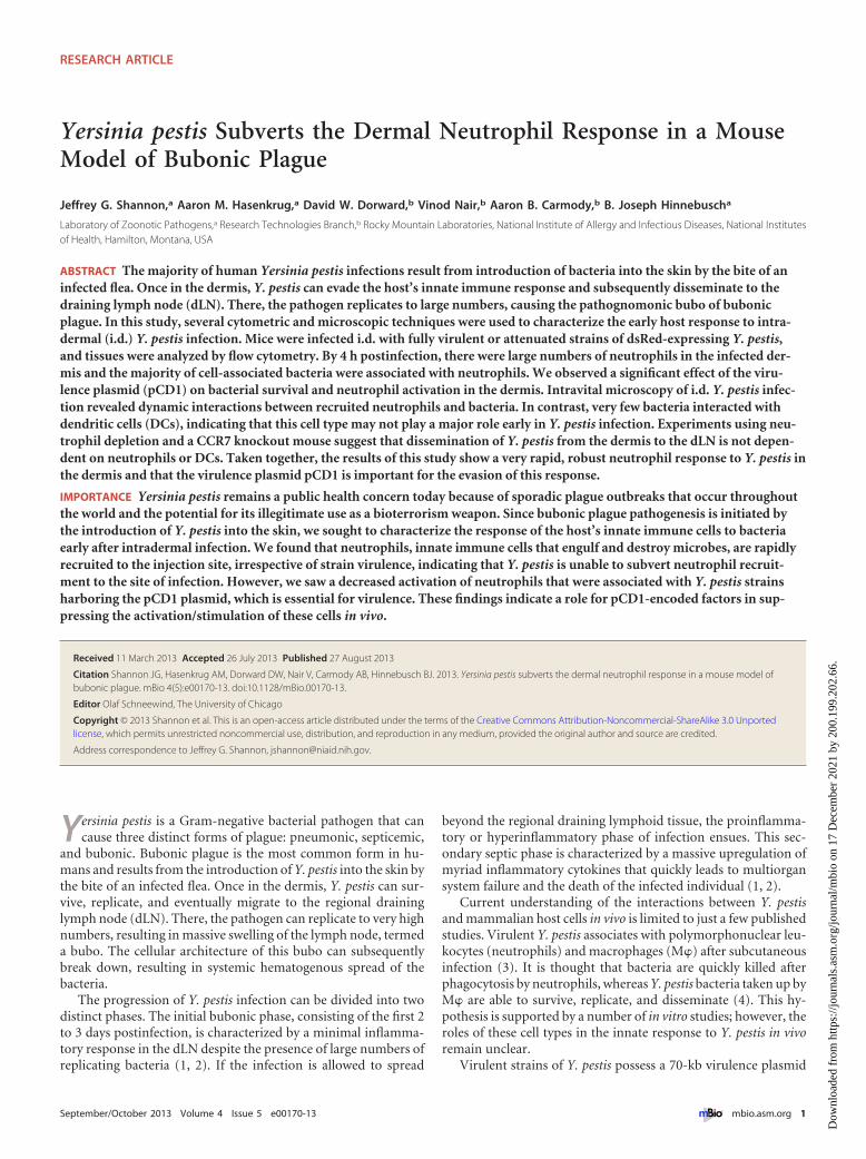
RESULTSY. pestis associates predominantly with neutrophils early afteri.d. infection. We used a mouse model of bubonic plague to de-termine which host cell types are recruited to the dermis early afterY. pestis infection. Mice were injected i.d. in the ear with 1 �105 CFU of a fully virulent Y. pestis strain, KIM6�/pCD1� (wildtype [WT]); attenuated strain KIM6�/pCD1� pgm�, which lacks
the virulence plasmid; or attenuated strain KIM5/pCD1� pgm,which possesses the virulence plasmid but lacks the pigmentationlocus (pgm) required for virulence by the i.d. or subcutaneousroute of infection (9). Additionally, groups of mice were injectedi.d. with a nonpathogenic strain of E. coli or phosphate-bufferedsaline (PBS) for comparison of the host response to innocuousbacteria or the injection itself. All of the bacterial strains possessedthe pTAC-dsRed plasmid, and dsRed expression was induced withisopropyl-�-D-thiogalactopyranoside (IPTG) prior to infection.The inoculum of 1 � 105 is at least 2 orders of magnitude morethan what an infected flea usually transmits (10); however, wefound this dose necessary for reliable and reproducible visualiza-tion of dsRed� populations by flow cytometry (data not shown).A representative example of the gating strategy used to excludebackground autofluorescence from our dsRed� populations isshown in Fig. S1A in the supplemental material.
At 4 h postinfection (hpi), mice were euthanized and single-cell suspensions of ear dermis were stained with a panel of fluo-rescently labeled antibodies (Abs) specific for mouse M�, neutro-phil, B cell, and DC surface markers and analyzed by flowcytometry. We observed an increase in the proportion of neutro-phils in some of the KIM6�- and KIM6�/pCD1-infected mice;however, the results were quite variable. Overall, neutrophilsmade up 5 to 10% of the total number of cells from the ears ofmost of the animals (Fig. 1A; see Fig. S1B in the supplementalmaterial). We did not see a significant increase in the M� propor-tion in the ear at this early time point (Fig. 1B; see Fig. S1C). B cells
FIG 1 Y. pestis associates with neutrophils recruited to the site of infection. Single-cell suspensions of mouse ear dermis were analyzed by flow cytometry afterthey were injected with PBS, E. coli, KIM5/pCD1�, KIM6� pgm�, or KIM6�/pCD1� (WT) or left uninjected. Total number of neutrophils (A) or M� (B) in theear dermis cell preparations at 4 hpi were determined by flow cytometry. Results are expressed as percentages of the total number of events that were either Ly6G�
or F4/80�, respectively. The percentage of dsRed� events that were also Ly6G� (C) or F4/80� (D) indicates the percentage of bacterium-associated cells that wereneutrophils or M�, respectively. Each symbol represents one mouse, and the line indicates the median. The results shown are the pooled data from a minimumof three separate experiments.
Shannon et al.
2 ® mbio.asm.org September/October 2013 Volume 4 Issue 5 e00170-13
Dow
nloa
ded
from
http
s://j
ourn
als.
asm
.org
/jour
nal/m
bio
on 1
7 D
ecem
ber
2021
by
200.
199.
202.
66.
and DCs were present in much lower numbers than M� and neu-trophils and did not increase over the levels found in uninfectedears (data not shown).
Injection of bacteria expressing the fluorescent protein dsRedallowed us to identify the host cell types the bacteria were associ-ating with in the dermis. At 4 hpi, we observed that �80% of thedsRed� events in the dermal cell suspensions were also positive forthe neutrophil specific marker Ly6G (Fig. 1C). Most of the dsRed�
Ly6G� events were F4/80�, indicating that a large proportion ofthe remaining bacteria were associated with M� (Fig. 1D). A smallpercentage of events were dsRed� Ly6G� F4/80� (data notshown). This may indicate some interaction between M� andneutrophils in the dermis but could also be an artifact of the cellisolation and staining procedure
Taken together, these data indicate that neutrophils may be thepredominant innate immune cell type encountered by Y. pestisvery early during the establishment of bubonic plague.
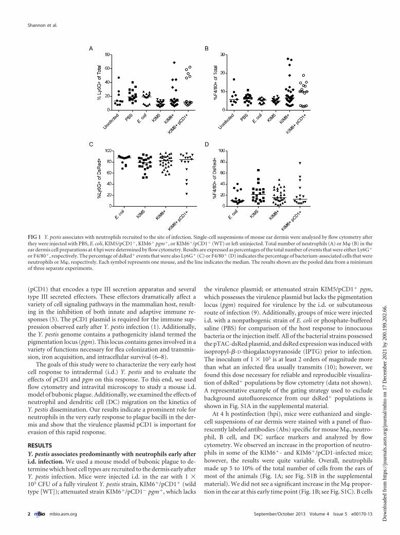
Y. pestis strains possessing the virulence plasmid pCD1 in-hibit neutrophil activation in vivo. Neutrophils undergo an ac-tivation process after contact with a variety of inflammatory stim-uli. CD11b is a �2 integrin that, together with CD18, formscomplement receptor 3, a molecule essential for extravasation ofneutrophils (11). Recognition of molecules such as bacterial lipo-polysaccharide (LPS) or formylated peptides, tumor necrosis fac-tor, complement component C5a, and leukotriene B4 by theircognate receptors on neutrophils results in mobilization ofCD11b from intracellular stores to the cell surface (12). We deter-mined the activation state of the dermal neutrophil population bymeasuring CD11b expression levels on the surface of Ly6G� cells.Analysis of the total population of neutrophils showed that infec-tion with the fully virulent KIM6�/pCD1� (WT) strain resulted inno measurable upregulation of CD11b expression, suggesting alack of neutrophil activation in the infected ear (Fig. 2A). In con-trast, the attenuated KIM5/pCD1� strain caused an increase intotal neutrophil CD11b� expression compared to the uninfectedcontrol but not the PBS-injected ear (Fig. 2A). Infection with the
KIM6� pgm� strain did not result in significantly increased neu-trophil activation; however, the results of these experiments werequite variable. The levels of activation seen in response to any ofthe Y. pestis strains were consistently lower than those observedafter infection with avirulent E. coli. Interestingly, when gating wasnarrowed to dsRed� events, thereby focusing only on cells thatwere associated with bacteria, we saw significantly less neutrophilactivation after infection with both of the pCD1-possessingstrains, KIM5/pCD1� and KIM6�/pCD1�, than after infectionwith KIM6� (Fig. 2B). Thus, the fully virulent strain of Y. pestisappears to subvert the normal activation process of all neutrophilsrecruited to the site of infection, whereas only the neutrophils thatdirectly associated with KIM5 showed lower levels of activation.
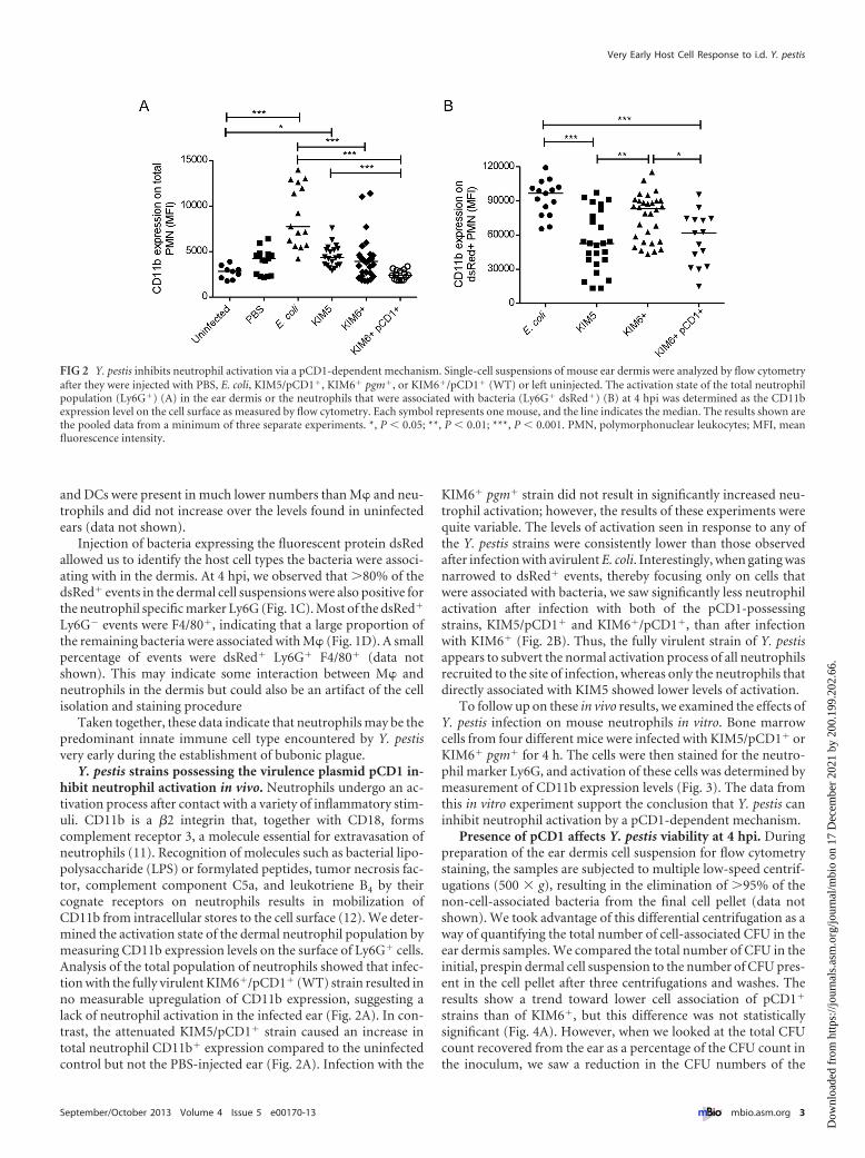
To follow up on these in vivo results, we examined the effects ofY. pestis infection on mouse neutrophils in vitro. Bone marrowcells from four different mice were infected with KIM5/pCD1� orKIM6� pgm� for 4 h. The cells were then stained for the neutro-phil marker Ly6G, and activation of these cells was determined bymeasurement of CD11b expression levels (Fig. 3). The data fromthis in vitro experiment support the conclusion that Y. pestis caninhibit neutrophil activation by a pCD1-dependent mechanism.
Presence of pCD1 affects Y. pestis viability at 4 hpi. Duringpreparation of the ear dermis cell suspension for flow cytometrystaining, the samples are subjected to multiple low-speed centrif-ugations (500 � g), resulting in the elimination of �95% of thenon-cell-associated bacteria from the final cell pellet (data notshown). We took advantage of this differential centrifugation as away of quantifying the total number of cell-associated CFU in theear dermis samples. We compared the total number of CFU in theinitial, prespin dermal cell suspension to the number of CFU pres-ent in the cell pellet after three centrifugations and washes. Theresults show a trend toward lower cell association of pCD1�
strains than of KIM6�, but this difference was not statisticallysignificant (Fig. 4A). However, when we looked at the total CFUcount recovered from the ear as a percentage of the CFU count inthe inoculum, we saw a reduction in the CFU numbers of the
FIG 2 Y. pestis inhibits neutrophil activation via a pCD1-dependent mechanism. Single-cell suspensions of mouse ear dermis were analyzed by flow cytometryafter they were injected with PBS, E. coli, KIM5/pCD1�, KIM6� pgm�, or KIM6�/pCD1� (WT) or left uninjected. The activation state of the total neutrophilpopulation (Ly6G�) (A) in the ear dermis or the neutrophils that were associated with bacteria (Ly6G� dsRed�) (B) at 4 hpi was determined as the CD11bexpression level on the cell surface as measured by flow cytometry. Each symbol represents one mouse, and the line indicates the median. The results shown arethe pooled data from a minimum of three separate experiments. *, P � 0.05; **, P � 0.01; ***, P � 0.001. PMN, polymorphonuclear leukocytes; MFI, meanfluorescence intensity.
Very Early Host Cell Response to i.d. Y. pestis
September/October 2013 Volume 4 Issue 5 e00170-13 ® mbio.asm.org 3
Dow
nloa
ded
from
http
s://j
ourn
als.
asm
.org
/jour
nal/m
bio
on 1
7 D
ecem
ber
2021
by
200.
199.
202.
66.
KIM6� strain present at 4 hpi, whereas the pCD1� strains hadincreased in number (Fig. 4B).
These data show that survival of these bacteria in the dermis iscompromised even at this very early time point, and although thedifferences were not significant, the pCD1� strain may associatewith host cells to a greater extent. However, it is important to notethat interpretation of the CFU cell association data is complicatedby the reduced survival/replication of the pCD1� strain in vivo. Incontrast, pCD1 possessing strains, even the attenuated KIM5
strain, show an increase in bacterial numbers and a trend towardreduced cell association at 4 hpi. Additionally, when we look atboth the CFU data in Fig. 4B and the neutrophil activation data inFig. 2, it is interesting that there is decreased neutrophil activationafter infection with the fully virulent KIM6�/pYV� strain despitethe presence of 5- to 10-fold more CFU in the mouse ear than ofKIM6�/pCD1� CFU.
Intravital microscopy reveals a robust neutrophil responseto i.d. Y. pestis. The flow cytometry data showing the large num-bers of neutrophils present in Y. pestis-infected dermis and theobservation that the vast majority of cell-associated bacteria wereassociated with neutrophils led us to examine the bacterium-neutrophil interactions in the dermis more thoroughly. To ac-complish this, we used an intravital confocal microscopy tech-nique to image transgenic mice that express high levels ofenhanced green fluorescent protein (eGFP) in neutrophils (LysM-eGFP mice) and, to a lesser extent, in cells of the monocyte lineage(13, 14). LysM-eGFP mice were infected i.d. in the ear with 1 � 103
dsRed-expressing bacteria and imaged from ~15 min postinfec-tion to �4 hpi. Because of a lack of suitable microscopy equip-ment in our biosafety level 3 (BSL-3) laboratory, we were limitedto using only the attenuated KIM5 and KIM6� strains of Y. pestisand avirulent E. coli for these studies.
A rapid influx of eGFPbright neutrophils was seen after injectionof the KIM6� pgm� strain (Fig. 5A; see Movie S1 in the supple-mental material). Most of the bacteria remained motionless untilthey associated with an eGFPbright cell. After association, many ofthe bacteria appeared to comigrate out of the field of view witheGFPbright cells. The neutrophil influx occurred rapidly after in-fection, with the first cells arriving at the injection site within thefirst 30 min postinfection. By 2 hpi, the neutrophils in the field ofview were too numerous to count and distinct association of asubset of bacteria with neutrophils was observed. In Movie S1R,the green channel has been turned off to allow better visualizationof the red bacteria. At 4 hpi, there were still bacteria that remainedstationary and apparently had not interacted with neutrophils.
Time-lapse images of the LysM-eGFP mice after infection withthe pCD1-possessing KIM5 strain yielded similar results (Fig. 5B;see Movie S2 in the supplemental material). Again we saw a rapid
FIG 3 In vitro confirmation that Y. pestis inhibits murine neutrophil acti-vation via a pCD1-dependent mechanism. The activation state of neutrophilsfrom mouse bone marrow was determined after mock infection or infectionwith KIM5 or KIM6� at an MOI of 2 or 20 in vitro. Flow cytometry was used todetermine the CD11b expression level on the surface of Ly6G� cells (neutro-phils) at 4 hpi. Cells from four C57BL/6 mice were analyzed. Each symbolrepresents one mouse, and the line indicates the median with the range. Theresults shown are from one experiment and are representative of three inde-pendent experiments. *, P � 0.05; **, P � 0.01; ***, P � 0.001. MFI, meanfluorescence intensity.
FIG 4 Reduced recovery of viable pCD1� Y. pestis from the ear dermis. (A) The number of bacterial CFU that were cell associated is shown as a percentage ofthe total number of CFU recovered from the ear at 4 hpi. (B) The total number of viable bacteria present in the ear dermis at 4 hpi is shown as a percentage of theactual inoculum. The results shown are pooled data from a minimum of three separate experiments. *, P � 0.05; ****, P � 0.0001.
Shannon et al.
4 ® mbio.asm.org September/October 2013 Volume 4 Issue 5 e00170-13
Dow
nloa
ded
from
http
s://j
ourn
als.
asm
.org
/jour
nal/m
bio
on 1
7 D
ecem
ber
2021
by
200.
199.
202.
66.
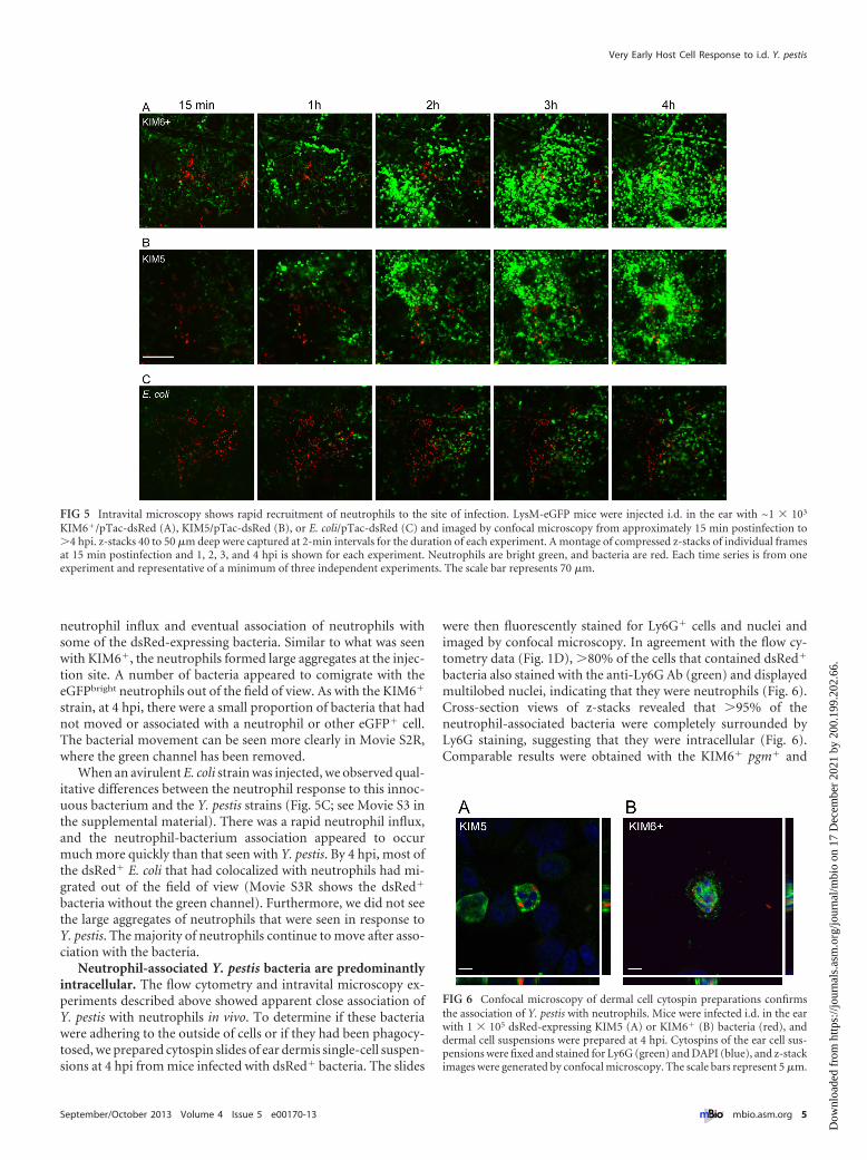
neutrophil influx and eventual association of neutrophils withsome of the dsRed-expressing bacteria. Similar to what was seenwith KIM6�, the neutrophils formed large aggregates at the injec-tion site. A number of bacteria appeared to comigrate with theeGFPbright neutrophils out of the field of view. As with the KIM6�
strain, at 4 hpi, there were a small proportion of bacteria that hadnot moved or associated with a neutrophil or other eGFP� cell.The bacterial movement can be seen more clearly in Movie S2R,where the green channel has been removed.
When an avirulent E. coli strain was injected, we observed qual-itative differences between the neutrophil response to this innoc-uous bacterium and the Y. pestis strains (Fig. 5C; see Movie S3 inthe supplemental material). There was a rapid neutrophil influx,and the neutrophil-bacterium association appeared to occurmuch more quickly than that seen with Y. pestis. By 4 hpi, most ofthe dsRed� E. coli that had colocalized with neutrophils had mi-grated out of the field of view (Movie S3R shows the dsRed�
bacteria without the green channel). Furthermore, we did not seethe large aggregates of neutrophils that were seen in response toY. pestis. The majority of neutrophils continue to move after asso-ciation with the bacteria.
Neutrophil-associated Y. pestis bacteria are predominantlyintracellular. The flow cytometry and intravital microscopy ex-periments described above showed apparent close association ofY. pestis with neutrophils in vivo. To determine if these bacteriawere adhering to the outside of cells or if they had been phagocy-tosed, we prepared cytospin slides of ear dermis single-cell suspen-sions at 4 hpi from mice infected with dsRed� bacteria. The slides
were then fluorescently stained for Ly6G� cells and nuclei andimaged by confocal microscopy. In agreement with the flow cy-tometry data (Fig. 1D), �80% of the cells that contained dsRed�
bacteria also stained with the anti-Ly6G Ab (green) and displayedmultilobed nuclei, indicating that they were neutrophils (Fig. 6).Cross-section views of z-stacks revealed that �95% of theneutrophil-associated bacteria were completely surrounded byLy6G staining, suggesting that they were intracellular (Fig. 6).Comparable results were obtained with the KIM6� pgm� and
FIG 5 Intravital microscopy shows rapid recruitment of neutrophils to the site of infection. LysM-eGFP mice were injected i.d. in the ear with ~1 � 103
KIM6�/pTac-dsRed (A), KIM5/pTac-dsRed (B), or E. coli/pTac-dsRed (C) and imaged by confocal microscopy from approximately 15 min postinfection to�4 hpi. z-stacks 40 to 50 �m deep were captured at 2-min intervals for the duration of each experiment. A montage of compressed z-stacks of individual framesat 15 min postinfection and 1, 2, 3, and 4 hpi is shown for each experiment. Neutrophils are bright green, and bacteria are red. Each time series is from oneexperiment and representative of a minimum of three independent experiments. The scale bar represents 70 �m.
FIG 6 Confocal microscopy of dermal cell cytospin preparations confirmsthe association of Y. pestis with neutrophils. Mice were infected i.d. in the earwith 1 � 105 dsRed-expressing KIM5 (A) or KIM6� (B) bacteria (red), anddermal cell suspensions were prepared at 4 hpi. Cytospins of the ear cell sus-pensions were fixed and stained for Ly6G (green) and DAPI (blue), and z-stackimages were generated by confocal microscopy. The scale bars represent 5 �m.
Very Early Host Cell Response to i.d. Y. pestis
September/October 2013 Volume 4 Issue 5 e00170-13 ® mbio.asm.org 5
Dow
nloa
ded
from
http
s://j
ourn
als.
asm
.org
/jour
nal/m
bio
on 1
7 D
ecem
ber
2021
by
200.
199.
202.
66.
KIM5/pCD1� strains of Y. pestis. Between 10 and 20% of thedsRed� bacteria were associated with Ly6G� cells (data notshown). The identity of these cells is unknown, but on the basis oftheir morphology and the flow cytometry data shown in Fig. 1E,they are likely M�.
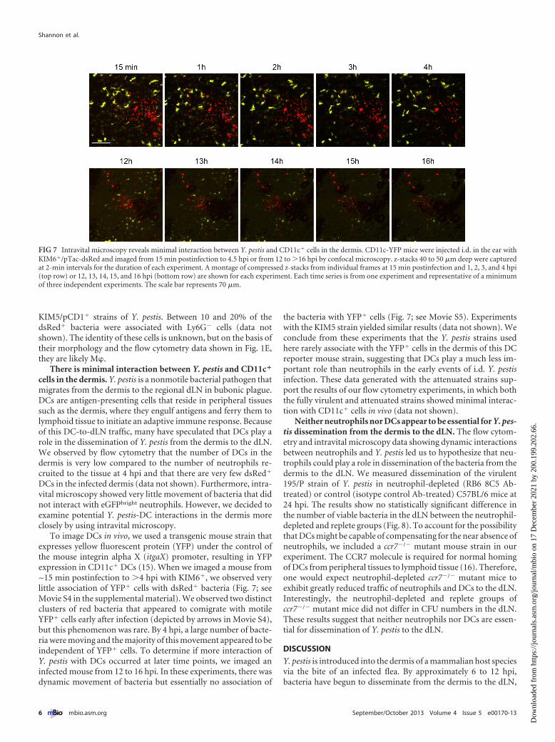
There is minimal interaction between Y. pestis and CD11c�
cells in the dermis. Y. pestis is a nonmotile bacterial pathogen thatmigrates from the dermis to the regional dLN in bubonic plague.DCs are antigen-presenting cells that reside in peripheral tissuessuch as the dermis, where they engulf antigens and ferry them tolymphoid tissue to initiate an adaptive immune response. Becauseof this DC-to-dLN traffic, many have speculated that DCs play arole in the dissemination of Y. pestis from the dermis to the dLN.We observed by flow cytometry that the number of DCs in thedermis is very low compared to the number of neutrophils re-cruited to the tissue at 4 hpi and that there are very few dsRed�
DCs in the infected dermis (data not shown). Furthermore, intra-vital microscopy showed very little movement of bacteria that didnot interact with eGFPbright neutrophils. However, we decided toexamine potential Y. pestis-DC interactions in the dermis moreclosely by using intravital microscopy.
To image DCs in vivo, we used a transgenic mouse strain thatexpresses yellow fluorescent protein (YFP) under the control ofthe mouse integrin alpha X (itgaX) promoter, resulting in YFPexpression in CD11c� DCs (15). When we imaged a mouse from~15 min postinfection to �4 hpi with KIM6�, we observed verylittle association of YFP� cells with dsRed� bacteria (Fig. 7; seeMovie S4 in the supplemental material). We observed two distinctclusters of red bacteria that appeared to comigrate with motileYFP� cells early after infection (depicted by arrows in Movie S4),but this phenomenon was rare. By 4 hpi, a large number of bacte-ria were moving and the majority of this movement appeared to beindependent of YFP� cells. To determine if more interaction ofY. pestis with DCs occurred at later time points, we imaged aninfected mouse from 12 to 16 hpi. In these experiments, there wasdynamic movement of bacteria but essentially no association of
the bacteria with YFP� cells (Fig. 7; see Movie S5). Experimentswith the KIM5 strain yielded similar results (data not shown). Weconclude from these experiments that the Y. pestis strains usedhere rarely associate with the YFP� cells in the dermis of this DCreporter mouse strain, suggesting that DCs play a much less im-portant role than neutrophils in the early events of i.d. Y. pestisinfection. These data generated with the attenuated strains sup-port the results of our flow cytometry experiments, in which boththe fully virulent and attenuated strains showed minimal interac-tion with CD11c� cells in vivo (data not shown).
Neither neutrophils nor DCs appear to be essential for Y. pes-tis dissemination from the dermis to the dLN. The flow cytom-etry and intravital microscopy data showing dynamic interactionsbetween neutrophils and Y. pestis led us to hypothesize that neu-trophils could play a role in dissemination of the bacteria from thedermis to the dLN. We measured dissemination of the virulent195/P strain of Y. pestis in neutrophil-depleted (RB6 8C5 Ab-treated) or control (isotype control Ab-treated) C57BL/6 mice at24 hpi. The results show no statistically significant difference inthe number of viable bacteria in the dLN between the neutrophil-depleted and replete groups (Fig. 8). To account for the possibilitythat DCs might be capable of compensating for the near absence ofneutrophils, we included a ccr7�/� mutant mouse strain in ourexperiment. The CCR7 molecule is required for normal homingof DCs from peripheral tissues to lymphoid tissue (16). Therefore,one would expect neutrophil-depleted ccr7�/� mutant mice toexhibit greatly reduced traffic of neutrophils and DCs to the dLN.Interestingly, the neutrophil-depleted and replete groups ofccr7�/� mutant mice did not differ in CFU numbers in the dLN.These results suggest that neither neutrophils nor DCs are essen-tial for dissemination of Y. pestis to the dLN.
DISCUSSION
Y. pestis is introduced into the dermis of a mammalian host speciesvia the bite of an infected flea. By approximately 6 to 12 hpi,bacteria have begun to disseminate from the dermis to the dLN,
FIG 7 Intravital microscopy reveals minimal interaction between Y. pestis and CD11c� cells in the dermis. CD11c-YFP mice were injected i.d. in the ear withKIM6�/pTac-dsRed and imaged from 15 min postinfection to 4.5 hpi or from 12 to �16 hpi by confocal microscopy. z-stacks 40 to 50 �m deep were capturedat 2-min intervals for the duration of each experiment. A montage of compressed z-stacks from individual frames at 15 min postinfection and 1, 2, 3, and 4 hpi(top row) or 12, 13, 14, 15, and 16 hpi (bottom row) are shown for each experiment. Each time series is from one experiment and representative of a minimumof three independent experiments. The scale bar represents 70 �m.
Shannon et al.
6 ® mbio.asm.org September/October 2013 Volume 4 Issue 5 e00170-13
Dow
nloa
ded
from
http
s://j
ourn
als.
asm
.org
/jour
nal/m
bio
on 1
7 D
ecem
ber
2021
by
200.
199.
202.
66.
where they rapidly multiply. Very little is known about the earlyhost cell response to Y. pestis in the dermis. We used flow cytom-etry, confocal microscopy, and several microbiologic techniquesto examine early Y. pestis-host cell interactions in vivo, with a focuson neutrophils and DCs. Additionally, we investigated the poten-tial roles of both neutrophils and DCs in bacterial disseminationfrom the dermis to the dLN.
The importance of neutrophils in the response to pathogens inthe dermis is well established. Neutrophils begin arriving at an i.d.injection site within minutes, even in response to an injection ofsterile PBS, showing that trauma or a break in the skin is all that isneeded for early neutrophil recruitment (14; J. G. Shannon, un-published observations). Several studies have examined the im-portance of neutrophils in bubonic plague pathogenesis. Janssenet al. observed viable Y. pestis within neutrophils from the perito-nea of infected guinea pigs (17). Sebbane et al. showed that neu-trophils are recruited to buboes in rats and Y. pestis genes essentialfor combating neutrophil-derived reactive nitrogen species arehighly upregulated in vivo (18). Y. pestis bacteria taken up by hu-man neutrophils in vitro are killed, but a small proportion of thebacteria remain viable and this survival is dependent on a bacterialtwo-component regulatory system (19, 20). Additionally, pCD1�
strains of Y. pestis inhibit the human neutrophil oxidative burstand apoptosis in vitro (21). Marketon et al. used a beta-lactamasereporter system to show that neutrophils, along with M� and DCs,are injected with Y. pestis type III secretion system effectors in vivo(22).
We found that �80% of the dsRed� bacteria in our ear dermissamples were associated with neutrophils at 4 hpi, and this asso-ciation was not affected by pCD1. Interestingly, only 15 to 30% of
the total number of recovered CFU were cell associated at this timepoint, indicating that there is a population of bacteria that remainextracellular throughout the early phase of infection. A caveat ofthese experiments is that a large dose of bacteria (1 � 105) wasneeded to generate consistent, reliable flow cytometry data. Bosioet al. did similar experiments using challenge doses of 500 or1,000 CFU and observed comparable cellular recruitment to thesite of infection (23). However, that study did not examine cellassociation, so we do not know if reducing the inoculum to a morebiologically relevant dose would affect the percentage of cell asso-ciation. It should also be noted that we were unable to determineif the dsRed fluorescence in these studies was from live or deadbacteria. We have found that heat-killed dsRed� Y. pestis bacteriamaintain fluorescence after uptake by neutrophils in vitro over thetime frame of this study (data not shown).
We also found that Y. pestis strains possessing pCD1 had asignificant effect on the activation status of neutrophils recruitedto the site of infection that had associated with dsRed� bacteria.This is consistent with a previous report showing decreased neu-trophil activation in response to virulent Y. pestis compared to apCD1� strain (23). Interestingly, infection with the attenuatedKIM5/pCD1� strain inhibited the activation of bacterium-associated neutrophils (dsRed�), but not the entire neutrophilpopulation, as is seen with fully virulent KIM6�/pCD1� (WT).Furthermore, infection with the KIM6� pgm� strain resulted inlower levels of neutrophil activation than did infection with E. coli.Thus, in addition to the known virulence factors present on pCD1,Y. pestis may possess non-pCD1-encoded mechanisms of reduc-ing neutrophil activation in vivo. One potential candidate is theability of Y. pestis to produce a nonstimulatory LPS molecule whengrown at 37°C (24). We are currently investigating the effects ofthis LPS modification on the neutrophil response in vivo.
Though we observed the majority (�80%) of the cell-associated Y. pestis bacteria in the dermis at 4 hpi were associatedwith neutrophils, most of the remaining bacteria were associatedwith M�. Y. pestis survives and replicates in monocytes and M� toa much greater extent than in neutrophils (25–27). It has beensuggested that ingestion by neutrophils is a dead end for the bac-teria and that only Y. pestis bacteria that are phagocytosed by M�go on to cause disease (4). Our intravital microscopy data, whilenot quantitative, show that bacteria that do not interact with neu-trophils remain stationary at the injection site, whereasneutrophil-associated bacteria traffic away from the site. Thus, itmay be possible that a small subset of Y. pestis bacteria surviveswithin neutrophils and that these cells contribute to dissemina-tion. It is also possible that all of the neutrophil-associated bacteriaare ultimately killed and that only bacteria that remain extracellu-lar go on to disseminate by a heretofore unknown mechanism.Clearly, a more detailed examination of the infected dermis isneeded to determine the ultimate fate of neutrophil- and M�-associated populations of Y. pestis bacteria.
Our intravital microscopy experiments with LysM-eGFP micerevealed qualitative differences in neutrophil recruitment toY. pestis infection versus E. coli infection. Many of the neutrophilsappeared to arrest their movement and form dense aggregates atthe site of Y. pestis infection. These aggregates appear similar to theneutrophil swarms observed by Chtanova et al. after Toxoplasmagondii infection (28). In contrast, the neutrophils recruited afterE. coli infection continued to move after contact with bacteria anddid not aggregate. We speculate that very different signals are pro-
FIG 8 Neutrophil depletion and lack of CCR7-mediated LN homing do notaffect the kinetics of Y. pestis dissemination. C57BL/6 or ccr7�/� mutant micewere injected intraperitoneally with 250 �g of anti-GR1 Ab () or a rat isotypecontrol Ab (�) at 24 and 4 h prior to infection. The mice were then infected i.d.with 1,000 CFU in the right hip. At 24 hpi, mice were euthanized and the dLNs(inguinal) were collected. Numbers of CFU in dLNs were determined by plat-ing on blood agar. The dashed line indicates the limit of detection of the assay(10 CFU/lymph node). k/o, knockout.
Very Early Host Cell Response to i.d. Y. pestis
September/October 2013 Volume 4 Issue 5 e00170-13 ® mbio.asm.org 7
Dow
nloa
ded
from
http
s://j
ourn
als.
asm
.org
/jour
nal/m
bio
on 1
7 D
ecem
ber
2021
by
200.
199.
202.
66.
duced by Y. pestis-infected tissue that are not found after infectionwith the more innocuous bacterium E. coli and that these signalsmay alter the behavior of the recruited neutrophils.
The intravital microscopy experiments described here show avery rapid, localized neutrophil response to Y. pestis infection.This is in contrast to our flow cytometric analysis of samples de-rived from an entire ear that show no significant increase in totalneutrophil numbers in the ear at 4 hpi and a previous report show-ing variable neutrophil numbers in the ear at early time pointspostinfection (23). This discrepancy highlights the importance ofstudying the discrete focus of infection in situ, as important as-pects of pathogenesis can be missed when one analyzes infectedtissues as a whole.
DCs are phagocytic antigen-presenting cells that serve as im-mune sentinels against tissue damage or invasion by potentialpathogens. Immature DCs reside in peripheral tissues like the der-mis. Upon contact with inflammatory signals, such as microbe-associated ligands of pattern recognition receptors or host-derived molecules associated with tissue damage, DCs undergo acomplex maturation process that results in, among many otherthings, upregulation of the chemokine receptor CCR7 (29). Thisleads to the migration of DCs along a chemokine gradient out ofthe peripheral tissues and into the lymphatics, where they caninitiate an adaptive immune response. This migration has led tospeculation that DCs are responsible for the dissemination ofY. pestis bacteria from the dermis to the dLN that occurs in bu-bonic plague (30, 31); however, minimal data exist to support thishypothesis. Our results suggest that DCs may not play a significantrole in the early phase of i.d. Y. pestis infection. On the basis of ourdata that show dynamic interactions between Y. pestis and neutro-phils in vivo, we explored the possibility that neutrophils play arole in dissemination. There is evidence that neutrophils cantransport other bacterial pathogens to the dLN (32); however,neutrophil depletion had no effect on Y. pestis dissemination inour experiments with a mouse model of bubonic plague. We havebeen unable to determine the mechanism of this dissemination,and it continues to be a subject of study in our laboratory.
The pCD1 genes encoding known virulence factors are ex-pressed at low levels during growth at the low ambient tempera-tures experienced while in the flea (33, 34). It is believed thatY. pestis requires 3 to 5 h of growth at 37°C to upregulate thepCD1-encoded type III secretion system and secreted effectors(34–36). Interestingly, even though we grew the bacteria at 22°Cprior to inoculation to mimic flea transmission, we observed ameasurable effect of the virulence plasmid on neutrophil activa-tion at 4 hpi. This suggests that, even at this early time point, thebacteria have produced these virulence factors to a level sufficientfor at least partial subversion of the host’s normal innate response.
We initiated these studies to elucidate the events that occur inthe dermis very early after infection with Y. pestis. Our resultsbuild upon the established importance of the pCD1 virulenceplasmid as a key virulence factor of this pathogen. Additionally,the data presented here highlight the importance of neutrophils inthe initial response to i.d. Y. pestis infection and indicate that sub-version of the normal innate neutrophil response is an importantcomponent of bubonic plague pathogenesis.
MATERIALS AND METHODSBacterial strains and plasmids. The fully virulent 195/P (37) and KIM6�/pCD1� (referred to as the WT) strains and the attenuated KIM5 (referred
to as pCD1� and pgm locus deficient) and KIM6� (referred to as pCD1virulence plasmid deficient and pgm�) strains were used in this study. TheWT (KIM6�/pCD1�) strain was generated by transformation of KIM6�
with the pCD1 plasmid into which a kanamycin resistance cassette hadbeen inserted as previously described (38). Similarly, this pCD1-kanr plas-mid was transformed into pCD1� (KIM5). Use of the pCD1-kanr plasmidallowed the selection of pCD1-containing bacteria by growth in the pres-ence of kanamycin, thus reducing problems associated with loss of pCD1under certain in vitro culture conditions. The E. coli strain used was theK-12-derived cloning strain TOP10 (Invitrogen, Carlsbad, CA). Allstrains were transformed by electroporation with the plasmid pTac-dsRed, which allows the expression of dsRed under the control of aninducible promoter. This pTac-dsRed plasmid was constructed by ampli-fying the dsRed gene from the pDsRed Express 2 vector (Clontech, Moun-tain View, CA) by PCR with forward primer 5= CGCTCGAGTAATGGATAGCACTGAGAACGTCA 3= and reverse primer 5= GCGAATTCGCGGCCGCTACTGGAACA 3=. The PCR product and the vector pTAC-MAT-Tag-2 (Sigma, St. Louis, MO) were digested with XhoI and EcoRI andligated to form pTac-dsRed.
Bacteria were grown in brain heart infusion (BHI) broth at 28°C with100 �g/ml carbenicillin to maintain the pTac-dsRed plasmid and75 �g/ml kanamycin to maintain pCD1-kanr where appropriate. IPTG(Sigma) was added at a concentration of 100 to 200 �M to induce dsRedexpression. Bacterial cultures were transferred to 22°C 12 h prior to infec-tion. Bacteria were washed in PBS, and Lysing Matrix H bead tubes (MPBiomedicals, Solon, OH) and a Fastprep Bio101 (Thermo, Fisher Scien-tific, Waltham, MA) were used to break up clumps of bacteria in culturesprior to injection. Bacteria were enumerated in a Petroff-Hausser count-ing chamber before preparation of the inocula in PBS. Serial dilutions ofinocula were plated on blood agar to determine the actual number of CFUinjected.
We have tested the virulence of the KIM6�/pYV� strain expressingdsRed in the mouse model of bubonic plague. Mice were challenged i.d.with 100 CFU of this strain. All mice exhibited terminal symptoms andwere euthanized between 3.5 and 5 days postinfection, similar to micechallenged with strains lacking the dsRed plasmid (data not shown). Allwork with the fully virulent KIM6�/pCD1� strain was performed in aBSL-3 or animal BSL-3 laboratory under a protocol approved by theRocky Mountain Laboratories Biosafety Committee and in accordancewith CDC select agent regulations.
Mice. C57BL/6J LysM-eGFP knock-in mice were originally created byT. Graf (Albert Einstein University, Bronx, NY) (13) and were bred byTaconic Laboratories under a contract with NIAID. C57BL/6J (stocknumber 000664) and ccr7�/� mutant (stock number 006621) mice werepurchased from The Jackson Laboratory (Bar Harbor, ME). Ten- To 20-week-old female mice were used in all experiments. All mice were main-tained at the Rocky Mountain Laboratories animal care facility underspecific-pathogen-free conditions.
Ethics statement. All animal studies were performed under protocolsadhering to guidelines established by the American Association for Labo-ratory Animal Science (AALAS) and the U.S. Public Health Service Officeof Laboratory Animal Welfare (PHS-OLAW). The protocols were re-viewed and approved by the Rocky Mountain Laboratories Animal Careand Use Committee (AALAS unit number 000462, PHS-OLAW numberA-4149-01).
Tissue processing for flow cytometry and microscopy. To evaluatethe cell association of Y. pestis by flow cytometry, mice were infected i.d. inthe ear with 1 � 105 bacteria suspended in 10 �l PBS. At 4 hpi, mice wereeuthanized and their ears were harvested. Ears that were injected with PBSalone or left unmanipulated and uninjected served as controls. Single-cellsuspensions were prepared as described in reference 23, with minor mod-ifications. Briefly, the ventral and dorsal ear dermal layers were separatedand incubated for 30 min at 37°C in digestion medium (complete RPMIsupplemented with 25 mM HEPES, 1.5 g/liter NaHCO3 [Invitrogen,Carlsbad, CA], 170 �g/ml Liberase [Roche, Indianapolis, IN], and
Shannon et al.
8 ® mbio.asm.org September/October 2013 Volume 4 Issue 5 e00170-13
Dow
nloa
ded
from
http
s://j
ourn
als.
asm
.org
/jour
nal/m
bio
on 1
7 D
ecem
ber
2021
by
200.
199.
202.
66.
50 �g/ml deoxyribonuclease I [Worthington Biochemical, Lakewood,NJ]). Following incubation, the dermal tissue was agitated in medium,filtered through a 30-�m nylon strainer, and washed with PBS. The cellsuspension was then treated with red blood cell lysis buffer (ammonium-chloride-potassium), and cells were washed with PBS and either pelletedfor cytospin and microscopy or resuspended in ice-cold flow cytometrybuffer (PBS supplemented with 2% heat-inactivated fetal bovine serum)for staining with fluorescent Abs. The Abs included V450-labeled anti-CD19, PerCP-Cy5.5-labeled anti-CD11c, and Alexa Fluor 488-, 647-, and700-labeled anti-CD11b, F4/80, and Ly6G (clone 1A8) Abs, respectively.All fluorochrome-conjugated monoclonal Abs were purchased from BDBiosciences (San Jose, CA) or eBioscience (San Diego, CA). All data werecollected on an LSRII flow cytometer (Becton Dickinson, San Jose, CA)and analyzed with FlowJo software (Treestar, Ashland, OR). To rule outthe possibility that some bacterium-host cell interactions were occurringduring the cell isolation and staining procedures, control experimentswere performed where bacteria were injected into the ear after the mousewas euthanized. The ear was then processed and stained exactly as de-scribed above. The results showed that minimal cell association occurredduring the procedure (data not shown). We have also established that cellscontaining only one dsRed� bacterium can be reliably detected by flowcytometry (data not shown).
Cells were prepared for confocal microscopy by using a cytospin 4(Thermo Scientific, Asheville, NC) (1,000 rpm, 4 min), followed by fixa-tion with 2% paraformaldehyde in PBS for 10 min at room temperature.Neutrophils were stained with rat anti-Ly6G Ab (Bio-X-Cell, West Leba-non, NH), followed by a goat anti-rat Alexa Fluor 488 secondary Ab (In-vitrogen). Nuclei were stained with 4=,6-diamidino-2-phenylindole(DAPI; Invitrogen). Images were acquired with a Zeiss LSM 510 Metaconfocal microscope, and image files were processed by Imaris 6.3.1 soft-ware (Bitplane, South Windsor, CT).
In vitro polymorphonuclear leukocyte activation assay. Wells of 24-well tissue culture plates were coated with 20% normal C57BL/6 mouseserum in PBS and incubated for 30 min at 37°C. Bone marrow cells fromC57BL/6 mice were then seeded at a density of 5 � 105/well in 500 �l ofRPMI medium without phenol red (Invitrogen). Cultures of KIM5 orKIM6� bacteria were grown overnight at 22°C in BHI medium and thenwashed and suspended in PBS. Cells were infected at an MOI of 2 or 20 ormock infected with PBS alone and incubated for 4 h at 37°C in 5% CO2.Cells were then harvested, stained with anti-Ly6G–Alexa Fluor 700 andanti-CD11b–Alexa Fluor 488 Abs, and analyzed by flow cytometry as de-scribed above.
Intravital microscopy. Either LysM-eGFP or CD11c-YFP mice wereanesthetized with an isoflurane-O2 mixture provided by nose cone andinjected i.d. in the ventral side of the ear with 1 � 103 cells of dsRed-expressing Y. pestis (KIM5 or KIM6�) or E. coli in 10 �l PBS. The infectedmouse was placed on a microscope stage insert containing a coverslip, andits ear was pressed against the coverslip ventral side down and taped inplace. The microscope stage was enclosed in an incubated chamber set at30°C to maintain the normal body temperature of mice. The ear wasimaged with a Zeiss LSM 510 Meta confocal microscope, and z-stackswere captured at 2-min intervals. Image files were processed by Imaris6.3.1 software (Bitplane, South Windsor, CT).
Bacterial dissemination assay. C57BL/6 or ccr7�/� mutant mice wereinjected with 250 �g of either the neutrophil-depleting rat anti-mouseGR1 (clone RB6-8C5; Bio-X-Cell) or rat isotype control (Bio-X-Cell) Abin 100 �l PBS at 24 and 4 h prior to infection. This strategy leads to thedepletion of �95% of the neutrophils from a mouse (see Fig. S2 in thesupplemental material). We found that treatment with the RB6 8C5 orisotype control Ab had no effect on the time to development of terminalsymptoms in CCR7 knockout or B6 mice after i.d. infection with WTY. pestis. At time zero, mice were injected i.d. in the right hip with 1 �103 CFU of Y. pestis strain 195/P in 25 �l PBS. All mice were euthanized at24 hpi, and blood, spleen, and dLN (right inguinal) were collected andstored at �80°C. The spleen and dLN tissues were disrupted with Lysing
Matrix H bead tubes (MP Biomedicals) and a mini-BeadBeater 1 (BioSpecProducts, Bartlesville, OK). The numbers of CFU in the blood and tissuesamples were determined by dilution and plating on blood agar plates. Allexperiments were done in a BSL-3 laboratory in compliance with the CDCselect agent regulations.
Cell association assay. Cell association and bacterial recovery assayswere done in parallel with the flow cytometry of ear dermis suspensions.Aliquots of 500 �l were taken from the 10-ml total single-cell suspensionsdescribed in the tissue-processing section. These aliquots represent theprespin samples. The remaining cell suspensions were then stained forflow cytometry. The staining procedure involves three low-speed centrif-ugation steps (500 � g, 5 min, 4°C) that remove �95% of the non-cell-associated bacteria (data not shown). After staining and before fixation,the cell pellets are resuspended in 4 ml flow cytometry buffer. Aliquots of500 �l representing the postspin samples were then taken. The prespinand postspin samples were disrupted with Lysing Matrix H bead tubes,diluted, and plated to determine the number of CFU present in eachsample. The total number of CFU recovered from the ear as a percentageof the inoculum and the percentage of cell-associated CFU were thencalculated.
Statistics. Data from experiments measuring levels of CD11b expres-sion on Ly6G� populations or measuring CFU cell association and bac-terial recovery were analyzed by one-way analysis of variance with Tukey’smultiple-comparison posttest. Data from the bacterial dissemination as-say were analyzed by the Kruskal-Wallis nonparametric test with Dunn’smultiple-comparison posttest. All data were analyzed and graphed byPrism5 software (GraphPad Software, San Diego, CA).
SUPPLEMENTAL MATERIALSupplemental material for this article may be found at http://mbio.asm.org/lookup/suppl/doi:10.1128/mBio.00170-13/-/DCSupplemental.
Movie S1, MOV file, 9.7 MB.Movie S1R, MOV file, 1.8 MB.Movie S2, MOV file, 9.7 MB.Movie S2R, MOV file, 2.1 MB.Movie S3, MOV file, 4.4 MB.Movie S3R, MOV file, 2.2 MB.Movie S4, MOV file, 7.5 MB.Movie S5, MOV file, 3.6 MB.Figure S1, TIFF file, 12.2 MB.Figure S2, TIFF file, 0.1 MB.
ACKNOWLEDGMENTS
We thank Chris Bosio, Scott Kobayashi, Shelly Robertson, and JustinSpinner for critical review of the manuscript; Anita Mora for technicalassistance with figures; and Michael Fay for review of our statistical anal-yses.
This work was supported by the Intramural Research Program of theNational Institute of Allergy and Infectious Diseases, National Institutesof Health.
REFERENCES1. Comer JE, Sturdevant DE, Carmody AB, Virtaneva K, Gardner D, Long
D, Rosenke R, Porcella SF, Hinnebusch BJ. 2010. Transcriptomic andinnate immune responses to Yersinia pestis in the lymph node duringbubonic plague. Infect. Immun. 78:5086 –5098.
2. Sebbane F, Gardner D, Long D, Gowen BB, Hinnebusch BJ. 2005.Kinetics of disease progression and host response in a rat model of bu-bonic plague. Am. J. Pathol. 166:1427–1439.
3. Lukaszewski RA, Kenny DJ, Taylor R, Rees DG, Hartley MG, OystonPC. 2005. Pathogenesis of Yersinia pestis infection in BALB/c mice: effectson host macrophages and neutrophils. Infect. Immun. 73:7142–7150.
4. Pujol C, Bliska JB. 2005. Turning Yersinia pathogenesis outside in: sub-version of macrophage function by intracellular yersiniae. Clin. Immunol.114:216 –226.
5. Viboud GI, Bliska JB. 2005. Yersinia outer proteins: role in modulation of
Very Early Host Cell Response to i.d. Y. pestis
September/October 2013 Volume 4 Issue 5 e00170-13 ® mbio.asm.org 9
Dow
nloa
ded
from
http
s://j
ourn
als.
asm
.org
/jour
nal/m
bio
on 1
7 D
ecem
ber
2021
by
200.
199.
202.
66.
host cell signaling responses and pathogenesis. Annu. Rev. Microbiol. 59:69 – 89.
6. Fetherston JD, Kirillina O, Bobrov AG, Paulley JT, Perry RD. 2010. Theyersiniabactin transport system is critical for the pathogenesis of bubonicand pneumonic plague. Infect. Immun. 78:2045–2052.
7. Hinnebusch BJ, Perry RD, Schwan TG. 1996. Role of the Yersinia pestishemin storage (hms) locus in the transmission of plague by fleas. Science273:367–370.
8. Pujol C, Grabenstein JP, Perry RD, Bliska JB. 2005. Replication ofYersinia pestis in interferon gamma-activated macrophages requires ripA,a gene encoded in the pigmentation locus. Proc. Natl. Acad. Sci. U. S. A.102:12909 –12914.
9. Une T, Brubaker RR. 1984. In vivo comparison of avirulent Vwa� andPgm� or Pstr phenotypes of yersiniae. Infect. Immun. 43:895–900.
10. Lorange EA, Race BL, Sebbane F, Joseph Hinnebusch B. 2005. Poorvector competence of fleas and the evolution of hypervirulence in Yersiniapestis. J. Infect. Dis. 191:1907–1912.
11. Mayadas TN, Cullere X. 2005. Neutrophil beta2 integrins: moderators oflife or death decisions. Trends Immunol. 26:388 –395.
12. Sengeløv H, Kjeldsen L, Diamond MS, Springer TA, Borregaard N.1993. Subcellular localization and dynamics of Mac-1 (alpha m beta 2) inhuman neutrophils. J. Clin. Invest. 92:1467–1476.
13. Faust N, Varas F, Kelly LM, Heck S, Graf T. 2000. Insertion of enhancedgreen fluorescent protein into the lysozyme gene creates mice with greenfluorescent granulocytes and macrophages. Blood 96:719 –726.
14. Peters NC, Egen JG, Secundino N, Debrabant A, Kimblin N, KamhawiS, Lawyer P, Fay MP, Germain RN, Sacks D. 2008. In vivo imagingreveals an essential role for neutrophils in leishmaniasis transmitted bysand flies. Science 321:970 –974.
15. Lindquist RL, Shakhar G, Dudziak D, Wardemann H, Eisenreich T,Dustin ML, Nussenzweig MC. 2004. Visualizing dendritic cell networksin vivo. Nat. Immunol. 5:1243–1250.
16. Förster R, Braun A, Worbs T. 2012. Lymph node homing of T cells anddendritic cells via afferent lymphatics. Trends Immunol. 33:271–280.
17. Janssen WA, Surgalla MJ. 1969. Plague bacillus: survival within hostphagocytes. Science 163:950 –952.
18. Sebbane F, Lemaître N, Sturdevant DE, Rebeil R, Virtaneva K, PorcellaSF, Hinnebusch BJ. 2006. Adaptive response of Yersinia pestis to extra-cellular effectors of innate immunity during bubonic plague. Proc. Natl.Acad. Sci. U. S. A. 103:11766 –11771.
19. Spinner JL, Cundiff JA, Kobayashi SD. 2008. Yersinia pestis type IIIsecretion system-dependent inhibition of human polymorphonuclearleukocyte function. Infect. Immun. 76:3754 –3760.
20. O’Loughlin JL, Spinner JL, Minnich SA, Kobayashi SD. 2010. Yersiniapestis two-component gene regulatory systems promote survival in hu-man neutrophils. Infect. Immun. 78:773–782.
21. Spinner JL, Seo KS, O’Loughlin JL, Cundiff JA, Minnich SA, BohachGA, Kobayashi SD. 2010. Neutrophils are resistant to Yersinia YopJ/P-induced apoptosis and are protected from ROS-mediated cell death by thetype III secretion system. PLoS One 5:e9279. doi:10.1371/journal.pone.0009279.
22. Marketon MM, DePaolo RW, DeBord KL, Jabri B, Schneewind O. 2005.
Plague bacteria target immune cells during infection. Science 309:1739 –1741.
23. Bosio CF, Jarrett CO, Gardner D, Hinnebusch BJ. 2012. Kinetics of theinnate immune response to Yersinia pestis after intradermal infection in amouse model. Infect. Immun. 80:4034 – 4045.
24. Rebeil R, Ernst RK, Gowen BB, Miller SI, Hinnebusch BJ. 2004. Vari-ation in lipid A structure in the pathogenic yersiniae. Mol. Microbiol.52:1363–1373.
25. Cavanaugh DC, Randall R. 1959. The role of multiplication of Pasteurellapestis in mononuclear phagocytes in the pathogenesis of flea-borneplague. J. Immunol. 83:348 –363.
26. Straley SC, Harmon PA. 1984. Yersinia pestis grows within phagolyso-somes in mouse peritoneal macrophages. Infect. Immun. 45:655– 659.
27. Straley SC, Harmon PA. 1984. Growth in mouse peritoneal macrophagesof Yersinia pestis lacking established virulence determinants. Infect. Im-mun. 45:649 – 654.
28. Chtanova T, Schaeffer M, Han SJ, van Dooren GG, Nollmann M,Herzmark P, Chan SW, Satija H, Camfield K, Aaron H, Striepen B,Robey EA. 2008. Dynamics of neutrophil migration in lymph nodes dur-ing infection. Immunity 29:487– 496.
29. Förster R, Davalos-Misslitz AC, Rot A. 2008. CCR7 and its ligands:balancing immunity and tolerance. Nat. Rev. Immunol. 8:362–371.
30. Robinson RT, Khader SA, Locksley RM, Lien E, Smiley ST, Cooper AM.2008. Yersinia pestis evades TLR4-dependent induction of IL-12(p40)2 bydendritic cells and subsequent cell migration. J. Immunol. 181:5560 –5567.
31. Zhang SS, Park CG, Zhang P, Bartra SS, Plano GV, Klena JD, SkurnikM, Hinnebusch BJ, Chen T. 2008. Plasminogen activator pla of Yersiniapestis utilizes murine DEC-205 (CD205) as a receptor to promote dissem-ination. J. Biol. Chem. 283:31511–31521.
32. Abadie V, Badell E, Douillard P, Ensergueix D, Leenen PJ, Tanguy M,Fiette L, Saeland S, Gicquel B, Winter N. 2005. Neutrophils rapidlymigrate via lymphatics after Mycobacterium bovis BCG intradermal vacci-nation and shuttle live bacilli to the draining lymph nodes. Blood 106:1843–1850.
33. Bacot AW, Martin CJ. 1914. Observations on the mechanism of thetransmission of plague by fleas. J. Hyg. 3:423– 439.
34. Cornelis GR, Wolf-xWatz H. 1997. The Yersinia Yop virulon: a bacterialsystem for subverting eukaryotic cells. Mol. Microbiol. 23:861– 867.
35. Burrows TW. 1956. An antigen determining virulence in Pasteurella pes-tis. Nature 177:426 – 427.
36. Burrows TW, Bacon GA. 1956. The basis of virulence in Pasteurella pestis:the development of resistance to phagocytosis in vitro. Br. J. Exp. Pathol.37:286 –299.
37. Chen TH, Foster LE, Meyer KF. 1961. Experimental comparison of theimmunogenicity of antigens in the residue of ultrasonated avirulent Pas-teurella pestis with a vaccine prepared with killed virulent whole organ-isms. J. Immunol. 87:64 –71.
38. Sun YC, Koumoutsi A, Jarrett C, Lawrence K, Gherardini FC, Darby C,Hinnebusch BJ. 2011. Differential control of Yersinia pestis biofilm for-mation in vitro and in the flea vector by two c-di-GMP diguanylate cycla-ses. PLoS One 6:e19267. doi:10.1371/journal.pone.0019267.
Shannon et al.
10 ® mbio.asm.org September/October 2013 Volume 4 Issue 5 e00170-13
Dow
nloa
ded
from
http
s://j
ourn
als.
asm
.org
/jour
nal/m
bio
on 1
7 D
ecem
ber
2021
by
200.
199.
202.
66.