wnt/wingless pathway activation is promoted by a … pathway activation is promoted by a critical...
TRANSCRIPT
| INVESTIGATION
Wnt/Wingless Pathway Activation Is Promoted by aCritical Threshold of Axin Maintained by the Tumor
Suppressor APC and the ADP-RibosePolymerase Tankyrase
Zhenghan Wang,*,1 Ofelia Tacchelly-Benites,*,1 Eungi Yang,* Curtis A. Thorne,†,2 Hisashi Nojima,‡
Ethan Lee,†,§ and Yashi Ahmed*,3
*Department of Genetics and the Norris Cotton Cancer Center, Geisel School of Medicine at Dartmouth College, Hanover, NewHampshire 03755, †Department of Cell and Developmental Biology and §Vanderbilt Ingram Cancer Center and Vanderbilt Instituteof Chemical Biology, Vanderbilt University Medical Center, Nashville, Tennessee 37232, and ‡The Francis Crick Institute, Mill Hill
Laboratory, London NW7 1AA, United Kingdom
ABSTRACT Wnt/b-catenin signal transduction directs metazoan development and is deregulated in numerous human congenitaldisorders and cancers. In the absence of Wnt stimulation, a multiprotein “destruction complex,” assembled by the scaffold proteinAxin, targets the key transcriptional activator b-catenin for proteolysis. Axin is maintained at very low levels that limit destructioncomplex activity, a property that is currently being exploited in the development of novel therapeutics for Wnt-driven cancers. Here, weuse an in vivo approach in Drosophila to determine how tightly basal Axin levels must be controlled for Wnt/Wingless pathwayactivation, and how Axin stability is regulated. We find that for nearly all Wingless-driven developmental processes, a three- to fourfoldincrease in Axin is insufficient to inhibit signaling, setting a lower-limit for the threshold level of Axin in the majority of in vivo contexts.Further, we find that both the tumor suppressor adenomatous polyposis coli (APC) and the ADP-ribose polymerase Tankyrase (Tnks)have evolutionarily conserved roles in maintaining basal Axin levels below this in vivo threshold, and we define separable domains inAxin that are important for APC- or Tnks-dependent destabilization. Together, these findings reveal that both APC and Tnks maintainbasal Axin levels below a critical in vivo threshold to promote robust pathway activation following Wnt stimulation.
KEYWORDS APC; Axin; Tankyrase; Wingless
THE Wnt/Wingless signal transduction pathway directsfundamental processes in metazoans, whereas Wnt path-
way deregulation underlies numerous human congenitaldisorders and cancers (MacDonald et al. 2009; Cleversand Nusse 2012). The development of .80% of colorectalcancers is triggered by inactivation of the tumor suppressor
adenomatous polyposis coli (APC), which results in theaberrant activation of Wnt signaling. APC is part of a “de-struction complex” that includes the scaffold protein Axin,and two kinases: glycogen synthase kinase 3 and caseinkinase 1a. Under basal conditions, the destruction com-plex targets the key transcriptional activator b-cateninfor proteasomal degradation. Following Wnt stimulation,destruction complex activity is inhibited, resulting in increasedconcentrations of cytoplasmic and nuclear b-catenin and thetranscriptional regulation of Wnt target genes (MacDonaldet al. 2009; Clevers and Nusse 2012).
Biochemical studies in Xenopus egg extracts revealed thatthe concentration of Axin is several magnitudes lower thanthat of other destruction complex components (Salic et al.2000; Lee et al. 2003). Because Axin is an essential scaffoldfor destruction complex assembly, its limiting concentrationwas proposed to dictate the amount of b-catenin that is
Copyright © 2016 by the Genetics Society of Americadoi: 10.1534/genetics.115.183244Manuscript received November 19, 2015; accepted for publication January 18, 2016;published Early Online March 10, 2016.Supplemental material is available online at www.genetics.org/lookup/suppl/doi:10.1534/genetics.115.183244/-/DC1.1These authors contributed equally to this work.2Present address: Department of Pharmacology, Green Center for Systems Biology,Simmons Cancer Center, University of Texas Southwestern Medical Center, Dallas,TX 75390.
3Corresponding author: Department of Genetics and the Norris Cotton Cancer Center,Geisel School of Medicine at Dartmouth College, HB 7400, Hanover, NH 03755.E-mail: [email protected]
Genetics, Vol. 203, 269–281 May 2016 269

targeted for degradation. Supporting this model, Axin over-expression inhibits Wnt signaling (Zeng et al. 1997; Hamadaet al. 1999; Willert et al. 1999), whereas Axin inactivationresults in the constitutive activation of the Wnt pathwayin vivo (Hamada et al. 1999; Willert et al. 1999).
The mechanisms controlling Axin stability are not fullyunderstood, but previous studies have implicated roles forAPC (Takacs et al. 2008), Protein Phosphatase 1 (Luo et al.2007) and the Wnt coreceptor LRP6 (Tolwinski et al. 2003;Cselenyi et al. 2008) in regulating Axin proteolysis. Morerecently, the ADP-ribose polymerase Tankyrase (Tnks) wasfound to target Axin for proteasomal degradation (Huanget al. 2009). Small molecule inhibitors of Tnks disrupt Wntsignaling in cultured colon carcinoma cells by stabilizing Axin(Chen et al. 2009; Huang et al. 2009) and impede the growthof Wnt pathway-dependent intestinal adenomas in mice (Waaleret al. 2012; Lau et al. 2013). These findings have suggested apromising new therapeutic strategy based on agents that in-crease Axin concentration to target Wnt-driven cancers.
Here, we investigate how tightly Axin levels must becontrolled to permit the activation of signaling followingWingless stimulation, and we examine the factors that regu-late Axin stability. We find that for nearly all Wingless-drivendevelopmental processes, a three- to fourfold increase in Axinwas insufficient to inhibit signaling, setting a lower limit forthe threshold level of Axin in the majority of in vivo contexts.Further, inactivation of Tnks increases Axin levels by twofold,which remain below the threshold at which signaling isinhibited in nearly all in vivo contexts. We find, however, thatincreases in Axin transcription that do not disrupt Winglesssignaling in wild-type flies are sufficient to inhibit Wingless-dependent developmental processes in Tnks mutants. Theseresults highlight the critical function of Tnks in bufferingAxin activity. Moreover, we demonstrate that like Tnks, APCalso has an evolutionarily conserved role in promoting Axindestabilization, and that separable proteolysis pathways re-quiring APC or Tnks function through distinct Axin domainsto promote Axin degradation. Together, these findings definethe in vivo threshold for Axin and reveal the important rolesof APC and Tnks inmaintaining Axin below this critical thresh-old to promote robust Wnt/Wingless pathway activation.
Materials and Methods
Fly stocks and transgenes
The BAC Axin-V5 was constructed using an Axin BAC clone(CH321-39B08) containing 110 kb surrounding the Axin locus(Gerlach et al. 2014). A V5 tag was inserted at the carboxyterminus of the Axin coding region using recombineering asdescribed previously (Venken et al. 2009) and verified bysequencing. The modified BAC was introduced using fC31-mediated integration at the VK30 (PBac{y[+]-attP-9A}VK00030) orVK33 (PBac{y[+]-attP-3B}VK00033)docking sites.
To generate the pUASTattB-AxinDTBD-V5 transgene,residues D-12 through K-32 were deleted by PCR-based
mutagenesis of pUASTattB-Axin-V5 (Yang et al. 2016) usingthe oligonucleotide: 59-GGT ATC TGC TAC CCC TTC GGTCAT ATG TTT CCG GAT TCC-39. The resulting AxinDTBD-V5fragment was digested with KpnI and XbaI and theninserted into the pUASTattB vector at the KpnI and XbaIsites. To generate the pUASTattB-AxinDRGS-V5 transgene, res-idues T-54 through Y-168 were deleted by PCR-based muta-genesis of pUASTattB-Axin-V5. The resulting AxinDRGS-V5fragment was digested with KpnI and XbaI and then insertedinto the pUASTattB vector at the KpnI and XbaI sites. Trans-genic flies were generated using site-specific integrationat the attP33 site using fC31-based integration (Bischofet al. 2007).
A complete deletion of the Axin gene, Axin18, was isolatedby FLP-mediated trans-recombination between FRT sites(Parks et al. 2004) in PBac{RB}Mgat2e01270 and PBac{WH}Axnf01654 (Exelixis Collection, Harvard Medical School). Po-tential deletions were identified by lethal complementationtests with the mutant allele Axins044230.
Other stocks are as follows: Tnks19 (Wang et al. 2016),Tnks503 (Wang et al. 2016), C765-Gal4 (Bloomington Dro-sophila Stock Center, BDSC) (Brand and Perrimon 1993),71B-Gal4 (BDSC) (Brand and Perrimon 1993), Axins044230
(Hamada et al. 1999), Apc233 (Takacs et al. 2008), Apc219.3
(Takacs et al. 2008), Apc1Q8 (Ahmed et al. 1998), hsFLP1(Golic and Lindquist 1989), FRT82B arm-lacZ (Vincentet al. 1994) (provided by J. Treisman, Skirball Institute,New York), hsFLP1 (Golic and Lindquist 1989), vestigial-Gal4 UAS-FLP (Chen and Struhl 1999), FRT82B ovoD1 (Chouand Perrimon 1992), and UAS-attB-Axin-V5 (Yang et al.2016). Canton-S flies were used as wild-type controls. Allcrosses were performed at 25� unless otherwise indicated.
Generation of somatic mitotic clones
Somatic mitotic mutant clones were generated by FLP-mediatedrecombination (Xu and Rubin 1993) using hsFLP1 or vestigial-Gal4 UAS-FLP. When using hsFLP1, clones were induced bysubjecting first and second instar larvae to a 37� heat shockfor 2 hr and were detected by the loss of expression of anarm-lacZ transgene in third instar larval wing imaginal discs.
Genotypes for generating mitotic clones
The genotypes for generating mitotic clones were as follows:
Tnks mutant clones expressing Axin-V5 with the 71B driver:hsFLP1/+; UAS-Axin-V5/+; FRT82B Tnks19/71B-Gal4FRT82B arm-lacZ.
Tnks mutant clones expressing Axin-V5 with the C765driver: hsFLP1/+; UAS-Axin-V5/+; FRT82B Tnks19/C765-Gal4 FRT82B arm-lacZ.
Apc1 Apc2 double mutant clones expressing Axin-V5with thevestigial driver: vestigial-Gal4 UAS-FLP /+; UAS-Axin-V5/+; FRT82B Apc233Apc1Q8/FRT82B arm-lacZ.
Apc1 Apc2 double mutant clones mutant clones expressingAxin-V5 with the C765 driver: hsFLP1/+; UAS-Axin-V5/+; FRT82B Apc233Apc1Q8/C765-Gal4 FRT82B arm-lacZ.
270 Z. Wang et al.

Generation of germline clones
Apc1 Apc2 double null mutant germline clones were gener-ated using the FLP-DFS technique (Chou and Perrimon 1992).First and second instar larvae of the genotype hsFLP1/+;FRT82B ovoD1/FRT82B Apc219.3 Apc1Q8 were heat shocked for2 hr and subsequently as adults were crossed to Apc219.3
Apc1Q8/TM6Bmales. Lysates for immunoblots were made fromtheir embryos at 0–2 hr of development.
Antibodies
The primary antibodies used were guinea pig anti-Axin(1:1000) (Wang et al. 2016), mouse anti-V5 (1:5000 forimmunoblots; Invitrogen), rabbit anti-V5 (1:2000 forimmunostaining; Abcam), mouse anti-Wingless (1:20, 4D4;Developmental Studies Hybridoma Bank, DSHB); mouseanti-Engrailed (1:100, 4D9; DSHB), guinea pig anti-Senseless(1:1000) (Nolo et al. 2000), guinea pig anti-Apc2 (1:5000)(Ahmed et al. 2002; Takacs et al. 2008), rabbit anti-b-gal (1:1000; MP Biomedicals), mouse anti-b-gal (1:1000;Promega), rabbit anti-GFP (1:200; Invitrogen), mouse anti-a-tubulin (1:4000; Sigma), and rabbit anti-Kinesin HeavyChain (1:10,000; Cytoskeleton).
Secondary antibodies used for immunostaining were goator donkey Alexa Fluor 488, 555, or 568 conjugates (1:400;Invitrogen), and goat Cy5 conjugates (1:200; Jackson Immuno-Research). Secondary antibodies used in immunoblots wereguinea pig and rat peroxidase conjugates (1:5000; JacksonImmunoResearch) or mouse and rabbit peroxidase conju-gates (1:10,000; Biorad).
Immunostaining, immunoblotting, and quantification
For immunostaining, third instar larval wing imaginal discswere dissected in PBS, fixed in 4% paraformaldehyde in PBSfor 20 min, and washed with PBS with 0.1% Triton X-100,followed by incubation in PBS with 0.5% Triton X-100 and10% BSA for 1 hr at room temperature. Incubation withprimary antibodies was performed at 4� overnight in PBSwith 0.5% Triton X-100. Incubation with secondary anti-bodies was for 2 hr at room temperature. Fluorescent imageswere obtained on a Nikon A1RSi confocal microscope andprocessed using Adobe Photoshop software. Quantificationof immnuostaining was performed with NIS Elements(Nikon). The same region of interest (ROI, with area of5–10 mm2) was placed in the adjacent cells with different ge-notypes (wild-type and 71B. Axin-V5 for Figure 2C; Axin-V5andAxin-V5; Tnks for Figure 2O).Mean intensitywas obtainedfor each ROI and the relative intensity was calculated forthe two correlated ROIs. A total of 20–30 measurements weredone for each experiment.
For immunostaining of embryos, embryos were dechorio-nated and then fixed in 3.7% formaldehyde and rehydratedin PBT (PBS with 0.1% Tween-20, and 1% BSA). Followingincubation for 1 hr in blocking solution (PBS with 0.1%Tween-20 and 10% BSA), embryos were incubated overnightat 4�with primary antibodies in PBT. After washingwith PTW
(PBS with 0.1% Tween-20), embryos were incubated withsecondary antibodies for 1 hr at room temperature. Embryoswere then washed with PTW and mounted in Prolong Gold(Invitrogen).
For larval lysates used in immunoblots, third instar larvaewere dissected to remove salivary glands, fat body, and guttissues in cold PBS. After removal of PBS, 43 Laemmli loadingbuffer was added and the lysates were vortexed brieflyand incubated for 5 min at 100� before SDS-PAGE analysis.Embryos were homogenized in 43 Laemmli loading buffer,and lysates were incubated at 100� for 5 min. Quantificationof immunoblots was performed with ImageJ (WayneRasband, National Institutes of Health).
Immunoprecipitation
For immunoprecipitation experiments, S2R+ cells were har-vested 48 hr after transfection, washed with 13 PBS, thenlysed in lysis buffer [50mMTris-HCl (pH 8.0), 100mMNaCl,1% NP-40, 10% glycerol, 1.5 mM EDTA (pH 8.0)] supple-mented with 1 mM ADP-HPD (Enzo Life Sciences) and phos-phatase and protease inhibitor cocktail (1:100, ThermoScientific). Lysates were incubated with mouse anti-V5 anti-body (Invitrogen) overnight at 4�, followed by addition ofprotein A/G-sepharose beads (Santa Cruz) for 1 hr at 4�.Beads were washed three times with wash buffer [50 mMTris-HCl (pH 8.0), 150 mM NaCl, 1% NP-40, 10% glycerol,1.5 mM EDTA (pH 8.0)] supplemented with 1 mM ADP-HPDand phosphatase and protease inhibitor cocktail (1:100), andboiled with 43 sample buffer supplemented with 0.1 M DTT.Samples were resolved by SDS-PAGE and immunoblottedwith the indicated antibodies.
Cell culture and transfection
S2R+ cells (Drosophila Genomics Resource Center) weremaintained at 25� in Schneider’s complete medium:Schneider’sDrosophilamediumwith L-glutamine (Gibco) sup-plemented with 10% FBS (Gibco) and 0.1 mg/ml penicillin/streptomycin (Invitrogen). Cells were transiently transfectedusing calcium-phosphate DNA precipitation (Graham andvan der Eb 1973).
Plasmids
Plasmids used for transfection ofDrosophila cellswere pAc5.1-Axin-V5, pAc5.1-AxinDTBD-V5, and pAc5.1-AxinDRGS-V5. Togenerate the plasmids pAc5.1-Axin-V5, pAc5.1-AxinDTBD-V5,and pAc5.1-AxinDRGS-V5, fragments encoding Axin-V5,AxinDTBD-V5, and AxinDRGS-V5 from pUASTattB-Axin-V5,pUASTattB-AxinDTBD-V5, and pUASTattB-AxinDRGS-V5, re-spectively, were digested using KpnI and XbaI. The resultingfragments were inserted into the pAc5.1 A vector (Invitrogen)at the KpnI and XbaI sites.
Double-stranded RNA generation andRNAi-mediated knockdown
Generation of double-stranded RNAs (dsRNAs) and dsRNA-mediated knockdown were performed as described previously
In Vivo Axin Threshold in Wnt Signaling 271

(Rogers and Rogers 2008). Briefly, DNA templates of 200–900 nucleotides in length targeting Axin or thewhite negativecontrol (Zhang et al. 2011) were generated by PCR fromgenomic DNA extracted from S2R+ cells. PCR templates con-tained T7 promoter sequences on both ends. The DNA tem-plates were amplified using the following primer pairs:
white: forward 59-T7-ACCTGTGGACGCCAAGG-39 and re-verse 59-T7-AAAAGAAGTCGACGGCTTC-39 and
Axin: forward 59-T7-CACAAAATAAAGAAGCAGCAGACGG-39and reverse 59-T7-ATTTGATTGTAGCTTTAACGGCTGG-39.
dsRNAs were transcribed from PCR-generated templatesusing the T7 Megascript kit (Ambion) according to themanufacturer’s instructions. For RNAi-mediated knockdown,S2R+ cells were plated in 10-cm2 plates with 2.5 ml of serum-free, antibiotic-free Schneider’s mediumwith L-glutamine.A total of 25 mg of each dsRNA was added to the mediumand cells were incubated with gentle rotation at room tem-perature for 1 hr. Following incubation, 2.5 ml of completemedium was added and cells were incubated at 25�. After24 hr, the medium was removed from the cells. This proce-dure was repeated once every 24 hr for a total of 96 hr.
Xenopus assays
Preparation of Xenopus egg extract and degradation assays,as well as immunodepletion and reconstitution of APC inXenopus egg extracts were performed as previously described(Salic et al. 2000). For Axin degradation, egg extracts weresupplemented with lithium chloride (25 mM) to enhanceturnover. APC antibodies were raised against recombinantMBP-APCm3 (amino acids 1342–2075 of Xenopus APC)expressed and purified using the baculovirus/Sf9 system,and the amount of APC added back to Xenopus egg extractswas quantified by immunoblotting and compared to a stan-dard curve of MBP-APCm3. MT-Axin and MT-AxinDRGSwere a gift from Frank Costantini (Columbia University,New York). AxinRGS encoding amino acids 1–216 of mouseAxin was cloned into the CS2+ plasmid. Labeled [35S] Axinand b-catenin for degradation assays was synthesized invitro using the TNT system (Promega). Capped mRNAsfor Xenopus embryo injections were synthesized from lin-earized plasmid DNA templates using mMessage mMachine(Ambion).
Data availability
The authors state that all data necessary for confirming theconclusions presented in the article are represented fullywithin the article.
Results
An in vivo threshold for Axin in Wingless signaling
To define the upper threshold for Axin levels that is compatiblewith Wingless pathway activation in physiological contexts,we sought to increase Axin levels in vivo to different extents.
However, the quantification of Axin levels has been challeng-ing, as endogenous Drosophila Axin had not been detectableby immunoblotting in previous studies, which was thought toresult from the very low Axin levels (Willert et al. 1999;Tolwinski et al. 2003). Thus, detection of endogenous Axinhad been dependent on a combination of immunoprecip-itation followed by immunoblotting, which made accuratequantification difficult (Willert et al. 1999; Tolwinski et al.2003; Peterson-Nedry et al. 2008). Recently, we overcamethis obstacle by generating a new polyclonal Axin antibodywith greatly improved sensitivity that permitted the detec-tion of endogenous Axin (Wang et al. 2016). The specificity ofthis Axin antibody was demonstrated by loss of Axin signal inlysates from cultured embryonic S2R+ cells subjected toRNAi-mediated Axin knockdown (Figure 1A).
Having established conditions that permitted detection ofendogenous Axin, we examined the effect of increasing Axinlevels on Wingless-dependent developmental processes. Toincrease Axin ubiquitously in vivo, we generated transgenicflies with a BAC clone that contained 110 kilobases surround-ing the Axin locus, such that Axin was expressed under thecontrol of its endogenous promoter/enhancers (Gerlach et al.2014). AV5 epitope tag was inserted at the carboxy terminusof the Axin coding sequence (Venken et al. 2009). BAC Axin-V5 was integrated at a single genomic site on either thesecond or the third chromosome using site-specific recombi-nation (Markstein et al. 2008). Whereas Axin inactivation isknown to result in fully penetrant embryonic lethality result-ing from the aberrant activation of the Wingless pathway(Hamada et al. 1999), expression of BAC Axin-V5 rescuedAxin null mutants to viability. The rescued adults appearedmorphologically wild-type, indicating complete recoveryof the many Wingless-dependent developmental processesrequired for normal development, and importantly, no Wing-less loss-of-function phenotypeswere observed. Thesefindingsindicated that the BAC Axin-V5 protein was fully functionaland also expressed at levels subject to physiological regulation.
We next examined how increases in Axin to different levelsaffected Wingless-dependent processes in vivo. In otherwisewild-type flies that were homozygous for the BAC Axin-V5transgene on the second or the third chromosome, the Axinprotein levels were increased by approximately twofold, asrevealed by immunoblotting of larval extracts (Figure 1,B and C). Further, in flies homozygous for the BAC Axin-V5transgene on both the second and the third chromosomes,the Axin protein levels were increased by three- to fourfold(Figure 1, B and C). Despite the increased Axin levels, nodefects in Wingless-dependent epidermal cell fate specifica-tion were observed in embryos, as revealed by the expressionof Wingless, the Wingless pathway target gene engrailed(Bejsovec and Martinez Arias 1991) (Figure 1, D–I), andby the embryonic hatch rate (Figure 1J). Further, nearlyall external structures in adults were morphologically in-distinguishable from wild type, indicating that Wingless-dependent developmental processes had not been disrupted.The only process for which we observed a defect was in the
272 Z. Wang et al.

patterning of the adult ventral abdomen. During pupation,Wingless signaling specifies the fate of cells that generatesternites, the bristle-bearing cuticular plates in the ventralabdomen, as well as specifying the cells that generate sen-sory bristles emanating from the sternites (Baker 1988). Atotal of 2% of the flies carrying BAC Axin-V5 on the secondor third chromosome, and 28% of the flies carrying BACAxin-V5 on both the second and third chromosome displayedloss of one or more sternal bristles, which is indicative ofWingless pathway inhibition (Figure 1, K–M). Together,these findings revealed that increases of three- to fourfoldabove the endogenous Axin level reach the threshold atwhich signaling is disrupted in one developmental context;however, the Axin threshold is higher than this for mostWingless-dependent processes during development, consis-tent with previous work (Peterson-Nedry et al. 2008).
Tankyrase promotes Wingless signaling by bufferingAxin activity
Axin stability is regulated in part by an ADP-ribose polymer-ase, Tankyrase (Tnks), which targets Axin for proteasomal
degradation (Huang et al. 2009). We sought to determinethe extent to which the control of basal Axin levels is de-pendent on Tnks in vivo. We isolated two Tnks null alleles(Wang et al. 2016) and confirmed that Axin protein levelswere increased in lysates from Tnks mutants by immuno-blotting with our Axin antibody (Figure 2A). Quantificationof immunoblots revealed a two- to threefold increase in Axinlevels in Tnks mutants, which is below the physiologicalthreshold at which Axin levels inhibit Wingless pathwayactivation. This conclusion may explain the observationsfrom recent work, which revealed that Tnks inactivationhad no effect on Wingless-dependent developmental pro-cesses unless Axin was concomitantly overexpressed at lev-els high enough to inhibit Wingless signaling (Feng et al.2014). In that background, Tnks loss further exacerbatedthe phenotypes that resulted from Axin overexpression.
However, we reasoned that overexpression of Axin in thisprior study, at levels that were high enough to abrogateWingless signaling, would likely have masked its physiolog-ical regulation; thus, we sought to examine the in vivo roleof Tnks in regulating Axin at levels that remained within
Figure 1 Threshold above whichAxin disrupts Wingless signaling.(A) Lysates from S2R+ cells treat-ed with mock, white (negativecontrol), or Axin dsRNAs weresubjected to immunoblot with Axinantibody. Axin antibody specifi-cally detected endogenous Axinin lysates treated with mock orwhite dsRNA. Tubulin was usedas a loading control. (B) Lysatesfrom third instar larvae of the in-dicated genotypes were subjectedto immunoblot with Axin anti-body. (C) Quantification of Axinprotein levels by immunoblot. Re-sults represent three indepen-dent experiments. Values indicatemean 6 SD. (D–I) stage 9 or 10wild-type embryos (D–F), andembryos expressing BAC Axin-V5 on both the second and thirdchromosomes (G–I) stained withantibodies against Wingless (Wg)and Engrailed (En). Images in (F)and (I) are higher magnificationviews of embryos in (E) and (H),respectively. Wg and En expres-sion patterns appeared indistin-guishable in wild-type embryos andembryos expressing BAC Axin-V5.(J) The hatch rate of wild-type em-bryos and embryos expressing BACAxin-V5. A total of 100 wild-typeembryos and 80 BAC Axin-V5 em-bryos were analyzed. (K) Ventralabdomen of wild-type females ex-
hibited normal organization of pleura (Pl), sternites, and sternal bristles (St, arrow). (L) Loss of sternal bristles and expansion of the pleura in fliesexpressing BAC Axin-V5 on both the second and third chromosomes. This phenotype was present with varying severity; shown is a representativeexample. (M) Percentage of flies exhibiting loss of sternal abdominal bristles.
In Vivo Axin Threshold in Wnt Signaling 273

physiological range. We examined Axin regulation in thelarval wing imaginal disc, where Wingless signaling is criticalfor growth and patterning and where inhibition of the Wing-less pathway results in well-characterized defects in cell fatespecification (Couso et al. 1994). We used the UAS/Gal4system (Brand and Perrimon 1993), which facilitated theconditional expression of Axin tagged with a V5 epitope(Yang et al. 2016), or mutant forms of Axin with deletions,for structure–function analysis. To express Axin at near-physiological levels in the wing disc, we screened for Gal4drivers that permitted Axin expression at levels that did notdisrupt Wingless-dependent wing development, and identi-fied two drivers that met these criteria: 71B and C765. Tocompare the relative level of Axin-V5 driven by 71B-Gal4with that of endogenous Axin, we took advantage of our
Axin antibody, which permits sensitive detection of endog-enous Axin by immunostaining of imaginal discs (Supple-mental Material, Figure S1). Wing discs expressing theAxin-V5 transgene were stained with both anti-V5 andanti-Axin antibodies. Quantification of the Axin and V5 sig-nals revealed a threefold increase in Axin levels in wing disccells expressing Axin-V5, by comparison with neighboringwild-type cells that did not express Axin-V5 (Figure 2C). Asexpected, under these conditions, both the expression of theWingless target gene senseless at the dorsoventral boundaryof the third instar larval wing imaginal disc, and themorphol-ogy of the adult wing margin were indistinguishable fromwild-type (Figure 2, D–G). These findings indicated that un-der these conditions, Axin was expressed at levels that weresubject to physiological regulation.
Figure 2 Tnks promotes Wingless signaling bypromoting Axin degradation. (A) Lysates fromthird instar larvae of the indicated genotypeswere subjected to immunoblot with Axin anti-body. Kinesin was used as a loading control. (B)Quantification of Axin protein levels by immu-noblots with wild-type or Tnks19 mutant larvae.Result represents four independent experi-ments. Values indicate mean 6 SD. (C) Quan-tification of Axin levels by immunostaining.Wing discs expressing Axin-V5 were stainedwith anti-V5 and anti-Axin antibodies. Axin pro-tein levels were measured by the intensity ofAxin antibody staining. Cells that did not ex-press Axin-V5 were identified by the absenceof V5 staining. Values indicate mean 6 SD(n = 30). (D and E) Confocal images of thirdinstar larval wing discs expressing Axin-V5 withthe 71B-Gal4 driver. Expression of Axin-V5 doesnot disrupt the Wingless target gene senseless(D), Wingless expression (E), or cell fate in theadult wing (F and G; 100% of flies had wild-type appearing wings, n = 20). (H and I) Expres-sion of Axin-V5 with the 71B-Gal4 driver inTnks null mutants (Tnks19/Tnks503) results in at-tenuation of Senseless expression (H, arrows)and a slight increase in the number of cellsexpressing Wingless (I, arrowheads). In theadult wing, expression of Axin-V5 in Tnks nullmutants causes loss of wing blade tissue (J), lossof sensory bristles at the margin (K, arrow), andextra bristles in the wing blade (K, arrowhead;100% of Tnks mutant flies displayed wing mar-gin defects, n = 35). (L–N) Wing disc expressingAxin-V5 labeled with a-V5 (green), a-b-gal(magenta), and merge. Absence of b-gal stain-ing marks Tnks19 mutant clones. The levels ofAxin-V5 inside Tnks19 mutant clones are in-creased compared to that of the surroundingwild-type tissue. Patchy expression from the71B driver likely accounts for the few cellswithin Tnks mutant clones in which the Axin-V5 level is not increased. (O) Quantification of
Axin protein levels by immunostaining with V5 antibody. Tnks mutant clones were induced in wing discs expressing the Axin-V5 transgene. Intensity ofV5 staining was measured inside and outside Tnks mutant clones. Values indicate mean 6 SD (n = 18). (P) Ventral abdomen of wild-type adult femaleexpressing Axin-V5with the 71B-Gal4 driver. Pleura (Pl), sternites (St), and sternal bristles (arrow) display normal organization. Some sternites and sternalbristles are lost in Tnks19/Tnks503 mutants (Q), indicating that Tnks promotes Wingless-dependent cell fate specification.
274 Z. Wang et al.
Having established conditions at which Axin was expressedwithin physiological range, we sought to determine the effectsof Tnks inactivation onAxin activity.We found that expressionof Axin-V5 in Tnks null mutants under the same conditionsresulted in loss of senseless at the dorsoventral boundary ofthe larval wing imaginal discs, loss of sensory bristles at theadult wing margin, notched wings, and ectopic bristles in thewing blade (Figure 2, H–K); each of these defects is indicativeof inhibition of Wingless signaling. Similar results were ob-served with the C765-Gal4 driver (see below, Figure 5, D–G),and thus we used these two drivers interchangeably in sub-sequent studies. These findings reveal that increases in Axinlevels that are compatible with normal signaling in wild-typeflies markedly inhibited signaling upon Tnks inactivation.
To identify the extent to which Axin levels were increasedin Tnks mutant wing discs cells, we compared Axin-V5 levelsin wild-type cells that were juxtaposed with Tnks null mutantcells. As expected, we found the levels of Axin-V5were higherin Tnksmutant clones compared with the surrounding tissue(Figure 2, L–N), consistent with the known role of Tnksin promoting Axin degradation. Quantification of the Axinimmunofluorescence intensity revealed a threefold increasein Axin-V5 levels in Tnks null mutant cells (Figure 2O). Asexpression of Axin-V5 in wild-type cells resulted in a three-fold increase above endogenous Axin levels, and eliminationof Tnks resulted in an additional threefold increase, we postu-lated that the threshold at which Axin levels inhibited Wing-less signaling in wing discs is between three and ninefoldabove the endogenous Axin level. These findings providedadditional evidence to support the hypothesis that Tnks-dependent Axin proteolysis buffers Axin activity.
To determinewhether Tnks has the same function in otherphysiological contexts, we examined a different developmen-tal stage and tissue. When transgenic Axin was expressedin the pupal abdomen with the 71B-Gal4 driver, Wingless-dependent cell fate specification was indistinguishable fromwild-type, as revealed by the size, morphology, spacing, andnumber of sternites and sternal bristles in adults (compareFigure 1K and Figure 2P). These findings indicated that thelevels of Axin expressed under these conditions remainedwithin the range that is subject to physiological regulation.In contrast, under the same conditions, the abdomens of Tnksnull mutant adults displayed a reduced number of sternitesand sternal bristles, a decreased size in the sternites thatremained, and expansion of the pleura (Figure 2Q). Thesephenotypes are indicative of loss of Wingless signaling, asobserved previously inwinglessmutants, or upon inactivationof the Wingless pathway transcriptional activators dTCF/Pangolin and Legless/BCL9 (Baker 1988; Brunner et al. 1997;Kramps et al. 2002). Thus, as observed in the wing, increasesin Axin transcription that are compatible with normal devel-opment in the wild-type abdomen inhibit Wingless-dependentdevelopmental processes following Tnks inactivation. Thesefindings provided evidence that Tnks-dependent Axin pro-teolysis serves to buffer Axin activity in multiple in vivocontexts.
APC-dependent Axin proteolysis controls basal Axinlevels in vivo
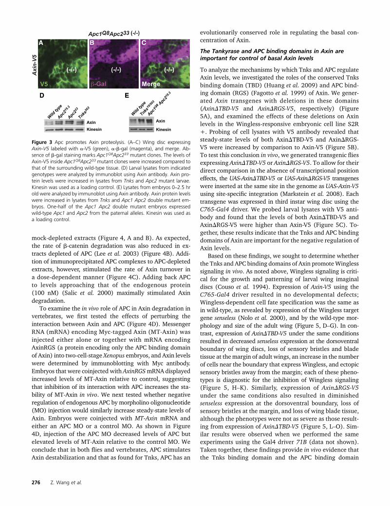
As we had found that Axin levels increased by only two- tothreefold following Tnks loss, we hypothesized that otherdegradation pathways also function to maintain Axin belowthe in vivo threshold compatible with Wingless signaling.Therefore, we further investigated our previous discoverythat the two fly homologs of the APC tumor suppressor facil-itate the control of basal Axin levels (Takacs et al. 2008).Consistent with our previous results, we found that in larvalwing discs expressing Axin-V5, the levels of Axin-V5 werehigher in Apc1 Apc2 double mutant clones as compared withsurrounding tissues (Figure 3, A–C). Importantly, the in-creased Axin staining intensity in Apc mutant clones wasdetected throughout the cell, revealing that the increasedstaining was not secondary to a change in the subcellularlocalization of Axin. These findings suggested that APCdestabilizes Axin.
To further test this conclusion, we utilized our new Axinantibodies, which allowed analysis of the effect of Apc inac-tivation on endogenous Axin levels with immunoblots, andthus provided an independent test of the regulation of Axinby Apc. Axin immunoblots revealed that by comparison withlysates from wild-type larvae, Axin levels were increased inlysates from Apc2mutant larvae to an extent similar to that inTnks null mutants (Figure 3D). We also sought to determinethe effect of simultaneous elimination of Apc1 and Apc2 onAxin levels; however, since loss of Apc1 and Apc2 results inlethality during larval development (Ahmed et al. 2002;Akong et al. 2002), we examined Axin levels in lysates fromApc1 Apc2 double null mutant embryos. By comparison withwild-type embryos, Axin levels were increased in Apc1 Apc2double mutants (Figure 3E), consistent with previous find-ings of increased Axin levels in Apc1 Apc2 double mutantclones in imaginal discs revealed by immunostaining (Takacset al. 2008). Of note, one-half of these mutant embryos havewild-type Apc supplied paternally; therefore, the level towhich Axin is increased following Apc inactivation is likelyhigher. Nonetheless, the increased Axin levels in Apc1 Apc2double mutant and Tnks mutant embryos were present by2 hr of development, which is prior to the onset of Winglessexpression. Together, these findings indicate that like Tnks,Apc also negatively regulates the basal levels of Axin, inde-pendently of Wingless exposure.
APC has an evolutionarily conserved role in regulatingAxin stability
To determine whether the regulation of Axin by APC is anevolutionarily conserved process, we reconstituted cytoplas-mic degradation of b-catenin and Axin in a cell-free systemusing Xenopus egg extracts as previously described (Salicet al. 2000; Lee et al. 2003). To determine if APC regu-lates Axin turnover in vertebrates, Xenopus egg extracts wereimmunodepleted of APC. APC-immunodepleted extractsresulted in a slower rate of Axin degradation compared to
In Vivo Axin Threshold in Wnt Signaling 275
mock-depleted extracts (Figure 4, A and B). As expected,the rate of b-catenin degradation was also reduced in ex-tracts depleted of APC (Lee et al. 2003) (Figure 4B). Addi-tion of immunoprecipitated APC complexes to APC-depletedextracts, however, stimulated the rate of Axin turnover ina dose-dependent manner (Figure 4C). Adding back APCto levels approaching that of the endogenous protein(100 nM) (Salic et al. 2000) maximally stimulated Axindegradation.
To examine the in vivo role of APC in Axin degradation invertebrates, we first tested the effects of perturbing theinteraction between Axin and APC (Figure 4D). MessengerRNA (mRNA) encoding Myc-tagged Axin (MT-Axin) wasinjected either alone or together with mRNA encodingAxinRGS (a protein encoding only the APC binding domainof Axin) into two-cell-stage Xenopus embryos, and Axin levelswere determined by immunoblotting with Myc antibody.Embryos that were coinjected with AxinRGSmRNA displayedincreased levels of MT-Axin relative to control, suggestingthat inhibition of its interaction with APC increases the sta-bility of MT-Axin in vivo. We next tested whether negativeregulation of endogenous APC by morpholino oligonucleotide(MO) injection would similarly increase steady-state levels ofAxin. Embryos were coinjected with MT-Axin mRNA andeither an APC MO or a control MO. As shown in Figure4D, injection of the APC MO decreased levels of APC butelevated levels of MT-Axin relative to the control MO. Weconclude that in both flies and vertebrates, APC stimulatesAxin destabilization and that as found for Tnks, APC has an
evolutionarily conserved role in regulating the basal con-centration of Axin.
The Tankyrase and APC binding domains in Axin areimportant for control of basal Axin levels
To analyze the mechanisms by which Tnks and APC regulateAxin levels, we investigated the roles of the conserved Tnksbinding domain (TBD) (Huang et al. 2009) and APC bind-ing domain (RGS) (Fagotto et al. 1999) of Axin. We gener-ated Axin transgenes with deletions in these domains(AxinDTBD-V5 and AxinDRGS-V5, respectively) (Figure5A), and examined the effects of these deletions on Axinlevels in the Wingless-responsive embryonic cell line S2R+. Probing of cell lysates with V5 antibody revealed thatsteady-state levels of both AxinDTBD-V5 and AxinDRGS-V5 were increased by comparison to Axin-V5 (Figure 5B).To test this conclusion in vivo, we generated transgenic fliesexpressing AxinDTBD-V5 or AxinDRGS-V5. To allow for theirdirect comparison in the absence of transcriptional positioneffects, the UAS-AxinDTBD-V5 or UAS-AxinDRGS-V5 transgeneswere inserted at the same site in the genome as UAS-Axin-V5using site-specific integration (Markstein et al. 2008). Eachtransgene was expressed in third instar wing disc using theC765-Gal4 driver. We probed larval lysates with V5 anti-body and found that the levels of both AxinDTBD-V5 andAxinDRGS-V5 were higher than Axin-V5 (Figure 5C). To-gether, these results indicate that the Tnks and APC bindingdomains of Axin are important for the negative regulation ofAxin levels.
Based on these findings, we sought to determine whetherthe Tnks and APC binding domains of Axin promoteWinglesssignaling in vivo. As noted above, Wingless signaling is criti-cal for the growth and patterning of larval wing imaginaldiscs (Couso et al. 1994). Expression of Axin-V5 using theC765-Gal4 driver resulted in no developmental defects;Wingless-dependent cell fate specification was the same asin wild-type, as revealed by expression of the Wingless targetgene senseless (Nolo et al. 2000), and by the wild-type mor-phology and size of the adult wing (Figure 5, D–G). In con-trast, expression of AxinDTBD-V5 under the same conditionsresulted in decreased senseless expression at the dorsoventralboundary of wing discs, loss of sensory bristles and bladetissue at themargin of adult wings, an increase in the numberof cells near the boundary that express Wingless, and ectopicsensory bristles away from the margin; each of these pheno-types is diagnostic for the inhibition of Wingless signaling(Figure 5, H–K). Similarly, expression of AxinDRGS-V5under the same conditions also resulted in diminishedsenseless expression at the dorsoventral boundary, loss ofsensory bristles at the margin, and loss of wing blade tissue,although the phenotypes were not as severe as those result-ing from expression of AxinDTBD-V5 (Figure 5, L–O). Sim-ilar results were observed when we performed the sameexperiments using the Gal4 driver 71B (data not shown).Taken together, these findings provide in vivo evidence thatthe Tnks binding domain and the APC binding domain
Figure 3 Apc promotes Axin proteolysis. (A–C) Wing disc expressingAxin-V5 labeled with a-V5 (green), a-b-gal (magenta), and merge. Ab-sence of b-gal staining marks Apc1Q8Apc233 mutant clones. The levels ofAxin-V5 inside Apc1Q8Apc233 mutant clones were increased compared tothat of the surrounding wild-type tissue. (D) Larval lysates from indicatedgenotypes were analyzed by immunoblot using Axin antibody. Axin pro-tein levels were increased in lysates from Tnks and Apc2 mutant larvae.Kinesin was used as a loading control. (E) Lysates from embryos 0–2.5 hrold were analyzed by immunoblot using Axin antibody. Axin protein levelswere increased in lysates from Tnks and Apc1 Apc2 double mutant em-bryos. One-half of the Apc1 Apc2 double mutant embryos expressedwild-type Apc1 and Apc2 from the paternal alleles. Kinesin was used asa loading control.
276 Z. Wang et al.

facilitate the control of basal Axin levels and the response toWingless stimulation.
Tankyrase and APC promote Axin destabilizationthrough distinct mechanisms
Next, we sought to compare the mechanisms by which Tnksand APC regulate Axin degradation. As our mutant clonalanalysis had revealed that Axin-V5 levels are regulated by bothTnks and Apc in vivo (Figure 2, L–N and Figure 3, A–C), thisprovided a sensitive in vivo assay for investigating the regu-lation of Axin by Apc and Tnks. To determine whether Tnksand APC function in the same Axin proteolysis pathwayin vivo, we determined whether the domains in Axin requiredfor Tnks- and APC-dependent Axin destabilization wereshared. To examine the role of the APC binding domain inAxin (RGS), we expressed AxinDRGS-V5 in wing imaginaldiscs. In contrast with Axin-V5 (Figure 3, A–C), the levelsof AxinDRGS-V5 did not increase in Apc1 Apc2 double mutant
clones; instead, equivalent AxinDRGS-V5 levels were de-tected inside and outside the clones (Figure 6, A–C). Thusas expected, deletion of the APC binding domain preventsthe ability of APC to negatively regulate Axin. We nexttested whether the RGS domain is also important for Tnks-dependent Axin regulation. In contrast with Apc1 Apc2 dou-ble mutant clones, the level of AxinDRGS-V5 increasedmarkedly in Tnks null mutant clones as compared to thesurrounding tissue (Figure 6, D–F). These results indicatedthat Tnks-mediated regulation of Axin does not requireAxin’s Apc binding domain. We conclude that the destabili-zation of Axin mediated by Tnks does not require Apc-dependent Axin degradation.
We next examined the importance of the Tnks bindingdomain (TBD) in Tnks- or APC-mediated Axin destabilization.As expected, deletion of the TBD abolished the ability of TnkstopromoteAxindegradation; incontrastwithAxin-V5(Figure2, L–N), the levels of AxinDTBD-V5 were indistinguishableinside and outside of Tnks null mutant clones (Figure 6, J–L).This finding confirmed that Tnks-mediated Axin degradationrequires the TBD. We next examined whether the TBD is alsoimportant for APC-dependent Axin degradation. In Apc1Apc2 double mutant null clones, the level of AxinDTBD-V5was the same as that found in the surrounding tissue (Figure6, G–I). These findings indicate, unexpectedly, that not onlyTnks-mediated degradation, but also Apc-mediated degrada-tion of Axin requires the Tnks binding domain in Axin.
The Axin TBD and RGS domains are juxtaposed (Figure5A). Thus, deletion of the Axin TBD might result in a confor-mational change in the Axin RGS domain that indirectly in-hibits the interaction between Axin and Apc. To address thispossibility, we determined whether deletion of the Axin TBDdisrupts the interaction between Axin and Apc. We expressedAxin-V5, AxinDTBD-V5, and AxinDRGS-V5 in DrosophilaS2R+ cells. Axin was immunoprecipitated with a V5 anti-body and the immunoprecipitates were probed with Apc2antibody. We detected Apc2 in immunoprecipitates of lysatesfrom cells expressing Axin-V5 and AxinDTBD-V5. In contrast,we did not detect Apc2 in immunoprecipitates of lysates fromcells expressing AxinDRGS-V5 (Figure 6M). These results in-dicate that the Axin TBD is not required for the physical in-teraction between Axin and APC, and thus that the Axin–APCinteraction is important, but not sufficient for APC-mediatedAxin degradation. Taken together, these findings indicatethat APC and Tnks promote Axin destabilization throughpartially distinct mechanisms.
Discussion
Our results demonstrate that regulation of the basal levels ofAxin is a dynamic process that requires the activity of the ADP-ribose polymerase Tnks and the tumor suppressor APC. Byincreasing the gene dosage of Axin, we found that endoge-nous Axin levels can increase by three- to fourfold withoutreaching the minimal threshold at which Wingless signalingis disrupted in nearly all developing tissues, consistent with
Figure 4 The destabilization of Axin by APC is conserved in vertebrates.(A) Immunoblotting revealed that the majority of endogenous APC wasremoved in APC-depleted Xenopus egg extracts. (B) Radiolabeled [35S]Axin or b-catenin was added to Xenopus egg extracts. At the indicatedtimes, samples were withdrawn and subjected to SDS-PAGE/autoradiography.The rate of Axin degradation is reduced in APC-depleted Xenopus eggextracts. (C) Apc immunoprecipitated from Xenopus egg extracts wastitrated into Apc-depleted egg extracts. Radiolabeled Axin was added,samples were removed after 3 hr for SDS-PAGE/autoradiography, and theamount of Axin remaining was quantified. The rate of Axin degradation isregulated by APC in a dose-dependent manner. Concentrations of APC innondepleted extracts (�100 nM) and amount of APC added back wascalculated as described in Lee et al. (2003). (D) APC regulates Axin turn-over in Xenopus embryos. (Top) mRNAs encoding AxinRGS or control(empty vector) plus MT-Axin mRNA (200 pg each) were coinjected intothe dorsal blastomeres of four-cell embryos. Embryos were lysed in RIPAbuffer at embryonic stage 7 and analyzed by Myc immunoblotting.(Bottom) Control or Apc MO (25 ng) plus mRNA encoding MT-AxinmRNAs (200 pg) was coinjected into the dorsal blastomeres of four-cell-stage embryos. Embryos were then lysed at stage 7 and analyzed byMyc immunoblotting.
In Vivo Axin Threshold in Wnt Signaling 277

previous work (Peterson-Nedry et al. 2008). These findingssupport the hypothesis that Axin levels regulate destructioncomplex activity, but also reveal that there exists a physiolog-ical range of at least three- to fourfold within which Axinlevels may fluctuate and yet remain compatible with the ac-tivation of the pathway following Wnt stimulation in all cells.This narrow range perhaps serves two essential functions ofAxin: (1) degradation of b-catenin to maintain its low levelsin the absence of Wnt ligands and (2) robust responsivenessto Wnt stimulation.
These results also provide evidence that Tnks promotesWingless signaling by maintaining Axin below this in vivo
threshold. Axin levels increase in the absence of Tnks,but remain below this threshold in nearly all developmentalcontexts. However, a relatively small increase in Axin expres-sion, which in itself has no effect on Wingless-dependent de-velopmental process in wild-type flies, is sufficient to result inclassicWingless loss-of-function phenotypes in Tnksmutants.These findings suggest that Tnks-dependent regulation buf-fers Axin activity and thus is likely important in specific in vivocontexts for promoting Wingless signaling.
Our previous work revealed that, whereas complete lossof Apc results in the constitutive activation of Wingless sig-naling, partial reduction in Apc levels resulted in Wingless
Figure 5 The Tnks and APC binding domains of Axin are important for promoting Wingless signaling. (A) Schematic representation of Axin-V5,AxinDTBD-V5, and AxinDRGS-V5. (B) Lysates of S2R+ cells transfected with the indicated plasmids (250 ng) were analyzed by immunoblotting using aV5 antibody. Deletion of the Tnks binding domain of Axin (AxinDTBD-V5) or the APC binding domain of Axin (AxinDRGS-V5) resulted in aberrantstabilization of Axin compared to wild-type controls (Axin-V5). (C) Lysates of third instar larvae expressing the indicated transgenes with C765-Gal4driver were analyzed by immunoblotting. AxinDTBD-V5 and AxinDRGS-V5 were stabilized compared with Axin-V5 protein. Confocal images of thirdinstar larval wing discs expressing Axin-V5 (D and E), AxinDTBD-V5 (H and I), or AxinDRGS-V5 (L and M) with the C765-Gal4 driver. Staining withWingless and Senseless antibodies shows that expression of Axin-V5 did not disrupt expression of the Wingless pathway target Senseless (D) or of thenumber of cells expressing Wingless (E, arrowheads), indicating that Axin-V5 was expressed at a level that is compatible with physiological regulation. Incontrast, expression of AxinDTBD-V5 resulted in loss of Senseless (H, arrows) and expansion in the number of cells expressing Wingless (I, arrowheads),indicating that Wingless signaling is inhibited by AxinDTBD-V5. Expression of AxinDRGS-V5 results in loss of Senseless (L, arrows), indicating thatAxinDRGS-V5 inhibits Wnt signaling. Adult wings expressing Axin-V5 (F and G), AxinDTBD-V5 (J and K), or AxinDRGS-V5 (N and O) using the C765-Gal4driver are shown. A total of 95% of wings expressing Axin-V5 (F and G) have normal morphology (n = 136), whereas 92% of wings expressingAxinDTBD-V5 (J and K) (n = 127) and 30% of wings expressing AxinDRGS-V5 (N and O) (n = 135) display loss of sensory bristles and tissue at the wingmargin (K and O, arrow), as well as extra bristles in the wing blade (K and O, arrowhead), which indicate inhibition of Wingless signaling.
278 Z. Wang et al.

loss-of-function phenotypes in multiple in vivo contexts, in-dicating that Apc has dual negative and positive roles inWingless signaling (Takacs et al. 2008). Our new ability todetect endogenous Axin by immunoblotting provided an in-dependent approach to test the hypothesis that negative reg-ulation of Axin levels contributes to the positive role of Apcin Wingless signaling (Takacs et al. 2008). Supporting thishypothesis, Axin levels were aberrantly increased in lysatesfrom Apcmutant embryos and larvae. In addition, our studiesin frog egg extracts and frog embryos suggest that the func-tion of APC in promoting Axin degradation is evolutionarilyconserved. Importantly, the role of APC in Axin degradation,like that of Tnks, is independent of Wingless stimulation.These results suggest that several pathways likely contributeto maintaining basal Axin levels below a critical concentra-tion, above which Wingless signaling is inhibited.
Our findings reveal that Tnks- and Apc-dependent proteol-ysis of Axin are achieved through partly separable mechanisms.
Tnks-mediated Axin destabilization requires the Tnks bind-ing domain of Axin, and thus their physical interaction.Apc-mediated Axin regulation is dispensable for the Tnks-dependent proteolysis of Axin (Figure 6). Conversely, Apc-mediated Axin regulation requires both the Tnks and APCbinding domains in Axin (Figure 6). Our analysis revealsthat the interaction between Axin and Apc is important,but not sufficient, for APC-mediated Axin degradation(Figure 6). Together, these results suggest that the APC-mediated regulation of Axin involves several distinct domainsin Axin and/or a specific Axin conformation.
Given the essential role of Wnt signaling in many funda-mental processes, and the requirement that Axin concentra-tions are maintained below a threshold level for the activationof signaling (Salic et al. 2000; Lee et al. 2003), it is not sur-prising that several degradation pathways have evolvedto ensure precise control of Axin levels (Luo et al. 2007;Cselenyi et al. 2008; Takacs et al. 2008; Huang et al. 2009).
Figure 6 APC- and Tnks-mediated regulation of Axin areachieved through partially separable mechanisms. Con-focal images of third instar larval wing imaginal discsstained with antibodies indicated at bottom left; genotypesare indicated on the right. (A–L) Wing disc labeled witha-V5 (green), a-b-gal (magenta), and merge. Absenceof b-gal staining marked Apc1Q8Apc233 mutant clones(A–C and G–I), and Tnks19 mutant clones (D–F and J–L).In contrast with Axin-V5 (Figure 3, A–C), the levels ofAxinDRGS-V5 did not increase inside Apc1Q8Apc233 mu-tant clones compared to the surrounding wild-type tissue(A–C). The levels of AxinDRGS-V5 inside Tnks19 mutantclones were increased compared to that of the surroun-ding wild-type tissue (D–F). In contrast with Axin-V5 (Figure3, A–C and Figure 2, L–N), the levels of AxinDTBD-V5 didnot increase in Apc1Q8Apc233 mutant clones (G–I) or inTnks19 mutant clones (J–L) compared to the surroundingwild-type tissue. (M) Immunoprecipitation with V5 anti-body from S2R+ cell lysates transfected with Axin-V5,AxinDTBD-V5, and AxinDRGS-V5. Apc2 was pulled downwith Axin-V5. Deletion of the Tnks binding domain ofAxin (AxinDTBD-V5) had no effect in the interaction be-tween Axin and Apc2. In contrast, deletion of the APCbinding domain of Axin (AxinDRGS-V5) inhibited the inter-action with Apc2.
In Vivo Axin Threshold in Wnt Signaling 279

Redundancy in Axin degradation pathways would provide acompensatory fail-safe mechanism to prevent an increase inAxin above its threshold and the resultant inhibition of sig-naling. Functional redundancy in Axin degradation pathwaysmay also explain why no defects in Wnt-dependent embryonicdevelopment were observed upon disruption of fish or fly Tnks(Huang et al. 2009; Feng et al. 2014;Wang et al. 2016). None-theless, the high degree of sequence conservation present inTnks homologs suggests that Tnks loss cannot be fully com-pensated by other pathways in all in vivo contexts. Indeed,small molecule inhibitor studies have indicated that Tnks isimportant for the Wnt-dependent regeneration of fins follow-ing injury in adult fish (Chen et al. 2009; Huang et al 2009).Moreover, our recentwork has revealed that regulation of Axinby Drosophila Tnks is required for Wingless target gene acti-vation and the Wingless-dependent control of intestinal stemcell proliferation in the adult midgut (Tian et al. 2016; Wanget al. 2016). Importantly, in the midgut, Tnks is essential fortarget gene activation in regions where the Wingless pathwayis activated at relatively low levels, but dispensable at highlevels (Wang et al. 2016), suggesting a critical role for Tnksin the amplification of signaling. Furthermore, we have foundthat Tnks not only targets Axin for proteolysis, but also pro-motes the central role of Axin in rapidWnt pathway activation(Yang et al. 2016).
If the basal concentration of human Axin, like that of flyAxin, is determined by several degradation pathways, thensmall molecule Tnks inhibitors will likely have the greatesttherapeutic efficacy in contexts for which Tnks-mediated Axinproteolysis has a predominant role in controlling Axin levels.For example, as APC activity is disrupted in the majority ofcolorectal carcinomas, APC-dependent Axin degradation islikely compromised in these cells; thus colon carcinoma cellsmight be particularly sensitive to treatment with Tnks inhib-itors, whereas the untransformed neighboring cells that con-tain wild-type APC levels would be less susceptible. Indeed,in mice for which APC activity has been disrupted by condi-tional targeting, daily treatment with a small molecule Tnksinhibitor for 3 weeks markedly reduced the proliferation ofcolonic adenoma cells, but resulted in little change in theproliferation rate or morphology of cells in the juxtaposedhealthy intestinal mucosa (Waaler et al. 2012). These studiesindicate that small molecule inhibitors of Axin degradationare promising agents for the targeted therapy ofWnt pathway-dependent diseases, and coupled with the work presentedhere, suggest that the conceptual framework needed to iden-tify new therapeutic agents in this category relies on our abilityto elucidate the distinct pathways that control endogenousAxin concentrations in vivo.
Acknowledgments
We thank the investigators listed in Materials and Methodsfor generously sharing reagents and V. Marlar for technicalassistance. We thank Claudio Pikielny, Ai Tian, and HassinaBenchabane for critical reading and thoughtful comments
on this manuscript. This work was funded by grantsfrom the National Institutes of Health (RO1CA105038 toY.A., R01GM081635 and R01GM103926 to E.L., andP40OD018537 to the Bloomington Drosophila Stock Cen-ter), the Emerald Foundation (to Y.A.), the Norris CottonCancer Center (to Y.A.), and the National Science Founda-tion (DBI-1039423 for the purchase of a Nikon A1RSi con-focal microscope).
Literature Cited
Ahmed, Y., S. Hayashi, A. Levine, and E. Wieschaus, 1998 Regula-tion of armadillo by a Drosophila APC inhibits neuronal apoptosisduring retinal development. Cell 93: 1171–1182.
Ahmed, Y., A. Nouri, and E. Wieschaus, 2002 Drosophila Apc1and Apc2 regulate Wingless transduction throughout develop-ment. Development 129: 1751–1762.
Akong, K., E. E. Grevengoed, M. H. Price, B. M. McCartney, M. A.Hayden et al., 2002 Drosophila APC2 and APC1 play overlap-ping roles in wingless signaling in the embryo and imaginaldiscs. Dev. Biol. 250: 91–100.
Baker, N. E., 1988 Transcription of the segment-polarity genewingless in the imaginal discs of Drosophila, and the phenotypeof a pupal-lethal wg mutation. Development 102: 489–497.
Bejsovec, A., and A. Martinez Arias, 1991 Roles of winglessin patterning the larval epidermis of Drosophila. Development113: 471–485.
Bischof, J., R. K. Maeda, M. Hediger, F. Karch, and K. Basler,2007 An optimized transgenesis system for Drosophila usinggerm-line-specific phiC31 integrases. Proc. Natl. Acad. Sci. USA104: 3312–3317.
Brand, A. H., and N. Perrimon, 1993 Targeted gene expression asa means of altering cell fates and generating dominant pheno-types. Development 118: 401–415.
Brunner, E., O. Peter, L. Schweizer, and K. Basler, 1997 pangolinencodes a Lef-1 homologue that acts downstream of Armadilloto transduce the Wingless signal in Drosophila. Nature 385:829–833.
Chen, B., M. E. Dodge, W. Tang, J. Lu, Z. Ma et al., 2009 Smallmolecule-mediated disruption of Wnt-dependent signaling intissue regeneration and cancer. Nat. Chem. Biol. 5: 100–107.
Chen, C. M., and G. Struhl, 1999 Wingless transduction by theFrizzled and Frizzled2 proteins of Drosophila. Development126: 5441–5452.
Chou, T. B., and N. Perrimon, 1992 Use of a yeast site-specificrecombinase to produce female germline chimeras in Drosoph-ila. Genetics 131: 643–653.
Clevers, H., and R. Nusse, 2012 Wnt/beta-catenin signaling anddisease. Cell 149: 1192–1205.
Couso, J. P., S. A. Bishop, and A. Martinez Arias, 1994 The wing-less signalling pathway and the patterning of the wing margin inDrosophila. Development 120: 621–636.
Cselenyi, C. S., K. K. Jernigan, E. Tahinci, C. A. Thorne, L. A. Leeet al., 2008 LRP6 transduces a canonical Wnt signal inde-pendently of Axin degradation by inhibiting GSK39s phosphor-ylation of beta-catenin. Proc. Natl. Acad. Sci. USA 105: 8032–8037.
Fagotto, F., E. Jho, L. Zeng, T. Kurth, T. Joos et al., 1999 Domainsof axin involved in protein-protein interactions, Wnt pathwayinhibition, and intracellular localization. J. Cell Biol. 145: 741–756.
Feng, Y., X. Li, L. Ray, H. Song, J. Qu et al., 2014 The Drosophilatankyrase regulates Wg signaling depending on the concentra-tion of Daxin. Cell. Signal. 26: 1717–1724.
280 Z. Wang et al.

Gerlach, J. P., B. L. Emmink, H. Nojima, O. Kranenburg, and M. M.Maurice, 2014 Wnt signalling induces accumulation of phos-phorylated beta-catenin in two distinct cytosolic complexes.Open Biol. 4: 140120.
Golic, K. G., and S. Lindquist, 1989 The FLP recombinase of yeastcatalyzes site-specific recombination in the Drosophila genome.Cell 59: 499–509.
Graham, F. L., and A. J. van der Eb, 1973 A new technique for theassay of infectivity of human adenovirus 5 DNA. Virology 52:456–467.
Hamada, F., Y. Tomoyasu, Y. Takatsu, M. Nakamura, S. Nagai et al.,1999 Negative regulation of Wingless signaling by D-axin, aDrosophila homolog of axin. Science 283: 1739–1742.
Huang, S. M., Y. M. Mishina, S. Liu, A. Cheung, F. Stegmeier et al.,2009 Tankyrase inhibition stabilizes axin and antagonizes Wntsignalling. Nature 461: 614–620.
Kramps, T., O. Peter, E. Brunner, D. Nellen, B. Froesch et al.,2002 Wnt/wingless signaling requires BCL9/legless-mediatedrecruitment of pygopus to the nuclear beta-catenin-TCF com-plex. Cell 109: 47–60.
Lau, T., E. Chan, M. Callow, J. Waaler, J. Boggs et al., 2013 Anovel tankyrase small-molecule inhibitor suppresses APC muta-tion-driven colorectal tumor growth. Cancer Res. 73: 3132–3144.
Lee, E., A. Salic, R. Kruger, R. Heinrich, and M. W. Kirschner,2003 The roles of APC and Axin derived from experimentaland theoretical analysis of the Wnt pathway. PLoS Biol. 1: E10.
Luo, W., A. Peterson, B. A. Garcia, G. Coombs, B. Kofahl et al.,2007 Protein phosphatase 1 regulates assembly and functionof the beta-catenin degradation complex. EMBO J. 26: 1511–1521.
MacDonald, B. T., K. Tamai, and X. He, 2009 Wnt/beta-cateninsignaling: components, mechanisms, and diseases. Dev. Cell 17:9–26.
Markstein, M., C. Pitsouli, C. Villalta, S. E. Celniker, and N. Perri-mon, 2008 Exploiting position effects and the gypsy retrovirusinsulator to engineer precisely expressed transgenes. Nat.Genet. 40: 476–483.
Nolo, R., L. A. Abbott, and H. J. Bellen, 2000 Senseless, a Znfinger transcription factor, is necessary and sufficient for sensoryorgan development in Drosophila. Cell 102: 349–362.
Parks, A. L., K. R. Cook, M. Belvin, N. A. Dompe, R. Fawcett et al.,2004 Systematic generation of high-resolution deletion cover-age of the Drosophila melanogaster genome. Nat. Genet. 36:288–292.
Peterson-Nedry, W., N. Erdeniz, S. Kremer, J. Yu, S. Baig-Lewiset al., 2008 Unexpectedly robust assembly of the Axin destruc-tion complex regulates Wnt/Wg signaling in Drosophila as re-vealed by analysis in vivo. Dev. Biol. 320: 226–241.
Rogers, S. L., and G. C. Rogers, 2008 Culture of Drosophila S2cells and their use for RNAi-mediated loss-of-function studiesand immunofluorescence microscopy. Nat. Protoc. 3: 606–611.
Salic, A., E. Lee, L. Mayer, and M. W. Kirschner, 2000 Control ofbeta-catenin stability: reconstitution of the cytoplasmic steps ofthe wnt pathway in Xenopus egg extracts. Mol. Cell 5: 523–532.
Takacs, C. M., J. R. Baird, E. G. Hughes, S. S. Kent, H. Benchabaneet al., 2008 Dual positive and negative regulation of winglesssignaling by adenomatous polyposis coli. Science 319: 333–336.
Tian, A., H. Benchabane, Z. Wang, and Y. Ahmed, 2016 Regulationof stem cell proliferation and cell fate specification by Wingless/Wnt signaling gradients enriched at adult intestinal compartmentboundaries. PLoS Genet. 12: e1005822.
Tolwinski, N. S., M. Wehrli, A. Rives, N. Erdeniz, S. DiNardo et al.,2003 Wg/Wnt signal can be transmitted through arrow/LRP5,6and Axin independently of Zw3/Gsk3beta activity. Dev. Cell 4:407–418.
Venken, K. J., J. W. Carlson, K. L. Schulze, H. Pan, Y. He et al.,2009 Versatile P[acman] BAC libraries for transgenesis studiesin Drosophila melanogaster. Nat. Methods 6: 431–434.
Vincent, J. P., C. H. Girdham, and P. H. O’Farrell, 1994 A cell-autonomous, ubiquitous marker for the analysis of Drosophilagenetic mosaics. Dev. Biol. 164: 328–331.
Waaler, J., O. Machon, L. Tumova, H. Dinh, V. Korinek et al.,2012 A novel tankyrase inhibitor decreases canonical Wnt sig-naling in colon carcinoma cells and reduces tumor growth inconditional APC mutant mice. Cancer Res. 72: 2822–2832.
Wang, Z., A. Tian, H. Benchabane, O. Tacchelly-Benites, E. Yanget al., 2016 The ADP-ribose polymerase Tankyrase regulatesadult intestinal stem cell proliferation during homeostasis inDrosophila. Development DOI: 10.1242/dev.127647.
Willert, K., C. Y. Logan, A. Arora, M. Fish, and R. Nusse, 1999 ADrosophila Axin homolog, Daxin, inhibits Wnt signaling. Devel-opment 126: 4165–4173.
Xu, T., and G. M. Rubin, 1993 Analysis of genetic mosaics in devel-oping and adult Drosophila tissues. Development 117: 1223–1237.
Yang, E., O. Tacchelly-Benites, Z. Wang, M. P. Randall, A. Tianet al., 2016 Wnt pathway activation by ADP-ribosylation. Nat.Commun. 7: 11430.
Zeng, L., F. Fagotto, T. Zhang, W. Hsu, T. J. Vasicek et al., 1997 Themouse Fused locus encodes Axin, an inhibitor of the Wnt sig-naling pathway that regulates embryonic axis formation. Cell90: 181–192.
Zhang, Y., S. Liu, C. Mickanin, Y. Feng, O. Charlat et al.,2011 RNF146 is a poly(ADP-ribose)-directed E3 ligase that reg-ulates axin degradation and Wnt signalling. Nat. Cell Biol. 13:623–629.
Communicating editor: M. F. Wolfner
In Vivo Axin Threshold in Wnt Signaling 281

GENETICSSupporting Information
www.genetics.org/lookup/suppl/doi:10.1534/genetics.115.183244/-/DC1
Wnt/Wingless Pathway Activation Is Promoted by aCritical Threshold of Axin Maintained by the Tumor
Suppressor APC and the ADP-RibosePolymerase Tankyrase
Zhenghan Wang, Ofelia Tacchelly-Benites, Eungi Yang, Curtis A. Thorne, Hisashi Nojima,Ethan Lee, and Yashi Ahmed
Copyright © 2016 by the Genetics Society of AmericaDOI: 10.1534/genetics.115.183244

Figure S1. Detection of Axin by immunostaining in imaginal discs
(A-C) Confocal images of larval third instar wing disc labeled with α-Axin (green)
and α-β-gal (magenta). Axin18 null mutant clones (marked by the absence of β-
gal, -/- in B) reveal the specificity of the Axin antibody. (C-E) Wing discs
expressing Axin-V5 were labeled with α-V5 (green) and α-Axin (magenta).
Higher magnification views of the boxed areas are shown in (C’-E’). Endogenous
Axin levels are measured in the cells that are not labeled by V5 antibody.