inhibition of the alternative complement activation pathway in
TRANSCRIPT
BioMed CentralJournal of Neuroinflammation
ss
Open AcceResearchInhibition of the alternative complement activation pathway in traumatic brain injury by a monoclonal anti-factor B antibody: a randomized placebo-controlled study in miceIris Leinhase1, Michal Rozanski1, Denise Harhausen1, Joshua M Thurman2, Oliver I Schmidt1, Amir M Hossini1, Mohy E Taha1, Daniel Rittirsch3, Peter A Ward3, V Michael Holers2, Wolfgang Ertel1 and Philip F Stahel*1,4Address: 1Department of Trauma and Reconstructive Surgery, Charité University Medical School, Campus Benjamin Franklin, 12200 Berlin, Germany, 2Departments of Medicine and Immunology, University of Colorado Health Sciences Center, Denver, CO 80262, USA, 3Department of Pathology, University of Michigan Medical School, Ann Arbor, MI 48109, USA and 4Department of Orthopedic Surgery, Denver Health Medical Center, University of Colorado School of Medicine, Denver, CO 80204, USA
Email: Iris Leinhase - [email protected]; Michal Rozanski - [email protected]; Denise Harhausen - [email protected]; Joshua M Thurman - [email protected]; Oliver I Schmidt - [email protected]; Amir M Hossini - [email protected]; Mohy E Taha - [email protected]; Daniel Rittirsch - [email protected]; Peter A Ward - [email protected]; V Michael Holers - [email protected]; Wolfgang Ertel - [email protected]; Philip F Stahel* - [email protected]
* Corresponding author
AbstractBackground: The posttraumatic response to traumatic brain injury (TBI) is characterized, in part,by activation of the innate immune response, including the complement system. We have recentlyshown that mice devoid of a functional alternative pathway of complement activation (factor B-/-mice) are protected from complement-mediated neuroinflammation and neuropathology after TBI.In the present study, we extrapolated this knowledge from studies in genetically engineered miceto a pharmacological approach using a monoclonal anti-factor B antibody. This neutralizing antibodyrepresents a specific and potent inhibitor of the alternative complement pathway in mice.
Methods: A focal trauma was applied to the left hemisphere of C57BL/6 mice (n = 89) using astandardized electric weight-drop model. Animals were randomly assigned to two treatmentgroups: (1) Systemic injection of 1 mg monoclonal anti-factor B antibody (mAb 1379) in 400 µlphosphate-buffered saline (PBS) at 1 hour and 24 hours after trauma; (2) Systemic injection ofvehicle only (400 µl PBS), as placebo control, at identical time-points after trauma. Sham-operatedand untreated mice served as additional negative controls. Evaluation of neurological scores andanalysis of brain tissue specimens and serum samples was performed at defined time-points for upto 1 week. Complement activation in serum was assessed by zymosan assay and by murine C5aELISA. Brain samples were analyzed by immunohistochemistry, terminal deoxynucleotidyltransferase dUTP nick-end labeling (TUNEL) histochemistry, and real-time RT-PCR.
Results: The mAb 1379 leads to a significant inhibition of alternative pathway complement activityand to significantly attenuated C5a levels in serum, as compared to head-injured placebo-treatedcontrol mice. TBI induced histomorphological signs of neuroinflammation and neuronal apoptosisin the injured brain hemisphere of placebo-treated control mice for up to 7 days. In contrast, the
Published: 2 May 2007
Journal of Neuroinflammation 2007, 4:13 doi:10.1186/1742-2094-4-13
Received: 19 March 2007Accepted: 2 May 2007
This article is available from: http://www.jneuroinflammation.com/content/4/1/13
© 2007 Leinhase et al; licensee BioMed Central Ltd. This is an Open Access article distributed under the terms of the Creative Commons Attribution License (http://creativecommons.org/licenses/by/2.0), which permits unrestricted use, distribution, and reproduction in any medium, provided the original work is properly cited.
Page 1 of 12(page number not for citation purposes)
Journal of Neuroinflammation 2007, 4:13 http://www.jneuroinflammation.com/content/4/1/13
systemic administration of an inhibitory anti-factor B antibody led to a substantial attenuation ofcerebral tissue damage and neuronal cell death. In addition, the posttraumatic administration of themAb 1379 induced a neuroprotective pattern of intracerebral gene expression.
Conclusion: Inhibition of the alternative complement pathway by posttraumatic administration ofa neutralizing anti-factor B antibody appears to represent a new promising avenue forpharmacological attenuation of the complement-mediated neuroinflammatory response after headinjury.
BackgroundTraumatic brain injury (TBI) represents a neuroinflamma-tory disease which is in large part mediated by an earlyactivation of the innate immune system [1-4]. In thisregard, the complement system has been identified as animportant early mediator of posttraumatic neuroinflam-mation [5-7]. Research strategies to prevent the neuroin-flammatory pathological sequelae of TBI have largelyfailed in translation to clinical treatment [8-14]. Thisnotion is exemplified by the recent failure of the "CRASH"trial (Corticosteroid randomization after significant headinjury). This large-scale multicenter, placebo-controlledrandomized study was designed to assess the effect ofattenuating the neuroinflammatory response after TBI byadministration of high-dose methylprednisolone [15].The trial was unexpectedly aborted after enrollment of10,008 patients based on the finding of a significantlyincreased mortality in the steroid cohort, compared to theplacebo control group [15]. These data imply that the"pan"-inhibition of the immune response by the use ofglucocorticoids represents a too broad and unspecificapproach for controlling neuroinflammation after TBI[16]. Thus, research efforts are currently focusing on morespecific and sophisticated therapeutic modalities, such asthe inhibition of the complement cascade [17-19]. Severalcomplement inhibitors have been investigated in experi-mental TBI models [20-26]. However, most modalities ofcomplement inhibition have focussed on interfering withthe cascade at the central level of the C3 convertases,where the three activation pathways merge (Fig. 1)[20,21,25-27]. Other approaches were designed to inhibitthe main inflammatory mediators of the complement cas-cade, such as the anaphylatoxin C5a [22,28-30]. Onlymore recently, increased attention was drawn to the "key"role of the alternative pathway in the pathophysiology ofdifferent inflammatory conditions outside the centralnervous system (CNS) [31-34]. We have recently reportedthat factor B knockout (fB-/-) mice, which are devoid of afunctional alternative pathway, show a significant neuro-protection after TBI, compared to head-injured wild-typemice [35]. These data served as a baseline for the presentstudy, where we extrapolated the positive findings in theknockout mice to a pharmacological approach. We there-fore used a neutralizing monoclonal anti-factor B anti-body which was recently described as a highly potent
inhibitor of the alternative pathway in mice [31,34,36,37]in the setting of a standardized model of closed headinjury [38].
MethodsAnimalsAll experiments were performed in adult male mice of theC57BL/6 strain (n = 89 in total) purchased from JacksonLaboratory (Bar Harbor, ME). The mice were bred in aselective pathogen-free (SPF) environment and understandardized conditions of temperature (21°C), humidity(60%), light and dark cycles (12:12 h), with food andwater provided ad libitum. Experiments were performed incompliance with the standards of the Federation of Euro-pean Laboratory Animal Science Association (FELASA) andwere approved by the institutional animal care committee(Landesamt für Arbeitsschutz, Gesundheitsschutz und tech-nische Sicherheit Berlin, Berlin, Germany, No. G0099/03).
Trauma modelMice were subjected to experimental TBI using a standard-ized weight-drop device, as previously described[26,35,39,40]. In brief, after induction of isofluraneanesthesia, the skull was exposed by a midline longitudi-nal scalp incision. The head was fixed and a 250 g weightwas dropped on the skull from a height of 2 cm, resultingin a focal blunt injury to the left hemisphere. After trauma,the mice received supporting oxygenation with 100% O2until fully awake. The extent of posttraumatic neurologi-cal impairment was assessed at defined time intervals aftertrauma (t = 1 h, 4 h, 24 h, and 7 days) using a standard-ized Neurological Severity Score (NSS), as described below.
Treatment protocolThe inhibitory monoclonal anti-factor B antibody (mAb1379) used in this study was previously described and theselected dosage was in the titrated range used in otherstudies on murine models of inflammation [34,36,37].The antibody itself does not have any complement-acti-vating properties. Mice were randomly assigned to twotreatment groups: (1) Systemic injection of 1 mg mAb1379 in 400 µl phosphate-buffered saline (PBS) at 1 hourand 24 hours after trauma; (2) Systemic injection of vehi-cle only (400 µl PBS), as placebo control, at identicaltime-points after trauma. Concealed allocation to the two
Page 2 of 12(page number not for citation purposes)
Journal of Neuroinflammation 2007, 4:13 http://www.jneuroinflammation.com/content/4/1/13
Page 3 of 12(page number not for citation purposes)
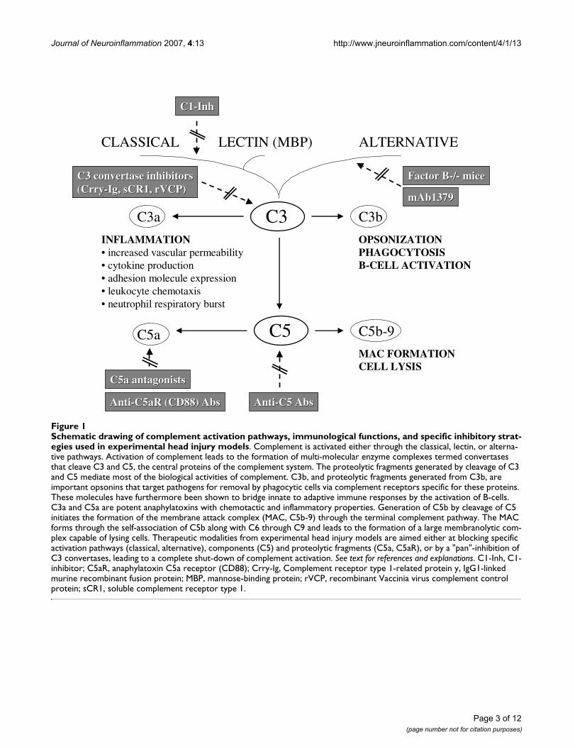
Schematic drawing of complement activation pathways, immunological functions, and specific inhibitory strategies used in experimental head injury modelsFigure 1Schematic drawing of complement activation pathways, immunological functions, and specific inhibitory strat-egies used in experimental head injury models. Complement is activated either through the classical, lectin, or alterna-tive pathways. Activation of complement leads to the formation of multi-molecular enzyme complexes termed convertases that cleave C3 and C5, the central proteins of the complement system. The proteolytic fragments generated by cleavage of C3 and C5 mediate most of the biological activities of complement. C3b, and proteolytic fragments generated from C3b, are important opsonins that target pathogens for removal by phagocytic cells via complement receptors specific for these proteins. These molecules have furthermore been shown to bridge innate to adaptive immune responses by the activation of B-cells. C3a and C5a are potent anaphylatoxins with chemotactic and inflammatory properties. Generation of C5b by cleavage of C5 initiates the formation of the membrane attack complex (MAC, C5b-9) through the terminal complement pathway. The MAC forms through the self-association of C5b along with C6 through C9 and leads to the formation of a large membranolytic com-plex capable of lysing cells. Therapeutic modalities from experimental head injury models are aimed either at blocking specific activation pathways (classical, alternative), components (C5) and proteolytic fragments (C5a, C5aR), or by a "pan"-inhibition of C3 convertases, leading to a complete shut-down of complement activation. See text for references and explanations. C1-Inh, C1-inhibitor; C5aR, anaphylatoxin C5a receptor (CD88); Crry-Ig, Complement receptor type 1-related protein y, IgG1-linked murine recombinant fusion protein; MBP, mannose-binding protein; rVCP, recombinant Vaccinia virus complement control protein; sCR1, soluble complement receptor type 1.
CLASSICAL LECTIN (MBP) ALTERNATIVE
C3 C3bC3a
C5a C5b-9C5
INFLAMMATION• increased vascular permeability • cytokine production • adhesion molecule expression• leukocyte chemotaxis, • neutrophil respiratory burst
OPSONIZATIONPHAGOCYTOSISB-CELL ACTIVATION
MAC FORMATIONCELL LYSIS
Factor Factor BB--//-- micemice
mAb1379mAb1379
C3 C3 convertase inhibitorsconvertase inhibitors((CrryCrry--IgIg, sCR1, , sCR1, rVCPrVCP))
AntiAnti--C5 C5 AbsAbs
C5a C5a antagonistsantagonists
AntiAnti--C5aR (CD88) C5aR (CD88) AbsAbs
C1C1--InhInh
Journal of Neuroinflammation 2007, 4:13 http://www.jneuroinflammation.com/content/4/1/13
treatment cohorts was performed after assessment of thebaseline NSS at 1 hour after trauma, in order to ensureequal injury severity between the groups. The systemic(i.p.) route of administration and the time window ofinjection were selected based on the breakdown of theblood-brain barrier (BBB) for up to 24 hours after trauma[38,41]. This allows a "time window" for peripherallyadministered compounds to reach the intrathecal com-partment and exert pharmacological effects in the CNS[26,39,40,42]. Furthermore, the systemic injection earlyafter trauma represents an approach with potential clini-cal implications. In order to induce a continuing comple-ment inhibition during the acute inflammatory phase inthe first days, injections were repeated at 24 hours.
Subgroups of mice (n = 10 per group and time-point)were euthanized by isoflurane anesthesia and decapitatedat t = 4 h, 24 h, and 7 days. Brains were immediatelyextracted, snap-frozen in liquid nitrogen and stored at -80°C until analysis by immunohistochemistry, TUNELhistochemistry and real-time RT-PCR. In addition, serumsamples were collected at identical time-points for deter-mination of complement activation levels. Sham-oper-ated and untreated normal mice served as negativecontrols.
Neurological Severity Score (NSS)A previously characterized 10-parameter score was usedfor assessment of posttraumatic neurological impairment,as described elsewhere in detail [41,43]. The NSS wasassessed in a blinded fashion by two different investiga-tors at the time-points t = 1 h, 4 h, 24 h, and 7 days aftertrauma. The score comprises 10 individual parameters,including tasks on motor function, alertness, and physio-logical behavior, whereby one point is given for failure ofthe task, and no point for succeeding. A maximum NSSscore of 10 points indicates severe neurological dysfunc-tion, with failure of all tasks.
Mouse C5a ELISASerum levels of the complement anaphylatoxin C5a weredetermined by a mouse-specific ELISA developed in thelaboratory of Dr. P.A. Ward (Ann Arbor, MI), as previ-ously described [35,44]. In brief, ELISA plates (Immulon4HBX, Thermo Labsystems, Milford, MA) were coatedwith 5 µg/ml of purified monoclonal anti-mouse C5a IgG(BD Pharmingen, San Diego, CA). After blocking of non-specific binding sites with 1% milk (Roth, Karlsruhe, Ger-many) in PBS (Gibco-Invitrogen, Carlsbad, CA) contain-ing 0.05% TWEEN 20 (Sigma-Aldrich), the plate wascoated with 100 µl of each serum diluted 1:20 (in 0.1%milk in PBS containing 0.05% TWEEN) and murinerecombinant mouse C5a at defined concentrations forestablishing the standard curve. After incubation and sub-sequent washing steps, biotinylated monoclonal anti-
mouse C5a antibody was added at 500 ng/ml (BDPharmingen) followed by washing steps and incubationwith streptavidin-peroxidase at 400 ng/ml (Sigma-Aldrich).
For colorimetric reaction, 0.4 mg/ml o-phenylenediaminedihydrochloride with 0.4 mg/ml urea hydrogen peroxidein 0.05 M phosphate citrate buffer (Sigma-Aldrich) wasadded and the color reaction was stopped with 3 M sulfu-ric acid. Absorbance was read at 490 nm using a "Spec-traMax 190" reader (Molecular Devices, Sunnyvale, CA).All samples were analyzed in duplicate and results werecalculated from the means of duplicate sample analysis.The standard curve was linear from 0.1 ng/ml to 50 ng/ml.
Quantification of alternative pathway complement activityAlternative pathway complement activity in mouse serumwas quantified as previously described [26,36]. Briefly, atthe above-mentioned defined time-points, whole bloodwas collected and spun down, serum was aliquoted andstored at -80°C until analyzed. Ten microlitres of serumfrom each animal was incubated with 109 zymosan parti-cles (Sigma-Aldrich, St. Louis, MO) at 37°C for 30 min ina master mix containing final concentrations of 5 mMMgCl2 and 10 mM EGTA and brought up to 100 µl in cal-cium-free PBS. C3 deposition on the particles wasdetected with a FITC-labeled antibody to C3 (Cappel,Durham, USA) diluted 1:100 and fluorescence was meas-ured by flow cytometry. Complement activity was calcu-lated using the formula:
ImmunohistochemistryImmunohistochemical stainings of serial coronal cryosec-tions (8 µm) of brain tissue were performed using abiotin/avidin/peroxidase technique with diaminobenzi-dine tetrahydrochloride as chromogen (Vector, Burlin-game, CA). The following primary antibodies were used ascell-markers: monoclonal anti-NeuN for neurons(1:2,000; Chemicon, Hampshire, UK); polyclonal rabbitanti-GFAP for astrocytes (1:100; Shandon Immunon,Pittsburgh, PA) and monoclonal rat anti-CD11b formicroglia and monocytes/macrophages (1:100; AccurateChemical, Westbury, NY). For negative control, non-immunized IgG (Vector) was used at equal dilutions.
TUNEL assayThe terminal deoxynucleotidyl transferase dUTP nick-endlabeling (TUNEL) technique was applied to determine theextent of neuronal cell death in tissue sections. Herefore,the commercially available ''Fluorescein In Situ Cell DeathDetection Kit'' (Roche Diagnostics GmbH, Mannheim,
100× −( [sample mean channel fluorescence background no serum ]])
( )positive control mean channel fluorescence background −
Page 4 of 12(page number not for citation purposes)
Journal of Neuroinflammation 2007, 4:13 http://www.jneuroinflammation.com/content/4/1/13
Germany) was used according to the manufacturer'sinstructions, as previously described [35]. In brief, slideswere dried for 30 min followed by fixation in 10% forma-lin solution at RT. After washing in PBS, sections wereincubated in ice-cold ethanol-acetic acid solution (3:1),washed in PBS and incubated with 3% Triton X-100 solu-tion for 60 min at RT for permeabilization. Slides werethen incubated with the TdT-enzyme in reaction buffercontaining fluorescein-dUTP for 90 min at 37°C. Nega-tive control was performed using only the reaction bufferwithout TdT enzyme. Positive controls were performed bydigesting with 500 U/ml DNase grade I solution (Roche).To preserve cells for comparison, slices were covered withVectashield® mounting medium containing 4',6'-diamino-2-phenylindole (DAPI; Vector). All sampleswere evaluated immediately after staining using an ''Axi-oskop 40'' fluorescence microscope (Zeiss, Germany) at460 nm for DAPI and 520 nm for TUNEL fluorescence.Data were analyzed by Alpha digi doc 1201 software(Alpha Innotech, San Leandro, CA).
Real-time RT-PCRChanges in the expression profiles of pro- and anti-apop-totic as well as complement-regulatory genes were deter-mined by semi-quantitative two-step real-time RT-PCRusing commercially available and custom-made murine-specific primers shown in table 1. This technique was pre-viously described [26]. In brief, brains were homogenizedper hemisphere in Qiazol® buffer (Qiagen, Hilden, Ger-many). RNA was isolated and further purified using RNe-asy® Mini-kits (Qiagen) and RNA concentrations weremeasured using a spectrophotometer (Bio-Rad, Munich,Germany). From each brain hemisphere, 2 µg RNA werereversed transcribed using random nonamer and oligo-dT16mer primers (Operon Biotechnologies, Cologne,Germany) with Omniscript® kits (Qiagen), according tothe manufacturer's instructions. Real-time RT-PCR wasperformed using validated commercially available andcustom designed primer-probe® sets (Qiagen) and opti-mized protocols on the Opticon® real-time PCR DetectionSystem (Bio-Rad). For quantification of gene expressionlevels, GAPDH amplicons were generated and used as a
house-keeping internal control gene. Relative gene expres-sion levels were calculated in relation to the correspond-ing GAPDH gene expression levels.
Statistical analysisStatistical analysis was performed using commerciallyavailable software (SPSS 9.0 for Windows™). Differencesin serum complement activity levels and in intracerebralgene expression levels between the groups were deter-mined by the unpaired Student's t-test. The repeatedmeasures analysis of variance (ANOVA) was used forassessing differences in neurological scores (NSS). A P-value < 0.05 was considered statistically significant.
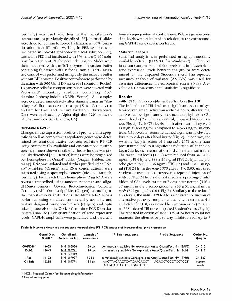
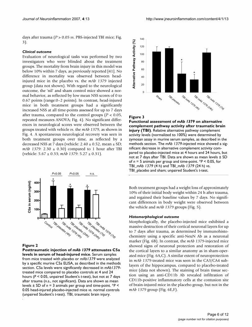
ResultsmAb 1379 inhibits complement activation after TBIThe induction of TBI lead to a significant extent of sys-temic complement activation within 4 hours after trauma,as revealed by significantly increased anaphylatoxin C5aserum levels (P < 0.05 vs. control, unpaired Student's t-test; Fig. 2). Peak C5a levels at 4 h after head injury wereas high as 450 ng/ml, compared to 42–53 ng/ml in con-trols. C5a levels in serum remained significantly elevatedfor up to 7 days after head injury (Fig. 2). In contrast, thesystemic (i.p.) injection of 1 mg mAb 1379 at one hourpost trauma lead to a significant reduction of anaphyla-toxin C5a levels in serum at 4 h and 24 h after head injury.The mean C5a levels (± SD) were reduced from 361 ± 59ng/ml (TBI 4 h) and 333 ± 29 ng/ml (TBI 24 h) in the pla-cebo group to 111 ± 36 ng/ml (TBI 4 h) and 118 ± 30 ng/ml (TBI 24 h) in the mAb 1379 group (P < 0.05, unpairedStudent's t-test; Fig. 2). However, a repeated injection ofmAb 1379 at 24 hours did not mediate a prolonged inhi-bition of C5a levels for up to 7 days after trauma (316 ±37 ng/ml in the placebo group vs. 265 ± 51 ng/ml in themAb 1379 group, P > 0.05; Fig. 2). Similarly to the reducedC5a levels, the mAb 1379 led to a significant reduction ofalternative pathway complement activity in serum at 4 hand 24 h after TBI, as assessed by zymosan assay (P < 0.05vs. PBS-injected TBI mice, unpaired Student's t-test; Fig. 3).The repeated injection of mAb 1379 at 24 hours could notmaintain the alternative pathway inhibition for up to 7
Table 1: Murine primer sequences used for real-time RT-PCR analysis of intracerebral gene expression
Gene ID at NCBI *
GeneBank Accession No.
Length of amplicons
Primer sequence Probe Sequence Order No. Qiagen
GAPDH# 14433 NM_008084 136 bp commercially available Genexpression Assay QuantiTect Mm_GAPD 241012Bcl-2 12043 NM_009741
NM_177410118 bp commercially available Genexpression Assay QuantiTect Mm_Bcl-2 241118
Fas 14102 NM_007987 96 bp commercially available Genexpression Assay QuantiTect Mm_ Tnfsf6 241122C1-Inh 12258 NM_009776 134 bp AACTTAGAACTCATCAACACCT
GTTATCTTCCACTTGGCACTCACACCTGCCTCGTCCT custom
made
* NCBI, National Center for Biotechnology Information# Housekeeping gene
Page 5 of 12(page number not for citation purposes)
Journal of Neuroinflammation 2007, 4:13 http://www.jneuroinflammation.com/content/4/1/13
days after trauma (P > 0.05 vs. PBS-injected TBI mice; Fig.3).
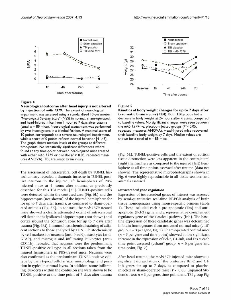
Clinical outcomeEvaluation of neurological tasks was performed by twoinvestigators who were blinded about the treatmentgroups. The mortality from brain injury in this model wasbelow 10% within 7 days, as previously reported [41]. Nodifference in mortality was observed between head-injured mice in the placebo vs. the mAb 1379 injectedgroup (data not shown). With regard to the neurologicaloutcome, the 'nil' and sham control mice showed a nor-mal behavior, as reflected by low mean NSS scores of 0 to0.67 points (range:0–2 points). In contrast, head-injuredmice in both treatment groups had a significantlyincreased NSS at all time-points assessed for up to 7 daysafter trauma, compared to the control groups (P < 0.05,repeated measures ANOVA; Fig. 4). No significant differ-ences in neurological scores were observed between thegroups treated with vehicle vs. the mAb 1379, as shown inFig. 4. A spontaneous neurological recovery was seen inboth treatment groups over time, as reflected by adecreased NSS at 7 days (vehicle: 2.40 ± 0.52, mean ± SD;mAb 1379: 2.30 ± 0.30) compared to 1 hour after TBI(vehicle: 5.67 ± 0.33; mAb 1379: 5.27 ± 0.31).
Both treatment groups had a weight loss of approximately10% of their initial body weight within 24 h after trauma,and regained their baseline values by 7 days. No signifi-cant differences in body weight were observed betweenthe vehicle and mAb 1379 groups (Fig. 5).
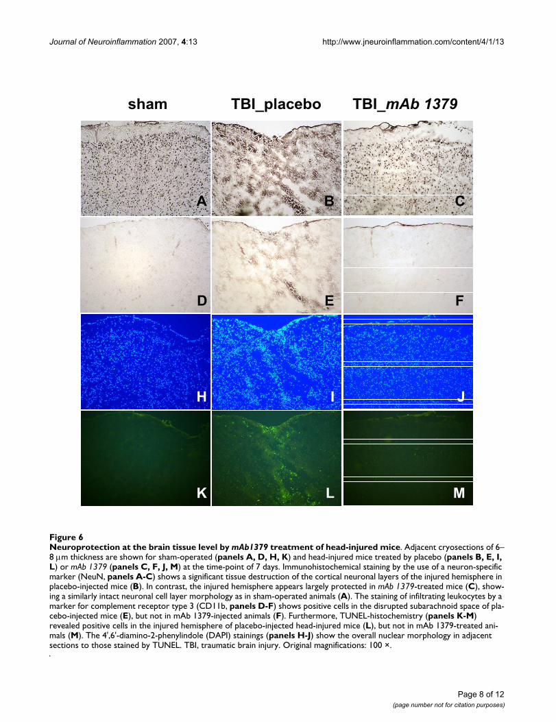
Histomorphological outcomeMorphologically, the placebo-injected mice exhibited amassive destruction of their cortical neuronal layers for upto 7 days after trauma, as determined by immunohisto-chemistry using a specific anti-NeuN Ab as a neuronalmarker (Fig. 6B). In contrast, the mAb 1379-injected miceshowed signs of neuronal protection and restoration ofthe cortical layers to a similar anatomy as in sham-oper-ated mice (Fig. 6A,C). A similar extent of neuroprotectionin mAb 1379-treated mice was seen in the CA3/CA4 sub-layers of the hippocampus, compared to placebo-treatedmice (data not shown). The staining of brain tissue sec-tion using an anti-CD11b Ab revealed infiltration ofCD11b-positive inflammatory cells at the contusion siteof brain-injured mice in the placebo group, but not in themAb 1379 group (Fig. 6E,F).
Functional assessment of mAb 1379 on alternative comple-ment pathway activity after traumatic brain injury (TBI)Figure 3Functional assessment of mAb 1379 on alternative complement pathway activity after traumatic brain injury (TBI). Relative alternative pathway complement activity levels (normalized to 100%) were determined by zymosan assay in murine serum samples, as described in the methods section. The mAb 1379-injected mice showed a sig-nificant decrease in alternative complement activity com-pared to placebo-injected mice at 4 hours and 24 hours, but not at 7 days after TBI. Data are shown as mean levels ± SD of n = 5 animals per group and time-point. *P < 0.05, for TBI_mAb 1379 (4 h) and TBI_mAb 1379 (24 h) vs. TBI_placebo and sham; unpaired Student's t-test.
0
20
40
60
80
100
120
140
Sham
TBI placebo
TBI mAb
1379 (4h)
Alt
ern
ati
ve
Pa
thw
ay A
cti
vit
y (
%)
TBI mAb
1379 (24h)
TBI mAb
1379 (7d)
*
*
Posttraumatic injection of mAb 1379 attenuates C5a levels in serum of head-injured miceFigure 2Posttraumatic injection of mAb 1379 attenuates C5a levels in serum of head-injured mice. Serum samples from mice treated with placebo or mAb1379 were analyzed by a specific murine C5a ELISA, as described in the methods section. C5a levels were significantly decreased in mAb1379-treated mice compared to placebo controls at 4 and 24 hours (P < 0.05, unpaired Student's t-test), but not at 7 days after trauma (n.s., not significant). Data are shown as mean levels ± SD of n = 3 animals per group and time-point. *P < 0.05 head-injured placebo-injected mice vs. normal controls (unpaired Student's t-test). TBI, traumatic brain injury.
C5a
in s
eru
m (
ng
/ml)
TBI placebo (4h)
TBI mAb 1379 (4h)
Control
TBI placebo (24h)
TBI mAb 1379 (24h)
TBI placebo (7d)
TBI mAb 1379 (7d)
0
100
200
300
400*
* *
P<0.05 P<0.05 n.s.
Page 6 of 12(page number not for citation purposes)
Journal of Neuroinflammation 2007, 4:13 http://www.jneuroinflammation.com/content/4/1/13
The assessment of intracerebral cell death by TUNEL his-tochemistry revealed a dramatic increase in TUNEL-posi-tive neurons in the injured left hemispheres of PBS-injected mice at 4 hours after trauma, as previouslydescribed for this TBI model [35]. TUNEL-positive cellswere detected within the contused area (Fig. 6L) and thehippocampus (not shown) of the injured hemisphere forfor up to 7 days after trauma, as compared to sham-oper-ated animals (Fig. 6K). In contrast, the mAb 1379 treatedmice showed a clearly attenuated extent of intracerebralcell death in the ipsilateral hippocampus (not shown) andcortex around the contusion zone for up to 7 days aftertrauma (Fig. 6M). Immunohistochemical staining of adja-cent sections to those analyzed by TUNEL histochemistryby cell markers for neurons (anti-NeuN), astrocytes (anti-GFAP), and microglia and infiltrating leukocytes (anti-CD11b), revealed that neurons were the predominantTUNEL-positive cell type in all sections taken from theinjured hemisphere in PBS-treated mice. Neurons werealso confirmed as the predominant TUNEL-positive cell-type by their typical cellular size, morphology, and posi-tion in typical neuronal layers. In addition, some infiltrat-ing leukocytes within the contusion site were shown to beTUNEL-positive at the time-point of 7 days after trauma
(Fig. 6L). TUNEL-positive cells and the extent of corticaltissue destruction were less apparent in the contralateral(right) hemisphere as compared to the injured (left) hem-isphere at all time-points assessed after trauma (data notshown). The representative microphotographs shown inFig. 6 were highly reproducible in all tissue sections andanimals assessed.
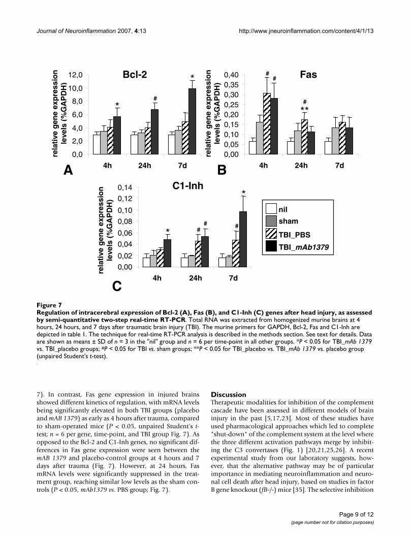
Intracerebral gene regulationExpression of intracerebral genes of interest was assessedby semi-quantitative real-time RT-PCR analysis of braintissue homogenates using mouse-specific primers (table1). These included each a pro-apoptotic (Fas) and anti-apoptotic (Bcl-2) gene and a representative complementregulatory gene of the classical pathway (Inh). The base-line expression of these candidate genes was determinedin brain homogenates from untreated normal mice („nil“group, n = 3 per gene, Fig. 7). Sham-operated control mice(n = 6 per gene and time-point) showed a non-significantincrease in the expression of Bcl-2, C1-Inh, and Fas at eachtime point assessed („sham“ group, n = 6 per gene andtime-point, Fig. 7).
After head trauma, the mAb1379-injected mice showed asignificant upregulation of the protective Bcl-2 and C1-Inh genes for up to 7 days, as compared to placebo-injected or sham-operated mice (P < 0.05, unpaired Stu-dent's t-test; n = 6 per gene, time-point, and TBI group Fig.
Kinetics of body weight changes for up to 7 days after trau-matic brain injury (TBI)Figure 5Kinetics of body weight changes for up to 7 days after traumatic brain injury (TBI). Both TBI groups had a decrease in body weight at 24 hours after trauma, compared to baseline values. No significant changes were seen between the mAb 1379- vs. placebo-injected groups (P > 0.05, repeated measures ANOVA). Head-injured mice recovered their baseline body weight by 7 days. Median values are shown for a total of n = 89 mice.
Time after trauma
24
25
26
27
28
29
30
31
32
pre-TBI 1h 4h 24h 7d
we
igh
t(g
)
Baseline 1h 4h 24h 7d
Normal mice
Sham operation
TBI placebo
TBI mAb 1379
Neurological outcome after head injury is not altered by injection of mAb 1379Figure 4Neurological outcome after head injury is not altered by injection of mAb 1379. The extent of neurological impairment was assessed using a standardized 10-parameter "Neurological Severity Score" (NSS) in normal, sham-operated, and head-injured mice from 1 hour to 7 days after trauma (total: n = 89 mice). Neurological assessment was performed by two investigators in a blinded fashion. A maximal score of 10 points corresponds to a severe neurological impairment, while a score of 0 points reflects normal behavior [41,43]. The graph shows median levels of the groups at different time-points. No statistically significant differences where found at any time-point between head-injured mice treated with either mAb 1379 or placebo (P > 0.05, repeated meas-ures ANOVA). TBI, traumatic brain injury.
NS
S
-1
0
1
23
4
5
6
7
1h 4h 24h 7d
Time after trauma
Normal miceSham operationTBI placeboTBI mAb 1379
1h 4h 24h 7d
Page 7 of 12(page number not for citation purposes)
Journal of Neuroinflammation 2007, 4:13 http://www.jneuroinflammation.com/content/4/1/13
Page 8 of 12(page number not for citation purposes)
Neuroprotection at the brain tissue level by mAb1379 treatment of head-injured miceFigure 6Neuroprotection at the brain tissue level by mAb1379 treatment of head-injured mice. Adjacent cryosections of 6–8 µm thickness are shown for sham-operated (panels A, D, H, K) and head-injured mice treated by placebo (panels B, E, I, L) or mAb 1379 (panels C, F, J, M) at the time-point of 7 days. Immunohistochemical staining by the use of a neuron-specific marker (NeuN, panels A-C) shows a significant tissue destruction of the cortical neuronal layers of the injured hemisphere in placebo-injected mice (B). In contrast, the injured hemisphere appears largely protected in mAb 1379-treated mice (C), show-ing a similarly intact neuronal cell layer morphology as in sham-operated animals (A). The staining of infiltrating leukocytes by a marker for complement receptor type 3 (CD11b, panels D-F) shows positive cells in the disrupted subarachnoid space of pla-cebo-injected mice (E), but not in mAb 1379-injected animals (F). Furthermore, TUNEL-histochemistry (panels K-M) revealed positive cells in the injured hemisphere of placebo-injected head-injured mice (L), but not in mAb 1379-treated ani-mals (M). The 4',6'-diamino-2-phenylindole (DAPI) stainings (panels H-J) show the overall nuclear morphology in adjacent sections to those stained by TUNEL. TBI, traumatic brain injury. Original magnifications: 100 ×.
A CB
H JI
D FE
K ML
TBI_mAb 1379TBI_placebosham
Journal of Neuroinflammation 2007, 4:13 http://www.jneuroinflammation.com/content/4/1/13
7). In contrast, Fas gene expression in injured brainsshowed different kinetics of regulation, with mRNA levelsbeing significantly elevated in both TBI groups (placeboand mAB 1379) as early as 4 hours after trauma, comparedto sham-operated mice (P < 0.05, unpaired Student's t-test; n = 6 per gene, time-point, and TBI group Fig. 7). Asopposed to the Bcl-2 and C1-Inh genes, no significant dif-ferences in Fas gene expression were seen between themAB 1379 and placebo-control groups at 4 hours and 7days after trauma (Fig. 7). However, at 24 hours, FasmRNA levels were significantly suppressed in the treat-ment group, reaching similar low levels as the sham con-trols (P < 0.05, mAb1379 vs. PBS group; Fig. 7).
DiscussionTherapeutic modalities for inhibition of the complementcascade have been assessed in different models of braininjury in the past [5,17,23]. Most of these studies haveused pharmacological approaches which led to complete"shut-down" of the complement system at the level wherethe three different activation pathways merge by inhibit-ing the C3 convertases (Fig. 1) [20,21,25,26]. A recentexperimental study from our laboratory suggests, how-ever, that the alternative pathway may be of particularimportance in mediating neuroinflammation and neuro-nal cell death after head injury, based on studies in factorB gene knockout (fB-/-) mice [35]. The selective inhibition
Regulation of intracerebral expression of Bcl-2 (A), Fas (B), and C1-Inh (C) genes after head injury, as assessed by semi-quanti-tative two-step real-time RT-PCRFigure 7Regulation of intracerebral expression of Bcl-2 (A), Fas (B), and C1-Inh (C) genes after head injury, as assessed by semi-quantitative two-step real-time RT-PCR. Total RNA was extracted from homogenized murine brains at 4 hours, 24 hours, and 7 days after traumatic brain injury (TBI). The murine primers for GAPDH, Bcl-2, Fas and C1-Inh are depicted in table 1. The technique for real-time RT-PCR analysis is described in the methods section. See text for details. Data are shown as means ± SD of n = 3 in the "nil" group and n = 6 per time-point in all other groups. *P < 0.05 for TBI_mAb 1379 vs. TBI_placebo groups; #P < 0.05 for TBI vs. sham groups; **P < 0.05 for TBI_placebo vs. TBI_mAb 1379 vs. placebo group (unpaired Student's t-test).
rela
tive
gen
eex
pre
ssio
nle
vels
(%G
AP
DH
)
TBI_mAb1379
TBI_PBS
nil
sham
0,0
2,0
4,0
6,0
8,0
10,0
12,0
4h 24h 7d
Bcl-2
*
*
#
Fas
rela
tive
gen
eex
pre
ssio
nle
vels
(%G
AP
DH
)
0,000,050,100,150,200,250,300,350,40
4h 24h 7d
**
# Fas
C1-Inh
rela
tive
gen
eex
pre
ssio
nle
vels
(%G
AP
DH
)
0,00
0,02
0,04
0,06
0,08
0,10
0,12
0,14
4h 24h 7d
*
*C1-Inh
#
##
#
#
A
C
B
Page 9 of 12(page number not for citation purposes)
Journal of Neuroinflammation 2007, 4:13 http://www.jneuroinflammation.com/content/4/1/13
of the alternative pathway only has received increasingattention in various inflammatory diseases outside theCNS, due to recent findings which support its essentialrole in contributing to secondary tissue injury [31,32].Based on our recent findings of a significant neuroprotec-tion in fB-/- mice after TBI, we sought to extrapolate thesefindings to a pharmacological model by targeted inhibi-tion of the alternative pathway [35]. We therefore used anewly available, highly specific and potent inhibitor ofthe alternative complement pathway, the mAb 1379 mon-oclonal anti-factor B antibody, in the identical head injurymodel. This antibody was previously shown to protectform inflammation and severity of disease in allergic air-way inflammation, renal ischemia/reperfusion syndrome,and anti-phospholipid antibody-induced pregnancy lossin mice [34,36,37].
In the present study, we randomized adult male C57BL/6to receive a systemic injection of either 1 mg mAb 1379 orplacebo (vehicle only) at 1 hour and 24 hours after closedhead injury. The selected dosage was in the titrated rangeused in previous studies on other murine models ofinflammation [34,36,37]. The systemic (i.p.) route ofadministration and the time window of injection wereselected based on the rationale that in this model system,the blood-brain barrier is breached as early as 1 hour aftertrauma, peaking at 4 hours, and persisting for up to 24hours [38,41]. These kinetics of blood-brain barrier open-ing offer a "time window of opportunity" for peripherallyadministered compounds to reach the intrathecal com-partment and exert pharmacological effects in theinflamed CNS, as previously shown for other pharmaco-logical agents [26,39,40,42]. Furthermore, the post-trauma systemic injection within 1 to 24 hours after injuryrepresents an approach with potential clinical implica-tions [10,14].
Our data demonstrate that the mAb 1379 represents apotent complement inhibitor after TBI, based on a signif-icant attenuation of alternative pathway complementactivity (zymosan assay) and a significant inhibition ofcomplement anaphylatoxin C5a levels (ELISA data) at 4and 24 hours after trauma, compared to placebo controls.However, while the injection of 1 mg mAb 1379 induceda complement inhibition for up to 24 hours, the repeatedinjection at this time-point was obviously not sufficientfor sustaining a prolonged inhibition of complement acti-vation until 7 days after injury. In other experimentalmodels of inflammation, we have recently found that thehepatic factor B synthesis is increased due to initiation ofthe acute-phase response, thus necessitating higher dosesof mAb 1379 for complete inhibition (Holers VM, Thur-man JM; unpublished observations).
Aside from the shortcoming of limited complement inhi-bition related to the half-life of the compound, compen-satory inflammatory reactions may also account for thelack of neurological improvement. These compensatoryeffects include the release of pro-inflammatory cytokinesin the injured brain, such as tumor necrosis factor (TNF)and of interleukins (IL) -1β, -8, -12, -18, and other medi-ators of neuroinflammation [2,45,46]. Finally, the neuro-logical score used in the present study (NSS), albeit widelyused with success in previous studies on this model sys-tem [26,27,39-43], may not be sensitive enough to detectsubtle changes in performance attributed to morphologi-cal alterations of cerebral tissue damage. Thus, other neu-rological testing systems may have to be applied in futurestudies to test the relevance of this compound in neuro-trauma in more detail, including the Morris water mazefor assessment of memory tasks.
Despite the lack of neurological improvement in the mAb1379-treated mice, we observed an impressive extent ofneuroprotection at the tissue level and a significant induc-tion of neuroprotective genes in the injured brain. Specif-ically, the mAb 1379-treated mice had an attenuatedextent of neuronal cell death and a preserved corticalmicroarchitecture for up to 7 days after head injury, com-pared to placebo controls. These promising findingsimply that with a modified protocol of mAb 1379 admin-istration, e.g. by higher doses or repeated injections every24 hours for the first week, may lead to an increased extentof cerebral neuroprotection which will likely influence theoutcome at a clinical-neurological level. Another strategycould involve the use of therapies targeted to the brainusing CR2-linked chimeras which might provide morecomplete local control of complement activation [47,48].This hypothesis will have to be tested in future experimen-tal studies.
ConclusionThe alternative pathway of complement activationappears to play a more crucial role in the pathophysiologyof complement-mediated neuroinflammation after TBIthan previously appreciated. In the present study, weextrapolated previous findings of neuroprotection in fac-tor B gene-deficient (fB-/-) mice [35] to a pharmacologicalapproach using a specific and potent inhibitor of the alter-native complement pathway (mAb 1379). The rand-omized treatment protocol used in this experimentalstudy on closed head injury in mice revealed the followingmAb 1379-mediated beneficial effects, as compared to pla-cebo controls:
(1) A significant attenuation of complement pathwayactivity at the level of the alternative pathway (zymosanassay) and overall at the level of anaphylatoxin formation(C5a ELISA).
Page 10 of 12(page number not for citation purposes)
Journal of Neuroinflammation 2007, 4:13 http://www.jneuroinflammation.com/content/4/1/13
(2) An impressive reduction of neuronal cell death(TUNEL) and a restoration of cortical cell layers in theinjured hemisphere (immunohistochemistry).
(3) A significant upregulation of candidate neuroprotec-tive genes in the injured hemisphere (real-time RT-PCR).
However, these neuroprotective effects at the tissue leveldid not extend to an improved neurological outcome or toreduced mortality in mAb 1379-treated mice, as comparedto placebo controls. The observation of elevated factor Blevels in the intrathecal compartment of severely head-injured patients further supports the pharmacologicalconcept of a specific inhibition of factor B [49]. However,prior to extrapolation to the clinical setting, further ani-mal studies will be required for determining the optimaldosage and injection intervals in experimental models ofhead injury.
AbbreviationsCentral nervous system (CNS); diaminobenzidine (DAB);4',6'-diamino-2-phenylindole (DAPI); enzyme-linkedimmunosorbent assay (ELISA); glial fibrillary acidic pro-tein (GFAP); neuron-specific nuclear protein (NeuN); o-phenylenediamine dihydrochloride (OPD); phosphate-buffered saline (PBS); real-time reverse transcriptasepolymerase chain reaction (real-time RT-PCR); room tem-perature (RT); sodium dodecyl sulfate-polyacrylamide gelelectrophoresis (SDS-PAGE); traumatic brain injury (TBI);terminal deoxynucleotidyl transferase (TdT); terminaldeoxynucleotidyl transferase biotin-dUTP nick end labe-ling (TUNEL).
Competing interestsDr. Holers receives consultation fees and stock from Tali-gen Therapeutics, which has licensed complement-basedtechnology currently submitted for patent from the Uni-versity of Colorado at Denver and Health Sciences Center.Drs. Thurman and Stahel are co-applicants on this patent.There are no other conflicting financial interests by any ofthe authors regarding the present project.
Authors' contributionsIL, OIS, WE, and PFS were responsible for conception andplanning of the experiments, performing of all animalexperiments, analysis of the data and writing of the man-uscript. VMH and JMT provided the anti-factor B antibodyand performed the zymosan assay. IL, AMH, MET, and MRperformed the TUNEL and immunohistochemistry exper-iments. IL, MR and DH performed the real-time RT-PCRanalyses. DR and PAW performed the murine C5a ELISAexperiments. All authors read and approved the finalmanuscript.
AcknowledgementsDr. Allison Williams is gratefully acknowledged for help with statistical anal-ysis of the data. We furthermore thank Claudia Conrad, Malte Pietzcker and Carlo Farah for excellent technical assistance. This project was previ-ously presented in part at the 123rdAnnual Congress of the German Society for Surgery (DGC), May 2–5, 2006, in Berlin, Germany, and at the XXI. Interna-tional Complement Workshop, October 20–27, 2006, in Beijing, China. Parts of this work have been published in abstract form in the proceedings of these scientific meetings. This study was supported by the German Research Foundation (DFG) grants No. STA 635/1-1, STA 635/1-2 (to PFS), and STA 635/2-1, STA 635/2-2 (to PFS, OIS, WE); NIH grants R01 AI31105 (to VMH) and K08 DK64790 (to JMT); grants GM 61656 and GM 029507 (to PAW).
References1. Stahel PF, Barnum SR: The role of the complement system in
CNS inflammatory diseases. Expert Rev Clin Immunol 2006,2:445-456.
2. Schmidt OI, Heyde CE, Ertel W, Stahel PF: Closed head injury - aninflammatory disease? Brain Res Rev 2005, 48(2):388-399.
3. Francis K, Van Beek J, Canova C, Neal JW, Gasque P: Innate immu-nity and brain inflammation: the key role of complement.Expert Rev Mol Med 2003, 2003:1-19.
4. Elward K, Gasque P: "Eat me" and "don't eat me" signals gov-ern the innate immune response and tissue repair in theCNS: emphasis on the critical role of the complement sys-tem. Mol Immunol 2003, 40(2-4):85-94.
5. Stahel PF, Morganti-Kossmann MC, Kossmann T: The role of thecomplement system in traumatic brain injury. Brain Res Rev1998, 27(3):243-256.
6. van Beek J, Elward K, Gasque P: Activation of complement in thecentral nervous system: roles in neurodegeneration and neu-roprotection. Ann N Y Acad Sci 2003, 992:56-71.
7. Morgan BP, Gasque P, Singhrao S, Piddlesden SJ: The role of com-plement in disorders of the nervous system. Immunopharmacol-ogy 1997, 38(1-2):43-50.
8. Maas AI, Steyerberg EW, Murray GD, Bullock R, Baethmann A, Mar-shall LF, Teasdale GM: Why have recent trials of neuroprotec-tive agents in head injury failed to show convincing efficacy?A pragmatic analysis and theoretical considerations. Neuro-surgery 1999, 44(6):1286-1298.
9. Reinert MM, Bullock R: Clinical trials in head injury. Neurol Res1999, 21(4):330-338.
10. Narayan RK, Michel ME, Ansell B, Baethmann A, Biegon A, BrackenMB, Bullock MR, Choi SC, Clifton GL, Contant CF, Coplin WM, Diet-rich WD, Ghajar J, Grady SM, Grossman RG, Hall ED, Heetderks W,Hovda DA, Jallo J, Katz RL, Knoller N, Kochanek PM, Maas AI, MajdeJ, Marion DW, Marmarou A, Marshall LF, McIntosh TK, Miller E, Moh-berg N, Muizelaar JP, Pitts LH, Quinn P, Riesenfeld G, Robertson CS,Strauss KI, Teasdale G, Temkin N, Tuma R, Wade C, Walker MD,Weinrich M, Whyte J, Wilberger J, Young AB, Yurkewicz L: Clinicaltrials in head injury. J Neurotrauma 2002, 19(5):503-557.
11. Royo NC, Shimizu S, Schouten JW, Stover JF, McIntosh TK: Pharma-cology of traumatic brain injury. Curr Opin Pharmacol 2003,3(1):27-32.
12. Doppenberg EM, Choi SC, Bullock R: Clinical trials in traumaticbrain injury: lessons for the future. J Neurosurg Anesthesiol 2004,16(1):87-94.
13. Wang KK, Larner SF, Robinson G, Hayes RL: Neuroprotection tar-gets after traumatic brain injury. Curr Opin Neurol 2006,19(6):514-519.
14. Marklund N, Bakshi A, Castelbuono DJ, Conte V, McIntosh TK: Eval-uation of pharmacological treatment strategies in traumaticbrain injury. Curr Pharm Des 2006, 12(13):1645-1680.
15. Roberts I, Yates D, Sandercock P, Farrell B, Wasserberg J, Lomas G,Cottingham R, Svoboda P, Brayley N, Mazairac G, Laloe V, Munoz-Sanchez A, Arango M, Hartzenberg B, Khamis H, Yutthakasemsunt S,Komolafe E, Olldashi F, Yadav Y, Murillo-Cabezas F, Shakur H,Edwards P: Effect of intravenous corticosteroids on deathwithin 14 days in 10008 adults with clinically significant headinjury (MRC CRASH trial): randomised placebo-controlledtrial. Lancet 2004, 364(9442):1321-1328.
Page 11 of 12(page number not for citation purposes)
Journal of Neuroinflammation 2007, 4:13 http://www.jneuroinflammation.com/content/4/1/13
16. Sauerland S, Maegele M: A CRASH landing in severe head injury.Lancet 2004, 364(9442):1291-1292.
17. Barnum SR: Inhibition of complement as a therapeuticapproach in inflammatory central nervous system (CNS)disease. Mol Med 1999, 5(9):569-582.
18. Kirschfink M: Controlling the complement system in inflam-mation. Immunopharmacology 1997, 38(1-2):51-62.
19. Harris CL, Fraser DA, Morgan BP: Tailoring anti-complementtherapeutics. Biochem Soc Trans 2002, 30(Pt 6):1019-1026.
20. Kaczorowski SL, Schiding JK, Toth CA, Kochanek PM: Effect of sol-uble complement receptor-1 on neutrophil accumulationafter traumatic brain injury in rats. J Cereb Blood Flow Metab1995, 15(5):860-864.
21. Hicks RR, Keeling KL, Yang MY, Smith SA, Simons AM, Kotwal GJ:Vaccinia virus complement control protein enhances func-tional recovery after traumatic brain injury. J Neurotrauma2002, 19(6):705-714.
22. Sewell DL, Nacewicz B, Liu F, Macvilay S, Erdei A, Lambris JD, SandorM, Fabry Z: Complement C3 and C5 play critical roles in trau-matic brain cryoinjury: blocking effects on neutrophilextravasation by C5a receptor antagonist. J Neuroimmunol2004, 155(1-2):55-63.
23. Kulkarni AP, Kellaway LA, Lahiri DK, Kotwal GJ: Neuroprotectionfrom complement-mediated inflammatory damage. Ann N YAcad Sci 2004, 1035:147-164.
24. Kulkarni AP, Kellaway LA, Kotwal GJ: Herbal complement inhib-itors in the treatment of neuroinflammation: future strategyfor neuroprotection. Ann N Y Acad Sci 2005, 1056:413-429.
25. Pillay NS, Kellaway LA, Kotwal GJ: Administration of vacciniavirus complement control protein shows significant cogni-tive improvement in a mild injury model. Ann N Y Acad Sci2005, 1056:450-461.
26. Leinhase I, Schmidt OI, Thurman JM, Hossini AM, Rozanski M, TahaME, Scheffler A, John T, Smith WR, Holers VM, Stahel PF: Pharma-cological complement inhibition at the C3 convertase levelpromotes neuronal survival, neuroprotective intracerebralgene expression, and neurological outcome after traumaticbrain injury. Exp Neurol 2006, 199(2):454-464.
27. Rancan M, Morganti-Kossmann MC, Barnum SR, Saft S, Schmidt OI,Ertel W, Stahel PF: Central nervous system-targeted comple-ment inhibition mediates neuroprotection after closed headinjury in transgenic mice. J Cereb Blood Flow Metab 2003,23(9):1070-1074.
28. Nataf S, Stahel PF, Davoust N, Barnum SR: Complement ana-phylatoxin receptors on neurons: new tricks for old recep-tors? Trends Neurosci 1999, 22(9):397-402.
29. Wong AK, Taylor SM, Fairlie DP: Development of C5a receptorantagonists. IDrugs 1999, 2(7):686-693.
30. Woodruff TM, Crane JW, Proctor LM, Buller KM, Shek AB, de VosK, Pollitt S, Williams HM, Shiels IA, Monk PN, Taylor SM: Therapeu-tic activity of C5a receptor antagonists in a rat model of neu-rodegeneration. Faseb J 2006, 20(9):1407-1417.
31. Holers VM, Thurman JM: The alternative pathway of comple-ment in disease: opportunities for therapeutic targeting. MolImmunol 2004, 41(2-3):147-152.
32. Thurman JM, Holers VM: The central role of the alternativecomplement pathway in human disease. J Immunol 2006,176(3):1305-1310.
33. Banda NK, Thurman JM, Kraus D, Wood A, Carroll MC, Arend WP,Holers VM: Alternative complement pathway activation isessential for inflammation and joint destruction in the pas-sive transfer model of collagen-induced arthritis. J Immunol2006, 177(3):1904-1912.
34. Taube C, Thurman JM, Takeda K, Joetham A, Miyahara N, CarrollMC, Dakhama A, Giclas PC, Holers VM, Gelfand EW: Factor B ofthe alternative complement pathway regulates develop-ment of airway hyperresponsiveness and inflammation. ProcNatl Acad Sci U S A 2006, 103(21):8084-8089.
35. Leinhase I, Holers VM, Thurman JM, Harhausen D, Schmidt OI, Pie-tzcker M, Taha ME, Rittirsch D, Huber-Lang M, Smith WR, Ward PA,Stahel PF: Reduced neuronal cell death after experimentalbrain injury in mice lacking a functional alternative pathwayof complement activation. BMC Neurosci 2006, 7:55.
36. Thurman JM, Kraus DM, Girardi G, Hourcade D, Kang HJ, Royer PA,Mitchell LM, Giclas PC, Salmon J, Gilkeson G, Holers VM: A novelinhibitor of the alternative complement pathway prevents
antiphospholipid antibody-induced pregnancy loss in mice.Mol Immunol 2005, 42(1):87-97.
37. Thurman JM, Royer PA, Ljubanovic D, Dursun B, Lenderink AM, Edel-stein CL, Holers VM: Treatment with an inhibitory monoclonalantibody to mouse factor B protects mice from induction ofapoptosis and renal ischemia/reperfusion injury. J Am SocNephrol 2006, 17(3):707-715.
38. Chen Y, Constantini S, Trembovler V, Weinstock M, Shohami E: Anexperimental model of closed head injury in mice: patho-physiology, histopathology, and cognitive deficits. J Neuro-trauma 1996, 13(10):557-568.
39. Yatsiv I, Grigoriadis N, Simeonidou C, Stahel PF, Schmidt OI, Alexan-drovitch AG, Tsenter J, Shohami E: Erythropoietin is neuropro-tective, improves functional recovery, and reduces neuronalapoptosis and inflammation in a rodent model of experimen-tal closed head injury. Faseb J 2005, 19(12):1701-1703.
40. Panikashvili D, Simeonidou C, Ben-Shabat S, Hanus L, Breuer A,Mechoulam R, Shohami E: An endogenous cannabinoid (2-AG)is neuroprotective after brain injury. Nature 2001,413(6855):527-531.
41. Stahel PF, Shohami E, Younis FM, Kariya K, Otto VI, Lenzlinger PM,Grosjean MB, Eugster HP, Trentz O, Kossmann T, Morganti-Koss-mann MC: Experimental closed head injury: analysis of neuro-logical outcome, blood-brain barrier dysfunction,intracranial neutrophil infiltration, and neuronal cell death inmice deficient in genes for pro-inflammatory cytokines. JCereb Blood Flow Metab 2000, 20(2):369-380.
42. Yatsiv I, Morganti-Kossmann MC, Perez D, Dinarello CA, Novick D,Rubinstein M, Otto VI, Rancan M, Kossmann T, Redaelli CA, TrentzO, Shohami E, Stahel PF: Elevated intracranial IL-18 in humansand mice after traumatic brain injury and evidence of neuro-protective effects of IL-18-binding protein after experimen-tal closed head injury. J Cereb Blood Flow Metab 2002,22(8):971-978.
43. Beni-Adani L, Gozes I, Cohen Y, Assaf Y, Steingart RA, BrennemanDE, Eizenberg O, Trembolver V, Shohami E: A peptide derivedfrom activity-dependent neuroprotective protein (ADNP)ameliorates injury response in closed head injury in mice. JPharmacol Exp Ther 2001, 296(1):57-63.
44. Huber-Lang M, Sarma JV, Zetoune FS, Rittirsch D, Neff TA, McGuireSR, Lambris JD, Warner RL, Flierl MA, Hoesel LM, Gebhard F,Younger JG, Drouin SM, Wetsel RA, Ward PA: Generation of C5ain the absence of C3: a new complement activation pathway.Nat Med 2006, 12(6):682-687.
45. Felderhoff-Mueser U, Schmidt OI, Oberholzer A, Buhrer C, Stahel PF:IL-18: a key player in neuroinflammation and neurodegener-ation? Trends Neurosci 2005, 28(9):487-493.
46. Lucas SM, Rothwell NJ, Gibson RM: The role of inflammation inCNS injury and disease. Br J Pharmacol 2006, 147 Suppl1:S232-40.
47. Qiao F, Atkinson C, Song H, Pannu R, Singh I, Tomlinson S: Comple-ment plays an important role in spinal cord injury and repre-sents a therapeutic target for improving recovery followingtrauma. Am J Pathol 2006, 169:1039-1047.
48. Atkinson C, Song H, Lu B, Qiao F, Burns TA, Holers VM, Tsokos GC,Tomlinson S: Targeted complement inhibition by C3d recog-nition ameliorates tissue injury without apparent increase insusceptibility to infection. J Clin Invest 2005, 115:2444-2453.
49. Kossmann T, Stahel PF, Morganti-Kossmann MC, Jones JL, BarnumSR: Elevated levels of the complement components C3 andfactor B in ventricular cerebrospinal fluid of patients withtraumatic brain injury. J Neuroimmunol 1997, 73(1-2):63-69.
Page 12 of 12(page number not for citation purposes)