white matter anatomy of the human deep brain revisited ... · white matter anatomy of the human...
TRANSCRIPT

O
WD
Ad
Ja
b
c
d
e
f
g
a
ARAA
KCDDFFW
MCCIFTS
0d
Neurochirurgie 57 (2011) 52–67
riginal article
hite matter anatomy of the human deep brain revisited with high resolutionTI fibre tracking
natomie de la substance blanche du cerveau profond revisitée en tractographie à partir’imagerie haute résolution par tenseur de diffusion
.-J. Lemairea,∗,b, G. Cosnardc, L. Sakkaa,b, C. Nutib,d, W. Gradkowskic,e, S. Mori f,g, L. Hermoyec,e
Service de neurochirurgie A, hôpital Gabriel-Montpied, CHU de Clermont-Ferrand, BP 69, 63003 Clermont-Ferrand cedex 1, FranceEA 3295, équipe de recherche en signal et imagerie médicale, Clermont université, université d’Auvergne, BP 10448, 63000 Clermont-Ferrand, FranceDiagnostic Radiology Unit, Saint-Luc University Hospital, université Catholique de Louvain, Brussels, BelgiumService de neurochirurgie, hôpital Bellevue, CHU de Saint-Étienne, 42055 Saint-Étienne, FranceImagilys SPRL, Brussels, BelgiumDepartment of Radiology and Radiological Science, Johns Hopkins University School of Medicine, Baltimore, MarylandF. M. Kirby Research Centre for Functional Brain Imaging, Kennedy Krieger Institute, Baltimore, Maryland
r t i c l e i n f o
rticle history:eceived 7 September 2010ccepted 21 March 2011vailable online 30 April 2011
eywords:onnectivityeep brainiffusion tensor imagingasciclesibre trackinghite matter
a b s t r a c t
Background and Purpose. – Deep white matter (WM) fascicles play a major, yet poorly understood, rolein the overall connectivity of human brain. Better knowledge of their anatomy is requisite to understandthe clinical correlates of their lesions and develop targeted treatments. We investigated whether MR-based diffusion tensor imaging (DTI) and fibre tracking could reveal in vivo, in explicit details, the 3DWM architecture within the subthalamic region and the internal capsule.Methods. – High-resolution DTI images were acquired on six healthy volunteers on a three Tesla MRscanner. We studied using single-subject analysis WM fascicles within the subthalamic region and theinternal capsule, as follows: DTI deterministic fibre tracking (FT) of fascicles; embedding fascicles inthe volume-rendered brain coupled with a triplanar view; rigorous anatomic labelling of each fascicleaccording to classical knowledge as described by pioneer neuroanatomists. Deterministic FT effects weretaken into account.Results. – We charted most of WM fascicles of the deep brain, in particular large and complex fasciclessuch as the basal forebrain bundle and the ansa lenticularis. A topographic classification of subthalamicfascicles was proposed into three groups: the cerebellorubral, the reticulo-dorsal and the tegmento-peripheral one.Conclusions. – Beyond to demonstrate the feasibility of imaging the deepest WM fascicles in vivo, ourresults pave the way for a better understanding of the brain connectivity and for developing targetedneuromodulation.
© 2011 Elsevier Masson SAS.
ots clés :
r é s u m é
Objectif. – Les faisceaux profonds de la substance blanche (SB) jouent un rôle majeur, encore incom-
Open access under CC BY-NC-ND license.
onnectivitéerveau profond
magerie par tenseur de diffusionaisceauxractographieubstance blanche
plètement étudié, dans la connectivité globale du cerveau. Une meilleure connaissance de leur anatomieest nécessaire pour comprendre l’impact de lésions et pour développer de nouveaux traitements ciblés.Nous avons cherché à évaluer l’imagerie par résonance magnétique (IRM) nucléaire en tenseur de diffu-sion (ITD) et la tractographie, pour explorer de manière détaillée l’architecture tridimensionnelle de laSB dans la région sous thalamique et la capsule interne.Méthode. – Des images ITD en haute résolution ont été acquises chez six volontaires sur une machineIRM à trois teslas. Les faisceaux de SB ont été étudies selon une approche par individu : tractographie
∗ Corresponding author.E-mail address: [email protected] (J.-J. Lemaire).
028-3770 © 2011 Elsevier Masson SAS.oi:10.1016/j.neuchi.2011.04.001
Open access under CC BY-NC-ND license.

J.-J. Lemaire et al. / Neurochirurgie 57 (2011) 52–67 53
déterministe des faisceaux à partir de l’ITD ; incorporation des faisceaux dans le cerveau reconstruit àpartir de l’IRM et analyse 3D triplanaire ; labellisation anatomique rigoureuse de chaque faisceau à partirdes connaissances anatomiques classiques. Les effets confondants en tractographie déterministique ontété intégrés.Résultat. – Nous avons cartographié la plupart des faisceaux du cerveau profond, en particulier ceuxlarges et complexes comme le faisceau basal du cerveau antérieur et l’anse lenticulaire. Une classificationtopographique en trois groupes des faisceaux sous-thalamiques est proposée : cérébello-rubral, réticulo-dorsal et tegmento-périphérique.Conclusion. – Au-delà de la faisabilité d’explorer les faisceaux les plus profonds de la SB, in vivo, nos résul-tats permettent d’envisager une meilleure compréhension de la connectivité cérébrale et le développentde neuromodulation ciblée.
Mass
1
dcbd(e2seathuca(Tdbdfaaibl(nm
wviitLpeo
ukctttgfA
to careful anatomical analysis (see below). Fibres that penetrated
© 2011 Elsevier
. Introduction
In the last decade, increased understanding of the role ofeep brain processes and connectivity in neurological and psy-hiatric disorders (Filley, 2001), as well as the success of deeprain stimulation in alleviating symptoms of several of theseisorders – movement disorders (Benabid et al., 1996), epilepsyAndrade et al., 2006), obsessive-compulsive disorder (Greenbergt al., 2006) and disorders of consciousness (Schiff et al.,007) – have fostered new research to better understand both thetructural and functional anatomy of the deep brain. The knowl-dge of the macroscopic structural anatomy that concerns nucleind their connectivity is pre-required to analyse or modulate func-ions in neurosurgical applications. Whereas major deep nucleiave been largely explored, little progress has been made in thenderstanding of connectivity supported by WM fascicles. Fasci-les of the human brain were described by pioneering anatomistst the end of the nineteenth and beginning of the twentieth centuryDejerine, 1901; Forel, 1877; Riley, 1953; Talairach et al., 1957).he human brain was extensively explored using post-mortemissections and histological analysis of pathologic and healthyrain samples. WM fascicles were also studied using anatomicissections after brain hardening techniques (see: Dejerine, 1901,or a review). Broadly it was the era of the “anatomo-clinicalpproach” that analysed relationships between clinical symptomsnd brain anatomy (Goetz, 2000). Thanks to the progress of med-cal imaging techniques, the detailed organization of the humanrain is now accessible in vivo, and used reliably in neurosurgery,
ike for the implantation of deep brain stimulation electrodesLemaire et al., 2007). However, in clinical practice, the 3D orga-ization of the fascicles of the deep brain is still difficult toaster.It is only recently that diffusion tensor imaging (DTI) coupled
ith fibre tracking has made it possible to reveal individual tracks inivo. Although the exact nature of the WM fascicles is still unknown,t comprises axons organized in bundles of fibres, arranged roughlyn parallel with each other, creating a homogenous anatomic struc-ure visible macroscopically or with low magnification (Hirano andlena, 1995). DTI, which is sensitive to the anisotropic diffusion ofrotons in the fascicles, can generate maps of the WM fibres (Bassert al., 1994; Mori and Zhang, 2006), yielding atlas representationsf main fascicles (Mori et al., 2005; Hermoye et al., 2006).
Deep white matter (WM) fascicles play a major, yet poorlynderstood, role in the overall connectivity of human brain. Betternowledge of their anatomy is requisite to understand the clini-al correlates of their lesions and develop targeted treatments. Inhis study, we have used high resolution DTI and deterministic fibreracking to reveal, in explicit details, the 3D WM architecture withinhe subthalamic region and the limbs of the internal capsule. Weive a meticulous description of the anatomical course of each of the
ascicles taking into account confounding deterministic FT effects.glossary is provided (Supplementary Material 1: glossary).
on SAS. Publie par Elsevier Masson SAS .
2. Materials and methods
2.1. Subjects
Six healthy volunteers (three males, three females; meanage = 31 years, range, 25–50 years) were included after InstitutionalReview Board approval and written informed consent provided.
2.2. MRI data acquisition
Images were acquired using a sensitivity encoding head coilon a 3-Tesla whole-body MR scanner (Achieva, Philips MedicalSystems, Best, Netherlands) equipped with explorer gradients (80mTesla/m) and an 8-element arrayed RF coil. For DTI, a single-shotspin-echo echo-planar sequence was used with diffusion gradi-ents in 32 non-collinear directions (b = 1000s/mm2). One referenceimage with the least diffusion weighting (b = 33 s/mm2) was alsoacquired. Twenty-four axial slices were acquired along the cranialbase from the pons to the caudate nucleus. Imaging parameterswere as follows: field of view = 240 mm, acquisition matrix (rectan-gular field of view) = 192 × 113 pixels, bandwidth = 1117 Hz/pixel,slice thickness = 1.5 mm, voxel size 1.25 × 1.25 × 1.5 mm3; allimages were zero-filled to a final reconstruction matrix of256 × 256 (reconstructed voxel size: 0.94 × 0.94 × 1.5 mm3); rep-etition time = 3800 ms; echo time = 93 ms; SENSE reductionfactor = 2.5, acquisition time = 4 min, 18 sec. To improve the signal-to-noise ratio, five identical datasets were acquired and averaged,leading to a total acquisition time of 21 min, 30 sec. A dual echoT2-weighted sequence (voxel size = 0.94 × 0.94 × 1.5 mm3) wasacquired for anatomical guidance (particularly the identificationof deep nuclei). The head was held in place by a vacuum-moldedcushion to minimize movement artifacts.
2.3. Diffusion tensor imaging fibre tracking
DTI analysis and fibre tracking were processed with DTI Studio(Jiang et al., 2006). Maps of the white matter tracts were computedby coding the main direction of diffusion of the protons in eachvoxel with the following convention: blue for inferior-superior (orvice versa), red for right-left, and green for anterior-posterior, withbrightness proportional to the fractional anisotropy (Jiang et al.,2006). Quantitative maps of the fractional anisotropy (FA maps)were also computed in grayscale: from black (FA = 0) for totallyisotropic-diffusion to white (FA = 1) for totally anisotropic diffusion.
Fibre tracking used a continuous tracking method (Mori and vanZijl, 2002; Mori et al., 1999), with a FA threshold of 0.25 and aturning-angle below 70◦. Tracking was performed from all pixelsinside the brain (brute force approach). To individualize each bun-dle, regions of interest (ROIs) were manually positioned according
Le libre accès sous CC BY-NC-ND licence.
each region-of-interest (ROI) were assigned to a specific fibre bun-dle associated with the ROI. Because most fascicles have a complex
54J.-J.Lem
aireet
al./Neurochirurgie
57(2011)
52–67
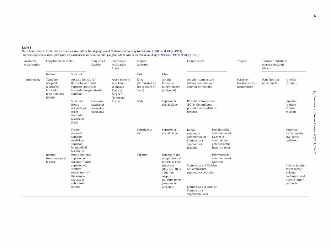
Table 1Main hemispheric white matter bundles around the basal ganglia and thalamus, according to Dejerine (1901) and Riley (1953).Principaux faisceaux hémisphériques de substance blanche autour des ganglions de la base et du thalamus, d’après Dejerine (1901) et Riley (1953).
Anatomicorganization
Longitudinal fascicles Long arcedfascicle
Short arcedassociativefibres
Corpuscallosum
Commissures Trigone Thalamic radiations(cortico-thalamicfibers)
Inferior Superior Part Fibre
Terminology Temporo-occipitalfascicle orfasciculuslongitudinalisinferior
Arcuate fascicle (ofBurdach), or frontalsuperior fascicle, orfasciculus longitudinalissuperior
Arced fibres ofArnold orU-shapedfibers ofMeynert(intergyralfibers)
Knee(terminated bythe rostrum orbeak)
Anteriorforceps orminor forceps(of Arnold)
Anterior commissure(AC) or Commissuraanterior or rostralis
Fornix ortractus cortico-mammillaris
Four fasciclesor peduncles
Anterior(frontal)
Superiorfronto-occipital, oroccip-itofrontalfascicle ofForel
Uncinatefascicle orfasciculusuncinatus
Body Superior ordorsal plane
Posterior commissure(PC) or Commissuraposterior or caudalis ordorsalis
Superior(parieto-fronto-rolandic)
Fronto-occipitalsuperiormedial, orsuperiorlongitudinalfascicle, orfronto-occipitalsuperior, oroccipito-frontalsuperior, or,stratumreticulatum ofthe coronaradiata, orsubcallosalbundle
Splenium orroll
Superior ordorsal plane
Dorsalsupraopticcommissure orCommissurasupraopticadorsalis
Pars dorsalis;commissure ofGanser orcommissureanterior of thehypothalamus
Posterior(occipitopari-etal; opticradiation)
Inferiorfronto-occipitalfascicle
Tapetum Belongs to theoccipitofrontalfascicle of Forel(Dejerine(Dejerine 1895,1901)) orcorpuscallosum fibers(commonlyaccepted)
Pars ventralis;commissure ofMeynert
Commissure of Guddenor Commissurasupraoptica ventralis
Inferior (occipi-totemporal):pulvinarcontingent andinferior-internpeduncle
Commissure of Forel orCommissurasupramamillaris

J.-J. Lemaire et al. / Neurochirurgie 57 (2011) 52–67 55
Fig. 1. The cerebellorubral group of subthalamic fibres (orange; subject no. 4): a: brachium conjonctivum (BC) and commissure of Wernekink (CW); b: uncrossed (u), bridgingcrossed (b) and decussating crossed (d) fibres of CW (the white arrow shows the origin of fibre tracking); c: section through the 3D block of mesencephalo-pontine fibres(pontine fibres, blue and purple; the reticulo-dorsal group of fibres, pink and beige); d: CW (white arrow) in an axial colour-coded map.Groupe cérébello-rubral des fibres sous-thalamiques (orange; sujet no. 4) : a : brachium conjonctivum (BC) et commissure de Wernekink (CW) ; b : fibres, non croisées (u), croisées ete tractob sur un
scw(bCr
2
teapapw(mPu(ritmac
n pont (b) et croisées et décussant (d) de CW (la flèche blanche indique l’origine de laleu et violet; groupe de fibres réticulo-dorsales, rose et beige) ; d : CW (flèche blanche)
hape, several ROIs (from 2 to 6) were used to generate each fascicleomposed of several fibre bundles. This anatomo-clinical approachas preferred to manually shaping bundles with logical operators
AND/NOT) after rough placement of ROIs. Each completed fibreundle was exported into graphical software (Avizo 5.0.0, Mercuryomputer Systems, MA, USA) for anatomical analysis on a volume-endered brain coupled with an orthogonal viewer.
.4. Anatomical analysis
The anatomic labelling of fascicles was realized in two steps:opographic analysis and comparison with classic anatomic knowl-dge (Supplementary Material 1: glossary). The topographicnalysis consisted in the exploration of both the extremities andaths of fascicles, locally in each region, e.g. subthalamus, and glob-lly (although limited by the stack of 24 axial slices going from theons to the caudate nucleus). For anatomical guidance, structuresere manually outlined on T2-weighted sequences or FA maps
Supplementary Material 2, which describes the anatomical land-arks); atlases (Morel et al., 1997; Olszewski and Baxter, 1954;
eel, 1954; Riley, 1953; Schaltenbrand and Bailey, 1959) were man-ally co-registered to the volume-rendered brain when necessarySupplementary Material 3: glossary, which describes the atlas co-egistrations). Image data sets (DTI, FA maps, and T2-weigthedmages) were manually co-registered (linear affine registration;
ranslation, rotation and scaling) minimizing the geometrical mis-atch caused by DTI distortion and carefully controlled by visualnalysis of several anatomic landmarks (nuclei, ventricles, corpusallosum and the envelope of the mesencephalon).
graphie) ; c : section dans le bloc 3D des fibres mésencéphalo-pontines (fibres pontines,e carte de direction codée par couleur dans un plan axial.
The final labelling was performed by comparing bundles offibres displayed on 3D rendering with 2D anatomic diagrams, draw-ings and classic anatomic knowledge (Supplementary Material 1:glossary). To facilitate the anatomic analysis, fibres were displayedas colored ribbons using the same colour for fibres that belongto the same fascicle (e.g. Figs. 1–3). For each subject, the finalanatomic labelling was accepted after complete 3D analysis of allthe fascicles within the two studied regions, i.e. the subthalamusand the internal capsule (anterior and posterior limbs; sublentic-ular and retrolenticular segments). Because of the intricatenessof deep brain fascicles we also explored fascicles related to thediencephalic-mesencephalic junction, whether they are anatomicneighbouring fibres or fibres embedded in the junction.
Practically, the individual subject analysis approach followeda learning curve to understand the 3D anatomy of WM fascicles(topography and organization) in one subject, then we identifiedand checked the WM bundles in the five others subjects. The clas-sical, anatomy or knowledge (Supplementary Material 1: glossary)used as a reference in this study is summarized in Tables 1–3. Theworkflow of the entire study is shown in Supplementary Material(Supplementary Material 4, which shows an overview of the work-flow). The broad qualitative analysis of the inter-subject variabilityof deep brain fascicles is provided.
3. Results
The subthalamic fibres were split into three topographicgroups – the cerebellorubral, reticulo-dorsal, and tegmento-peripheral. This new organization was proposed after meticulous

56J.-J.Lem
aireet
al./Neurochirurgie
57(2011)
52–67Table 2Main white matter bundles of the internal capsule, according to Dejerine (1901) and Riley (1953).Principaux faisceaux de substance blanche de la capsule interne, d’après Dejerine (1901) et Riley (1953).
Anatomicorganization
Anterior arm;lenticulo-caudatesegment
Knee or genu Posterior arm; lenticulo-optic segment Retrolenticularsegment
Sublenticularsegment
Superior tracts Peduncular tracts Lenticulateradiate fibres
Other
Terminology Fronto-thalamic projections Corticodiencephalic Corticomesencephalic Lenticulo-thalamicradiations(dorsallocation)
Fascicle ofTürck (orTürck-Meynert) ortractus tem-poropontinusor fascicletemporo-occipito-protuberantial(of Flechsig)
Opticradiations (ofGratiolet) orradiatio optica;visual corticalfascicle(including theMeyer’s loop)
Fascicle ofTürck
Lenticulo-caudatefibers
Geniculatefascicle
Corticopontine(including thefronto-pontinefascicle)
Strio-luysienradiations
Fronto-pontine fascicle (tractus) of Arnold
Corticothalamic Pyramidal tract Ansalenticularis
Fascicle ofArnold ortemporo-thalamicfascicle ofArnold
Lenticularfascicle (ofForel)
Table 3Other brain fascicles, according to Dejerine (1901) and Riley (1953).Autres faisceaux, d’après Dejerine (1901) et Riley (1953).
Anatomicorganization
Lenticular radiate fibres Forel’s fields Forebrainfascicles
Peduncular fascicle Others
Strio-thalamicradiations
Strio-subthalamicradiations
Tegmental area Longitudinal fascicles ofthe diencephalo-mesencephalicregion
Superiorcerebellarfascicles
Terminology Contains theAnsapeduncularis
Lenticularfascicle (H2)
Strio-luysienfascicle
H1 fascicle(dorsal) orthalamicfascicle
Fronto-mesencephalicbundle
Fascicle of Türck
Corticopontine tract ortractus corticopontinus
Medial longitudinalfascicle or fasciculuslongitudinalis medialis
Ansalenticularis orAnsa of thelentiformisnucleus
H2 fascicle(ventral) orlenticularfascicle
Basal (ormedial)forebrainbundle ormedialtelencephalicfascicle orfasciculustelencephalicusmedialis
Pyramidal tract or tractuspyramidalis
Central tegmental tractor tractus tegmentaliscentralis
Brachiumconjunctivumwith itsdecussation(tegmentum,dorsal andventral parts)or Wernekink(“horseshoe”)decussation
Forel’s H field(pre-rubral)
Aberrant fibers(medial); Themore medialfibers appearpredominantlycrossed.
E.g. : (1)Stratumintermedium;(2) Obliquefascicle, fascicle“en écharpe” ofFéré (fasciclecircumligatusor obliquus)
Lemniscus Medial orLemniscussensibilis orReil median
Lateral orLemniscusacusticus

J.-J. Lemaire et al. / Neurochir
Fig. 2. The reticulo-dorsal group of subthalamic fibres: a: (subject no. 4) left: basalforebrain bundle (BFB), central tegmental tract (CTT), longitudinal medial fascicle(LMF) and the ventral tegmental decussation of Forel (FD); Top right, FD, sectionthrough the 3D block of mesencephalo-pontine fibres; bottom right, axial colour-coded map showing FD (white arrow); b: (subject no. 1) front views of BFB (gold):left, BFB (anteriorly to the mamillary bodies; black dotted arrows; registration:Schaltenbrand and Bailey’s atlas (Schaltenbrand and Bailey, 1959), semi-schematicdrawing from Ingram (Peel, 1954)) and the superior, SD, and inferior, ID, divisionsof ansa lenticularis, are depicted (white arrows); right, BFB with the pre- (PO) andretro- optic (RO, black arrows) contingents, the representation according to Ingram(Peel, 1954) at the optic area is also depicted (*).Groupe réticulo-dorsal des fibres sous-thalamiques : a : (sujet no. 4) à gauche : faisceaubasal du cerveau antérieur (BFB), tractus tegmental central (CTT), faisceau longitudi-nal médial (LMF) et décussation tegmentale ventrale de Forel (FD) ; en haut à droite,FD, section dans le bloc 3D des fibres mésencéphalo-pontines ; en bas à droite, cartede direction codée par couleur dans un plan axial montrant FD (flèche blanche) ; b :(sujet no. 1) vues de face du BFB (doré) : à gauche, BFB (en avant des corps mamillaires ;flèches pointillées noires ; correspondance avec l’atlas de Schaltenbrand et Bailey (1959),dessin semi-schématique d’après Ingram (Peel, 1954)), et les divisions supérieure, SD,atd
awcgant
nd inférieure, ID, de l’anse lenticulaire (flèches blanches) ; à droite, BFB avec les con-ingents pré- (PO) and rétro- optiques (RO, flèches noires), ainsi que la représentation’après Ingram (Peel, 1954) dans la région optique (*).
nalysis of the overall WM architecture showing the threeell-organized groups, systematically identifiable. The internal
apsule contained projection tracts (Supplementary Material 1:
lossary) to deep nuclei and the brainstem (the lower brainstemnd spinal cord fibres could not be followed), and the largest,on-commissural, connective bundle of the basal deep brain,he ansa lenticularis (Supplementary Material 1: glossary). Theurgie 57 (2011) 52–67 57
fascicles related to those of the diencephalic-mesencephalicjunction, whether they are anatomic neighbouring fibres or fibresembedded in the junction are also described.
3.1. Subthlamus and internal capsule
3.1.1. The cerebellorubral groupBrachium conjonctivum fibres went mainly to the capsule of the
red nucleus and the ventrolateral thalamus via the commissure ofWernekink (“horse shoe commissure”). Most of medial brachiumconjonctivum fibres bridged the two superior cerebellar pedun-cles (bridging crossed fibres) rather than decussating from one sideto another (decussating crossed fibres), whereas the lateral fibresoften did not cross the midline (uncrossed fibres) (Fig. 1). The fibresof red nucleus core were connected mainly with the ventrolateralthalamus through the pre-lemniscal radiations (SupplementaryMaterial 1: glossary).
3.1.2. The reticulo-dorsal groupThe ventral fibres of the reticulo-dorsal group were condensed
along the midline (few crossing fibres) forming the ventral tegmen-tal decussation of Forel (Fig. 2); superiorly they reached the inferiordivision of the ansa lenticularis and frontal projections. The centraltegmental tract, longitudinal medial fascicle and basal forebrainbundle (BFB; Supplementary Material 1: glossary) were identifiedmore dorsally following the curvature of the region (Fig. 2).
3.1.3. The tegmento-peripheral groupAt the periphery of tegmentum (Supplementary Material
1:glossary), we defined two systems (partially overlapped) theanteromedial and posterolateral (Fig. 3). The anteromedial con-sisted of:
• the inferior division of the ansa lenticularis;• frontal fibres, the most anterior and medial fibres forming the
oblique fascicle;• some of the lemniscal fibres;• the so-called aberrant pyramidal fibres (Supplementary Material
1: glossary).
The posterolateral system consisted of: fibres of the posteriorlimb of the internal capsule, some passing through the subthalamicnucleus (STN) and the substantia nigra (SN); and most of lemnis-cal fibres. The two systems, along with thalamus-related fibres(see below), were included in the Forel’s fields (SupplementaryMaterial 1: glossary) and in the substance Q region (Schaltenbrandand Bailey, 1959).
3.1.4. The anterior limb of the internal capsuleFronto-thalamic fibres projected to the anterior, medial and dor-
sal thalamic nuclei. We noticed that fronto-thalamic fibres formeda fronto-pulvinar fascicle (not described classically in humans),located superiorly, travelling through the stratum zonale (Fig. 4).Fronto-tegmental and ansa lenticularis fibres were intermingledin the medial and superior region of the cerebral peduncles. Theinferior fibres of the anterior limb joined the genu of the internalcapsule.
3.1.5. The posterior limb of the internal capsuleThis limb contained mainly extrapyramidal projection fibres
to the thalamus, subthalamus, tegmentum and pons. The pontinefibres followed a classic organization (Fig. 5). The superior tha-
lamic radiations tracked from the ventral thalamus (Fig. 6) wereorganized according to the subcompartmentalization of thalamus(Morel et al., 1997). The ventral anterior region projected fibresto the frontal lobe through the superior thalamic peduncle and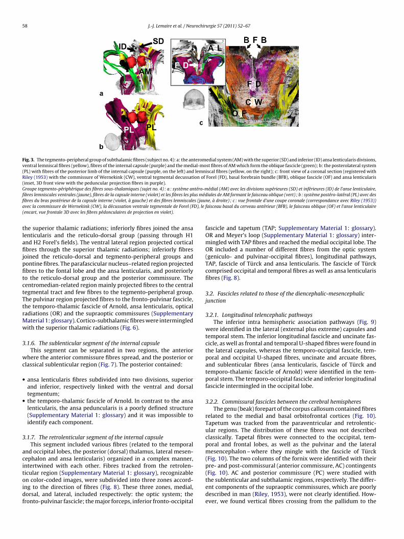
58 J.-J. Lemaire et al. / Neurochirurgie 57 (2011) 52–67
Fig. 3. The tegmento-peripheral group of subthalamic fibres (subject no. 4): a: the anteromedial system (AM) with the superior (SD) and inferior (ID) ansa lenticularis divisions,ventral lemniscal fibres (yellow), fibres of the internal capsule (purple) and the medial-most fibres of AM which form the oblique fascicle (green); b: the posterolateral system(PL) with fibres of the posterior limb of the internal capsule (purple, on the left) and lemniscal fibres (yellow, on the right); c: front view of a coronal section (registered withRiley (1953) with the commissure of Wernekink (CW), ventral tegmental decussation of Forel (FD), basal forebrain bundle (BFB), oblique fascicle (OF) and ansa lenticularis(inset, 3D front view with the peduncular projection fibres in purple).Groupe tegmento-périphérique des fibres sous-thalamiques (sujet no. 4) : a : système antéro-médial (AM) avec les divisions supérieures (SD) et inférieures (ID) de l’anse lenticulaire,fibres lemniscales ventrales (jaune), fibres de la capsule interne (violet) et les fibres les plus médiales de AM formant le faisceau oblique (vert) ; b : système postéro-latéral (PL) avec desfi s (jaua D), le(
tlafijpfitctTtrMw
3
wc
•
•
3
acitoidf
bres du bras postérieur de la capsule interne (violet, à gauche) et des fibres lemniscalevec la commissure de Wernekink (CW), la décussation ventrale tegmentale de Forel (Fencart, vue frontale 3D avec les fibres pédonculaires de projection en violet).
he superior thalamic radiations; inferiorly fibres joined the ansaenticularis and the reticulo-dorsal group (passing through H1nd H2 Forel’s fields). The ventral lateral region projected corticalbres through the superior thalamic radiations; inferiorly fibres
oined the reticulo-dorsal and tegmento-peripheral groups andontine fibres. The parafascicular nucleus–related region projectedbres to the fontal lobe and the ansa lenticularis, and posteriorlyo the reticulo-dorsal group and the posterior commissure. Theentromedian-related region mainly projected fibres to the centralegmental tract and few fibres to the tegmento-peripheral group.he pulvinar region projected fibres to the fronto-pulvinar fascicle,he temporo-thalamic fascicle of Arnold, ansa lenticularis, opticaladiations (OR) and the supraoptic commissures (Supplementaryaterial 1: glossary). Cortico-subthalamic fibres were intermingledith the superior thalamic radiations (Fig. 6).
.1.6. The sublenticular segment of the internal capsuleThis segment can be separated in two regions, the anterior
here the anterior commissure fibres spread, and the posterior orlassical sublenticular region (Fig. 7). The posterior contained:
ansa lenticularis fibres subdivided into two divisions, superiorand inferior, respectively linked with the ventral and dorsaltegmentum;the temporo-thalamic fascicle of Arnold. In contrast to the ansalenticularis, the ansa peduncularis is a poorly defined structure(Supplementary Material 1: glossary) and it was impossible toidentify each component.
.1.7. The retrolenticular segment of the internal capsuleThis segment included various fibres (related to the temporal
nd occipital lobes, the posterior (dorsal) thalamus, lateral mesen-ephalon and ansa lenticularis) organized in a complex manner,ntertwined with each other. Fibres tracked from the retrolen-icular region (Supplementary Material 1: glossary), recognizable
n color-coded images, were subdivided into three zones accord-ng to the direction of fibres (Fig. 8). These three zones, medial,orsal, and lateral, included respectively: the optic system; theronto-pulvinar fascicle; the major forceps, inferior fronto-occipitalne, à droite) ; c : vue frontale d’une coupe coronale (correspondance avec Riley (1953))faisceau basal du cerveau antérieur (BFB), le faisceau oblique (OF) et l’anse lenticulaire
fascicle and tapetum (TAP; Supplementary Material 1: glossary).OR and Meyer’s loop (Supplementary Material 1: glossary) inter-mingled with TAP fibres and reached the medial occipital lobe. TheOR included a number of different fibres from the optic system(geniculo- and pulvinar-occipital fibres), longitudinal pathways,TAP, fascicle of Türck and ansa lenticularis. The fascicle of Türckcomprised occipital and temporal fibres as well as ansa lenticularisfibres (Fig. 8).
3.2. Fascicles related to those of the diencephalic-mesencephalicjunction
3.2.1. Longitudinal telencephalic pathwaysThe inferior intra hemispheric association pathways (Fig. 9)
were identified in the lateral (external plus extreme) capsules andtemporal stem. The inferior longitudinal fascicle and uncinate fas-cicle, as well as frontal and temporal U-shaped fibres were found inthe lateral capsules, whereas the temporo-occipital fascicle, tem-poral and occipital U-shaped fibres, uncinate and arcuate fibres,and sublenticular fibres (ansa lenticularis, fascicle of Türck andtemporo-thalamic fascicle of Arnold) were identified in the tem-poral stem. The temporo-occipital fascicle and inferior longitudinalfascicle intermingled in the occipital lobe.
3.2.2. Commissural fascicles between the cerebral hemispheresThe genu (beak) forepart of the corpus callosum contained fibres
related to the medial and basal orbitofrontal cortices (Fig. 10).Tapetum was tracked from the paraventricular and retrolentic-ular regions. The distribution of these fibres was not describedclassically. Tapetal fibres were connected to the occipital, tem-poral and frontal lobes, as well as the pulvinar and the lateralmesencephalon – where they mingle with the fascicle of Türck(Fig. 10). The two columns of the fornix were identified with theirpre- and post-commissural (anterior commissure, AC) contingents(Fig. 10). AC and posterior commissure (PC) were studied with
the sublenticular and subthalamic regions, respectively. The differ-ent components of the supraoptic commissures, which are poorlydescribed in man (Riley, 1953), were not clearly identified. How-ever, we found vertical fibres crossing from the pallidum to the
J.-J. Lemaire et al. / Neurochirurgie 57 (2011) 52–67 59
Fig. 4. Anterior limb of the internal capsule: a: anterior limb (white) and genu (gold) fibres (subject no. 1), extrapyramidal fibres reaching the posterior limb are also visible(arrows) (the whole posterior limb is displayed in purple on one side); b: fronto-pulvinar fascicle (purple) (subject no.1); c,d: fibres located in the antero-medial region ofthe cerebral peduncle (subject no. 2), horizontal sections, where ansa lenticularis (yellow) and frontal (white and gold) fascicles intermingle (section C is below section D).Bras antérieur de la capsule interne : a : fibres (sujet no. 1) du bras antérieur (blanc) et du genou (doré), des fibres extrapyramidales atteignant le bras postérieur sont aussi visibles(flèches) (l’ensemble du bras postérieur est représenté en violet sur un côté) ; b : faisceau fronto-pulvinar (violet) (sujet no.1) ; c,d : fibres localisées dans la région antéro-médiale dupédoncule cérébral (sujet no. 2), coupes horizontales, où l’anse lenticulaire (jaune) et des faisceaux frontaux (blanc et doré) se mélangent (la coupe C est au dessous de la coupe D).
Fig. 5. Posterior limb of the internal capsule, brain stem fibres: a: pontine (PF) and aberrant pyramidal fibres (AP) (subject no. 1); b: the PF group comprise (subject no. 5): (i)pyramidal (cortico- pontine and spinal) contingent through the pontine nuclei area (purple, inset depicts nuclear-related fibres only), (ii) crossing fibres (red), (iii) anteriorpeduncular fibres (orange), and (iv) posterolateral fibres of the middle cerebellar peduncle (green; brachium pontis).Bras postérieur de la capsule interne, fibres du tronc cérébral : a : fibres (sujet no. 1) pontines (PF) et pyramidales aberrantes (AP) ; b : le groupe PF comprend (sujet no. 5) : (i) uncontingent pyramidal (cortico-pontin et spinal) traversant la région des noyaux du pont (violet ; dans l’encart seules des fibres nucléaires sont représentées), (ii) des fibres croisées(rouge), (iii) des fibres pédonculaires antérieures (orange), et (iv) des fibres postéro-latérales du pédoncule cérébelleux moyen (vert ; brachium pontis).

60 J.-J. Lemaire et al. / Neurochirurgie 57 (2011) 52–67
Fig. 6. Posterior limb of the internal capsule (registration with the slice 3 mm above the horizontal AC-PC plan (Morel et al., 1997)); a: thalamic fibres of the superior thalamicradiations (subject no. 1): ventral anterior (purple), ventral median (gold), ventral lateral (orange), center median (yellow), para fascicularis (green) and pulvinar (blue)contingents; b: thalamic fibres of the superior thalamic radiations (STR) (subject no. 1): projection fibres (white) are cut horizontally at the upper brainstem, the fascicleof Arnold (FA) and the optic radiations (OR) are visible; c: cortico-subthalamic fibres (subject no. 4): cerebellorubral group (orange); mammillothalamic bundle (red); ansalenticularis -related fibres (white); lemniscal-related fibres (yellow) and reticulo-dorsal group (beige and pink); d: cortico-subthalamic fibres (subject no. 4): relationshipswith the superior thalamic radiations (STR) within the posterior limb (coronal section along red nucleus-related fibres).Bras postérieur de la capsule interne (correspondance avec la coupe 3 mm au dessus du plan horizontal AC-PC (Morel et al., 1997)) : a : fibres thalamiques des radiations thalamiquessupérieures (sujet no. 1) : contingents, ventral antérieur (violet), ventral médian (doré), ventral latéral (orange), central médian (jaune), para fasciculaire (vert) and pulvinarien (bleue);b : fibres thalamiques des radiations thalamiques supérieures (STR) (sujet no. 1) : des fibres de projection (blanc) sont sectionnées horizontalement au niveau de la partie haute dutronc cérébral, le faisceau d’Arnold (FA) et les radiations optiques (OR) sont visibles ; c : fibres cortico-sous-thalamiques (sujet no. 4) : groupe cérébello-rubral (orange) ; faisceaum édian( ostérie
o(
3
tn
•
•
•
3
cfirt
amillo-thalamique (rouge) ; fibres de l’anse lenticulaire (blanc) ; fibres du lemnisque msujet no. 4) : relations avec les radiations thalamiques supérieures (STR) dans le bras p
ptic tract that might be related to this commissural networkFig. 10).
.2.3. Radiate fibresThe radiate fibres were explored in each region (tracked from
he caudate nucleus, putamen, pallidum, thalamus, subthalamicucleus and substantia nigra). We only found:
inferior (basal) pallidal fibres joining the ansa lenticularis and ACthat were often intermingled with the supraoptic commissurefibres;short caudate fibres that joined the anterior limb of the internalcapsule;short intra putaminal fibres.
.2.4. Nucleus-related subthalamic fibresNucleus-related fibres (Fig. 11) form an inhomogeneous group
haracterized by gray matter tracking (for red nucleus-relatedbres: see manuscript). The tegmental pedunculopontine nucleus-elated fibres reached the intralaminar thalamus and mingled withhe central tegmental tract, whereas others reached rubral bun-
(jaune) and groupe réticulo-dorsal (beige et rose) ; d : fibres cortico-sous-thalamiquesur (coupe coronale le long des fibres du noyau rouge).
dles. The subthalamic nucleus-related fibres reached the superiorthalamic radiations, the ansa lenticularis and the reticulo-dorsalgroup. The substantia nigra-related fibres reached the frontal supe-rior thalamic radiations and the pontine fibres. Finally, fibres linkedwith substance Q were intermingled with the tegmento-peripheralgroup.
3.2.5. Inter-subject variabilityThe same observers performed the analysis on the six sub-
jects; globally, the same connectivity patterns were identifiable.However, we noticed several differences (subjective analysis; notquantified) in term of fascicle thickness. The relative thickness offascicles belonging to the same region, were different from onesubject to another:
• within the retrolenticular region, between the TAP and fascicleof Türck, the posterior fibres of AC and ansa lenticularis and the
inferior longitudinal pathways;• within the medial, anterior and superior region of brain pedun-cles, between the ansa lenticularis, the anterior (and genu) andthe posterior limbs of the internal capsule;

J.-J. Lemaire et al. / Neurochirurgie 57 (2011) 52–67 61
Fig. 7. Sublenticular portion of the internal capsule (subject no. 5): a: superior; b: inferior, views: ansa lenticularis (yellow) with its superior (SD) and inferior (ID) divisions,along with anterior commissure (blue), temporo-thalamic fascicle of Arnold (FA), fascicle of Türck (FT), supraoptic commissure-related fibres (SO), optic radiations (OR) andtapetum (TAP) and fronto-tegmental projections (f).Portion sous lenticulaire de la capsule interne (sujet no. 5). Vues a : supérieure; et b : inférieure : anse lenticulaire (jaune) avec ses divisions supérieure (SD) et inférieures (ID),commissure blanche antérieure (bleu), faisceau temporo-thalamique d’Arnold (FA), faisceau de Türck (FT), fibres des commissures supra-optiques (SO), radiations optiques (OR) ettapetum (TAP) et fibres de projection fronto-tegmentales (f).
Fig. 8. Retrolenticular segment of the internal capsule: a: location of the retrolenticular region (LGB, lateral geniculate body), yellow, orange and purple ROIs defined onan axial colour-coded map (inset overview) (subject no. 2); b: fronto-pulvinar fascicle (FP), major forceps of corpus callosum (MAF), optic system (OS), inferior longitudinalfascicle (IFO) and tapetum (TAP), for information the trigone is displayed (red); c: optical radiations (OR) and Meyer’s loop (ML) (subject no. 1); d: fascicle of Türck (FT) andfascicle of Arnold (FA).Portion rétro-lenticulaire de la capsule interne ; a : localisation (sujet no. 2) de la région rétro-lenticulaire (LGB, corps géniculé latéral) avec 3 ROIs jaune, orange et violette, définies surune carte de direction codée par couleur dans un plan axial (encart, vue d’ensemble) ; b : faisceau fronto-pulvinarien (FP), forceps major du corpus callosum (MAF), système optique(OS), faisceau longitudinal inférieur (IFO) et tapetum (TAP), le trigone est représenté pour information (rouge) ; c : radiations optiques (OR) et boucle de Meyer (ML) (sujet no 1) ; d :faisceau de Türck (FT) et faisceau d’Arnold (FA).

62 J.-J. Lemaire et al. / Neurochirurgie 57 (2011) 52–67
Fig. 9. Telencephalic longitudinal pathways (TLPs) and related fascicles: Türck’s (FT), Arnold’s (FA), and insular (INS): a: TLPs: (1) tracks of the lateral capsules (Subject no.1) are shown in white, the U-shaped fibres, the uncinate fascicle (UF) and the inferior fronto-occipital fascicle (IFO); fascicles of the temporal stem are shown in gold, thetemporo-occipital fascicle (TOF), U-shaped fibres and the arcuate fascicle (AF); b: the relationship between the IFO, INS and UF is depicted (left, subject no. 2; right, subjectno. 3); c: Layers of fibres facing the ventricular trigone (Subject no. 1): [1] superficial temporal fibres; [2] intermediate fibres, mainly IFO plus TOF fibres, and rare tapetalfibres (blue); and [3] deep tapetal fibres.Voies longitudinales télencéphaliques (TLPs) et faisceaux en rapport : faisceaux de Türck (FT), d’Arnold (FA) et insulaire (INS) : a : TLPs : (1) fibres des capsules latérales (sujet no. 1)e faisceaa c : Cout lques fi
•
•
•
(w
n blanc, fibres en U, faisceau unciné (UF) et faisceau fronto-occipital inférieur (IFO) ;rqué (AF) ; b : Relations entre IFO, INS and UF (gauche, sujet no. 2 ; droite, sujet no. 3) ;emporales [2], fibres intermédiaires, principalement des fibres du IFO et du TOF, et que
within the lateral subthalamus, between the lemniscal and aber-rant pyramidal fibres;within the cerebellorubral route, between crossed and uncrossed(commissure of Wernekink) fibres;within the pulvinar, between fronto-pulvinar fibres, the OR andtemporo-thalamic fascicle of Arnold.
The small commissure of Forel was identified in two of six casesFig. 10). The radiate fibres (Supplementary Material 1: glossary)ere rarely identifiable except below the pallidum.
ux du lobe temporal en doré, faisceau temporo-occipital (TOF), fibres en U et faisceauches de fibres en regard du carrefour ventriculaire (sujet no. 1) : [1] fibres superficiellesbres du tapetum (bleu) ; et [3] des fibres profondes du tapetum.
4. Discussion
We analysed in explicit details the 3D WM architecture ofthe deep brain using high resolution DTI and deterministic fibretracking. Interplay between anatomy textbooks and our data,visualized on 2D slices and on 3D reconstructions in the volume-
rendered brain, allowed us to iteratively refine the fibre tracking ofeach individual fascicle. This test-re-test approach (back and forthbetween fibre tracking and anatomic analysis), while subjectiveand qualitative, was nonetheless very informative. We visualized
J.-J. Lemaire et al. / Neurochirurgie 57 (2011) 52–67 63
Fig. 10. Commissural (and related) fascicles between the cerebral hemispheres (from A to F, subjects no. 1, 3, 2, 1, 2 and 6): a: the minor forceps (white; orbitofrontal fibresthrough the beak, arrows), AC (blue), PC (pale blue) and trigonal fibres (pre- (P) and retro- (R) commissural contingents, red; b: commissure of Forel (FC) with the superior(SD) and inferior (ID) divisions of the ansa lenticularis (pallidum, PAL; peduncle, PED); c: major forceps (MAF) and tapetum (TAP) (white arrows) facing the ventricular trigone(VT) that reaches the posterior thalamus (LGB, lateral geniculate body; optic tract, gold) and the MAF; d: shown are the supposed (see text) supraoptic commissure-relatedfibres (SO); e: fascicle of Türck (FT), tapetum (TAP) plus temporal (T) and occipital (O) fibres, pontine fibres (white) and fibres of the posterior limb (blue). f. T (green), O(white) and TAP (purple) fibres.Faisceaux commissuraux (et ceux en rapport) entre les hémisphères cérébraux (de A à F, sujets no. 1, 3, 2, 1, 2 et 6) : a : forceps mineur (blanc ; fibres orbito-frontales passant par lebec, flèches), AC (bleu), PC (bleu pâle) et fibres du trigones (contingents pré- (P) and rétro- (R) commissuraux, rouge ; b : commissure de Forel (FC) avec les divisions supérieure (SD)e jeur (Mr AF ; dT c) et fi
ansmotio
t inférieures (ID) de l’anse lenticulaire (pallidum, PAL ; pédoncule, PED) ; c : forceps maejoignent le thalamus postérieur (LGB, corps géniculé latéral ; voie optique, doré) et Mürck (FT), tapetum (TAP) et fibres temporales (T) et occipitales (O), fibres du pont (blan
nd detailed most of the deep WM bundles described by pio-eering neuroanatomists including large and complex fasciclesuch as the basal forebrain bundle and the ansa lenticularis. Thisodern in vivo analysis leads to important insight into the 3D
rganization of this complex WM architecture. Our findings lento propose a topographic classification of subthalamic fasciclesnto three groups (Supplementary Material 5, which shows theverview of the whole organisation of deep brain WM fascicles): the
AF) et tapetum (TAP) (flèches blanches) en regard du carrefour ventriculaire (VT) qui: fibres en lien avec les commissures supra-optiques (voir texte) (SO) ; e : faisceau debres du bras postérieur (bleu) ; f : fibres T (vert), O (blanc) et du TAP (violet).
cerebellorubral, the reticulo-dorsal and the tegmento-peripheralone.
4.1. Diffusion tensor imaging and fibre tracking analysis
With an optimised sequence, limited coverage, and a relativelylong yet clinically acceptable scanning time, we reached a voxel vol-ume of 1.25 × 1.25 × 1.5 mm3. This is less than half the volume used

64 J.-J. Lemaire et al. / Neurochirurgie 57 (2011) 52–67
Fig. 11. Nucleus-related fibres (subject no. 1): a: pedunculopontine nucleus ROI superimposed on Olszewski and Baxter (1954)’s atlas (bold dark line); inset left and rightROIs (white circles) on colour-coded image; b: pedunculopontine nucleus-related fibres (pink); c,d: red nucleus-(red), subthalamus- (yellow), SN- (white) and substance Q(green)-related fibres.Fibres issues des noyaux (sujet no. 1) : a : ROI du noyau pédonculo-pontin tegmental localisée sur l’atlas d’Olszewski et Baxter (1954) (trait noir épais) et en encart ROIs droite etg r coulc tance
iNsft
wiabttrtiMrc
icjaeppd2
auche (cercles blancs, tegmentum droit et gauche) sur une carte de direction codée pa,d : fibres issues du noyau rouge (rouge), du noyau sous-thalamique (jaune), de la subs
n other DTI studies of the same region (Habas and Cabanis, 2007;agae-Poetscher et al., 2004), but we maintained an equivalent or
uperior signal-to-noise ratio. This enabled us to study fasciclesrom 2 to 7 mm diameter (Supplementary Material 6, which giveshe rough diameter of small fascicles).
DTI images are acquired with an echo-planar imaging sequence,hich is prone to susceptibility artifacts. The use of parallel imag-
ng (sensitivity encoding) (Jaermann et al., 2004) minimized thesertifacts. However, at the current state of technology they cannote totally avoided. In our study, this phenomenon disturbed fibreracking on a case-by-case basis in two ways: artifacts of the ven-ral pons and distortion of colour-coded maps in the fronto-callosalegion. The former precluded tracking in the ventral pons; whereashe latter was taken into account when comparing T2 versus DTImages (anatomic landmarks were useful, like those contoured).
ovement artifacts also decrease image quality, especially at highesolution; however they should have been limited by the vacuumushion used in this study.
The direct identification of the entire length of deep fascicless generally not possible on colour-coded maps because of theomplex structural anatomy at the diencephalo-mesencephalicunction. The understanding of 3D organization of WM fasciclesfforded by our study shows that at a macroscopic level it is how-ver possible to identify certain fascicles when they cross the axial
lane (Fig. 12). This is more complex when fascicles interweave, aroblem which is well known at the infra millimeter level whenifferent fibres (i.e. axons) cross in the same voxel (Oouchi et al.,007; Tuch et al., 2003). However the difficulty to identify veryeur dans un plan axial ; b : fibres issues du noyau pédonculo-pontin tegmental (rose) ;noire (blanc) et de la substance Q (vert).
small fibres, kissing or crossing, was minimized in our macroscopicstudy by using small voxels while seeking only supra millimetrediameter connectivity (Supplementary Material 6, which gives therough diameter of small fascicles). Nevertheless, the problematicof the differentiation of kissing and crossing fibre is still a limita-tion of anatomical studies based on deterministic DTI fibre tracking,including our study. Partial biologic and anatomic knowledge ofhuman brain fascicles added with still on-going research on math-ematical models used for the display of fascicles preclude any firmand definitive conclusion about the detailed biologic organizationhidden behind DTI fibre tracking technique. This challenge fostersthe research of new methodologies (Leow et al., 2009).
Finally, even though averaging and spatial normalization of theDTI datasets into a standard atlas (Jones et al., 2002) could havebeen a useful statistical tool to confirm the repeatability of ourresults; it would have also reduced the benefits of the high res-olution of individual datasets and made the tracking of small WMfascicles described in this study likely impossible. This approachwould certainly be interesting in the future in parallel with clini-cally useful individual subject analysis studies.
4.2. Analysis of white matter fascicles
Our results using in vivo technique give new topographic
insights into this complex region.Within the subthalamus, our findings highlight the ambigu-ity concerning the crossing of the cerebellorubral paths (Dejerine,1901; Laget, 1973; Nieuwenhuys et al., 1979; Olszewski and Baxter,

J.-J. Lemaire et al. / Neurochirurgie 57 (2011) 52–67 65
Fig. 12. Analysis of axial colour-coded map: a: locations of bundles are shown from superior (left) to inferior (right) plans; b: locations of ROIs for the grey matter analysis: theprojection of 3D structures is on the left, along with the red nucleus (RN) and substantia nigra (SN), the projection of the pedunculopontine nucleus (tegmental, PPN) is on theright, a colour-coded map reconstructed along a section of Olszewski and Baxter (1954)’s atlas is presented in the inset. Abbreviations: AC, anterior commissure; Acq, aqueductof the brain; AF, Arcuate fascicle; AL*, Ansa lenticularis located outside the sublenticular region; BC, Brachium conjunctivum; MAF, MIF, forceps major (MA) and minor (MI) ofthe corpus callosum; Fo, Fornix; Fh, Frontal horn of the lateral ventricle; IcAl, Internal capsule (IC), anterior limb; Icg, IC, genu; IcPl, IC, posterior limb; IcRl, IC, retro-lenticularregion; IcSl, IC, sub-lenticular region; IFO, Inferior fronto-occipital fascicle; INS, Insular fascicles; Lem, Lemniscus; Lfof + UF, Longitudinal fronto-occipital fascicle plus uncinatefascicle; LteT, Longitudinal telencephalic tracts; BFB, Basal forebrain bundle; Mtb, Mamillothalamic bundle; OR, Optic radiations (broad meaning; see text); PAL, Pallidum;PC, Posterior commissure; PED, Peduncular bundles; PPN, Pedunculopontine nucleus (Tegmental); RD, Reticulo dorsal group (see text); Rlr, Retrolenticular region; RN, Rednucleus; SQ, Substance Q; SN, Substantia nigra; ST, Striatum (caudate nucleus and putamen); TAP, Tapetum; TOF, Temporo-occipital fascicle; Tha, Thalamus; ThaVent, Ventralregion of the thalamus; VT, Trigone of the lateral ventricle; V3, Third ventricle.Analyse de carte de direction codée par couleur dans un plan axial : a : localisations des faisceaux de la partie supérieure (gauche) à la partie inférieure (droite) du cerveau ; b :localisations des ROIs pour l’analyse de la substance grise : à gauche, projection des structures 3D, passant par le noyau rouge (RN) et la substance noire (SN) ; à droite, projection dunoyau pédunculo-pontin tegmental (PPN) avec en encart une carte de direction codée par couleur reconstruite dans un plan passant par la coupe de l’atlas d’Olszewski et Baxter (1954).Abréviations : AC, commissure blanche antérieure ; Acq, aqueduc du cerveau ; AF, faisceau arqué ; AL*, anse lenticulaire localisée hors de la région sous lenticulaire ; BC, Brachiumconjunctivum ; MAF, MIF, forceps majeur (MA) et mineur (MI) du corps calleux ; Fo, fornix ; Fh, corne ventriculaire frontale ; IcAl, bras antérieur de la capsule interne (IC) ; Icg, genou(IC) ; IcPl, bras postérieur (IC) ; IcRl, région rétro-lenticulaire (IC) ; IcSl, région sous-lenticulaire (IC) ; IFO, faisceau fronto-occipital ; INS, faisceaux insulaires ; Lem, lemnique ; Lfof + UF,faisceau longitudinal fronto-occipital et faisceau unciné ; LteT, faisceaux longitudinaux télencephaliques ; BFB, faisceau basal du cerveau antérieur ; Mtb, faisceau mamillo-thalamique ;O che pog , substT VT, ca
1bta(ta
sndfi
R, radiations optiques (au sens large ; voir texte) ; PAL, pallidum ; PC, commissure blanroupe réticulo-dorsal (voir texte) ; Rlr, région rétro-lenticulaire ; RN, noyau rouge ; SQOF, faisceau temporo-occipital ; Tha, thalamus ; ThaVent, région ventral du thalamus ;
954; Riley, 1953), notably complicated by the intermingling ofrachium conjonctivum fibres with tegmental fascicles (Fig. 1). Allhe published characteristics of human BFB fit with our results,llowing for the first time in vivo the description of the entire BFBFig. 2). The tegmento-peripheral group shows a double organiza-ion related to the reticular system, the cerebellorubral group, thensa lenticularis and the lemniscuses.
Concerning the projection fibres of the internal capsule, we
uggest the existence of a new fascicle, the fronto-pulvinar, con-ecting the frontal polar cortices with the pulvinar that was alreadyescribed in monkeys (Petrides and Pandya, 2007). The projectionbres of the posterior limb appear to primarily consist of extrapyra-stérieure ; PED,faisceaux pédonculaires ; PPN, noyau pédonculo-pontin tegmental ; RD,ance Q ; SN, substance noire ; ST, striatum (noyau caudé et putamen) ; TAP, Tapetum ;rrefour du ventricule latérale ; V3, troisième ventricule.
midal fibres (the occipital and temporal cortices project through theretrolenticular region). The limited volume of imaged brain (36 mmthickness) prevented any firm conclusion concerning the detailedcortical connections of fibres passing through the posterior limb.
We found that the surface of the thalamus, wrapped laterallyby thalamocortical fibres forming the superior thalamic radia-tions (passing through the reticular nucleus or Arnold’s net), isalso coated with other extrapyramidal projection fibres that pass
through the subthalamus (Fig. 6). We also found a large bundle thatfits the description by Dejerine (Dejerine, 1901) of a rich networkof myelinated fibres connected with the subthalamic region (theregion of “Ruban de Reil médian”).
6 rochir
cgooseta
twa
Non&cc
dcccttTa
pd(mbsaFt
buRwipu1ttong(1
toaepdrcob
6 J.-J. Lemaire et al. / Neu
Within the sub- and retro-lenticular segments of internalapsule, our findings show the complexity of OR made of theeniculo-calcarine tract, TAP, fronto-occipital fascicle, temporo-ccipital fascicle, uncinate fascicle, ansa lenticularis and fasciclef Türck (Fig. 8). This might explain the variability noticed withtatistical anatomic approaches (Bürgel et al., 1999), and the over-stimation of the Meyer’s loop (Yamamoto et al., 2005). Theopography and organization of the ansa lenticularis is well defined,nd for the first time totally revealed in vivo (Fig. 7).
The organization of the telencephalic longitudinal pathways andhe neighboring uncinate fascicles described here is in agreementith the classic organization, as well as DTI fiber tracking of the
rcuate fascicle and temporo-occipital fascicles (Catani et al., 2005).We collected new information about the AC, PC and fornix.
otably, we visualized the pre- and post-commissural contingentsf the fornix, the former being connected to the basal forebrain,amely the preoptic area (or prothalamus) on the Schaltenbrand
Bailey atlas (Schaltenbrand and Bailey, 1959). However, theomplexity of this region (Heimer, 2000) does not permit any con-lusions regarding a precise connection.
The TAP is a complex structure, inherited from topographicescriptive anatomy, which can be split into its differentomponents: the temporo-occipital – the commonly acceptedommissure between the temporal and occipital lobes; and a newonnectivity, inter-thalamic (between the two pulvinars), towardshe frontal lobes (through the inferior longitudinal fascicle) andhe upper brainstem (dorso-laterally, mingled with the fascicle ofürck). The TAP and the others fibres facing the ventricular trigonere organized in three layers identifiable on DTI slices (Fig. 9).
Our study of radiate fibres did not provide detailed results,erhaps due to: the spreading of fibres through large isotropic bun-les beyond the geometric resolution of our DTI tracking systeme.g., the stratum pedunculi); or the unknown connectivity of grey
atter that might be hidden by mainstream WM tracking of neigh-ouring, or crossing, fascicles. It seems that our inability to trackmall fascicles (e.g., mamillo-peduncular and retroflexus fascicles),s well as the variable visibility of small structures (commissure oforel), is linked to the limited geometric resolution of our DTI fibreracking.
Concerning nucleus-related fibres, an important connectionetween the pedunculopontine nucleus and subthalamic nucleussing a probabilistic approach to balance the low accuracy ofOI positioning has been described (Aravamuthan et al., 2007)hereas our results, using individual-based approach, show lim-
ted connectivity (Fig. 11). The high density of fibres around theedunculopontine nucleus embedded in the mesencephalic retic-lar formation (Nieuwenhuys et al., 1979; Olszewski and Baxter,954) could explain this discrepancy. This complexity, along withhe lack of information about grey matter fibres, brings into ques-ion fine connectivity revealed by statistical approaches. Resultsf the other grey matter structures we explored (subthalamicucleus, substantia nigra, and substance Q) illustrate the limits ofrey matter tracking; the red nucleus is an exception because itcore and capsule) classically consists primarily of fibres (Dejerine,901).
Finally, the design of the study, the evaluation of the poten-ial of DTI fiber tracking to explore and analyse the complexityf deep brain, did not allow quantifying the inter-subject vari-bility. This could be done more easily after detailed descriptivexplorations, like this work. Although subjective, we found mor-hologic differences in connectivity that may not predict ofifferences in functionality. Given the complexities of human neu-
oanatomy, such variability could be significant. The variationsoncerning small fascicles can be explained by technical limitationsf our study and/or by true variability in the organization of WMundles.urgie 57 (2011) 52–67
5. Conclusion
In conclusion, we charted in vivo most of the tracts describedin the deep brain, including large and complex fascicles such asthe basal forebrain bundle and the ansa lenticularis. In functionaland minimally invasive neurosurgery, the possibility to map bothgrey and white matter structures deep inside the brain will havemajor implications. Today, the deposition of electricity in deepbrain stimulation (Herzog et al., 2007), tomorrow the depositionof other forms of energy inside the brain, the targeted delivery ofdrugs, devices, or cells, will necessitate both a deep understandingof the underlying anatomy and the possibility to acquire detailedimages before, during and after the treatment. More generally,the progressive understanding of the brain connectivity is partic-ularly challenging for the description of the human connectome(Bullmore and Sporns, 2009).
Disclosure of interest
No author received financial support in conjunction with thegeneration of the submission; no author has any personal or insti-tutional financial interest in materials, or devices described intheir submissions; Laurent Hermoye and Wojciech Gradkowski are,respectively, the head and an employee of Imagilys; this study doesnot use or promote Imagilys’s products, there is no conflict of inter-est between Imagilys and this study.
Acknowledgments
This study was supported by the Institute for the Encourage-ment of Scientific Research and Innovation of Brussels (IRSIB),Government of Brussels-Capital Region, Belgium, the Fond Nationalde la Recherche Scientifique (FNRS), Belgium, and NIH/NIA grant#AG20012. We thank Andrew Frew (PhD, UCLA, USA) for his edi-torial advice.
We want to pay homage to all the pioneers in neuroanatomy fortheir extensive and meticulous work that has fostered research inthis field and yielded a tremendous amount of pertinent and robustdata.
Appendix A. Supplementary data
Supplementary data associated with this article can be found, inthe online version, at doi:10.1016/j.neuchi.2011.04.001.
References
Andrade, D.M., et al., 2006. Long-term follow-up of patients with thalamic deepbrain stimulation for epilepsy. Neurology 66 (10), 1571–1573.
Aravamuthan, B.R., et al., 2007. Topography of cortical and subcortical connectionsof the human pedunculopontine and subthalamic nuclei. NeuroImage 37 (3),694–705.
Basser, P.J., Mattiello, J., LeBihan, D., 1994. MR diffusion tensor spectroscopy andimaging. Biophysical Journal 66 (1), 259–267.
Benabid, A.L., et al., 1996. Chronic electrical stimulation of the ventralis intermediusnucleus of the thalamus as a treatment of movement disorders. Journal of Neu-rosurgery 84 (2), 203–214.
Bürgel, U., et al., 1999. Mapping of histologically identified long fibre tracts in humancerebral hemispheres to the MRI volume of a reference brain: position andspatial variability of the optic radiation. NeuroImage 10 (5), 489–499.
Bullmore, E., Sporns, O., 2009. Complex brain networks: graph theoretical analy-sis of structural and functional systems. Nature reviews. Neuroscience 10 (3),186–198.
Catani, M., Jones, D.K., ffytche, D.H., 2005. Perisylvian language networks of thehuman brain. Annals of Neurology 57 (1), 8–16.
Dejerine, J., 1901. Anatomie des centres nerveux (Tomes 1 and 2). Rueff et Cie, Paris.Filley, C., 2001. The behavioural neurology of white matter. Oxford University Press,
Oxford.Forel, A., 1877. Untersuchungen über die haubenregion und ihre oberen verknüp-
fungen im gehirne des menschen und einiger säugethiere, mit beiträgen

rochir
G
G
H
H
H
H
H
J
J
J
LL
L
J.-J. Lemaire et al. / Neu
zu den methoden der gehirnuntersuchung. Archiv für Psychiatrie und Ner-venkrankheiten 7, 393–495.
oetz, C.G., 2000. Battle of the titans: Charcot and Brown-Séquard on cerebral local-ization. Neurology 54 (9), 1840–1847.
reenberg, B.D., et al., 2006. Three-year outcomes in deep brain stimulation forhighly resistant obsessive-compulsive disorder. Neuropsychopharmacology:Official Publication of the American College of Neuropsychopharmacology 31(11), 2384–2393.
abas, C., Cabanis, E.A., 2007. Cortical projection to the human red nucleus: com-plementary results with probabilistic tractography at 3T. Neuroradiology 49 (9),777–784.
eimer, L., 2000. Basal forebrain in the context of schizophrenia brain research. BrainResearch Reviews 31 (2–3), 205–235.
ermoye, L., et al., 2006. Pediatric diffusion tensor imaging: normal database andobservation of the white matter maturation in early childhood. NeuroImage 29(2), 493–504.
erzog, J., et al., 2007. Stimulation of subthalamic fibre tracts reduces dyskinesiasin STN-DBS. Movement Disorders: Official Journal of the Movement DisorderSociety 22 (5), 679–684.
irano, A., Llena, J.F., 1995. Morphology of central nervous system axons. In:Waxman, S.G., Kocsis, J.D., Stys, P.K. (Eds.), The axon: structure, function andpathophysiology. New York Oxford, pp. 49–67.
aermann, T., et al., 2004. SENSE-DTI at 3 T. Magnetic Resonance in Medicine: OfficialJournal of the Society of Magnetic Resonance in Medicine/Society of MagneticResonance in Medicine 51 (2), 230–236.
iang, H., et al., 2006. DtiStudio: resource program for diffusion tensor computationand fiber bundle tracking. Computer Methods and Programs in Biomedicine 81(2), 106–116.
ones, D.K., et al., 2002. Spatial normalization and averaging of diffusion tensor MRIdata sets. NeuroImage 17 (2), 592–617.
aget, P., 1973. Elements de neuro-anatomie fonctionnelle. Masson, Paris.emaire, J., et al., 2007. Brain mapping in stereotactic surgery: a brief overview from
the probabilistic targeting to the patient-based anatomic mapping. NeuroImage37 (Suppl 1), S109–S115.
eow, A.D., et al., 2009. The tensor distribution function. Magnetic Resonancein Medicine: Official Journal of the Society of Magnetic Resonance inMedicine/Society of Magnetic Resonance in Medicine 61 (1), 205–214.
urgie 57 (2011) 52–67 67
Morel, A., Magnin, M., Jeanmonod, D., 1997. Multiarchitectonic and stereotacticatlas of the human thalamus. The Journal of Comparative Neurology 387 (4),588–630.
Mori, S., et al., 1999. Three-dimensional tracking of axonal projections in the brainby magnetic resonance imaging. Annals of Neurology 45 (2), 265–269.
Mori, S., et al., 2005. MRI atlas of human white matter. Elsevier B.V, Amsterdam.Mori, S., Zhang, J., 2006. Principles of diffusion tensor imaging and its applications
to basic neuroscience research. Neuron 51 (5), 527–539.Mori, S., van Zijl, P.C.M., 2002. Fibre tracking: principles and strategies – a technical
review. NMR in Biomedicine 15 (7–8), 468–480.Nagae-Poetscher, L.M., et al., 2004. High-resolution diffusion tensor imaging of the
brain stem at 3 T. American Journal of Neuroradiology 25 (8), 1325–1330.Nieuwenhuys, R., Voogd, J., Huijzen, C., 1979. The human central nervous system: a
synopsis and atlas. Springer-Verlag, Berlin.Olszewski, J., Baxter, D., 1954. Cytoarchitecture of the brain stem. Karger, Basel.Oouchi, H., et al., 2007. Diffusion anisotropy measurement of brain white matter
is affected by voxel size: underestimation occurs in areas with crossing fibres.American Journal of Neuroradiology 28 (6), 1102–1106.
Peel, T., 1954. The neuroanatomical basis for clinical neurology. McGraw-Hill BookCompany, New York.
Petrides, M., Pandya, D.N., 2007. Efferent association pathways from the rostral pre-frontal cortex in the macaque monkey. The Journal of Neuroscience: The OfficialJournal of the Society for Neuroscience 27 (43), 11573–11586.
Riley, H., 1953. An atlas of the basal ganglia brain stem and spinal cord. Williams &Wilkins, Baltimore.
Schaltenbrand, G., Bailey, P., 1959. Introduction to stereotaxis with an atlas of thehuman brain Georg. Thieme Verlag, Stuttgart.
Schiff, N.D., et al., 2007. Behavioural improvements with thalamic stimulation aftersevere traumatic brain injury. Nature 448 (7153), 600–603.
Talairach, J., et al., 1957. Atlas d’anatomie stéréotaxique. Repérage radiologiqueindirect des noyaux gris centraux des régions mésencéphalo-sous-optiques ethypothalamiques de l’homme. Masson et Cie, Paris.
Tuch, D.S., et al., 2003. Diffusion MRI of complex neural architecture. Neuron 40 (5),885–895.
Yamamoto, T., et al., 2005. Tractography to depict three layers of visual field tra-jectories to the calcarine gyri. American Journal of Ophthalmology 140 (5),781–785.