what follows ’simple’ protein identification? protein
TRANSCRIPT
1
What Follows ’Simple’ Protein Identification?
• Defining N- and C-termini• Classification of splice variants• Characterization of protein modifications
Protein Characterization
Protein Modifications• Identification of modified protein• Localization of modification• Structure elucidation of the modification
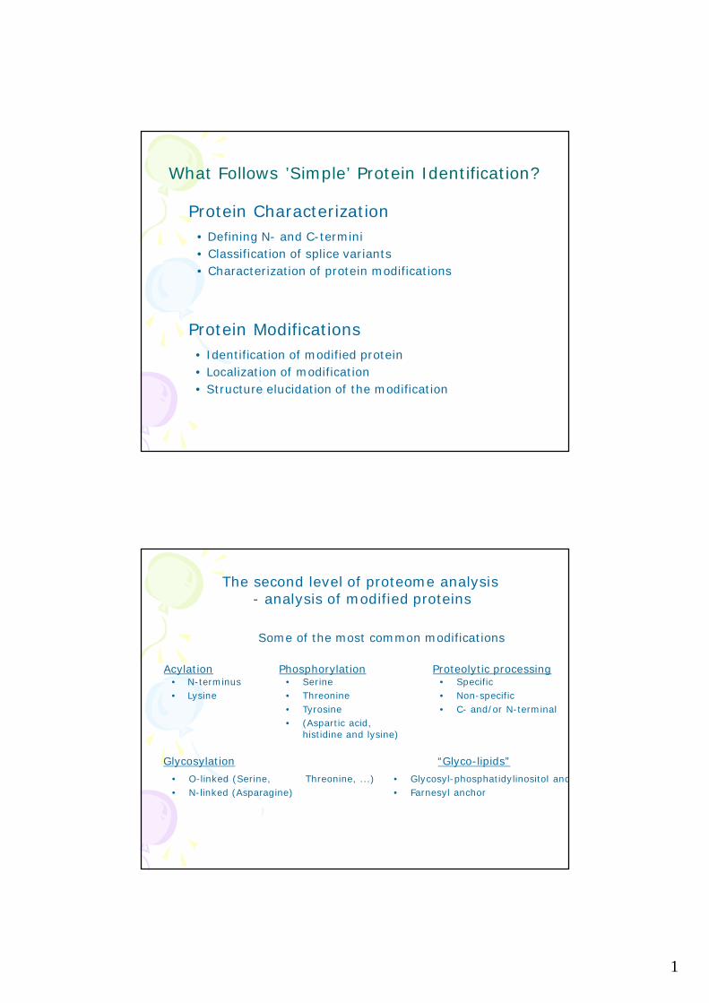
The second level of proteome analysis- analysis of modified proteins
Some of the most common modifications
• N-terminus• Lysine
Acylation• Specific• Non-specific• C- and/or N-terminal
Proteolytic processing
• O-linked (Serine, Threonine, ...)• N-linked (Asparagine)
Glycosylation
Phosphorylation• Serine• Threonine• Tyrosine• (Aspartic acid,
histidine and lysine)
“Glyco-lipids”• Glycosyl-phosphatidylinositol anchor• Farnesyl anchor

2
•Specific detection in gelsRadiolabelingFluorescent labelingWestern blottingModification specific stains
•Affinity fishingImmune precipitation of proteinsAffinity purification of proteins/peptides
-phosphopeptide isolation: IMAC, TiO2-phosphoprotein isolation: phosphospecific antibodies
Selective tagging followed by affinity purification
•Selective mass spectrometryPrecursor ion scanningNeutral loss scanningStable isotope labeling
Analysis of modified proteins
Mass analyser types
3
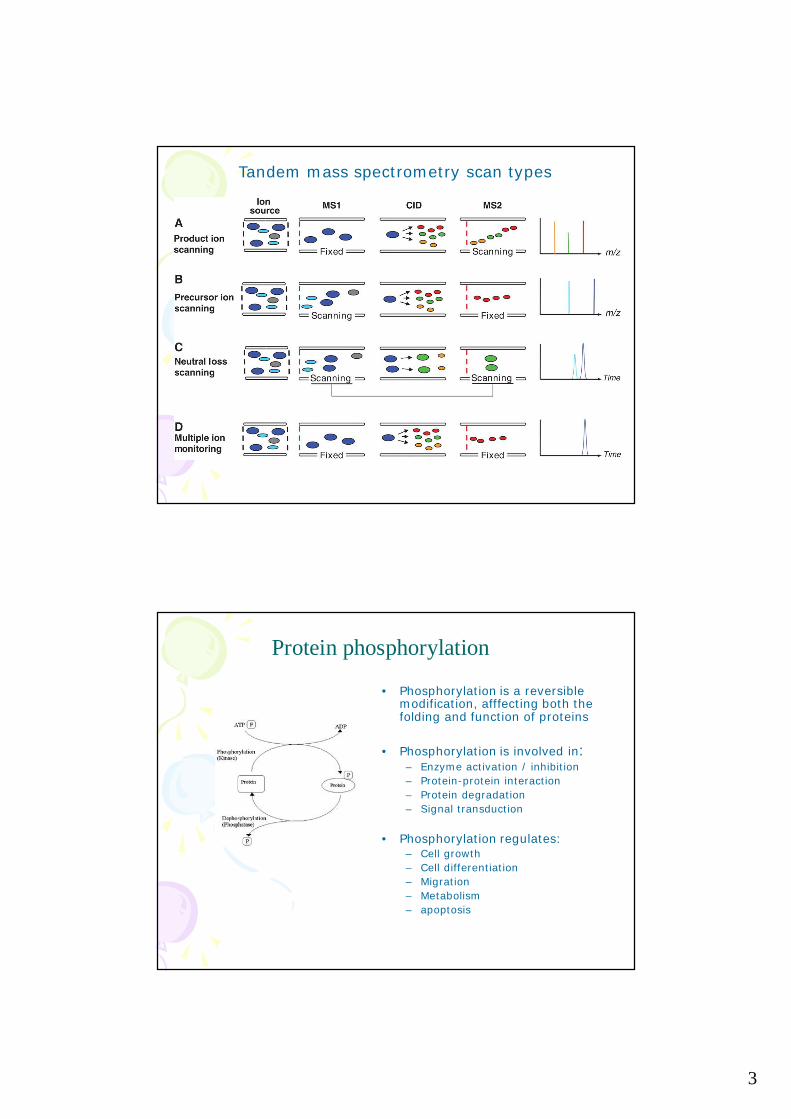
Tandem mass spectrometry scan types
Protein phosphorylation
• Phosphorylation is a reversiblemodification, afffecting both thefolding and function of proteins
• Phosphorylation is involved in:– Enzyme activation / inhibition– Protein-protein interaction– Protein degradation– Signal transduction
• Phosphorylation regulates:– Cell growth– Cell differentiation– Migration– Metabolism– apoptosis
4
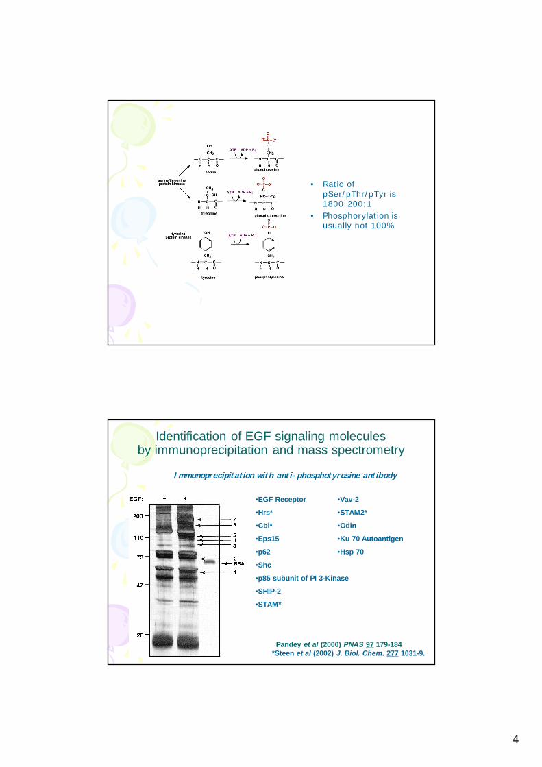
• Ratio ofpSer/pThr/pTyr is1800:200:1
• Phosphorylation isusually not 100%
Identification of EGF signaling moleculesby immunoprecipitation and mass spectrometry
Immunoprecipitation with anti-phosphotyrosine antibody
•EGF Receptor
•Hrs*
•Cbl*
•Eps15
•p62
•Shc
•p85 subunit of PI 3-Kinase
•SHIP-2
•STAM*
•Vav-2
•STAM2*
•Odin
•Ku 70 Autoantigen
•Hsp 70
Pandey et al (2000) PNAS 97 179-184*Steen et al (2002) J. Biol. Chem. 277 1031-9.
5
Phosphorylation site analysis by MALDI-TOF
• Digestion of phosphorylated protein by trypsin• MALDI-TOF mass analysis of peptides and
comparison– to database calculated masses– to nonphosphorylated peptide masses
• To find a peptide with a mass addition of 80(=HPO3)
• Phosphatase treatment of the sample on the MALDIplate should result in the disappearance phosphategroup
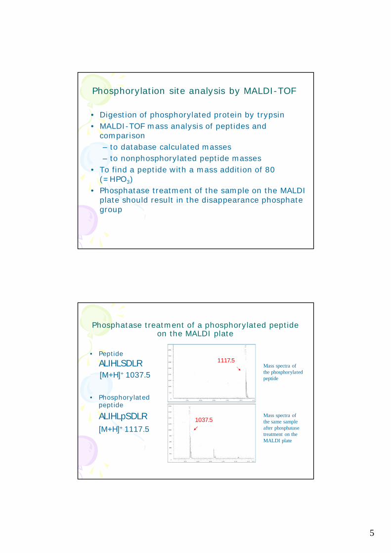
Phosphatase treatment of a phosphorylated peptideon the MALDI plate
• PeptideALIHLSDLR[M+H]+ 1037.5
• Phosphorylatedpeptide
ALIHLpSDLR[M+H]+ 1117.5
Mass spectra ofthe phosphorylatedpeptide
Mass spectra ofthe same sampleafter phosphatasetreatment on theMALDI plate
1117.5
1037.5

6
Problems in phosphorylation site analysis(by MALDI-TOF)
• Phosphorylated peptides ionize poorly in a positivemode
• Phosphorylated peptides are supressed in a totalprotein digest analyses– enrichment of phosphopeptides– prefractioning the digest into peptides (HPLC)
Selective enrichment of phosphopeptides
• IMAC (Immobilized metal-ion affinitychromatography)
• SAX + IMAC• SCX + IMAC• TiO2
• SCX + TiO2• HILIC (Hydrophilic interaction chromatography) +
IMAC• IMAC + TiO2 (SIMAC)� a-pTyr ab + IMAC• ZrO2, other metal oxides• Calcium-precipitation• Phosphoramidate chemistry
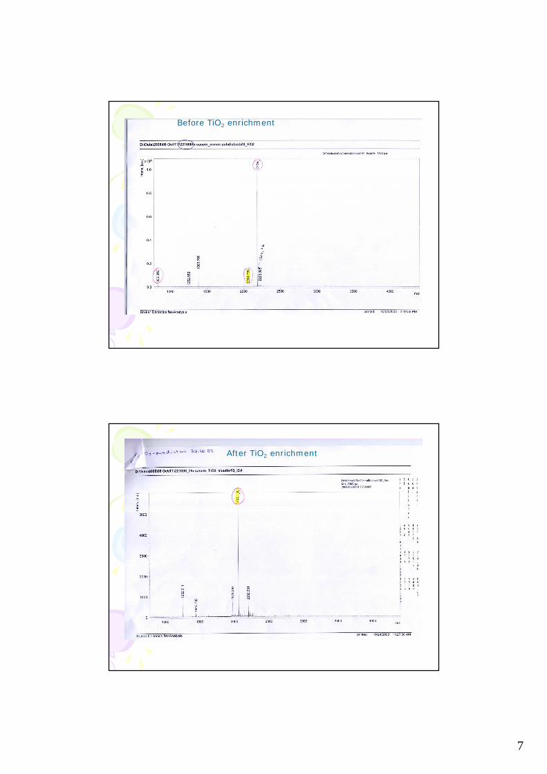
7
Before TiO2 enrichment
After TiO2 enrichment

8
D.
Phosphopeptides enriched using IMAC
followed by sequencingby ESI-MS/MS
A. Stensballe & O.N. Jensen
MALDI mass map of in-geldigested protein (CK2)
Verification of phosphorylation
Proteomics levels
Expression proteomicsWhich gene products are expressed, when and how much
PTM-omics, ”Modificomics”Which variants are present of each protein, when and how much
Cell map proteomics,”Interactomics”Who interacts, when and where
9
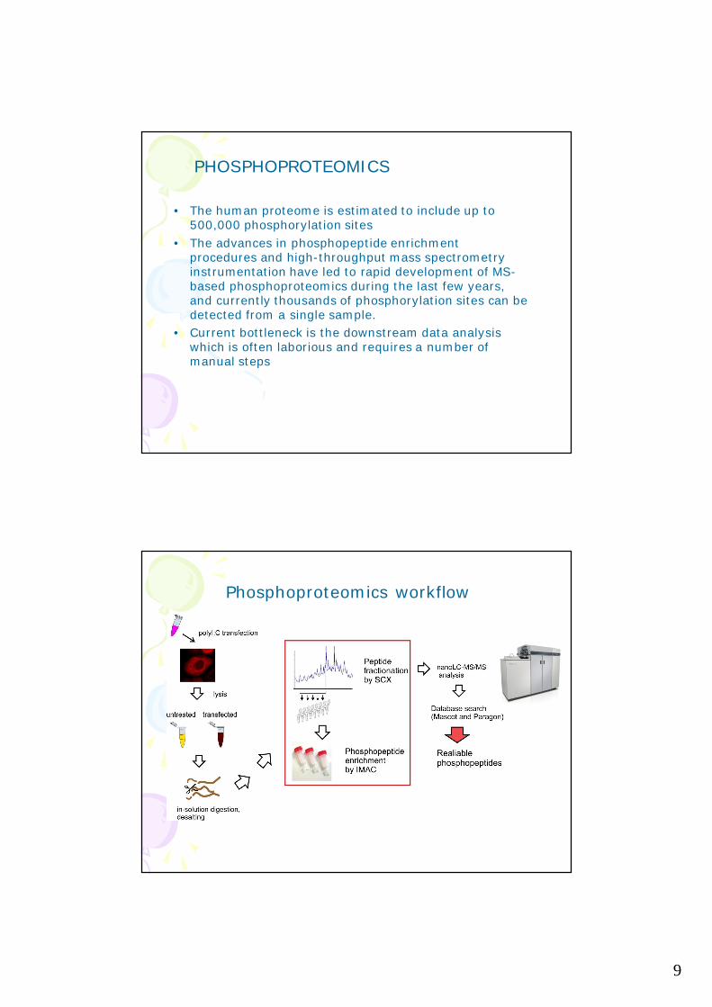
• The human proteome is estimated to include up to500,000 phosphorylation sites
• The advances in phosphopeptide enrichmentprocedures and high-throughput mass spectrometryinstrumentation have led to rapid development of MS-based phosphoproteomics during the last few years,and currently thousands of phosphorylation sites can bedetected from a single sample.
• Current bottleneck is the downstream data analysiswhich is often laborious and requires a number ofmanual steps
PHOSPHOPROTEOMICS
Phosphoproteomics workflow
10
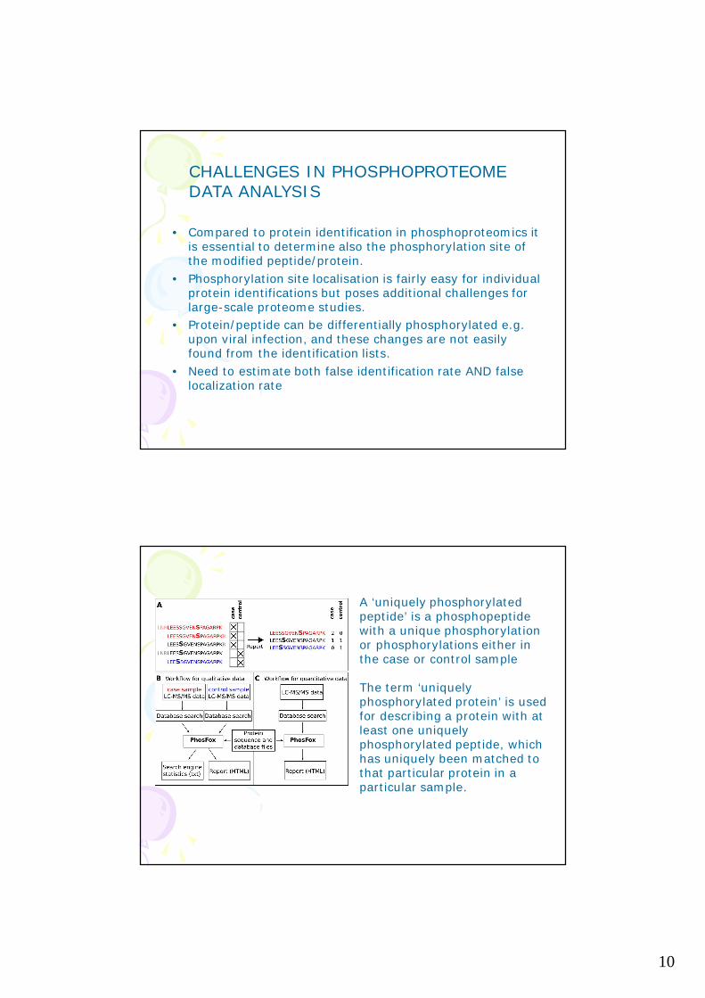
• Compared to protein identification in phosphoproteomics itis essential to determine also the phosphorylation site ofthe modified peptide/protein.
• Phosphorylation site localisation is fairly easy for individualprotein identifications but poses additional challenges forlarge-scale proteome studies.
• Protein/peptide can be differentially phosphorylated e.g.upon viral infection, and these changes are not easilyfound from the identification lists.
• Need to estimate both false identification rate AND falselocalization rate
CHALLENGES IN PHOSPHOPROTEOMEDATA ANALYSIS
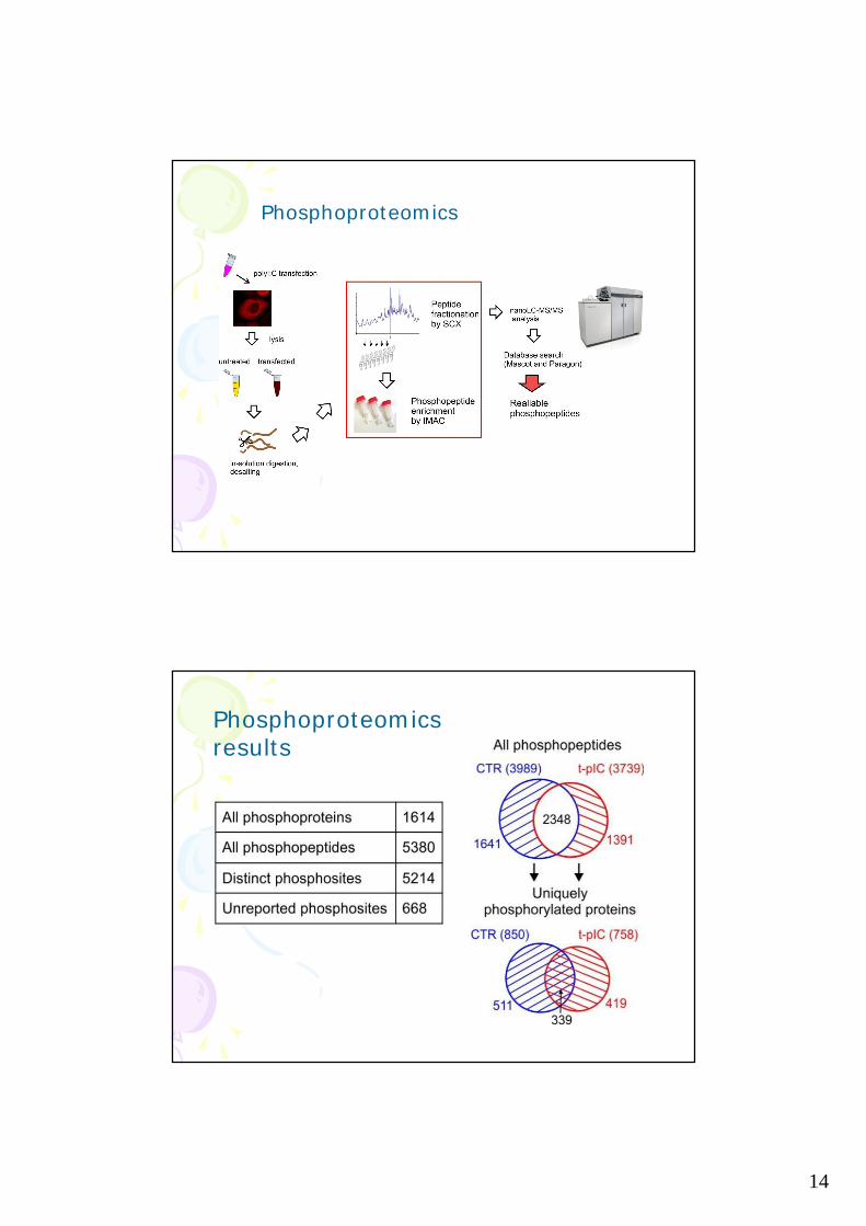
A ‘uniquely phosphorylatedpeptide’ is a phosphopeptidewith a unique phosphorylationor phosphorylations either inthe case or control sample
The term ‘uniquelyphosphorylated protein’ is usedfor describing a protein with atleast one uniquelyphosphorylated peptide, whichhas uniquely been matched tothat particular protein in aparticular sample.
11
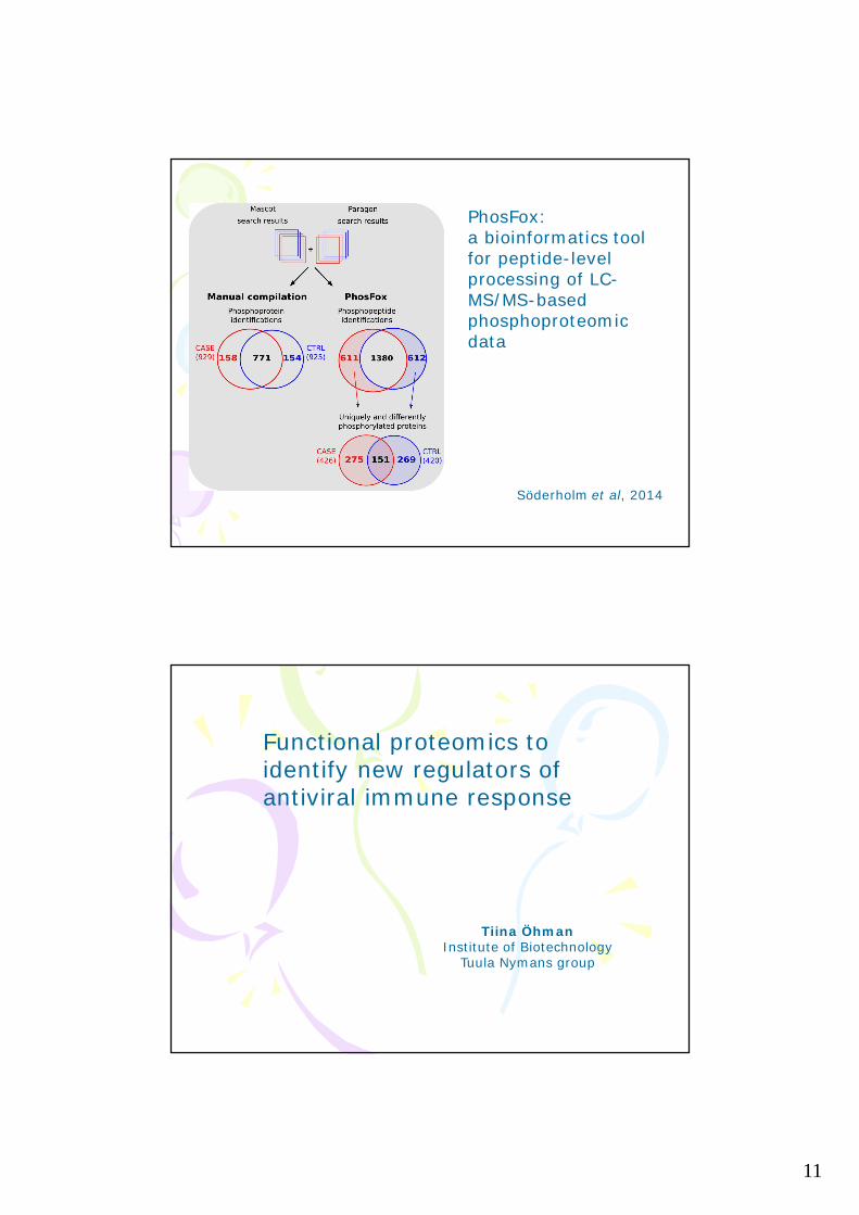
PhosFox:a bioinformatics toolfor peptide-levelprocessing of LC-MS/MS-basedphosphoproteomicdata
Söderholm et al, 2014
Functional proteomics toidentify new regulators ofantiviral immune response
Tiina ÖhmanInstitute of Biotechnology
Tuula Nymans group
12
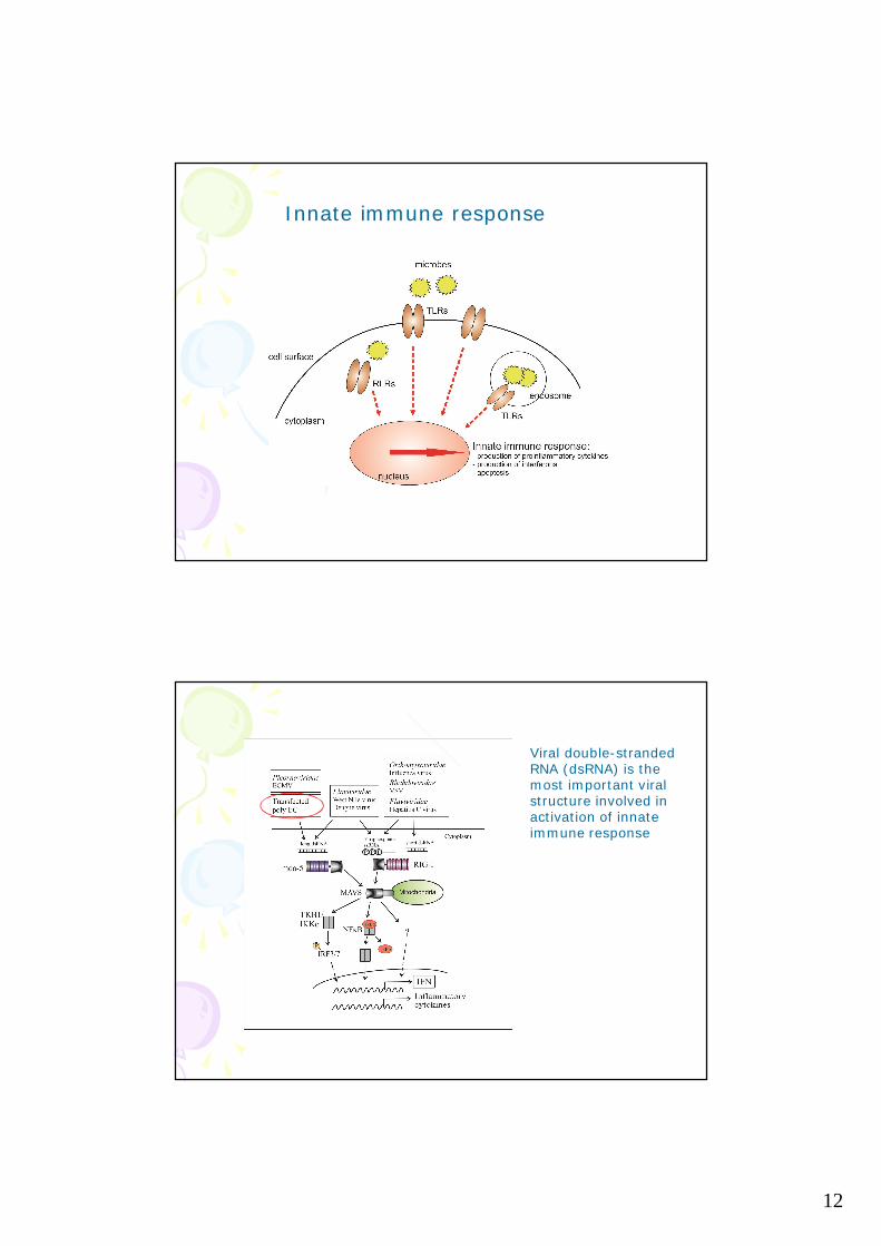
Innate immune response
Viral double-strandedRNA (dsRNA) is themost important viralstructure involved inactivation of innateimmune response

13
Cytosolic RNA recognition pathway activates14-3-3 protein mediated signaling and caspase-dependent disruption of cytoskeleton networkin human keratinocytes
Öhman et al, 2010
Aims of this study:-to characterize the signaling pathways activated in dsRNA-stimulated keratinocytes-to identify new players in antiviral innate immune responses
For this, we used14-3-3 affinity chromatographyand phosphoproteomicscombined with bioinformaticsand functional studies
14
Phosphoproteomics
Phosphoproteomicsresults
15
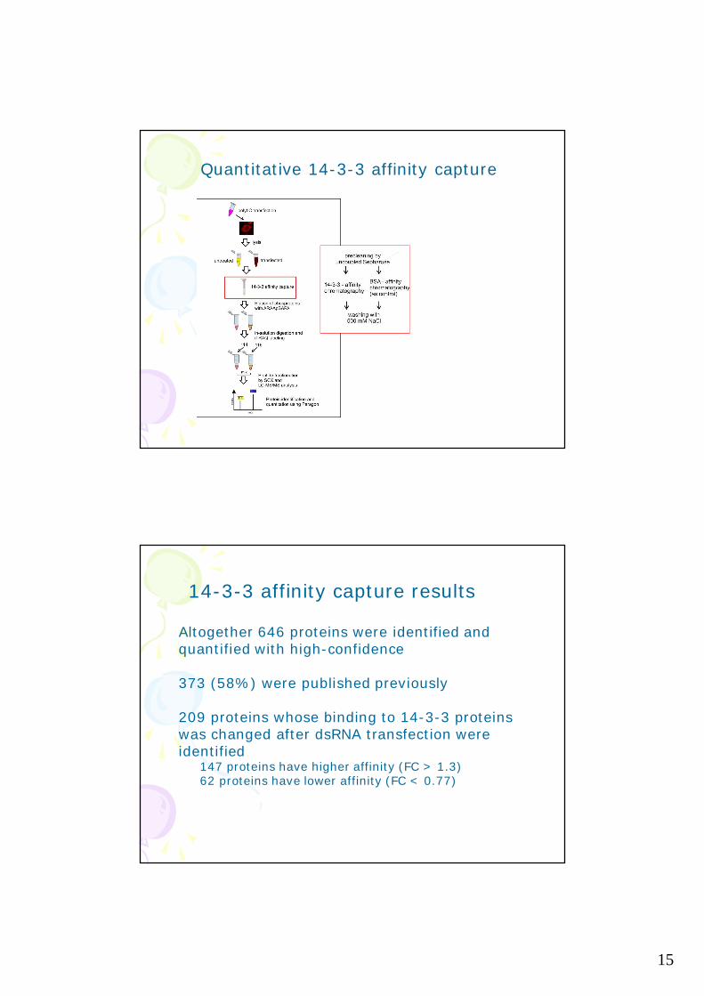
Quantitative 14-3-3 affinity capture
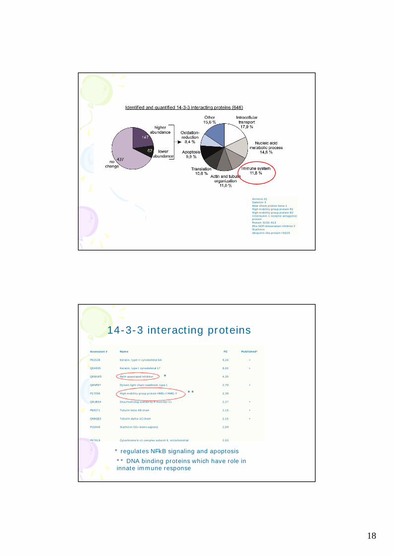
Altogether 646 proteins were identified andquantified with high-confidence
373 (58%) were published previously
209 proteins whose binding to 14-3-3 proteinswas changed after dsRNA transfection wereidentified
147 proteins have higher affinity (FC > 1.3)62 proteins have lower affinity (FC < 0.77)
14-3-3 affinity capture results
16
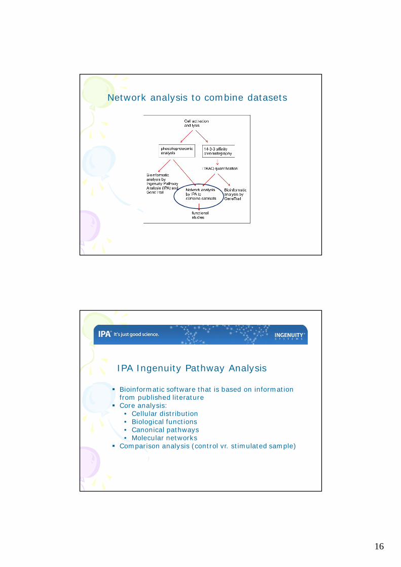
Network analysis to combine datasets
IPA Ingenuity Pathway Analysis
§ Bioinformatic software that is based on informationfrom published literature
§ Core analysis:• Cellular distribution• Biological functions• Canonical pathways• Molecular networks
§ Comparison analysis (control vr. stimulated sample)
17
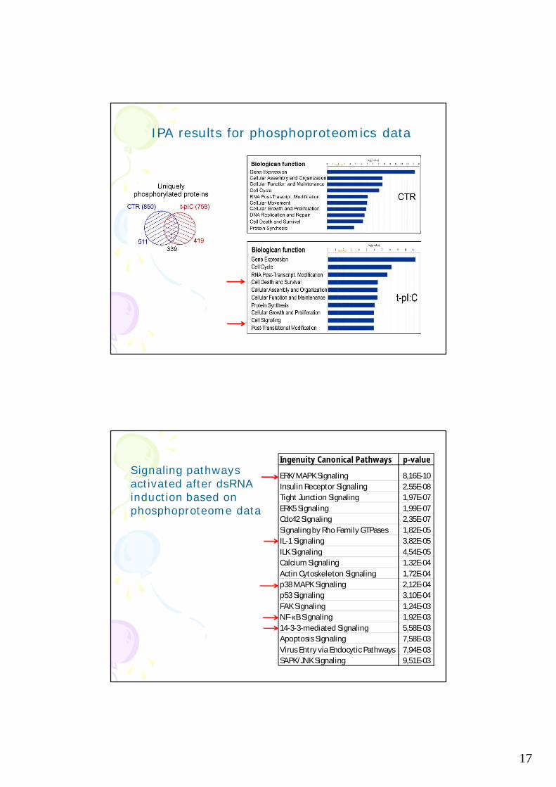
IPA results for phosphoproteomics data
Ingenuity Canonical Pathways p-value
ERK/MAPK Signaling 8,16E-10Insulin Receptor Signaling 2,55E-08Tight Junction Signaling 1,97E-07ERK5 Signaling 1,99E-07Cdc42 Signaling 2,35E-07Signaling by Rho Family GTPases 1,82E-05IL-1 Signaling 3,82E-05ILK Signaling 4,54E-05Calcium Signaling 1,32E-04Actin Cytoskeleton Signaling 1,72E-04p38 MAPK Signaling 2,12E-04p53 Signaling 3,10E-04FAK Signaling 1,24E-03NF-κB Signaling 1,92E-0314-3-3-mediated Signaling 5,58E-03Apoptosis Signaling 7,58E-03Virus Entry via Endocytic Pathways 7,94E-03SAPK/JNK Signaling 9,51E-03
Signaling pathwaysactivated after dsRNAinduction based onphosphoproteome data
18
Annexin A1Galectin-3Heat shock protein beta-1High mobility group protein B1High mobility group protein B2Interleukin-1 receptor antagonistproteinProtein S100-A13Rho GDP-dissociation inhibitor 2StathminUbiquitin-like protein ISG15
Accession # Name FC Published*
P02538 Keratin, type II cytoskeletal 6A 9,24 +
Q04695 Keratin, type I cytoskeletal 17 8,62 +
Q8WUF5 RelA-associated inhibitor 4,35
Q9NP97 Dynein light chain roadblock-type 1 2,70 +
P17096 High mobility group protein HMG-I/HMG-Y 2,39
Q9UBS4 DnaJ homolog subfamily B member 11 2,27 +
P68371 Tubulin beta-4B chain 2,15 +
Q9BQE3 Tubulin alpha-1C chain 2,15 +
P16949 Stathmin OS=Homo sapiens 2,09
P07919 Cytochrome b-c1 complex subunit 6, mitochondrial 2,03
* regulates NFkB signaling and apoptosis** DNA binding proteins which have role ininnate immune response
14-3-3 interacting proteins
*
**
19
RelA-associated inhibitor, RAI
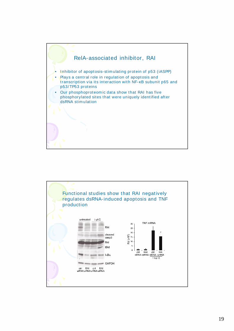
• Inhibitor of apoptosis-stimulating protein of p53 (iASPP)• Plays a central role in regulation of apoptosis and
transcription via its interaction with NF-κB subunit p65 andp53/TP53 proteins
• Our phosphoproteomic data show that RAI has fivephosphorylated sites that were uniquely identified afterdsRNA stimulation
Functional studies show that RAI negativelyregulates dsRNA-induced apoptosis and TNFproduction
20
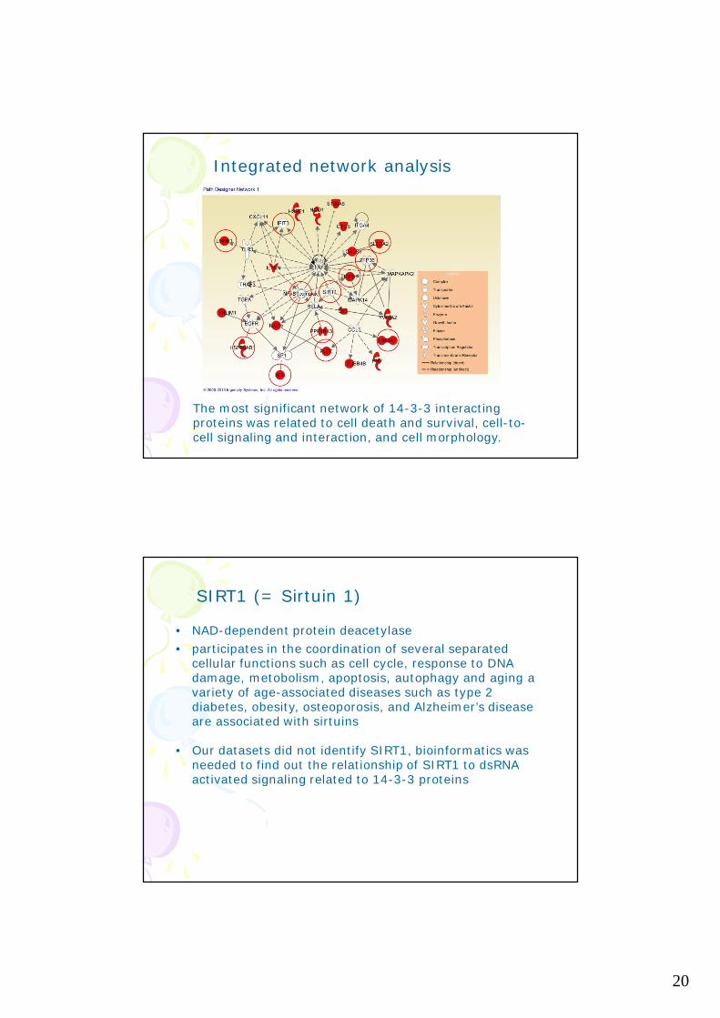
The most significant network of 14-3-3 interactingproteins was related to cell death and survival, cell-to-cell signaling and interaction, and cell morphology.
Integrated network analysis
SIRT1 (= Sirtuin 1)
• NAD-dependent protein deacetylase• participates in the coordination of several separated
cellular functions such as cell cycle, response to DNAdamage, metobolism, apoptosis, autophagy and aging avariety of age-associated diseases such as type 2diabetes, obesity, osteoporosis, and Alzheimer’s diseaseare associated with sirtuins
• Our datasets did not identify SIRT1, bioinformatics wasneeded to find out the relationship of SIRT1 to dsRNAactivated signaling related to 14-3-3 proteins

21
Sirtuin 1 regulates NFkB-signaling and cytokineproduction upon dsRNA stimulation
SIRT1 negatively regulates dsRNA-inducedapoptosis
Similarly, Sirtuin 1 negatively regulatesinnate immune responses in EMCV(Encephalomyocarditis virus) infected cells!

22
CONLUSIONS:RAI and Sirtuin 1 were identified as novelregulators of antiviral innate immune responsesFunctional studies showed that RAI inhibits dsRNA-induced apoptosis and contributes to dsRNA-induced TNF cytokine responseSirtuin 1 is a central molecule regulated by 14-3-3proteins and functional studies show that itnegatively regulates virus-induced cytokineproduction and protects cells from apoptosis inviral-infected keratinocytes