what are the normal flora
DESCRIPTION
What Are the Normal FloraTRANSCRIPT

What are the normal flora?
It may or may not surprise you to find out that your body is host to billions of bacteria of many different kinds. These bacteria exist in many different parts of the body, and usually do not cause any problems for the host body. The following is a list of the main sites for bacteria that consitute the normal flora.
The skin, especially the moist areas, such as the groin and between the toes. The respiratory tract, particularly the nose.
Urinary tract.
The digestive tract, i.e. the mouth, the terminal ileum and the colon.
There are many different types of relationship that the body can have with the normal flora. These are
Mutualism. In a mutualist relationship, both the host and the microbe benefit from the relationship. The best example if this is E.Coli. This organism lives in the intestines, where it receives nourishment, and in turn produces Vitamin K, which the human body requires for the process of blood clotting.
Commensalism. A commensalist relationship is where one partner of the relationship benefits, and the other partner is neither benefitted nor harmed.
Parasitism. A parasitic relationship is where one organism benefits at the expense of the host. The cost to the host can vary from slight to fatal. An external parasite (ectoparasite) is said to cause infestation, an internal parasite (endoparasite) is said to cause infection.
Pathogenic. A pathogenic relationship is where an organism causes damage to the host during infection. An Opportunistic Pathogen causes disease in a host that is physically impaired or debilitated. Normally the opportunist organism is harmless, but it takes advantage when the hosts defences are impaired, for example when the normal flora have been destroyed by antibiotics, or when the immune system has been suppressed by drug treatment or by other illnesses.
The relationships between the human host and most normal flora usually fall under the the category of Mutualist relationships. The benefit to the bacteria is that they have a place to survive and multiply. The benefits to the human host are as follows
The hosts ability to nourish itself is increased. The bacteria may produce vitamins (such as B and K), and may break down food stuffs that are normally indigestible by the host into components that can be digested.
The host is protected against infection by pathogenic organisms. This happens in several ways. Firstly, the normal flora occupy all of the available niches for bacteria, thus presenting the invading pathogen with the problem of finding somewhere to anchor itself. Secondly, the normal flora may outcompete the invader for the available food, thus starving the invader and preventing it from multiplying. Thirdly, some members of the normal flora produce antibacterial chemicals (bacteriocins) as a side product of their metabolism, thus generating a local antibiotic effect.

Are the normal flora always beneficial? Definitely not! If they remain in the site with which they are usually associated, the normal flora is usually beneficial. However, some members of the normal flora are also opportunistic pathogens, or are pathogenic if they turn up at a site with which they are not normally associated. Bacteroides bacteria, which normally reside in the intestines, may produce abscesses if they penetrate into deeper tissues via traumatic or surgical wounds. E.coli, a normal inhabitant of the gastrointestinal tract, is the most common cause of urinary tract infections.
normal flora induce an immune response from the hostAntibodies to the normal flora exist in our bodies, but at lower concentrations than would exist for pathogenic bacteria. They provide a "sparring partner" for our bodies that keeps our immune systems in tune. The precise role that our immune systems take in regulating the populations of the normal flora is not known.
Human flora
From Wikipedia, the free encyclopedia
The human flora is the assemblage of microorganisms that reside on the surface and in deep layers of skin, in the saliva and oral mucosa, and in the gastrointestinal tracts. They include bacteria, fungi and archaea. Some of these organisms are known to perform tasks that are useful for the human host, while the majority have no known beneficial or harmful effect. Those that are expected to be present, and that under normal circumstances do not cause disease, but instead participate in maintaining health, are deemed members of the normal flora,[1] or microbiota. An effort to better describe the microflora of humans has been initiated; see Human microbiome project.
Though widely known as "microflora", this is technically a misnomer since the word root "flora" pertains to plants and biota refers to the total collection of organisms in a particular ecosystem. Thus recently, the more appropriate term "microbiota" is applied, though its use has not eclipsed the entrenched use and recognition of "flora" with regard to bacteria and other microorganisms. Both terms are being used in different textbooks.
Studies in 2009 questioned whether the decline in biota (including microfauna) as a result of human intervention might impede human health[2]
Bacterial flora
Culturable and nonculturable bacteria
Traditionally, bacteria have been described by how they grow - what they grow on, color of the colony and so forth. More recently, bacteria have been described on the basis of DNA
sequencing. One common finding is that the number of bacteria - both in terms of diversity (number of different types) and mass (total number of cells) is very different when a surface is sampled for culturable bacteria or sampled for DNA. DNA evidence suggests that well described species - in essence, species that can be cultured - constitute <10% of the total bacterial population seen with DNA based techniques; that is, most of the bacteria present on the human skin or in the gut are species known only by their DNA, and, are species that until very recently were completely unknown to science.
It is estimated that 500 to 1000 species of bacteria live in the human gut[3] and a roughly similar number on the skin.[4][5] Bacterial cells are much smaller than human cells, and there are at least ten times as many bacteria as human cells in the body (approximately 10 14 versus 1013).[6][7] Though members of the flora are found on all surfaces exposed to the environment (on the skin and eyes, in the mouth, nose, small intestine), the vast majority of bacteria live in the large intestine.
Many of the bacteria in the digestive tract, collectively referred to as the gut flora, are able to break down certain nutrients such as carbohydrates that humans otherwise could not digest. The majority of these commensal bacteria are anaerobes, meaning they survive in an environment with no oxygen. Bacteria of the normal flora can act as opportunistic pathogens at times of lowered immunity.[1]
Escherichia coli (a.k.a. E. coli) is a bacterium that lives in the colon; it is an extensively studied model organism and probably the best understood bacterium of all.[8] Certain mutated strains of these gut bacteria do cause disease; an example is E. coli O157:H7.
A number of types of bacteria, such as Actinomyces viscosus and A. naeslundii, live in the mouth, where they are part of a sticky substance called plaque. If this is not removed by brushing, it hardens into calculus (also called tartar). The same bacteria also secrete acids that dissolve tooth enamel, causing tooth decay.
The vaginal microflora consist mostly of various lactobacillus species. It was long thought that the most common of these species was Lactobacillus acidophilus, but it has later been shown that the most common one is L. iners followed by L. crispatus. Other lactobacilli found in the vagina are L. delbruekii and L. gasseri. Disturbance of the vaginal flora can lead to bacterial vaginosis.
Archaean flora
Archaea are present in the human gut, but in contrast to the enormous variety of bacteria in this organ, the number of archaeal species are much more limited.[9] The dominant group is the methanogens, particularly Methanobrevibacter smithii and Methanosphaera stadtmanae.[10] However, colonization by methanogens is variable and only about 50% of humans have easily-detectable populations of these organisms.[11]
[edit] No apparent effect on health
As of 2007, no clear examples of archaeal pathogens are known,[12][13] although a relationship has been proposed between the presence of some methanogens and human periodontal disease.[14]

Flora by anatomical area
Skin floraMain article: Skin flora
A study of 20 skin sites on each of 10 healthy humans found 205 identified genera in nineteen bacterial phyla, with most sequences assigned to four phyla: Actinobacteria (51.8%), Firmicutes (24.4%), Proteobacteria (16.5%), and Bacteroidetes (6.3%)[18].
[edit] Nasal flora
[edit] Oral cavityMain article: Oral microbiology
Throat CultureIntroductionThe human mouth has numerous and varied organisms as part of itsnormal flora. Both aerobes and anaerobes flourish is this warm, moistenvironment. Virtually every type of microorganism can be found in the mouth.The most prevalent are the viridans streptococci. These alpha hemolyticorganisms account for most of the organisms that grow aerobically in a throatculture. In addition to these Gram positive cocci, numerous species ofStaphylococcus may also be found. Neisseria, Branhamella and the anaerobicVeillonella comprise the majority of the Gram negative cocci found in the mouth.Various Gram negative bacilli, such as Haemophilus species and Klebsiellapneumoniae, are also present. The nonpathogenic Corynebacterium, ordiphtheroids, are also alpha hemolytic. Diphtheroids are pleomorphic Grampositive bacilli. Spirochetes, a few yeasts and occasional protozoa round out thenormal mouth flora. These organisms are commensals that probably protect usfrom other organisms that may enter our mouths. The presence of our normalflora prevents other organisms from finding space or nutrients to support theirgrowth.While our normal flora potentially protects us from certain diseases, theydo contribute to one. The organisms of the mouth contribute to the developmentof dental caries. Certain organisms adhere to the teeth forming a network forothers to adhere. These organisms produce the plaque found on your teeth.Some of the organisms involved in plaque metabolize sugars found in the mouthto acids that etch the tooth enamel and weaken it. If the tooth enamel isdamaged, organisms can penetrate to the pulp of the tooth damaging it. Regularremoval of these organisms and plaque helps prevent tooth decay.
Normal Flora

Typically, when one says "I have an infection" they mean to say "I have a disease", however the latter is not quite so socially acceptable. In fact, we are all "infected" with a variety of microorganisms throughout our entire lives. Incredibly, our bodies are actually composed of more bacterial cells than human cells; while the human body is made up of about 1013 human cells, we harbor near 1014 bacteria. This group of organisms, traditionally referred to as "normal flora" (although they are not plants) is composed of a fairly stable set of genera, mostly anaerobes. While each person has a relatively unique set of normal flora, members of the Streptococcus and Bacteroides make up a large percentage of the inhabitants. These organisms contribute to our existence in several ways. These normal flora may:
Help us by competing with pathogens such as Salmonella Help us by providing vitamins or eliminating toxins (e.g. Bacteroides)
Harm us by promoting disease (e.g. dental caries)
Cause neither help nor harm (e.g. "commensals").
One of the most important functions of our normal flora is to protect us from highly pathogenic organisms. For example, in a normal (bacterially inhabited animal), about 106 Salmonella must be ingested in order to cause disease. However, when an animal has been maintained in a sterile environment all of its life (a "gnotobiotic" animal), the same level of disease can be produced by as few as 10 Salmonella. This dramatic difference is simply due to competition.
To a microorganism, the human body seems very much like the planet Earth seems to us. Just like our planet, our bodies contain numerous different environments, ranging from dry deserts (e.g. the forearm) to tropical forests (e.g. the perineum) to extremely hostile regions (e.g. the intestinal tract). Each environment possesses certain advantages and disadvantages and different microorganisms have adapted to certain regions of the body for their particular needs. This page will examine these regions and describe the types of microorganisms found in each. You may review these regions by clicking on the human body "map" shown below.
Skin Flora The surface of the skin itself comprises several distinct environments. Areas such as the axilla (armpit), the perineum (groin) and the toe webs provide typically moister regions for bacterial growth. These "tropical forest" environments often harbor the largest diversity amongst the skin flora. Typical organisms include Staphylococcus aureus, Corynebacterium and some Gram-negative bacteria. The bulk of the human skin surface, however, is much drier and is predominantly inhabited by Staphylococcus epidermidis and Propionobacterium.
Oral Cavity and Nasopharyngeal Flora Streptococci predominate in the oral cavity and nasopharyngeal regions but one can also find other anaerobes and species of Neisseria. Many potential pathogens may also be found in the nasopharynx of a healthy individual, providing a reservoir for infection of others. These pathogens include Streptococcus pneumoniae, Neisseria meningitidis and Haemophilus influenzae.
Intestinal Flora The intestinal tract is a rather hostile environment for microorganisms yet the bulk of our normal flora inhabit this region of the body. In fact, the colon may contain 109 to 1011 bacteria per gram of material. Most (95 - 99.9%) of these are anaerobes, represented by Bacteroides, Bifidobacterium,

anaerobic streptococci and Clostridium. These organisms inhibit the growth of other pathogens but some can be opportunistic (e.g. C. difficile can produce pseudomembranous colitis).
Urogenital Flora The urogenital tract is normally sterile with the exception of the vagina and the distal 1 cm of the urethra. Various members of the genus Lactobacillus predominate in the vagina. These organisms generally lower the pH to around 4-5, which is optimal for the lactobacilli but inhibitory for the growth of many other bacteria. Loss of this protective effect by antibiotic therapy can lead to infection by Candida ("yeast infection"). The urethra may contain predominantly skin microorganisms including staphylococci, streptococci and diphtheroids.
The Normal Flora
In a healthy animal, the internal tissues, e.g. blood, brain, muscle, etc., are normally free of microorganisms. However, the surface tissues, i.e., skin and mucous membranes, are constantly in contact with environmental organisms and become readily colonized by various microbial species. The mixture of organisms regularly found at any anatomical site is referred to as the normal flora, except by researchers in the field who prefer the term "indigenous microbiota". The normal flora of humans consists of a few eucaryotic fungi and protists, but bacteria are the most numerous and obvious microbial components of the normal flora.
Figure 1. Gram stain of a species of Micrococcus, commonly isolated from the skin and nasal membranes of humans.
The predominant bacterial flora of humans are shown in Table 1. This table lists only a fraction of the total bacterial species that occur as normal flora of humans. A recent experiment that used 16S RNA probes to survey the diversity of

bacteria in dental plaque revealed that only one percent of the total species found have ever been cultivated. Similar observations have been made with the intestinal flora. Also, this table does not indicate the relative number or concentration of bacteria at a particular site. If you are reading online, you can skip this table and use it as an ongoing reference. To continue this article, scroll to the bottom of the Table notes to Associations Between Humans and the Normal Flora
Table 1. Bacteria commonly found on the surfaces of the human body.
BACTERIUM SkinCon-junc-tiva
Nose Pharynx MouthLower
GI
Ant. ure-thra
Vagina
Staphylococcus epidermidis (1) ++ + ++ ++ ++ + ++ ++Staphylococcus aureus* (2) + +/- + + + ++ +/- +
Streptococcus mitis + ++ +/- + +
Streptococcus salivarius ++ ++
Streptococcus mutans* (3) + ++
Enterococcus faecalis* (4) +/- + ++ + +
Streptococcus pneumoniae* (5) +/- +/- + + +/-
Streptococcus pyogenes* (6) +/- +/- + + +/- +/-
Neisseria sp. (7) + + ++ + + +
Neisseria meningitidis* (8) + ++ + +
Enterobacteriaceae*(Escherichia
coli) (9) +/- +/- +/- + ++ + +
Proteus sp. +/- + + + + + +
Pseudomonas aeruginosa* (10) +/- +/- + +/-
Haemophilus influenzae* (11) +/- + + +
Bacteroides sp.* ++ + +/-
Bifidobacterium bifidum (12) ++
Lactobacillus sp. (13) + ++ ++ ++
Clostridium sp.* (14) +/- ++
Clostridium tetani (15) +/-
Corynebacteria (16) ++ + ++ + + + + +
Mycobacteria + +/- +/- + +
Actinomycetes + +
Spirochetes + ++ ++
Mycoplasmas + + + +/- +
++ = nearly 100 percent + = common (about 25 percent) +/- = rare (less than 5%) * = potential pathogen
Table 1 Notes (1) The staphylococci and corynebacteria occur at every site listed. Staphylococcus epidermidis is highly adapted to the diverse environments of its human host. S. aureus is a potential pathogen. It is a leading cause of bacterial disease in humans. It can be transmitted from the nasal membranes of an asymptomatic carrier to a susceptible host.

S. epidermidis. Scanning EM. CDC.
(2) Many of the normal flora are either pathogens or opportunistic pathogens, The asterisks indicate members of the normal flora a that may be considered major pathogens of humans.
S. aureus. Gram stain.
(3) Streptococcus mutans is the primary bacterium involved in plaque formation and initiation of dental caries. Viewed as an opportunistic infection, dental disease is one of the most prevalent and costly infectious diseases in the United States.
Streptococcus mutans. Gram stain. CDC
(4) Enterococcus faecalis was formerly classified as Streptococcus faecalis. The bacterium is such a regular a component of the intestinal flora, that many European countries use it as the standard indicator of fecal pollution, in the same way we use E. coli in the U.S. In recent years, Enterococcus faecalis has emerged as a significant, antibiotic-resistant, nosocomial pathogen.

Vancomycin Resistant Enterococcus faecalis. Scanning E.M. CDC
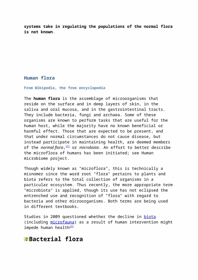
(5) Streptococcus pneumoniae is present in the upper respiratory tract of about half the population. If it invades the lower respiratory tract it can cause pneumonia. Streptococcus pneumoniae causes 95 percent of all bacterial pneumonia.
Streptococcus pneumoniae. Direct fluorescent antibody stain. CDC.
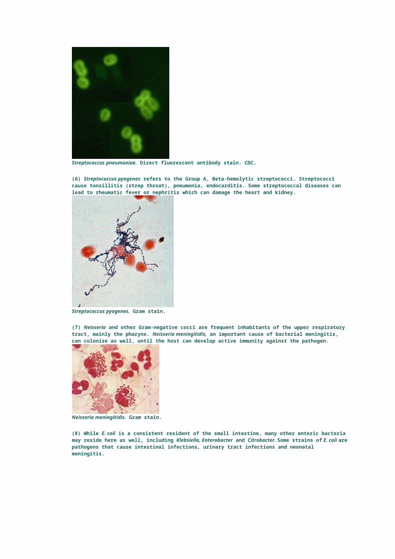
(6) Streptococcus pyogenes refers to the Group A, Beta-hemolytic streptococci. Streptococci cause tonsillitis (strep throat), pneumonia, endocarditis. Some streptococcal diseases can lead to rheumatic fever or nephritis which can damage the heart and kidney.
Streptococcus pyogenes. Gram stain.
(7) Neisseria and other Gram-negative cocci are frequent inhabitants of the upper respiratory tract, mainly the pharynx. Neisseria meningitidis, an important cause of bacterial meningitis, can colonize as well, until the host can develop active immunity against the pathogen.

Neisseria meningitidis. Gram stain.
(8) While E. coli is a consistent resident of the small intestine, many other enteric bacteria may reside here as well, including Klebsiella, Enterobacter and Citrobacter. Some strains of E. coli are pathogens that cause intestinal infections, urinary tract infections and neonatal meningitis.
E. coli. Scanning E.M. Shirley Owens. Center for Electron Optics. Michigan State University.
(9) Pseudomonas aeruginosa is the quintessential opportunistic pathogen of humans that can invade virtually any tissue. It is a leading cause of hospital-acquired (nosocomial) Gram-negative infections, but its source is often exogenous (from outside the host).
Colonies of Pseudomonas aeruginosa growing on an agar plate. The most virulent Pseudomonas species produce mucoid colonies and green pigments such as this isolate.
(10) Haemophilus influenzae is a frequent secondary invader to viral influenza, and was named accordingly. The bacterium was the leading cause of meningitis in infants and children until the recent development of the Hflu type B vaccine.

Haemophilus influenzae. Gram stain.
(11) The greatest number of bacteria are found in the lower intestinal tract, specifically the colon and the most prevalent bacteria are the Bacteroides, a group of Gram-negative, anaerobic, non-sporeforming bacteria. They have been implicated in the initiation colitis and colon cancer.
Bacteroides fragilis. Gram stain.
(12) Bifidobacteria are Gram-positive, non-sporeforming, lactic acid bacteria. They have been described as "friendly" bacteria in the intestine of humans. Bifidobacterium bifidum is the predominant bacterial species in the intestine of breast-fed infants, where it presumably prevents colonization by potential pathogens. These bacteria are sometimes used in the manufacture of yogurts and are frequently incorporated into probiotics.
Bifidobacterium bifidum. Gram stain
(13) Lactobacilli in the oral cavity probably contribute to acid formation that leads to dental caries. Lactobacillus acidophilus colonizes the vaginal epithelium during child-bearing years and establishes the low pH that inhibits the growth of pathogens.

Lactobacillus species and a vaginal squaemous epithelial cell. CDC
(14) There are numerous species of Clostridium that colonize the bowel. Clostridium perfringens is commonly isolated from feces. Clostridium difficile may colonize the bowel and cause "antibiotic-induced diarrhea" or pseudomembranous colitis.
Clostridium perfringens. Gram stain.
(15) Clostridium tetani is included in the table as an example of a bacterium that is "transiently associated" with humans as a component of the normal flora. The bacterium can be isolated from feces in 0 - 25 percent of the population. The endospores are probably ingested with food and water, and the bacterium does not colonize the intestine.
Clostridium tetani. Gram stain.
(16) The corynebacteria, and certain related propionic acid bacteria, are consistent skin flora. Some have been implicated as a cause of acne. Corynebacterium diphtheriae, the agent of diphtheria, was considered a member of the normal flora before the widespread use of the diphtheria toxoid, which is used to immunize against the disease.
Corynebacterium diphtheriae. No longer a part of the normal flora.
Associations Between Humans and the Normal Flora
E. coli is the best known bacterium that regularly associates itself with humans, being an invariable component of the human intestinal tract. Even though E. coli is the most studied of all bacteria, and we know the exact location and sequence of 4,288 genes on its chromosome, we do not fully understand its ecological relationship with humans.
In fact, not much is known about the nature of the associations between humans and their normal flora, but they are thought to be dynamic interactions rather than associations of mutual indifference. Both host and bacteria are thought to derive benefit from each other, and the associations are, for the most part, mutualistic. The normal flora derive from their host a steady supply of nutrients, a stable environment, and protection and transport. The host obtains from the normal flora certain nutritional and digestive benefits, stimulation of the development and activity of immune system, and protection against colonization and infection by pathogenic microbes.
While most of the activities of the normal flora benefit their host, some of the normal flora are parasitic (live at the expense of their host), and some are pathogenic (capable of producing disease). Diseases that are produced by the normal flora in their host may be called endogenous diseases. Most endogenous bacterial diseases are opportunistic infections, meaning that the the organism must be given a special opportunity of weakness or let-down in the host defenses in order to infect. An example of an opportunistic infection is chronic bronchitis in smokers wherein normal flora bacteria are able to invade the weakened lung.
Sometimes the relationship between a member of the normal flora an its host cannot be deciphered. Such a relationship where there is no apparent benefit or harm to either organism during their association is referred to as a commensal relationship. Many of the normal flora that are not predominant in their habitat, even though always present in low numbers, are thought of as commensal bacteria. However, if a presumed commensal relationship is studied in detail, parasitic or mutualistic characteristics often emerge.
Tissue specificity
Most members of the normal bacterial flora prefer to colonize certain tissues and not others. This "tissue specificity" is usually due to properties of both the host and the bacterium. Usually, specific bacteria colonize specific tissues by one or another of these mechanisms.
1. Tissue tropism is the bacterial preference or predilection for certain tissues for growth. One explanation for tissue tropism is that the host provides essential nutrients and growth factors for the bacterium, in addition to suitable oxygen, pH, and temperature for growth.

Lactobacillus acidophilus, informally known as "Doderlein's bacillus" colonizes the vagina because glycogen is produced which provides the bacteria with a source of sugar that they ferment to lactic acid.
2. Specific adherence Most bacteria can colonize a specific tissue or site because they can adhere to that tissue or site in a specific manner that involves complementary chemical interactions between the two surfaces. Specific adherence involves biochemical interactions between bacterial surface components (ligands or adhesins) and host cell molecular receptors. The bacterial components that provide adhesins are molecular parts of their capsules, fimbriae, or cell walls. The receptors on human cells or tissues are usually glycoprotein molecules located on the host cell or tissue surface.
Figure 2. Specific adherence involves complementary chemical interactions between the host cell or tissue surface and the bacterial surface. In the language of medical microbiologist, a bacterial "adhesin" attaches covalently to a host "receptor" so that the bacterium "docks" itself on the host surface. The adhesins of bacterial cells are chemical components of capsules, cell walls, pili or fimbriae. The host receptors are usually glycoproteins located on the cell membrane or tissue surface.
Some examples of adhesins and attachment sites used for specific adherence to human tissues are described in the table below.
Table 2. Examples of bacterial specific adherence to host cells or tissue.
Bacterium Bacterial adhesin Attachment site
Streptococcus pyogenes
Cell-bound protein (M-protein)
Pharyngeal epithelium
Streptococcus mutans
Cell- bound protein (Glycosyl transferase)
Pellicle of tooth
Streptococcus salivarius
Lipoteichoic acidBuccal epithelium of tongue
Streptococcus pneumoniae
Cell-bound protein (choline-binding protein)
Mucosal epithelium
Staphylococcus aureus
Cell-bound protein Mucosal epithelium
Neisseria gonorrhoeae
N-methylphenyl- alanine piliUrethral/cervical epithelium
Enterotoxigenic E. coli
Type-1 fimbriae Intestinal epithelium
Uropathogenic E. coli
P-pili (pap) Upper urinary tract

Bordetella pertussisFimbriae ("filamentous hemagglutinin")
Respiratory epithelium
Vibrio cholerae N-methylphenylalanine pili Intestinal epithelium
Treponema pallidum
Peptide in outer membrane Mucosal epithelium
Mycoplasma Membrane protein Respiratory epithelium
Chlamydia UnknownConjunctival or urethral epithelium
3. Biofilm formation Some of the indigenous bacteria are able to construct biofilms on a tissue surface, or they are able to colonize a biofilm built by another bacterial species. Many biofilms are a mixture of microbes, although one member is responsible for maintaining the biofilm and may predominate.
Figure 3. Cartoon depicting biofilm formation. Biofilms usually occur when one bacterial species attaches specifically or non specifically to a surface, and then secretes carbohydrate slime (exopolymer) that imbeds the bacteria and attracts other microbes to the biofilm for protection or nutritional advantages.
The classic biofilm that involves components of the normal flora of the oral cavity is the formation of dental plaque on the teeth. Plaque is a naturally-constructed biofilm, in which the consortia of bacteria may reach a thickness of 300-500 cells on the surfaces of the teeth. These accumulations subject the teeth and gingival tissues to high concentrations of bacterial metabolites, which result in dental disease.
The Composition of the Normal Flora
The normal flora of humans are exceedingly complex and consist of more than 200 species of bacteria. The makeup of the normal flora may be influenced by various factors, including genetics, age, sex, stress, nutrition and diet of the individual.
Three developmental changes in humans, weaning, the eruption of the teeth, and the onset and cessation of ovarian functions, invariably affect the composition of the normal flora in the intestinal tract, the oral cavity, and the vagina, respectively. However, within the limits of these fluctuations, the bacterial flora of humans is sufficiently constant to a give general description of the situation.
A human first becomes colonized by a normal flora at the moment of birth and passage through the birth canal. In utero, the fetus is sterile, but when the mother's water breaks and the birth process begins, so does colonization of the body surfaces. Handling and feeding of the infant after birth leads to establishment of a stable normal flora on the skin, oral cavity and intestinal tract in about 48 hours.

It has been calculated that a human adult houses about 1012 bacteria on the skin, 1010 in the mouth, and 1014 in the gastrointestinal tract. The latter number is far in excess of the number of eucaryotic cells in all the tissues and organs which comprise a human. The predominant bacteria on the surfaces of the human body are listed in Table 3. Informal names identify the bacteria in this table. Formal taxonomic names of organisms are given in Table 1.
Table 3. Predominant bacteria at various anatomical locations in adults.
Anatomical Location Predominant bacteria
Skin staphylococci and corynebacteria
Conjunctiva sparse, Gram-positive cocci and Gram-negative rods
Oral cavity
teeth streptococci, lactobacilli
mucous membranes streptococci and lactic acid bacteria
Upper respiratory tract
nares (nasal membranes)
staphylococci and corynebacteria
pharynx (throat) streptococci, neisseria, Gram-negative rods and cocci
Lower respiratory tract none
Gastrointestinal tract
stomach Helicobacter pylori (up to 50%)
small intestine lactics, enterics, enterococci, bifidobacteria
colon bacteroides, lactics, enterics, enterococci, clostridia, methanogens
Urogenital tract
anterior urethra sparse, staphylococci, corynebacteria, enterics
vagina lactic acid bacteria during child-bearing years; otherwise mixed
Normal Flora of the Skin The adult human is covered with approximately 2 square meters of skin. The density and composition of the normal flora of the skin varies with anatomical locale. The high moisture content of the axilla, groin, and areas between the toes supports the activity and growth of relatively high densities of bacterial cells, but the density of bacterial populations at most other sites is fairly low, generally in 100s or 1000s per square cm. Most bacteria on the skin are sequestered in sweat glands.
The skin microbes found in the most superficial layers of the epidermis and the upper parts of the hair follicles are Gram-positive cocci (Staphylococcus epidermidis and Micrococcus sp.) and corynebacteria such as Propionibacterium sp. These are generally nonpathogenic and considered to be commensal, although mutualistic and parasitic roles have been assigned to them. For example, staphylococci and propionibacteria produce fatty acids that inhibit the growth of fungi and yeast on the skin. But, if Propionibacterium acnes, a normal inhabitant of the skin, becomes trapped in hair follicle, it may grow rapidly and cause inflammation and acne.
Sometimes potentially pathogenic Staphylococcus aureus is found on the face and hands in individuals who are nasal carriers. This is because the face and hands are likely to become inoculated with the bacteria on the nasal membranes. Such individuals may autoinoculate themselves with the pathogen or spread it to other individuals or foods.
Normal Flora of the Conjunctiva A variety of bacteria may be cultivated from the normal conjunctiva, but the number of organisms is usually small. Staphylococcus epidermidis and certain coryneforms (Propionibacterium acnes) are dominant. Staphylococcus aureus, some streptococci, Haemophilus sp. and Neisseria sp. are occasionally found. The conjunctiva is kept moist and healthy by the continuous secretions from the lachrymal glands. Blinking wipes the conjunctiva every few seconds mechanically washing away foreign objects including bacteria. Lachrymal secretions (tears) also contain bactericidal substances including lysozyme. There is little or no opportunity for microorganisms to colonize the conjunctiva without special mechanisms to attach to the epithelial surfaces and some ability to withstand attack by lysozyme.
Pathogens which do infect the conjunctiva (e.g. Neisseria gonorrhoeae and Chlamydia trachomatis) are thought to be able to specifically attach to the conjunctival epithelium. Newborn infants may be especially prone to bacterial

attachment. Since Chlamydia and Neisseria might be present on the cervical and vaginal epithelium of an infected mother, silver nitrate or an antibiotic may be put into the newborn's eyes to avoid infection after passage through the birth canal.
Figure 4. Colonies of Propionibacterium acnes, found on skin and the conjunctiva.
Normal Flora of the Respiratory Tract A large number of bacterial species colonize the upper respiratory tract (nasopharynx). The nares (nostrils) are always heavily colonized, predominantly with Staphylococcus epidermidis and corynebacteria, and often (in about 20% of the general population) with Staphylococcus aureus, this being the main carrier site of this important pathogen. The healthy sinuses, in contrast are sterile. The pharynx (throat) is normally colonized by streptococci and various Gram-negative cocci. Sometimes pathogens such as Streptococcus pneumoniae, Streptococcus pyogenes, Haemophilus influenzae and Neisseria meningitidis colonize the pharynx.
The lower respiratory tract (trachea, bronchi, and pulmonary tissues) is virtually free of microorganisms, mainly because of the efficient cleansing action of the ciliated epithelium which lines the tract. Any bacteria reaching the lower respiratory tract are swept upward by the action of the mucociliary blanket that lines the bronchi, to be removed subsequently by coughing, sneezing, swallowing, etc. If the respiratory tract epithelium becomes damaged, as in bronchitis or viral pneumonia, the individual may become susceptible to infection by pathogens such as H. influenzae or S. pneumoniae descending from the nasopharynx.
Normal Flora of the Urogenital Tract Urine is normally sterile, and since the urinary tract is flushed with urine every few hours, microorganisms have problems gaining access and becoming established. The flora of the anterior urethra, as indicated principally by urine cultures, suggests that the area my be inhabited by a relatively consistent normal flora consisting of Staphylococcus epidermidis, Enterococcus faecalis and some alpha-hemolytic streptococci. Their numbers are not plentiful, however. In addition, some enteric bacteria (e.g. E. coli, Proteus) and corynebacteria, which are probably contaminants from the skin, vulva or rectum, may occasionally be found at the anterior urethra.
The vagina becomes colonized soon after birth with corynebacteria, staphylococci, streptococci, E. coli, and a lactic acid bacterium historically named "Doderlein's bacillus" (Lactobacillus acidophilus). During reproductive life, from puberty to menopause, the vaginal epithelium contains glycogen due to the actions of circulating estrogens. Doderlein's bacillus predominates, being able to metabolize the glycogen to lactic acid. The lactic acid and other products of metabolism inhibit colonization by all except this lactobacillus and a select number of lactic acid bacteria. The resulting low pH of the vaginal epithelium prevents establishment by most other bacteria as well as the potentially-pathogenic yeast, Candida albicans. This is a striking example of the protective effect of the normal bacterial flora for their human host.

Figure 5. A Lactobacillus species, possibly Doderlein's bacillus, in association with a vaginal epithelial cell.
Normal Flora of the Oral Cavity The presence of nutrients, epithelial debris, and secretions makes the mouth a favorable habitat for a great variety of bacteria. Oral bacteria include streptococci, lactobacilli, staphylococci and corynebacteria, with a great number of anaerobes, especially bacteroides.
The mouth presents a succession of different ecological situations with age, and this corresponds with changes in the composition of the normal flora. At birth, the oral cavity is composed solely of the soft tissues of the lips, cheeks, tongue and palate, which are kept moist by the secretions of the salivary glands. At birth the oral cavity is sterile but rapidly becomes colonized from the environment, particularly from the mother in the first feeding. Streptococcus salivarius is dominant and may make up 98% of the total oral flora until the appearance of the teeth (6 - 9 months in humans). The eruption of the teeth during the first year leads to colonization by S. mutans and S. sanguis. These bacteria require a nondesquamating (nonepithelial) surface in order to colonize. They will persist as long as teeth remain. Other strains of streptococci adhere strongly to the gums and cheeks but not to the teeth. The creation of the gingival crevice area (supporting structures of the teeth) increases the habitat for the variety of anaerobic species found. The complexity of the oral flora continues to increase with time, and bacteroides and spirochetes colonize around puberty.

Figure 6. Various streptococci in a biofilm in the oral cavity.
The normal bacterial flora of the oral cavity clearly benefit from their host who provides nutrients and habitat. There may be benefits, as well, to the host. The normal flora occupy available colonization sites which makes it more difficult for other microorganisms (nonindigenous species) to become established. Also, the oral flora contribute to host nutrition through the synthesis of vitamins, and they contribute to immunity by inducing low levels of circulating and secretory antibodies that may cross react with pathogens. Finally, the oral bacteria exert microbial antagonism against nonindigenous species by production of inhibitory substances such as fatty acids, peroxides and bacteriocins.
On the other hand, the oral flora are the usual cause of various oral diseases in humans, including abscesses, dental caries, gingivitis, and periodontal disease. If oral bacteria can gain entrance into deeper tissues, they may cause abscesses of alveolar bone, lung, brain, or the extremities. Such infections usually contain mixtures of bacteria with Bacteroides melaninogenicus often playing a dominant role. If oral streptococci are introduced into wounds created by dental manipulation or treatment, they may adhere to heart valves and initiate subacute bacterial endocarditis.
Figure 7. Colonies of E. coli growing on EMB agar.
Normal Flora of the Gastrointestinal Tract The bacterial flora of the gastrointestinal (GI) tract of animals has been studied more extensively than that of any other site. The composition differs between various animal species, and within an animal species. In humans, there are differences in the composition of the flora which are influenced by age, diet, cultural conditions, and the use of antibiotics. The latter greatly perturbs the composition of the intestinal flora.
In the upper GI tract of adult humans, the esophagus contains only the bacteria swallowed with saliva and food. Because of the high acidity of the gastric juice, very few bacteria (mainly acid-tolerant lactobacilli) can be cultured from the normal stomach. However, at least half the population in the United States is colonized by a pathogenic bacterium, Helicobacter pylori. Since the 1980s, this bacterium has been known to be the cause of gastric ulcers, and it is probably a cause of gastric and duodenal cancer as well. The Australian microbiologist, Barry Marshall, received

the Nobel Prize in Physiology and Medicine in 2005, for demonstrating the relationship between Helicobacter and gastric ulcers.
Figure 8. Helicobacter pylori. ASM
The proximal small intestine has a relatively sparse Gram-positive flora, consisting mainly of lactobacilli and Enterococcus faecalis. This region has about 105 - 107 bacteria per ml of fluid. The distal part of the small intestine contains greater numbers of bacteria (108/ml) and additional species, including coliforms (E. coli and relatives) and Bacteroides, in addition to lactobacilli and enterococci.
The flora of the large intestine (colon) is qualitatively similar to that found in feces. Populations of bacteria in the colon reach levels of 1011/ml feces. Coliforms become more prominent, and enterococci, clostridia and lactobacilli can be regularly found, but the predominant species are anaerobic Bacteroides and anaerobic lactic acid bacteria in the genus Bifidobacterium (Bifidobacterium bifidum). These organisms may outnumber E. coli by 1,000:1 to 10,000:1. Sometimes, significant numbers of anaerobic methanogens (up to 1010/gm) may reside in the colon of humans. This is our only direct association with archaea as normal flora. The range of incidence of certain bacteria in the large intestine of humans is shown in Table 4 below.
Table 4. Bacteria found in the large intestine of humans.
BACTERIUM RANGE OF INCIDENCEBacteroides fragilis 100Bacteroides melaninogenicus 100Bacteroides oralis 100Lactobacillus 20-60Clostridium perfringens 25-35Clostridium septicum 5-25Clostridium tetani 1-35Bifidobacterium bifidum 30-70Staphylococcus aureus 30-50Enterococcus faecalis 100Escherichia coli 100Salmonella enteritidis 3-7Klebsiella sp. 40-80Enterobacter sp. 40-80Proteus mirabilis 5-55Pseudomonas aeruginosa 3-11Peptostreptococcus sp. ?commonPeptococcus sp. ?common
At birth the entire intestinal tract is sterile, but bacteria enter with the first feed. The initial colonizing bacteria vary with the food source of the infant. In breast-fed infants, bifidobacteria account for more than 90% of the total intestinal bacteria. Enterobacteriaceae and enterococci are regularly present, but in low proportions, while bacteroides, staphylococci, lactobacilli and clostridia are practically absent. In bottle-fed infants, bifidobacteria are not predominant. When breast-fed infants are switched to a diet of cow's milk or solid food, bifidobacteria are progressively joined by enterics, bacteroides, enterococci lactobacilli and clostridia. Apparently, human milk contains a growth factor that enriches for growth of bifidobacteria, and these bacteria play an important role in preventing colonization of the infant intestinal tract by non indigenous or pathogenic species.

Figure 9. Clostridium difficile. Gram stain. The growth of "C. diff" in the intestinal tract is normally held in check by other members of the normal flora. When antibiotics given for other infections cause collateral damage to the normal intestinal flora, the clostridium may be able to "grow out" and produce a serious diarrheal syndrome called pseudomembranous colitis. This is an example of an "antibiotic induced diarrheal disease".
The composition of the flora of the gastrointestinal tract varies along the tract (at longitudinal levels) and across the tract (at horizontal levels) where certain bacteria attach to the gastrointestinal epithelium and others occur in the lumen. There is frequently a very close association between specific bacteria in the intestinal ecosystem and specific gut tissues or cells (evidence of tissue tropism and specific adherence). Gram-positive bacteria, such as the streptococci and lactobacilli, are thought to adhere to the gastrointestinal epithelium using polysaccharide capsules or cell wall teichoic acids to attach to specific receptors on the epithelial cells. Gram-negative bacteria such as the enterics may attach by means of specific fimbriae which bind to glycoproteins on the epithelial cell surface.
It is in the intestinal tract that we see the greatest effect of the bacterial flora on their host. This is due to their large mass and numbers. Bacteria in the human GI tract have been shown to produce vitamins and may otherwise contribute to nutrition and digestion. But their most important effects are in their ability to protect their host from establishment and infection by alien microbes and their ability to stimulate the development and the activity of the immunological tissues.
On the other hand, some of the bacteria in the colon (e.g. Bacteroides) have been shown to produce metabolites that are carcinogenic, and there may be an increased incidence of colon cancer associated with these bacteria. Alterations in the GI flora brought on by poor nutrition or perturbance with antibiotics can cause shifts in populations and colonization by nonresidents that leads to gastrointestinal disease.
Skin flora
From Wikipedia, the free encyclopediaJump to: navigation, search

Depiction of the human body and bacteria that predominate
The skin flora are the microorganisms which reside on the skin. Most research has been upon those that reside upon the 2 square meters of human skin. Many of them are bacteria of which there are around 1000 species upon human skin from 19 phyla.[1][2] The total number of bacteria on an average human has been estimated at 1012.[3] Most are found in the superficial layers of the epidermis and the upper parts of hair follicles.
Skin flora are usually non-pathogenic, and either commensals (are not harmful to their host) or mutualistic (offer a benefit). The benefits bacteria can offer include preventing transient pathogenic organisms from colonizing the skin surface, either by competing for nutrients, secreting chemicals against them, or stimulating the skin's immune system.[4] However, resident microbes can cause skin diseases and enter the blood system creating life threatening diseases particularly in immunosuppressed people.[4] Hygiene to control such flora is important in preventing the transmission of antibiotic resistant hospital-acquired infections.
A major nonhuman skin flora is Batrachochytrium dendrobatidis, a chytrid and non-hyphal zoosporic fungus that causes chytridiomycosis, infectious disease thought to be responsible for the decline in amphibian populations.
Species variety

[edit] Bacteria
Scanning electron microscope image of Staphylococcus epidermidis one of roughly a thousand bacteria species present on human skin. Though usually not pathogenic, it can cause skin infections and even life threatening illnesses in those that are immunocompromised.
The estimate of the number of species present on skin bacteria has been radically changed by the use of 16S ribosomal RNA to identify bacterial species present on skin samples direct from their genetic material. Previously such identification had depended upon microbiological culture upon which many varieties of bacteria did not grow and so were hidden to science.[1]
Staphylococcus epidermidis and Staphylococcus aureus were thought from cultural based research to be dominant. However 16S ribosomal RNA research finds that while common, these species make up only 5% of skin bacteria.[5] However, skin variety provides a rich and diverse habit for bacteria. Most come from four phyla: Actinobacteria (51.8%), Firmicutes (24.4%), Proteobacteria (16.5%), and Bacteroidetes (6.3%).
ecology of the 20 sites on the skin studied in the Human Microbiome {Project

There are three main ecological areas: moist, dry and sebaceous. Propionibacteria and Staphylococci species were the main species in sebaceous areas. In moist places on the body Corynebacteria together with Staphylococci dominate. In dry areas, there is a mixture of species but b-Proteobacteria and Flavobacteriales are dominant. Ecologically, sebaceous areas had greater species richness than moist and dry one. The areas with least similarity between people in species were the spaces between fingers, the spaces between toes, axillae, and umbilical cord stump. Most similarly were beside the nostril, nares (inside the nostril), and on the back.[1]
Frequency of the best studied skin microbes[4]
Organism observations
Staphylococcus epidermidis Common, occasionally pathogenic
Staphylococcus aureus Infrequent, usually pathogenic
Staphylococcus warneri Infrequent, occasionally pathogenic
Streptococcus pyogenes Infrequent, usually pathogenic
Streptococcus mitis Frequent, occasionally pathogenic
Propionibacterium acnes Frequent, occasionally pathogenic
Corynebacterium spp. Frequent, occasionally pathogenic
Acinetobacter johnsonii Frequent, occasionally pathogenic
Pseudomonas aeruginosa Infrequent, occasionally pathogenic
[edit] Fungal
A study of the area between toes in a 100 young adults found 14 different genera of fungi. These include yeasts such as Candida albicans, Rhodotorula rubra, Torulopsis and Trichosporon cutaneum, dermatophytes (skin living fungi) such as Microsporum gypseum, and Trichophyton rubrum and nondermatophyte fungi (opportunistic fungi that can live in skin) such as Rhizopus stolonifer, Trichosporon cutaneum, Fusarium, Scopulariopsis brevicaulis, Curvularia, Alternaria alternata, Paecilomyces, Aspergillus flavus and Penicillium species.[6]
Relationship to host
Skin microflora can be commensals, mutualistic or pathogens. Often they can be all three depending upon the strength of the person's immune system.[4] Research upon the immune system in the gut and lungs has shown that microflora aids immunity development: however such research has only started upon whether this is the case with the skin.[4] Pseudomonas

aeruginosa is an example of a mutualistic bacteria that can turn into a pathogen and cause disease: if gains entry into the blood system it can result in inflections in bone, joint, gastrointestinal, and respiratory systems. It can also cause dermatitis. However, Pseudomonas aeruginosa produces antimicrobial substances such as pseudomonic acid that are exploited commercially such as Mupirocin. This works against staphylococcal and streptococcal infections. Pseudomonas aeruginosa also produces substances that inhibit the growth of fungus species such as Candida krusei, Candida albicans, Torulopsis glabrata, Saccharomyces cerevisiae and Aspergillus fumigatus. [7] It can also inhibit the growth of Helicobacter pylori.[8] So important is its antimicrobial actions that it has been noted that "removing P. aeruginosa from the skin, through use of oral or topical antibiotics, may inversely allow for aberrant yeast colonization and infection."[4]
Another aspect of bacteria is the generation of body odor. Sweat is odorless but Propionibacteria in adolescent and adult sebaceous glands can turn its amino acids into propionic acid. Staphylococcus epidermidis create the other source of body odor: isovaleric acid (3-methyl butanoic acid).[9] In addition to these, people with strong foot odor this is due to Bacillus subtilis.[10]
Skin defenses
Antimicrobial peptides
The skin creates antimicrobial peptides such as cathelicidins that control the proliferation of skin microbes. Cathelicidins not only reduce microbe numbers directly but also cause the secretion of cytokine release which induces inflammation, angiogenesis, and reepithelialization. Conditions such as atopic dermatitis have been linked to the suppression in cathelicidin production. In rosacea abnormal processing of cathelicidin cause inflammation. Psoriasis has been linked self-DNA created from cathelicidin peptides that causes autoinflammation. A major factor controlling cathelicidin is vitamin D3.[11]
Acidity
The superficial layers of the skin are naturally acidic (pH 4-4.5) due lactic acid in sweat and produced by skin bacteria.[12] At this pH mutualistic flora such as Staphylococci, Micrococci, Corynebacterium and Propionibacteria grow but not transient bacteria such as Gram negative bacteria like Escherichia and Pseudomonas or Gram positive ones such as Staphylococcus aureus or Candida albicans.[12] Another factor effecting the growth of pathological bacteria is that the antimicrobial substances secreted by the skin are enhanced in acidic conditions.[12] In alkaline conditions, bacteria cease to be attached to the skin and are more readily shed. It has been observed that the skin also swells under alkaline conditions and opens up allowing move to the surface.[12]
Immune system
If activated, the immune system in the skin produces cell-mediated immunity against microbes such as dermatophytes (skin fungi).[13] One reaction is to increase stratum corneum turnover and so shed the fungus from the skin surface. Skin fungi such as Trichophyton rubrum have evolved to create substances that limit the immune response to them.[13] The shedding of skin is a general means to control the build up flora upon the skin surface.

Clinical
Skin diseases
Microorganisms play a role in noninfectious skin diseases such as atopic dermatitis,[14] rosacea, psoriasis,[15] and acne [16] Damaged skin can cause nonpathogenic bacteria to become pathogenic.[17]
Infected devices
Skin microbes are source of infected medical devices such as catheters.[18]
Hygiene
Contagion
Skin flora do not readily pass between people: 30 seconds of moderate friction and dry hand contact results in a transfer of only 0.07% of natural hand flora from naked with a greater percentage from gloves.[19]
Removal
The most effective (60 to 80% reduction) antimicrobial washing is with ethanol, isopropanol, and n-propanol. Viruses are most affected by high (95%) concentrations of ethanol, while bacteria are more affected by n-propanol.[20]
Unmedicated soaps is not very effective as illustrated by the following data. Health care workers washed their hands once in nonmedicated liquid soap for 30 seconds. The students/technicians for 20 times.[21]
Skin flora upon two hospital groups in colony-forming units per mL.
group and hand skin condition unwashed washed
Health care workers healthy 3.47 3.15
Health care workers damaged 3.33 3.29
Students/technicians healthy 4.39 3.54
Students/technicians damaged 4.58 4.43
An important use of hand washing is to prevent the transmission of antibiotic resistant skin flora that cause hospital-acquired infections such as Methicillin-resistant Staphylococcus aureus. While such flora have become antibiotic resistant to due antibiotics there is no evidence that recommended antiseptics or disinfectants selects for antibiotic-resistant

organisms when used in hand washing.[22] However, many strains of organisms are resistant to some of the substances used in antibacterial soaps such as Triclosan.[22]
One survey of bar soaps in dentist clinics found they all had their own flora and on average from two to five different genera of microorganisms with those used most more likely to have more species varieties.[23] Another survey of bar soaps in public toilets found even more flora.[24] Another study found that very dry soaps are not infected while all are that rest in pools of water.[25] However, research upon soap that was specially infected found that soap flora do not transmit to the hands.[26]
Damaged skin
Washing skin repeatedly can damage the protective external layer and cause transepidermal loss of water. This can be seen in roughness characterized by scaling and dryness, itchiness, dermatitis provoked by microorganisms and allergens penetrating the corneal layer and redness. Wearing gloves can cause further problems since it produces a humid environment favoring the growth of microbes and also contains irritants such as latex and talcum powder.[27]
Hand washing can damage skin because the stratum corneum top layer of skin consists of 15 to 20 layers of keratin disks, corneocytes, each of which is each surrounded by a thin film of skin lipids which can be removed by alcohols and detergents.[28]
Damaged skin defined by extensive cracking of skin surface, widespread reddening or occasional bleeding has also been found to be more frequently colonized by Staphylococcus hominis and these were more likely to methicillin resistant.[27] Though not related to greater antibiotic resistance, damaged skin was also more like to be colonized by Staphylococcus aureus, gram-negative bacteria, Enterococci and Candida.[27]
Comparison with other flora
The skin flora is different from that of the gut which is predominately Firmicutes and Bacteroidetes.[29] There is also low level of variation between people that is not found in gut studies.[5] Both gut and skin flora however lack the diversity found in soil flora.[1]
Some of the more common complications from Staph infections include toxic shock syndrome, skin infections, and pneumonia. A more serious infection occurs when Staph enters the bloodstream, this is known as bacteremia. Identified as a lethal threat
What types of diseases are caused by Staph?
Skin infections (see above) are the most common type of disease produced by Staphylococcus. Staph infections of the skin can progress to impetigo (a crusting of the skin) or cellulitis (inflammation of the connective tissue under the skin, leading to swelling and

redness of the area). In rare cases, a serious complication known as scalded skin syndrome (see below) can develop. In breastfeeding women, Staph can result in mastitis (inflammation of the breast) or in abscess of the breast. Staphylococcal breast abscesses can release bacteria into the mother's milk.
When the bacteria enter the bloodstream and spread to other organs, a number of serious infections can occur. Spread of the organisms to the bloodstream is known as bacteremia or sepsis. Staphylococcal pneumonia predominantly affects people with underlying lung disease and can lead to abscess formation within the lungs. Infection of the heart valves (endocarditis) can lead to heart failure. Spread of Staphylococci to the bones can result in severe inflammation of the bones known as osteomyelitis. Staphylococcal sepsis (widespread infection of the bloodstream) is a leading cause of shock and circulatory collapse, leading to death, in people with severe burns over large areas of the body.
Staphylococcal food poisoning is an illness of the bowels that causes nausea, vomiting, diarrhea, and dehydration. It is caused by eating foods contaminated with toxins produced by Staphylococcus aureus. Symptoms usually develop within one to six hours after eating contaminated food. The illness usually lasts for one to three days and resolves on its own. Patients with this illness are not contagious, since toxins are not transmitted from one person to another.
Toxic shock syndrome is an illness caused by toxins secreted by Staph aureus bacteria growing under conditions in which there is little or no oxygen. Toxic shock syndrome is characterized by the sudden onset of high fever, vomiting, diarrhea, and muscle aches, followed by low blood pressure (hypotension), which can lead to shock and death. There may be a rash resembling sunburn, with peeling of skin. Toxic shock syndrome was originally described and still occurs especially in menstruating women using tampons
Who is at risk for Staph infections?
Anyone can develop a Staph infection, although certain groups of people are at greater risk, including newborn infants, breastfeeding women, and people with chronic conditions such as diabetes, cancer, vascular disease, and lung disease. Injecting drug users, those with skin injuries or disorders, intravenous catheters, surgical incisions, and those with a weakened immune system all have an increased risk of developing Staph infections.
Differential Growth Media
A differential medium distinguishes between different types of bacteria based on some characteristic of the bacteria that is growing on it. Typically there is a color change that results from certain bacterial metabolic products reacting with substances or chemicals that have been added to the media.

Selective and Differential Media
If a bacterial growth medium is selective, that means that it grows only certain types of microbes while inhibiting the growth of other types of microbes. A growth medium is considered differential if, when specific microbes are present, the medium, or bacterial colonies themselves, exhibit a color change that provides information about their identity.