visual pathways in the chiasm - home | fondazione g.b. bietti · intracranial relationships of the...
TRANSCRIPT


Visual pathways in the chiasmVisual pathways in the chiasm
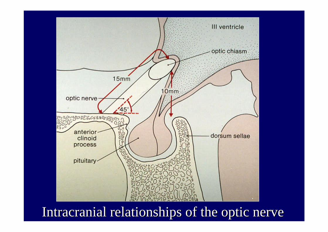
Intracranial relationships of the optic nerveIntracranial relationships of the optic nerve

Fixation of the chiasmFixation of the chiasm

••ChiasmaticChiasmatic pathologiespathologies
••The The functionfunction of the of the opticoptic chiasmchiasm maymay bebe alteredalteredbyby the the presencepresence of :of :
1) 1) TumorsTumors, , 2) 2) InflammatoryInflammatory lesionslesions, , 3) 3) DemyelinatingDemyelinating lesionslesions
4) 4) ArteroArtero--venousvenous malformationsmalformations involvinginvolving the the sellar sellar regionregion..

••EtiologyEtiology::••1) 1) TumorsTumors:: (25% of (25% of chiasmaticchiasmatic pathologiespathologies):):-- pituitary adenoma (50%): secreting (with pituitary adenoma (50%): secreting (with endocrinologicalendocrinologicalsymptomssymptoms, , CushingCushing’’s s diseasedisease, amenorrea, , amenorrea, infertilityinfertility, acromegalia, , acromegalia, impotenceimpotence –– prolactin in males) and non secretingprolactin in males) and non secreting-- craniopharyngiomacraniopharyngioma (25%): (25%): approximatelyapproximately 8% of 8% of brainbrain tumorstumors(13% in (13% in childrenchildren))-- meningiomameningioma (10%): ((10%): (oftenoften FosterFoster-- KennedyKennedy syndromesyndrome))-- gliomaglioma (7%): (7%): astrocytomaastrocytoma gradesgrades 1 and 21 and 2••DiagnosisDiagnosis:: MRI and CTMRI and CT••Therapy:Therapy: surgical removalsurgical removal••2) Suprasellar aneurisms2) Suprasellar aneurisms (aneurismatic (aneurismatic dilationdilation of one of the of one of the vesselsvessels of of WillisWillis’’ss polygonpolygon))••DiagnosisDiagnosis:: MRI. CT, MRI. CT, brainbrain angiographyangiography

3) Inflammation and infections- frontal sfenoidal sinus mucocele- sarcoidosis- tubercolosis- syphilis
4) Empty sell syndrome :- primary: extension of the subaracnoid space inside the sella turcica through a congenitally incomplete sellar diaphragm (2-4 % of the normal population)- secondary: after exeresis of pituitary adenoma or after pituitaryapoplexy following optic chiasm prolapse.

••SymptomsSymptoms: : ••1. 1. ReductionReduction in visual in visual functionfunction, , whichwhich oftenoften representsrepresentsthe first the first ifif notnot onlyonly symptomsymptom. The . The evolutionevolution isis slow and slow and maymay lastlast monthsmonths or or yearsyears. Visual . Visual defectsdefects are are almostalmost neverneverassociatedassociated toto painpain. .
••2. The visual 2. The visual fieldfield defectdefect isis generallygenerally bitemporalbitemporal butbut non non characteristicscharacteristics defectsdefects maymay bebe presentpresent
••3. 3. BinocularBinocular diplopia diplopia secondarysecondary toto paralysisparalysis of the 3rd, of the 3rd, 4th, or 6th CN due 4th, or 6th CN due toto compressioncompression or or invasioninvasion of the of the cavernouscavernous sinussinus byby the the lesionlesion

••SymptomsSymptoms: :
••4. 4. HeadacheHeadache, , whichwhich isis nevernever presentpresent asas onsetonsetsymptomsymptom exceptexcept after after pituitarypituitary apoplexiaapoplexia, , isispresentpresent in 13% of in 13% of casescases affectedaffected byby chiasmaticchiasmaticsyndromesyndrome..
5. 5. EndocrinologicalEndocrinological dysfunctionsdysfunctions, , secondarysecondary toto the the presencepresence of a of a secretingsecreting pituitarypituitary adenoma, and adenoma, and whichwhich varyvary dependingdepending on the on the hormonehormone producedproducedbyby the the tumortumor (GH, (GH, prolactinprolactin, TSH or ACTH), TSH or ACTH)

••The The mainmain signsign of a of a chiasmaticchiasmatic syndromesyndrome isis a a visual visual fieldfield defectdefect, , whichwhich isis generallygenerally bitemporalbitemporal. .
••HoweverHowever, , whenwhen the the sizesize of the of the tumortumor isis smallsmall (<10 mm), (<10 mm), suchsuch asasforfor a microadenoma, or a microadenoma, or ifif therethere isis no no suprasellarsuprasellar extensionextension, , itit isisdifficultdifficult forfor a a chiasmaticchiasmatic compressioncompression toto show. show. IndeedIndeed, the , the distancedistance separatingseparating the the opticoptic chiasmchiasm fromfrom the sellar the sellar diaphragmdiaphragm isisequalequal toto 10 mm. 10 mm.
••AnatomicalAnatomical variationsvariations in the position of the in the position of the opticoptic chiasmchiasm withwithrespectrespect toto the sella the sella turcicaturcica and the and the differentdifferent modalitiesmodalities of of growthgrowth of of expansiveexpansive lesionslesions in in thisthis regionregion justifyjustify the the heterogeneityheterogeneity of visual of visual fieldfield defectsdefects. . IndeedIndeed, , itit isis possiblepossible toto observeobserve the the presencepresence of:of:

••1.1. CentralCentral fieldfield defectdefect,, whenwhen the the lesionlesiondeterminesdetermines compressioncompression at the at the levellevel of the of the prepre--chiasmaticchiasmatic visual visual pathwayspathways, , generallygenerallymonolateralmonolateral..
••2. 2. JunctionalJunctional fieldfield defectdefect,, or or anterioranterior chiasmaticchiasmaticsyndromesyndrome, , whenwhen the the presencepresence of a of a centralcentral scotoma scotoma in one in one eyeeye isis associatedassociated toto a a superiorsuperior temporaltemporaldefectdefect in the in the contralateralcontralateral eyeeye, due , due toto compressioncompressionof the of the mostmost anterioranterior portionportion of the of the opticoptic chiasmchiasm

JunctionalJunctional ((ChiasmalChiasmal) field defect) field defect

CT scan of a CT scan of a suprasellarsuprasellar meningiomameningiomaproducing producing junctionaljunctional field defectfield defect
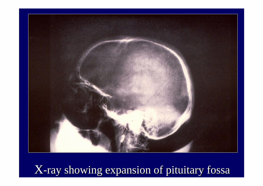
XX--ray showing expansion of pituitary ray showing expansion of pituitary fossafossa

CT san of a cystic CT san of a cystic chromophobechromophobe adenomaadenoma

33. . BitemporalBitemporal hemianopsiahemianopsia,, whichwhich maymay bebecomplete or incomplete, complete or incomplete, symmetricalsymmetrical or or asymmetricalasymmetrical; ; thisthis representsrepresents the the mostmost typicaltypicalvisual visual fieldfield defectdefect in the in the presencepresence of of lesionslesions of the of the opticoptic chiasmchiasm. . ItIt isis the the resultresult of the of the involvementinvolvement of of crossedcrossed fibersfibers, , originatingoriginating fromfrom the the nasalnasal retina, retina, whichwhich are are localizedlocalized in the in the centralcentral partpart of the of the chiasmchiasm..4. 4. HomonymousHomonymous hemianopsiahemianopsia secondarysecondary toto the the compressioncompression of the of the opticoptic tracttract due due toto lesionslesionswhichwhich extendextend posteriorlyposteriorly withwith respectrespect toto the the chiasmchiasm..

BitemporalBitemporal field defectfield defect

••OpticOptic disc pallor disc pallor occursoccursin in advancedadvanced stagesstages
••SeeSee--sawsaw--nystagmusnystagmusisis sometimessometimes presentpresent in in
suprasellarsuprasellar tumorstumors

Topographical arrangement of retinal nerve fibres Topographical arrangement of retinal nerve fibres with congenital homonymous with congenital homonymous hemianopsiahemianopsia

Grooves in retinal nerve fibre layerGrooves in retinal nerve fibre layer