ventrolateral motor thalamus abnormal connectivity in ... · constantin tuleasca et al....
TRANSCRIPT

Original Article
Ventrolateral Motor Thalamus Abnormal Connectivity in Essential Tremor Before and
After Thalamotomy: A Resting-State Functional Magnetic Resonance Imaging Study
Constantin Tuleasca1,2,4,5, Elena Najdenovska2, Jean Regis6, Tatiana Witjas7, Nadine Girard8, Jerome Champoudry6,
Mohamed Faouzi9, Jean-Philippe Thiran3-5, Meritxell Bach Cuadra2,4, Marc Levivier1,5, Dimitri Van De Ville10,11
-OBJECTIVE: To evaluate functional connectivity (FC) ofthe ventrolateral thalamus, a common target for drug-resistant essential tremor (ET), resting-state data wereanalyzed before and 1 year after stereotactic radiosurgicalthalamotomy and compared against healthy controls (HCs).
-METHODS: In total, 17 consecutive patients with ETand 10HCs were enrolled. Tremor network was investigated usingthe ventrolateral ventral (VLV) thalamic nucleus as the regionof interest, extracted with automated segmentation frompretherapeutic diffusion magnetic resonance imaging. Tem-poral correlations of VLV at whole brain level were evaluatedby comparing drug-naïve patients with ET with HCs, andlongitudinally, 1 year after stereotactic radiosurgical thala-motomy. 1 year thalamotomy MR signature was alwayslocated inside VLV and did not correlate with any of FC mea-sures (P > 0.05). This suggested presence of longitudinalchanges in VLV FC independently of theMR signature volume.
-RESULTS: Pretherapeutic ET displayed altered VLV FC withleft primary sensory-motor cortex, pedunculopontine nucleus,
Key words- Essential tremor- fMRI- Motor thalamus- Resting state- Stereotactic radiosurgery- Thalamotomy- Ventrointermediate nucleus
Abbreviations and AcronymsADL: Activities of daily livingBA: Brodmann areaDBS: Deep-brain stimulationDWI: Diffusion-weighted imagingET: Essential tremorFC: Functional connectivityFEW: Family-wise correctionfMRI: Functional magnetic resonance imagingHC: Healthy controlHIFU: High-focused ultrasoundION: Inferior olivary nucleusMRI: Magnetic resonance imagingRS: RadiosurgerySD: Standard deviationSRS-T: Stereotactic radiosurgical thalamotomy
WORLD NEUROSURGERY 113: e453-e464, MAY 2018
dorsal anterior cingulate, left visual association, and leftsuperior parietal areas. Pretherapeutic negative FC withprimary somatosensory cortex and pedunculopontine nu-cleus correlated with poorer baseline tremor scores(Spearman [ 0.04 and 0.01). Longitudinal study displayedchanges within right dorsal attention (frontal eye-fieldsand posterior parietal) and salience (anterior insula) net-works, as well as areas involved in hand movementplanning or language production.
-CONCLUSIONS: Our results demonstrated that patientswith ET and HCs differ in their left VLV FC to primary so-matosensory and supplementary motor, visual association,or brainstem areas (pedunculopontine nucleus). Longitu-dinal changes display reorganization of dorsal attentionand salience networks after thalamotomy. Beside atten-tional gateway, they are also known for their major role infacilitating a rapid access to the motor system.
Vim: Ventrointermediate nucleusVLV: Ventral lateral ventral (thalamic nucleus)
From the 1Neurosurgery Service and Gamma Knife Center, Centre Hospitalier UniversitaireVaudois, Lausanne, Switzerland; 2Medical Image Analysis Laboratory (MIAL) and Departmentof Radiology-Center of Biomedical Imaging (CIBM), Centre Hospitalier Universitaire Vaudoisand University of Lausanne, Lausanne, Switzerland; 3Department of Radiology, CentreHospitalier Universitaire Vaudois, Lausanne, Switzerland; 4Signal Processing Laboratory (LTS5), Ecole Polytechnique Fédérale de Lausanne (EPFL), Lausanne, Switzerland; 5Faculty ofBiology and Medicine, University of Lausanne, Lausanne, Switzerland; 6Stereotactic andFunctional Neurosurgery Service and Gamma Knife Unit, and 7Neurology Department, CHUTimone, Marseille, France; 8AMU, CRMBM UMR CNRS 7339, Faculté de Médecine andAPHM, Hopital Timone, Department of Diagnostic and Interventional Neuroradiology,Marseille, France; 9Centre for Clinical Epidemiology, Institute of Social and PreventiveMedicine, Lausanne, Switzerland; 10Faculty of Medicine, University of Geneva, Geneva,Switzerland; and 11Medical Image Processing Laboratory, Ecole Polytechnique Fédérale deLausanne (EPFL), Lausanne, Switzerland
To whom correspondence should be addressed: Constantin Tuleasca, M.D., M.D.-Ph.D.candidate; Dimitri Van de Ville, Ph.D.[E-mail: [email protected]; [email protected]]
Elena Najdenovska and Jean Régis are coefirst authors.
Citation: World Neurosurg. (2018) 113:e453-e464.https://doi.org/10.1016/j.wneu.2018.02.055
Journal homepage: www.WORLDNEUROSURGERY.org
Available online: www.sciencedirect.com
1878-8750/$ - see front matter ª 2018 Elsevier Inc. All rights reserved.
www.WORLDNEUROSURGERY.org e453

ORIGINAL ARTICLE
CONSTANTIN TULEASCA ET AL. VENTROLATERAL THALAMUS RESTING-STATE CONNECTIVITY
INTRODUCTION
ssential tremor (ET) is the most prevalent movement dis-order in the adult population.1-3 Initially regarded as an
Eindividual illness, it is nowadays suggested as a family ofdiseases.4 The pathophysiology is still poorly understood.3,5
One hypothesis for tremor generation suggests a central role of theinferior olivary nucleus (ION).6 In addition, recent findings usingresting-state functional magnetic resonance imaging (fMRI)showed that pretherapeutic interconnectivity strength between theION and bilateral motor cortex is predictive for tremor arrest afterthalamotomy.7 This hypothesis is based on the fact that ION wouldproduce an abnormal rhythmic output, affecting synchronization ofPurkinje cell firing,8 propagated through the cerebellothalamictract,9,10 tuning motor activity.11 Independently of the tremor origin(ION vs. cerebellum), the abnormal rhythmic output travels fromdentate cerebellar nucleus to the contralateral M1 area, passingthrough the ventrointermediate nucleus (Vim) (e.g., “tremor ax”).12
In fact, the Vim has been successfully targeted in tremor since thepioneering thermocoagulation performed by Hassler,13 furthercontinuing with the stereotactic radiofrequency thalamotomy14 andmore recently deep-brain stimulation (DBS),15,16 the standard of care.An alternative to open surgical procedures, radiosurgery (RS),
which aims at the same target (e.g., Vim), has a high level ofevidence.17-19 Unlike radiofrequency thalamotomy and DBS, RS doesnot have the possibility of intraoperative confirmation and induces adelayed clinical and radiologic effect, up to 1 year after the proced-ure.17 More recently, high-focused ultrasound (HIFU), whichproduces a controlled thermocoagulation, has demonstrated itssafety and efficacy, with an immediate clinical and radiologic effect.20
fMRI is a valuable, noninvasive technique, that allows exploringbrain networks in healthy and pathologic conditions, includingET.21-24 Resting-state fMRI, in particular, evaluates interactionsbetween segregated brain areas in the absence of an explicit task.Resting-state activity is observed through changes in spontaneousfluctuations of blood-oxygen-level-dependent signal.25 The formercan be acquired with minimal patient compliance, which unlocksnew possibilities for application in the clinical realm.26
Here, we used resting-state fMRI to describe the anterolateralmotor thalamus temporal correlations at the whole brain level(seed-to-voxel analysis). Function connectivity (FC) derived fromresting-state fMRI time-courses was analyzed pretherapeutically,before stereotactic radiosurgical thalamotomy (SRS-T, ascompared with healthy controls [HCs]) and 1 year later. Thestudied region-of-interest seed was the ventrolateral ventral nu-cleus (VLV; nomenclature form Morel et al.27), as Vim is notdirectly visible on current 1.5- and 3-Tesla magnetic resonanceimaging (MRI) acquisitions. The VLV was obtained by using anewly automated, robust, and reproducible method for thalamusclustering published by our group.28 This method exclusivelyexplores local thalamic diffusion properties across both HCs andpatients with ET (pretherapeutic data).28
Our primary aims in this study were 1) to compare VLV FC inHCs versus pretherapeutic ET; and 2) to evaluate longitudinalchanges 1 year after SRS-T (as compared with pretherapeutic), toaccount for the delayed clinical effect.29
Our first hypothesis was that pretherapeutically FC is impairedwithin the previously described tremor network, based on recent
e454 www.SCIENCEDIRECT.com WORLD NE
fMRI studies and existing physiopathologic theories.1,22,23 How-ever, in addition to the main role of Vim in tremor propagationand its altered thalamocortical connectivity in ET,23 recent studieshave specified an increased FC of sensory-motor and saliencenetworks in patients with ET compared with HCs.30 Our secondhypothesis was that SRS-T would not only generate changeswithin the thalamocortical network but also produce a functionalreorganization of salience networks.
MATERIALS AND METHODS
ParticipantsWe included 17 consecutive patients (right-handed, drug-resistant,drug-naïve during study neuroimaging protocol) treated only withleft unilateral SRS-T between September 2014 and August 2015 atMarseille University Hospital, Marseille, France. All were referredby a neurologist specialized in movement disorders (T.W.). Clin-ical diagnosis was ET in all cases.Only patients meeting inclusion criteria analyzed here were
included: confirmed diagnosis of ET, able to give formal approval,drug-resistance after adequate trials, age between 18 and 80 years,and targeted thalamic area apparent on pretherapeutic MRI. Pa-tients with mixed or Parkinsonian tremor were excluded, as wellas those with previous contralateral SRS-T, epilepsy, brain tumors,or stroke. The main indication for SRS-T rather than DBS/ste-reotactic thalamotomy was medical comorbidities, advanced age,or patient refusal of DBS. Ten HCs (age- and sex-matched) alsowere enrolled (age: mean 70.4 years; median 71 years; range 59e83years; male to female ratio: 4:6).
Standard Protocol Approvals, Registrations, and Patient ConsentsThe Ethical Committee of the Timone University Hospital (CPPRB1) provided formal approval. Written informed consent was ob-tained for all cases. The ongoing trial started in September 2014.
Tremor and Cognitive AssessmentTremor severity was assessed with the questionnaire designed byBain et al.31 (e.g., activities of daily living [ADL])31 and tremorscore on the right-treated hand from the Fahn-Tolosa-MarinTremor Rating Scale.32 Tremor assessment was standardlyperformed at baseline and 1 year after SRS-T to account fordelayed clinical effect.29
Cognitive assessment was performed with the Mattis DementiaRating Scale33 and was not statistically different before (mean135.9) and 1 year after SRS-T (mean 135.5, P > 0.05).
Stereotactic Radiosurgical Thalamotomy ProcedureStereotactic radiosurgical thalamotomy was performed by thesame neurosurgeon (J.R.). After application of the Leksell Coor-dinate Frame G (Elekta AB, Stockholm, Sweden), under localanesthesia,29 all patients underwent both stereotactic computedtomography and MRI. Indirect targeting was performed in allcases with the use of uniform and standard methodology byGuiot’s diagram, placed 2.5 mm above the anteriorcommissureeposterior commissure line and 11 mm lateral tothe wall of the third ventricle. A single 4-mm isocenter was used,and a maximal prescription dose of 130 Gy.
UROSURGERY, https://doi.org/10.1016/j.wneu.2018.02.055

Figure 1. Schematic illustration of the data approach. (1.1.) Thalamussegmentation is done with our previously published methodology andfurther ventral lateral ventral (VLV) is extracted as a region of interest forsubsequent seed-to-voxel analysis. (1.2.) Seed-to-voxel connectivity mapsare obtained in healthy controls (HC; 1.2.1.) and in patients with essentialtremor (ET) before and after stereotactic radiosurgical thalamotomy (SRT);the thalamotomy (in white) is coregistered with the VLV (in red) and wasalways present inside the cluster (1.2.2.). VA, ventral anterior; MD,
medio-dorsal; VLD, ventral-lateral dorsal; CL-LP-PuM, central lateral-lateralposterior-pulvinar medial; Pu, pulvinar. The image appearing in Figure 1.1.1. isan adapted version of the Figures 4 and 5 from our paper “Battistella G, etal. Robust thalamic nuclei segmentation method based on local diffusionmagnetic resonance properties. Brain Struct Funct 2017;222:2203-221628”;the figure has been adapted under the terms of the Creative CommonsAttribution 4.0 International License (http://creativecommons.org/licenses/by/4.0/).
ORIGINAL ARTICLE
CONSTANTIN TULEASCA ET AL. VENTROLATERAL THALAMUS RESTING-STATE CONNECTIVITY
Image AcquisitionPretherapeutic neuroimaging included structural noninjectedT1-weighted MRI, diffusion-weighted imaging (DWI), and resting-state fMRI. Post-therapeutic neuroimaging at 1 year after SRS-Tincluded structural gadolinium-injected T1-weighted (for bettervisualization of thalamotomy MR signature) and resting-statefMRI.Neuroimaging was done on a head-only 3T MRI scanner
(Siemens Medical Solutions, Erlangen, Germany) with a 32-channelreceive-only phased-array head coil. The following parameters wereemployed: for the high-resolution structural, a 3D T1-weighted,repetition time/echo time¼ 2300/2.98 milliseconds, isotropic voxelof 1 mm3, 160 slices; DWI was acquired with 72 gradient directionsand b¼ 1000 s/mm2; T2*-weighted fast echo planar imaging (blood-oxygen-level-dependent contrast): repetition time/echo time ¼ 3.3seconds/30 milliseconds/90�, voxel size 4� 4� 4mm, 300 volumesacquired per subject, 46 interleaved axial slices. Same parameterswere applied in HC (including for DWI and resting-state fMRI).The resting-state fMRI experiments, acquired with no explicit
task, consisted of a 10-minute run in which participant were askedto relax with their eyes closed without falling asleep or engaging incognitive or motor tasks. Patients and HC were monitored duringscanning to ensure maintenance of the eyes-closed and the awakestate.
WORLD NEUROSURGERY 113: e453-e464, MAY 2018
Resting-State fMRI PreprocessingNeuroimaging data were analyzed in Lausanne (Switzerland) bypersons not involved in patient selection, treatment, orpost-therapeutic evaluation (C.T., E.N., M.B.C., and D.V.D.V.).Processing of fMRI data was performed with the use of differentstandard software suites: SPM12 (http://www.fil.ion.ucl.ac.uk/spm/, London, United Kingdom) implemented in MATLABR2016a (Mathworks, Natick, Massachusetts, USA), FSL (FMRIBSoftware Library v5.0. Analysis Group, FMRIB, Oxford, UnitedKingdom), and FreeSurfer (Massachusetts General Hospital,Boston, Massachusetts, USA). The preprocessing pipeline of thefMRI data encompassed standard procedures, including motioncorrection (FSL McFlirt function) with selection of the middlefMRI image as the reference one, correction of geometricdistortions using the individual field map, signal stabilization,coregistration of the T1-to-fMRI, spatial smoothing (full width athalf maximum ¼ 3 mm), temporal band pass filtering (0.01e0.10Hz), and regression of the average signals from both the whitematter and the cerebrospinal fluid.
Resting-State fMRI Frame Censoring, of Particular Importance forPatients with ETHead motion during the MRI acquisitions, even if ofsubmillimetric amplitude, has been already demonstrated as
www.WORLDNEUROSURGERY.org e455

Figure 2. Illustration of the left ventral lateral ventral (VLV) functionalconnectivity (FC) in healthy controls (HCs) versus patients with essentialtremor (ET). (A.A.1.) HC. (A.A.2.) Pretherapeutic ET. (A.A.3.) HCs,pretherapeutic ET. (A.A.4.) Pretherapeutic ET e HC; illustration of the left
VLV FC pretherapeutically and 1 year after stereotactic radiosurgicalthalamotomy (SRS-T). (B.B.1.) Pretherapeutic ET. (B.B.2.) One year afterSRS-T. (B.B.3.) Pretherapeutic e 1 year after SRS-T. (B.B.4.) One year afterSRS-T e pretherapeutic.
ORIGINAL ARTICLE
CONSTANTIN TULEASCA ET AL. VENTROLATERAL THALAMUS RESTING-STATE CONNECTIVITY
nonphysiological source of spurious brain connectivity inresting-state fMRI data.34 We computed Power’s framewisedisplacement index for each time point.34 When it exceeded 0.5mm, the corresponding frame was “scrubbed” along with 2proceeding and 2 following ones (for a total of 5 for onetime-point exceeding the upper limit allowed). Only the remain-ing frames were further considered for analysis. Here, the meannumber of frames taken out was 35 (median 15, range 0e135).
Resting-State fMRI Data AnalysisFor both left and right VLV, FC maps were generated individuallyusing the REST35 by a seed-to-voxel approach. Furthermore, theywere normalized applying the Montreal Neurological Institutetemplate using SPM 12 (London, United Kingdom).The resulting subject-level spatial maps were statistically
analyzed with SPM 12, using: 1) 2-sample t test for evaluating the
e456 www.SCIENCEDIRECT.com WORLD NE
left versus the right VLV FC; 2) 2-sample t test for assessing HCversus ET; and 3) paired t test for evaluating the FC betweenpretherapeutic and 1 year after SRS-T. We first applied anuncorrected height threshold of P < 0.001 followed by a P < 0.05family-wise correction (FWE)- or false discovery rateecorrectedcluster-size threshold. All the presented graphs (includingboxplots and scatter plots) were made with Stata (version 11,StataCorp LLC, College Station, Texas, USA). The DWI analysisand thalamus segmentation can be seen in Figure 1, 1.1. (1.1.1. and1.1.2.; Supplementary Materials).
Automated 3T difussion MRI-Based VLV SegmentationA short and simplified overview of DWI analysis and thalamussegmentation can be seen in Figure 1, 1.1. (1.1.1. and 1.1.2.) andwithin the Supplementary Materials.
UROSURGERY, https://doi.org/10.1016/j.wneu.2018.02.055
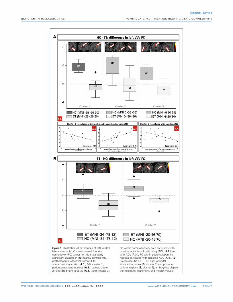
Figure 3. Illustration of differences of left ventrallateral ventral (VLV) seed-to-voxel functionconnectivity (FC) values for the statisticallysignificant clusters in (A) healthy controls (HC) epretherapeutic essential tremor (ET):somatosensory cortex (A.1., left, cluster 1),pedunculopontine nucleus (A.1., center, cluster2), and Brodmann area 32 (A.1., right, cluster 3);
FC within somatosensory area correlates withbaseline activities of daily living (ADL; A.2.) andwith ADL (A.3.); FC within pedunculopontinenucleus correlates with baseline ADL (A.4.). (B)Pretherapeutic ET e HC: right occipitalassociation cortex (B, cluster 1) and posteriorparietal regions (B, cluster 2); all boxplots displaythe minimum, maximum, and median values.
WORLD NEUROSURGERY 113: e453-e464, MAY 2018 www.WORLDNEUROSURGERY.org e457
ORIGINAL ARTICLE
CONSTANTIN TULEASCA ET AL. VENTROLATERAL THALAMUS RESTING-STATE CONNECTIVITY
ORIGINAL ARTICLE
CONSTANTIN TULEASCA ET AL. VENTROLATERAL THALAMUS RESTING-STATE CONNECTIVITY
Lesion Location After ThalamotomyThe classical “ring-enhancing” MR signature,29 visualized 1 yearafter SRS-T, was always located inside the left VLV36 cluster(Figure 1, 1.2., 1.2.1., 1.2.2.).
Clinical CharacteristicsMean age was 70.1 years (range 49e82; 5 males, 12 females). Meanduration of symptoms was 38 years (range 6e70 years). The meanbaseline ADL was 29.1 (standard deviation [SD] 12, range 13e49).The mean decrease in points at 1 year after SRS-T was �23.06 (SD11.9, range 2 to � 45). The mean baseline tremor score on thetreated hand was 18.6 (SD 5.5, range 8e30). The mean decrease inpoints at 1 year after SRS-T was �11.4 (SD 4.1, range �4 to �19).The mean time to tremor improvement was 3.32 months (SD 2.7,range 0.5e10 months).
Radiologic Characteristics: Thalamotomy VolumeThe 1 year thalamotomy MR signature volume has been drawn onT1 (gadolinium-injected) MR 1 year after SRS-T, which isconsidered the definitive radiologic answer in our experience.19 Toensure the accuracy of volume calculation, each patient’s MRI at 1-year follow-up was imported in the Leksell GammaPlan software(Elekta AB) and coregistered with the therapeutic images. Weprojected on the MR-signature the 90-Gy isodose line, whichcorresponds to the final radiologic response, in our previouslypublished experience.19,37 Manually, with the segmentation tools,the draw was made for the individual cases. The “volume” moduleinside the station was used to extract the values.The mean 1 year thalamotomy MR signature volume after SRS-T
was 0.125 mL (SD 0.162, range 0.002e0.600 mL). There was nocorrelation between decrease in ADL or head tremor improvementand lesion volume (P > 0.05), but the former related with tremorscore on the treated hand drop (P ¼ 0.01). The FC values withinthe relevant clusters showed no statistically significant correlationwith lesion volume (P > 0.05).
RESULTS
We evaluated the impact of age, disease duration, or volumelesion, and we report no statistically significant correlation(Spearman > 0.05) with FC values. Furthermore, no statisticallysignificant differences in FC between left and right VLV nucleuswere found.
VLV FC in Pretherapeutic Drug-Naïve Patients with ETDrug-naïve patients with ET, compared with HCs (Figure 2A, A.1.eA.4., Figure 3A and B, and Table 1) showed decreased (mediannegative value) FC between the left VLV and the following clusters:left primary somatosensory area (inferior part, pFWE-cor ¼ 0.035),pedunculopontine nucleus (pFWE-cor ¼ 0.003), and dorsal anteriorcingulate cortex (Brodmann area [BA] 32, pFWE-cor ¼ 0.000;Figure 3, A.1.; Table 1). Furthermore, a decreased pretherapeutic FCwith the primary somatosensory cortex (Spearman ¼ 0.04;Figure 3, A.2.; Table 1) and pedunculopontine nucleus(Spearman ¼ 0.01; Figure 3, A.4.) correlated with baseline ADL. Adecrease in points of ADL 1 year after SRS-T correlated with an in-crease in FC between left VLV and primary somatosensory cortex(Spearman ¼ 0.01, Figure 3, A.3.; Table 1).
e458 www.SCIENCEDIRECT.com WORLD NE
Drug-naïve patients with ET, compared with HCs, revealedincreased left FC with left visual association cortex (BA19,pFWE-cor ¼ 0.005) and left superior parietal regions (BA 7,pFWE-cor ¼ 0.014).
VLV FC at 1 Year After SRS-TA decrease from pretherapeutic positive FC to a median value closeto zero 1 year after SRS-T (Figure 2, B.1.eB.4.; Figure 4, A.1.eA.4.;Table 1) was found for the following clusters: right insular andorbitofrontal cortex (BA 47, pFWE-cor ¼ 0.000), right BA 40(posterior parietal, supramarginal gyrus, pFWE-cor ¼ 0.002), leftBA 13 (anterior insula, pFWE-cor ¼ 0.000), and right BA 44 and 8(inferior frontal gyrus and frontal-eye fields, pFWE-cor ¼ 0.002). Incontrast, an increase from pretherapeutic negative FC to a medianclose to zero 1 year after SRS-T is reported for left VLV FC with rightsupplementary motor area (puncor ¼ 0.015; Figure 4B and Table 1).
DISCUSSION
In the present study, we evaluated the tremor network using aseed-to-voxel approach on resting-state fMRI data, as functionalimaging had been widely used as an alternative for evaluatingsegregated brain processes.38-40 We focus on FC of the mostcommonly used surgical target for tremor, the ventrolateral motorthalamus. With regard to HCs versus pretherapeutic ET FC, wereport as statistically significant: primary somatosensory, visualassociation, and anterior cingulate cortex, as well as pedunculo-pontine nucleus. The longitudinal changes 1 year after SRS-Trelate to dorsal attention (frontal eye-fields and parts of the dor-sal premotor cortex and posterior parietal areas41,42) and salience (i.e.,insula) networks. Additional FC changes are present within severaldistinct clusters: language-related areas (BA 47), meaning andphonology (BA 40), or selective response suppression in go/no-gotasks and hand movements areas (all for BA 44).With regard to differences between HCs and patients with ET,
previous resting-state fMRI studies have provided noteworthyfindings. Buijink et al.22 usedmotor-task fMRI and electromyographyand reported FC decrease in patients with ET between cortical andcerebellar motor regions. Gallea et al.43 analyzed resting-state fMRIdata and reported FC alterations in supplementary motor areas,which were considered consequence of a cerebellar defect and actingto attempt to reduce tremor in motor output by reducing commu-nications with M1 hand areas. More recently, Fang et al.23 used also aregion-of-interest approach and evaluated time-courses of Vim. Theauthors reported FC increase between the Vim andM1 area, as well asa decrease with the cerebellum. Additional resting-state fMRI studiesused other data-driven approaches.21,30
We report multiple network alterations of left VLV prether-apeutic FC in patients with ET. At the cortical level, some of therepresentative regions have already been reported by previousstudies. This includes the primary somatosensory/somatomotorcortex,44 which is responsible for integration of somatic sensation,visual stimuli, and movement planning,45 or BA 32, involved in“Stroop” task.46 We further report newly discovered FCalterations of the left VLV with left posterior parietal BA 7 andleft visual association cortex BA 19. The BA 7 is involved inlocating objects in the space and represents a point ofconvergence between vision and proprioception; its presence
UROSURGERY, https://doi.org/10.1016/j.wneu.2018.02.055

Table 1. Overview of the Main Left VLV FC Results
Set-Level Cluster-Level Peak-Level MNI, Anatomical Area
P pFWE-cor pFDR-cor kc puncor pFWE-cor pFDR-cor T/P ZE puncor puncor
HCeET patients
0.000 0.035 0.007 205 0.002 0.019 0.030 7.19 5.16 0.000 e28 e26 20, L sensory-mot
0.003 0.001 332 0.000 0.187 0.109 5.93 4.57 0.000 0 e36 e36, peduncle-pontine
0.000 0.000 648 0.000 0.250 0.113 5.76 4.48 0.000 e8 20 24, L BA 32
ET patientseHC
0.000 0.005 0.008 314 0.000 0.676 0.566 5.04 4.10 0.000 e34 e78 12, L BA 19
0.014 0.012 254 0.001 0.820 0.566 4.82 3.97 0.000 e20 e46 70, L BA 7
Pretherapeutice1 year after SRS-T (ET patients)
0.000 0.000 0.000 1099 0.000 0.217 0.783 7.23 4.68 0.000 42 24 e8, R BA 47
0.002 0.001 298 0.000 0.341 0.783 6.80 4.53 0.000 56 e38 24, R BA 40
0.000 0.000 660 0.000 0.571 0.783 6.25 4.32 0.000 e34 18 0, L BA 13
0.002 0.001 289 0.000 0.952 0.784 5.29 3.92 0.000 48 16 34, R BA 44 and 8
1 year after SRS-Tepretherapeutic (ET patients)
0.308 0.105 85 0.015 0.883 0.583 5.55 4.03 0.000 22 24 22, R SMA
VLV, ventral lateral ventral; FC, functional connectivity; FWE, family-wise correction; cor, corrected; FDR, false discovery rate; uncor, uncorrected P value; T/P, height threshold; ZE, Z scored; MNI,Montreal Neurological Institute; HC, healthy control; ET, essential tremor; L, left; BA, Brodmann area; SRS-T, stereotactic radiosurgical thalamotomy; R, right; SMA, supplementary motor area.
ORIGINAL ARTICLE
CONSTANTIN TULEASCA ET AL. VENTROLATERAL THALAMUS RESTING-STATE CONNECTIVITY
suggests a functional alteration of sensorial networks in patientswith ET. The left BA 19 presence is somewhat surprising, mostprobably by polysynaptic connections, and would expressescurrently underestimated alterations of visual networks inpatients ET.We also report altered FC of the pedunculopontine nucleus,
which is responsible for modulation of gait (initiation, mainte-nance, modulation, and other stereotyped motor behaviors). Thepedunculopontine nucleus has been classically explored in DBSfor patients with Parkinson disease with axial symptoms lessresponsive to subthalamic nucleus stimulation and is consideredhighly interconnected with the pallido-thalamo-cortical circuit.47
In the context of patients with ET, alterations in FC betweenthis structure and the motor thalamus are most probably relatedto other neurologic features, already acknowledged, mainlydeficits on both balance (the ability to maintain the body withinits base of support) and gait.48 An additional argument in favorof this statement is the found correlation betweenpretherapeutic ADL and FC with this structure, being know thatADL is global score including also aspects related to gate andposture.An added value of this report is the display of longitudinal
changes in FC 1 year after SRS-T. For dorsal attention and saliencenetwork, FC exhibited a decrease from positive median values toones close to zero after SRS-T. This would support the fact thatpretherapeutic global increase in FC described by other authors inthese networks30 is an adaptive change during ET disease course.Thalamotomy done by RS is generating a progressive functionalreorganization of these systems, with further decrease of an
WORLD NEUROSURGERY 113: e453-e464, MAY 2018
originally probably adaptive and compensatory hyperactivitythought to balance tremor appearance and disease progression.The same type of changes described in the previous 2 networks
is applicable to the left VLV FC with anterior insula, which isinvolved in salience and warrants further attention. It is wellacknowledged that the insula is the bottom-up detection of salientevents, allowing switching between other large-scale networks tofacilitate access to attention and working memory resources whena salient event is detected. Moreover and of high relevance, theanterior insula has a strong additional functional coupling withanterior cingulate cortex, facilitating rapid access to the motorsystem.49 It would so act as an integral hub in the generation ofappropriate behavioral responses to salient stimuli.49 Sridharanet al.50 revealed that the right anterior insula plays a critical andcausal role in interchanging between 2 other major networks(central attention and default-mode network), known to demon-strate competitive interactions during cognitive information pro-cessing. Across stimulus modalities, this structure would play acritical and causal role in activating the central attention anddeactivating the default-mode network.50 These entire featureswould give the insula a balance capacity between salience andmotor networks.We also report FC changes with the right supplementary motor
area, which has multiple roles, including postural stability duringstance or walking, bimanual coordination, or the initiation ofinternally generated as opposed to stimulus driven movement.51
Only 2 previous resting-state fMRI studies, not related toRS,52,53 evaluated the effect of HIFU thermocoagulation on brainnetworks up to 3 months after the procedure. A major difference is
www.WORLDNEUROSURGERY.org e459

Figure 4. Illustration of differences of left ventrallateral ventral (VLV) seed-to-voxel functionalconnectivity (FC) values for the statisticallysignificant clusters in (A) pretherapeutic e 1 yearafter stereotactic radiosurgical thalamotomy
(SRS-T): right insular and orbito-frontal (A.1.),posterior parietal cortex (A.2.), left anterior insularcortex (A.3.), right inferior frontal gyrus, andfrontal-eye fields (A.4.). (B) 1 year after SRS-T epretherapeutic: right supplementary motor area.
e460 www.SCIENCEDIRECT.com WORLD NEUROSURGERY, https://doi.org/10.1016/j.wneu.2018.02.055
ORIGINAL ARTICLE
CONSTANTIN TULEASCA ET AL. VENTROLATERAL THALAMUS RESTING-STATE CONNECTIVITY

ORIGINAL ARTICLE
CONSTANTIN TULEASCA ET AL. VENTROLATERAL THALAMUS RESTING-STATE CONNECTIVITY
that HIFU, unlike RS, produces an apparent lesion and clinicaleffect immediately. The delayed effect of RS could account forbrain networks reorganization and allow for plasticity.It is currently considered that a dense network of axonal path-
ways interconnects structurally segregated and functionallyspecialized specific brain regions.54 It has been advocated thatstructural connectivity patterns and functional interactionsbetween different regions of cortex are meaningfullycorrelated.54 Furthermore, strong FC commonly observedbetween regions with no direct structural connection.55 FCchanges might be also due to tissue disruption, with furtherinvolvement of the specific white matter tracts, eventuallyaffected by Vim SRS-T. For instance, in patients with ET treatedwith HIFU, diffusion tensor imaging revealed changes over timenot only at the thermocoagulation site, but also in distant areas ofthe brain. After corrections for multiple comparisons, only remotediffusion tensor imaging changes were correlated with the clinicalimprovement.56 Functional studies using fluorodeoxyglucoseepositron emission tomography also confirmed remote effectsafter Vim DBS in ET. These effects involved the cerebellum, inpatients with posttherapeutic ataxia, suggesting thus possibleclinical effects.57
We have recently reported tremor recovery as related to distantsites changes after SRS-T using resting-state fMRI and whole-brain analysis without prior assumption (i.e., independentcomponent analysis58,59). We reported 2 networks that presentedstatistically significant interconnectivity with visual clusters: oneincluded the bilateral motor network, frontal eye-fields, and leftcerebellum lobule VI, of which interconnectivity strength withright visual BA 19 related to tremor arrest after SRS-T; the secondincluded reminiscent of the salience network, which showedaltered interconnectivity strength with right fusiform gyrus andV5.60
Our study design has several potential limitations, although wehave taken many precautions. One, related to study design, is theuse of resting-state data, which might not be directly related to
WORLD NEUROSURGERY 113: e453-e464, MAY 2018
motor performance; however, we aimed at studying networkchanges in the absence of a task. A second limitation is the smallnumber of subjects. A third one is the lack of data on longitudinalchanges (at 1 year) in the HC group; nevertheless, recent studieshave advocated the reproducibility of functional networks acrossmultiple sessions, including 1 year apart.61 Furthermore, theneurologic evaluation was not blinded. Also, at which exact timepoint SRS-T RS induces these changes in brain networks re-mains unknown. Another aspect is related to resting-state fMRIresolution. Regarding the former, tiny anatomical structures, suchas the pedunculopontine nucleus, are reasonable to assume butillustrate this limitation.In conclusion, the present resting-state fMRI analysis has
allowed us, using longitudinal study after SRS-T, to depict for thefirst time FC reorganization of several major brain networks,which cannot be easily captured by other existing techniques. Wepostulate that the commonly targeted ventrolateral thalamus fordrug-resistant ET would act as a mediator after the intervention,inducing major changes in dorsal attention, salience, and sup-plementary motor networks. The insula would act like a hub indownregulating the relationship between all these aforementionedstructurally segregated, yet functionally highly interconnected,systems. Pretherapeutic hyperactivity of the attentional networksmight be an adaptive change in ET during the disease course, aspreviously postulated by other authors. By SRS-T, it is generated a“functional reset” of these circuits, and they are brought back to a“normal state.” We postulate that a more pre-eminent insular roleas well as of other structures related to attention exists, and thismight further interact with motor-related systems for normalmotor and cognitive homeostasis.
ACKNOWLEDGMENTS
We acknowledge the important contribution of Axelle Cretol, fromMarseille University Hospital (CHU Timone), Marseille, France,who, as research assistant, kept the database up-to-date.
REFERENCES
1. Deuschl G, Elble R. Essential tremor—neurode-generative or nondegenerative disease towards aworking definition of ET. Mov Disord. 2009;24:2033-2041.
2. Louis ED. Clinical practice. Essential tremor.N Engl J Med. 2001;345:887-891.
3. Louis ED. Essential tremor. Lancet Neurol. 2005;4:100-110.
4. Louis ED. Essential tremors: a family of neuro-degenerative disorders? Arch Neurol. 2009;66:1202-1208.
5. Sharifi S, Nederveen AJ, Booij J, vanRootselaar AF. Neuroimaging essentials inessential tremor: a systematic review. NeuroimageClin. 2014;5:217-231.
6. Llinas RR. The olivo-cerebellar system: a key tounderstanding the functional significance ofintrinsic oscillatory brain properties. Front NeuralCircuits. 2013;7:96.
7. Tuleasca C, Najdenovska E, Régis J, Witjas T,Girard N, Champoudry J, et al. Pretherapeuticfunctional neuroimaging predicts tremor arrestafter thalamotomy [e-pub ahead of print]. ActaNeurol Scand https://doi.org/10.1111/ane.12891,Accessed January 7 2018.
8. Hansel C. Reading the clock: how Purkinje cellsdecode the phase of olivary oscillations. Neuron.2009;62:308-309.
9. Bhalsing KS, Saini J, Pal PK. Understanding thepathophysiology of essential tremor throughadvanced neuroimaging: a review. J Neurol Sci.2013;335:9-13.
10. Passamonti L, Novellino F, Cerasa A, Chiriaco C,Rocca F, Matina MS, et al. Altered cortical-cerebellar circuits during verbal working memoryin essential tremor. Brain. 2011;134:2274-2286.
11. Popa T, Russo M, Vidailhet M, Roze E, Lehéricy S,Bonnet C, et al. Cerebellar rTMS stimulation mayinduce prolonged clinical benefits in essentialtremor, and subjacent changes in functional
www
connectivity: an open label trial. Brain Stimul. 2013;6:175-179.
12. Middleton FA, Strick PL. Basal ganglia and cere-bellar loops: motor and cognitive circuits. Brain ResBrain Res Rev. 2000;31:236-250.
13. Hassler R. The influence of stimulations and co-agulations in the human thalamus on the tremorat rest and its physiopathologic mechanism. ProcSecond Intl Congr Neuropath. 1955:637-642.
14. Goldman MS, Ahlskog JE, Kelly PJ. The symp-tomatic and functional outcome of stereotacticthalamotomy for medically intractable essentialtremor. J Neurosurg. 1992;76:924-928.
15. Benabid AL, Pollak P, Gao D, Hoffmann D,Limousin P, Gay E, et al. Chronic electrical stim-ulation of the ventralis intermedius nucleus of thethalamus as a treatment of movement disorders.J Neurosurg. 1996;84:203-214.
16. Schuurman PR, Bosch DA, Bossuyt PM,Bonsel GJ, van Someren EJ, de Bie RM, et al.A comparison of continuous thalamic stimulation
.WORLDNEUROSURGERY.org e461

ORIGINAL ARTICLE
CONSTANTIN TULEASCA ET AL. VENTROLATERAL THALAMUS RESTING-STATE CONNECTIVITY
and thalamotomy for suppression of severetremor. N Engl J Med. 2000;342:461-468.
17. Campbell AM, Glover J, Chiang VL, Gerrard J,Yu JB. Gamma knife stereotactic radiosurgicalthalamotomy for intractable tremor: a systematicreview of the literature. Radiother Oncol. 2015;114:296-301.
18. Kondziolka D, Ong JG, Lee JY, Moore RY,Flickinger JC, Lunsford LD. Gamma Knife thala-motomy for essential tremor. J Neurosurg. 2008;108:111-117.
19. Witjas T, Carron R, Azulay JP, Regis J. Gamma-knife Thamamotomy for Intractable Tremors:Clinical Outcome and Correlations with Neuro-imaging Features. Paper presented at: MDS 17thInternational Congress of Parkinson’s Disease andMovement Disorders. June 16e20, 2013; Sydney,Australia.
20. Elias WJ, Lipsman N, Ondo WG, Ghanouni P,Kim YG, Lee W, et al. A randomized trial offocused ultrasound thalamotomy for essentialtremor. N Engl J Med. 2016;375:730-739.
21. Benito-León J, Louis ED, Romero JP, Hernández-Tamames JA, Manzanedo E, Álvarez-Linera J, et al.Altered functional connectivity in essential tremor:a resting-state fMRI study. Medicine. 2015;94:e1936.
22. Buijink AW, van der Stouwe AM, Broersma M,Sharifi S, Groot PF, Speelman JD, et al. Motornetwork disruption in essential tremor: a func-tional and effective connectivity study. Brain. 2015;138:2934-2947.
23. Fang W, Chen H, Wang H, Zhang H, Puneet M,Liu M, et al. Essential tremor is associated withdisruption of functional connectivity in the ventralintermediate NucleuseMotor CortexeCerebellumcircuit. Hum Brain Mapp. 2016;37:165-178.
24. Lenka A, Bhalsing KS, Panda R, Jhunjhunwala K,Naduthota RM, Saini J, et al. Role of alteredcerebello-thalamo-cortical network in the neuro-biology of essential tremor. Neuroradiology. 2017;59:157-168.
25. Biswal B, Yetkin FZ, Haughton VM, Hyde JS.Functional connectivity in the motor cortex ofresting human brain using echo-planar MRI. MagnReson Med. 1995;34:537-541.
26. Fox MD, Greicius M. Clinical applications ofresting state functional connectivity. Front SystNeurosci. 2010;4:19.
27. Morel A, Magnin M, Jeanmonod D. Multi-architectonic and stereotactic atlas of the humanthalamus. J Comp Neurol. 1997;387:588-630.
28. Battistella G, Najdenovska E, Maeder P,Ghazaleh N, Daducci A, Thiran JP, et al. Robustthalamic nuclei segmentation method based onlocal diffusion magnetic resonance properties.Brain Struct Funct. 2017;222:2203-2216.
29. Witjas T, Carron R, Krack P, Eusebio A,Vaugoyeau M, Hariz M, et al. A prospective single-blind study of Gamma Knife thalamotomy fortremor. Neurology. 2015;85:1562-1568.
e462 www.SCIENCEDIRECT.com
30. Fang W, Chen H, Wang H, Zhang H, Liu M,Puneet M, et al. Multiple resting-state networksare associated with tremors and cognitive featuresin essential tremor. Mov Disord. 2015;30:1926-1936.
31. Bain PG, Findley LJ, Atchison P, Behari M,Vidailhet M, Gresty M, et al. Assessing tremorseverity. J Neurol Neurosurg Psychiatry. 1993;56:868-873.
32. Fahn S, Tolosa E, Marin C. Clinical rating scale fortremor. In: Jankovic J, Tolosa E, eds. Parkinson’sDisease and Movement Disorders. Baltimore: Urbanand Schwarzenberg; 1988:225-234.
33. Schmidt R, Freidl W, Fazekas F, Reinhart B,Grieshofer P, Koch M, et al. The Mattis DementiaRating Scale: normative data from 1,001 healthyvolunteers. Neurology. 1994;44:964-966.
34. Power JD, Barnes KA, Snyder AZ, Schlaggar BL,Petersen SE. Spurious but systematic correlationsin functional connectivity MRI networks arisefrom subject motion. NeuroImage. 2012;59:2142-2154.
35. Song XW, Dong ZY, Long XY, Li SF, Zuo XN,Zhu CZ, et al. REST: a toolkit for resting-statefunctional magnetic resonance imaging data pro-cessing. PLoS One. 2011;6:e25031.
36. Najdenovska E, Tuleasca C, Bloch J, Maeder P,Girard N, Witjas T, et al. Exploring local diffusionMRI properties for vim localization: evaluation inclinical cases. J Neurol Surg A Cent Eur Neurosurg.2017;78:S1-S22.
37. Regis J, Carron R, Park M. Is radiosurgery aneuromodulation therapy? A 2009 Fabrikant awardlecture. J Neurooncol. 2010;98:155-162.
38. Logothetis NK, Pauls J, Augath M, Trinath T,Oeltermann A. Neurophysiological investigationof the basis of the fMRI signal. Nature. 2001;412:150-157.
39. Hipp JF, Siegel M. BOLD fMRI correlation reflectsfrequency-specific neuronal correlation. Curr Biol.2015;25:1368-1374.
40. Zhuge W, Ben-Galim P, Hipp JA, Reitman CA.Efficacy of MRI for assessment of spinal trauma:correlation with intraoperative findings. J SpinalDisord Tech. 2015;28:147-151.
41. Fox MD, Corbetta M, Snyder AZ, Vincent JL,Raichle ME. Spontaneous neuronal activity dis-tinguishes human dorsal and ventral attentionsystems. Proc Natl Acad Sci USA. 2006;103:10046-10051.
42. Vossel S, Geng JJ, Fink GR. Dorsal and ventralattention systems: distinct neural circuits butcollaborative roles. Neuroscientist. 2014;20:150-159.
43. Gallea C, Popa T, García-Lorenzo D,Valabregue R, Legrand AP, Marais L, et al.Intrinsic signature of essential tremor in thecerebello-frontal network. Brain. 2015;138:2920-2933.
44. Muthuraman M, Heute U, Deuschl G, Raethjen J.The central oscillatory network of essentialtremor. Conf Proc IEEE Eng Med Biol Soc. 2010;2010:154-157.
WORLD NEUROSURGERY, http
45. Borich MR, Brodie SM, Gray WA, Ionta S,Boyd LA. Understanding the role of the primarysomatosensory cortex: opportunities for rehabili-tation. Neuropsychologia. 2015;79:246-255.
46. Cerasa A, Passamonti L, Novellino F, Salsone M,Gioia MC, Morelli M, et al. Fronto-parietal over-activation in patients with essential tremor duringStroop task. Neuroreport. 2010;21:148-151.
47. Zhang J, Wang ZI, Baker KB, Vitek JL. Effect ofglobus pallidus internus stimulation on neuronalactivity in the pedunculopontine tegmental nu-cleus in the primate model of Parkinson’s disease.Exp Neurol. 2012;233:575-580.
48. Arkadir D, Louis ED. The balance and gait dis-order of essential tremor: what does this mean forpatients? Ther Adv Neurol Disord. 2013;6:229-236.
49. Menon V, Uddin LQ. Saliency, switching, atten-tion and control: a network model of insulafunction. Brain Struct Funct. 2010;214:655-667.
50. Sridharan D, Levitin DJ, Menon V. A critical rolefor the right fronto-insular cortex in switchingbetween central-executive and default-mode net-works. Proc Natl Acad Sci USA. 2008;105:12569-12574.
51. Roland PE, Larsen B, Lassen NA, Skinhoj E.Supplementary motor area and other cortical areasin organization of voluntary movements in man.J Neurophysiol. 1980;43:118-136.
52. Jang C, Park HJ, Chang WS, Pae C, Chang JW.Immediate and longitudinal alterations of func-tional networks after thalamotomy in essentialtremor. Front Neurol. 2016;7:184.
53. Park HJ, Pae C, Friston K, Jang C, Razi A,Zeidman P, et al. Hierarchical dynamic causalmodeling of resting-state fMRI reveals longitudi-nal changes in effective connectivity in the motorsystem after thalamotomy for essential tremor.Front Neurol. 2017;8:346.
54. Hagmann P, Cammoun L, Gigandet X, Meuli R,Honey CJ, Wedeen VJ, et al. Mapping the struc-tural core of human cerebral cortex. PLoS Biol.2008;6:e159.
55. Honey CJ, Sporns O, Cammoun L, Gigandet X,Thiran JP, Meuli R, et al. Predicting humanresting-state functional connectivity from struc-tural connectivity. Proc Natl Acad Sci USA. 2009;106:2035-2040.
56. Wintermark M, Huss DS, Shah BB, Tustison N,Druzgal TJ, Kassell N, et al. Thalamic connectivityin patients with essential tremor treated with MRimaging-guided focused ultrasound: in vivo fibertracking by using diffusion-tensor MR imaging.Radiology. 2014;272:202-209.
57. Reich MM, Brumberg J, Pozzi NG, Marotta G,Roothans J, Åström M, et al. Progressive gaitataxia following deep brain stimulation foressential tremor: adverse effect or lack of efficacy?Brain. 2016;139:2948-2956.
58. Beckmann C, DeLuca M, Devlin J, Smith S. In-vestigations into resting-state connectivity using
s://doi.org/10.1016/j.wneu.2018.02.055
ORIGINAL ARTICLE
CONSTANTIN TULEASCA ET AL. VENTROLATERAL THALAMUS RESTING-STATE CONNECTIVITY
independent component analysis. Philos Trans RSoc Lond B Biol Sci. 2005;360:1001-1013.
59. Calhoun VD, Adali T, Pearlson GD, Pekar JJ.A method for making group inferences fromfunctional MRI data using independent compo-nent analysis. Hum Brain Mapp. 2001;14:140-151.
60. Tuleasca C, Najdenovska E, Régis J, Witjas T,Girard N, Champoudry J, et al. Clinical responseto Vim’s thalamic stereotactic radiosurgery foressential tremor is associated with distinctivefunctional connectivity patterns. Acta Neurochir(Wien). 2018;160:611-624.
WORLD NEUROSURGERY 113: e453-e46
61. Guo CC, Kurth F, Zhou J, Mayer EA, Eickhoff SB,Kramer JH, et al. One-year test-retest reliability ofintrinsic connectivity network fMRI in olderadults. NeuroImage. 2012;61:1471-1483.
Conflict of interest statement: The work was supported inpart by the Swiss National Science Foundation (SNSF-205321e157040), in part by the Centre d’ImagerieBioMédicale (CIBM) of the University of Lausanne (UNIL), theSwiss Federal Institute of Technology Lausanne (EPFL), theUniversity of Geneva (UniGe), the Centre Hospitalier
4, MAY 2018 www
Universitaire Vaudois (CHUV), the Hôpitaux Universitaires deGenève (HUG), and the Leenaards and Jeantet Foundationsand in part by the CHU Timone, Marseille, France.
Received 9 November 2017; accepted 9 February 2018
Citation: World Neurosurg. (2018) 113:e453-e464.https://doi.org/10.1016/j.wneu.2018.02.055
Journal homepage: www.WORLDNEUROSURGERY.org
Available online: www.sciencedirect.com
1878-8750/$ - see front matter ª 2018 Elsevier Inc. Allrights reserved.
.WORLDNEUROSURGERY.org e463

SUPPLEMENTARY MATERIALS
AUTOMATED 3T DIFUSSION MRI-BASED VENTROLATERAL VENTRAL
(VLV) SEGMENTATIONIn the field of an automated thalamic parcellation, several researchgroups have explored diffusion-weighted imaging information,although mainly by using coarse diffusion features.1-5 Addressingthis limitation, recently our group has proposed an approachbased on spherical harmonics representation of the orientationdistribution functions,6 which provides finer details of thediffusion process within a voxel. With this approach, wesubdivide the thalamus, in a robust and reproducible manner,into 7 groups of nuclei closely matching the anatomy by theMorel’s atlas.7 Among those 7 groups is the VLV thalamic part,which encompasses in its vast surface majority theventrointermediate nucleus and therefore is used as a region orinterest in the presented study.The thalamic mask, required for the parcellation, was first ob-
tained from FreeSurfer (Massachusetts General Hospital, Boston,Massachusetts, USA), cortical and subcortical subdivision8
performed on each individual T1-weighted image and then,
according to Battistella et al.,6 was refined by the use ofinformation from both the probability map representing thecerebrospinal fluid and the fractional anisotropy map.Standard preprocessing steps were applied both for patients
with essential tremor (ET; pretherapeutic, in absence of thala-motomy) and healthy controls, on diffusion-weighted imaging. Inpatients with ET, this included denoising, eddy current, andmotion correction and in healthy control patients denoising, biasfield, eddy current, and motion correction.6,9,10 The compensationof the echo planar imaging distortion was corrected via a nonlinearimage registration between the respective T1-weighted andfractional anisotropy map using FSL FNIRT (non linearregistration).9
An identical procedure was applied for VLV clustering for bothpatients with ET and healthy controls, as reported by Battistellaet al.6 The orientation distribution function coefficients for thefinal thalamic subdivision, which provided the used VLV cluster,were calculated with FSL’s qboot function using maximumspherical harmonics order of 66. The clustering itself was doneusing the modified k-means framework6 for each subject in itsindividual diffusion-image space.
REFERENCES
1. Behrens TE, Johansen-Berg H, Woolrich MW,Smith SM, Wheeler-Kingshott CA, Boulby PA,et al. Non-invasive mapping of connections be-tween human thalamus and cortex using diffusionimaging. Nat Neurosci. 2003;6:750-757.
2. Deoni SC, Rutt BK, Parrent AG, Peters TM. Seg-mentation of thalamic nuclei using a modified k-means clustering algorithm and high-resolutionquantitative magnetic resonance imaging at 1.5T. Neuroimage. 2007;34:117-126.
3. Mang SC, Busza A, Reiterer S, Grodd W,Klose AU. Thalamus segmentation based on thelocal diffusion direction: a group study. MagnReson Med. 2012;67:118-126.
4. Wiegell MR, Tuch DS, Larsson HB, Wedeen VJ.Automatic segmentation of thalamic nuclei fromdiffusion tensor magnetic resonance imaging.Neuroimage. 2003;19:391-401.
5. Ziyan U, Tuch D, Westin CR. Segmentation ofthalamic nuclei from DTI using spectral clus-tering. Med Image Comput Assist Interv. 2006;4191:807-814.
6. Battistella G, Najdenovska E, Maeder P,Ghazaleh N, Daducci A, Thiran JP, et al. Robustthalamic nuclei segmentation method based onlocal diffusion magnetic resonance properties.Brain Struct Funct. 2017;222:2203-2216.
7. Morel A, Magnin M, Jeanmonod D. Multi-architectonic and stereotactic atlas of the humanthalamus. J Comp Neurol. 1997;387:588-630.
8. Fischl B, Salat DH, Busa E, Albert M, Dieterich M,Haselgrove C, et al. Whole brain segmentation:automated labeling of neuroanatomical structuresin the human brain. Neuron. 2002;33:341-355.
9. Smith SM, Jenkinson M, Woolrich MW,Beckmann CF, Behrens TE, Johansen-Berg H,et al. Advances in functional and structural MRimage analysis and implementation as FSL. Neu-roimage. 2004;23(suppl 1):S208-219.
10. Zhang Y, Brady M, Smith S. Segmentation ofbrain MR images through a hidden Markovrandom field model and the expectation-maximization algorithm. IEEE Trans Med Imaging.2001;20:45-57.
e464 www.SCIENCEDIRECT.com WORLD NEUROSURGERY, https://doi.org/10.1016/j.wneu.2018.02.055
ORIGINAL ARTICLE
CONSTANTIN TULEASCA ET AL. VENTROLATERAL THALAMUS RESTING-STATE CONNECTIVITY