vanadate increases glucocorticoid receptor-mediated gene
TRANSCRIPT

Journal of Steroid Biochemistry & Molecular Biology 80 (2002) 35–47
Vanadate increases glucocorticoid receptor-mediated gene expression:a novel mechanism for potentiation of a steroid receptor
Sergio Li Calzi, Sumudra Periyasamy, Da-Pei Li, Edwin R. Sánchez∗Department of Pharmacology, Medical College of Ohio, 3035 Arlington Avenue, Toledo, OH 43614-5804, USA
Accepted 5 October 2001
Abstract
Transition metal oxyanions, such as molybdate, tungstate and vandadate, have been shown to prevent in vitro hormone-induced activationof the glucocorticoid receptor (GR) by blocking dissociation of the GR/heat shock protein heterocomplex. In this work, we report a noveleffect of vanadate: in vivo potentiation ofGR-mediated gene expression. In cells stably-transfected with complex (mouse mammary tumorvirus (MMTV)) or minimal GR-regulated CAT reporters, treatment with 500�M vanadate causedCAT gene expression to dramaticallyincrease, even at saturating concentrations of dexamethasone; while no such effect was seen in response to RU486 antagonist. Similartreatment with molybdate had no effect on GR activity, suggesting that the response to vanadate was not a general property of transitionmetal oxyanions. Treatment with vanadate after hormone-induced nuclear translocation of the GR also caused potentiation, demonstratingthat vanadate was acting on a post-transformation event, perhaps by affecting the transactivation function of DNA-bound GR. Paradoxically,vanadate caused an apparent but temporary “loss” of GR protein immediately after treatment (as measured by loss of reactivity to BuGR2antibody and of hormone-binding capacity) that returned to normal at approximately 8 h post-treatment, suggesting that potentiation of GRtransactivation function (as measured by our CAT assays) was probably occurring during the later stages (8–24 h) of this assay. However, gelshift analyses revealed that vanadate could induce binding of the hormone-free GR to glucocorticoid response element (GRE)-containingoligonucleotides immediately after treatment. Thus, the rapid vanadate-induced “loss” of GR was not due to degradation of GR protein.Yet, vanadate in the absence of hormone had no effect on CAT reporter expression, demonstrating that this form of the GR still requiresagonist for its enhanced transcriptional activity. As an indication of the potential mechanism of vanadate action, vanadate was found todramatically stimulate the mitogen-activated protein kinases, ERK-1 and ERK-2. In addition, vanadate potentiation ofGR reporter geneexpression was completely blocked by the tyrosine kinase inhibitor herbimycin A. Taken as a whole, our results suggest that vanadatecan have dramatic and complex effects on GR structure and function, resulting in hormone-free activation of GR DNA-binding function,as well as alterations to the BuGR2 epitope and hormone-binding domains—while at the same time stimulating tyrosine phosphorylationpathways controllingGR-mediated gene transcription. © 2002 Elsevier Science Ltd. All rights reserved.
Keywords: Vanadate; Steroid receptor; Gene expression
1. Introduction
The glucocorticoid receptor (GR) is a hormone-dependenttranscription factor that serves to positively or negativelyregulate specific gene expression. In the absence of hor-mone, the GR is known to reside in the cytoplasm of mostcells as a heteromeric complex containing the Hsp90-basedchaperone complex (for a comprehensive review of thisand related topics, see [1]). Based on this structure andlocalization, it is generally accepted that the major stages ofthe GR signaling pathway include the following sequenceof events: hormone-binding, dissociation of the GR/Hspcomplex, translocation to the nucleus of hormone-bound
∗ Corresponding author. Tel.:+1-419-383-4182; fax:+1-419-383-2871.E-mail address: [email protected] (E.R. Sanchez).
GR, binding by GR to high-affinity response elementswithin chromatin, and, finally, enhancement of Pol II-basedtranscription through mediation by co-activators.
As a result of numerous studies done under cell-freeconditions, it has been suggested that sodium molybdateand other transition metal oxyanions (tungstate and vana-date) could be used to effectively block the second stage ofthis signal pathway, namely, hormone-induced dissociationof the GR heterocomplex (a process also referred to as“transformation”). In these studies, addition of molybdate,vanadate or tungstate to cytosols has been shown to preventGR transformation, not only in response to hormone, butalso in response to elevated temperatures, salt, or precipita-tion with heparin or ammonium sulfate [2–8]. Addition ofmolybdate after the transformation event has been shownto not cause re-association of the GR/Hsp90 complex, nor
0960-0760/02/$ – see front matter © 2002 Elsevier Science Ltd. All rights reserved.PII: S0960-0760(01)00180-7

36 S. Li Calzi et al. / Journal of Steroid Biochemistry & Molecular Biology 80 (2002) 35–47
to prevent subsequent binding of GR to DNA [8], suggest-ing that these oxyanions act to prevent an irreversible stepin the transformation process. Evidence suggests that thesite of action for this effect is a low-affinity, ATP-bindingsite on Hsp90, as both vanadate and permolybdate can alterthe conformation of purified Hsp90 in a manner similar tothat seen with azido-ATP [9–12].
Although the above observations suggest that molybdateand similar compounds should be effective inhibitors of GRsignaling in vivo, few reports exist to support this notion.In the original work of its kind, Samuels and co-workersshowed that treatment of intact GH1 cells with 30 mMsodium molybdate (presumably by simply adding molybdateto the medium) reduced the rate of nuclear accumulationof hormone-bound GR [13], as measured by fractionationstudies of cells incubated with radiolabeled hormone. Yangand DeFranco used liposome-mediated delivery of moly-bdate into cells, achieved similar results by demonstratingthat molybdate could block both hormone-induced disso-ciation of the GR/Hsp90 complex and nuclear import ofGR [14]. In general, therefore, these limited in vivo studiessupport the mechanism of action proposed for these com-pounds under cell-free conditions, namely, stabilization ofthe GR heterocomplex against hormone-induced transfor-mation.
Because of this history, we were recently surprised to findthat vanadate treatment of intact cells can also have a stim-ulatory effect on GR activity. This was discovered as part ofour efforts to use sodium vanadate to inhibit heat shock fac-tor (HSF1) activity in stressed cells [15]. In that work, HSF1inactivation was achieved through a mechanism that wasconsistent with the known ability of vanadate to act as aninhibitor of tyrosine phosphatases [16–18], resulting in sub-sequent inhibition of HSF1 via MAPK (ERK-2)-mediatedphoshorylation [19,20]. However, in cells stably-transfectedwith a GR-responsive CAT reporter, we have observed thatvanadate treatment under non-stress conditions greatlyincreases hormone-inducedCAT gene expression. This ob-servation has led to the following question: Is potentiationof GR activity by vanadate mediated by an effect on theHsp90-based chaperone complex or by a novel mechanisminvolving phosphorylation events controlling GR transacti-vation? In the course of addressing this question, we haveuncovered an unusual and paradoxical mechanism in whichvanadate initially causes a temporary loss of GR that is func-tional for hormone-binding and BuGR2 antibody reactivity,followed by a large increase in the transactivation functionof DNA-bound GR. As these effects were not seen in cellstreated with molybdate, and as the vanadate effect is seen forGR that has already undergone hormone-induced nucleartranslocation, it is clear that vanadate potentiation is not op-erating via the traditional effects of these compounds at theuntransformed GR heterocomplex. Instead, it appears thatvandate potentiation of GR transactivation function is theresult of phosphorylation mechanisms that either controlthe intrinsic activity of the DNA-bound GR, or the activity
of the Pol II complex, including co-activators, that respondto GR.
2. Results
2.1. Potentiation of GR-mediated reporter geneexpression by sodium vanadate
In previous work by our laboratory [15], studying theeffects of heat shock on GR activity, we used sodium vana-date to inhibit HSF1 activity in stressed cells by a mechanisminvolving MAPK (ERK-1 and ERK-2)-mediated phospho-rylation of HSF1 [19,20]. In those studies, the presumptivetarget of vanadate action was the dual-specificity MAPKphosphatase, MKP1, inhibition of which by vanadate hasbeen shown to induce MAPK activity [17,18]. In the courseof establishing control conditions for those experiments, wetreated L929 cells stably-transfected with the pMMTV-CATreporter (LMCAT2 cells) with vanadate in the expectationthat this drug should either be neutral with respect to GR ac-tion, or should inhibit the GR-based on the well-documentedeffects of vanadate and similar oxyanions on GR activityin vitro [2–8] and in vivo [13,14]. To our surprise, neitheroutcome was observed. Instead, vanadate treatment ofLMCAT2 cells resulted in a dose-dependent potentiationof dexamethasone-induced pMMTV-CAT expression(Fig. 1A), with increased activity seen with vanadate rangingfrom 50 to 500�M. A potential explanation for this effectwas that vanadate at these seemingly-high concentrationswas actually a form of chemical shock, such as that seen withsodium arsenite, which we have previously shown will causesimilar increases of GR activity in these same cells [21,22].However, this mechanism was clearly not operating, as vana-date treatment (500�M) of L929 cells stably-transfectedwith an HSF1-responsive CAT reporter was found to inhibitheat and arsenite induction of HSF1 activity [15].
As the mouse mammary tumor virus (MMTV) promo-ter (≈1200 bp) contains many potential binding sites fortransacting factors other than GR (any one of which couldbe responsible for potentiation of GR action through co-operative binding at the promoter), we tested the effectsof vanadate in cells stably-transfected with the minimalpGRE2EIB-CAT reporter (LGRECAT cells), in which CATexpression is solely controlled by two synthetic glucocorti-coid response elements (GREs) and a TATA box [23]. In theexperiments of Fig. 1B, LGRECAT cells were left untreatedor were treated with 500�M vanadate prior to exposureto increasing concentrations of dexamethasone. The resultsshow that vanadate potentiation of dexamethasone-inducedCAT expression is concentration-dependent, with the largestincreases occurring at 100 nM and 1�M hormone. As max-imal dexamethasone-induced CAT activity in these cells (inthe absence of vanadate treatment) occurs at 1�M dexam-ethasone, it is apparent that vanadate treatment is a way inwhich latent activity on the part of GR can be unmasked.

S. Li Calzi et al. / Journal of Steroid Biochemistry & Molecular Biology 80 (2002) 35–47 37
Fig. 1. Sodium vanadate increases GR-mediatedCAT gene expression controlled by complex and minimal promoters, even at saturating concentrationsof dexamethasone. Panel A: L929 cells stably-transfected with a CAT reporter construct controlled by the complex MMTV–LTR promoter (LMCAT2cells) were subjected to the indicated conditions prior to measurement of CAT activity. (C): no treatment; Dex: 1�M dexamethasone for 20 h; SV+ Dex:50–500�M sodium vanadate for 2.5 h followed by washing and additional culture in the presence of 1�M dexamethasone for 20 h. The results shownrepresent the means± S.E.M. of six independent experiments. Panel B: L929 cells stably-transfected with a CAT reporter construct controlled by thesynthetic, pGRE2E1B minimal promoter (LGRECAT cells) were left untreated (light bars) or were treated with 500�M sodium vanadate for 2.5 h (darkbars). After washing, cells were incubated with increasing concentrations of dexamethasone for 20 h followed by measurement of CAT activity. Theresults shown represent the mean± S.E.M. of three independent experiments.
More importantly, the results also demonstrate that thevanadate effect cannot be explained by the involvement ofany other transacting factor, suggesting that unmasking ofthis latent activity requires only GR, or non-DNA-bindingfactors controlling GR action.
To further characterize the hormone requirements of thisresponse, we tested whether potentiation of GR by vanadatecould also be seen in the presence of RU486 antagonist(Fig. 2A). As expected, vanadate treatment caused a dra-matic increase in dexamethasone-induced CAT expression,while RU486 alone had no effect on GR transactivity. Incells exposed to vanadate and RU486, an increase in theRU486 response was observed. However, the magnitude of
Fig. 2. Vanadate potentiation of GR-mediated CAT expression is agonist specific and does not occur with sodium molybdate. Panel A: LGRECAT cellswere subjected to the indicated conditions followed by assay for CAT activity. The results shown represent the mean± S.E.M. of four independentexperiments. (C): no treatment; Dex: 1�M dexamethasone for 20 h; SV/Dex: 500�M sodium vanadate for 2.5 h followed by washing and culture in thepresence of 1�M dexamethasone for 20 h; RU: 1�M RU486 for 20 h; SV/RU: 500�M sodium vanadate for 2.5 h followed by washing and culture inthe presence of 1�M RU486 for 20 h. Panel B: LGRECAT cells were treated as above, except that 500�M sodium molybdate (SM) was used. Theresults shown represent the mean± S.E.M. of two independent experiments. (C): no treatment; Dex: 1�M dexamethasone for 20 h; SM/Dex: 500�Msodium molybdate for 2.5 h followed by washing and culture in the presence of 1�M dexamethasone for 20 h; Dex/SM: 1�M dexamethasone for 2 hfollowed by 500�M sodium molybdate for 2.5 h, washing, and culture in the presence of 1�M dexamethasone for 20 h.
CAT activity under this condition was still much lower thanthat seen with dexamethasone+ vanadate. We conclude,therefore, that vanadate cannot dramatically overcome theantagonist activity of RU486 and that vanadate’s action asa potentiator requires agonist-bound receptor.
As stated earlier, the vast preponderance of evidencesuggests that vanadate, like molybdate, should act to blockhormone-induced transformation of GR heterocomplexes,leading to a blockade of all downstream events. Yet, in oursystem, vanadate appears to have the opposite effect, sug-gesting that this drug is acting by a distinct mechanism. Ifso, we were interested to know if sodium molybdate also be-haved like vanadate in our system. To test this, we prepared

38 S. Li Calzi et al. / Journal of Steroid Biochemistry & Molecular Biology 80 (2002) 35–47
stocks of molybdate in the same manner as our active sodiumvanadate solutions, followed by treatment of LGRECATcells (Fig. 2B). The results show that pretreatment of cellswith 500�M sodium molybdate has little or no effect onGR-mediated CAT expression, especially when compared tovanadate at the same concentration (Figs. 1B and 2A). Thus,vanadate, as used in this study, appears to be operating bya mechanism not involving the typical actions of transitionmetal oxyanions to stabilize GR heterocomplexes.
2.2. Vanadate alters GR conformation, leadingto temporary loss of BuGR2 antibody reactivityand hormone-binding function
As an initial test of the mechanism by which vanadatecauses potentiation of the GR, we attempted to measurethe effects of vanadate on GR nuclear translocation. Thiswas done by fractionation of cells, followed by immune-purification and Western-blot analysis using the BuGR2monoclonal antibody against GR (Fig. 3). As expected, theresults show that hormone-free GR is predominantly foundin the cytosolic fraction of cells, while hormone treatment(1�M Dex, 1 h) results in GR that is predominantly in thenuclear fraction (Fig. 3A). To our surprise, however, vana-date treatment resulted in no detectable GR. This ability ofvanadate to apparently cause a complete loss of GR pro-tein was highly consistent and could be seen whether cellswere treated with vanadate alone (SV condition), or treatedwith vanadate either before or after exposure to hormone(see SV/Dex and Dex/SV conditions of Fig. 3A). The lattercondition was particularly interesting, as it suggested thatvanadate was causing “loss” of GR after it had alreadytranslocated to the nucleus (compare with Dex condition).However, one possible explanation for this result was that
Fig. 3. Vanadate causes rapid loss of BuGR2-reactive GR. Panel A: LGRECAT cells subjected to the indicated conditions were fractionated into cytosolic(C) and nuclear (N) portions by salt extraction followed by immunoadsorption of GR and Western-blotting using BuGR2 antibody, see text. Resultsare representative of five independent experiments. mGR: baculovirus-expressed mouse GR; (C): no treatment; Dex: 1�M dexamethasone for 1 h; SV:500�M sodium vanadate for 2.5 h; SV/Dex: 500�M sodium vanadate for 2.5 h followed by 1�M dexamethasone for 1 h; Dex/SV: 1�M dexamethasonefor 1 h followed by 500�M sodium vanadate for 2.5 h; HC= IgG heavy chain. Panel B: LGRECAT cells were fractionated as in Panel A, except GR wasextracted by SDS followed by immunoadsorption of GR and Western-blotting using BuGR2 antibody. Dex: 1�M dexamethasone for 1 h; SV: 500�Msodium vanadate for 2.5 h; mGR: baculovirus-expressed mouse GR. Panel C: LGRECAT cells were incubated with 500�M vanadate for 0, 15, 30, 60and 120 min followed by preparation of cytosolic extracts containing equal protein and Western-blotting with BuGR2 antibody. Results are representativeof two independent experiments.
vanadate was somehow causing the GR to become tightlybound within the nucleus, even in the absence of hormone.The experiments of Fig. 3A used salt extraction (400 mMNaCl) to remove GR from the nuclear pellet. As a morestringent means by which to extract GR, we used a methodinvolving solubilization of nuclei with SDS (Fig. 3B). Here,also the results were the same—lack of BuGR2-reactiveGR following vanadate treatment.
Based on these data, it seemed highly unlikely that tightnuclear binding was responsible for these results. Instead,the most logical hypothesis was that vanadate was sim-ply causing loss of GR through degradation. However, ifdegradation of GR was occurring, it had to be operatingfor both the hormone-free GR present in the cytoplasm andthe hormone-bound GR localized to the nucleus (Fig. 3A).Moreover, vanadate-induced “loss” of GR was observedimmediately after the 2.5 h vanadate treatment (Fig. 3A),suggesting a faster-acting mechanism than that typicallyassociated with degradative processes. To measure exactlyhow fast vanadate was acting on the GR, we subjectedLGRECAT cells to a time-course of exposure to vanadate,followed by measurement of GR reactivity to BuGR2 anti-body in cytosols (Fig. 3C). The results show a complete lossof BuGR2-reactive GR within 30 min of vanadate treatment.Thus, this effect of vanadate on the GR is very rapid.
Given these results, we decided to test the possibility thatvanadate was simply causing an alteration of the BuGR2epitope, making it unreactive to antibody. If true, this mech-anism would mean that we should be able to label the GRby other means, such as through use of radio-labeled lig-and. As our first test of this, we incubated cytosols fromcontrol and vanadate-treated cells with the covalent ligand,[3H]dexamethasone 21-mesylate (Fig. 4). Once more theresults were unexpected—loss of mesylate-labeled GR in
S. Li Calzi et al. / Journal of Steroid Biochemistry & Molecular Biology 80 (2002) 35–47 39
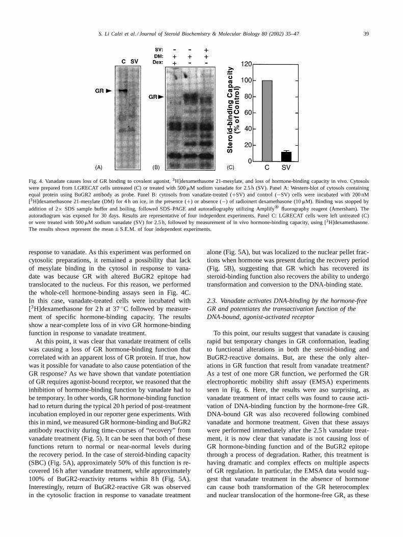
Fig. 4. Vanadate causes loss of GR binding to covalent agonist,3H]dexamethasone 21-mesylate, and loss of hormone-binding capacity in vivo. Cytosolswere prepared from LGRECAT cells untreated (C) or treated with 500�M sodium vanadate for 2.5 h (SV). Panel A: Western-blot of cytosols containingequal protein using BuGR2 antibody as probe. Panel B: cytosols from vanadate-treated (+SV) and control (−SV) cells were incubated with 200 nM[3H]dexamethasone 21-mesylate (DM) for 4 h on ice, in the presence (+) or absence (−) of radioinert dexamethasone (10�M). Binding was stopped byaddition of 2× SDS sample buffer and boiling, followed SDS–PAGE and autoradiography utilizing Amplify® fluorography reagent (Amersham). Theautoradiogram was exposed for 30 days. Results are representative of four independent experiments. Panel C: LGRECAT cells were left untreated (C)or were treated with 500�M sodium vanadate (SV) for 2.5 h, followed by measurement of in vivo hormone-binding capacity, using [3H]dexamethasone.The results shown represent the mean± S.E.M. of four independent experiments.
response to vanadate. As this experiment was performed oncytosolic preparations, it remained a possibility that lackof mesylate binding in the cytosol in response to vana-date was because GR with altered BuGR2 epitope hadtranslocated to the nucleus. For this reason, we performedthe whole-cell hormone-binding assays seen in Fig. 4C.In this case, vanadate-treated cells were incubated with[3H]dexamethasone for 2 h at 37◦C followed by measure-ment of specific hormone-binding capacity. The resultsshow a near-complete loss of in vivo GR hormone-bindingfunction in response to vanadate treatment.
At this point, it was clear that vanadate treatment of cellswas causing a loss of GR hormone-binding function thatcorrelated with an apparent loss of GR protein. If true, howwas it possible for vanadate to also cause potentiation of theGR response? As we have shown that vandate potentiationof GR requires agonist-bound receptor, we reasoned that theinhibition of hormone-binding function by vanadate had tobe temporary. In other words, GR hormone-binding functionhad to return during the typical 20 h period of post-treatmentincubation employed in our reporter gene experiments. Withthis in mind, we measured GR hormone-binding and BuGR2antibody reactivity during time-courses of “recovery” fromvanadate treatment (Fig. 5). It can be seen that both of thesefunctions return to normal or near-normal levels duringthe recovery period. In the case of steroid-binding capacity(SBC) (Fig. 5A), approximately 50% of this function is re-covered 16 h after vanadate treatment, while approximately100% of BuGR2-reactivity returns within 8 h (Fig. 5A).Interestingly, return of BuGR2-reactive GR was observedin the cytosolic fraction in response to vanadate treatment
alone (Fig. 5A), but was localized to the nuclear pellet frac-tions when hormone was present during the recovery period(Fig. 5B), suggesting that GR which has recovered itssteroid-binding function also recovers the ability to undergotransformation and conversion to the DNA-binding state.
2.3. Vanadate activates DNA-binding by the hormone-freeGR and potentiates the transactivation function of theDNA-bound, agonist-activated receptor
To this point, our results suggest that vanadate is causingrapid but temporary changes in GR conformation, leadingto functional alterations in both the steroid-binding andBuGR2-reactive domains. But, are these the only alter-ations in GR function that result from vanadate treatment?As a test of one more GR function, we performed the GRelectrophoretic mobility shift assay (EMSA) experimentsseen in Fig. 6. Here, the results were aso surprising, asvanadate treatment of intact cells was found to cause acti-vation of DNA-binding function by the hormone-free GR.DNA-bound GR was also recovered following combinedvanadate and hormone treatment. Given that these assayswere performed immediately after the 2.5 h vanadate treat-ment, it is now clear that vanadate is not causing loss ofGR hormone-binding function and of the BuGR2 epitopethrough a process of degradation. Rather, this treatment ishaving dramatic and complex effects on multiple aspectsof GR regulation. In particular, the EMSA data would sug-gest that vanadate treatment in the absence of hormonecan cause both transformation of the GR heterocomplexand nuclear translocation of the hormone-free GR, as these

40 S. Li Calzi et al. / Journal of Steroid Biochemistry & Molecular Biology 80 (2002) 35–47
Fig. 5. Loss and recovery of GR SBC and BuGR2 antibody reactivity during a sodium vanadate post-treatment time-course. LGRECAT cells weresubjected to the indicated conditions, followed by measurement of in vivo SBC or Western-blot analysis of GR in cytosolic (C) and nuclear (N) fractions.Panel A: effect of vanadate on BuGR2-reactivity and SBC during hormone-free recovery. (−D): no treatment; (+D): 1�M dexamethasone for 1 h; SV:500�M sodium vanadate for 2.5 h; SV/2 through 18: 500�M sodium vanadate for 2.5 h followed by washing and recovery in the absence of hormonefor 2, 8, 12, 16 and 18 h. SBC results represent the mean± S.E.M. of two independent experiments. Western-blot results are representative of twoindependent experiments. Panel B: effect of vanadate on BuGR2-reactivity during recovery in the presence of hormone. (+D): 1�M dexamethasone for1 h; SV: 500�M sodium vanadate for 2.5 h; SV/2 and 8: 500�M sodium vanadate for 2.5 h followed by washing and recovery in the presence of 1�Mdexamethasone for 2 and 8 h.
Fig. 6. Sodium vanadate activates DNA binding by the hormone-free GR.LGRECAT cells were subjected to the indicated conditions followed bypreparation of nuclear extracts and EMSA assay for GR DNA-binding.(C): no treatment; (D): 1�M dexamethasone for 1 h; D+Ab: 1�M dexam-ethasone for 1 h, followed by incubation of nuclear extracts with BuGR2antibody against GR prior to EMSA assay; SV: 500�M sodium vanadatefor 2.5 h; SV/Dex: 500�M sodium vanadate for 2.5 h followed by 1�Mdexamethasone for 1 h. Panel A: autoradiogram of typical results. Arrowindicates major band corresponding to oligo-bound GR. Panel B: quan-titation of results by densitometric scanning of the autoradiograms. Theresults shown represent the mean±S.E.M. of 11 independent experiments.
EMSA assays were performed solely on nuclear extracts.Vanadate-induced transformation of the GR heterocomplexwould be consistent with the ability of vanadate to causeloss of GR hormone-binding function (Fig. 4), since a largenumber of reports support a model in which GR interac-tion with Hsp90 is required for maintenance of receptorsteroid-binding function (see [1]).
Based on the above, it is appears that vanadate is havingmultiple and, seemingly, diametrically-opposed actions onGR function—temporary loss of hormone-binding, yet in-creasedGR-mediated reporter gene activity. In an attemptto clarify how vanadate was increasing GR-mediated tran-
Fig. 7. Sodium vanadate increases GR-mediated CAT expression bothbefore and after agonist-induced activation of the receptor. LGRECATcells were subjected to the indicated conditions followed by assay forCAT activity. The results shown represent the mean± S.E.M. of 8–12independent experiments. (C): no treatment; Dex: 1�M dexamethasonefor 20 h; SV: 500�M sodium vanadate for 2.5 h; SV/Dex: 500�M sodiumvanadate for 2.5 h followed by washing and culture in the presence of1�M dexamethasone for 20 h; Dex/SV: 1�M dexamethasone for 2 hfollowed by 500�M sodium vanadate for 2.5 h, washing, and culture inthe presence of 1�M dexamethasone for 15.5 h.
S. Li Calzi et al. / Journal of Steroid Biochemistry & Molecular Biology 80 (2002) 35–47 41
scription, we decided to test if the potentiation effect ofvanadate on GR-induced CAT activity could be seen afterhormone-induced activation of the receptor. In other words,does vanadate increase GR activity when it is alreadybound with hormone and to its high-affinity sites withinpromoters? To achieve this, we subjected LGRECAT cellsto vanadate treatment either before or after incubation withhormone treatment (Fig. 7). The results show approximatelythe same level of potentiation of GR-mediated CAT expres-sion by vanadate, regardless of order of addition. As the
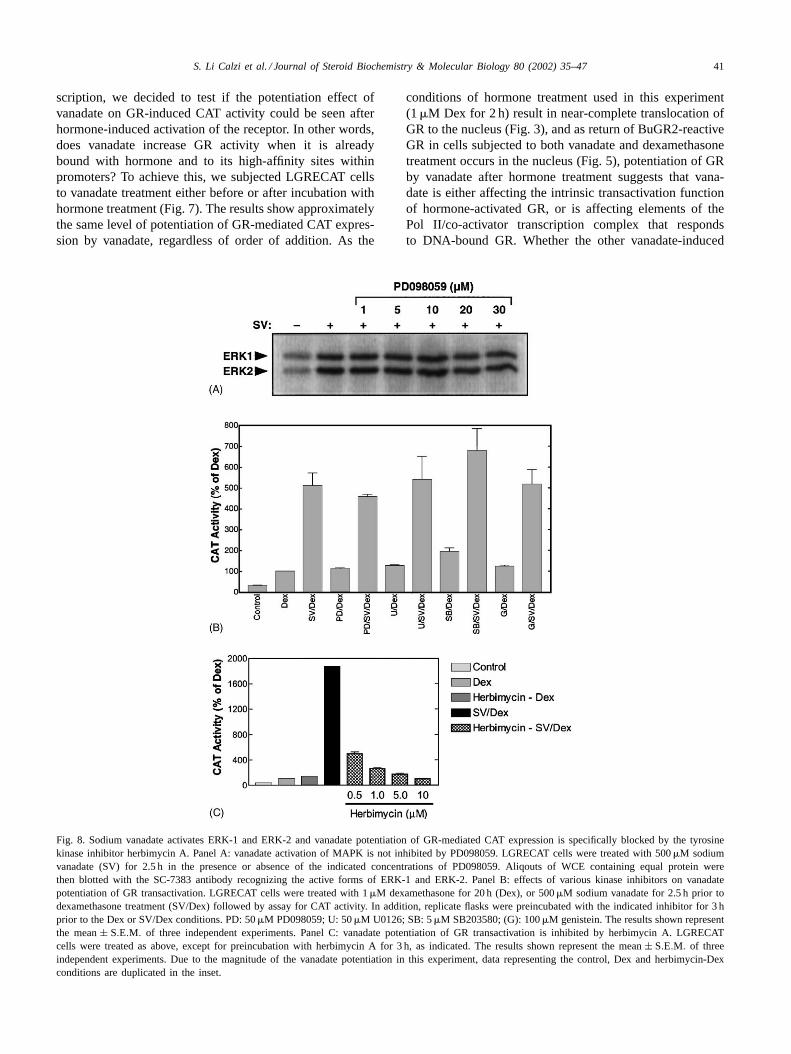
Fig. 8. Sodium vanadate activates ERK-1 and ERK-2 and vanadate potentiation of GR-mediated CAT expression is specifically blocked by the tyrosinekinase inhibitor herbimycin A. Panel A: vanadate activation of MAPK is not inhibited by PD098059. LGRECAT cells were treated with 500�M sodiumvanadate (SV) for 2.5 h in the presence or absence of the indicated concentrations of PD098059. Aliqouts of WCE containing equal protein werethen blotted with the SC-7383 antibody recognizing the active forms of ERK-1 and ERK-2. Panel B: effects of various kinase inhibitors on vanadatepotentiation of GR transactivation. LGRECAT cells were treated with 1�M dexamethasone for 20 h (Dex), or 500�M sodium vanadate for 2.5 h prior todexamethasone treatment (SV/Dex) followed by assay for CAT activity. In addition, replicate flasks were preincubated with the indicated inhibitor for 3 hprior to the Dex or SV/Dex conditions. PD: 50�M PD098059; U: 50�M U0126; SB: 5�M SB203580; (G): 100�M genistein. The results shown representthe mean± S.E.M. of three independent experiments. Panel C: vanadate potentiation of GR transactivation is inhibited by herbimycin A. LGRECATcells were treated as above, except for preincubation with herbimycin A for 3 h, as indicated. The results shown represent the mean± S.E.M. of threeindependent experiments. Due to the magnitude of the vanadate potentiation in this experiment, data representing the control, Dex and herbimycin-Dexconditions are duplicated in the inset.
conditions of hormone treatment used in this experiment(1�M Dex for 2 h) result in near-complete translocation ofGR to the nucleus (Fig. 3), and as return of BuGR2-reactiveGR in cells subjected to both vanadate and dexamethasonetreatment occurs in the nucleus (Fig. 5), potentiation of GRby vanadate after hormone treatment suggests that vana-date is either affecting the intrinsic transactivation functionof hormone-activated GR, or is affecting elements of thePol II/co-activator transcription complex that respondsto DNA-bound GR. Whether the other vanadate-induced

42 S. Li Calzi et al. / Journal of Steroid Biochemistry & Molecular Biology 80 (2002) 35–47
changes in GR function (hormone-binding function andBuGR2-reactivity) play a role in the potentiation mecha-nism is still not clear, as these early changes, especiallythe altered reactivity to BuGR2 antibody, may contribute toincreased transcriptional activity that can only be detectedat the later time at which our CAT assays are performed.
2.4. Vanadate activates MAPK and vanadate potentiationof GR transactivity is blocked by the tyrosine kinaseinhibitor herbimycin A
Given the well-documented role of vanadate as an inhi-bitor of protein phosphatases, we set out to test the role ofprotein phosphorylation in the mechanism by which thiscompound increases GR transactivity. As an initial test ofthis idea, we measured activation of the ERK-1 and ERK-2MAPK family members through use of an antibody againstthe active, phosphorylated forms of these enzymes. In theWestern-blot experiment of Fig. 8A, it can be seen that vana-date treatment of the LGRECAT cells dramatically increasesthe amounts of active ERK-1 and ERK-2. In the hopes thatan inhibitor of ERK could, therefore, be used to determinethe involvement of these kinases in the vanadate potentiationof GR, we tested whether vanadate activation of ERK-1 andERK-2 could be blocked by the MAPK inhibitor PD098059.These results are also seen in the blot of Fig. 8A and demon-strate that PD098059 has no effect on this response. As theprincipal target of PD098059 is known to be MAPK kinase(the immediate upstream regulator of MAPK; also known
Fig. 9. Model for potentiation ofGR-mediated gene expression by sodium vanadate. We have shown that potentiation ofGR-mediated reporter geneexpression by sodium vanadate does not result from stabilization of the GR/Hsp90 heterocomplex. Rather, it is a process that occurs after agonist-inducednuclear translocation of the receptor and that can be completely blocked by tyrosine kinase inhibitor (herbimycin A). Based on these results, we proposethat vanadate potentiation of GR occurs through phosphorylation events that target the GR or elements of the Pol II/co-regulator transcription complexthat interact with DNA-bound GR.
as MEK), these results may simply indicate that vanadateactivation of MAPK (presumably through inhibition ofphosphatases, such as MKP1) is an event that is downstreamand, consequently, independent of MAPK kinase activity.
As a way in which to screen for other potential inhibitors,we used our CAT assay to measure the effects of variousinhibitors on vanadate potentiation of GR-mediatedCATgene expression (Fig. 8B). It can be seen that PD098059had no effect on this response—a result that was consis-tent with the inability of this compound to block vanadateactivation of ERK-1 and ERK-2. A more selective andpotent inhibitor of MAPK kinase, U0126, also had no effect.Similarly, no effect was seen in response to treatmentwith SB203580 (a selective p38 kinase inhibitor) or withgenistein (a non-selective tyrosine kinase inhibitor). How-ever, inhibition of the vanadate effect on GR transactivitywas seen in response to pretreatment of the LGRECATcells with herbimycin A (Fig. 8C). These results show aconcentration-dependent effect of herbimycin A on thisresponse, with 10�M drug showing 100% inhibition.Interestingly, 0.5�M drug caused approximately 70%inhibition, suggesting that even lower concentrations ofthis compound would still be efficacious. Of importance isthe fact that herbimycin A had no inhibitory effect on GRactivity in the absence of vanadate (Dex alone), suggestingthat herbimycin A was principally counter-acting the effectof vanadate, rather than affecting the GR mechanism itself.As will be discussed in more detail later, this observationis also important as it indicates that herbimycin A is not

S. Li Calzi et al. / Journal of Steroid Biochemistry & Molecular Biology 80 (2002) 35–47 43
targeting Hsp90, as has been demonstrated in some systems[24,25] but, rather, is most likely acting through its acceptedrole as a tyrosine kinase inhibitor.
3. Discussion
Most of the early discoveries concerning the structure ofhormone-free steroid receptors would not have been possi-ble without the use of sodium molybdate and related oxyan-ions as a means by which to stabilize receptor complexesduring purification. In addition to this use, transitional metaloxyanions have also been used to prevent transformationof receptor heterocomplexes in response to hormone or avariety of other in vitro conditions. Based on these obser-vations, it has been generally accepted that transition metaloxyanions should act as inhibitors of steroid receptor sig-naling within the intact cell. In this study, we show that theexpected effect of these compounds is not seen under in vivoconditions. Instead, we demonstrate that sodium vanadatehas a stimulatory effect on the transcriptional enhancementactivity of hormone-bound GR.
Because potentiation of GR-mediatedCAT gene expres-sion by vanadate was not as expected, it seemed unlikelythat vanadate was acting by targeting the Hsp90-basedchaperone complex. Several of our observations support thisconclusion. First, a similar effect on GR-mediatedCAT geneexpression was not seen in response to sodium molybdate(Fig. 2), even though molybdate was prepared in the samemanner as our active vanadate solutions. It should be pointedout here that even vanadate has no activity on the GR of L929cells when the stock solutions are not prepared by boilingat pH 10 (data not shown). Thus, it is the monomeric formof vanadate, rather than decavanadate and other oligomericforms, that is essential for activity—a result that is consistentwith the use of these compounds as phosphatase inhibitors[16]. Second, potentiation of GR transactivity occurred bothbefore and after hormone-induced translocation of the GR tothe nucleus (Figs. 3 and 7). Thus, vanadate was clearly hav-ing an effect on GR that had, not only undergone dissociationfrom Hsp90, but had already become bound to high-affinitysites within chromatin. Third, vanadate caused an imme-diate loss (followed by recovery) of GR hormone-bindingcapacity in vivo (Figs. 4 and 5)—a result that is not con-sistent with the known ability of these compounds to sta-bilize the GR heterocomplex against loss of this function[26,27].
Based on the above, it appears that vanadate targets astage of GR action that is distal to transformation of the GR/Hsp90 heterocomplex (Fig. 9). Although our data indicatesthat vanadate may achieve this through a phosphorylationmechanism (see following sections for discussion), it is notcertain that vanadate entry into the cell is required for thiseffect. In the earliest experiments of this kind, Samuels andco-workers [13] were able to inhibit GR transformation andtranslocation by apparently adding molybdate to the medium
of GH1 cells, although no mention was made of how themolybdate stocks were prepared. A short time after that pub-lication, we tried to duplicate their results by addition of var-ious concentrations of molybdate to intact L929 cells—to noavail. Instead, we now find that only vanadate that is preparedto yield a high proportion of monomers (Na3VO4·nH2O)has any effect on the GR of these cells. Although vanadateprepared in this fashion has been used to target various en-zymatic activities within cells [16], especially tyrosine phos-phatases [17,18], proof that this action requires intracellularentry, rather than a membrane-based signaling event, has not,to our knowledge, been reported. Yet, studies using vanadateto inhibit intracellular phosphatases typically use very highconcentrations of this compound (100–500�M), perhapsbecause it is inefficient in gaining entry into the cell. Indeed,the actual effective intracellular concentration of vanadatein our system is at present unknown. It is interesting to notethat Defranco and co-workers, in a study of the in vivo ef-fects of molybdate on the GR [14], used liposome-mediateddelivery of molybdate into the cells, presumably becausesimple addition of the compound to the culture medium wasineffective. In that work, the authors report that molybdatecould effectively block hormone-induced transformationand translocation of the GR. Thus, once inside the cell,molybdate, at least, appears to act much as it does invitro.
If vanadate, in our system, is not affecting GR transactiva-tion function through the Hsp90-based chaperone complex,what is its mechanism of action? Because vanadate altersBuGR2 antibody reactivity, it is tempting to speculate thatthis may occur through modification of the BuGR2 epitope,perhaps as a result of phosphorylation. However, the verychanges that are induced in GR structure/function by vana-date have made a direct test of this hypothesis difficult, asour standard antibody or steroid-affinity purification tech-niques would not be effective on this form of the GR. Yet,other aspects of our data also point to a role for phosphoryla-tion events in the vanadate response. Clearly, vanadate at theconcentrations we employ can dramatically increase MAPKactivity, particularly that of ERK-2 (Fig. 8). More impor-tantly, potentiation of the GR by vanadate was completelyblocked by herbimycin A—a widely-used tyrosine kinaseinhibitor. It should be noted that the concentrations of her-bimycin A employed (0.5–10�M) had no obvious cyto-toxic effects on these cells. Curiously, genistein, anotherwidely-used tyrosine kinase inhibitor did not inhibit thevanadate effect on GR. Although it is not clear why thisdiscrepancy should occur, it is true that these compoundshave very distinct modes of action, with herbimycin Ahaving a benzoquinone moiety that is thought to covalentlybind target kinase enzymes at thiol groups [28]. Moreover,similar discrepancies between herbimycin A and genisteinhave been reported, especially with respect to the actionsof transcription factors. In one such report, the stimulatoryeffect of herbimycin A on constitutive (non-stress) expres-sion of heat shock proteins was not observed following

44 S. Li Calzi et al. / Journal of Steroid Biochemistry & Molecular Biology 80 (2002) 35–47
similar treatment with genistein [29]. Recent reports haveshown that both herbimycin A and geldanamycin (anotherbenzoquinone antibiotic) can bind Hsp90 and inactivate itsactivity as a molecular chaperone. Based on this property,both compounds have been found to specifically inhibit theprotein tyrosine kinase, pp60v-src, by preventing its nor-mal interaction with Hsp90 [24], while geldanamycin hasbeen shown to inhibit GR signaling in a similar fashion[25]. However, our data does not support this mechanismof action, as herbimycin A had no effect onGR reportergene activity induced solely by dexamethasone (Fig. 8).Thus, this compound appears to specifically counteract theactions of vanadate on the GR.
A variety of reports support the notion that vanadateacts on the GR through tyrosine phosphorylation pathways.Examples include, the ability of tyrosine kinase-activatinggrowth factors, such as EGF and IGF-1, to activate orpotentiate the transactivation functions of androgen, pro-gesterone, estrogen and GRs [30–33]. In their work withthe androgen receptor, Reinikainen et al. [33] showed thatandrogen-dependent transcription activity in CV-1 and Helacells could be increased, not only by EGF and IGF-1, butalso by vanadate. Interestingly, although EGF did not alterthe hormone-binding response of this receptor, vanadatetreatment caused a significant reduction in this function thatwas reversible by treatment with genistein. Thus, the seem-ingly oppositional effects of vanadate on GR that we reportare not unique to our cells or receptor, suggesting that thesignal mechanism which mediates the vanadate stimulusis conserved, at least between human, simian and murinecells. That vanadate can actually stimulate tyrosine phos-phorylation while modulating GR action has been shownby DeFranco and co-workers [34]. In this work, molybdateor vanadate treatment of permeabilized cells caused bothan increase in overall tyrosine phosphorylation of proteinswithin the nucleus and stimulation of nuclear export ofhormone-free receptors. Like the other studies mentioned,here too, the effects of vanadate were blocked by tyrosinekinase inhibitors (genistein and tyrphostin).
Although the above observations provide good circum-stantial evidence for cross-talk of steroid receptors with tyro-sine phosphorylation pathways, direct evidence of how thiscross-talk leads to increased transcriptional activity hasbeen lacking. In what may be the first evidence of this kind,Rowan et al. [35,36] showed that steroid receptor co-acti-vator-1 (SRC-1) contains several consensus sequences forphosphorylation by MAPK (ERK-1 and ERK-2), threeof which were phosphorylated in vitro by ERK-2. Moreimportantly, evidence was provided that potentiation ofligand-dependent, progesterone receptor-mediated tran-scription by EGF treatment of intact cells was mediatedby SRC-1. As most studies on cross-talk between steroidreceptors and phosphorylation signal pathways have failedto demonstrate altered phosphorylation of the receptor itself[37,38], it is likely that control at the level of co-regulatorsmay provide the basis for understanding, not only how
vanadate is controlling GR action in our system, but alsothe general nature of transcription factor cross-talk withphosphorylation cascades.
4. Materials and methods
4.1. Materials
[3H]dexamethasone, [3H]dexamethasone 21-mesylate,[3H]acetate and [125I]conjugates of goat anti-mouse IgG andgoat anti-rabbit IgG were obtained from ICN Radiochemi-cals. Sodium vanadate, sodium molybdate, dexamethasone,G418 (Geneticin) antibiotic, acetyl CoA synthetase, acetylcoenzyme A (CoA), ATP, Tris, Hepes, EDTA, proteinA-Sepharose, DMEM powdered medium, horseradish per-oxidase conjugates of goat anti-mouse and goat anti-rabbitIgG were from Sigma. Iron-supplemented newborn calfserum and dialyzed fetal bovine serum were from Hyclone.Immobilon PVDF membranes were obtained from MilliporeCorp. Lipofectin reagent was obtained from BRL. TheBuGR2 monoclonal antibody against GR was purchasedfrom Affinity Bioreagents, Inc. The SC-7383 monoclonalantibody against phosphorylated (active) ERK was obtainedfrom Santa Cruz Biotechnology, Inc.
The pMMTV-CAT plasmid contains the completeMMTV–long terminal repeat promoter (LTR) upstream ofCAT [39]. Hormonally driven expression of CAT by thisreporter is controlled by GREs residing within the LTRregion [40]. The pGRE2E1B-CAT minimal reporter is com-posed of two synthetic GREs derived from the tyrosineaminotransferase (TAT) promoter linked to the adenovirusE1B TATA sequence [23].
4.2. Cell culture and vanadate treatment
The various CAT reporter-expressing cells lines wereestablished as previously described [21,22]. Briefly, mouseL929 cells were co-transfected with pSV2neo and atwo-fold excess of pMMTV-CAT (LMCAT2 cells), orpGRE2E1B-CAT (LGRECAT cells) using lipofectin as car-rier. This was followed by selection for stably-transfected,cloned cell lines using G418 (Geneticin) antibiotic at0.4 mg/ml. Once established, all cell lines were grown inan atmosphere of 5% CO2 at 37◦C in DMEM contain-ing 0.2 mg/ml G418 and 10% iron-supplemented newborncalf serum. For all experiments, the newborn calf serumwas stripped of endogenous steroids by extraction withdextran-coated charcoal. Most experiments were performedon cells that were at or near confluence; although similarresults were obtained with sub-confluent cultures.
In the experiments involving sodium vanadate, the drugwas typically added to the culture media for the indicatedinterval prior to hormone treatment, followed by washingand continued culture as indicated for each experiment.Vanadate was prepared according to the method of Gordon

S. Li Calzi et al. / Journal of Steroid Biochemistry & Molecular Biology 80 (2002) 35–47 45
[16] by dissolving sodium orthovanadate (Na3VO4·nH2O) to500 mM in sterile water and adjusting to approximately pH10. To insure the presence of the active monomers, the vana-date stock solution was heated to boiling until the solutionwas clear.
4.3. CAT assay
Measurement of CAT enzyme activity was performedaccording to the method of Nordeen et al. [41] with minormodifications. In this assay, a reaction mixture containingacetyl CoA synthetase, [3H]sodium acetate, CoA and ATPis briefly preincubated to enzymatically generate labeledacetyl CoA A from CoA and labeled acetate. Acetylationof chloramphenicol was then initiated by adding cell lysatecontaining CAT enzyme. The reaction was stopped by ex-traction with cold benzene and 75% of the organic phasewas counted. Cell lysates were prepared by sequential freez-ing and thawing in 0.25 M Tris, 5 mM EDTA (pH 7.5) andcentrifugation at 14,000× g. Aliquots of lysate containingequal protein content were added to the enzymatic reactionmixtures.
4.4. Cellular fractionation and immunoadsorption of GR
Assay for subcellular localization of GR by fractionationwas performed by two methods.
4.4.1. Salt extraction methodReplicate flasks (75 cm2) of LGRECAT cells subjected to
a variety of vanadate and dexamethasone conditions werefractionated into cytosolic and nuclear portions by DounceA homogenization in hypotonic buffer (10 mM Hepes, 1 mMEDTA, pH 7.4). After centrifugation at 1000× g for 5 min,the cytosolic fraction was saved and the nuclear pellet waswashed two times by resuspension and pelleting in hypo-tonic buffer containing 250 mM sucrose to separate nucleifrom cellular membranes. Hypotonic buffer was then addedto both the pellet and cytosolic fractions to a final volume of0.5 ml. Each fraction was made 0.4 M for NaCl by the addi-tion of 0.5 ml of a 0.8 M stock solution and incubated on icewith occasional vortexing for 1 h. After salt treatment, thenuclear pellets and cytosols were centrifuged at 16,000× g
and the supernatants saved. BuGR2 anti-GR monoclonal an-tibody (40�l) was added to both the cytosolic and nuclearextract fractions and incubated on ice overnight. Each sam-ple was then adsorbed in batch to protein A-Sepharose for4 h at 4◦C, washed three times with TEG buffer (10 mMTES, 1 mM EDTA, 10% w/v glycerol, 50 mM NaCl, pH 7.6)and the proteins extracted with 2× SDS sample buffer.
4.4.2. SDS extraction methodCells were fractionated by Dounce homogenization,
as described above. Cytosols and nuclear pellets werethen solubilized and denatured by addition of SDS (final
concentration= 2.5%) and heating at 95◦C. After coolingand addition of Triton X-100 (final concentration= 2.5%),each sample was immunoadsorbed with BuGR2 antibody,as described in the earlier sections.
4.5. Gel electrophoresis and quantitative Western-blotting
Samples were resolved by electrophoresis in 7% poly-acrylamide SDS gels as described by Laemmli [42], fol-lowed by transfer to Imobilon® PVDF membranes. Therelative amounts of GR and MAPK (ERK-1 and ERK-2)were determined via a quantitative Western-blotting tech-nique previously described [43], involving incubation ofthe blots with primary antibody (BuGR2 for GR andSC-7383 antibody for active ERK-1 and ERK-2), followedby peroxidase- and [125I]-conjugated counter antibodies.After color development, the blots were exposed to KodakXAR-5 film with an intensifying screen at−70◦C.
4.6. Electrophoretic mobility shift assay (EMSA)
Nuclear extracts from control and treated LGRE cells(one 75 cm2 flask each) were prepared as originally de-scribed by Dignam et al. [44], followed by measurementof protein content (BCA assay, Pierce). DNA mobility shiftassays were performed as originally described by Sen andBaltimore [45]. The MMTV GRE [46] consists of base pairsfrom −191 to−159 of the MMTV–LTR and has the follow-ing sequence: 5′-GTT, TAT, GGT, TAC, AAA, CTG, TTC,TTA, AAA, CAA, GGA-3 ′. The GRE oligonucleotideswere end-labeled with [�-32P] using T4-polynucleotidekinase. EMSA assays were performed by mixing 10�gof nuclear extract with 0.1 ng (40,000 cpm) of32P-labeledGRE oligonucleotides and 1�g of poly (dI-dC) in 1× gelshift buffer (10 mM Tris–HCl, pH 7.5, 50 mM NaCl, 1 mMMgCl2, 0.5 mM EDTA, 0.5 mM dithiothreitol, 4% glycerol)in a final volume of 20�l. The reactions were carried out atroom temperature for 30 min, and protein–DNA complexeswere analyzed on a 4% polyacrylamide gel in 0.5× TBE.The gels were run at 4◦C for 2 h at 175 V and were ex-posed to Kodak XAR-5 film with an intensifying screen at−80◦C. The identity of GR was confirmed by addition ofcompetitive unlabeled GRE oligonucleotide or by additionof monoclonal antibody (BuGR2) specific for GR.
4.7. In vivo hormone-binding assay
Cells were incubated with 10 nM [3H]dexamethasone for2 h at 37◦C in presence or absence of 10�M unlabeleddexamethasone. After three washes of the cells with Hank’sbuffered saline solution (HBSS, pH 7.4) to eliminate un-bound ligand, cells were harvested in HBSS and centrifugedat 1000× g for 10 min. The cell pellet was then rapidlyfrozen in dry ice/ethanol and stored at−80◦C for 30 min,followed by resuspension in four volumes of whole-cell

46 S. Li Calzi et al. / Journal of Steroid Biochemistry & Molecular Biology 80 (2002) 35–47
extract (WCE) buffer (20 mM Hepes pH 7.9, 25% glycerol,0.42 M NaCl, 1.5 mM MgCl2, 0.2 mM EDTA, 0.5 mMPMS, 0.5 mM DTT). After incubation on ice for 5 min andcentrifugation at 100,000× g for 10 min, the supernatantswere collected and transferred to scintillation vials andcounted. Steroid-binding capacities, measured in dpm, werenormalized to mg of protein.
Acknowledgements
We thank Drs. Keith Schlender and Lee Faber (MedicalCollege of Ohio) for insightful comments. This investiga-tion was supported by a National Institutes of Health Grant(DK43867) to E.R. Sanchez.
References
[1] W.B. Pratt, D.O. Toft, Steroid receptor interactions with heat shockprotein and immunophilin chaperones, Endocrinol. Rev. 18 (1997)306–360.
[2] K.L. Leach, M.K. Dahmer, N.D. Hammond, J.J. Sando, W.B. Pratt,Molybdate inhibition of glucocorticoid receptor inactivation andtransformation, J. Biol. Chem. 254 (1979) 11884–11890.
[3] W.A. McBlain, D.O. Toft, G. Shyamala, Transformation of mammarycytoplasmic glucocorticoid receptor under cell-free conditions,Biochemistry 20 (1981) 6790–6798.
[4] N. Murakami, T.M. Quattrociocchi, S.P. Healy, V.K. Moudgil, Effectsof sodium tungstate on the nuclear uptake of glucocorticoid-receptorcomplex from rat liver, Arch. Biochem. Biophys. 214 (1982) 326–334.
[5] W.A. McBlain, G. Shyamala, Heparin-mediated inactivation andtransformation of mammary cytoplasmic glucocorticoid receptor,J. Steroid Biochem. 20 (1984) 1211–1220.
[6] D.B. Mendel, J.E. Bodwell, B. Gametchu, R.W. Harrison, A.Munck, Molybdate-stabilized non-activated glucocorticoid-receptorcomplexes contain a 90 kDa non-steroid-binding phosphoprotein thatis lost on activation, J. Biol. Chem. 261 (1986) 3758–3763.
[7] M. Denis, A.C. Wikstrom, J.A. Gustafsson, The molybdate-stabi-lized nonactivated glucocorticoid receptor contains a dimer of Mr90,000 non-hormone-binding protein, J. Biol. Chem. 262 (1987)11803–11806.
[8] E.R. Sanchez, S. Meshinchi, W. Tienrungroj, M.J. Schlesinger, D.O.Toft, W.B. Pratt, Relationship of the 90 kDa murine heat shockprotein to the untransformed and transformed states of the L cellglucocorticoid receptor, J. Biol. Chem. 262 (1987) 6986–6991.
[9] P. Csermely, C.R. Kahn, The 90 kDa heat shock protein (Hsp90)possesses an ATP-binding site and autophosphorylating activity, J.Biol. Chem. 266 (1991) 4943–4950.
[10] P. Csermely, J. Kajtar, M. Hollosi, G. Jalsovszky, S. Holly, C.R.Kahn, P. Gergely Jr., C. Soti, K. Mihaly, J. Somogyi, ATP inducesa conformational change of the 90 kDa heat shock protein (Hsp90),J. Biol. Chem. 268 (1993) 1901–1907.
[11] Z. Li, P.K. Srivastava, Tumor rejection antigen gp96/grp94 is anATPase: implications for protein folding and antigen presentation,Embo. J. 12 (1993) 3143–3151.
[12] C. Soti, L. Radics, I. Yahara, P. Csermely, Interaction of vanadateoligomers and permolybdate with the 90 kDa heat-shock protein,Hsp90, Eur. J. Biochem. 255 (1998) 611–617.
[13] B.M. Raaka, M. Finnerty, E. Sun, H.H. Samuels, Effects of molybdateon steroid receptors in intact GH1 cells: evidence for dissociation ofan intracellular 10 S receptor oligomer prior to nuclear accumulation,J. Biol. Chem. 260 (1985) 14009–14015.
[14] J. Yang, D.B. DeFranco, Assessment of glucocorticoid receptorheat-shock protein 90 interactions in vivo during nucleocytoplasmictrafficking, Mol. Endocrinol. 10 (1996) 3–13.
[15] D.P. Li, S. Periyasamy, T.J. Jones, E.R. Sanchez, Heat and chemicalshock potentiation of glucocorticoid receptor transactivation requiresheat shock factor (HSF1) activity: modulation of HSF by vanadateand wortmannin, J. Biol. Chem. 275 (2000) 26058–26065.
[16] J.A. Gordon, Use of vanadate as protein-phosphotyrosine phosphataseinhibitor, Meth. Enzymol. 201 (1991) 477–482.
[17] H. Sun, C.H. Charles, L.F. Lau, N.K. Tonks,MKP1, an imme-diate early gene product, is a dual-specificity phosphatase thatdephosphorylates MAP kinase in vivo, Cell 75 (1993) 487–493.
[18] Y. Ward, S. Gupta, P. Jensen, M. Wartmann, R.J. Davis, K. Kelly,Control of MAP kinase activation by the mitogen induced threonine/tyrosine phosphatase PAC1, Nature 367 (1994) 651–654.
[19] B. Chu, F. Soncin, B.D. Price, M.A. Stevenson, S.K. Calderwood,Sequential phosphorylation by mitogen-activated protein kinase andglycogen synthase kinase 3 represses transcriptional activation byheat shock factor 1, J. Biol. Chem. 271 (1996) 30847–30857.
[20] J. Kim, A. Nueda, Y.H. Meng, W.S. Dynan, N.F. Mivechi, Analysisof the phosphorylation of human heat shock transcription factor 1 byMAP kinase family members, J. Cell. Biochem. 67 (1997) 43–54.
[21] D.P. Li, S. Li Calzi, E.R. Sanchez, Inhibition of heat shockfactor activity prevents heat shock potentiation of glucocorticoidreceptor-mediated gene expression, Cell Stress Chaperones 4 (1999)223–234.
[22] E.R. Sanchez, J.L. Hu, S.J. Zhong, P. Shen, M.J. Green, P.R. Housley,Potentiation of glucocorticoid receptor-mediated gene expression byheat and chemical shock, Mol. Endocrinol. 8 (1994) 408–421.
[23] V.E. Allgood, R.H. Oakley, J.A. Cidlowski, Modulation by VitaminB6 of glucocorticoid receptor-mediated gene expression requirestranscription factors in addition to the glucocortcoid receptor, J. Biol.Chem. 268 (1993) 20870–20876.
[24] L. Whitesell, E.G. Mimnaugh, B. De Costa, C.E. Myers,L.M. Neckers, Inhibition of heat shock protein Hsp90–pp60v-srcheteroprotein complex formation by benzoquinone ansamycins:essential role for stress proteins in oncogenic transformation, Proc.Natl. Acad. Sci. U.S.A. 91 (1994) 8324–8328.
[25] L. Whitesell, P. Cook, Stable and specific binding of heat shockprotein 90 by geldanamycin disrupts glucocorticoid receptor functionin intact cells, Mol. Endocrinol. 10 (1996) 705–712.
[26] E.H. Bresnick, E.R. Sanchez, W.B. Pratt, Relationship betweenglucocorticoid receptor steroid-binding capacity and association ofthe Mr 90,000 heat shock protein with the unliganded receptor,J. Steroid Biochem. 30 (1988) 267–269.
[27] E.H. Bresnick, F.C. Dalman, E.R. Sanchez, W.B. Pratt, Evidence thatthe 90 kDa heat shock protein is necessary for the steroid-bindingconformation of the L cell glucocorticoid receptor, J. Biol. Chem.264 (1989) 4992–4997.
[28] Y. Uehara, H. Fukazawa, Y. Murakami, S. Mizuno, Irreversibleinhibition of pp60v-src tyrosine kinase activity by herbimycin A andits abrogation by sulfhydryl compounds, Biochem. Biophys. Res.Commun. 163 (1989) 803–809.
[29] R.S. Hegde, J. Zuo, R. Voellmy, W.J. Welch, Short circuiting stressprotein expression via a tyrosine kinase inhibitor, herbimycin A,J. Cell. Physiol. 165 (1995) 186–200.
[30] S. Krusekopf, A. Chauchereau, E. Milgrom, D. Henderson, A.C.Cato, Co-operation of progestational steroids with epidermal growthfactor in activation of gene expression in mammary tumor cells,J. Steroid Biochem. Mol. Biol. 40 (1991) 239–245.
[31] S.M. Aronica, B.S. Katzenellenbogen, Stimulation of estrogenreceptor-mediated transcription and alteration in the phosphorylationstate of the rat uterine estrogen receptor by estrogen, cyclic adenosinemonophosphate, and insulin-like growth factor I, Mol. Endocrinol.7 (1993) 743–752.
[32] S.K. Nordeen, M.L. Moyer, B.J. Bona, The coupling of multiplesignal transduction pathways with steroid response mechanisms,Endocrinology 134 (1994) 1723–1732.

S. Li Calzi et al. / Journal of Steroid Biochemistry & Molecular Biology 80 (2002) 35–47 47
[33] P. Reinikainen, J.J. Palvimo, O.A. Janne, Effects of mitogenson androgen receptor-mediated transactivation, Endocrinology 137(1996) 4351–4357.
[34] J. Yang, J. Liu, D.B. DeFranco, Subnuclear trafficking of gluco-corticoid receptors in vitro: chromatin recycling and nuclear export,J. Cell. Biol. 137 (1997) 523–538.
[35] B.G. Rowan, N. Garrison, N.L. Weigel, B.W. O’Malley, 8-Bromo-cyclic AMP induces phosphorylation of two sites in SRC-1 thatfacilitate ligand-independent activation of the chicken progesteronereceptor and are critical for functional co-operation between SRC-1and CREB binding protein, Mol. Cell. Biol. 20 (2000) 8720–8730.
[36] B.G. Rowan, N.L. Weigel, B.W. O’Malley, Phosphorylation of steroidreceptor co-activator-1. Identification of the phosphorylation sitesand phosphorylation through the mitogen-activated protein kinasepathway, J. Biol. Chem. 275 (2000) 4475–4483.
[37] P.N. Rangarajan, K. Umesono, R.M. Evans, Modulation of gluco-corticoid receptor function by protein kinase A, Mol. Endocrinol. 6(1992) 1451–1457.
[38] W. Bai, B.G. Rowan, V.E. Allgood, B.W. O’Malley, N.L. Weigel,Differential phosphorylation of chicken progesterone receptor inhormone-dependent and ligand-independent activation, J. Biol. Chem.272 (1997) 10457–10463.
[39] M. Danielsen, J.P. Northrop, G.M. Ringold, The mouse glucocorti-coid receptor: mapping of functional domains by cloning, sequencing
and expression of wild-type and mutant receptor proteins, Embo.J. 5 (1986) 2513–2522.
[40] A.C. Cato, R. Miksicek, G. Schutz, J. Arnemann, M. Beato, Thehormone regulatory element of mouse mammary tumour virusmediates progesterone induction, Embo. J. 5 (1986) 2237–2240.
[41] S.K. Nordeen, P.P.I. Green, D.M. Fowlkes, A rapid, sensitive, andinexpensive assay for chloramphenicol acetyltransferase, DNA 6(1987) 173–178.
[42] U.K. Laemmli, Cleavage of structural proteins during the assemblyof bacteriophage T4, Nature 227 (1970) 680–685.
[43] W. Tienrungroj, E.R. Sanchez, P.R. Housley, R.W. Harrison, W.B.Pratt, Glucocorticoid receptor phosphorylation, transformation, andDNA-binding, J. Biol. Chem. 262 (1987) 17342–17349.
[44] J.D. Dignam, R.M. Lebovitz, R.G. Roeder, Accurate transcriptioninitiation by RNA polymerase II in a soluble extract from isolatedmammalian nuclei, Nucl. Acids Res. 11 (1983) 1475–1489.
[45] R. Sen, D. Baltimore, Inducibility of kappa immunoglobulinenhancer-binding protein Nf-kappa B by a posttranslational mecha-nism, Cell 47 (1986) 921–928.
[46] F. Payvar, D. DeFranco, G.L. Firestone, B. Edgar, O. Wrange, S.Okret, J.A. Gustafsson, K.R. Yamamoto, Sequence-specific bindingof glucocorticoid receptor to MTV DNA at sites within and upstreamof the transcribed region, Cell 35 (1983) 381–392.