glucocorticoid-mediated regulation of tight junction...
TRANSCRIPT

Glucocorticoid-mediated Regulation of Tight Junction Organization in Human Endometrial Cancer Cells
By
Bhumika Kapadia
A dissertation submitted in partial satisfaction of the
Requirements for the degree of
Doctor of Philosophy
In
Endocrinology
In the
Graduate Division
Of the
University of California, Berkeley
Committee in charge:
Professor Gary L. Firestone, Chair Professor Jen-Chywan (Wally) Wang
Professor Terry E. Machen
Fall 2013


1
ABSTRACT
Glucocorticoid-mediated Regulation of Tight Junction Organization in Human Endometrial Cancer Cells
By
Bhumika Kapadia
Doctor of Philosophy in Endocrinology
University of California, Berkeley
Professor Gary L. Firestone, Chair
Communication between cells is important to multi-cellular organisms for processes such as cell survival, development, proliferation, differentiation, adhesion, and migration. Junctional complexes that are comprised of intracellular and intercellular protein components govern these cell-cell interactions. The loss of junctional integrity is implicated in the development and progression of cancer. Although intracellular signaling molecules such as phosphatases and kinases and extracellular signaling molecules such as steroids hormones and growth factors have been shown to regulate cellular junctions, the exact mechanism by which these junctional complexes are assembled, disassembled, and maintained is largely unknown. Glucocorticoids have been shown to regulate junctional complexes in a variety of tissue types. However, their effect on the human endometrium, a tissue that is primarily regulated by steroid hormones, has not been evaluated. Using human endometrial cancer cells, the present work details the effects of glucocorticoids on junctional organization and delineates glucocorticoid-mediated signaling cascades that regulate protein complexes within these cellular junctions. We show that treatment of Ishikawa cells, a well-established human endometrial cancer cell line, with dexamethasone (DEX), a synthetic glucocorticoid, results in organization of tight junctional proteins ZO-1 and occludin to the cell periphery. This effect does not involve changes in cell cycle progression and is attenuated upon treatment with RU-486, a glucocorticoid receptor (GR) antagonist. Furthermore, DEX treatment stimulates the interaction between ZO-1 and occludin that occurs before the localization to the cellular membrane. Total protein expression levels of the GR, ZO-1, occludin and other junctional proteins such as E-cadherin and beta-catenin do not change. The DEX-dependent increase in interaction between ZO-1 and occludin and the subsequent localization of the proteins at the cell periphery is a co-dependent process that requires the expression of both ZO-1 and occludin. An evaluation of various kinases known to regulate interactions between ZO-1 and occludin reveals that activation of AMP-activated protein kinase (AMPK), a serine/threonine kinase, is required for the DEX-induced organization of the ZO-1 and occludin to the cell

2
periphery, as siRNA mediated knockdown of AMPK reverses the effect. Inhibition of c-Src, a non-receptor protein tyrosine kinase, increases the localization of ZO-1 and occludin to the cell membrane where as the inhibition of protein kinase C (PKC), a serine/threonine kinase, disrupts the membranous organization of the two proteins. Activation of AMPK and inhibition of c-Src and PCK also results in changes in interaction between ZO-1 and occludin. Gene expression profile of Ishikawa cells reveals that Kruppel-like factor (KLF9), a transcription factor that is associated with a differentiated endometrium, is significantly up-regulated upon treatment with DEX. Clinical data suggests that loss-of-expression of KLF9 leads to endometrial carcinogenesis. We show that knockdown of KLF9 disrupts the DEX-induced organization of ZO-1 and occludin to the cell periphery and attenuates the interactions between the two proteins. Similarly, gene expression level of GRIM-19, a tumor-suppressor and a known inhibitor of c-Src kinase is also increased upon treatment with DEX. si-RNA mediated knockdown of GRIM-19 disrupts DEX-induced organization of ZO-1 and occludin to the cell periphery and attenuates the interactions between the two proteins. The effect is somewhat reversed upon addition of SRC-inhibitor1 in cells where GRIM-19 is knocked-down. Taken together, our work shows that DEX confers a differentiated phenotype with a lower tumorigenic potential in human endometrial cancer cells by increasing gene expression levels of KLF9 and GRIM-19 that leads to alterations in the dynamics of ZO-1 and occludin through a mechanism that involves kinases associated with regulating interactions between the two tight junctional proteins.

i
ACKNOWLEDGEMENTS
I would like to thank my advisor, Dr. Gary Firestone. Gary, thank you for the opportunity to be a part of your lab, for your mentorship, the great conversations, and for caring both on a professional and a personal level. Thank you also for always making me feel forever-young. I am glad we got to celebrate our 90th birthday together. I look forward to celebrating our 100th and, if science lets us retain our youth, then our 200th!
Dr. Terry Machen, you are a great professor and an even better mentor. Thank you for being on my qualifying exam committee, my dissertation committee, and for the opportunity to teach with you. Your enthusiasm for science and teaching is so genuine and so inspiring.
I am grateful to Dr. Jen-Chyan Wally Wang. Thank you for your helpful comments. I will always remember the time you spent after my qualifying exam to give me advice on my project.
Amanpreeet Kaur, thank you for your patience, your support, and your dedication. Thank you also for putting up with my fast-paced gibberish, for always keeping me on my toes with your intellectual curiosity, and for always, always responding to my 2AM phone calls! Most importantly, thank you for your positivity. You being in the lab made everything run much more smoothly.
Ida Aronchik, thank you for everything. Kevin Poindexter and Jeanne Quirit, thank you for the technical support, for the endless encouragement, and for the hours and hours of amazing conversation. Tony Tin and Kalvin Tran thank you for being there when I needed your help. To the rest of the Firestone Lab members: Danielle Munce, Anna Park, Aishwarya Kundu, Kristina Hargraves, Christine Kyauk, Michelle Khouri, Andrew Gabrielson, Sheila Aryana, Susanne Mathew, and Janice Xu, it truly was a pleasure coming to work with all of you!
I would also like to extend my deepest gratitude to my friends for letting me share my life with them. Lastly, I would like to thank my family for their love, trust, and support. Your unwavering faith in me drives me forward. Thank you!
This has been a true adventure and I am thankful for the lessons and memories that I get to cherish for a lifetime!

ii
I dedicate this work to the people that I love so dearly –
You know who you are and I am so thankful to have you in my life.

iii
TABLE OF CONTENTS List of Figures …………...………….………………………………………………………...iv General Introduction ……………………………….............………………………….... … vi Chapter I The apical junction complex in human endometrial cancer cells and the development of a model system responsive to glucocorticoids ……...................... ..1 Abstract ……………………….................................…….......…………........2 Introduction ...…….………………………………........………………….........3 Materials and Methods .....…………………………......………………….......8 Results .………………………………………………………….....................12 Discussion …………….………………………………………………............20 References ……..…………...……………………………………….........…..22 Chapter II Glucocorticoids alter the dynamics of tight junctional proteins in human endometrial cancer cells …………..……...…………………………………...................25 Abstract …….…….…………..……………………………………….............26 Introduction …...…….………………………………………………...............27 Materials and Methods ………………………………………………............ 30 Results ………...………………………………………………………........... .33 Discussion …...……..…………………………………………………............53 References ……….……………………………………………………...........55 Chapter III Glucocorticoid-mediated regulation of tight junctional proteins requires serine/threonine and tyrosine kinases and up-regulation of KLF9 and GRIM-19 gene expression …………….....……………………….……...…....................................58 Abstract ….....……………..………………………………………….............59 Introduction ……….………………………………...………………...............60 Materials and Methods ..……………………………………………............. 63 Results …………………………………………………………………........... 66 Discussion …………………………………..………………………….......... …80 References …………………………………………..…………………......... 8484 Chapter IV Conclusion and Future Directions .............................................................................88 Conclusion and Future Directions ............................................................ 89

iv
LIST OF FIGURES General Introduction Figures Figure 1: Cellular junctions ……..……………………………………..….................... vi Figure 2: The apical junction complex .………………..……………….....................viii Figure 3: Characteristics of cancer ……………………......................................…... x Figure 4: Signaling to the nucleus via tight junctions...……...…............................. xii Figure 5: Signaling to the nucleus via adherens junctions …………...................... xiii Chapter I Figures Figure 6: Proposed model for DEX-induced regulation of cell-cell interactions in rat
mammary epithelial tumor cells (Con8)…….………….............................. 5 Figure 7: Glucocorticoid receptor signaling …………………………………….....….. 66 Figure 8 Measurement of transepithelial electrical resistance (TER)...................... 9 Figure 9: Transepithelial electrical resistance measurement in Ishikawa cells.......12 Figure 10: Localization of beta-catenin and ZO-1 in Ishikawa cells treated with
DEX…………………………………………………………………………… 14 Figure 11: Cell cycle profile of Ishikawa cells treated with DEX.…….............…….. 15 Figure 12: Expression of beta-catenin and ZO-1 protein in DEX-treated Ishikawa
cells …………………………………………..…………………............…… 1616 Figure 13: Protein-protein interactions between beta-catenin and E-cadherin in DEX-
treated Ishikawa cells............................................................................. 1717 Figure 14: Ectopic expression of human epidermal growth factor receptor 2 (HER2)
in Ishikawa cell ….……………………………...…..…………................… 1818 Figure 15: Localization of ZO-1 in Ishi-HER2 cells………………………............…… 19 Chapter II Figures Figure 16: Cell cycle profile of Ishi-Neo and Ishi-HER2 cells treated with DEX….... 34 Figure 17: Time course of localization of ZO-1 and occludin in Ishi-Neo and Ishi-
HER2 cells treated with DEX……………..................................………… 37 Figure 18: Dose response analysis of localization of ZO-1 and occludin in Ishi-Neo
and Ishi-HER2 cells treated with DEX…................................................. 39 Figure 19: DEX-induced organization of ZO-1 and occludin to cell periphery in Ishi-
HER2 cells is prevented in the presence of Ru-486…………………...... 4040 Figure 20: Expression of junctional proteins in DEX-treated Ishi-Neo and Ishi-HER2
cells…................................................................................................... 40 Figure 21: DEX increases protein-protein interaction between ZO-1 and occludin in
DEX-treated Ishi-HER2 cells……............................................................42 Figure 22: Co-dependent localization of ZO-1 and occludin in Ishi-HER2 cells after
treatment with DEX….………………….........................................………46
v
Figure 23: DEX alters expression levels of proteins involved in differentiation and tumorigenicity…………………............................................………….......48
Figure 24: Localization of ZO-1 in Ishi-HER2 cells treated with DEX in the combination with cisplatin…………………...........................………….....50
Figure 25: Cell cycle profile of Ishi-HER2 cells treated with DEX in combination with cisplatin………………………………….....................................................52
Chapter III Figures Figure 26: AMPK, PKC, and c-Src alter the localization of ZO-1 and occludin in Ishi-
HER2 cells after treatment with DEX.………..…………….................…...67 Figure 27: siRNA mediated knockdown of AMPK disrupts DEX-induced localization
of ZO-1 and occludin to the cell periphery in Ishi-HER2 cells….…..........69 Figure 28: DEX-induced increase in protein-protein interaction between ZO-1 and
occludin in Ishi-HER2 DEX involves changes in phosphorylation state of occludin-.......…………………………………………………………………..70
Figure 29: AMPK, PKC, and c-Src alter the DEX-induced increase in protein-protein interaction between ZO-1 and occludin in Ishi-HER2 cells………....……71
Figure 30: Effect of progesterone on the localization of ZO-1 and occludin in and Ishi-HER2 cells…….…………….…………………………………..………..73
Figure 31: Progesterone increases protein-protein interaction between ZO-1 and occludin in Ishi-HER2 cell...............................…………………..…………74
Figure 32: DNA microarray analysis of Ishi-HER2 cells treated with and without DEX………………………………………………………………………….....75
Figure 33: Expression of KLF9 and GRIM-19 transcripts in DEX-treated Ishi-HER2 cells…………………………………………………………………........……77
Figure 34: siRNA mediated knockdown of KLF9 and GRIM-19 disrupts DEX-induced localization of ZO-1 and occludin to the cell periphery in Ishi-HER2 cells.….............………....………………………………………………….....78
Figure 35: si-RNA mediated knockdown of KLF9 and GRIM-19 results in changes in protein-protein interaction between ZO-1 and occludin in Ishi-HER2 treated with DEX.……….............………………………………………....…79
Figure 36: Proposed model for the DEX-mediated regulation of tight junctional proteins ZO-1 and occludin in human endometrial cancer cells..…….....82

vi
INTRODUCTION Cell-cell Interactions & The Apical Junctional Complex
Cell-cell interactions are important to multi-cellular organisms in both physiological and pathological states. For example, the assembly of tissues and their organization into organs would not be possible without the expression of junctional complexes that allow cells to interact with other cells and with the extracellular matrix. Interactions between cells, between cells and the extracellular matrix, and between cells and the external environment are important to many physiological processes such as cell survival, development, proliferation, differentiation, adhesion and migration [1]. The four major intercellular junctions are comprised of multi-protein complexes that include both intracellular and intercellular transmembrane protein components, and can be functionally categorized into occluding junctions (tight junctions), anchoring junctions (adherens junctions and desmosomes), and communicating junctions (gap junctions) (Figure 1). Occluding junctions include tight junctions, which seal cells together to prevent para-cellular transport of small molecules. Anchoring junctions serve to attach cells to each other and to the extra-cellular matrix. These junctions include adherens junctions, which serve as connection sites for actin filaments and desmosomes, which serve as connection sites for intermediate filaments. Lastly, communicating junctions include gap junctions, which serve to mediate passage of chemical and electrical signals between connected cells [2]. Figure 1 Cellular Junctions. The four major intercellular junctions are functionally categorized into occluding junctions (tight junctions), anchoring junctions (adherens junctions and desmosomes) and communicating junctions (gap junctions).

vii
Intercellular junctions have highly dynamic structures that can be coordinately regulated in response to diverse sets of extracellular, intracellular and metabolic signals [3, 4]. They dismantle through a regulatory network during normal, physiological processes such as development, wound healing, and tissue repair. In such processes the integrity of junctional complexes is reestablished. Because junctional complexes regulate cell proliferation, differentiation, adhesion, death and movement, disruption of cell-cell interactions that leads to a permanent loss of junctional integrity results in cancer development and progression [5].
The focus of this work is on the junctional complexes within epithelium because in no other tissue type is the maintenance of junctional integrity more important than in epithelial cells. Carcinomas, cancers that are derived from epithelial cells, constitute approximately 90% of all human cancers and many of these result from disruptions of junctional complexes. In multicellular organisms, the epithelium is located at the exterior of our bodies and at the interface of the external and internal surfaces of our tissues and organs [6]. The epithelium not only forms a barrier that protects against harmful external stimuli, but also plays role in absorption, secretion, trans-cellular transport, and sensation. To carry out these important functions, epithelial cells have developed intercellular junctions that are highly polarized in nature giving rise to distinct apical and basolateral domains. These junctional complexes include adherens junctions and tight junctions, which together constitute the apical junctional complex [7]. Both junctions in the epithelium are composed of protein complexes that are made up of intracellular proteins and transmembrane adhesion proteins (Figure 2).

viii
Figure 2 The apical junction complex. Epithelial cells have highly polarized intercellular junctions that give rise to distinct apical and basolateral domains. These junctions are composed of intracellular proteins and transmembrane adhesion proteins. The apical junction complex is includes adherens junctions and tight junctions.
Adherens junctions are located on the basal side of tight junctions such that the
plasma membranes of two adjacent cells are held together by transmembrane adhesion proteins of the cadherin family. Intracellularly, there are actin filaments that are adjacent to the adhesion belt and are connected to the plasma membrane through intracellular anchor proteins that include proteins of the catenin family. Cadherins are single-pass transmembrane glycoproteins that associate in with other cadherins in the plasma membrane to form dimmers and trimers and bind cells together through the interaction with adjacent cadherin molecules. The conserved cytoplasmic tails of cadherins interact with intracellular catenins, which serves to link this complex to the actin cytoskeleton. Cadherins form complexes with β-catenin through their armadillo repeats, whereas α-catenin binds directly to β-catenin. TJs are located in the most apical portion of the cell and are composed of sealing strands that are made up of transmembrane proteins that interact with one another to restrict the passage of ions and small molecules. Some of the major proteins in TJs are the transmembrane proteins occludins, claudins and junctional adhesion molecules (JAMs), and the intracellular peripheral membrane proteins called zonula occluden (ZO) proteins. The extracellular loop of occludins interacts with JAM and claudins and the cytoplasmic tail interacts with ZO-1. Claudins also interact with ZO-1 and recruit occludins to TJs. The ZO proteins are members of the membrane-associated guanylate kinase homologs (MAGUK) family with

ix
characteristic PDZ, SH3 and GUK domains [8]. The ZO proteins are known to act as scaffolding proteins that link AJ and TJ transmembrane and cytoplasmic proteins and coordinate their interactions with the actin cytoskeleton. The protein-binding motifs within the N-terminus of ZO-1 direct the interactions with all of the other TJ proteins and some AJ proteins. The C-terminus motif directs interactions with actin or other actin-binding proteins [9]. It has been shown that actin dynamics is altered when ZO-1 is depleted following calcium switch that induce TJ formation [10]. Lastly, ZO-1 not only binds other AJ proteins, but is also involved in formation of AJs. This has been demonstrated in cells lacking ZO-1, which have defects in AJ assembly [11].
Despite the existing understanding of the structure and function of proteins that
make up the apical junction complex, how AJs and TJs are assembled, disassembled, and maintained is largely unknown. There are several complex mechanisms by which the dynamic regulation of AJs and TJs can occur. Some of these include proteolysis, phosphorylation of key components, endocytosis, or transcriptional regulation in response to extracellular cues [12]. Extensive work has been done by our lab to identify the signaling pathway involved in hormonal regulation of apical junction formation. We have shown that glucocorticoids, one class of steroid hormones, induce AJ formation and TJ sealing in rat mammary epithelial tumor cells (Con8) using components of various signaling cascades [13-15].
Glucocorticoids and their effects on junctional complexes have been studied in a
variety of tissue types. However, there is little known about the effects of glucocorticoids on cellular junctions in the human endometrium. The endometrium is a dynamic tissue that undergoes hundreds of cycles of regeneration, differentiation, and shedding during the course of a woman’s reproductive years. The regulation of these cycles is under the control of steroid hormones [16]. In the normal endometrium, glucocorticoids have been shown to play both negative and positive roles during implantation and throughout pregnancy [17]. Moreover, a study that set out to determine the gene expression profile of human endometrial cancer cells upon treatment with progesterone and dexamethasone, found that dexamethasone has growth inhibitory effects in endometrial cancer [18]. Thus, glucocorticoids play an important role in both the normal and the cancerous endometrium. As is the case in many cancers, the loss of junctional integrity is also associated with endometrial cancer. For example, decreased expression of occludins and the eventual loss of tight junctions was correlated with the progression of human endometrial carcinomas and their malignant potential [19]. Considering this observation and the role of glucocorticoids in the context of the endometrium, the present work examines the effects of dexamethasone (DEX), a synthetic glucocorticoid, on junctional complexes within the epithelium using cultured human endometrial cancer cells. Endometrial Cancer & Ishikawa Cells Like other forms of cancer, endometrial cancer also manifests changes in cell

x
physiology that are characteristic of malignant growth because mammalian cells share common molecular machinery for proliferation, differentiation, and death (Figure 3). When a normal cell transforms into a cancerous one, it does so by acquiring a set of defined characteristics. The first of such features is the ability to grow in a self-sufficient manner without depending on exogenous mitogenic factors. A cancer cell achieves this by synthesizing its own growth factors, by increasing the expression of growth factor receptors, or by altering the expression of extracellular matrix receptors that promote transmission of growth signaling pathways. Alternatively, tumor cells can become insensitive to anti-growth signals by disrupting critical cell cycle regulators that leads to uncontrolled proliferation and lack of differentiation. Cancer cells also achieve the ability to evade apoptosis by up-regulation of anti-apoptotic genes or induction of pathways that confer a survival signal. While normal cells have a finite number of doublings, another characteristic of cancer cells is that they have a limitless replicative potential allowing them to be immortalized. By altering gene expression such that there is an increase in angiogenic inducers and a decrease in inhibitors, cancer cells are also able to sustain angiogenesis, which allows them to obtain oxygen and nutrients necessary for survival. Lastly, cancer cells achieve the ability to invade tissues that leads to metastasis [20]. This ability is acquired through alteration in protein complexes that are at points of cell-cell contacts. Figure 3 Hallmarks of cancer. This figure is adapted from Hanahan & Weinberg (2011). Normal cells transform into cancerous ones by acquiring a set of characteristics. These include the ability to grow in a self-sufficient manner, become insensitive to anti-growth signals, evade apoptosis, have a limitless replicative potential, sustain angiogenesis, and invade tissues and metastasize.
xi
According to the American Cancer Society, endometrial cancer is the most common cancer of the female reproductive organs in the United States. In 2013, it is estimated that there will be 49,560 new cases of endometrial cancer and that 8,190 women will die from the disease [21]. Endometrial cancer is the cancer that forms in the lining of the uterus and it is the fourth common malignancy in women after breast, lung, and colon cancer. It is classified into two groups: type I endometrioid tumors and type II primarily serous tumors. Type I is linked to excess estrogen, obesity, and hormone-receptor positivity. Type II is more common among older, non-obese women. Current treatment options include radiotherapy, chemotherapy, hormone therapy, molecular targeted therapy or surgery [22, 23].
The endometrium is a highly hormone-responsive tissue. The discovery of
endocrine factors that play a role in endometrial adenocarcinomas and elucidation of molecular pathways that can regulate these disruptors such that the cancer can be prevented or treated hold great value to targeted therapy. Considering the hallmarks of cancer outlined above and the nature of the endometrium, a good model system to study to endometrium cancer is necessary. Endometrial tumorigenesis models in inbred animals, tumor cell lines, and transgenic mice have been used to study endometrial cancer [24]. Our model system for the present work is a well-differentiated human endometrial adenocarcinoma cell line known as Ishikawa cells. These cells are estrogen, progesterone, and glucocorticoid receptor positive and have been shown to be highly hormone responsive. They have been widely studied to elucidate the molecular mechanisms of hormone action for drug discovery and development [24, 25]. Molecular Signaling, Endometrial Cancer & The Junctional Complexes A study that performed a genomic, transcriptional and proteomic characterization of endometrial carcinomas found dysregulated expression of several genes that play an important role in cell proliferation, adhesion, motility, and differentiation. For example, endometrial cancer has more mutations in the PI3K/AKT pathway than any other pathway. Other notable genes that are frequently mutated include HER2, WNT, KRAS, PTEN, and TP53 [26, 27]. HER2, which is a member of the epidermal growth factor receptor (EGFR/ErbB) family, is of particular interest because stimulation of the EGFR pathway leads to increases in cell proliferation, up-regulation of vascular endothelial growth factors, prevention of apoptosis, and enhancement of tumor cell mobility, adhesion, and invasion. HER2 is overexpressed in several types of cancers, including endometrial carcinomas. The rate of overexpression and amplification of the HER2 gene ranges from 17 to 38% [28-32]. The dynamic regulation of junctional complexes involves various signal transduction pathways, many of which lie downstream of HER2/neu. Some of the signaling molecules involved include phosphatidylinositol 3-kiase (PI3K)/Akt, mitogen-activated protein (MAP) kinase, and protein kinase C, Rho, myosin light chain kinase among many others [33]. Crosstalk between various signaling cascades leads to

xii
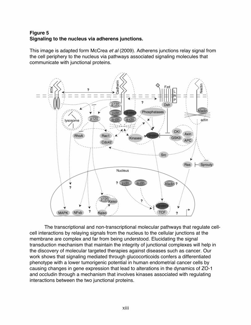
activation of phosphatases and kinases that eventually regulate the assembly, disassembly and maintenance of junctional complexes (Figure 4 and 5). Figure 4 Signaling to the nucleus via tight junctions. This image is adapted form McCrea et al (2009). Tight junctions relay signal from the cell periphery to the nucleus via pathways associated signaling molecules that communicate with junctional proteins.
xiii
Figure 5 Signaling to the nucleus via adherens junctions. This image is adapted form McCrea et al (2009). Adherens junctions relay signal from the cell periphery to the nucleus via pathways associated signaling molecules that communicate with junctional proteins.
The transcriptional and non-transcriptional molecular pathways that regulate cell-cell interactions by relaying signals from the nucleus to the cellular junctions at the membrane are complex and far from being understood. Elucidating the signal transduction mechanism that maintain the integrity of junctional complexes will help in the discovery of molecular targeted therapies against diseases such as cancer. Our work shows that signaling mediated through glucocorticoids confers a differentiated phenotype with a lower tumorigenic potential in human endometrial cancer cells by causing changes in gene expression that lead to alterations in the dynamics of ZO-1 and occludin through a mechanism that involves kinases associated with regulating interactions between the two junctional proteins.

xiv
References 1. Trosko, J.E. and R.J. Ruch, Cell-cell communication in carcinogenesis. Front
Biosci, 1998. 3: p. d208-36. 2. Franke, W.W., Discovering the molecular components of intercellular junctions--a
historical view. Cold Spring Harb Perspect Biol, 2009. 1(3): p. a003061. 3. Giepmans, B.N. and S.C. van Ijzendoorn, Epithelial cell-cell junctions and plasma
membrane domains. Biochim Biophys Acta, 2009. 1788(4): p. 820-31. 4. McCrea, P.D., D. Gu, and M.S. Balda, Junctional music that the nucleus hears:
cell-cell contact signaling and the modulation of gene activity. Cold Spring Harb Perspect Biol, 2009. 1(4): p. a002923.
5. Ebnet, K., Organization of multiprotein complexes at cell-cell junctions. Histochem Cell Biol, 2008. 130(1): p. 1-20.
6. Tanos, B. and E. Rodriguez-Boulan, The epithelial polarity program: machineries involved and their hijacking by cancer. Oncogene, 2008. 27(55): p. 6939-57.
7. Wang, Q. and B. Margolis, Apical junctional complexes and cell polarity. Kidney Int, 2007. 72(12): p. 1448-58.
8. Hartsock, A. and W.J. Nelson, Adherens and tight junctions: structure, function and connections to the actin cytoskeleton. Biochim Biophys Acta, 2008. 1778(3): p. 660-9.
9. Fanning, A.S. and J.M. Anderson, Zonula occludens-1 and -2 are cytosolic scaffolds that regulate the assembly of cellular junctions. Ann N Y Acad Sci, 2009. 1165: p. 113-20.
10. Ikenouchi, J., et al., Requirement of ZO-1 for the formation of belt-like adherens junctions during epithelial cell polarization. J Cell Biol, 2007. 176(6): p. 779-86.
11. McNeil, E., C.T. Capaldo, and I.G. Macara, Zonula occludens-1 function in the assembly of tight junctions in Madin-Darby canine kidney epithelial cells. Mol Biol Cell, 2006. 17(4): p. 1922-32.
12. Niessen, C.M., Tight junctions/adherens junctions: basic structure and function. J Invest Dermatol, 2007. 127(11): p. 2525-32.
13. Buse, P., et al., Glucocorticoid-induced functional polarity of growth factor responsiveness regulates tight junction dynamics in transformed mammary epithelial tumor cells. J Biol Chem, 1995. 270(47): p. 28223-7.
14. Buse, P., et al., Transforming growth factor-alpha abrogates glucocorticoid-stimulated tight junction formation and growth suppression in rat mammary epithelial tumor cells. J Biol Chem, 1995. 270(12): p. 6505-14.
15. Wong, V., et al., Glucocorticoid down-regulation of fascin protein expression is required for the steroid-induced formation of tight junctions and cell-cell interactions in rat mammary epithelial tumor cells. J Biol Chem, 1999. 274(9): p. 5443-53.
16. Gargett, C.E., R.W. Chan, and K.E. Schwab, Hormone and growth factor signaling in endometrial renewal: role of stem/progenitor cells. Mol Cell Endocrinol, 2008. 288(1-2): p. 22-9.
17. Michael, A.E. and A.T. Papageorghiou, Potential significance of physiological and pharmacological glucocorticoids in early pregnancy. Hum Reprod Update, 2008. 14(5): p. 497-517.

xv
18. Davies, S., et al., Gene regulation profiles by progesterone and dexamethasone in human endometrial cancer Ishikawa H cells. Gynecol Oncol, 2006. 101(1): p. 62-70.
19. Tobioka, H., et al., Occludin expression decreases with the progression of human endometrial carcinoma. Hum Pathol, 2004. 35(2): p. 159-64.
20. Hanahan, D. and R.A. Weinberg, Hallmarks of cancer: the next generation. Cell, 2011. 144(5): p. 646-74.
21. Siegel, R., D. Naishadham, and A. Jemal, Cancer statistics, 2013. CA Cancer J Clin, 2013. 63(1): p. 11-30.
22. Emons, G., et al., Hormonal interactions in endometrial cancer. Endocr Relat Cancer, 2000. 7(4): p. 227-42.
23. Rose, P.G., Endometrial carcinoma. N Engl J Med, 1996. 335(9): p. 640-9. 24. Vollmer, G., Endometrial cancer: experimental models useful for studies on
molecular aspects of endometrial cancer and carcinogenesis. Endocr Relat Cancer, 2003. 10(1): p. 23-42.
25. Nishida, M., The Ishikawa cells from birth to the present. Hum Cell, 2002. 15(3): p. 104-17.
26. Kandoth, C., et al., Integrated genomic characterization of endometrial carcinoma. Nature, 2013. 497(7447): p. 67-73.
27. Biscuola, M., et al., Oncogene alterations in endometrial carcinosarcomas. Hum Pathol, 2013. 44(5): p. 852-9.
28. Kohlberger, P., et al., Prognostic value of immunohistochemically detected HER-2/neu oncoprotein in endometrial cancer. Cancer Lett, 1996. 98(2): p. 151-5.
29. Nazeer, T., et al., Multivariate survival analysis of clinicopathologic features in surgical stage I endometrioid carcinoma including analysis of HER-2/neu expression. Am J Obstet Gynecol, 1995. 173(6): p. 1829-34.
30. Riben, M.W., et al., Identification of HER-2/neu oncogene amplification by fluorescence in situ hybridization in stage I endometrial carcinoma. Mod Pathol, 1997. 10(8): p. 823-31.
31. Rolitsky, C.D., et al., HER-2/neu amplification and overexpression in endometrial carcinoma. Int J Gynecol Pathol, 1999. 18(2): p. 138-43.
32. Livasy, C.A., et al., EGFR expression and HER2/neu overexpression/amplification in endometrial carcinosarcoma. Gynecol Oncol, 2006. 100(1): p. 101-6.
33. Gonzalez-Mariscal, L., R. Tapia, and D. Chamorro, Crosstalk of tight junction components with signaling pathways. Biochim Biophys Acta, 2008. 1778(3): p. 729-56.

1
Chapter I
The apical junction complex in human endometrial cancer cells and the development of a model system responsive to glucocorticoids

2
Abstract Cell-cell interactions are important to both the physiology and pathology of multi- cellular organisms and are governed by junctional complexes. Cellular junctions are dynamic structures comprised of both intracellular and intercellular protein complexes. The loss of junctional integrity is implicated in the development and progression of cancer. Although intracellular signaling molecules such as phosphatases and kinases, and extracellular signaling molecules such as steroid hormones and growth factors, have been shown to regulate cellular junctions, the exact mechanism by which these junctional complexes are assembled, disassembled, and maintained is largely unknown. Glucocorticoids have been shown to regulate junctional complexes in a variety of tissue types. However, their effect on the human endometrium, a tissue that is primarily regulated by steroid hormones, has not been evaluated. Using human endometrial cancer cells, the present work details the effects of glucocorticoids on junctional organization. We show that treatment of Ishikawa cells, a well-established human endometrial cancer cell line, with dexamethasone (DEX), a synthetic glucocorticoid, results in only minor changes in the subcellular localization of junctional proteins ZO-1 and beta-catenin. Transepithelial electrical resistance (TER), a technique used to evaluate the formation of functional junctions, does not increase upon treatment of Ishikawa cells with DEX. Furthermore, there are no changes in interaction between the junctional proteins nor are there changes in the total protein expression levels of ZO-1 and beta-catenin. Because HER2 is over-expressed in endometrial cancer and many signaling components downstream of HER2 are involved in the regulation of junctional complexes, evaluating junctional complexes after expression of exogenous HER2 and delineating the signaling cascade downstream of HER2 that may be involved in glucocorticoid induced organization of junctional complexes is important, as the information has great therapeutic potential for the treatment of cancer.

3
Introduction
Cell-cell interactions are indispensable for a wide range of complex cellular and physiological processes, and selective disruptions in intercellular communication and contact can trigger tissue dysfunction and the onset of a variety of physiological disorders. Intercellular junctions control the nature and efficacy of cell-cell interactions, and in recent years, the structure and function of proteins that make up these junctional complexes have been intensely examined in many mammalian cell systems [1]. The four major intercellular junctions are comprised of multi-protein complexes that include both intracellular and intercellular transmembrane protein components, and can be functionally categorized into communicating junctions (gap junctions), anchoring junctions (adherens junctions and desmosomes), and occluding junctions (tight junctions) [2]. In no other tissue type is the maintenance of junctional integrity more important than in epithelial cells. Carcinomas, cancers that are derived from epithelial cells, constitute 90% of all human cancers and many of these result from disruptions of junctional complexes. In multicellular organisms, the epithelium is located at the exterior of our bodies and at the interface of the external and internal surfaces of our tissues and organs [3]. The epithelium not only forms a barrier that protects against harmful external stimuli, but also plays role in absorption, secretion, trans-cellular transport, and sensation. To carry out these important functions, epithelial cells have developed intercellular junctions that are highly polarized in nature giving rise to distinct apical and basolateral domains. These junctional complexes include adherens junctions and tight junctions, which together make up the apical junctional complex [4].
Junctional complexes are composed of both intracellular and intercellular protein
components. Tight junctions seal cells together to prevent para-cellular transport of small molecules and also serve as barriers against diffusion of membrane proteins between the apical and the basolateral domains of the plasma membrane such that polarity is maintained. The major proteins in tight junction are transmembrane proteins occludins, claudins and junctional adhesion molecules (JAMs), and intracellular peripheral membrane proteins called zonula occluden (ZO) proteins [5]. Adherens junctions and desmosomes are anchoring junctions that serve to attach cells to each other and to the extra-cellular matrix. Adherens junctions are made up of transmembrane proteins of the cadherin family that bind cells together through interaction with adjacent E-cadherin molecules. The conserved cytoplasmic tails of cadherins interact with intracellular catenins, which serves to link this complex to the actin cytoskeleton [6].
Despite the existing understanding of the structure and function of proteins that constitute the apical junction complex, how adherens junctions and tight junctions are assembled, disassembled, and maintained is largely unknown. Intercellular junctions have highly dynamic structures that can be coordinately regulated in response to diverse sets of extracellular, intracellular and metabolic signals. Some of these include proteolysis, phosphorylation of key components, endocytosis, or transcriptional regulation in response to extracellular cues [7-9]. Steroids and other small molecule

4
hormone ligands act through nuclear receptors [10-12], and have also emerged as an important class of regulators of intercellular junctional complexes that can efficiently coordinate the function and accessibility of junctional components and control the assembly, disassembly, and maintenance of intercellular junctions. Despite the relatively limited mechanistic information, intracellular nuclear receptors have been demonstrated to regulate the dynamics of cell-cell interactions through both primary and secondary transcriptional signaling and through nontranscriptional membrane effects that target the expression, modification, stability, function and/or localization of specific structural and/or accessory components of junctional complexes, depending on the physiological and tissue context.
Our lab has shown that glucocorticoids, one class of steroid hormones, induce
adherens junction formation and tight junction sealing in rat mammary epithelial tumor cells (Con8) using components of various signaling cascades [13-15]. Treatment of Con8 cells with dexamethasone (DEX), a synthetic glucocorticoid results in up-regulation of β-catenin protein and transcript expression and down-regulation of the phosphorylated form of β-catenin [16]. To further elucidate the signaling pathway involved in glucocorticoid-induced regulation of apical junction formation, factors involved in up-regulation of β-catenin were examined. In the absence of glucocorticoids, β-catenin is phosphorylated by GSK-3 and phosphorylated β-catenin is either transported into the nucleus or is degraded in the cytoplasm. In either scenario, it is inaccessible to the apical junction complex. In the presence of glucocorticoids, a stable non-phosphorylated form of β-catenin is produced and localized to the cell periphery where it plays a role in AJ organization that leads to TJ sealing. Stabilization of β-catenin is carried out by a novel mechanism in which glucocorticoid induced Sgk (serum-and glucocorticoid-induced protein kinase) activity phosphorylates GSK-3, which signals the phosphorylation and ubiquitin-mediated degradation of GSK-3. This in turn, inhibits phosphorylation of β-catenin allowing it to play a role in AJ assembly and subsequently TJ sealing [17] (Figure 6).

5
Figure 6 Proposed model for DEX-induced regulation of cell-cell interactions in rat mammary epithelial tumor cells (Con8). This diagram is adapted form Failor et al (2010). In the absence of glucocorticoids, β-catenin is phosphorylated by GSK-3 and phosphorylated β-catenin is either transported into the nucleus or is degraded in the cytoplasm. In either scenario, it is inaccessible to the apical junction complex. In the presence of glucocorticoids, a stable and un-phosphorylated form of β-catenin is produced and localized to the cell periphery where it plays a role in AJ organization that leads to TJ sealing.
Glucocorticoids, Signaling Through the Glucocorticoid Receptor, & the Endometrium
Glucocorticoids received their name because they are able to promote the conversion of proteins and lipids into glucose when the hypothalamic-pituitary-adrenal (HPA) axis is activated upon stress [18]. Glucocorticoids paly a variety of roles in the context physiology, including a role in metabolism, regulating tissue-specific activities in immune function, the inflammatory response, embryogenesis, behavior, stress, cell proliferation and survival [19]. The actions of glucocorticoids are mediated by the glucocorticoid receptor (GR), which is part of a superfamily of steroid receptors. These receptors are ligand-inducible transcription factors that control physiological function by
6
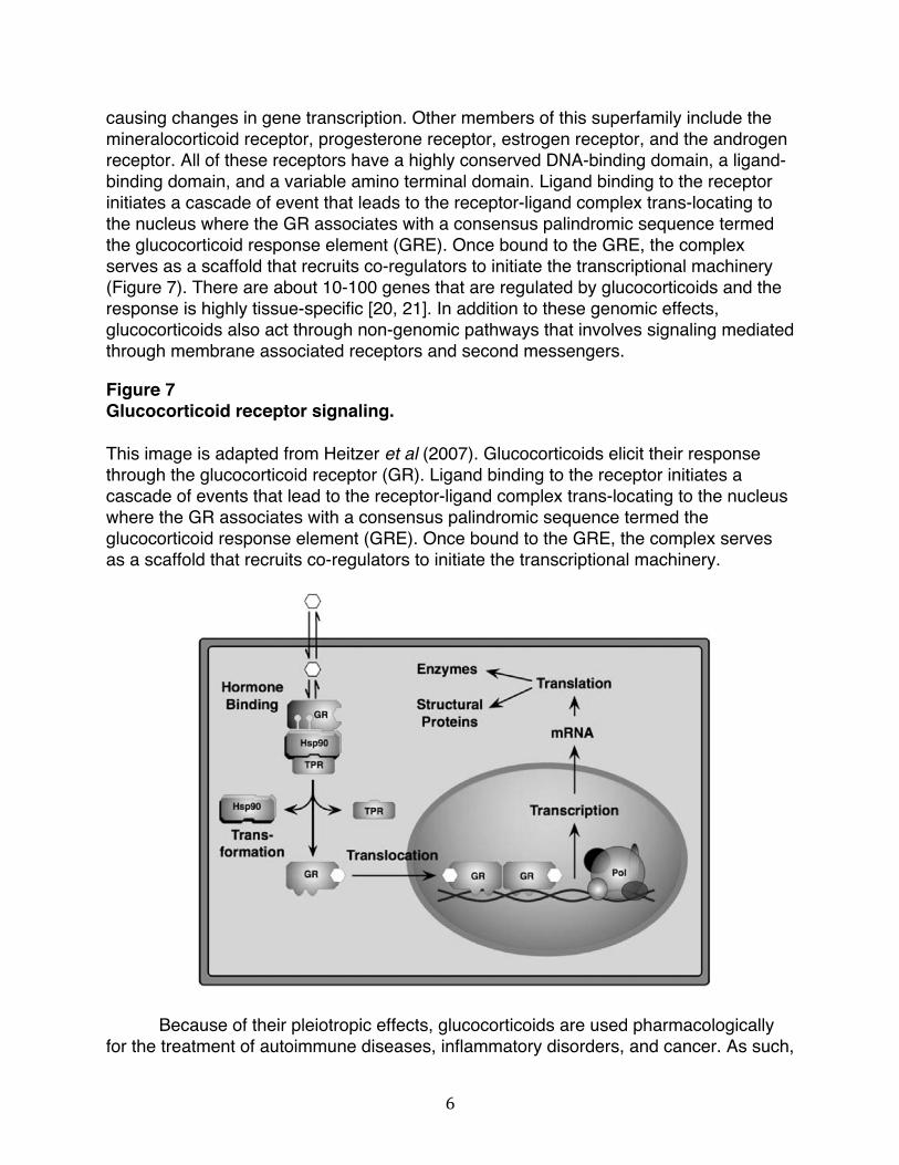
causing changes in gene transcription. Other members of this superfamily include the mineralocorticoid receptor, progesterone receptor, estrogen receptor, and the androgen receptor. All of these receptors have a highly conserved DNA-binding domain, a ligand-binding domain, and a variable amino terminal domain. Ligand binding to the receptor initiates a cascade of event that leads to the receptor-ligand complex trans-locating to the nucleus where the GR associates with a consensus palindromic sequence termed the glucocorticoid response element (GRE). Once bound to the GRE, the complex serves as a scaffold that recruits co-regulators to initiate the transcriptional machinery (Figure 7). There are about 10-100 genes that are regulated by glucocorticoids and the response is highly tissue-specific [20, 21]. In addition to these genomic effects, glucocorticoids also act through non-genomic pathways that involves signaling mediated through membrane associated receptors and second messengers.
Figure 7 Glucocorticoid receptor signaling. This image is adapted from Heitzer et al (2007). Glucocorticoids elicit their response through the glucocorticoid receptor (GR). Ligand binding to the receptor initiates a cascade of events that lead to the receptor-ligand complex trans-locating to the nucleus where the GR associates with a consensus palindromic sequence termed the glucocorticoid response element (GRE). Once bound to the GRE, the complex serves as a scaffold that recruits co-regulators to initiate the transcriptional machinery.
Because of their pleiotropic effects, glucocorticoids are used pharmacologically for the treatment of autoimmune diseases, inflammatory disorders, and cancer. As such,

7
synthetic glucocorticoids are the most widely prescribed drug worldwide [22]. One such synthetic glucocorticoid is dexamethasone (DEX). DEX is highly potent derivative of the endogenous glucocorticoid, cortisol, and is used to in the treatment of cancer along with chemotherapy to prevent allergic reactions to drugs, to treat nausea associated with chemotherapy, and to reduce edema for tumors in the brain, spinal cord, and bones. In addition to this, DEX is used to reduce inflammation, treat allergies or asthma, autoimmune disorders or arthritis, endocrine disorders, and to prevent transplant rejection [23]. Our interest is in the effects of DEX on junctional complexes that mediate cell-cell interactions, as dysregulated expression of these complexes is the cause many of these diseases. For example, loss of tight junction integrity has been implicated in a variety of cancers, including breast, bladder, colorectal, gastric, esophageal, gynecological, lung, prostrate, melanoma, pancreatic, liver, thyroid, and oral cancers [24]. And although the effects of glucocorticoids have been studied in a variety of organs and tissue types including liver, pancreas, heart, brain, breast, intestine, and lung [25-29], very little is known about the effects of glucocorticoids on cellular junctions within the endometrium.
The endometrium is the innermost layer that lines the uterus. The human
endometrium is the most dynamic tissue within the body and it goes through hundreds of rounds of proliferation, differentiation, and death. The regulation of these cycles is under the control of steroids hormones [30]. During the menstrual cycle the endometrium proliferates under the influence of estrogen and the cycle of building and shedding the endometrial lining is regulated by the actions of estrogen and progesterone [31]. In the normal endometrium, glucocorticoids play a role during implantation and through pregnancy [32]. Using endometrial cells, it has been shown that glucocorticoids regulate thousands of genes that are involved in embryonic pathways and give insight into the mechanism of glucocorticoids within the endometrium [33]. Another study that set out to determine the gene expression profile of human endometrial cancer cells upon treatment with progesterone and DEX, found that DEX has growth inhibitory effects in the endometrial cancer [34]. As is the case in many cancers, the loss of junctional integrity is also associated with endometrial cancer. As an example, a low expression level of claudin-7 in endometrial cancer cells is indicative of a late stage tumor. When claudin-7 levels are restored, proliferation and invasion of endometrial cancer cells is inhibited [35]. Considering this and the fact that glucocorticoids have been shown to play a variety of roles within the endometrium as well as influence the dynamics of junctional complexes, it is intriguing to evaluate the effects of glucocorticoids on cellular junctions within the endometrium. As such, our work examines the effects of DEX on junctional complexes using human endometrial cancer cells.

8
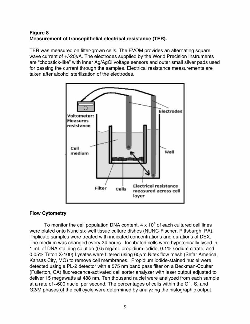
Materials & Methods Cell Culture
Ishikawa cells were grown in Dulbecco’s modified Eagles Medium, supplemented with 10% fetal bovine serum (all media components purchased from Lonza, Allendale, NJ and cell culture plates purchased from NUNC-Fischer, Pittsburgh, PA), 10 µg/ml insulin, 50 U/ml penicillin, 50 U/ml streptomycin, and 2 mmol/l L-glutamine (Sigma-Aldrich, St. Louis, MO). The cells were maintained at subconfluency in a humidified air chamber at 37˚C containing 5% CO2. A 100 mmol/l stock solution of DEX (Sigma-Aldrich, St. Louis, MO; catalog number D1756-500mg) was dissolved in dimethyl sulfoxide (DMSO) (Sigma-Aldrich, St. Louis, MO; catalog number DD2650), and then diluted in the ratio 1:1000 in media before culture plate application. Before each drug treatment, cells were washed in ice cold phosphate-buffered saline (PBS) (obtained form Lonza, Allendale, NJ). Cell Culture and Measurement of Transepithelial Electrical Resistance
Con8 rat mammary epithelial tumor cells were used as positive controls and were
routinely grown to 100% confluency on Nunc permeable supports or Nunc tissue culture plates (Fisher Scientific, Santa Clara, CA) in DMEM/F-12 supplemented with 10% calf serum and penicillin/streptomycin and maintained at 37 C in a humid atmosphere of air/CO2 (95:5). Ishikawa cells were grown in Dulbecco’s modified Eagles Medium, supplemented with 10% fetal bovine serum (all media components purchased from Lonza, Allendale, NJ), 10 µg/ml insulin, 50 U/ml penicillin, 50 U/ml streptomycin, and 2 mmol/l L-glutamine (obtained from Sigma-Aldrich, St. Louis, MO). These cells were cultured in serum free medium for 24 h before and during all experiments, and the cell culture medium was routinely changed every 24 h. Cells were treated with or without the DEX, at a final concentration of 10 µM (prepared as 100mM stock in DMSO). The formation of tight junction was monitored by measuring transepithelial electrical resistance (TER), using an EVOM Epithelial Voltohmmeter (World Precision Instruments, Sarasota, FL). TER was measured on filter-grown cells, allowed to cool to room temperature. The EVOM provides an alternating square wave current of +/-20µA. The electrodes supplied by the World Precision Instruments are “chopstick-like” with inner Ag/AgCl voltage sensors and outer small silver pads used for passing the current through the samples (Figure 8). Electrical resistance measurements were taken daily after alcohol sterilization of the electrode. Calculations for ohms X cm2 were determined by multiplying the area by the monolayer (0.49 cm2 for the 10-mm filters).
9
Figure 8 Measurement of transepithelial electrical resistance (TER). TER was measured on filter-grown cells. The EVOM provides an alternating square wave current of +/-20µA. The electrodes supplied by the World Precision Instruments are “chopstick-like” with inner Ag/AgCl voltage sensors and outer small silver pads used for passing the current through the samples. Electrical resistance measurements are taken after alcohol sterilization of the electrodes.
Flow Cytometry
To monitor the cell population DNA content, 4 x 10⁴ of each cultured cell lines were plated onto Nunc six-well tissue culture dishes (NUNC-Fischer, Pittsburgh, PA). Triplicate samples were treated with indicated concentrations and durations of DEX. The medium was changed every 24 hours. Incubated cells were hypotonically lysed in 1 mL of DNA staining solution (0.5 mg/mL propidium iodide, 0.1% sodium citrate, and 0.05% Triton X-100) Lysates were filtered using 60µm Nitex flow mesh (Sefar America, Kansas City, MO) to remove cell membranes. Propidium iodide-stained nuclei were detected using a PL-2 detector with a 575 nm band pass filter on a Beckman-Coulter (Fullerton, CA) fluorescence-activated cell sorter analyzer with laser output adjusted to deliver 15 megawatts at 488 nm. Ten thousand nuclei were analyzed from each sample at a rate of ~600 nuclei per second. The percentages of cells within the G1, S, and G2/M phases of the cell cycle were determined by analyzing the histographic output

10
with the multicycle computer program MPLUS, provided by Phoenix Flow Systems (San Diego, CA), in the Cancer Research Laboratory Microchemical Facility at the University of California at Berkeley. Indirect Immunofluorescence
Cells were grown and indicated treatments performed on two-well chamber slides from Nalgene Nunc International. The cells were fixed with 3.75% formaldehyde in PBS for 15 min at room temperature. After three additional washes with PBS, the plasma membrane was permeabilized with 0.1% Triton-X-100, 10 mM Tris-HCl, pH 7.5, 120 mM sodium chloride, 25 mM potassium chloride, 2 mM EGTA, and 2 mM EDTA) for 10 min at room temperature. Slides were incubated with 3% bovine serum albumin (Sigma-Aldrich) before incubation with primary antibodies. Rabbit anti-ZO-1 (61-7300) and Mouse anti-beta-catenin (18-0226) purchased from life Technologies/Invitrogen, San Diego, CA) were used at a 1:400 dilution. Secondary Alexa 488 anti-rabbit and Texas Red- anti-mouse antibodies (Molecular Probes, Inc., Eugene, OR) were used at 1:400 dilutions each. Stained cells were mounted with Vectashield mounting medium containing 4,6-diamidino-2-phenylindole (DAPI) (Vector Laboratories, Burlingame, CA). Stained and mounted cells were then processed with an Axioplan epifloruescence microscope (Carl Zeiss, Thornwood, NY). The images were acquired and processed by M1/Hamamatsu Orca and QImaging MicroPublisher color cameras. Contrast and brightness settings were chosen so that all pixels were in the linear range. Western Blots
After the indicated treatments, cells were harvested in radioimmune precipitation assay buffer (150 mM NaCl, 0.5% deoxycholate, 0.1% NoNidet-p40 (Nonidet P-40, Flulta Biochemitra, Switzerland), 0.1% SDS, 50 mM Tris) containing protease and phosphatase inhibitors (50 g/ml phenylmethylsulfonyl fluoride, 10 g/mL aprotinin, 5 g/mL leupeptin, 0.1 g/mL NaF, 1 mM dithiothreitol, 0.1 mM sodium orthovanadate, and 0.1 mM glycerol phosphate). Equal amounts of total cellular protein were mixed with loading buffer (25% glycerol, 0.075% SDS, 1.25 ml of 14.4M βmercaptoethanol, 10% bromphenol blue, 3.13% 0.5M Tris-HCl, and 0.4% SDS (pH 6.8)) and fractionated on 10% polyacrylamide/0.1% SDS resolving gels by electrophoresis. Rainbow marker (Amersham Biosciences) was used as the molecular weight standard. Proteins were electrically transferred to nitrocellulose membranes (Micron Separations, Inc., Westboro, MA) and blocked for 1 hour with Western wash buffer5% NFDM (10 mM Tris-HCl (pH 8.0), 150 mM NaCl, and 0.05% Tween 20, 5% nonfat dry milk). Protein blots were subsequently incubated for overnight at 4oC in primary antibodies. The antibodies used were as follows, Rabbit anti-ZO-1, (61-7300) and Mouse anti-beta-catenin (18-0226) purchased from Life Technologies/Invitrogen, San Diego, CA), Rabbit anti-E-cadherin (sc7870) was purchased from Santa Cruz Biotechnology and diluted in the ratio 1:1000 in TBST. Rabbit anti-actin (AANO1; Cytoskeleton, Denver CO) was diluted 1:1000 in TBST and used as a gel-loading control. The working concentration for all

11
antibodies was 1 µg/mL in Western wash buffer. Immunoreactive proteins were detected after incubation with horseradish peroxidase conjugated secondary antibody diluted to 3 x 10⁴ in Western wash buffer (goat anti-rabbit IgG, rabbit anti-goat IgG, and rabbit anti-mouse IgG (Bio-Rad)). Blots were treated with enhanced chemiluminescence reagents (PerkinElmer Life Sciences), and all proteins were detected by autoradiography. Equal protein loading was confirmed by Ponceau S staining of blotted membranes.
Co-Immunoprecipitation
Ishikawa cells were cultured on growth medium with DEX for the indicated times and then rinsed twice with PBS, harvested, and stored as dry pellets at −80°C. Cells were lysed for 15 min in IP buffer (50 mΜ Tris-HCl (pH 7.4), 150 mΜ NaCl, and 0.1% Triton X-100) containing protease inhibitors, 10 µg/ml aprotonin, 5 µg/ml leupeptin, 0.1 µg/ml NaF, 10 µg/ml β-glycerophosphate, and 0.1 mM sodium orthovanadate). Samples were diluted to 1 mg of protein in 1 ml of IP buffer. Samples were precleared for 1 hour at 4°C with 40 µl of a 1:1 slurry of protein G-Sepharose beads (GE health BioSciences AB). Precleared samples were then incubated with 50 µg of mouse anti-beta-catenin overnight at 4°C. Immunoprecipitated protein was eluted from beads by addition of gel loading buffer (50 mM Tris-HCl, pH 6.8, 2% SDS, 10% glycerol, 1% β-mercaptoethanol, 12.5 mM EDTA, 0.02 mM bromophenol blue) and heating the sample at 100°C for 5 min. Samples were analyzed by Western blot.
Expression Plasmids and Transfections
Human cytomegalovirus (CMV)-HER2 expression plasmid was a kind gift from
Dr. Leonard Bjeldanes, (Department of Nutritional Sciences and Toxicology, University of California at Berkeley). Transfection of expression vectors were performed using Superfect transfection reagent from QIAGEN per the manufacturers’ recommended protocol.

12
Results
Our previous work demonstrated that treatment of rat Con8 mammary epithelial cells with DEX results in the formation of functional tight junctions. To determine if DEX induces a similar phenotype in human endometrial cancer cells, we treated Ishikawa cells with or without 1μM DEX during a 5-day time course. Tight junction sealing was monitored using monolayer transepithelial electrical resistance. The electrical resistance across a confluent monolayer of cells is directly proportional to the sealing of the tight junctions between cells [13]. In Con8 cells, DEX strongly induced TER of the cell monolayer, where during the entire time course of the experiment, the TER remained low in the absence of DEX. In contrast, in Ishikawa cells, the steroid induction of tight junction sealing did not occur. DEX treatment in these cells did not cause an increase in TER compared to untreated cells (Figure 9). This result indicates that treatment of Ishikawa cells with DEX does not induce the formation of functional tight junctions.
Figure 9 Transepithelial electrical resistance measurement in Ishikawa cells. Con8 cells (top panel) and Ishikawa cells (bottom panel) were grown to 100% confluency on Nunc permeable supports and treated with or without DEX for 5 d. TER was measured every 24 h starting at the beginning of the steroid treatment. The results are an average of three independent experiments.

13
Ishikawa cells do not form functional tight junctions, as demonstrated by the lack of an increase in TER measurement. However, it is possible that DEX can induce changes in junctional proteins that alter cell-cell interactions without a tight junction sealing. To test this hypothesis, Ishikawa cells were treated with or without DEX for 24, 48, and 72 h and the localization of the adherens junction protein beta-catenin and the tight junction protein ZO-1 were examined by indirect immunofluorescence microscopy. In Ishikawa cells treated with DEX, both beta-catenin and ZO-1 showed diffuse staining with some localization to the cell periphery. The same was true for cells that were treated with the vehicle control, DMSO (Figure 10). DEX treatment in these cells did not significantly change the localization of beta-catenin and ZO-1 compared to untreated cells.

14
Figure 10 Localization of beta-catenin and ZO-1 in Ishikawa cells treated with DEX. Ishikawa cells grown to 100% confluency and treated with or without DX for 24, 48, and 72 h. Indirect immunofluorescence microscopy was used to visualize beta-catenin (an adherens junction protein, center) and ZO-1 (tight junction protein, right). DAPI staining was used to visualize DNA (the nucleus, left).

15
Given that DEX induces cell-cycle arrest and inhibits cell proliferation in a number of cancer cell lines [36-38], it is possible that DEX induces a growth arrest in Ishikawa cells and confers a more differentiated phenotype by causing changes in cell cycle regulators that could alter junctional dynamics. To test this hypothesis, Ishikawa cells were treated with or without DEX for 24, 48, and 72 h and nuclear DNA stained with propodium iodide. The samples were prepared in triplicates and quantified by flow cytometry. As shown in Figure 11, Ishikawa cells treated with DEX have same cell cycle profile as cells treated with DMSO, the vehicle control. The results were the same in the presence and absence of serum. This indicates that DEX does not alter cell cycle progression in Ishikawa cells. Figure 11 Cell cycle profile of Ishikawa cells treated with DEX. Ishikawa cells were treated with and without DEX for 24, 48, and 72 h and the cell population DNA content was quantified by flow cytomertry. The bar graphs show the average DNA content corresponding to the cell cycle phases of three independent experiments.
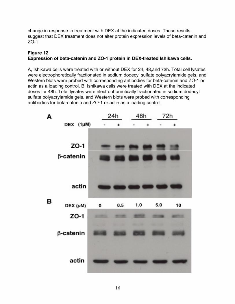
Changes in total levels of proteins that are part of the junctional complex could lead to changes in the dynamics of cell-cell interactions. We tested whether the expression level of beta-catenin and ZO-1 were affected by DEX treatment. Total levels of junctional proteins were examined in Ishikawa cells treated with or without 1μM DEX for 24, 48, 72h (Figure 12A). To determine the correct dose for treatment, Ishikawa cells were treated for 48h at 0, 0.5, 1.0, 5.0, and 10μM DEX (Figure 12B). As shown in Figure 12A, Western blots revealed that the protein levels of beta-catenin and ZO-1 did not change in response to treatment with DEX over the entire duration of the treatment. Similarly, as shown in Figure 12B, total protein levels of beta-catenin and ZO-1 did not
16
change in response to treatment with DEX at the indicated doses. These results suggest that DEX treatment does not alter protein expression levels of beta-catenin and ZO-1. Figure 12 Expression of beta-catenin and ZO-1 protein in DEX-treated Ishikawa cells. A, Ishikawa cells were treated with or without DEX for 24, 48,and 72h. Total cell lysates were electrophoretically fractionated in sodium dodecyl sulfate polyacrylamide gels, and Western blots were probed with corresponding antibodies for beta-catenin and ZO-1 or actin as a loading control. B, Ishikawa cells were treated with DEX at the indicated doses for 48h. Total lysates were electrophorectically fractionated in sodium dodecyl sulfate polyacrylamide gels, and Western blots were probed with corresponding antibodies for beta-catenin and ZO-1 or actin as a loading control.
17
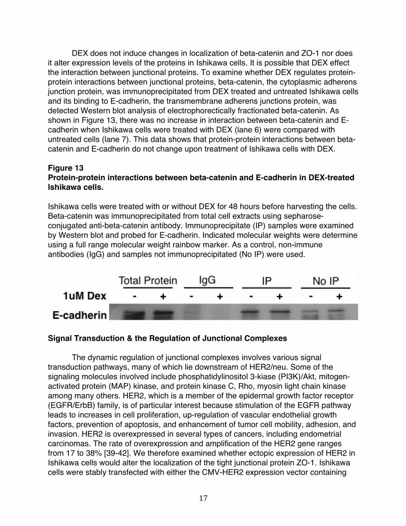
DEX does not induce changes in localization of beta-catenin and ZO-1 nor does it alter expression levels of the proteins in Ishikawa cells. It is possible that DEX effect the interaction between junctional proteins. To examine whether DEX regulates protein-protein interactions between junctional proteins, beta-catenin, the cytoplasmic adherens junction protein, was immunoprecipitated from DEX treated and untreated Ishikawa cells and its binding to E-cadherin, the transmembrane adherens junctions protein, was detected Western blot analysis of electrophorectically fractionated beta-catenin. As shown in Figure 13, there was no increase in interaction between beta-catenin and E-cadherin when Ishikawa cells were treated with DEX (lane 6) were compared with untreated cells (lane 7). This data shows that protein-protein interactions between beta-catenin and E-cadherin do not change upon treatment of Ishikawa cells with DEX. Figure 13 Protein-protein interactions between beta-catenin and E-cadherin in DEX-treated Ishikawa cells. Ishikawa cells were treated with or without DEX for 48 hours before harvesting the cells. Beta-catenin was immunoprecipitated from total cell extracts using sepharose-conjugated anti-beta-catenin antibody. Immunoprecipitate (IP) samples were examined by Western blot and probed for E-cadherin. Indicated molecular weights were determine using a full range molecular weight rainbow marker. As a control, non-immune antibodies (IgG) and samples not immunoprecipitated (No IP) were used.
Signal Transduction & the Regulation of Junctional Complexes
The dynamic regulation of junctional complexes involves various signal transduction pathways, many of which lie downstream of HER2/neu. Some of the signaling molecules involved include phosphatidylinositol 3-kiase (PI3K)/Akt, mitogen-activated protein (MAP) kinase, and protein kinase C, Rho, myosin light chain kinase among many others. HER2, which is a member of the epidermal growth factor receptor (EGFR/ErbB) family, is of particular interest because stimulation of the EGFR pathway leads to increases in cell proliferation, up-regulation of vascular endothelial growth factors, prevention of apoptosis, and enhancement of tumor cell mobility, adhesion, and invasion. HER2 is overexpressed in several types of cancers, including endometrial carcinomas. The rate of overexpression and amplification of the HER2 gene ranges from 17 to 38% [39-42]. We therefore examined whether ectopic expression of HER2 in Ishikawa cells would alter the localization of the tight junctional protein ZO-1. Ishikawa cells were stably transfected with either the CMV-HER2 expression vector containing

18
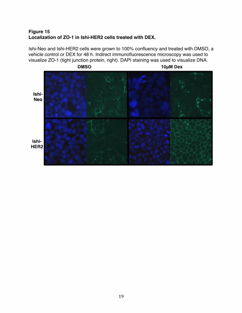
the neomycin resistance gene, or the control vector comprised of the CMV-Neomycin gene, forming the Ishi-HER2 and Ishi-NEO cells, respectively. Western blot analysis demonstrates that Ishi-HER2 cells expressed significantly higher levels of HER2 compared to the control CMV-Neo cell (Figure 14). Ishi-HER2 and Ishi-Neo cells were treated with or without DEX for 48h and localization of ZO-1 was examine by indirect immunofluorescence microscopy. We chose to primarily focus on ZO-1 because the ZO proteins are known to act as scaffolding proteins that link AJ and TJ transmembrane and cytoplasmic proteins and coordinate their interactions with the actin cytoskeleton. The protein-binding motifs within the N-terminus of ZO-1 direct the interactions with all of the other TJ proteins and some AJ proteins. The C-terminus motif directs interactions with actin or other actin-binding proteins [43]. As shown in Figure 15, in Ishi-Neo cells localization of ZO-1 was diffused throughout the cells in the presence and absence of steroid treatment. In Ishi-HER2 cells localization of ZO-1 was diffuse in the absence of DEX. However, ZO-1 was localized exclusively to the cell periphery in DEX-treated cells. These results indicated that DEX induces localization of ZO-1to cell periphery in Ishikawa cells that over-express HER2. Figure 14 Ectopic expression of human epidermal growth factor receptor 2 (HER2) in Ishikawa cell. Cultured Ishikawa cells were transfected with either empty vector control CMV-Neo or CMV-HER2. Over-expression of HER2 was verified by Western blot analysis. Total lysates were electrophorectically fractionated in sodium dodecyl sulfate polyacrylamide gels, and Western blots were probed with corresponding antibodies for HER2 or HSP 90 as a loading control.
19
Figure 15 Localization of ZO-1 in Ishi-HER2 cells treated with DEX. Ishi-Neo and Ishi-HER2 cells were grown to 100% confluency and treated with DMSO, a vehicle control or DEX for 48 h. Indirect immunofluorescence microscopy was used to visualize ZO-1 (tight junction protein, right). DAPI staining was used to visualize DNA.

20
Discussion
Our previous studies have shown that glucocorticoids induce a steroid-regulated pathway that stimulates adherens junction formation and tight junction sealing in Con8 rat mammary epithelial tumor cells. This occurs through a glucocorticoid-regulated cascade that stabilizes an un-phosphorylated form of bate-catenin that is accessible to be localized to its site of function in the apical junction complex. Here we uncover the effects of DEX on junctional complexes in Ishikawa cells, a human endometrial cancer cell line.
Hormones are known to modify epithelial permeability. For example, junctional sealing of the mammary gland during lactation is stimulated by corticosterones [44]. Within the endometrium, when proliferation is intensive tight junction proteins are not concentrated in the region of the apical junction complex. This is in agreement with cancerous tissues, in which proliferation rates are high and junctional proteins are diffuse throughout the cytoplasm or not sufficiently expressed [45-48]. Using rat mammary epithelial cells, we show that TER, a measure of functional tight junctions formation, increases in response to DEX. However, in Ishikawa cells TER remained unchanged, indicating that functional tight junctions do not form in human endometrial cancer cells in response to DEX. Because the endometrium is a tissue that is highly regulated by hormones and the cyclic changes in serum steroid hormone levels correlate with morphological changes in the endometrial lining, it is possible that the endometrium is incapable of forming functional tight junctions at specific phases of the menstrual cycle.
An increase in TER is not the only determinant of functional tight junction formation. Changes in localization of junctional proteins can also indicate the presence of somewhat leaky junctions. In Ishikawa cells, we observed that DEX did not alter the localization of junctional proteins nor did it change the expression level of proteins within cellular junctions. Additionally, DEX had no effect on the dynamics of protein-protein interactions that regulate junctional complexes. Cell cycle progression of Ishikawa cells treated with DEX also remained unaltered. However, as we have shown ectopic expression of HER2 in Ishikawa cells resulted in a dramatic organization of ZO-1 to the cell periphery upon treatment with DEX. Taken together, our results indicate that DEX does not changes the dynamics of junctional complexes in Ishikawa cells but does induce a significant organization of ZO-1 to the cell periphery when these cells over-express HER2. Because the dynamic regulation of junctional complexes involves various signal transduction pathways, it is conceivable that other signaling components are involved in the regulation of junctional complexes with Ishikawa cells. Many of the signaling cascades that regulate the dynamics of cellular junctions lie downstream of HER2/neu. Some of the signaling molecules involved include phosphatidylinositol 3-kiase (PI3K)/Akt, mitogen-activated protein (MAP) kinase, and protein kinase C, Rho, myosin light chain kinase among many others [49]. As such, the DEX-induced organization of ZO-1 to the cell periphery only in Ishi-HER2 cells may prove to be an important component that drives the regulation of junctional complexes.

21
Proper functioning of the cell is dependent on the integrity of cellular junctions and loss of junctional integrity is associated with diseases such as cancer. Understanding how glucocorticoids regulate apical junction complex organization in Ishi-HER2 cells will help determine the signaling pathways that control cell-cell interactions in human endometrium cancer cells. Future studies that delineate the signal transduction mechanisms that maintain integrity of junctional complexes will help in the discovery of molecular targeted therapies against this disease.

22
References
1. Ebnet, K., Organization of multiprotein complexes at cell-cell junctions. Histochem Cell Biol, 2008. 130(1): p. 1-20.
2. Franke, W.W., Discovering the molecular components of intercellular junctions--a historical view. Cold Spring Harb Perspect Biol, 2009. 1(3): p. a003061.
3. Tanos, B. and E. Rodriguez-Boulan, The epithelial polarity program: machineries involved and their hijacking by cancer. Oncogene, 2008. 27(55): p. 6939-57.
4. Wang, Q. and B. Margolis, Apical junctional complexes and cell polarity. Kidney Int, 2007. 72(12): p. 1448-58.
5. Schneeberger, E.E. and R.D. Lynch, The tight junction: a multifunctional complex. Am J Physiol Cell Physiol, 2004. 286(6): p. C1213-28.
6. Miyoshi, J. and Y. Takai, Structural and functional associations of apical junctions with cytoskeleton. Biochim Biophys Acta, 2008. 1778(3): p. 670-91.
7. Niessen, C.M., Tight junctions/adherens junctions: basic structure and function. J Invest Dermatol, 2007. 127(11): p. 2525-32.
8. Giepmans, B.N. and S.C. van Ijzendoorn, Epithelial cell-cell junctions and plasma membrane domains. Biochim Biophys Acta, 2009. 1788(4): p. 820-31.
9. McCrea, P.D., D. Gu, and M.S. Balda, Junctional music that the nucleus hears: cell-cell contact signaling and the modulation of gene activity. Cold Spring Harb Perspect Biol, 2009. 1(4): p. a002923.
10. Aranda, A. and A. Pascual, Nuclear hormone receptors and gene expression. Physiol Rev, 2001. 81(3): p. 1269-304.
11. Lonard, D.M. and W. O'Malley B, Nuclear receptor coregulators: judges, juries, and executioners of cellular regulation. Mol Cell, 2007. 27(5): p. 691-700.
12. Hilser, V.J. and E.B. Thompson, Structural dynamics, intrinsic disorder, and allostery in nuclear receptors as transcription factors. J Biol Chem, 2011. 286(46): p. 39675-82.
13. Buse, P., et al., Transforming growth factor-alpha abrogates glucocorticoid-stimulated tight junction formation and growth suppression in rat mammary epithelial tumor cells. J Biol Chem, 1995. 270(12): p. 6505-14.
14. Buse, P., et al., Glucocorticoid-induced functional polarity of growth factor responsiveness regulates tight junction dynamics in transformed mammary epithelial tumor cells. J Biol Chem, 1995. 270(47): p. 28223-7.
15. Wong, V., et al., Glucocorticoid down-regulation of fascin protein expression is required for the steroid-induced formation of tight junctions and cell-cell interactions in rat mammary epithelial tumor cells. J Biol Chem, 1999. 274(9): p. 5443-53.
16. Guan, Y., et al., Glucocorticoids control beta-catenin protein expression and localization through distinct pathways that can be uncoupled by disruption of signaling events required for tight junction formation in rat mammary epithelial tumor cells. Mol Endocrinol, 2004. 18(1): p. 214-27.
17. Failor, K.L., et al., Glucocorticoid-induced degradation of glycogen synthase kinase-3 protein is triggered by serum- and glucocorticoid-induced protein kinase and Akt signaling and controls beta-catenin dynamics and tight junction formation in mammary epithelial tumor cells. Mol Endocrinol, 2007. 21(10): p. 2403-15.
23
18. Smith, S.M. and W.W. Vale, The role of the hypothalamic-pituitary-adrenal axis in neuroendocrine responses to stress. Dialogues Clin Neurosci, 2006. 8(4): p. 383-95.
19. Lee, S.R., et al., Glucocorticoids and their receptors: insights into specific roles in mitochondria. Prog Biophys Mol Biol, 2013. 112(1-2): p. 44-54.
20. Cosio, B.G., A. Torrego, and I.M. Adcock, [Molecular mechanisms of glucocorticoids]. Arch Bronconeumol, 2005. 41(1): p. 34-41.
21. Heitzer, M.D., et al., Glucocorticoid receptor physiology. Rev Endocr Metab Disord, 2007. 8(4): p. 321-30.
22. Gross, K.L. and J.A. Cidlowski, Tissue-specific glucocorticoid action: a family affair. Trends Endocrinol Metab, 2008. 19(9): p. 331-9.
23. Rhen, T. and J.A. Cidlowski, Antiinflammatory action of glucocorticoids--new mechanisms for old drugs. N Engl J Med, 2005. 353(16): p. 1711-23.
24. Martin, T.A. and W.G. Jiang, Loss of tight junction barrier function and its role in cancer metastasis. Biochim Biophys Acta, 2009. 1788(4): p. 872-91.
25. Firestone, G.L. and B.J. Kapadia, Minireview: regulation of gap junction dynamics by nuclear hormone receptors and their ligands. Mol Endocrinol, 2012. 26(11): p. 1798-807.
26. Yukitatsu, Y., et al., Decreased expression of VE-cadherin and claudin-5 and increased phosphorylation of VE-cadherin in vascular endothelium in nasal polyps. Cell Tissue Res, 2013. 352(3): p. 647-57.
27. Kimura, K., et al., Protective effect of dexamethasone against hypoxia-induced disruption of barrier function in human corneal epithelial cells. Exp Eye Res, 2011. 92(5): p. 388-93.
28. Hermanns, M.I., et al., Lung epithelial cell lines in coculture with human pulmonary microvascular endothelial cells: development of an alveolo-capillary barrier in vitro. Lab Invest, 2004. 84(6): p. 736-52.
29. Blecharz, K.G., D. Drenckhahn, and C.Y. Forster, Glucocorticoids increase VE-cadherin expression and cause cytoskeletal rearrangements in murine brain endothelial cEND cells. J Cereb Blood Flow Metab, 2008. 28(6): p. 1139-49.
30. Prianishnikov, V.A., A functional model of the structure of the epithelium of normal, hyperplastic and malignant human endometrium: a review. Gynecol Oncol, 1978. 6(5): p. 420-8.
31. Ferenczy, A. and C. Bergeron, Histology of the human endometrium: from birth to senescence. Ann N Y Acad Sci, 1991. 622: p. 6-27.
32. Michael, A.E. and A.T. Papageorghiou, Potential significance of physiological and pharmacological glucocorticoids in early pregnancy. Hum Reprod Update, 2008. 14(5): p. 497-517.
33. Whirledge, S., X. Xu, and J.A. Cidlowski, Global gene expression analysis in human uterine epithelial cells defines new targets of glucocorticoid and estradiol antagonism. Biol Reprod, 2013. 89(3): p. 66.
34. Davies, S., et al., Gene regulation profiles by progesterone and dexamethasone in human endometrial cancer Ishikawa H cells. Gynecol Oncol, 2006. 101(1): p. 62-70.
35. Li, X., et al., Downregulation of claudin-7 potentiates cellular proliferation and invasion in endometrial cancer. Oncol Lett, 2013. 6(1): p. 101-105.

24
36. Chung, Y.J., et al., Anti-proliferative effect and action mechanism of dexamethasone in human medullary thyroid cancer cell line. Endocr Res, 2011. 36(4): p. 149-57.
37. Kullmann, M.K., et al., The p27-Skp2 axis mediates glucocorticoid-induced cell cycle arrest in T-lymphoma cells. Cell Cycle, 2013. 12(16): p. 2625-35.
38. Li, H., et al., Glucocorticoid receptor and sequential P53 activation by dexamethasone mediates apoptosis and cell cycle arrest of osteoblastic MC3T3-E1 cells. PLoS One, 2012. 7(6): p. e37030.
39. Kohlberger, P., et al., Prognostic value of immunohistochemically detected HER-2/neu oncoprotein in endometrial cancer. Cancer Lett, 1996. 98(2): p. 151-5.
40. Nazeer, T., et al., Multivariate survival analysis of clinicopathologic features in surgical stage I endometrioid carcinoma including analysis of HER-2/neu expression. Am J Obstet Gynecol, 1995. 173(6): p. 1829-34.
41. Riben, M.W., et al., Identification of HER-2/neu oncogene amplification by fluorescence in situ hybridization in stage I endometrial carcinoma. Mod Pathol, 1997. 10(8): p. 823-31.
42. Rolitsky, C.D., et al., HER-2/neu amplification and overexpression in endometrial carcinoma. Int J Gynecol Pathol, 1999. 18(2): p. 138-43.
43. Fanning, A.S. and J.M. Anderson, Zonula occludens-1 and -2 are cytosolic scaffolds that regulate the assembly of cellular junctions. Ann N Y Acad Sci, 2009. 1165: p. 113-20.
44. Nguyen, D.A., A.F. Parlow, and M.C. Neville, Hormonal regulation of tight junction closure in the mouse mammary epithelium during the transition from pregnancy to lactation. J Endocrinol, 2001. 170(2): p. 347-56.
45. Li, D. and R.J. Mrsny, Oncogenic Raf-1 disrupts epithelial tight junctions via downregulation of occludin. J Cell Biol, 2000. 148(4): p. 791-800.
46. Kramer, F., et al., Genomic organization of claudin-1 and its assessment in hereditary and sporadic breast cancer. Hum Genet, 2000. 107(3): p. 249-56.
47. Chlenski, A., et al., Organization and expression of the human zo-2 gene (tjp-2) in normal and neoplastic tissues. Biochim Biophys Acta, 2000. 1493(3): p. 319-24.
48. Hoover, K.B., S.Y. Liao, and P.J. Bryant, Loss of the tight junction MAGUK ZO-1 in breast cancer: relationship to glandular differentiation and loss of heterozygosity. Am J Pathol, 1998. 153(6): p. 1767-73.
49. Gonzalez-Mariscal, L., R. Tapia, and D. Chamorro, Crosstalk of tight junction components with signaling pathways. Biochim Biophys Acta, 2008. 1778(3): p. 729-56.

25
Chapter II
Glucocorticoids alter the dynamics of tight junctional proteins in human endometrial cancer cells

26
Abstract
The protein complexes within cellular junctions are an array of integral and peripheral proteins that associate with a diverse set of molecules involved in signaling cascades. Cells are able to transmit information from the point of cell-cell contact to the cell’s interior through these cascades. This interplay between junctional protein complexes and signaling pathways is important in the regulation of gene expression that coordinates many physiological events such as cell death, differentiation, and proliferation. Crosstalk between various signaling cascades leads to activation of phosphatases and kinases that eventually regulate the assembly, disassembly and maintenance of junctional complexes. The dynamic regulation of junctional complexes involves various signal transduction pathways that lie downstream of the human epidermal growth factors receptor 2 (HER2). We have shown that DEX induces organization of tight junctional proteins ZO-1 and occludin to the cell periphery in human endometrial cancer cells only upon ectopic expression of HER2. Here, we evaluate protein-protein interactions between ZO-1 and occludin and consider the role of other components that might be involved in the DEX-mediated, HER2-dependent organization of ZO-1 and occludin to the cell periphery. We show that treatment of Ishikawa cells with DEX results in an increase in interaction between ZO-1 and occludin without changes in total protein expression levels of the junctional proteins. The increase in interaction and subsequent localization of ZO-1 and occludin to the cell periphery is a co-dependent process that requires the expression both proteins. Because the interaction between ZO-1 and occludin affect the properties of tight junctions and is primarily regulated by phosphorylation events, it is intriguing to further evaluate the signaling pathways that might be involved observed effects.

27
Introduction
Intercellular junctions have highly dynamic structures that can be coordinately regulated in response to diverse sets of extracellular, intracellular and metabolic signals. Some of these include proteolysis, phosphorylation of key components, endocytosis, or transcriptional regulation in response to extracellular cues [1]. Because the complexes within cellular junctions are an array of integral and peripheral proteins that associate with a diverse set of molecule involved in signaling cascades, cells are able to transmit information from the point of cell-cell contact to the cell’s interior. This interplay between junctional protein complexes and signaling pathways is important in the regulation of gene expression that coordinates many physiological events such as cell death, differentiation, and proliferation.
Crosstalk between various signaling cascades leads to activation of phosphatases and kinases that eventually regulate the assembly, disassembly and maintenance of junctional complexes. Despite the relatively limited mechanistic information, intracellular nuclear receptors have been demonstrated to regulate the dynamics of cell-cell interactions through both primary and secondary transcriptional signaling and through non-transcriptional membrane effects that target the expression, modification, stability, function and/or localization of specific structural and/or accessory components of junctional complexes, depending on the physiological and tissue context [2]. In addition to nuclear receptors, the dynamic regulation of junctional complexes involves various signal transduction pathways that lie downstream of the human epidermal growth factors receptor 2 (HER2). Some of these signaling molecules include phosphatidylinositol 3-kiase (PI3K)/Akt, mitogen- activated protein (MAP) kinase, and protein kinase C, among many others [3]. For example, it has been shown that inhibition of PKC leads to disruption of TJ assembly. This effect leads to a defect in the sorting of ZO-1 and parallels a decrease in its phosphorylation, indicating PKC is involved in the assembly of functional TJs and that ZO-1 is one of its direct targets [4]. It also known that DEX, a synthetic glucocorticoid, increases both the mRNA and protein levels of human epidermal growth factor receptor 2/neu (HER2/neu) in Ishikawa cells [5]. In ovarian cancer cells, DEX is shown to have an inhibitory effect on cell proliferation in addition to having a similar effect on the mRNA levels of HER2/neu as those seen in Ishikawa cells [6]. We have shown that DEX induces organization of tight junctional proteins ZO-1 and occludin to the cell periphery in human endometrial cancer cells only upon ectopic expression of HER2. Next, we further evaluate protein-protein interactions between ZO-1 and occludin and consider the role of other components that might be involved in the DEX-mediated, HER2-dependent organization of ZO-1 and occludin to the cell periphery. Human Epidermal Growth Factor Receptor Signaling The epidermal growth factor receptor (EGFR) is a member of the ErbB family of four structurally related receptor tyrosine kinases: HER1, HER2, HER3, and HER4. The receptors are located on the cell membrane and interact with a variety of ligands,

28
including epidermal growth factor, transforming growth factor alpha, and amphiregulin. Structurally, the receptors have a cysteine-rich extracellular ligand binding site, a transmembrane domain, and an intracellular domain that has tyrosine kinase catalytic activity. The receptors exist as monomers that form receptor hertero-dimers or homo-dimers upon ligand binding. Dimerization triggers intrinsic intracellular tyrosine kinase activity that lead to auto-phosphorylation of tyrosine residues on the C-terminal domain of the receptors. This in turn activates downstream signal transduction cascades that lead to cell proliferation and growth [7]. Many cancers are associated with aberrant activation of the EGFR. This can result from mutation of the receptors, its over-expression or from abnormal receptor stimulation [8]. HER2 is considered an orphan receptor because it has no known ligands. Additionally, unlike other member of the receptor family, the extra-cellular domain of the HER2 remains constitutively active such that it always ready to dimerize with other receptor family members. These heterodimers can bind growth factors. On the intracellular side, HER2 contains many interacting domains including the src homology 2(SH2), phosphotyrosine-binding domain (PTB), among others [9]. The HER2 gene is amplified in many cancers including breast, cervical, colon, esophageal, lung, pancreatic, and endometrial [10-12]. Varying levels of over-expression of HER2 within these cancers poses a challenge for treatment options that are based on HER2 and other tyrosine kinase inhibitors. For example, well differentiated cell line models of endometrial cancer respond more robustly to EGFR and are more sensitive to HER2 inhibitors than less differentiated models [13]. Thus, it is known that endometrial cancers have the ability to respond to therapies based on HER2. This warrants further research into the signaling components associated with HER2 that take into consideration the variability of HER2 gene amplification and offer targeted treatment options. ZO-1 & Occludin Protein-Protein Interaction
Occludin is an integral membrane protein and ZO-1 is a membrane associated, cytoplasmic protein. The cytoplasmic C-terminal domain of occludin directly associates with the SH3-U5-GuK domains of ZO-1 [14, 15]. The interaction between ZO-1 and occludin affect the properties of tight junctions and is primarily regulated by phosphorylation events. Specifically, occludin phosphorylation tends to govern tight junction formation, structure, and function. There is a cluster of 11 amino acids that are close to the ZO-1 binding site in the C-terminal cytoplasmic domain of occludin that serve as phosphorylation sites for kinases including c-Src, PKC, and CK2 [16]. Several studies have shown that within the resting epithelium, occludin is highly phosphorylated at ser/thr residues [17-19]. Level of Tyr phosphorylation tends to be low in the resting epithelium [20]. Dephosphorylation of ser/thr residues is associated with disassembly of tight junctions and phosphorylation of ser/thr residues is associated with assembly of tight junctions. In contrast to this, Tyr phosphorylation is associated with disruption of interaction between ZO-1 and occludin. When the C-terminal domain of occludin was Tyr-phosphorylated by incubating it with c-Src, ZO-1 binding was reduced [20, 21]. In

29
MDCK cells, activation of c-Src induces disruption of tight junction that is ameliorated upon expression of a kinase-inactive c-Src [22]. The mechanism by which these phosphorylation events regulate junctional complexes is not well understood. However, a variety of studies have shown that serine, threonine, and tyrosine kinases are involved in mediating the interaction between ZO-1 and occludin and therefore maintain the integrity of junctional complexes. Using human endometrial cancer cells, we have shown a DEX-mediated, HER2 dependent organization of ZO-1 and occludin to the cell periphery. We further evaluate protein-protein interactions between ZO-1 and occludin and consider the role of serine, threonine, and tyrosine kinases that might be involved in junctional regulation.

30
Material & Methods
Cell Culture
Ishikawa cells were grown in Dulbecco’s modified Eagles Medium, supplemented with 10% fetal bovine serum (all media components purchased from Lonza, Allendale, NJ and cell culture plates purchased from NUNC-Fischer, Pittsburgh, PA), 10 µg/ml insulin, 50 U/ml penicillin, 50 U/ml streptomycin, and 2 mmol/l L-glutamine (obtained from Sigma-Aldrich, St. Louis, MO). The cells were maintained at subconfluency in a humidified air chamber at 37˚C containing 5% CO2. A 100 mmol/l stock solution of DEX (purchased from Sigma-Aldrich, St. Louis, MO; catalog number D1756-500mg) was dissolved in DMSO (purchased from Sigma-Aldrich, St. Louis, MO; catalog number DD2650), and then diluted in the ratio 1:1000 in media before culture plate application. A 10mM stock solution of RU-486 (Milfepristone, purchased from Sigma-Aldrich, St. Louis, MO; catalog number M8046) was dissolved in DMSO to give a final concentration of 1nM. Cisplatin (catalog number P4394) and N,N-Dimethylformamide (catalog number 319937) were purchased from Sigma-Aldrich. Before each drug treatment, cells were washed in ice cold phosphate-buffered saline (PBS) (obtained form Lonza, Allendale, NJ). Expression Plasmids and Transfections
Human cytomegalovirus (CMV)-HER2 expression plasmid was a kind gift from
Dr. Leonard Bjeldanes, (Department of Nutritional Sciences and Toxicology, University of California at Berkeley). Transfection of expression vectors were performed using Superfect transfection reagent from QIAGEN per the manufacturers’ recommended protocol. Flow Cytometry
To monitor the cell population DNA content, 4 x 10⁴ of each cultured cell lines were plated onto Nunc six-well tissue culture dishes (NUNC-Fischer, Pittsburgh, PA). Triplicate samples were treated with indicated concentrations and durations of DEX. The medium was changed every 24 hours. Incubated cells were hypotonically lysed in 1 mL of DNA staining solution (0.5 mg/mL propidium iodide, 0.1% sodium citrate, and 0.05% Triton X-100) Lysates were filtered using 60µm Nitex flow mesh (Sefar America, Kansas City, MO) to remove cell membranes. Propidium iodide-stained nuclei were detected using a PL-2 detector with a 575 nm band pass filter on a Beckman-Coulter (Fullerton, CA) fluorescence-activated cell sorter analyzer with laser output adjusted to deliver 15 megawatts at 488 nm. Ten thousand nuclei were analyzed from each sample at a rate of ~600 nuclei per second. The percentages of cells within the G1, S, and G2/M phases of the cell cycle were determined by analyzing the histographic output with the multicycle computer program MPLUS, provided by Phoenix Flow Systems (San Diego, CA), in the Cancer Research Laboratory Microchemical Facility at the University of California at Berkeley.

31
Indirect Immunofluorescence & Quantification of Staining
Cells were grown and indicated treatments performed on two-well chamber slides from Nalgene Nunc International. The cells were fixed with 3.75% formaldehyde in PBS for 15 min at room temperature. After three additional washes with PBS, the plasma membrane was permeabilized with 0.1% Triton-X-100, 10 mM Tris-HCl, pH 7.5, 120 mM sodium chloride, 25 mM potassium chloride, 2 mM EGTA, and 2 mM EDTA) for 10 min at room temperature. Slides were incubated with 3% bovine serum albumin (Sigma-Aldrich) before incubation with primary antibodies. Rabbit anti-ZO-1 (61-7300) purchased from life Technologies/Invitrogen Rabbit anti-ZO-1, (61-7300) and Mouse anti-occludin (sc-133255) purchased from Santa Sruc Biotechnologies, Santa Cruz, CA) were used at a 1:400 dilution. Secondary Alexa 488 anti-rabbit and Texas Red- anti-mouse antibodies (Molecular Probes, Inc., Eugene, OR) were used at 1:400 dilutions each. Stained cells were mounted with Vectashield mounting medium containing 4,6-diamidino-2-phenylindole (DAPI) (Vector Laboratories, Burlingame, CA). Stained and mounted cells were then processed with an Axioplan epifloruescence microscope (Carl Zeiss, Thornwood, NY). The images were acquired and processed by M1/Hamamatsu Orca and QImaging MicroPublisher color cameras. Contrast and brightness settings were chosen so that all pixels were in the linear range.
To quantify the average ZO-1 length per cell, four fields were randomly selected
from each coverslip, and the total length of ZO-1 at cell junctions in each field was outlined manually, followed by measurement on ImageJ software (National Institutes of Health). Cell numbers were counted for each field by using 4,6-diamidino-2-phenylindole staining to reveal nuclei, and the mean ZO-1 length per cell was calculated. Western Blots
After the indicated treatments, cells were harvested in radioimmune precipitation assay buffer (150 mM NaCl, 0.5% deoxycholate, 0.1% NoNidet-p40 (Nonidet P-40, Flulta Biochemitra, Switzerland), 0.1% SDS, 50 mM Tris) containing protease and phosphatase inhibitors (50 g/ml phenylmethylsulfonyl fluoride, 10 g/mL aprotinin, 5 g/mL leupeptin, 0.1 g/mL NaF, 1 mM dithiothreitol, 0.1 mM sodium orthovanadate, and 0.1 mM glycerol phosphate). Equal amounts of total cellular protein were mixed with loading buffer (25% glycerol, 0.075% SDS, 1.25 ml of 14.4M βmercaptoethanol, 10% bromphenol blue, 3.13% 0.5M Tris-HCl, and 0.4% SDS (pH 6.8)) and fractionated on 10% polyacrylamide/0.1% SDS resolving gels by electrophoresis. Rainbow marker (Amersham Biosciences) was used as the molecular weight standard. Proteins were electrically transferred to nitrocellulose membranes (Micron Separations, Inc., Westboro, MA) and blocked for 1 hour with Western wash buffer5% NFDM (10 mM Tris-HCl (pH 8.0), 150 mM NaCl, and 0.05% Tween 20, 5% nonfat dry milk). Protein blots were subsequently incubated for overnight at 4oC in primary antibodies. The antibodies

32
used were as follows: anti-GR was a kind gift from Dr. Jen-Chywan (Wally) Wang, anti-ZO-1, (61-7300) and anti-beta-catenin (18-0226) purchased from Life Technologies/Invitrogen, San Diego, CA, anti-E-cadherin (sc7870) anti-occludin (sc-133255), anti- nucleostemin (sc-67012) were purchased from Santa Cruz Biotechnology, Santa Cruz, CA, anti-stat5A (RB095PO) purchased from Neomarkers and anti-HER2 (2165) purchased from Cell Signaling, Danvers, MA and diluted in the ratio 1:1000 in TBST. Rabbit anti-actin (AANO1; Cytoskeleton, Denver CO) was diluted 1:1000 in TBST and used as a gel-loading control. The working concentration for all antibodies was 1 µg/mL in Western wash buffer. Immunoreactive proteins were detected after incubation with horseradish peroxidase conjugated secondary antibody diluted to 3 x 10⁴ in Western wash buffer (goat anti-rabbit IgG, rabbit anti-goat IgG, and rabbit anti-mouse IgG (Bio-Rad)). Blots were treated with enhanced chemiluminescence reagents (PerkinElmer Life Sciences), and all proteins were detected by autoradiography. Equal protein loading was confirmed by Ponceau S staining of blotted membranes.
Co-Immunoprecipitation
Ishikawa cells were cultured on growth medium with DEX for the indicated times and then rinsed twice with PBS, harvested, and stored as dry pellets at −80°C. Cells were lysed for 15 min in IP buffer (50 mΜ Tris-HCl (pH 7.4), 150 mΜ NaCl, and 0.1% Triton X-100) containing protease inhibitors, 10 µg/ml aprotonin, 5 µg/ml leupeptin, 0.1 µg/ml NaF, 10 µg/ml β-glycerophosphate, and 0.1 mM sodium orthovanadate). Samples were diluted to 1 mg of protein in 1 ml of IP buffer. Samples were precleared for 1 hour at 4°C with 40 µl of a 1:1 slurry of protein G-Sepharose beads (GE health BioSciences AB). Precleared samples were then incubated with 50 µg of rabbit anti-ZO-1 overnight at 4°C. Immunoprecipitated protein was eluted from beads by addition of gel loading buffer (50 mM Tris-HCl, pH 6.8, 2% SDS, 10% glycerol, 1% β-mercaptoethanol, 12.5 mM EDTA, 0.02 mM bromophenol blue) and heating the sample at 100°C for 5 min. Samples were analyzed by Western blot. Small Interfering RNA (siRNA)
Cells were grown and indicated treatments performed on 6-cm tissue culture plates from Nalgene Nunc International (Rochester, CA). Once cells reached 50% confluence, transfection with siRNA constructs was performed following transfection reagent manufacturer's protocol using HiPerfect (purchased from QIAGEN, Valencia, CA) with control siRNA or specific siRNA against ZO-1 (sc-29829) and occludin (sc-36117) purchased from Santa Cruz Biotechnologies, Santa Cruz, CA, Valencia, CA.

33
Results We have demonstrated that treatment of Ishi-HER2 cells with the synthetic glucocorticoid DEX results in a significant re-localization of ZO-1 to the cell periphery. Given that DEX influences cell cycle regulators in various cell types, we wanted to determine whether or not the changes in localization of ZO-1 in Ishi-HER2 are due to changes in cell cycle progression. To test this hypothesis, Ishi-HER2 and Ishi-Neo cells were treated with or without DEX for 24, 48, and 72 h and nuclear DNA stained with propodium iodide. The samples were prepared in triplicates and quantified by flow cytometry. As shown in Figure 16, Ishi-Neo cells (Figure 16A) treated with DEX have the same cell cycle profile as cells treated with DMSO, the vehicle control. The results were the same for Ishi-HER2 cells (Figure 16B). This indicates that DEX does not alter cell cycle progression in Ishikawa cells.

34
Figure 16 Cell cycle profile of Ishi-Neo and Ishi-HER2 cells treated with DEX. Ishi-Neo (A) and Ishi-HER2 (B) cells were treated with and without DEX for 24, 48, and 72 h and the cell population DNA content was quantified by flow cytomertry. The bar graphs show the average DNA content corresponding to the cell cycle phases of three independent experiments. C, A representative flow histogram of Ishi-Neo and Ishi-HER2 cells treated with or without DEX for 48h is shown in the lower panel.

35
The ZO proteins are known to act as scaffolding proteins that link AJ and TJ transmembrane and cytoplasmic proteins and coordinate their interactions with the actin cytoskeleton. The protein-binding motifs within the N-terminus of ZO-1 direct the interactions with all of the other TJ proteins and some AJ proteins. The C-terminus motif directs interactions with actin or other actin-binding proteins [23]. It has been shown that actin dynamics is altered when ZO-1 is depleted following calcium switch that induce TJ formation [24]. ZO- 1 not only binds other AJ proteins, but is also involved in formation of AJs. This has been demonstrated in cells lacking ZO-1, which have defects in AJ assembly [25]. Because ZO-1 is a scaffold that brings together various components of cellular junctions, it is intriguing to determine if treatment of Ishi-HER2 cells with DEX has a similar effect on other junctional proteins. We therefore, treated Ishi-HER2 cells with and without 10μM DEX for 24, 48, and 72h and evaluated changes in localization of the tight junctional proteins ZO-1 and occludin by indirect immunofluorescences

36
microscopy. In Ishi-Neo cells, localization of ZO-1 and occludin was diffuse throughout the cells in the presence and absence of steroid treatment at all time points tested. In Ishi-HER2 cells localization of ZO-1 and occludin was diffuse in the absence of DEX. However, both ZO-1 and occludin were localized exclusively to the cell periphery in the presence of DEX. This effect is maximal 48 hours after treatment (Figure 17A). The percent of cells with ZO-1 and occludin organization to the cell periphery were quantified using ImageJ as described by Zhang et al [26] (Figure 17B). These results indicate that DEX induces a significant change in localization of ZO-1 and occludin to the cell periphery in Ishi-HER2 cells 48h after treatment.

37
Figure 17 Time course of localization of ZO-1 and occludin in Ishi-Neo and Ishi-HER2 cells treated with DEX. A, Ishi-Neo and Ishi-HER2 cells were grown to 100% confluency and treated with DMSO, a vehicle control or DEX for 24, 48, and 72h. Indirect immunofluorescence microscopy was used to visualize ZO-1 and occludin (tight junction proteins). DAPI staining was used to visualize DNA. B, Quantification of ZO-1 and occludin relocation to cell-cell junctions.

38
In a similar experiment, Ishi-Neo and Ishi-HER2 cells were treated with DEX to determine the proper dose required to produce optimal effects on junctional complexes. We treated Ishi-Neo and Ishi-HER2 cells for 48h at 0, 0.1, 1.0, and 10μM DEX and evaluated changes in localization of the tight junctional proteins and ZO-1 and occludin by indirect immunofluorescence microscopy. As shown in Figure 18, in Ishi-Neo cells, localization of ZO-1 and occludin was diffuse throughout the cells in the presence and absence of steroid treatment at all indicated doses tested. In Ishi-HER2 cells localization of ZO-1 and occludin was diffuse in the absence of DEX. However, both ZO-1 and occludin were localized maximally to the cell periphery in the presence of 10μM DEX. The percent of cells with ZO-1 and occludin organization to the cell periphery were quantified using ImageJ as described above (Figure 18B). Taken together, these results suggest that treatment of Ishi-HER2 cells with DEX induces ZO-1 and occludin to organize to the cell periphery. This effect is maximal 48h treatment and at a dose of 10μM DEX.

39
Figure 18 Dose response analysis of localization of ZO-1 and occludin in Ishi-Neo and Ishi-HER2 cells treated with DEX. A, Ishi-Neo and Ishi-HER2 cells were grown to 100% confluency and treated with DMSO, a vehicle control or DEX for 48h at 0, 0.1, 1.0, and 10μM DEX. Indirect immunofluorescence microscopy was used to visualize ZO-1 and occludin (tight junction proteins). DAPI staining was used to visualize DNA. B, Quantification of ZO-1 and occludin relocation to cell-cell junctions.

40
The actions of glucocorticoids are mediated by the glucocorticoid receptor (GR), which is part of a superfamily of steroid receptors. These receptors are ligand-inducible transcription factors that control physiological function by causing changes in gene transcription. The GR has a highly conserved DNA-binding domain, a ligand-binding domain, and a variable amino terminal domain. Ligand binding to the receptor initiates a cascade of event that leads to the receptor-ligand complex trans-locating to the nucleus where the GR associates with a consensus palindromic sequence termed the glucocorticoid response element (GRE). Once bound to the GRE, the complex serves as a scaffold that recruits co-regulators to initiate the transcriptional machinery [27]. To determine whether the effects of DEX-induced organization of ZO-1 and occludin in Ishi-HER2 cells is mediated through the GR, we treated Ishi-HER2 cells with and without 10μM DEX in presence and absence of RU486, a GR antagonist. As shown in Figure 19, in Ishi-HER2 cells localization of ZO-1 and occludin is diffused in the absence of DEX. However, both ZO-1 and occludin are localized maximally to the cell periphery in the presence of 10μM DEX. This effect is attenuated in the presence of 1μM RU486. Taken together, these results suggest that treatment of Ishi-HER2 cells with DEX induces ZO-1 and occludin to organize to the cell periphery and this effect is mediated through the GR receptor.
Figure 19 DEX-induced organization of ZO-1 and occludin to cell periphery in Ishi-HER2 cells is prevented in the presence of Ru-486. Ishi-HER2 cells were grown to 100% confluency and treated with DMSO, a vehicle control or 10μM DEX for 48h in the presence and absence of RU486. Indirect immunofluorescence microscopy was used to visualize ZO-1 and occludin (tight junction proteins). DAPI staining was used to visualize DNA.

41
Our work thus far shows that DEX mediates the organization of ZO-1 and occludin to the cell periphery. It is probable that these changes are a result of altered expression levels of junctionally-associated proteins. To evaluate whether DEX affects expression levels of various junctional proteins, total protein levels were examined in Ishi-Neo and Ishi-HER2 cells with and without 10μM DEX over a 12, 24, 48, and 72h time course. As shown in Figure 20A, Western blots revealed that the protein expression of levels of the adherens junctions proteins beta-catenin and E-cadherin and the tight junction proteins ZO-1 and occludin do not change after treatment of Ishi-Neo cells with or without DEX at any of the indicated time points. Similarly, total proteins expression levels of these antibodies also do not change in the Ishi-HER2 cell with or without the steroid. These results indicate that the observed DEX-induced localization of ZO-1 and occludin to the cell periphery in Ishi-HER2 is not a result of changes in the expression level of proteins associated with junctional complexes.

42
Figure 20 Expression of junctional proteins in DEX-treated Ishi-Neo and Ishi-HER2 cells. A, Ishi-Neo cells were treated with or without DEX for 12, 24, 48, and 72h. Total cell lysates were electrophoretically fractionated in sodium dodecyl sulfate polyacrylamide gels, and Western blots were probed with indicated antibodies or HSP90 as a loading control. B, Ishi-HER2 cells were treated with or without DEX for 12, 24, 48,and 72h. Total cell lysates were electrophoretically fractionated in sodium dodecyl sulfate polyacrylamide gels, and Western blots were probed with indicated antibodies or actin as a loading control.

43
The DEX-mediated localization of ZO-1 and occludin to the cell periphery in Ishi-HER2 cells does not alter the total expression levels of junctional proteins. We therefore determine whether or not these effects are a result of changes in the interaction between proteins associated with the apical junction complex. To examine whether DEX regulates protein-protein interactions between junctional proteins, ZO-1, the cytoplasmic tight junction protein, was immunoprecipitated from Ishi-Neo and Ishi-HER2 cells treated and untreated with DEX for 12, 24, 48, and 72h. Its binding to E-cadherin, beta-catenin, and occludin was detected using Western blot analysis of electrophorectically fractionated ZO-1. As shown in Figure 21A, there is no increase in interaction between ZO-1 and E-cadherin, beta-catenin, or occludin in Ishi-Neo cells treated or untreated with DEX at any of the indicated time points. The same is true for untreated, Ishi-HER2 cells. When Ishi-HER2 cells are treated with DEX there is significant increase in interaction between ZO-1 and occludin starting at 24h after treatment. This increase is sustained for up to 72h post treatment with the steroid. There is no such increase in interaction between ZO-1 and the adherens junction proteins beta-catenin or E-cadherin. These data reveal that protein-protein interactions between ZO-1 and occludin increases upon treatment of Ishi-HER2 cells with DEX and that this effect is specific to tight junctional protein, as interaction because beta-catenin and E-cadherin do not.
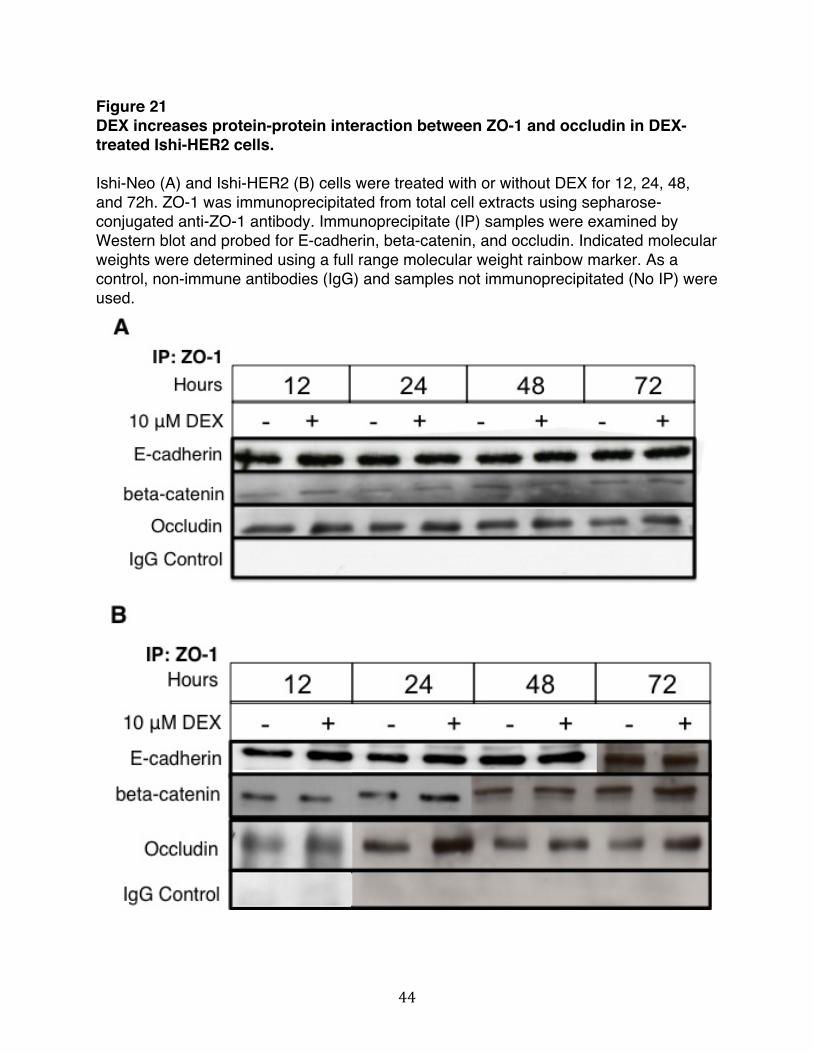
44
Figure 21 DEX increases protein-protein interaction between ZO-1 and occludin in DEX-treated Ishi-HER2 cells. Ishi-Neo (A) and Ishi-HER2 (B) cells were treated with or without DEX for 12, 24, 48, and 72h. ZO-1 was immunoprecipitated from total cell extracts using sepharose-conjugated anti-ZO-1 antibody. Immunoprecipitate (IP) samples were examined by Western blot and probed for E-cadherin, beta-catenin, and occludin. Indicated molecular weights were determined using a full range molecular weight rainbow marker. As a control, non-immune antibodies (IgG) and samples not immunoprecipitated (No IP) were used.

45
Treatment of Ishi-HER2 cells with DEX promotes an increase in interaction between ZO-1 and occludin. This increase in protein-protein interaction is specific to tight junctional proteins and does not involve changes in expression of levels of total protein or alteration in interaction between other proteins within the apical junction complex. In light of this, whether DEX induced ZO-1-occludin interaction drives the localization of the two proteins to the cell periphery is an intriguing issue, To test the dynamics of this interaction and evaluate whether the interaction between ZO-1 and occludin is co-dependent process that requires the expression of both proteins, we examined the localization of ZO-1 and occludin after siRNA mediated knockdown of ZO-1 and occludin. Ishi-HER2 cells were treated with or without DEX in the presence and absence of siRNA targeted towards either ZO-1 or occludin and a scrambled sequence of the siRNA target sequence. Localization of ZO-1 and occludin was examined using direct immunofluorescence microscopy. As shown in Figure 22, Ishi-HER2 cells that are treated with DMSO, a vehicle control, there is no localization of ZO-1 or occludin to periphery. When these cells are treated with DEX and transfected with a scrambled sequence of the siRNA target sequence, ZO-1 and occludin localize to the cell periphery. When occludin is knocked-down, there is a small amount of occludin that is localized to the cell periphery. Interestingly, the knockdown of occludin also prevents ZO-1 from localizing to the cell periphery. Similarly, the knockdown of ZO-1 prevents occludin from localizing to the cell periphery. This indicates that the interaction between ZO-1 and occludin and their subsequent localization to the periphery is a co-dependent process that requires both proteins to be expressed.

46
Figure 22 Co-dependent localization of ZO-1 and occludin in Ishi-HER2 cells after treatment with DEX. Ishi-HER2 cells were transfected with control scrambled siRNA or siRNA targeted against ZO-1 and occludin. The cells were grown to 100% confluency and treated with or without DEX for 48 hours. Indirect immunofluorescence microscopy was used to visualize ZO-1 and occludin localization and amount of knockdown of the two protein was determined using intensity of signal detected.
The expression of ZO-1 and occludin is important for maintaining the integrity of cellular junctions. The junctional proteins interact with a plethora of signaling proteins that regulate junction assembly and function and epithelial proliferation and differentiation. The down-regulation of occludin is seen in various tumors, including breast cancer, hepatocellular carcinoma, and gastric cancer. It is also associated with loss of proper cell-cell adhesion and differentiation [28-31]. Loss of occludin results in decreased cell-cell adhesion, altered epidermal differentiation, and reduced susceptibility to induction of apoptosis [32]. Expression of different isoforms of ZO-1 is also correlated with a differentiated phenotype in various cancers and non-cancer experimental models including intestinal tumor cells, lung cells, pancreatic stem cells and endothelial cells [33]. These findings suggest that loss of ZO-1 and occludin is associated with tumorigenesis and loss of differentiation. We have shown that treatment of Ishi-HER2 with DEX results in an increase in protein-protein interactions between ZO-1 and occludin and this increase in interaction is followed by a co-dependent localization of the two proteins to the periphery. To determine whether or not this DEX-induced organization of ZO-1 and occludin also
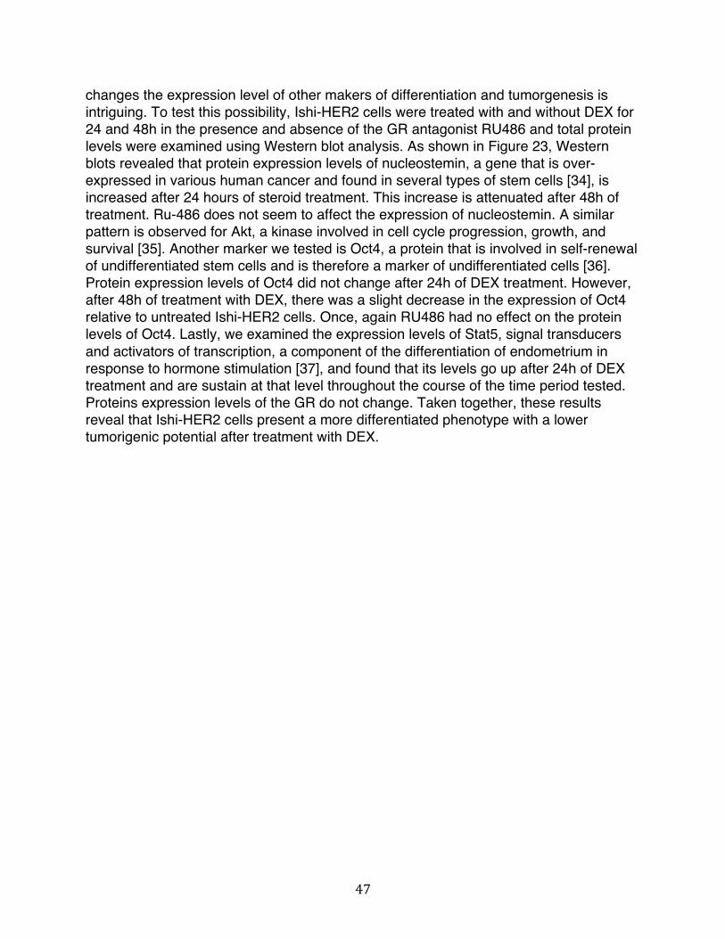
47
changes the expression level of other makers of differentiation and tumorgenesis is intriguing. To test this possibility, Ishi-HER2 cells were treated with and without DEX for 24 and 48h in the presence and absence of the GR antagonist RU486 and total protein levels were examined using Western blot analysis. As shown in Figure 23, Western blots revealed that protein expression levels of nucleostemin, a gene that is over-expressed in various human cancer and found in several types of stem cells [34], is increased after 24 hours of steroid treatment. This increase is attenuated after 48h of treatment. Ru-486 does not seem to affect the expression of nucleostemin. A similar pattern is observed for Akt, a kinase involved in cell cycle progression, growth, and survival [35]. Another marker we tested is Oct4, a protein that is involved in self-renewal of undifferentiated stem cells and is therefore a marker of undifferentiated cells [36]. Protein expression levels of Oct4 did not change after 24h of DEX treatment. However, after 48h of treatment with DEX, there was a slight decrease in the expression of Oct4 relative to untreated Ishi-HER2 cells. Once, again RU486 had no effect on the protein levels of Oct4. Lastly, we examined the expression levels of Stat5, signal transducers and activators of transcription, a component of the differentiation of endometrium in response to hormone stimulation [37], and found that its levels go up after 24h of DEX treatment and are sustain at that level throughout the course of the time period tested. Proteins expression levels of the GR do not change. Taken together, these results reveal that Ishi-HER2 cells present a more differentiated phenotype with a lower tumorigenic potential after treatment with DEX.

48
Figure 23 DEX alters expression levels of proteins involved in differentiation and tumorigenicity in Ishi-HER2 cells. Ishi-HER2 cells were treated with or without DEX for 24 and 48h. Total cell lysates were electrophoretically fractionated in sodium dodecyl sulfate polyacrylamide gels, and Western blots were probed with indicated antibodies or actin as a loading control.
DEX and The Combinational Effects of the Anti-tumor Drug Cisplatin on Junctional Complexes in Ishi-HER2 Cells Cisplatin is a member of a family of platinum-based anti-cancer agents. It is one of the most potent chemotherapy drugs widely used to treat a variety of cancers, including testicular, ovarian, bladder, head and neck, esophageal, small and non-small cell lung, breast, cervical, stomach and prostate cancers, as well as Hodgkin’s and non-Hodgkin’s lymphomas, neuroblastoma, sarcomas, multiple myeloma, melanoma, and mesothelioma [38]. Cisplatin treatment is also highly effective against endometrial cancer [39]. Additionally, the effects of cisplatin have been evaluated on junction complexes using various cancer cell models. Specifically, using cancerous endometrial cells obtained from females with endometrial endometriod adenocarcinoma, it has been shown that there is a significant reduction of tumor growth and a significant decrease in claudin-4, a tight junction protein that is unregulated in uterine serous papillary cancer [40]. In light of this, we wanted to determine whether or not DEX, in combination with Cisplatin, would have a differentiating effect on Ihis-HER2 by a mechanism that involves

49
changes in cell-cell interactions. To test this, we treated Ishi-HER2 cells with and without 10μM DEX 48 in the presence and absence of 0.5 and 1.0μM cisplatin and evaluated changes in localization of the tight junctional protein ZO-1 by indirect immunofluorescence microscopy. As shown in Figure 24A, In Ishi-HER2 cells localization of ZO-1 is diffused in the absence of DEX and cisplatin. However, ZO-1 is localized exclusively to the cell periphery in the presence of 10μM DEX. At 0.5 and 1.0μM cisplatin, ZO-1 is minimally localized to the cell periphery. However, at 0.5 and 1.0μM cisplatin in combination of DEX, there is increase in localization of ZO-1 to the cell periphery that is higher than in the samples treated with cisplatin alone. The percent of cells with ZO-1 organized to the cell periphery were quantified using ImageJ (Figure 24B). In addition to this, we also measure the change in perimeter of cells treated with DEX and cisplatin (Figure 24C). We observed a significant increase in the size of Ishi-HER2 cells treated with 0.5μM and 1.0μM cisplatin. The size of the cells treated with 1.0μM cisplatin in combination with 10μM DEX we significantly larger than any other groups tested. These results indicate that cisplatin alone does not significantly effect the localization of ZO-1. However, cisplatin in combination with DEX results in significant localization of ZO-1 to the cell periphery in Ishi-HER2 cells 48h after treatment with the drugs.
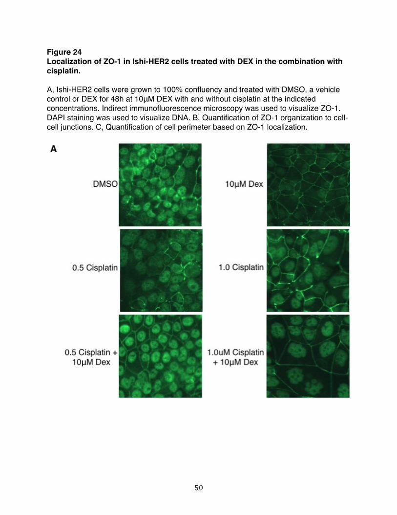
50
Figure 24 Localization of ZO-1 in Ishi-HER2 cells treated with DEX in the combination with cisplatin. A, Ishi-HER2 cells were grown to 100% confluency and treated with DMSO, a vehicle control or DEX for 48h at 10μM DEX with and without cisplatin at the indicated concentrations. Indirect immunofluorescence microscopy was used to visualize ZO-1. DAPI staining was used to visualize DNA. B, Quantification of ZO-1 organization to cell-cell junctions. C, Quantification of cell perimeter based on ZO-1 localization.

51
Platinum compounds such as cisplatin are known to damage cancer cells by inducing apoptosis by activating several signal transduction pathways. This has been shown to be mediated by inhibiting DNA synthesis and repair in ways that result in cell cycle arrest at G1, S, or G2-M phase [41]. To determine whether or not cisplatin in combination with DEX alters cell cycle progression, Ishi-HER2 cells were treated with or without either 1.0μM or 10μM DEX (dissolved in DMSO) for 48h in the presence and absence of 0.1, 0.5, 1.0, and 2.0μM cisplatin (dissolved in DMF) and nuclear DNA was stained with propodium iodide. The samples were prepared in triplicates and quantified by flow cytometry. As shown in Figure 25, Ishi-HER2 cells treated with DEX have the same cell cycle profile as cells treated with DMSO or DMF, the vehicle controls. Cells treated with cisplatin have a significant, dose-dependent increase in the G2/M and S phase of the cell cycle with concurrent decrease in the G1 phase. This trend continues up to 2.0μM cisplatin, as which point there is significant decrease in the G2/M and S

52
phase relative to cells treated at lower concentrations of cisplatin. When cells are treated at the indicated concentration of cisplatin in presence of either 1. 0μM or 10μM DEX, a similar dose-dependent increase in G2/M and S phase is observed. The increase in less than that seen in cells treated with cisplatin alone. Taken together, these data indicate that cisplatin alters cell cycle progression in Ishi-HER2 cells by increasing the population of cells in the G2/M and S phase of the cell cycle. When these cells are treated with cisplatin in combination with DEX, the increase is sustained at much lower level, but is still higher than cells treated with DEX alone. Figure 25 Cell cycle profile of Ishi-HER2 cells treated with DEX in combination with cisplatin. Ishi-HER2 cells were treated with and without DEX and cisplatin at the indicated doses and population DNA content was quantified by flow cytomertry. The bar graphs show the average DNA content corresponding to the cell cycle phases of three independent experiments.

53
Discussion
We have shown that DEX induces organization of tight junctional proteins ZO-1 and occludin to the cell periphery in human endometrial cancer cells only upon ectopic expression of HER2. Here, we show that treatment of Ishi-HER2 cells with DEX results in an increase in interaction between ZO-1 and occludin without changes in total protein expression levels of the junctional proteins. This increase in interaction and subsequent localization of ZO-1 and occludin to the cell periphery is a co-dependent process that requires the expression of both proteins. It is interesting to note that the increase in interaction between the two proteins occurs prior to their localization to the cell membrane. It is possible that the length of time required for the observed changes in ZO-1 and occludin dynamics is a result of changes in gene transcription caused by DEX.
Interaction between ZO-1 and occludin affect the properties of tight junctions and
is primarily regulated by phosphorylation events. Generally, increase in tyrosine phosphorylation tends to disrupt junctional integrity and an increase in serine/threonine phosphorylation tends to enhance junctional integrity by altering the manner in which occludin interacts with ZO-1 [42]. In light of this, it is intriguing to consider the role of serine, threonine, and tyrosine kinases that might be involved in the DEX-induced organization of ZO-1 and occludin to the cell periphery in Ishi-HER2 cells. Additionally, although our data reveal that the increase in protein-protein interactions between ZO-1 and occludin upon treatment of Ishi-HER2 cells with DEX is specific to tight junctional proteins, we cannot ignore the possibility that interactions between other proteins that are part of the apical junction complexes may be influenced by DEX.
As mentioned previously, loss of ZO-1 and occludin is associated with tumorigenesis and loss of differentiation. Our work has not only demonstrated that treatment of Ishi-HER2 cells results in an increase in interaction between the two proteins, but we have also shown that other makers of differentiation and tumorigenicity are altered upon treatment with DEX. Protein expression levels of nucleostemin, Oct4, Akt, and Stat5a were variably altered in Ishi-HER2 cells after treatment with DEX. A more thorough evaluation of these proteins is warranted in order to further elucidate their role in causing a differentiated phenotype. Not much is known about each of these proteins and their role in the regulation of cellular junctions. Because their levels are altered in response to DEX in Ishi-HER2 that show a dramatic organization of ZO-1 and occludin to the cell periphery, it is conceivable that they might also paly a role in regulating junctional complexes.
The aim of anti-cancer drugs is to inhibit the growth of cancer cells. Clinically,
platinum-based compounds are used as adjuvant therapy of cancer and are the most commonly used compounds for a wide range of malignancies [43-45]. Specifically, cisplatin induces tumor cell death by interfering with signal transduction pathways involved in transcription and DNA replication [46]. Because cisplatin resistance is a

54
common phenomenon, combination therapies have become routine for the treatment of multiple cancers. Our work reveals that treatment of Ishi-HER2 cells with DEX in combination with cisplatin results in localization of ZO-1 to the cell periphery. This change in localization if not seen in cells treated with cisplatin alone. The size of the cells treated with 1.0μM cisplatin in combination with 10μM DEX we significantly larger than any other groups tested. Additionally, cisplatin alters cell cycle progression in Ishi-HER2 cells by increasing the population of cells in the G2/M and S phase of the cell cycle. When these cells are treated with cisplatin in combination with DEX, the increase is sustained at a much lower level, but is still higher than cells treated with DEX alone. G2/M and S phase cell cycle arrest due to DNA damage leads to apoptosis of cells or inhibition of cell proliferation. The combined effect of DEX and cisplatin on cell cycle progression on Ishi-HER2 cells warrants further research.
Taken together, we show that treatment of Ishi-HER2 cells with DEX changes the dynamics of tight junctional proteins ZO-1 and occludin such that the cells present a more differentiated phenotype. It is known that endometrial cancers have the ability to respond to therapies based on HER2. This warrants further research into the signaling components associated with HER2 that take into consideration the variability of HER2 gene amplification and offer targeted treatment options so that the treatment of endometrial cancer can be more manageable.

55
References 1. Spadaro, D., et al., The control of gene expression and cell proliferation by the
epithelial apical junctional complex. Essays Biochem, 2012. 53: p. 83-93. 2. McCrea, P.D., D. Gu, and M.S. Balda, Junctional music that the nucleus hears:
cell-cell contact signaling and the modulation of gene activity. Cold Spring Harb Perspect Biol, 2009. 1(4): p. a002923.
3. Gonzalez-Mariscal, L., R. Tapia, and D. Chamorro, Crosstalk of tight junction components with signaling pathways. Biochim Biophys Acta, 2008. 1778(3): p. 729-56.
4. Stuart, R.O. and S.K. Nigam, Regulated assembly of tight junctions by protein kinase C. Proc Natl Acad Sci U S A, 1995. 92(13): p. 6072-6.
5. Markogiannakis, E., et al., Estrogens and glucocorticoids induce the expression of c-erbB2/NEU receptor in Ishikawa human endometrial cells. Life Sci, 1997. 61(11): p. 1083-95.
6. Karlan, B.Y., et al., Glucocorticoids stabilize HER-2/neu messenger RNA in human epithelial ovarian carcinoma cells. Gynecol Oncol, 1994. 53(1): p. 70-7.
7. Rubin, I. and Y. Yarden, The basic biology of HER2. Ann Oncol, 2001. 12 Suppl 1: p. S3-8.
8. Roskoski, R., Jr., The ErbB/HER receptor protein-tyrosine kinases and cancer. Biochem Biophys Res Commun, 2004. 319(1): p. 1-11.
9. Schulze, W.X., L. Deng, and M. Mann, Phosphotyrosine interactome of the ErbB-receptor kinase family. Mol Syst Biol, 2005. 1: p. 2005 0008.
10. Blume-Jensen, P. and T. Hunter, Oncogenic kinase signalling. Nature, 2001. 411(6835): p. 355-65.
11. Salomon, D.S., et al., Epidermal growth factor-related peptides and their receptors in human malignancies. Crit Rev Oncol Hematol, 1995. 19(3): p. 183-232.
12. Yarden, Y. and M.X. Sliwkowski, Untangling the ErbB signalling network. Nat Rev Mol Cell Biol, 2001. 2(2): p. 127-37.
13. Albitar, L., et al., EGFR isoforms and gene regulation in human endometrial cancer cells. Mol Cancer, 2010. 9: p. 166.
14. Furuse, M., et al., Direct association of occludin with ZO-1 and its possible involvement in the localization of occludin at tight junctions. J Cell Biol, 1994. 127(6 Pt 1): p. 1617-26.
15. Nusrat, A., et al., The coiled-coil domain of occludin can act to organize structural and functional elements of the epithelial tight junction. J Biol Chem, 2000. 275(38): p. 29816-22.
16. Dorfel, M.J. and O. Huber, A phosphorylation hotspot within the occludin C-terminal domain. Ann N Y Acad Sci, 2012. 1257: p. 38-44.
17. Sakakibara, A., et al., Possible involvement of phosphorylation of occludin in tight junction formation. J Cell Biol, 1997. 137(6): p. 1393-401.
18. Wong, V., Phosphorylation of occludin correlates with occludin localization and function at the tight junction. Am J Physiol, 1997. 273(6 Pt 1): p. C1859-67.

56
19. Seth, A., et al., Protein phosphatases 2A and 1 interact with occludin and negatively regulate the assembly of tight junctions in the CACO-2 cell monolayer. J Biol Chem, 2007. 282(15): p. 11487-98.
20. Rao, R.K., et al., Tyrosine phosphorylation and dissociation of occludin-ZO-1 and E-cadherin-beta-catenin complexes from the cytoskeleton by oxidative stress. Biochem J, 2002. 368(Pt 2): p. 471-81.
21. Kale, G., et al., Tyrosine phosphorylation of occludin attenuates its interactions with ZO-1, ZO-2, and ZO-3. Biochem Biophys Res Commun, 2003. 302(2): p. 324-9.
22. Basuroy, S., et al., Expression of kinase-inactive c-Src delays oxidative stress-induced disassembly and accelerates calcium-mediated reassembly of tight junctions in the Caco-2 cell monolayer. J Biol Chem, 2003. 278(14): p. 11916-24.
23. Fanning, A.S. and J.M. Anderson, Zonula occludens-1 and -2 are cytosolic scaffolds that regulate the assembly of cellular junctions. Ann N Y Acad Sci, 2009. 1165: p. 113-20.
24. Ikenouchi, J., et al., Requirement of ZO-1 for the formation of belt-like adherens junctions during epithelial cell polarization. J Cell Biol, 2007. 176(6): p. 779-86.
25. McNeil, E., C.T. Capaldo, and I.G. Macara, Zonula occludens-1 function in the assembly of tight junctions in Madin-Darby canine kidney epithelial cells. Mol Biol Cell, 2006. 17(4): p. 1922-32.
26. Zhang, L., et al., AMP-activated protein kinase regulates the assembly of epithelial tight junctions. Proc Natl Acad Sci U S A, 2006. 103(46): p. 17272-7.
27. Schaaf, M.J. and J.A. Cidlowski, Molecular mechanisms of glucocorticoid action and resistance. J Steroid Biochem Mol Biol, 2002. 83(1-5): p. 37-48.
28. Martin, T.A., R.E. Mansel, and W.G. Jiang, Loss of occludin leads to the progression of human breast cancer. Int J Mol Med, 2010. 26(5): p. 723-34.
29. Orban, E., et al., Different expression of occludin and ZO-1 in primary and metastatic liver tumors. Pathol Oncol Res, 2008. 14(3): p. 299-306.
30. Tobioka, H., et al., Occludin expression decreases with the progression of human endometrial carcinoma. Hum Pathol, 2004. 35(2): p. 159-64.
31. Tobioka, H., et al., Expression of occludin, a tight-junction-associated protein, in human lung carcinomas. Virchows Arch, 2004. 445(5): p. 472-6.
32. Rachow, S., et al., Occludin is involved in adhesion, apoptosis, differentiation and Ca2+-homeostasis of human keratinocytes: implications for tumorigenesis. PLoS One, 2013. 8(2): p. e55116.
33. Ciana, A., et al., A dynamic ratio of the alpha+ and alpha- isoforms of the tight junction protein ZO-1 is characteristic of Caco-2 cells and correlates with their degree of differentiation. Cell Biol Int, 2010. 34(6): p. 669-78.
34. Tsai, R.Y. and L. Meng, Nucleostemin: a latecomer with new tricks. Int J Biochem Cell Biol, 2009. 41(11): p. 2122-4.
35. Martelli, A.M., et al., The Emerging Role of the Phosphatidylinositol 3-Kinase/ Akt/Mammalian Target of Rapamycin Signaling Network in Cancer Stem Cell Biology. Cancers (Basel), 2010. 2(3): p. 1576-1596.
36. Sterneckert, J., S. Hoing, and H.R. Scholer, Concise review: Oct4 and more: the reprogramming expressway. Stem Cells, 2012. 30(1): p. 15-21.

57
37. Mak, I.Y., et al., Regulated expression of signal transducer and activator of transcription, Stat5, and its enhancement of PRL expression in human endometrial stromal cells in vitro. J Clin Endocrinol Metab, 2002. 87(6): p. 2581-8.
38. Florea, A.M. and D. Busselberg, Cisplatin as an anti-tumor drug: cellular mechanisms of activity, drug resistance and induced side effects. Cancers (Basel), 2011. 3(1): p. 1351-71.
39. Carey, M.S., et al., Systematic review of systemic therapy for advanced or recurrent endometrial cancer. Gynecol Oncol, 2006. 101(1): p. 158-67.
40. Pan, X.Y., et al., Overexpression of claudin-4 may be involved in endometrial tumorigenesis. Oncol Lett, 2013. 5(4): p. 1422-1426.
41. Desoize, B. and C. Madoulet, Particular aspects of platinum compounds used at present in cancer treatment. Crit Rev Oncol Hematol, 2002. 42(3): p. 317-25.
42. Rao, R., Occludin phosphorylation in regulation of epithelial tight junctions. Ann N Y Acad Sci, 2009. 1165: p. 62-8.
43. Olszewski, U. and G. Hamilton, A better platinum-based anticancer drug yet to come? Anticancer Agents Med Chem, 2010. 10(4): p. 293-301.
44. Shah, N. and D.S. Dizon, New-generation platinum agents for solid tumors. Future Oncol, 2009. 5(1): p. 33-42.
45. Zhang, J., et al., Status of bi- and multi-nuclear platinum anticancer drug development. Anticancer Agents Med Chem, 2010. 10(4): p. 272-82.
46. Sedletska, Y., M.J. Giraud-Panis, and J.M. Malinge, Cisplatin is a DNA-damaging antitumour compound triggering multifactorial biochemical responses in cancer cells: importance of apoptotic pathways. Curr Med Chem Anticancer Agents, 2005. 5(3): p. 251-65.
58
Chapter III
Glucocorticoid-mediated regulation of tight junctional proteins requires serine/threonine and tyrosine kinases and up-regulation of KLF9 and GRIM-19
gene expression

59
Abstract
The dynamic regulation of junctional complexes involves various signal transduction pathways that lie downstream of the human epidermal growth factors receptor 2 (HER2). The present work shows that treatment of Ishikawa cells expressing exogenous HER2 induces the organization of tight junctional proteins ZO-1 and occludin to the cell periphery upon treatment with the synthetic glucocorticoid, DEX. We further show that DEX induces an increase interaction between ZO-1 and occludin that requires the expression of both ZO-1 and occludin, indicating that the localization of the two proteins is a co-dependent process. Here, we delineate the involvement of other hormones and kinases downstream of HER2 that might be involved in the regulation of ZO-1 and occludin interaction. Because DEX is a steroid and associates with the GR to induce changes in gene transcription, we evaluate changes in gene expression of Ishikawa cells treated with DEX that lead to the altered dynamics of ZO-1 and occludin. Because the interaction between ZO-1 and occludin is primarily regulated by phosphorylation events, we evaluate the role of kinases downstream of HER2 in the DEX mediated effects on ZO-1 and occludin. We show that activation of AMP-activated protein kinase (AMPK) AMPK and inhibition of c-Src enhances the organization of ZO-1 and occludin to the cell periphery in the presence of DEX in Ishikawa cells. In contrast, inhibition of protein kinase C (PKC) disrupts this organization and attenuates the increase in protein-protein interaction between ZO-1 and occludin that was seen upon activation of AMPK and c-Src. Furthermore, gene expression profile of Ishikawa cells reveals that Kruppel-like factor (KLF9), a transcription factor that is associated with a differentiated endometrium, is significantly up-regulated upon treatment with DEX. We show that knockdown of KLF9 disrupts the DEX-induced organization of ZO-1 and occludin to the cell periphery and attenuates the interactions between the two proteins. Similarly, gene expression levels of GRIM-19, a tumor-suppressor and a known inhibitor of Src kinase is also increased upon treatment with DEX. si-RNA mediated knockdown of GRIM-19 disrupts DEX-induced organization of ZO-1 and occludin to the cell periphery and attenuates the interactions between the two proteins. The effect is somewhat reversed upon addition of SRC-inhibitor1 in cells where GRIM-19 is knocked-down. Taken together, our work shows that DEX confers a differentiated phenotype with a lower tumorigenic potential in human endometrial cancer cells by increasing gene expression levels of KLF9 and GRIM-19 that leads to alterations in the dynamics of ZO-1 and occludin through a mechanism that involves kinases associated with regulating interactions between the two tight junctional proteins.

60
Introduction
The interplay between junctional protein complexes and signaling pathways is important in the regulation of gene expression that coordinates many physiological events such as cell death, differentiation, and proliferation. Crosstalk between various signaling cascades leads to activation of phosphatases and kinases that eventually regulate the assembly, disassembly and maintenance of junctional complexes. Despite the relatively limited mechanistic information, intracellular nuclear receptors have been demonstrated to regulate the dynamics of cell-cell interactions through both primary and secondary transcriptional signaling and through non-transcriptional membrane effects that target the expression, modification, stability, function and/or localization of specific structural and/or accessory components of junctional complexes, depending on the physiological and tissue context [1]. In addition to nuclear receptors, the dynamic regulation of junctional complexes involves various signal transduction pathways that lie downstream of the human epidermal growth factors receptor 2 (HER2). Some of these signaling molecules include phosphatidylinositol 3-kiase (PI3K)/Akt, mitogen- activated protein (MAP) kinase, and protein kinase C, among many others [2]. The present work shows that tight junctional proteins ZO-1 and occludin organize to the cell periphery upon treatment of Ishikawa cells, a human endometrial cancer cell line, with the synthetic glucocorticoids, DEX and upon ectopic expression of HER2. We further show that DEX induces an increase interaction between ZO-1 and occludin that requires the expression of both ZO-1 and occludin, indicating that the localization of the two proteins is a co-dependent process. Next, we delineate the involvement of other hormones and kinases downstream of HER2 that might be involved in the regulation of ZO-1 and occludin interaction. Because DEX is a steroid and associates with the GR to induces changes in gene transcription, we evaluate changes in gene expression of Ishikawa cells treated with DEX that leads to the altered dynamics of ZO-1 and occludin.
Kinases & the Regulation of Tight Junctional Proteins
AMP-activated protein kinase
AMP-activated protein kinase (AMPK) is a serine-threonine kinase that plays a role in energy metabolism and cell polarity. As such, AMPK is activated upon cellular stress and results in the stimulation of fatty acid oxidation, inhibition of cholesterol synthesis, lipogenesis, and triglyceride synthesis and modulation of insulin secretion by the pancreases. Structurally, AMPK is a heterotrimeric protein with three subunits, alpha, beta, and gamma. The gamma subunit detects shifts in the ratio of AMP:ATP. When the ratio of AMP:ATP is high, AMP binds to the gamma subunit inducing a confirmation change in the protein that exposes a catalytic domain that in on the alpha subunit. AMPK is then rendered active when it is phosphorylated on a threonine residue by an upstream kinase known as LKB1. Once activated, AMPK inhibits anabolic pathways and activate catabolic pathways and leads to an increase in ATP production and inhibition of mTOR, which in turn inhibits cells proliferation and protein synthesis [3-5].

61
In addition to the role of AMPK in various cancers, the role of AMPK has been evaluated in endometrial cancer [6]. Furthermore, AMPK is also involved in regulating junctional complexes. Inflammatory bowl disease is caused by an impaired epithelial barrier functions. AMPK is activated in response to cellular stress that occurs during inflammation. Using intestinal epithelial cells, it has been shown that knockdown of AMPK prevents the decrease in ZO-1 and occludin that is caused in response to inflammation [7]. In MDCK cells, activation of AMPK using 5-aminoimidazole-4-carboxamide ribonucleoside (AICAR), an AMP analog, facilitates tight junction assembly as demonstrated by the relocation of ZO-1 to the cell periphery [8]. Expression of kinase-dead mutant of AMPK inhibits tight junction assembly, where as AICAR protects tight junctions from disassembly. This is demonstrated by measuring TER and ZO-1 localization to the tight junction in MDCK [9].
Protein kinase C
Protein kinase is a family of serine/threonine kinases that is activated by diacylglycerol. There are at least ten isoforms of PKC and they are differentially expressed within tissues and cellular compartments and their signaling is regulated by their expression, localization, and phosphorylation status. PKC is involved in signal transduction pathways that regulate cell migration, polarity, differentiation, survival, death, and proliferation. As such, dysregulated expression or activity of PKC has been implicated in a variety of cancers [10-12].
PKC is also involved in directly regulating the activity of junctional proteins. For example, in human pancreatic cancer cells it has been shown that gene expression of tight junctional proteins ZO-1, occludin, claudin, and JAMs are regulated via a PKC signal pathway [13]. Additionally, inhibition of PKC leads to disruption of TJ assembly. This effect leads to a defect in the sorting of ZO-1 and parallels a decrease in its phosphorylation, indicating PKC is involved in the assembly of functional TJs and that ZO-1 is one of its direct targets [14]. Furthermore, PKC is directly involved in phosphorylation of occludin. A novel isoform of PKC has been shown to interact with the C-terminal domain of occludin and phosphorylate it on conserved threonine residues [15].
c-Src tyrosine kinase
c-Src is a non-receptor tyrosine kinase that is localized to the intracellular membranes of cells. There are eight members of within the src family o tyrosine kinases and they are activated along with activation of transmembrane receptors, including EGFR. They play a role in cell growth, division, differentiation, survival, apoptosis, the immune response, and cell adhesion, movement and endocytosis [16-19]. As such, dysregulated expression and activity of c-Src is implicated in many diseases such as cancer. Elevated levels of Src kinase activity has been reported in a variety of cancer including breast, colon, brain, esophagus, pancreas, lung, renal, squamous cell carcinoma of the head and neck, leukemia, ovarian, prostate, and melanomas [20].

62
Because c-Src and EFGRs are over-expressed in many of the same tumors, it is possible that the two synergize to promote cancer cell growth [21]. In fact, both in vitro and in vivo studies have shown that HER2 associates with the SH2 domain of z-Src in a tyrosine phosphorylation-dependent manner [22, 23].
Tyrosine phosphorylation of occludin is involved in regulating its interaction with other junctional proteins and c-Src appears to play a direct role in mediating the phosphorylation. In Madin-Darby canine kidney (MDCK) cells, c-Src phosphorylates occludin at a unique motif that is involved in the regulation of ZO-1 binding. c-Src phosphorylate Tyr-398 and Tyr-402 in human occludin and this results in the loss interaction between occludin and ZO-1 and disruption of tight junction [24].
In the present work, we examine the role of these kinases in the regulation of ZO-1 and occludin and then evaluate changes in gene expression to delineate the mechanism by which DEX alters the dynamics of these tight junctional proteins in human endometrial cancer cells.

63
Material & Methods
Cell Culture
Ishikawa cells were grown in Dulbecco’s modified Eagles Medium, supplemented with 10% fetal bovine serum (all media components purchased from Lonza, Allendale, NJ and cell culture plates purchased from NUNC-Fischer, Pittsburgh, PA), 10 µg/ml insulin, 50 U/ml penicillin, 50 U/ml streptomycin, and 2 mmol/l L-glutamine (obtained from Sigma-Aldrich, St. Louis, MO). The cells were maintained at subconfluency in a humidified air chamber at 37˚C containing 5% CO2. A 100 mmol/l stock solution of DEX (D1756-500mg) was dissolved in DMSO (DD2650), and then diluted in the ratio 1:1000 in media before culture plate application. A 10mM stock solution of RU-486 (Milfepristone, M8046) was dissolved in DMSO to give a final concentration of 1nM. A 100mM stock solution of AICAR (A9978) was dissolved in water, and then diluted in the ratio 1:100 in media. A 10mM stock solution of G6983 (G1918) was dissolved in DMSO and then diluted 1:1000 in media. A 10mM stock solution of Src inhibitor (S2075) was dissolved in DMSO and diluted in the ratio 1:1000 in media before culture plate application. A 100 mmol/l stock solution of progesterone (purchased from Sigma-Aldrich, St. Louis, MO; catalog number PO130-25G) was dissolved in DMSO and then diluted in the ratio 1:1000 in media before culture plate application. All of the above drugs were purchased from Sigma-Aldrich, St. Louis, MO. Before each drug treatment, cells were washed in ice cold phosphate-buffered saline (PBS) (obtained form Lonza, Allendale, NJ). Indirect Immunofluorescence
Cells were grown and indicated treatments performed on two-well chamber slides from Nalgene Nunc International. The cells were fixed with 3.75% formaldehyde in PBS for 15 min at room temperature. After three additional washes with PBS, the plasma membrane was permeabilized with 0.1% Triton-X-100, 10 mM Tris-HCl, pH 7.5, 120 mM sodium chloride, 25 mM potassium chloride, 2 mM EGTA, and 2 mM EDTA) for 10 min at room temperature. Slides were incubated with 3% bovine serum albumin (Sigma-Aldrich) before incubation with primary antibodies. Anti-GR was a kind gift from Dr. Jen-Chywan (Wally) Wang, anti-ZO-1 (61-7300) purchased from Life Technologies/Invitrogen, anti-occludin (sc-133255), anti-AMPK (sc-130394), anti-GRIM-19 (sc-99086) purchased from Santa Sruc Biotechnologies, Santa Cruz, CA) and anti-KLF9 (AV31369-50ug) purchased from Sigma-Aldrich were used at a 1:400 dilution. Secondary Alexa 488 anti-rabbit and Texas Red- anti-mouse antibodies (Molecular Probes, Inc., Eugene, OR) were used at 1:400 dilutions each. Stained cells were mounted with Vectashield mounting medium containing 4,6-diamidino-2-phenylindole (DAPI) (Vector Laboratories, Burlingame, CA). Stained and mounted cells were then processed with an Axioplan epifloruescence microscope (Carl Zeiss, Thornwood, NY). The images were acquired and processed by M1/Hamamatsu Orca and QImaging MicroPublisher color cameras. Contrast and brightness settings were chosen so that all pixels were in the linear range.
64
Small Interfering RNA (siRNA)
Cells were grown and indicated treatments performed on 6-cm tissue culture plates from Nalgene Nunc International (Rochester, CA). Once cells reached 50% confluence, transfection with siRNA constructs was performed following transfection reagent manufacturer's protocol using HiPerfect (purchased from QIAGEN, Valencia, CA) with control siRNA (sc-37007)or specific siRNA against AMPK (sc-45312), KLF9 (sc-37716) and GRM19 (sc-60765) purchased from Santa Cruz Biotechnologies, Santa Cruz, CA, Valencia, CA. Microarray Analysis Cells treated with indicated doses of DEX and duration were harvested in Trizol (Invitrogen), and total RNA was extracted according to the manufacturer’s protocol. Gene array was performed using GeneChip Human U133 2.0 Array (Affymetrix, CA, USA) by QB3 Berkeley Core Research Facility’s Functional Genomics Laboratory at the University of California, Berkeley. Reverse Transcription and Polymerase Chain Reaction
Cells treated with indicated doses of DEX and duration were harvested in Trizol (Invitrogen), and total RNA was extracted according to the manufacturer’s protocol. This was quantified and 1 mg of total RNA was used for reverse transcription using Mu-MLV reverse transcriptase (Invitrogen) and random hexamers according to manufacturer’s protocol. The cDNA pool was used (2 ml) in polymerase chain reaction and was amplified with primers of the following sequences: KLF9 Forward: 5’-TACTGCACACTGGTCACCATC-3’ KLF9 Reverse: 5’-GTCACTCCTCATGAAGCGCTT-3’ GRIM-19 Forward: 5’-ACCGGAAGTGTGGGATACTG-3’ GRIM-19 Reverse: 5’-GCTCACGGTTCCACTTCATT-3’ SRC Forward: 5’-TCCAGATTGTCAACAACACAGA-3’ SRC Reverse: 5’-TTCTCTGCATTGAGCAGTAA-3’ GAPDH Forward: 5’-TGAAGGTCGGAGTCAACGGATTTG-3’ GAPDH Reverse: 5’-CATGTGGGCCATGAGGTCCACCAC-3’ 20 mL of this reaction was electrophoreased on a 1% agarose gel and visualized using a UV transilluminator. Western Blots
After the indicated treatments, cells were harvested in radioimmune precipitation assay buffer (150 mM NaCl, 0.5% deoxycholate, 0.1% NoNidet-p40 (Nonidet P-40, Flulta Biochemitra, Switzerland), 0.1% SDS, 50 mM Tris) containing protease and phosphatase inhibitors (50 g/ml phenylmethylsulfonyl fluoride, 10 g/mL aprotinin, 5 g/mL
65
leupeptin, 0.1 g/mL NaF, 1 mM dithiothreitol, 0.1 mM sodium orthovanadate, and 0.1 mM glycerol phosphate). Equal amounts of total cellular protein were mixed with loading buffer (25% glycerol, 0.075% SDS, 1.25 ml of 14.4M βmercaptoethanol, 10% bromphenol blue, 3.13% 0.5M Tris-HCl, and 0.4% SDS (pH 6.8)) and fractionated on 10% polyacrylamide/0.1% SDS resolving gels by electrophoresis. Rainbow marker (Amersham Biosciences) was used as the molecular weight standard. Proteins were electrically transferred to nitrocellulose membranes (Micron Separations, Inc., Westboro, MA) and blocked for 1 hour with Western wash buffer5% NFDM (10 mM Tris-HCl (pH 8.0), 150 mM NaCl, and 0.05% Tween 20, 5% nonfat dry milk). Protein blots were subsequently incubated for overnight at 4oC in primary antibodies. The antibodies used were as follows: anti-GR was a kind gift from Dr. Jen-Chywan (Wally) Wang, anti-ZO-1, (61-7300) purchased from Life Technologies/Invitrogen, San Diego, CA, anti-occludin (sc-133255), anti-ampk (sc-130394) anti-GRIM-19 (sc-99086) purchased from Santa Cruz Biotechnologies, Santa Cruz, CA) and anti-KLF9 (AV31369-50ug) purchased from Sigma-Aldrich were and diluted in the ratio 1:1000 in TBST. Rabbit anti-actin (AANO1; Cytoskeleton, Denver CO) was diluted 1:1000 in TBST and used as a gel-loading control. The working concentration for all antibodies was 1 µg/mL in Western wash buffer. Immunoreactive proteins were detected after incubation with horseradish peroxidase conjugated secondary antibody diluted to 3 x 10⁴ in Western wash buffer (goat anti-rabbit IgG, rabbit anti-goat IgG, and rabbit anti-mouse IgG (Bio-Rad)). Blots were treated with enhanced chemiluminescence reagents (PerkinElmer Life Sciences), and all proteins were detected by autoradiography. Equal protein loading was confirmed by Ponceau S staining of blotted membranes.
Co-Immunoprecipitation
Ishikawa cells were cultured on growth medium with DEX in the presence and absence of the indicated chemical activators and inhibitors for the designated times and then rinsed twice with PBS, harvested, and stored as dry pellets at −80°C. Cells were lysed for 15 min in IP buffer (50 mΜ Tris-HCl (pH 7.4), 150 mΜ NaCl, and 0.1% Triton X-100) containing protease inhibitors, 10 µg/ml aprotonin, 5 µg/ml leupeptin, 0.1 µg/ml NaF, 10 µg/ml β-glycerophosphate, and 0.1 mM sodium orthovanadate). Samples were diluted to 1 mg of protein in 1 ml of IP buffer. Samples were precleared for 1 hour at 4°C with 40 µl of a 1:1 slurry of protein G-Sepharose beads (GE health BioSciences AB). Precleared samples were then incubated with 50 µg of rabbit anti-ZO-1 or anti-occludin overnight at 4°C. Immunoprecipitated protein was eluted from beads by addition of gel loading buffer (50 mM Tris-HCl, pH 6.8, 2% SDS, 10% glycerol, 1% β-mercaptoethanol, 12.5 mM EDTA, 0.02 mM bromophenol blue) and heating the sample at 100°C for 5 min. Samples were analyzed by Western blot. Monoclonal anti-phosphotyrosine antibody (P5872-200UL) and monoclonal anti-phosphoserine (P5747) antibodies were purchased from Sigma-Aldrich.
66
Results We have shown that treatment of Ishi-HER2 cells with DEX results in an increase in protein-protein interactions between ZO-1 and occludin and this increase in interaction is followed by a co-dependent localization of the two proteins to the cell periphery. Additionally, we have shown that the DEX-induced membranous organization of ZO-1 and occludin, an indication of a differentiated phenotype, also changes the protein expression levels of other makers of differentiation and tumorgenesis. The junctional proteins serve as a docking site for adaptors with multiple protein-protein interaction domains and form a network that links the junctions to the actin cytoskeleton. This network in turn serves to recruit different types of signaling proteins such as kinases and phosphatase that regulate junctional assembly and epithelial proliferation and differentiation [25].
Because we see a DEX-induced increase in interaction between ZO-1 and occludin in Ishi-HER2, we wanted to test whether or not this increase in protein-protein interaction is due to the activity of tyr/ser/thr kinases. In order to test this, we implemented the use of various chemical activators and inhibitors of specific kinases known to play a role in the regulation of junctional complexes. These include 5-aminoimidazole-4-carboxamide ribonucleoside (AICAR), an AMP analog that activates AMPK, G6983, a pan-specific PKC inhibitor, and src inihibitor-1, a potent src kinase inhibitor. Initially, we wanted to see if the activation or inhibition of these kinases alters the DEX-induced organization of ZO-1 an occludin to the cell periphery. To this end, we treated Ishi-HER2 with and without DEX for 48h in the presence and absence of AICAR (ser/thr kinase), PKC (ser/thr kinase) inhibitor, and Src (tyr kinase) inhibitor. Localization of ZO-1 and occludin was examined using direct immunofluorescence microscopy. As shown in Figure 26, in Ishi-HER2 cells that are treated with DMSO, a vehicle control, there is no localization of ZO-1 or occludin to periphery. When these cells are treated with DEX there is a significant increase in localization of ZO-1 and occludin to the cell periphery. In the presence of AICAR, there is some organization of the ZO-1 and occludin to the cell periphery in the absence of the steroid. However, in the presence of DEX and AICAR, there is enhancement of in the organization of ZO-1 and occludin to membrane. This effect is reversed upon the treatment of Ishi-HER2 cells with DEX in the presence and absence of G6983. When these cells are treated with DEX in the presence of a src inhibitor, there is a dramatic organization of ZO-1 and occludin to the cell periphery that is higher than that seen with DEX alone. This indicates that the interaction between ZO-1 and occludin and their subsequent localization to the periphery is a co-dependent process that involves the activation and inhibition of the ser/thr/tyr kinases AMPK, PKC, and c-Src.

67
Figure 26 AMPK, PKC, and c-Src alter the localization of ZO-1 and occludin in Ishi-HER2 cells after treatment with DEX.
Ishi-HER2 cells were grown to 100% confluency and treated with or without DEX for 48 hours in the presence and absence of AICAR, an AMPK (ser/thr kinase) activator, PKC (ser/thr kinase) inhibitor, and Src (tyr kinase) inhibitor. Indirect immunofluorescence microscopy was used to visualize ZO-1 and occludin localization. DAPI staining was used to visualize DNA.
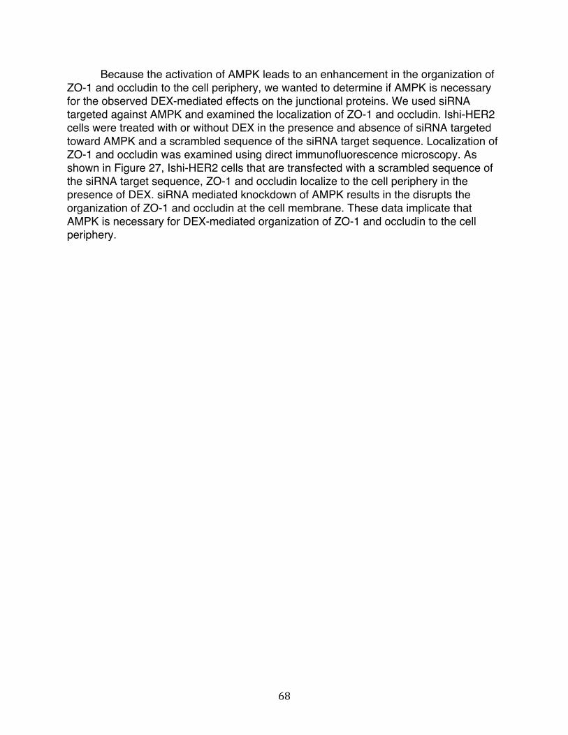
68
Because the activation of AMPK leads to an enhancement in the organization of ZO-1 and occludin to the cell periphery, we wanted to determine if AMPK is necessary for the observed DEX-mediated effects on the junctional proteins. We used siRNA targeted against AMPK and examined the localization of ZO-1 and occludin. Ishi-HER2 cells were treated with or without DEX in the presence and absence of siRNA targeted toward AMPK and a scrambled sequence of the siRNA target sequence. Localization of ZO-1 and occludin was examined using direct immunofluorescence microscopy. As shown in Figure 27, Ishi-HER2 cells that are transfected with a scrambled sequence of the siRNA target sequence, ZO-1 and occludin localize to the cell periphery in the presence of DEX. siRNA mediated knockdown of AMPK results in the disrupts the organization of ZO-1 and occludin at the cell membrane. These data implicate that AMPK is necessary for DEX-mediated organization of ZO-1 and occludin to the cell periphery.
69
Figure 27 siRNA mediated knockdown of AMPK disrupts DEX-induced localization of ZO-1 and occludin to the cell periphery in Ishi-HER2 cells. Ishi-HER2 cells were transfected with control scrambled siRNA or siRNA targeted against AMPK. The cells were grown to 100% confluency and treated with or without DEX for 48 hours. Indirect immunofluorescence microscopy was used to visualize ZO-1 and occludin localization. Amount of knockdown of AMPK was determined using intensity of signal detected.
Previous research and our results here indicate that one major class of signaling molecules that regulate tight junction structure, function, and dynamics are kinases. Specifically, the phosphorylation of occludin is tightly regulated and plays an important role in tight junction assembly and disassembly. Generally, increase in tyrosine phosphorylation tends to disrupt junctional integrity and an increase in serine/threonine phosphorylation tends to enhance junctional integrity by altering the manner in which occludin interacts with ZO-1 [26]. Because have shown that ser/thr/tyr kinases are involved in the regulation of ZO-1 and occludin dynamics, we wanted to evaluate the phosphorylation state of occludin. Ishi-HER2 cells were treated with and without DEX in the presence and absence of Src-inhibitor for 24h and occludin was immunoprecipitated

70
using sepharose beads. Phospho-ser and phosphor-tyr levels were detected using Western blot analysis of electrophorectically fractionated occludin and antibodies against phosphor-ser and phosphor-tyr. As shown in Figure 28, there is no increase the phosphorylation state of tyr residues of occludin in Ishi-HER2 treated with or without DEX. As expected, there is slight decrease in phosphor-tyr in the presence of Src-inhibitor. There is slight in increase in the phosphorylation state of ser residues of occludin upon treatment with DEX relative to cells treated with a vehicle control. This increase is sustained in the presence of Src-inhibitor. These data reveal that the observed increase in protein-protein interaction between ZO-1 and occludin might be due to changes in phosphorylation state of ser residues of occludin and may involve the inhibition of c-Src. Figure 28 DEX-induced increase in protein-protein interaction between ZO-1 and occludin in Ishi-HER2 DEX involves changes in phosphorylation state of occludin. Ishi-HER2 cells were treated with or without DEX for 24h. Occludin was immunoprecipitated from total cell extracts using sepharose-conjugated anti-occludin antibody. Immunoprecipitates (IP) samples were examined by Western blot and probed for phosphor-ser and phosphor-tyr. Indicated molecular weights were determine using a full range molecular weight rainbow marker. As a control, non-immune antibodies (IgG) were used.
Thus far, we have shown that AMPK, c-Src, and PKC are involved in the DEX-induced organization of ZO-1 and occludin to the cell periphery and that this effect results in changes in phosphorylation state of ser residues of occludin. To determine whether or not these kinases effect the protein-protein interaction between ZO-1 and occludin, occludin immunoprecipitated from Ishi-HER2 cells treated and without DEX 24h in the presence and absence of AICAR, G6983, Src-inhibitor1, and the GR antagonist RU486. Its binding to occludin was detected using Western blot analysis of

71
electrophorectically fractionated ZO-1. As shown in Figure 29, there is no increase in interaction between ZO-1 and occludin in Ishi-HER2 cells treated with the vehicle control. When Ishi-HER2 cells are treated with DEX there is significant increase in interaction between ZO-1 and occludin. In the presence of AICAR, an AMPK activator, there is an increase in the interaction between the two proteins that is equal in amount that seen in DEX treated cells without AICAR. This effect is attenuated in the presence of a PKC inhibitor. In the presence of Src-inhibitor1 there is an increase in interaction between ZO-1 and occludin in the absence of DEX relative to cells treated with DMSO, the vehicle control. In the presence of both the c-Src inhibitor and DEX, there is an increase interaction between ZO-1 and occludin that is higher than that seen in the cells treated with DEX alone. These data reveal that protein-protein interactions between ZO-1 and occludin increase upon treatment of Ishi-HER2 cells with DEX and that this increase involves AMPK, c-Src and PKC. Activation of AMPK, a ser/thr kinase, sustains the increase between the two proteins with or without the steroid. Inhibition of PKC, ser/thr kinase, attenuates this effect. In contrast, inhibition of c-Src, a tyr kinase, cause a slight increase in interaction between the two proteins even in the absence of DEX. A slightly higher increase in interaction is seen in the presence of both Src-inhibitor1 and DEX. Figure 29 AMPK, PKC, and c-Src alter the DEX-induced increase in protein-protein interaction between ZO-1 and occludin in Ishi-HER2 cells. Ishi-HER2 cells were treated with or without DEX 24h in the presence and absence of AICAR, G6983, and Src-inhibitor 1. ZO-1 was immunoprecipitated from total cell extracts using sepharose-conjugated anti-ZO-1 antibody. Immunoprecipitated (IP) samples were examined by Western blot and probed for occludin. Indicated molecular weights were determined using a full range molecular weight rainbow marker. As a control, non-immune antibodies (IgG) were used.
Because glucocorticoids, by nature have a permissive effect in that they allow a process to proceed at a maximum rate without themselves have little effect on the process itself [27], we wanted to determine if other hormones have a similar effect on tight junction dynamics in human endometrial cancer cells as the ones we have seen in
72
response to DEX. Progesterone is of particular interest to us because it plays an important role in the physiology of the endometrium. It plays a role during the endometrial cycle that is critical to the health and function of the endometrium. Uterine receptivity, embryo attachment, pregnancy, and the secretory phase of the menstrual cycle in health and disease are all processes that are regulated by progesterone within the endometrium [28]. The mechanism of action of progesterone in the endometrium is complex. Like other steroid hormones, progesterone works through a progesterone receptor (PR) and binds to its progesterone response element to elicits its effects. The PR consists of two isoforms, PR-A and PR-B, that come from a single gene [29]. They have distinct activation properties that are tissue and cell type specific [30]. Differential expression of the two isoforms have been reported, as the physiological consequences are a result of the specific co-regulators and nuclear accessory proteins that are found in different tissues and cells [31]. In order to examine the effects of progesterone on junctional complexes, we treated Ishi-HER2 cells at 0, 0.1, 1.0 and 10μM progesterone for 24, 48, and 72h and evaluated changes in localization of the tight junctional proteins ZO-1 by indirect immunofluorescence microscopy. As shown in Figure 30A, in Ishi-HER2 cells localization of ZO-1 is diffused in the absence of DEX at times points and doses tested. However, ZO-1 is localized exclusively to the cell periphery in the presence of DEX after 24h, at a dose of 10μM progesterone and 48h, at a dose of 1μM progesterone. The effect is maximal at a dose of 1μM progesterone and after 48h of treatment. The localization of occludin and ZO-1 was evaluated at this optimal dose and duration of treatment. As shown in Figure 30B, both ZO-1 and occludin localize to the cell periphery in the presence of the steroid and are diffused throughout the cells in the absence of progesterone. Treatment with DEX was used as a positive control. Taken together, these data reveal that progesterone, just as DEX, induces organization of ZO-1 and occludin to cell periphery in Ishi-HER2 cells.

73
Figure 30 Effect of progesterone on the localization of ZO-1 and occludin in Ishi-HER2 cells. A, Ishi-HER2 cells were grown to 100% confluency and treated with DMSO, a vehicle control or progesterone for 24, 48, and 72h at 0, 0.1, 1.0, and 10μM progesterone. Indirect immunofluorescence microscopy was used to visualize ZO-1. B, Ishi-HER2 cells were grown to 100% confluency and treated with DMSO, a vehicle control or progesterone at 1.0μM or DEX at 10μM for 48h. Indirect immunofluorescence microscopy was used to visualize ZO-1 and occludin.

74
To determine whether or not progesterone effects protein-protein interaction between ZO-1 and occludin, ZO-1 was immunoprecipitated from Ishi-HER2 cells treated and without 1μM progesterone for 24h. Cells were treated for 24h because an increase in interaction between ZO-1 and occludin is observed in Ishi-HER2 cells in response to DEX. Its binding to occludin was detected using Western blot analysis of electrophorectically fractionated ZO-1. As shown in Figure 31, there is no increase in interaction between ZO-1 and occludin in Ishi-HER2 cells treated with the vehicle control. When Ishi-HER2 cells are treated with DEX there is significant increase in interaction between ZO-1 and occludin. In the presence of progesterone, there is an increase in the interaction between the two proteins that is equal in amount that seen in DEX treated cells. This implicates that progesterone plays a role in the dynamics of tight junctions regulation regulating the interaction between ZO-1 and occludin. Figure 31 Progesterone increases protein-protein interaction between ZO-1 and occludin in Ishi-HER2 cell. Ishi-HER2 cells were treated with or without DEX and with or without progesterone for 24h. ZO-1 was immunoprecipitated from total cell extracts using sepharose-conjugated anti-ZO-1 antibody. Immunoprecipitated (IP) samples were examined by Western blot and probed for occludin. Indicated molecular weights were determined using a full range molecular weight rainbow marker. As a control, non-immune antibodies (IgG) were used.
The actions of glucocorticoids are mediated by the glucocorticoid receptor (GR), which is part of a superfamily of steroid receptors. These receptors are ligand-inducible transcription factors that control physiological function by causing changes in gene transcription. The mineralocorticoid receptor, progesterone receptor, estrogen receptor, and the androgen receptor are other members of this super family. All of these receptors have a highly conserved DNA-binding domain, a ligand-binding domain, and a variable amino terminal domain. Ligand binding to the receptor initiates a cascade of event that leads to the receptor-ligand complex trans-locating to the nucleus where the GR associates with a consensus palindromic sequence termed the glucocorticoid response element (GRE). Once bound to the GRE, the complex serves as a scaffold

75
that recruits co-regulators to initiate the transcriptional machinery and cause changes in gene transcription [32, 33].
We have shown that DEX alters the dynamics of tight junctional proteins ZO-1
and occludin in Ishi-HER2 through a signaling cascade that involves various kinases. DEX is a steroid hormone that binds to the GR and causes changes in gene transcription through its association with the GRE. To determine the specific genes that might involved in the DEX-mediated regulation of junctional complexes, we perform a DNA microarray analysis using the GeneChip Human Genome U133A 2.0 Array (Affymetrix) that represents 14,500 well-characterized human genes. We treated Ishi-HER2 cells with and without DEX for 48h and extracted total RNA according to the manufacturer’s protocol. The treated and untreated samples of the cells were sent for DNA microarray analysis. Of the all genes that were tested, there were not many that were significantly up- or down-regulated in response to DEX. Figure 32 lists the genes whose expression was the most altered in response to DEX. Figure 32 DNA microarray analysis of Ishi-HER2 cells treated with and without DEX. Total RNA was collected from Ishi-HER2 cells treated with and without DEX and a DNA microarray was performed using GeneChip Human Genome U133A 2.0 chip. A few of the genes most altered are shown.

76
Of the genes that were affected by treatment of Ishi-HER2 cells with DEX, Kruppel-like factor 9 (KLF9) is of particular interest. As shown in Figure 32, the expression of KLF9 increases by more than two fold in Inshi-HER2 cells upon treatment with DEX. KLF9 belongs to the SP/Kruppel-like family of transcription factors that function as transcriptional activators, repressors, or both by binding to GC/GT-rich sequences within target gene promoters through homologous zinc finger motifs [34]. They play a role in the regulation of genes involved in cell migration, growth, differentiation, and apoptosis [35, 36]. As such, KLFs have been implicated in a variety of cancers, including cancers of the nervous system, as well as skin, pancreatic, lung, ovarian, prostate, breast, endometrial, liver, bladder, gastric, intestinal and colon cancer [37].
KLFs can influence steroid hormone signaling through transcriptional networks that involves the nuclear receptor family of transcription factors. Because sensitivity to steroid hormone actions is the cause of many endocrine-related malignancies, KLFs act as tumor suppressors and or oncogenes under different cellular contexts [38]. The uterine endometrium epithelium goes through cycles of proliferation and differentiation and a subset of KLF family members have been linked to physiological changes associated with reproductive events such as pregnancy [39]. The role of KLF9 has been studied in the endometrium in relationship both the estrogen and the progesterone receptor. KLF9 represses ligand-activated ER alpha signaling through its ability to inhibit ERalpha expression and selectively regulate ERalpha transactivation in Ishikawa endometrial adenocarcinoma cells [40]. KLF9 also interact with the progesterone receptor, preferentially to the PR-B isoform, in Ishikawa cells [41, 42]. As such, KLF9 coordinately influences the expression of multiple genes regulated by estrogen and progesterone.
Lastly, attenuated expression of KLF9 has been linked to high endometrial tumor
grade, suggesting that KLF9 is involved in endometrial pathogenesis. Mice with a null mutation of KLF9 had altered patterns of proliferation and apoptosis in a variety of endometrial cells tested [43]. In light of this, we wanted to verify the DNA microarray results with RT-PCR to confirm the increase in expression of KLF9 in response to DEX in Ishi-HER2 cells. To this end, we treated Ishi-HER2 cells with and without DEX for 24 and 48h and examined the expression of KLF9 using RT-PCR. As shown in Figure 33, there is increase in KLF9 gene expression upon treatment of Ishi-HER2 cells with DEX at both 24 and 48h after treatment with the steroid. We also evaluated the gene expression levels of GRIM-19 (Gene associated with Retinoid-Interferon induced Mortality 19). The expression of GRIM-19 is of interest to us because GRIM-19 is a well-known tumor suppressor [44, 45]. Mutations of GRIM-19 and loss of protein expression has been observed in a variety of cancers including breast, thyroid, kidney, prostate, cervical carcinomas, among others. In-vivo studies have demonstrated that injection with GRIM-19 dramatically inhibits tumor growth [46]. In Ishi-HER2 cells that were treated with and without DEX, there was an increase in expression of GRIM-19 transcript at both 24 and 48 hours after treatment (Figure 33). These data reveal that

77
DEX induces gene expression of KLF9 and GRIM-19 in Ishi-HER2 cells. Gene expression levels of c-Src were detected because, as we have demonstrated, it is a kinase that is involved in the regulation of junctional complexes. As shown in Figure 33, gene expression levels of c-Src do not change in the presence and absence of DEX. Figure 33 Expression of KLF9 and GRIM-19 transcripts in DEX-treated Ishi-HER2 cells. Total RNA was collected from Ishi-HER2 cells were treated with and without DEX for 24 and 48h. RT-PCR was used to detect the levels of mRNA transcripts for KLF9, c-Src, and GRIM-19 or GAPDH as a loading control. PCR products were visualized using a 1% agarose gel stained with ethidium bromide.
To further evaluate the role of KLF9 and GRIM-19 in the regulation of junctional complexes, we used siRNA targeted against KLF9 and GRIM-19 and examined the localization of ZO-1 and occludin. Ishi-HER2 cells were treated with or without DEX in the presence and absence of siRNA targeted toward either KLF9 or GRIM-19 and a scrambled sequence of the siRNA target sequence. Localization of ZO-1 and occludin was examined using direct immunofluorescence microscopy. As shown in Figure 34, Ishi-HER2 cells that are transfected with a scrambled sequence of the siRNA target sequence, ZO-1 and occludin localize to the cell periphery in the presence of DEX. siRNA mediated knockdown of KLF9 and GRIM-19 results in the disrupts the organization of ZO-1 and occludin at the cell membrane. These data implicate that KLF9 and GRIM-19 are necessary for DEX-mediated organization of ZO-1 and occludin to the cell periphery.
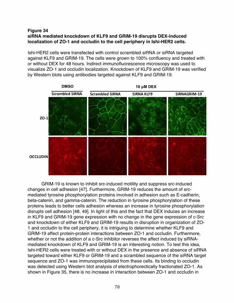
78
Figure 34 siRNA mediated knockdown of KLF9 and GRIM-19 disrupts DEX-induced localization of ZO-1 and occludin to the cell periphery in Ishi-HER2 cells. Ishi-HER2 cells were transfected with control scrambled siRNA or siRNA targeted against KLF9 and GRIM-19. The cells were grown to 100% confluency and treated with or without DEX for 48 hours. Indirect immunofluorescence microscopy was used to visualize ZO-1 and occludin localization. Knockdown of KLF9 and GRIM-19 was verified by Western blots using antibodies targeted against KLF9 and GRIM-19.
GRIM-19 is known to inhibit src-induced motility and suppress src-induced
changes in cell adhesion [47]. Furthermore, GRIM-19 reduces the amount of src-mediated tyrosine phosphorylation proteins involved in adhesion such as E-cadherin, beta-catenin, and gamma-catenin. The reduction in tyrosine phosphorylation of these proteins leads to better cells adhesion whereas an increase in tyrosine phosphorylation disrupts cell adhesion [48, 49]. In light of this and the fact that DEX induces an increase in KLF9 and GRIM-19 gene expression with no change in the gene expression of c-Src and knockdown of either KLF9 and GRIM-19 results in disruption in organization of ZO-1 and occludin to the cell periphery, it is intriguing to determine whether KLF9 and GRIM-19 affect protein-protein interactions between ZO-1 and occludin. Furthermore, whether or not the addition of a c-Src inhibitor reverses the affect induced by siRNA-mediated knockdown of KLF9 and GRIM-19 is an interesting notion. To test this idea, Ishi-HER2 cells were treated with or without DEX in the presence and absence of siRNA targeted toward either KLF9 or GRIM-19 and a scrambled sequence of the siRNA target sequence and ZO-1 was immunoprecipitated from these cells. Its binding to occludin was detected using Western blot analysis of electrophorectically fractionated ZO-1. As shown in Figure 35, there is no increase in interaction between ZO-1 and occludin in

79
Ishi-HER2 cells treated with the vehicle control. When Ishi-HER2 cells are treated with DEX there is significant increase in interaction between ZO-1 and occludin. This increase in interaction is attenuated upon siRNA-mediated knockdown of KLF9 and GRIM-19. The effect is somewhat reversed upon addition of SRC-inhibitor1 in cells where GRIM-19 is knocked-down. Figure 35 si-RNA mediated knockdown of KLF9 and GRIM-19 results in changes in protein-protein interaction between ZO-1 and occludin in Ishi-HER2 treated with DEX. Ishi-HER2 cells were treated with or without DEX, with or without Src-inhibitor 1, and in the presence and absence of siRNA targeted toward either KLF9 or GRIM-19 and a scrambled sequence of the siRNA target sequence for 24h. ZO-1 was immunoprecipitated from total cell extracts using sepharose-conjugated anti-ZO-1 antibody. Immunoprecipitated (IP) samples were examined by Western blot and probed for occludin (top panel). Knockdown of KLF9 and GRIM-19 were determined using Western blot analysis from whole cell lysates with antibodies targeted against KLF9 and GRIM-19 (bottom panel). Indicated molecular weights were determined using a full range molecular weight rainbow marker. As a control, non-immune antibodies (IgG) were used. Hsp90 was used as a loading control.

80
Discussion
Crosstalk between junctional protein complexes and signaling cascades leads to activation of phosphatases, kinases, and other signaling molecules that eventually regulate the assembly, disassembly and maintenance of junctional complexes. The present work shows that tight junctional proteins ZO-1 and occludin organize to the cell periphery upon treatment of cultured human endometrial cancer cells with the synthetic glucocorticoids, DEX. This requires the ectopic expression of HER2. We further show that DEX induces an increase interaction between ZO-1 and occludin that requires the expression of both ZO-1 and occludin, indicating that the localization of the two proteins is a co-dependent process. Here, we show that activation of AMP-activated protein kinase (AMPK) AMPK and inhibition of c-Src enhances the organization of ZO-1 and occludin to the cell periphery in the presence of DEX in Ishi-HER2 cells. In contrast, inhibition of protein kinase C (PKC) disrupts this organization and attenuates the increase in protein-protein interaction between ZO-1 and occludin that was upon activation of AMPK and c-Src. Furthermore, gene expression profile of Ishikawa cells reveals that Kruppel-like factor (KLF9), a transcription factor that is associated with a differentiated endometrium, is significantly up-regulated upon treatment with DEX. We show that knockdown of KLF9 disrupts the DEX-induced organization of ZO-1 and occludin to the cell periphery and attenuates the interactions between the two proteins. Similarly, gene expression levels of GRIM-19, a tumor-suppressor and a known inhibitor of Src kinase is also increased upon treatment with DEX. si-RNA mediated knockdown of GRIM-19 disrupts DEX-induced organization of ZO-1 and occludin to the cell periphery and attenuates the interactions between the two proteins. The effect is somewhat reversed upon addition of SRC-inhibitor-1 in cells where GRIM-19 is knocked-down. Taken together, our work shows that DEX confers a differentiated phenotype with a lower tumorigenic potential in human endometrial cancer cells by increasing gene expression levels of KLF9 and GRIM-19 that leads to alterations in the dynamics of ZO-1 and occludin through a mechanism that involves kinases associated with regulating interactions between the two tight junctional proteins.
As illustrated in Figure 36, we propose that in the presence of DEX (bottom
panel) there is an increase in gene expression of KLF9 and GRIM-19 in Ishi-HER2 cells. GRIM-19, a known inhibitor of the tyrosine kinase c-Src inhibitor, inhibits c-Src which in turn results in an increase in interaction between ZO-1 and occludin and their subsequent, co-dependent organization to the cell periphery. The increase in interaction between the two tight junctional proteins is likely due to the involvement of ser/thr kinases AMPK and PKC, as phosphorylation state of serine residue on occludin is slightly higher in cells treated with DEX. In the absence of DEX (top panel), c-Src is uninhibited and free to phosphorylate tyrosine residues of target proteins. Given that when the C-terminal domain of occludin is tyr-phosphorylated by incubating it with c-Src, ZO-1 binding to occludin is reduced [50, 51], it is conceivable that c-Src phosphorylates tyr residues such that it disrupts the junctional integrity. Although we did not observe a change in the tyr-phosphorylation state of occludin, it is possible that tyrosine restudies

81
of other junctional proteins is increased and this prevents ZO-1 and occludin from localizing to cell periphery. Tyr-phosphorylation of adherens junctions is important to the regulation of tight junctional proteins. Therefore, phosphorylation state of other junctional proteins and the activity level of c-Src, AMPK, and PKC need to be further examined. Disruption of adherens junctions proteins is known to disrupts tight junctional proteins

82
Figure 36 Proposed model for the DEX-mediated regulation of tight junctional proteins ZO-1 and occludin in human endometrial cancer cells. In the presence of DEX (bottom panel) there is an increase in gene expression of GRIM-19 in Ishi-HER2 cells. GRIM-19 inhibits c-Src which in turn results in an increase in interaction between ZO-1 and occludin and their subsequent, co-dependent organization to the cell periphery. In the absence of DEX (top panel), c-Src is uninhibited and free to phosphorylate tyrosine residues of target proteins and, in turn disrupt junctional integrity.

83
KLF9 is known to interact with steroid receptors. Specifically, KLF9 interacts with the progesterone receptor in endometrial cells and mediates progesterone responsiveness of target gene by direct interactions with the receptor [52]. The DEX-induced increased in gene expression level of KLF9 that we see warrants further studies in which the interaction of KLF9 with the GR is examined. We have shown that knockdown of KLF9 results in loss of junctional integrity. Additionally, we have shown that progesterone increases interaction between ZO-1 and occludin. Because both progesterone and KLF9 play a crucial role in the context of the endometrium, further delineating the mechanism by which they regulate junctional proteins is paramount to understanding the DEX-induced affect on cellular junctions in Ishi-HER2 cells. From the data presented here, it can be inferred that signaling molecules, such as the above-mentioned kinases, and hormone receptors systems can regulate junctional complexes in Ishi-HER2 cells in a way that lowers tumorigenicity. This information is invaluable in the discovery of molecularly targeted therapy against endometrial cancer.

84
References 1. McCrea, P.D., D. Gu, and M.S. Balda, Junctional music that the nucleus hears:
cell-cell contact signaling and the modulation of gene activity. Cold Spring Harb Perspect Biol, 2009. 1(4): p. a002923.
2. Gonzalez-Mariscal, L., R. Tapia, and D. Chamorro, Crosstalk of tight junction components with signaling pathways. Biochim Biophys Acta, 2008. 1778(3): p. 729-56.
3. Hardie, D.G., F.A. Ross, and S.A. Hawley, AMPK: a nutrient and energy sensor that maintains energy homeostasis. Nat Rev Mol Cell Biol, 2012. 13(4): p. 251-62.
4. Forcet, C. and M. Billaud, Dialogue between LKB1 and AMPK: a hot topic at the cellular pole. Sci STKE, 2007. 2007(404): p. pe51.
5. Jansen, M., et al., LKB1 and AMPK family signaling: the intimate link between cell polarity and energy metabolism. Physiol Rev, 2009. 89(3): p. 777-98.
6. Brown, K.A., N.U. Samarajeewa, and E.R. Simpson, Endocrine-related cancers and the role of AMPK. Mol Cell Endocrinol, 2013. 366(2): p. 170-9.
7. Scharl, M., et al., AMP-activated protein kinase mediates the interferon-gamma-induced decrease in intestinal epithelial barrier function. J Biol Chem, 2009. 284(41): p. 27952-63.
8. Zhang, L., et al., AMP-activated protein kinase regulates the assembly of epithelial tight junctions. Proc Natl Acad Sci U S A, 2006. 103(46): p. 17272-7.
9. Zheng, B. and L.C. Cantley, Regulation of epithelial tight junction assembly and disassembly by AMP-activated protein kinase. Proc Natl Acad Sci U S A, 2007. 104(3): p. 819-22.
10. Nishizuka, Y., The molecular heterogeneity of protein kinase C and its implications for cellular regulation. Nature, 1988. 334(6184): p. 661-5.
11. Nishizuka, Y., Studies and prospectives of the protein kinase c family for cellular regulation. Cancer, 1989. 63(10): p. 1892-903.
12. Breitkreutz, D., et al., Protein kinase C family: on the crossroads of cell signaling in skin and tumor epithelium. J Cancer Res Clin Oncol, 2007. 133(11): p. 793-808.
13. Yamaguchi, H., et al., Transcriptional control of tight junction proteins via a protein kinase C signal pathway in human telomerase reverse transcriptase-transfected human pancreatic duct epithelial cells. Am J Pathol, 2010. 177(2): p. 698-712.
14. Stuart, R.O. and S.K. Nigam, Regulated assembly of tight junctions by protein kinase C. Proc Natl Acad Sci U S A, 1995. 92(13): p. 6072-6.
15. Suzuki, T., et al., PKC eta regulates occludin phosphorylation and epithelial tight junction integrity. Proc Natl Acad Sci U S A, 2009. 106(1): p. 61-6.
16. Brown, M.T. and J.A. Cooper, Regulation, substrates and functions of src. Biochim Biophys Acta, 1996. 1287(2-3): p. 121-49.
17. Thomas, S.M. and J.S. Brugge, Cellular functions regulated by Src family kinases. Annu Rev Cell Dev Biol, 1997. 13: p. 513-609.

85
18. Wilde, A., et al., EGF receptor signaling stimulates SRC kinase phosphorylation of clathrin, influencing clathrin redistribution and EGF uptake. Cell, 1999. 96(5): p. 677-87.
19. Okada, M., Regulation of the SRC family kinases by Csk. Int J Biol Sci, 2012. 8(10): p. 1385-97.
20. Homsi, J., C. Cubitt, and A. Daud, The Src signaling pathway: a potential target in melanoma and other malignancies. Expert Opin Ther Targets, 2007. 11(1): p. 91-100.
21. Biscardi, J.S., et al., Tyrosine kinase signalling in breast cancer: epidermal growth factor receptor and c-Src interactions in breast cancer. Breast Cancer Res, 2000. 2(3): p. 203-10.
22. Luttrell, D.K., et al., Involvement of pp60c-src with two major signaling pathways in human breast cancer. Proc Natl Acad Sci U S A, 1994. 91(1): p. 83-7.
23. Muthuswamy, S.K., et al., Mammary tumors expressing the neu proto-oncogene possess elevated c-Src tyrosine kinase activity. Mol Cell Biol, 1994. 14(1): p. 735-43.
24. Elias, B.C., et al., Phosphorylation of Tyr-398 and Tyr-402 in occludin prevents its interaction with ZO-1 and destabilizes its assembly at the tight junctions. J Biol Chem, 2009. 284(3): p. 1559-69.
25. Matter, K., et al., Mammalian tight junctions in the regulation of epithelial differentiation and proliferation. Curr Opin Cell Biol, 2005. 17(5): p. 453-8.
26. Rao, R., Occludin phosphorylation in regulation of epithelial tight junctions. Ann N Y Acad Sci, 2009. 1165: p. 62-8.
27. Granner, D.K., The role of glucocorticoid hormones as biological amplifiers. Monogr Endocrinol, 1979. 12: p. 593-611.
28. Lessey, B.A., Two pathways of progesterone action in the human endometrium: implications for implantation and contraception. Steroids, 2003. 68(10-13): p. 809-15.
29. Kastner, P., et al., Two distinct estrogen-regulated promoters generate transcripts encoding the two functionally different human progesterone receptor forms A and B. EMBO J, 1990. 9(5): p. 1603-14.
30. Conneely, O.M. and B.M. Jericevic, Progesterone regulation of reproductive function through functionally distinct progesterone receptor isoforms. Rev Endocr Metab Disord, 2002. 3(3): p. 201-9.
31. Giangrande, P.H., et al., The opposing transcriptional activities of the two isoforms of the human progesterone receptor are due to differential cofactor binding. Mol Cell Biol, 2000. 20(9): p. 3102-15.
32. Cosio, B.G., A. Torrego, and I.M. Adcock, [Molecular mechanisms of glucocorticoids]. Arch Bronconeumol, 2005. 41(1): p. 34-41.
33. Heitzer, M.D., et al., Glucocorticoid receptor physiology. Rev Endocr Metab Disord, 2007. 8(4): p. 321-30.
34. Black, A.R., J.D. Black, and J. Azizkhan-Clifford, Sp1 and kruppel-like factor family of transcription factors in cell growth regulation and cancer. J Cell Physiol, 2001. 188(2): p. 143-60.
35. Jensen, D.E., et al., Distinct roles for Sp1 and E2F sites in the growth/cell cycle regulation of the DHFR promoter. J Cell Biochem, 1997. 67(1): p. 24-31.
86
36. Cook, T., et al., Molecular cloning and characterization of TIEG2 reveals a new subfamily of transforming growth factor-beta-inducible Sp1-like zinc finger-encoding genes involved in the regulation of cell growth. J Biol Chem, 1998. 273(40): p. 25929-36.
37. Bureau, C., et al., Expression and Function of Kruppel Like-Factors (KLF) in Carcinogenesis. Curr Genomics, 2009. 10(5): p. 353-60.
38. Simmen, R.C., et al., The emerging role of Kruppel-like factors in endocrine-responsive cancers of female reproductive tissues. J Endocrinol, 2010. 204(3): p. 223-31.
39. Simmen, R.C., et al., Molecular markers of endometrial epithelial cell mitogenesis mediated by the Sp/Kruppel-like factor BTEB1. DNA Cell Biol, 2002. 21(2): p. 115-28.
40. Velarde, M.C., et al., Kruppel-like factor 9 is a negative regulator of ligand-dependent estrogen receptor alpha signaling in Ishikawa endometrial adenocarcinoma cells. Mol Endocrinol, 2007. 21(12): p. 2988-3001.
41. Velarde, M.C., et al., Progesterone receptor transactivation of the secretory leukocyte protease inhibitor gene in Ishikawa endometrial epithelial cells involves recruitment of Kruppel-like factor 9/basic transcription element binding protein-1. Endocrinology, 2006. 147(4): p. 1969-78.
42. Zhang, X.L., et al., Selective interactions of Kruppel-like factor 9/basic transcription element-binding protein with progesterone receptor isoforms A and B determine transcriptional activity of progesterone-responsive genes in endometrial epithelial cells. J Biol Chem, 2003. 278(24): p. 21474-82.
43. Velarde, M.C., et al., Null mutation of Kruppel-like factor9/basic transcription element binding protein-1 alters peri-implantation uterine development in mice. Biol Reprod, 2005. 73(3): p. 472-81.
44. Zhang, J., et al., The cell death regulator GRIM-19 is an inhibitor of signal transducer and activator of transcription 3. Proc Natl Acad Sci U S A, 2003. 100(16): p. 9342-7.
45. Lufei, C., et al., GRIM-19, a death-regulatory gene product, suppresses Stat3 activity via functional interaction. EMBO J, 2003. 22(6): p. 1325-35.
46. Moreira, S., et al., GRIM-19 function in cancer development. Mitochondrion, 2011. 11(5): p. 693-9.
47. Sun, P., et al., GRIM-19 inhibits v-Src-induced cell motility by interfering with cytoskeletal restructuring. Oncogene, 2009. 28(10): p. 1339-47.
48. Behrens, J., et al., Loss of epithelial differentiation and gain of invasiveness correlates with tyrosine phosphorylation of the E-cadherin/beta-catenin complex in cells transformed with a temperature-sensitive v-SRC gene. J Cell Biol, 1993. 120(3): p. 757-66.
49. Kalakonda, S., et al., Tumor suppressive protein gene associated with retinoid-interferon-induced mortality (GRIM)-19 inhibits src-induced oncogenic transformation at multiple levels. Am J Pathol, 2007. 171(4): p. 1352-68.
50. Rao, R.K., et al., Tyrosine phosphorylation and dissociation of occludin-ZO-1 and E-cadherin-beta-catenin complexes from the cytoskeleton by oxidative stress. Biochem J, 2002. 368(Pt 2): p. 471-81.

87
51. Kale, G., et al., Tyrosine phosphorylation of occludin attenuates its interactions with ZO-1, ZO-2, and ZO-3. Biochem Biophys Res Commun, 2003. 302(2): p. 324-9.
52. Zhang, D., et al., Direct interaction of the Kruppel-like family (KLF) member, BTEB1, and PR mediates progesterone-responsive gene expression in endometrial epithelial cells. Endocrinology, 2002. 143(1): p. 62-73.

88
Chapter IV
Conclusion and Future Directions

89
Conclusion and Future Directions Cancer is complex. The development of genomic instability within cells paves the way for tumor progression by altering intracellular signaling networks that regulate the functions of a cell. Reprograming of these signaling pathways enables normal cells to acquire characteristics of cancer cells and allows them to survive, proliferate, and eventually metastasize. Each cancer manifests itself with unique features making it difficult to study, diagnose, and treat. Despite this challenge, a growing understanding of cancer cell biology sets the stage for the discovery of novel treatment regimes that provide personalized, molecularly targeted therapy.
Our work focuses on cellular junctions and their role in carcinogenesis. Proper cell-cell communication is key to regulating cell proliferation, differentiation, apoptosis, movement, and adhesion. In epithelial cells, organization of cell-cell interactions is carried out by cellular junctions that are composed of multiple protein complexes. Junctional proteins serve as docking sites for adaptors with multiple protein-protein interaction domains and form a network that serves to recruit different types of signaling proteins that regulate junctional assembly and epithelial proliferation and differentiation. Because junctional complexes regulate such important physiological processes, disruption of cell-cell interactions that leads to a permanent loss of junctional integrity results in the development and progression of a variety of cancers. Here we evaluate the effects of glucocorticoids on junctional complexes in human endometrial cancer cells. We show that tight junctional proteins ZO-1 and occludin organize to the cell periphery upon treatment of cultured human endometrial cancer cells with the synthetic glucocorticoids, DEX. Because this requires the ectopic expression of HER2, an open question to be discovered is the amount of HER2 over-expression that is required for the Ishikawa cells to be sensitive to DEX treatment. This would be clinically significant, as HER2 is amplified to varying degrees in different types of endometrial cancers. Furthermore, we show that DEX induces an increase in interaction between ZO-1 and occludin that requires the expression of both ZO-1 and occludin, indicating that the localization of the two proteins is a co-dependent process. Given that the apical junction complex is composed to many different proteins, an area of interest would be to investigate the effects of DEX on other proteins that part of adherens and tight junctions. Although we show that the DEX-induced effect is specific to tight junctional proteins, it is probable that DEX can alter interactions between beta-catenin and occludin, for example. It is interesting to note that the increase in interaction between the two proteins occurs prior to their localization to the cell membrane. It is possible that the length of time required for the observed changes in ZO-1 and occludin dynamics is a result of changes in gene transcription caused by DEX. The reason for the delay in interaction between ZO-1 and occludin and the their subsequent localization to the cell periphery remains to be discovered.

90
Signaling components downstream of HER2 play a role in the regulation of cell-cell interactions and one major class of signaling molecules that regulate tight junction structure, function, and dynamics are kinases. Here, we examine the role of three kinases, AMPK, c-Src, and PKC. We show that activation of AMP-activated protein kinase (AMPK) AMPK and inhibition of c-Src enhances the organization of ZO-1 and occludin to the cell periphery in the presence of DEX in Ishi-HER2 cells. In contrast, inhibition of protein kinase C (PKC) disrupts this organization and attenuates the increase in protein-protein interaction between ZO-1 and occludin that was upon activation of AMPK and c-Src. Determining the precise role of these kinases remains to be discovered. It will be interesting to determine whether or not AMPK, c-Src, or PKC are involved in directly regulating the phosphorylation of occludin.
The actions of glucocorticoids are mediated by the glucocorticoid receptor (GR).
These receptors are ligand-inducible transcription factors that control physiological function by causing changes in gene transcription. Gene expression profile of Ishi-HER2 cells reveals that Kruppel-like factor (KLF9), a transcription factor that is associated with a differentiated endometrium, is significantly up-regulated upon treatment with DEX. We show that knockdown of KLF9 disrupts the DEX-induced organization of ZO-1 and occludin to the cell periphery and attenuates the interactions between the two proteins. Similarly, gene expression level of GRIM-19, a tumor-suppressor and a known inhibitor of c-Src kinase is also increased upon treatment with DEX. si-RNA mediated knockdown of GRIM-19 disrupts DEX-induced organization of ZO-1 and occludin to the cell periphery and attenuates the interactions between the two proteins. The effect is somewhat reversed upon addition of SRC-inhibitor1 in cells where GRIM-19 is knocked-down. In light of this, it is critical to determine whether or not kinase activity of c-Src is correlated with expression of GRIM-19. Additionally, the precise role of KLF9 and GRMI19 in mediating the DEX-induced effect on ZO-1 and occludin also warrants further investigation. Specifically, discovering whether or not KLF9 or GRIMP19 interact directly with the GR or interact with other transcription factors is key to uncovering the precise mechanism by which DEX elicits the observed effects. Lastly, in light of our observation that cisplatin and progesterone affect ZO-1 and occludin dynamics, it is crucial to further study the effects of DEX in combination with other anti-cancer drugs and hormone receptor systems. Because both progesterone and KLF9 play a crucial role in the context of the endometrium, further research into delineating the mechanism by which they regulate junctional proteins is paramount to understanding the DEX-induced affect on cellular junctions in Ishi-HER2 cells.
Taken together, the present work shows that DEX confers a differentiated
phenotype with a lower tumorigenic potential in human endometrial cancer cells by increasing gene expression levels of KLF9 and GRIM-19 that leads to alterations in the dynamics of ZO-1 and occludin through a mechanism that involves kinases associated with regulating interactions between the two tight junctional proteins. Transcriptional and non-transcriptional molecular pathways that regulate cell-cell interactions by relaying signals from the nucleus to the cellular junctions at the membrane are complex and far

91
from being understood. Elucidating the signal transduction mechanisms that maintain integrity of cellular junctions has profound implication for the diagnosis and treatment of cancer.