glucocorticoid associated osteoporosis
TRANSCRIPT
1
Glucocorticoid Associated Osteoporosis
Prevention and Treatment
Jonathan Graf, MD
Professor of Clinical Medicine UCSF
Division of Rheumatology, San Francisco General Hospital
Traditional rheumatology practice
2
Question #1
Bone loss in GIO is bimodal and occurs rapidly in an early phase (weeks) and more gradually in a later phase. Which one of the following primarily occurs in the early/rapid phase?
A. Increased osteoblast apoptosis (cell death)
B. Increased osteoclast activity
C. Decreased osteoblast activation
D. Combination of A,B,C
3
Glucocorticoids: Some toxicities
DiabetesCataractsHTNWeight gainFluid retentionPUDMyopathyPsychiatric
OSTEOPOROSIS & OSTEONECROSIS50% of patients
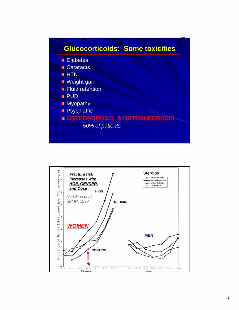
HIGH
MEDIUM
CONTROL
Steroids
WOMEN
MEN
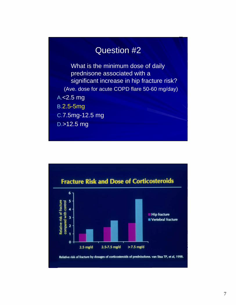
Fracture risk increases with AGE, GENDER, and Dose
*
Van Staa et al, JBMR, 1998

4
Glucocorticoid Effects of Bone
Increased apoptosis of osteocytes (bone quality decreased before BMD)
Prolonged osteoclast survival and decreased osteoclastogenesis
Increased Osteoclast bone resporption
Decreased osteoblastogenesis and increased apoptosis
Multiple pathways affected!!!! Increased bone resportion relative to decreased bone formation and decreased overall bone quality:
Weinstein R. NEJM 2011;365:62-70
GLUCOCORTICOIDS: “Double Whammy to Bones”
Osteoclast /Pro-resorptive effects (early)– Decrease OPG– Increase RANK-L osteoclast #,
activity, lifespan
Anti bone-formation effects (late)Decrease osteoblast and osteoclast formation Decrease osteoblast lifespanEnhance apoptosis in osteocytes
Biophasic effect: Osteoclast effects are early and osteoblast are late

5
GLUCOCORTICOID - INDUCED OSTEOPOROSIS
Early phase of rapid bone loss (Pro-resorption)– As early as 2 mos into therapy
– Resorption markers elevated
– Pts on high dose prednisone can lose 15-20% of trabecular bone (spine) in 5-7 mos
Slower phase of bone loss (Anti-Formation)– Continues indefinitely
– Trabecular bone especially vulnerable
Glucocorticoid Effects on Remodeling/Strength
Manolagas, JBMR, 2000
X many…
6
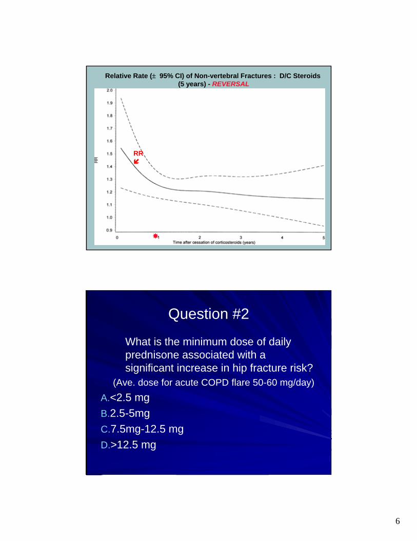
Relative Rate ( 95% CI) of Non-vertebral Fractures : D/C Steroids(5 years) - REVERSAL
RR
Question #2
What is the minimum dose of daily prednisone associated with a significant increase in hip fracture risk?
(Ave. dose for acute COPD flare 50-60 mg/day)
A.<2.5 mg
B.2.5-5mg
C.7.5mg-12.5 mg
D.>12.5 mg
7
Question #2
What is the minimum dose of daily prednisone associated with a significant increase in hip fracture risk?
(Ave. dose for acute COPD flare 50-60 mg/day)
A.<2.5 mg
B.2.5-5mg
C.7.5mg-12.5 mg
D.>12.5 mg
8
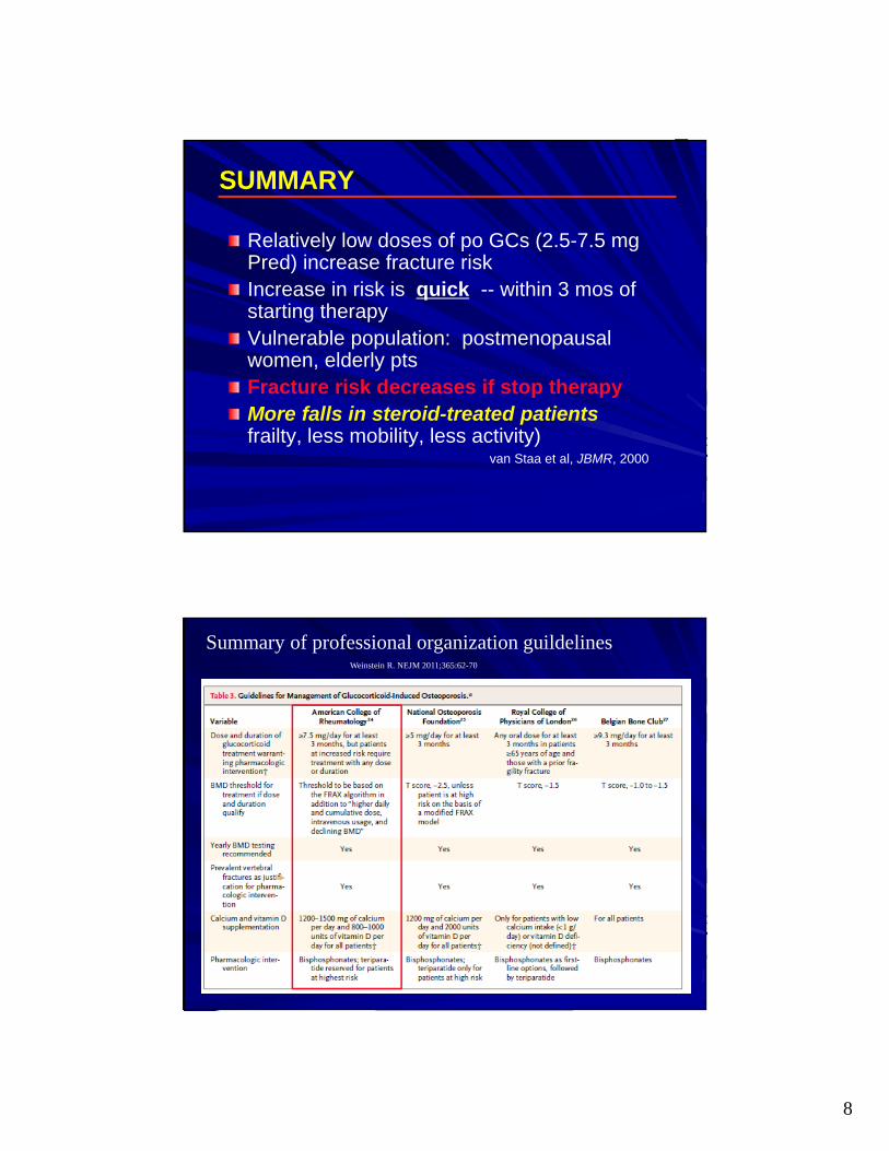
SUMMARY
Relatively low doses of po GCs (2.5-7.5 mg Pred) increase fracture riskIncrease in risk is quick -- within 3 mos of starting therapyVulnerable population: postmenopausal women, elderly ptsFracture risk decreases if stop therapyMore falls in steroid-treated patients (more frailty, less mobility, less activity)
van Staa et al, JBMR, 2000
Summary of professional organization guildelinesWeinstein R. NEJM 2011;365:62-70

9
2017 American College of Rheumatology Guidelines on GIOP
Systematic literature review summarizes evidence for risks/benefit of GIOP 1) Risk Assessment 2) treatment and 3) Follow up using G.R.A.D.E. methodology
– Adults
– Women of childbearing potential
– Adults requiring very high doses of glucocorticoids
– Adults with organ transplants
– Children ages 4-17
Principles Reflected in ACR 2017 guidelines
GIO does not occur in a vacuum but interacts with risk factors other than corticosteroids
Guided by risk/benefit assessment: risk assessed using FRAX score risk or other risk calculator PLUS correction/adjustment for prednisone dosage
Risk calculators stratifying GC use as low/high (<>7.5 mg/day) underestimate risk associated with higher daily GC doses
Observational data suggest risk to young women and children receiving high dose GC’s
10
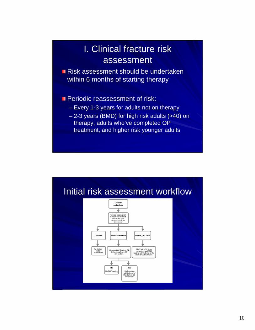
I. Clinical fracture risk assessment
Risk assessment should be undertaken within 6 months of starting therapy
Periodic reassessment of risk:– Every 1-3 years for adults not on therapy
– 2-3 years (BMD) for high risk adults (>40) on therapy, adults who’ve completed OP treatment, and higher risk younger adults
Initial risk assessment workflow
11
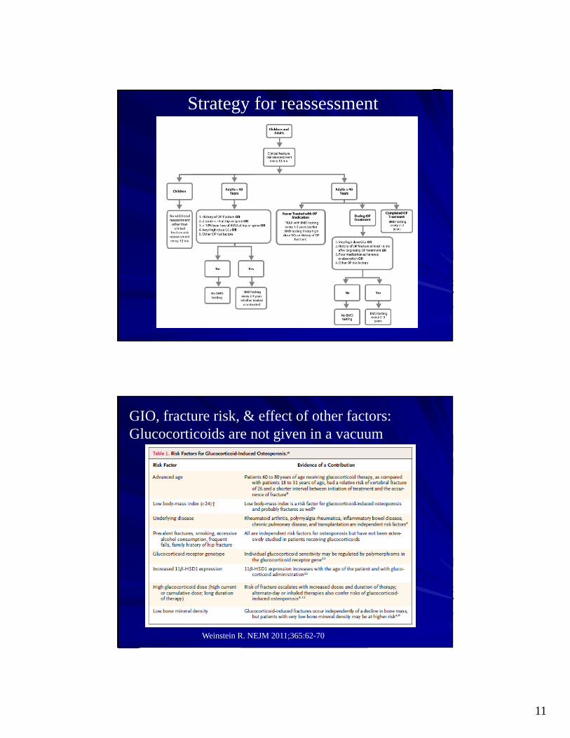
Strategy for reassessment
GIO, fracture risk, & effect of other factors:Glucocorticoids are not given in a vacuum
Weinstein R. NEJM 2011;365:62-70

12
ACR 2017 Guidelines definition of risk:
Recommendations are conditional for moderate risk & strong for high risk
II. Treatment: Moderate/high risk
Women not of childbearing potential and men:(order of preference)
1.Oral bisphosphonate2.IV bisphosphonate3.Teriparatide4.Denosumab5.Raloxifene (PMP women as last resort)

13
Treatment: Moderate/high risk
Women of childbearing potential (conditional):(order of preference)
1.Oral bisphosphonate2.Teriparatide
3.IV bisphosphonate (high risk only)4.Denosumab (high risk only)
Treatment: High dose GCsAdults > 30 years
Initial prednisone dose > 30 mg/day or cumulative dose > 5 grams in 1 year
1.Oral bisphosphonate2.IV bisphosphonate3.Teriparatide4.Denosumab5.Raloxifene (PMP women as last resort)
14
III: Follow up
After 5 years of treatment:– Continue therapy if still assessed at mod/high
risk.
After discontinuing GC treatment– Mod/high risk: continue treatment
– Low risk: discontinue treatment
What about therapeutic “failures?”
Question #3
True or False:
There have been no head to head comparative effectiveness studies of bisphosphonates for GIO:
A.True
B.False

15
Question #3
True or False:
There have been no head to head comparative effectiveness studies of bisphosphonates for GIO:
A.True
B.False
What about Zoledronic Acid? Better than other bisphosphonates?: Horizon
Reid et al. Lancet. 2009 Apr 11;373(9671):1253-63
1 year randomized double blind, double dummy, non-inferiority
833 patients– Subdivided into
treatment groups based on duration of steroid therapy (>< 3 months)
IV ZA 5mg vs. PO Risedronate 5mg
Primary endpoint: BMD LS spine
16
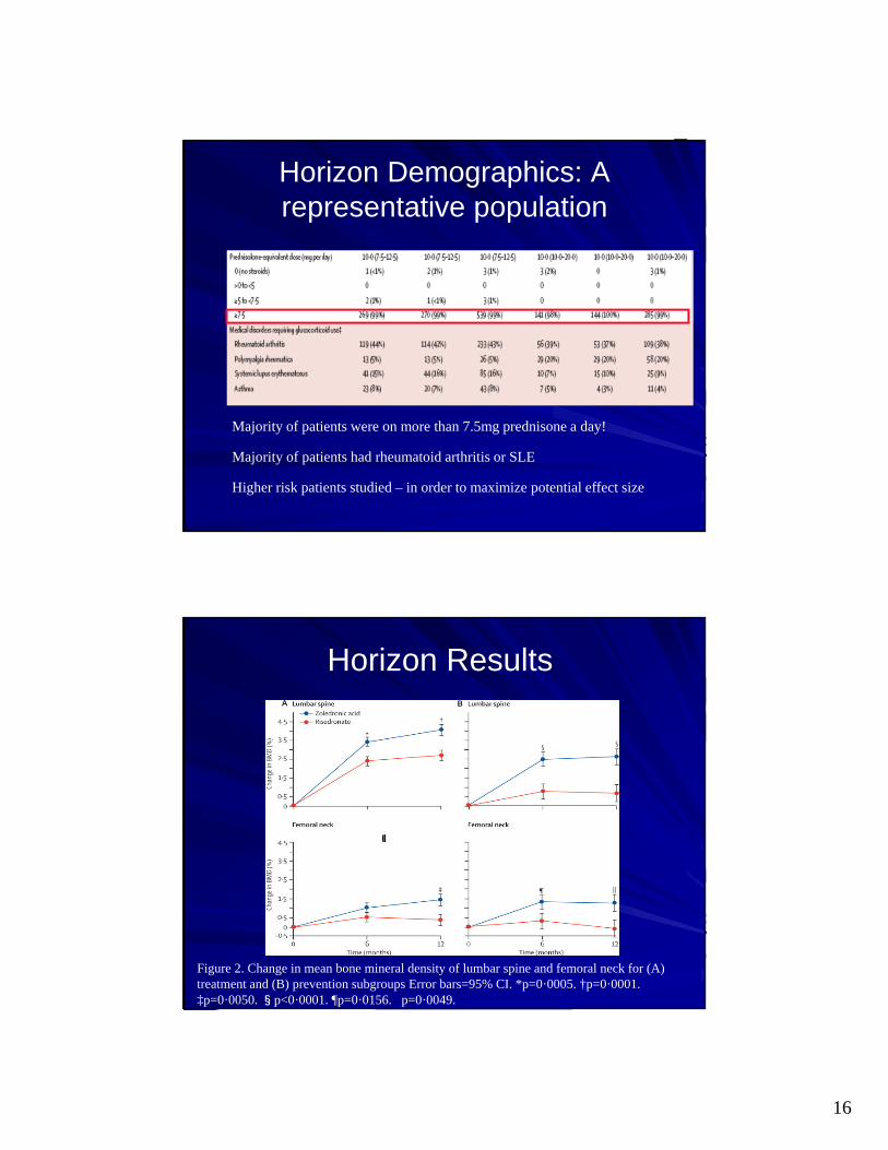
Horizon Demographics: A representative population
Majority of patients were on more than 7.5mg prednisone a day!
Majority of patients had rheumatoid arthritis or SLE
Higher risk patients studied – in order to maximize potential effect size
Horizon Results
Figure 2. Change in mean bone mineral density of lumbar spine and femoral neck for (A) treatment and (B) prevention subgroups Error bars=95% CI. *p=0·0005. †p=0·0001. ‡p=0·0050. §p<0·0001. ¶p=0·0156. p=0·0049.

17
PTH vs. bisphosponates: Beneficial for GIO? (2008)
Saag et al. NEJM 2007;357:2028-39
36 Month randomized double blinded controlled
18 month interim analysis
428 patients studied– 22-89 years of age– Treated with GC’s for at
least three months– Prednisone equivalent of 5
mg/day or more– 20 mcg/d PTH vs. 10 mg/d
alendronate– Everyone continued
Ca/VitD
Who were the Patients?BMD<-2.0 or <1.0 + fragility fracture (Higher risk patients)
Exclusions: Standard for PTH Use
Two treatment groups similar (n=214 both)
Alendronate PTH
Age 57.3 56.1
Prednisone dose 7.8 7.5
Non Vert Frag Fx 20.1% 19.6%
BMD T score LS Spine -2.6 -2.5
BMD T score Hip -1.9 -2.0
18
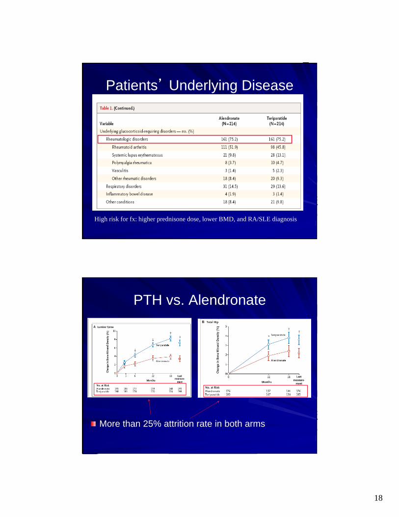
Patients’ Underlying Disease
High risk for fx: higher prednisone dose, lower BMD, and RA/SLE diagnosis
PTH vs. Alendronate
More than 25% attrition rate in both arms
19
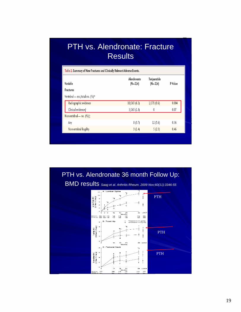
PTH vs. Alendronate: Fracture Results
PTH vs. Alendronate 36 month Follow Up:
BMD results Saag et al. Arthritis Rheum. 2009 Nov;60(11):3346-55
PTH
PTH
PTH

20
PTH vs. Alendronate: 36 month fracture follow up
Saag et al. Arthritis Rheum. 2009 Nov;60(11):3346-55
At 36 months, statistically significant reduction in both radiographic and clinical vertebral fractures
No significant differences in non-vertebral fractures
PTH for GC induced Osteoporosis: Summary
PTH appears to improve BMD in GC assoc. osteoporosis– Evidence suggests superior increases in BMD vs.
alendronate at hip, femoral neck, and LS spine
However, high attrition rate in this study – Nearly 50% drop out in both arms by 36 months– Appears to be of benefit both for clinical and
radiographically defined fractures at 36 months– BUT….overall rate of clinical fracture is low in both
groups
Decrease in fractures limited to vertebral fractures but non-significant for non-vertebral fractures
21
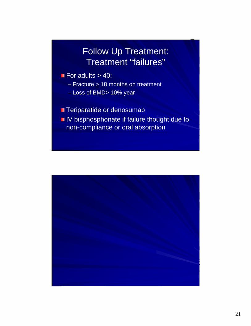
Follow Up Treatment:Treatment “failures”
For adults > 40:– Fracture > 18 months on treatment
– Loss of BMD> 10% year
Teriparatide or denosumab
IV bisphosphonate if failure thought due to non-compliance or oral absorption