use of indirectly immobilized recombinant p17 antigen for detection of antibodies to hiv-1 by enzyme...
TRANSCRIPT
© 1999 Wiley-Liss, Inc.
Journal of Clinical Laboratory Analysis 13:9–18 (1999)
Use of Indirectly Immobilized Recombinant p17 Antigen forDetection of Antibodies to HIV-1 by Enzyme Immunoassay
Setsuko Ishikawa, 1 Kazuya Hashinaka, 1 Seiichi Hashida, 1 Shinichi Oka, 2
and Eiji Ishikawa 1*1Department of Biochemistry, Miyazaki Medical College, Miyazaki, Japan
2AIDS Clinical Center, International Medical Center of Japan, Tokyo, Japan
Recombinant HIV-1 p17 antigen (rp17)and maltose binding protein-rp17 fusionprotein (MBP-rp17) were immobilized indifferent ways: rp17 and MBP-rp17 wereimmobilized directly onto polystyrenebeads by physical adsorption, and biotinyl-rp17, biotinyl-MBP-rp17, and 2,4-dinitro-phenyl (DNP)-MBP-rp17 were immobilizedindirectly onto polystyrene beads, whichhad been coated with streptavidin alone,with biotinyl-bovine serum albumin andstreptavidin and with (anti-2,4-dinitro-phenyl group) IgG. These immobilizedantigens were tested by incubation withdiluted serum from an HIV-1 seropositivesubject in the absence and presence ofserum from HIV-1 seronegative subjectsand, after washing, with rp17 β-D-galac-tosidase conjugate. Higher positive signals(fluorescence intensities for bound -β-D-galactosidase activity) and less serum in-terference were obtained with indirectly
immobilized antigens than with directly im-mobilized ones. Enzyme immunoassayusing biotinyl-MBP-rp17 indirectly immo-bilized onto polystyrene beads, which hadbeen coated sequentially with biotinyl-bovine serum albumin and streptavidin,was approximately 1,000-fold more sen-sitive than that using directly immobilizedrp17 antigen and Western blotting for p17band. This enzyme immunoassay indi-cated positivity in HIV-1 seroconversionserum panels as early as or even earlierthan conventional methods and consider-ably earlier than Western blotting for HIV-1 p17 band. In addition, the sensitivity wasfurther improved approximately 10-fold byincubation with shaking for immunoreactionsand by increase of both the number of poly-styrene beads and the volume of serumsamples used per assay. J. Clin. Lab. Anal.13:9–18, 1999. © 1999 Wiley-Liss, Inc.
Key Words: human immunodeficiency virus type 1; antibodies to p17; enzyme immunoassay;serum interference; immobilization of antigen; seroconversion
INTRODUCTION
Antibodies to HIV-1 env, pol, and gag proteins in serumhave been detected by ELISA and Western blotting. The sen-sitivities of the conventional ELISA with gp41 and reversetranscriptase (RT) as antigens and Western blotting for gp41and RT (p66 and p51) bands were 100% in asymptomaticcarriers, 86–100% in patients with AIDS-related complex(ARC), and 77–100% in patients with AIDS (1–3). The sen-sitivities of the conventional ELISA using rp24 as antigenand Western blotting for p24 band were 63–92% in asymp-tomatic carriers, 50–97% in patients with ARC, and 49–77%in patients with AIDS (1–3). The earliest positive bands afterthe infection were for p24, p55, gp120, and gp160 (4–6). Bycontrast, the sensitivity of Western blotting for p17 band wasmuch lower (41% in asymptomatic carriers, 30% in patientswith ARC, and 14% in patients with AIDS) (1), and p17 bandremained negative for significant periods of time after posi-
tive bands for p24, p55, and env proteins appeared followingthe infection (4–6).
Recently, a highly sensitive enzyme immunoassay for an-tibodies to HIV-1 in urine has been reported using indirectlyimmobilized recombinant HIV-1 p17 antigen (rp17). 2,4-Dinitrophenyl-rp17 was indirectly immobilized onto polysty-rene beads, which had been coated with affinity-purified(anti-2,4-dinitrophenyl group) IgG, and biotinyl-rp17 wasindirectly immobilized onto polystyrene beads, which hadbeen coated successively with biotinyl-bovine serum albu-min and streptavidin (7). The sensitivity was 1,000- to 3,000-fold higher than that of Western blotting for HIV-1 p17 band.
*Correspondence to: Eiji Ishikawa, M.D., Ph.D., Professor of Biochemistry,Department of Biochemistry, Miyazaki Medical College, Kiyotake, Miyazaki889-1692, Japan.
Received 14 August 1998; Accepted 14 August 1998
10 Ishikawa et al.
This report describes not only more detailed effects of in-directly immobilized rp17 on the sensitivity and serum inter-ference in enzyme immunoassay for antibodies to HIV-1 p17antigen, but also earlier detection than by conventional meth-ods of antibodies to HIV-1 in HIV-1 seroconversion serumpanels by enzyme immunoassay using indirectly immobilizedrp17 and 100 µL of serum.
MATERIALS AND METHODS
Buffer
The regularly used buffer was 10 mmol/L sodium phos-phate buffer, pH 7.0, containing 1.0 mmol/L MgCl2, 0.1%bovine serum albumin (fraction V, Intergen Company, Pur-chase, NY) and 0.1% NaN3 (buffer A).
Maltose Binding Protein-HIV-1 p17 AntigenFusion Protein and HIV-1 p17 Antigen
Maltose binding protein-HIV-1 p17 antigen fusion protein(MBP-rp17) was produced in Escherichia coli transformedwith expression plasmid carrying the corresponding DNA,and MBP-rp17 and recombinant HIV-1 p17 antigen (rp17)were purified as described previously (8). The recombinantproviral clone used (pNL4-3) (9) contained DNA from HIV-1 isolates NY5 (GenBank accession number HIVNL43) andLAV (10), and the sequence for p17 derived from NY5.
Biotinyl- and 2,4-Dinitrophenyl-MBP-rp17
MBP-rp17 was mercaptoacetylated and was reacted with6-maleimidohexanoyl-biocytin and aN-6-maleimidohexanoyl-eN-2,4-dinitrophenyl-L-lysine, which had been prepared byreaction of N-hydroxysuccinimidyl-6-maleimidohexanoatewith biocytin and eN-2,4-dinitrophenyl-L-lysine (7).
Biotinyl-rp17 Antigen
Antigen rp17 was mercaptoacetylated and reacted with 6-maleimidohexanoyl-biocytin (7).
rp17-b-D-Galactosidase Conjugate
Mercaptoacetylated rp17 was conjugated to β-D-galactosi-dase (EC 3.2.1.23) from Escherichia coli (BoehringerMannheim, GmbH, Mannheim, Germany) using 2,4-phenyl-enedimaleimide (11).
Protein-Coated Polystyrene Beads
Polystyrene beads (3.2 mm in diameter, Immuno Chemi-cal Inc., Okayama, Japan) were coated with rp17-related pro-teins in seven different ways (Fig. 1). Polystyrene beads werecoated with biotinyl-bovine serum albumin (100µg/mL) (12),affinity-purified (anti-2,4-dinitrophenyl group) IgG (100 µg/mL) (13), streptavidin (Bethesda Research Laboratories,
Life Technologies, MD) (5 to 50 µg/mL), rp17 (1 to 100µg/mL), and MBP-rp17 (1 to 100 µg/mL) by physical ad-sorption as described previously (14). Biotinyl-bovine se-rum albumin-coated polystyrene beads were further coatedwith streptavidin (100 µg/mL). For this coating, 0.1 mol/Lsodium phosphate buffer, pH 7.5, containing 0.1% NaN3 wasused. For indirect coating, individual polystyrene beadscoated with streptavidin and streptavidin-biotinyl-bovineserum albumin were incubated at 4°C overnight with biotinyl-rp17 (100–40,000 fmol) and biotinyl-MBP-rp17 (50–1,000fmol) in 0.15 mL of buffer A containing 0.1 mol/L NaCl, andindividual polystyrene beads coated with affinity-purified(anti-2,4-dinitrophenyl group) IgG were incubated at 4°Covernight with 2,4-dinitrophenyl MBP-rp17 (50–300 fmol)in 0.15 mL of the same buffer. These protein-coated polysty-rene beads were stored in buffer A containing 0.1 mol/L NaClat 4°C until use.
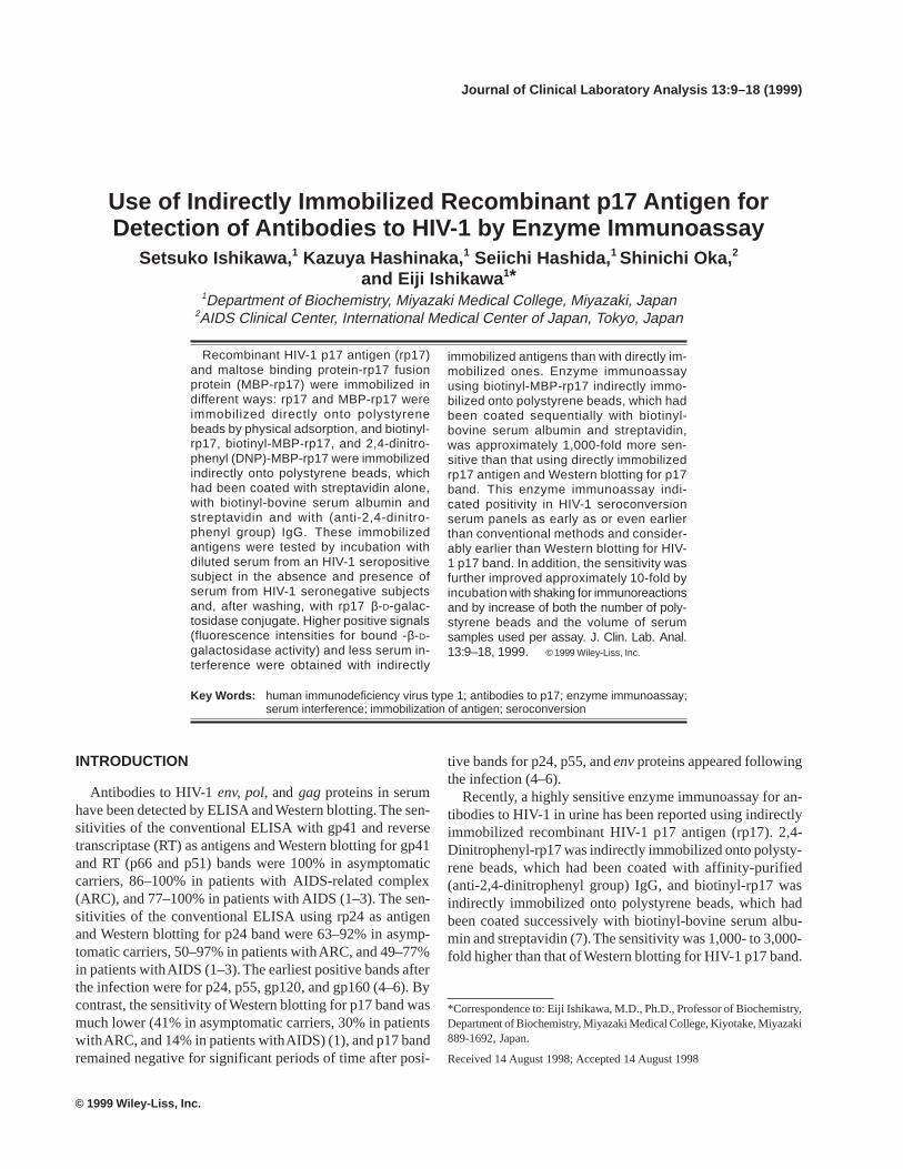
Fig. 1. Immobilization of rp17 antigens onto polystyrene beads. A and B,directly immobilized rp17 antigen and maltose binding protein (MBP)-rp17fusion protein; C, D, E, F and G, indirectly immobilized biotinyl-MBP-rp17, biotinyl-rp17 antigen and 2,4-dinitrophenyl (DNP)-MBP-rp17. STRAV,streptavidin; BSA, bovine serum albumin. The orientations of moleculesare only possibilities.

Immunoassay for Anti-HIV-1 p17 Antibodies 11
Enzyme Immunoassays I, II, and III for Antibodiesto HIV-1 p17 Antigen
Enzyme immunoassay I
Individual polystyrene beads coated with protein(s) wereincubated at room temperature overnight with sample mix-tures containing serum and were washed twice with 2 mL ofbuffer A containing 0.1 mol/L NaCl. The final volume ofsample mixtures was made to 150 µL with buffer A contain-ing 0.4 mol/L NaCl. The washed polystyrene beads were in-cubated at room temperature for 3.5 hr with 50 µg of inactiveβ-D-galactosidase (Mutein, Boehringer Mannheim GmbH)and 100 fmol of rp17-β-D-galactosidase conjugate in 150 µLof buffer A containing 0.4 mol/L NaCl. After washing as de-scribed above, β-D-galactosidase activity bound to the poly-styrene beads was assayed at 30°C for 1 hr by fluorometryusing 4-methylumbelliferyl-β–D-galactoside as substrate (15).The fluorescence intensity was measured by adjusting thatof 1 × 10⁻⁸ mol/L 4-methylumbelliferone to 100 with aspectrofluorophotometer (RF-510, Shimadzu Corporation,Kyoto, Japan).
Enzyme immunoassay II
Enzyme immunoassay II was performed in the same wayas enzyme immunoassay I except that each incubation for theimmunoreactions was performed at room temperature withshaking for 1 hr.
Enzyme immunoassay III
Five polystyrene beads coated with protein(s) were incu-bated for 0.5–2.0 hr with serum samples in a total volume of140 µL containing 50 µg of inactive β-D-galactosidase, andthe incubation was further continued for 0.5–2.0 hr after ad-dition of 100 fmol of rp17-β-D-galactosidase conjugate in 10µL. The buffer used was buffer A containing 0.4 mol/L NaCl.When 100 µL of serum samples was used, the concentrationof NaCl in the reaction mixtures was adjusted to 0.4 mol/Lassuming that in serum to be 0.15 mol/L. The incubationswere performed at room temperature with shaking through-out. Bound β-D-galactosidase activity was assayed at 30°Cfor 1 hr.
Other Immunological Methods
The conventional EIA for antibodies to HIV-1 was per-formed using a commercial kit with two recombinant pro-teins of HIV-1 (gp41 and p24) as antigens (AbbottRecombinant HIV-1/HIV-2 3rd Generation EIA, Abbott Labo-ratories, North Chicago, IL). The volume of serum used was150 µL in a total volume of 200 µL. The gelatin particle ag-glutination test for antibodies to HIV-1 was performed usinga commercial kit with a lysate of HIV-1 as antigen (SERODIA-HIV, Fujirebio Inc., Tokyo, Japan). Serum samples were di-
luted at least 16-fold with the diluent included in the kit andwere mixed with the same volume of gelatin particle solu-tion. Western blotting for antibody IgGs to HIV-1 was per-formed using a commercial kit preblotted with nine proteinsof HIV-1 (gp160, gp120, p66, p55, p51, gp41, p31, p24, andp17) (Ortho HIV Western Blot Kit, Ortho Diagnostic Sys-tems Inc., Raritan, NJ). Serum samples were diluted 101-foldwith the diluent included in the kit and were incubated with anitrocellulose membrane preblotted with the nine proteins.
HIV-1 Seroconversion Serum Panels
Three HIV-1 seroconversion serum panels (SV-0111, SV-0211, and SV-0241) were obtained from North AmericanBiologicals (Miami, FL), and two HIV-1 seroconversion se-rum panels (Panel J and Panel K) were obtained from BostonBiomedica, Inc. (West Bridgewater, MA).
Serum Samples From HIV-1 Seronegative andSeropositive Subjects
Serum samples were collected from 150 HIV-1 seronega-tive subjects (102 males aged 20–77 yr and 48 females aged26–68 yr) and two HIV-1 seropositive subjects (one maleasymptomatic carrier aged 22 yr and one female asymptom-atic carrier aged 39 yr) and were stored at –20°C until use.These serum samples were tested by the gelatin particle ag-glutination, and the seropositivity was confirmed by West-ern blotting.
RESULTS AND DISCUSSION
Higher Signals With Indirectly Immobilized rp17Antigens
Recombinant HIV-1 p17 antigen (rp17) and maltose-bind-ing protein-HIV-1 p17 fusion protein (MBP-rp17) were im-mobilized onto polystyrene beads in seven different ways.First, rp17 and MBP-rp17 were immobilized directly ontopolystyrene beads by physical adsorption (Fig. 1A and B).Second and third, biotinyl-rp17 and biotinyl-MBP-rp17 wereimmobilized indirectly by incubation with polystyrene beads,which had been coated with streptavidin by physical adsorp-tion (Fig. 1C and D). Fourth and fifth, biotinyl-rp17 andbiotinyl-MBP-rp17 were immobilized indirectly by incuba-tion with polystyrene beads, which had been coated withbiotinyl-bovine serum albumin by physical adsorption andstreptavidin (Fig. 1E and F). Seventh, 2,4-dinitrophenyl-MBP-rp17 was immobilized indirectly by incubation with polysty-rene beads, which had been coated with affinity-purified(anti-2,4-dinitrophenyl group) IgG (Fig. 1G).
These immobilized antigens were tested by enzyme im-munoassay I. Serum from an HIV-1 seropositive subject wasdiluted with buffer A containing 0.4 mol/L NaCl 1,000-foldfor directly immobilized antigens and 10,000-fold for indi-rectly immobilized antigens, and individual polystyrene beads
12 Ishikawa et al.
on which rp17 had been immobilized in various ways as de-scribed above were incubated overnight with the diluted se-rum samples (10 µL) and, after washing, for 3.5 hr withrp17-β-D-galactosidase conjugate (Tables 1–4). Bound β-D-galactosidase activity was assayed at 30°C for 1 hr.
The highest signals (fluorescence intensities for bound β-D-galactosidase activity) in the presence of the diluted serumwere obtained with biotinyl-MBP-rp17 indirectly immobi-lized onto polystyrene beads, which had been coated withbiotinyl-bovine serum albumin and streptavidin (Fig. 1F).Slightly lower signals were obtained with biotinyl-rp17 indi-rectly immobilized onto polystyrene beads, which had beencoated with biotinyl-bovine serum albumin and streptavidin(Fig. 1E) and 2,4-dinitrophenyl-MBP-rp17 indirectly immo-bilized onto polystyrene beads, which had been coated withaffinity-purified (anti-2,4-dinitrophenyl group) IgG (Fig. 1G)(Table 1). The lowest signals were observed with directlyimmobilized rp17 (Fig. 1A) (Table 2). Signals with otherimmobilized antigens were much higher than the lowest sig-nals but much lower than the highest signals and were higherin the order of directly immobilized MBP-rp17 (Fig. 1B) testedwith the 1,000-fold diluted serum, biotinyl-rp17 indirectlyimmobilized onto streptavidin-coated polystyrene beads (Fig.1C) and biotinyl-MBP-rp17 indirectly immobilized onto
streptavidin-coated polystyrene beads (Fig. 1D) tested with10,000-fold diluted serum (Tables 2–4). Signals with directlyimmobilized MBP-rp17 varied to great extents (Table 2).Nonspecific signals in the absence of the diluted serum weresimilar with all the immobilized antigens.
Thus, signals in the presence of the diluted serum tendedto be enhanced by increasing the number of protein moleculesbetween rp17 molecules and solid surface as summarized inFigure 1 and Table 5.
Less Serum Interference With Less Amounts ofrp17 Antigens for Immobilization and MoreIndirectly Immobilized rp17 Antigens
In order to examine serum interference in enzyme immu-noassay I, the directly and indirectly immobilized antigenswere tested as described above in the presence of serum (10µL) from HIV-1 seronegative subjects, and the results werecompared with those described above (Tables 1–4). Degreeof serum interference tended to increase with increasingamounts or concentrations of antigens for immobilization.With biotinyl-rp17 and biotinyl-MBP-rp17 indirectly immo-bilized onto polystyrene beads, which had been coated withbiotinyl-bovine serum albumin by physical adsorption and
TABLE 1. Signal by Enzyme Immunoassay I for Antibodies to p17 Antigen in the Absence and Presence of Serum UsingBiotinyl-Maltose Binding Protein (MBP)-rp17 Fusion Protein and Biotinyl-rp17 Antigen Immobilized Onto Streptavidin-Biotinyl-Bovine Serum Albumin-Coated Polystyrene Beads and 2,4-Dinitrophenyl (DNP)-MBP-rp17 Fusion ProteinImmobilized Onto (Anti-DNP) IgG-Coated Polystyrene Beads
Serum from Signal (fluorescence intensity for
Indirectly Amount of HIV-1 bound β-D-galactosidase activity) with
immobilized antigen for seronegative Diluted serum from anantigen immobilization subjects Buffer HIV-1 seropositive subject
fmol/tube µL (%)Biotinyl-MBP-rp17 50 0 22 2,667 (100)Fig. 1F 100 0 20 2,382 (100)
50 10 23 2,250 ± 141 (84)100 10 24 1,990 ± 47 (83)
Biotinyl-rp17 100 0 – 1,294 –Fig. 1E 300 0 19 1,772 (100)
500 0 24 1,871 (100)1,000 0 25 1,493 (100)
100 10 – 973 ± 10 –300 10 15 1,361 ± 99 (77)500 10 17 1,277 ± 40 (68)
1,000 10 21 846 ± 105 (56)DNP-MBP-rp17 50 0 31 1,610 (100)Fig. 1G 100 0 31 1,990 (100)
300 0 26 1,682 (100)50 10 19 704 ± 118 (44)
100 10 22 622 ± 170 (31)300 10 17 232 ± 129 (14)
Serum from an HIV-1 seropositive subject was diluted 10,000-fold with buffer A containing 0.4 mol/L NaCl, and immobilized antigens were tested byenzyme immunoassay I using 10 µL of the diluted serum in the absence and presence of serum samples (10 µL) from three HIV-1 seronegative subjects.Signals in the presence of both the diluted serum and serum samples from the seronegative subjects were expressed as means ± SD (n = 3), and other signalsare means of duplicate determinations. Specific signals (differences between signals in the presence and absence of the diluted serum) in the presence ofserum samples from the seronegative subjects are shown as percentages of those in their absence in parentheses.
Immunoassay for Anti-HIV-1 p17 Antibodies 13
TABLE 2. Signal by Enzyme Immunoassay I for Antibodies to p17 Antigen in the Absence and Presence of Serum UsingDirectly Immobilized rp17 Antigen and Maltose Binding Protein (MBP)-rp17 Fusion Protein
Concentration Serum from Signal (fluorescence intensity for
Directly of antigen HIV-1 bound β-D-galactosidase activity) with
immobilized for seronegative Diluted serum from anantigen immobilization subjects Buffer HIV-1 seropositive subject
µg/mL µL (%)rp17 1 0 38 50 (100)Fig. 1A 5 0 36 49 (100)
25 0 42 51 (100)50 0 45 51 (100)
100 0 42 51 (100)1 10 16 28 ± 6.0 (100)5 10 15 23 ± 3.4 (62)
25 10 17 18 ± 0.5 (11)50 10 17 19 ± 0.7 (33)
100 10 19 19 ± 3.9 (0)MBP-rp17 1 0 34 142 ± 31 (100)Fig. 1B 5 0 35 652 ± 566 (100)
25 0 45 1,391 ± 274 (100)50 0 45 1,849 ± 560 (100)
100 0 41 778 ± 554 (100)1 10 14 168 ± 72 (143)5 10 14 169 ± 75 (25)
25 10 18 54 ± 6.0 (2.7)50 10 16 47 ± 3.1 (1.7)
100 10 18 43 ± 4.9 (3.4)
Experimental procedures and expression of results are as described in Table 1, except that serum from an HIV-1 seropositive subject used in Table 1 wasdiluted 1,000-fold and that signals with MBP-rp17 in the absence of serum samples from HIV-1 seronegative subjects are means ± SD (n = 3).
TABLE 3. Signal by Enzyme Immunoassay I for Antibodies to p17 Antigen in the Absence and Presence of Serum UsingBiotinyl-rp17 Antigen Indirectly Immobilized Onto Streptavidin-Coated Polystyrene Beads (Fig. 1C)
Amount Serum from Signal (fluorescence intensity for
Concentration of of biotinyl-rp17 HIV-1 bound β-D-galactosidase activity) with
streptavidin for antigen for seronegative Diluted serum from animmobilization immobilization subjects Buffer HIV-1 seropositive subject
µg/mL fmol/tube µL (%)5 1,000 0 41 128 (100)5 3,000 0 43 218 (100)5 10,000 0 44 335 (100)5 40,000 0 – 40 –
25 1,000 0 45 115 (100)25 3,000 0 40 236 (100)25 10,000 0 42 311 (100)25 40,000 0 – 71 –50 1,000 0 41 102 (100)50 3,000 0 46 216 (100)50 10,000 0 40 367 (100)50 40,000 0 – 58 –5 1,000 10 12 23 ± 2.5 (12.6)5 3,000 10 12 25 ± 3.2 (7.4)5 10,000 10 13 26 ± 1.1 (4.5)
25 1,000 10 11 25 ± 9.6 (20)25 3,000 10 12 24 ± 3.0 (6.1)25 10,000 10 14 32 ± 6.5 (6.7)50 1,000 10 11 25 ± 6.3 (23)50 3,000 10 12 25 ± 3.5 (7.6)50 10,000 10 12 27 ± 5.6 (4.6)
Experimental procedures and expression of results are as described in Table 1.
14 Ishikawa et al.
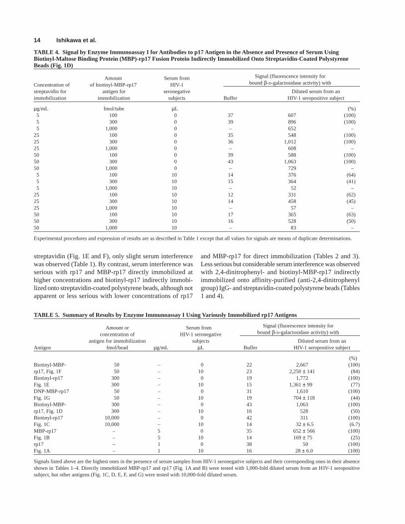
TABLE 4. Signal by Enzyme Immunoassay I for Antibodies to p17 Antigen in the Absence and Presence of Serum UsingBiotinyl-Maltose Binding Protein (MBP)-rp17 Fusion Protein Indirectly Immobilized Onto Streptavidin-Coated PolystyreneBeads (Fig. 1D)
Amount Serum from Signal (fluorescence intensity for
Concentration of of biotinyl-MBP-rp17 HIV-1 bound β-D-galactosidase activity) with
streptavidin for antigen for seronegative Diluted serum from animmobilization immobilization subjects Buffer HIV-1 seropositive subject
µg/mL fmol/tube µL (%)5 100 0 37 607 (100)5 300 0 39 896 (100)5 1,000 0 – 652 –
25 100 0 35 548 (100)25 300 0 36 1,012 (100)25 1,000 0 – 608 –50 100 0 39 588 (100)50 300 0 43 1,063 (100)50 1,000 0 – 729 –5 100 10 14 376 (64)5 300 10 15 364 (41)5 1,000 10 – 52 –
25 100 10 12 331 (62)25 300 10 14 458 (45)25 1,000 10 – 57 –50 100 10 17 365 (63)50 300 10 16 528 (50)50 1,000 10 – 83 –
Experimental procedures and expression of results are as described in Table 1 except that all values for signals are means of duplicate determinations.
TABLE 5. Summary of Results by Enzyme Immunoassay I Using Variously Immobilized rp17 Antigens
Amount or Serum from Signal (fluorescence intensity for
concentration of HIV-1 seronegative bound β-D-galactosidase activity) with
antigen for immobilization subjects Diluted serum from anAntigen fmol/bead µg/mL µL Buffer HIV-1 seropositive subject
(%)Biotinyl-MBP- 50 – 0 22 2,667 (100)rp17, Fig. 1F 50 – 10 23 2,250 ± 141 (84)Biotinyl-rp17 300 – 0 19 1,772 (100)Fig. 1E 300 – 10 15 1,361 ± 99 (77)DNP-MBP-rp17 50 – 0 31 1,610 (100)Fig. 1G 50 – 10 19 704 ± 118 (44)Biotinyl-MBP- 300 – 0 43 1,063 (100)rp17, Fig. 1D 300 – 10 16 528 (50)Biotinyl-rp17 10,000 – 0 42 311 (100)Fig. 1C 10,000 – 10 14 32 ± 6.5 (6.7)MBP-rp17 – 5 0 35 652 ± 566 (100)Fig. 1B – 5 10 14 169 ± 75 (25)rp17 – 1 0 38 50 (100)Fig. 1A – 1 10 16 28 ± 6.0 (100)
Signals listed above are the highest ones in the presence of serum samples from HIV-1 seronegative subjects and their corresponding ones in their absenceshown in Tables 1–4. Directly immobilized MBP-rp17 and rp17 (Fig. 1A and B) were tested with 1,000-fold diluted serum from an HIV-1 seropositivesubject, but other antigens (Fig. 1C, D, E, F, and G) were tested with 10,000-fold diluted serum.
streptavidin (Fig. 1E and F), only slight serum interferencewas observed (Table 1). By contrast, serum interference wasserious with rp17 and MBP-rp17 directly immobilized athigher concentrations and biotinyl-rp17 indirectly immobi-lized onto streptavidin-coated polystyrene beads, although notapparent or less serious with lower concentrations of rp17
and MBP-rp17 for direct immobilization (Tables 2 and 3).Less serious but considerable serum interference was observedwith 2,4-dinitrophenyl- and biotinyl-MBP-rp17 indirectlyimmobilized onto affinity-purified (anti-2,4-dinitrophenylgroup) IgG- and streptavidin-coated polystyrene beads (Tables1 and 4).
Immunoassay for Anti-HIV-1 p17 Antibodies 15
TABLE 6. Sensitivity of Enzyme Immunoassays I and III Using Directly Immobilized rp17 Antigen and Biotinyl-MaltoseBinding Protein-rp17 Fusion Protein (Biotinyl-MBP-rp17) Indirectly Immobilized Onto Streptavidin-Biotinyl-Bovine SerumAlbumin (BSA)-Coated Polystyrene Beads and Western Blotting for p17 Band
Dilution withSignal by enzyme immunoassay using
pooled serum Directly Biotinyl-MBP-rp17-
from HIV-1 immobilized streptavidin-biotinyl-BSA
seronegative rp17 antigen coated polystyrene beads WesternSerum subjects Serum volume used Serum volume used blotting forno. (fold) 10 µL 10 µL 100 µL p17 band
1 to 70 – 11 ± 1.3 (SD) 16 ± 3.6 (SD) 8.1 ± 1.3 (SD)(10 to 14, n = 5) (7.4 to 24, n = 50) (4.3 to 11, n = 20)
71 3 × 10⁵ – 29 – NG105 – 67 – NG
3 × 10⁴ – 178 – NG104 – 568 – NG
3 × 10³ 12 1,683 – NG103 14 4,789 – NG
3 × 10² 16 9,399 – PS102 24 13,411 – PS
72 3 × 10⁶ – – 53 –106 – – 152 –
3 × 10⁵ – 70 436 NG105 – 193 – NG
3 × 10⁴ – 610 – NG104 11 1,898 – NG
3 × 10³ 10 5,286 – NG103 10 11,433 – NG
3 × 10² 14 17,063 – PS102 22 21,566 – PS
Serum nos. 1–70 were from HIV-1 seronegative subjects. Serum nos. 71 and 72 from HIV-1 seropositive subjects were diluted with pooled serum contain-ing an equal volume of serum samples from 10 HIV-1 seronegative subjects. The concentration of rp17 antigen used for immobilization was 25 µg/mL, andthe amount of biotinyl-MBP-rp17 used for immobilization was 50 fmol/bead. Ten µL and 100 µL of the diluted serum samples were tested by enzymeimmunoassays I and III, respectively. NG, negative; PS, positive.
Thus, serum interference in enzyme immunoassay I tendedto be alleviated to greater extents with increasing numbers ofprotein molecules between rp17 molecules and solid surfaceas summarized in Figure 1 and Table 5.
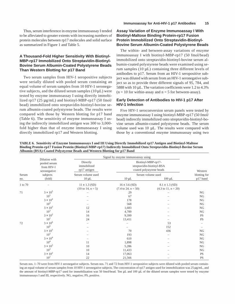
A Thousand-Fold Higher Sensitivity With Biotinyl-MBP-rp17 Immobilized Onto Streptavidin-Biotinyl-Bovine Serum Albumin-Coated Polystyrene BeadsThan Western Blotting for p17 Band
Two serum samples from HIV-1 seropositive subjectswere serially diluted with pooled serum containing anequal volume of serum samples from 10 HIV-1 seronega-tive subjects, and the diluted serum samples (10 µL) weretested by enzyme immunoassay I using directly immobi-lized rp17 (25 µg/mL) and biotinyl-MBP-rp17 (50 fmol/bead) immobilized onto streptavidin-biotinyl-bovine se-rum albumin-coated polystyrene beads. The results werecompared with those by Western blotting for p17 band(Table 6). The sensitivity of enzyme immunoassay I us-ing the indirectly immobilized antigen was 300 to 3,000-fold higher than that of enzyme immunoassay I usingdirectly immobilized rp17 and Western blotting.
Assay Variation of Enzyme Immunoassay I WithBiotinyl-Maltose Binding Protein-rp17 FusionProtein Immobilized Onto Streptavidin-Biotinyl-Bovine Serum Albumin-Coated Polystyrene Beads
The within- and between-assay variations of enzymeimmunoassay I with biotinyl-MBP-rp17 (50 fmol/bead)immobilized onto streptavidin-biotinyl-bovine serum al-bumin-coated polystyrene beads were examined using se-rum samples (10 µL) containing three different levels ofantibodies to p17. Serum from an HIV-1 seropositive sub-ject was diluted with serum from an HIV-1 seronegative sub-ject so as to provide three different signals of 95, 784, and5888 with 10µL. The variation coefficients were 1.2 to 4.3%(n = 10 for within-assay and n = 5 for between-assay).
Early Detection of Antibodies to HIV-1 p17 AfterHIV-1 Infection
Five HIV-1 seroconversion serum panels were tested byenzyme immunoassay I using biotinyl-MBP-rp17 (50 fmol/bead) indirectly immobilized onto streptavidin-biotinyl-bo-vine serum albumin-coated polystyrene beads. The serumvolume used was 10 µL. The results were compared withthose by a conventional enzyme immunoassay using two
16 Ishikawa et al.
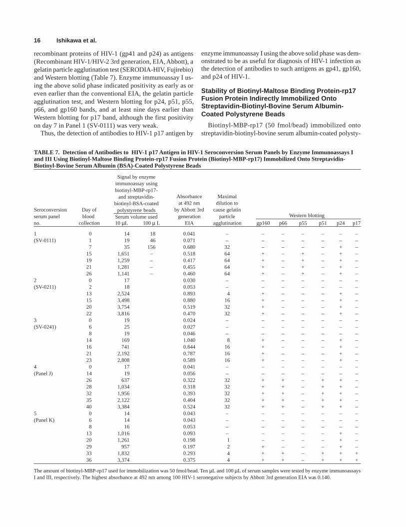
TABLE 7. Detection of Antibodies to HIV-1 p17 Antigen in HIV-1 Seroconversion Serum Panels by Enzyme Immunoassays Iand III Using Biotinyl-Maltose Binding Protein-rp17 Fusion Protein (Biotinyl-MBP-rp17) Immobilized Onto Streptavidin-Biotinyl-Bovine Serum Albumin (BSA)-Coated Polystyrene Beads
Absorbance Maximalat 492 nm dilution to
Seroconversion Day of by Abbott 3rd cause gelatinserum panel blood generation particle Western blotting
no. collection 10 µL 100 µ L EIA agglutination gp160 p66 p55 p51 p24 p17
1 0 14 18 0.041 – – – – – – –(SV-0111) 1 19 46 0.071 – – – – – – –
7 35 156 0.680 32 – – – – + –15 1,651 – 0.518 64 + – + – + –19 1,259 – 0.417 64 + – + – + –21 1,281 – 0.455 64 + – + – + –26 1,141 – 0.460 64 + – + – + –
2 0 17 0.030 – – – – – – –(SV-0211) 2 18 0.053 – – – – – – –
13 2,524 0.893 4 + – – – + –15 3,498 0.880 16 + – – – + –20 3,754 0.519 32 + – – – + –22 3,816 0.470 32 + – – – + –
3 0 19 0.024 – – – – – – –(SV-0241) 6 25 0.027 – – – – – – –
8 19 0.046 – – – – – – –14 169 1.040 8 + – – – + –16 741 0.844 16 + – – – + –21 2,192 0.787 16 + – – – + –23 2,808 0.589 16 + – – – + –
4 0 17 0.041 – – – – – – –(Panel J) 14 19 0.056 – – – – – – –
26 637 0.322 32 + + – + + –28 1,034 0.318 32 + + – + + –32 1,956 0.393 32 + + – + + –35 2,122 0.404 32 + + – + + –40 3,384 0.524 32 + + – + + –
5 0 14 0.043 – – – – – – –(Panel K) 6 14 0.043 – – – – – – –
8 16 0.053 – – – – – – –13 1,016 0.093 – – – – – + –20 1,261 0.198 1 – – – – + –29 957 0.197 2 + – – – + –33 1,832 0.293 4 + + – + + +36 3,374 0.375 4 + + – + + +
The amount of biotinyl-MBP-rp17 used for immobilization was 50 fmol/bead. Ten µL and 100 µL of serum samples were tested by enzyme immunoassaysI and III, respectively. The highest absorbance at 492 nm among 100 HIV-1 seronegative subjects by Abbott 3rd generation EIA was 0.140.
recombinant proteins of HIV-1 (gp41 and p24) as antigens(Recombinant HIV-1/HIV-2 3rd generation, EIA, Abbott), agelatin particle agglutination test (SERODIA-HIV, Fujirebio)and Western blotting (Table 7). Enzyme immunoassay I us-ing the above solid phase indicated positivity as early as oreven earlier than the conventional EIA, the gelatin particleagglutination test, and Western blotting for p24, p51, p55,p66, and gp160 bands, and at least nine days earlier thanWestern blotting for p17 band, although the first positivityon day 7 in Panel 1 (SV-0111) was very weak.
Thus, the detection of antibodies to HIV-1 p17 antigen by
enzyme immunoassay I using the above solid phase was dem-onstrated to be as useful for diagnosis of HIV-1 infection asthe detection of antibodies to such antigens as gp41, gp160,and p24 of HIV-1.
Stability of Biotinyl-Maltose Binding Protein-rp17Fusion Protein Indirectly Immobilized OntoStreptavidin-Biotinyl-Bovine Serum Albumin-Coated Polystyrene Beads
Biotinyl-MBP-rp17 (50 fmol/bead) immobilized ontostreptavidin-biotinyl-bovine serum albumin-coated polysty-
Signal by enzymeimmunoassay usingbiotinyl-MBP-rp17-
and streptavidin-biotinyl-BSA-coated
polystyrene beadsSerum volume used

Immunoassay for Anti-HIV-1 p17 Antibodies 17
rene beads were stored in buffer A containing 0.1 mol/L NaClat 4°C for up to 194 days and were tested by enzyme immu-noassay II. Serum from an HIV-1 seropositive subject wasdiluted 104-fold with buffer A containing 0.4 mol/L NaCl,and individual polystyrene beads were incubated with thediluted serum (10 µL) in the presence and absence of pooledserum (10 µL) containing an equal volume of serum samplesfrom 15 HIV-1 seronegative subjects and, after washing, withrp17-β-D-galactosidase conjugate. Each incubation was per-formed at room temperature for 1 hr with shaking. The signalwas not significantly changed during the storage.
Shortening of Incubation Time forImmunoreactions
In order to detect anti-HIV-1 p17 antibodies within a shorterperiod of time, time courses of immunoreactions in enzymeimmunoassay III using biotinyl-MBP-rp17 (50 fmol/bead)immobilized onto streptavidin-biotinyl-bovine serum albu-min-coated polystyrene beads were examined (Fig. 2). Se-
rum from an HIV-1 seropositive subject was diluted 104-foldwith buffer A containing 0.4 mol/L NaCl, and five of the poly-styrene beads per assay tube were incubated with 10 µL ofthe diluted serum for 0.5, 1.0, or 2.0 hr with shaking, and theincubation was further continued for 2.0 hr after addition ofrp17-β-D-galactosidase conjugate. β-D-Galactosidase activ-ity bound to the five polystyrene beads reached a value, within2 hr, similar to that obtained by enzyme immunoassay I, inwhich each of the polystyrene bead was incubated with 10µL of the diluted serum overnight and, after washing, withrp17-β-D-galactosidase conjugate for 3.5 hr. Alternatively, fiveof the polystyrene beads were incubated with 10 µL of thediluted serum for 2.0 hr with shaking, and the incubation wasfurther continued for 0.5, 1.0, or 2.0 hr after addition of rp17-β-D-galactosidase conjugate. Bound β-D-galactosidase activ-ity reached a value close to the maximum within 1.0 hr. Theseresults indicated that the sensitivity of enzyme immunoassayIII, in which five polystyrene beads were incubated with shak-ing for less than 2 hr with serum samples and, without wash-ing, for less than 1 hr with rp17-β-D-galactosidase conjugate,could be similar to that of enzyme immunoassay I, in whichone polystyrene bead was incubated without shaking over-night with serum samples and, after washing, for 3.5 hr withrp17-β-D-galactosidase conjugate.
Further Improvement of Sensitivity by Increasingthe Volume of Serum Samples
On the basis of the above results, serum from an HIV-1 se-ropositive subject was serially diluted with pooled serum con-taining an equal volume of serum samples from 10 HIV-1seronegative subjects and were tested by enzyme immunoassayIII. Five polystyrene beads coated with streptavidin-biotinyl-bovine serum albumin and subsequently with 50 fmol ofbiotinyl-MBP-rp17 were incubated with 100 µL of the dilutedserum samples at room temperature for 1.5 hr with shaking andthe incubation was further continued for 1.5 hr after addition ofrp17-β-D-galactosidase conjugate (Table 6). The sensitivity wasimproved approximately 10-fold as compared with that of en-zyme immunoassay I, in which one piece/tube of the same poly-styrene beads was incubated without shaking overnight withthe diluted serum (10 µL) and, after washing, for 3.5 hr withrp17-β-D-galactosidase conjugate. The improved sensitivity wasapproximately 10,000-fold higher than that of Western blottingfor p17 band. This improvement made possible earlier detec-tion of antibodies to p17 in an HIV-1 seroconversion serumpanel. Signals on days 1 and 7 in Panel 1 (SV-0111) becameunequivocally positive, although not clearly positive or onlyweakly positive by enzyme immunoassay I (Table 7).
REFERENCES
1. Baur A, Vornhagen R, Korn K et al. Viral culture and p24 antigenemiaof human immunodeficiency virus (HIV)-infected individuals correlatedwith antibody profiles determined with recombinant polypeptides of allHIV-1 open-reading frames. J Infect Dis 1992;165:419–426.
Fig. 2. Time courses of immunoreactions in enzyme immunoassay III us-ing biotinyl-maltose binding protein-rp17 fusion protein (50 fmol/bead) im-mobilized onto streptavidin-biotinyl-bovine serum albumin-coatedpolystyrene beads. Serum from an HIV-1 seropositive subject was diluted10,000-fold with buffer A containing 0.4 mol/L NaCl. Incubations were per-formed at room temperature with shaking throughout. A, Five of the poly-styrene beads were incubated with the diluted serum samples (10 µL) for0.5, 1.0, or 2.0 hr, and the incubation was further continued for 2.0 hr afteraddition of 100 fmol of rp17-β-D-galactosidase conjugate; B, Five of thepolystyrene beads were incubated with the diluted serum samples for 2.0 hr,and the incubation was further continued for 0.5, 1.0, or 2.0 hr after additionof 100 fmol of rp17-β-D-galactosidase conjugate.
18 Ishikawa et al.
2. Chiang CS, Grove T, Cooper M, et al. Development of a confirmatoryenzyme-linked immunosorbent assay for HIV-1 antibodies. Clin Chem1989;35:946–952.
3. Filice G, Soldini L, Orsolini P, et al. Sensitivity and specificity of anti-HIV ELISA employing recombinant (p24, p66. gp120) and synthetic(gp41) viral antigenic peptides. Microbiologica 1991;14:185–194.
4. Soriano V, Tor J, Ribera A, Muga R. Synthetic peptide immunoassay indiagnosis of primary HIV infection. Vox Sang 1990;58:228–230.
5. Ulstrup JC, Skaug K, Figenschau KJ, Ørstavik I, Bruun JN, Petersen G.Sensitivity of Western blotting (compared with ELISA and immuno-fluorescence) during seroconversion after HTLV-III infection. Lancet1986;i:1151–1152.
6. Weber B, Hess G, Enzensberger R, et al. Multicenter evaluation of thenovel ABN Western blot (immunoblot) system in comparison with anenzyme-linked immunosorbent assay and a different Western blot. J ClinMicrobiol 1992;30:691–697.
7. Ishikawa S, Hashinaka K, Hashida S, Oka S, Ishikawa E. Sensitive en-zyme immunoassay of antibodies to HIV-1 p17 antigen using indirectlyimmobilized recombinant p17 for diagnosis of HIV-1 infection. J ClinLab Anal 1998;12:343–350.
8. Ishikawa S, Hashida S, Hashinaka K, Adachi A, Oka S, Ishikawa E.Ultrasensitive and rapid enzyme immunoassay (thin aqueous layer im-mune complex transfer enzyme immunoassay) for antibody IgG to HIV-1 p17 antigen. J Clin Lab Anal 1998;12:179–189.
9. Adachi A, Gendelman HE, Koenig S, et al. Production of acquired im-munodeficiency syndrome-associated retrovirus in human and non-human cells transfected with an infectious molecular clone. J Virol1986;59:284–291.
10. Wain-Hobson S, Sonigo P, Danos O, Cole S, Alizon M. Nucleotide se-quence of the AIDS virus, LAV. Cell 1985;40:9–17.
11. Hashida S, Hirota K, Hashinaka K, et al. Detection of antibody IgG toHIV-1 in urine by sensitive enzyme immunoassay (immune complextransfer enzyme immunoassay) using recombinant proteins as antigensfor diagnosis of HIV-1 infection. J Clin Lab Anal 1993;7:353–364.
12. Hashida S, Hashinaka K, Nishikata I, et al. Measurement of human im-munodeficiency virus type 1 p24 in serum by an ultrasensitive enzymeimmunoassay, the two-site immune complex transfer enzyme immu-noassay. J Clin Microbiol 1995;33:298–303.
13. Hashida S, Tanaka K, Yamamoto N, Uno T, Yamaguchi K, Ishikawa E.Detection of one attomole of [Arg8]-vasopressin by novel noncompeti-tive enzyme immunoassay (hetero-two-site complex transfer enzymeimmunoassay). J Biochem 1991;110:486–492.
14. Ishikawa E, Kato K. Ultrasensitive enzyme immunoassay. Scand JImmunol 1978;8(Suppl. 7):43–55.
15. Imagawa M, Hashida S, Ohta Y, Ishikawa E. Evaluation of β-D-galac-tosidase from Escherichia coli and horseradish peroxidase as labels bysandwich enzyme immunoassay technique. Ann Clin Biochem1984;21:310–317.