unit two anatomy & physiology the cells, tissues & skin to... · anatomy & physiology...
TRANSCRIPT
www.aromaflex.co.nz Telephone: 03 545 6218
UNIT TWO
__________________________ ANATOMY & PHYSIOLOGY
THE CELLS, TISSUES & SKIN

Aromaflex Academy, Development House, 1st Floor, 280 – 282 Trafalgar Street, Nelson. 7010 NZ. [email protected]
Unit 2 – Introduction to the Human Body
2
Certificate of Anatomy & Physiology Course Notes: 1998 First Revised Edition 2000 Second Revised Edition 2003 Third Revised Edition 2008 Fourth Revised Edition 2013 Fifth Revised Edition Published by Aromaflex Academy, Development House, First Floor 280-282 Trafalgar Street, Nelson, New Zealand References to updating material in Fourth Revised Edition: A Textbook of Anatomy and Physiology, 2nd Edition; William Arnold-Taylor MSc PhD Ross & Wilson Anatomy and Physiology in Health and Illness, 7th Edition; Kathleen J. W. Wilson OBE Human Anatomy and Physiology, 3rd Edition.; Elaine N. Marieb Principles of Anatomy and Physiology, 8th Edition; Gerald J. Tortora and Sandra Reynolds Grabowski The Human Body in Health and Disease, 9th Edition; Cohen and Woods. A massage Therapists Guide to Pathology,; Werner and Benjamin. An Introductory Guide to Anatomy & Physiology; Louise Tucker NCEA Level 1 Chemistry; Dorothy Kane Year 10 Science: Paul King,Tania Lineham, Marea Goode, Jenny Pollock, Cara Scott, Mark Standley All rights reserved. No part of the publication may be reproduced, stored in a retrieval system, or transmitted, in any form or by any means, electronic, mechanical, photocopying, recording or otherwise, without the prior permission of the copyright holder. Aromaflex Academy asserts the moral right to be identified as author of this work.
UNIT TWO.
Aromaflex Academy, Development House, 1st Floor, 280 – 282 Trafalgar Street, Nelson. 7010 NZ. [email protected]
Unit 2 – Introduction to the Human Body
3
INTRODUCTION TO THE HUMAN BODY It is a good idea for therapists to have a general knowledge of anatomy and physiology before performing any work on the body. We need to know the basic structure, functions and workings of the body. If we are thoroughly familiar with the body in its ‘normal’ state it becomes much easier to detect any imbalances. Sometimes it might be more appropriate to refer a client to a doctor or other health professional. We need to know what we are talking about when speaking with other health professionals. The more knowledgeable you are about your profession the more respect you’ll gain from both clients and other health therapists. ANATOMY - deals with the gross structure of the body and the relationship of various parts one to another. PHYSIOLOGY - concerns the normal functions performed by the various cells, tissues and systems in the body. PATHOLOGY - is the scientific study of changes in organs, tissues and cells produced by disease. CELLS & TISSUES -are the building blocks of the whole body. The hierarchy of structural organisation: The simplest level is the chemical level and at this level are: Atoms - tiny building blocks of matter that combine to form molecules such as water, sugar, and proteins. Molecules - in turn, associate in specific ways to form the basic components of the microscopic cells. Cells - are the smallest units of living things, and the human body develops from one microscopic cell. (see section on cells and tissues later.) Groups of similar cells that have a common function combine to form tissues. Tissues - combine to form organs. Organs - that co-operate and work closely together to accomplish a common purpose are said to be part of a particular organ system. Organ system - Such as the digestive system or the cardiovascular system. The highest level of organisation is the organism. Organism - is the living human being. The organismal level represents the sum total of all structural levels working in unison to promote life. The 11 principal organ systems are: • The Integumentary System, (the skin).
Aromaflex Academy, Development House, 1st Floor, 280 – 282 Trafalgar Street, Nelson. 7010 NZ. [email protected]
Unit 2 – Introduction to the Human Body
4
• The Skeletal System. • The Muscular System. • The Nervous System • The Endocrine System. • The Circulatory System. • The Immune System. • The Respiratory System. • The Digestive System. • The Urinary System. • The Reproductive System.
Homeostasis: Homeostasis describes the body’s ability to maintain relatively stable internal conditions even though there is continuous change in the outside world. The literal translation means “unchanging” but the term does not really mean a static or unchanging state. Rather it indicates a balance in which internal conditions change and vary, but always within relatively narrow limits. The body is in homeostasis when its needs are adequately met and its functions are occurring smoothly. Virtually every organ system is involved in maintaining this delicate balance. Heart activity, blood pressure, essential supply of nutrients, accumulation of wastes and volume of body fluids must be constantly monitored and adjusted. Homeostasis is so important that most disease is considered to be a result of its disturbance. Communication in the body is essential for homeostasis, and this takes place chiefly via the endocrine and nervous system. Metabolic Rate: Metabolic rate and body temperature tend to rise and fall together. When nutritional materials are oxidised in the cells of the body, energy is released, some in the form of heat. Energy may be used immediately to do work or it may be stored The metabolic rate is the rate at which energy is released from the nutrients inside the cells. i.e. energy used by the body per hour is the metabolic rate. The higher the body temperature, the higher the metabolic rate. This means the metabolic rate is increased during fever. Basal metabolic rate (BMR) is the rate of metabolism when the individual is resting, and has not had a meal for 12 hours. In this state the release of energy is sufficient to meet only the essential needs of the vital organs, e.g. the heart, lungs, liver and kidneys. BMR is also an indicator of how much thyroid hormone is present as this hormone is one of the main regulators of metabolism. So what is energy? A definition of energy is – ‘the ability to do work or to put matter into motion’. All living things need a constant supply of energy in order to grow and function. All our cells and the various parts of each individual cell, such as a muscle cell, participate in the various stages of metabolism and life processes. Factors affecting BMR.
• Age. • Gender. • Stress. • The size of the individual. • Muscular activity. • Hormones, especially thyroxine produced by the thyroid gland.

Aromaflex Academy, Development House, 1st Floor, 280 – 282 Trafalgar Street, Nelson. 7010 NZ. [email protected]
Unit 2 – Introduction to the Human Body
5
Body Temperature. This reflects the balance between heat production and heat loss, and is normally 36.6 - 37.8C. which is optimal for physiological activities. At rest, most of the body heat is produced by the body’s core organs and glands, especially the liver. It is these internal cavities that have the highest temperature. The body’s shell, the skin, is usually the coolest. Blood serves as the major heat exchange agent between the core and the shell. The hypothalamus (in the brain) acts as the body’s thermostat. The body uses 4 mechanisms to lose and gain heat:
• Radiation. Close to half of the body’s heat loss occurs by radiation. e.g. people in a cold room warm the room, or sunbathing warms the body.
• Conduction. Heat is transferred between objects that are in direct contact with each other. e.g. a hot tub where the water transfers heat to our skin, or our buttocks will warm the seat of a chair.
• Convection occurs when moving air or water gives, or takes heat away from something. E.g When the body loses heat to the surrounding air – This is due to the air surrounding the body being cooler than the body itself.
• Evaporation, the conversion of water to water- vapour, requires the absorption of heat. E.g During exercise heat is lost from the body with the evaporation of sweat.
. Heat promoting mechanisms include; • The constriction of blood vessels in the skin. The skin is the body’s most important
mechanism for body heat regulation. It is the heat exchange surface. • Increase in metabolic rate. • Shivering. • Activity of glands, the liver. • Increased activity of the thyroid gland. If environmental cold is prolonged the thyroid gland is
stimulated to produce more thyroxine. • Muscular activity. • Posture. This can be changed to reduce the exposed body surface. Heat loss mechanisms: Heat may be removed from the body in the following ways; • Blood vessels in the skin dilate, allowing heat loss through radiation, conduction and
convection. • Sweating occurs when greater heat loss is needed thus heat is lost by evaporation. • Expiration and inspiration are also ways in which the body loses heat. • Cold food and drink is warmed by the body on ingestion and the body heat is thus reduced. • Even our posture can aid heat loss to some extent. When we are hot we may stretch out and
try and expose more of our skin surface to the surrounding air. Heat Stroke occurs when the body can’t rid itself of surplus heat. Body temperature reaches a point where thermo-regulatory systems become ineffective, a potentially lethal condition. Heat Exhaustion from profuse sweating, is indicated by a raised temperature, low blood pressure and collapse. Anatomical Position of the body: To describe body parts and position accurately we must have an initial reference point and use indications of direction. The anatomical reference point is a standard body position called the anatomical position. In the anatomical position the body is erect with the feet together, and arms by the sides. The palms of the hands face forward with thumbs pointing away from the body.
Aromaflex Academy, Development House, 1st Floor, 280 – 282 Trafalgar Street, Nelson. 7010 NZ. [email protected]
Unit 2 – Introduction to the Human Body
6
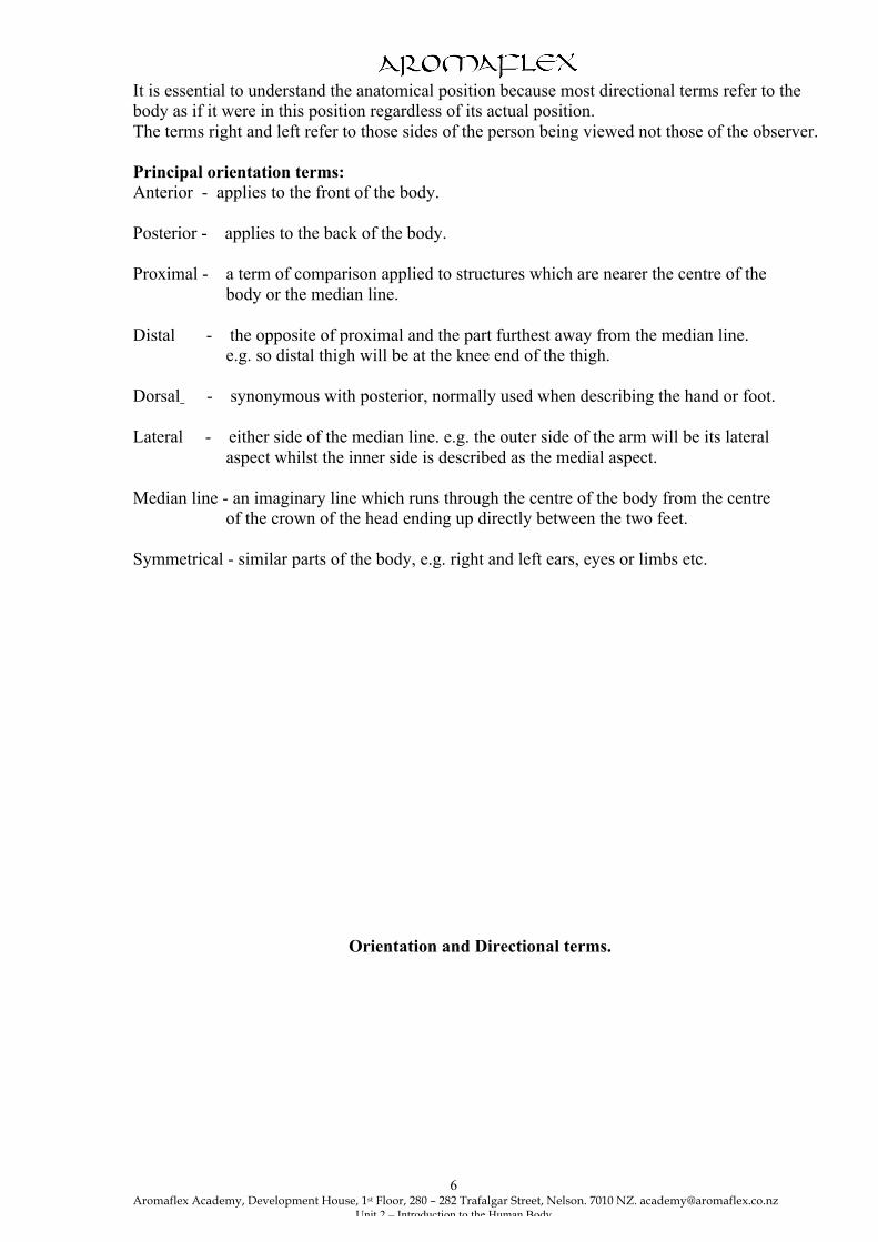
It is essential to understand the anatomical position because most directional terms refer to the body as if it were in this position regardless of its actual position. The terms right and left refer to those sides of the person being viewed not those of the observer. Principal orientation terms: Anterior - applies to the front of the body. Posterior - applies to the back of the body. Proximal - a term of comparison applied to structures which are nearer the centre of the body or the median line. Distal - the opposite of proximal and the part furthest away from the median line. e.g. so distal thigh will be at the knee end of the thigh. Dorsal - synonymous with posterior, normally used when describing the hand or foot. Lateral - either side of the median line. e.g. the outer side of the arm will be its lateral aspect whilst the inner side is described as the medial aspect. Median line - an imaginary line which runs through the centre of the body from the centre of the crown of the head ending up directly between the two feet. Symmetrical - similar parts of the body, e.g. right and left ears, eyes or limbs etc.
Orientation and Directional terms.

Aromaflex Academy, Development House, 1st Floor, 280 – 282 Trafalgar Street, Nelson. 7010 NZ. [email protected]
Unit 2 – Introduction to the Human Body
7
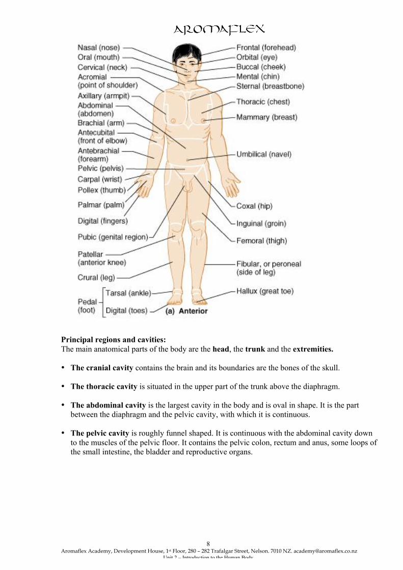
Surface anatomy: Anterior regional terms.
Aromaflex Academy, Development House, 1st Floor, 280 – 282 Trafalgar Street, Nelson. 7010 NZ. [email protected]
Unit 2 – Introduction to the Human Body
8
Principal regions and cavities: The main anatomical parts of the body are the head, the trunk and the extremities. • The cranial cavity contains the brain and its boundaries are the bones of the skull. • The thoracic cavity is situated in the upper part of the trunk above the diaphragm. • The abdominal cavity is the largest cavity in the body and is oval in shape. It is the part
between the diaphragm and the pelvic cavity, with which it is continuous. • The pelvic cavity is roughly funnel shaped. It is continuous with the abdominal cavity down
to the muscles of the pelvic floor. It contains the pelvic colon, rectum and anus, some loops of the small intestine, the bladder and reproductive organs.
Aromaflex Academy, Development House, 1st Floor, 280 – 282 Trafalgar Street, Nelson. 7010 NZ. [email protected]
Unit 2 – Introduction to the Human Body
9
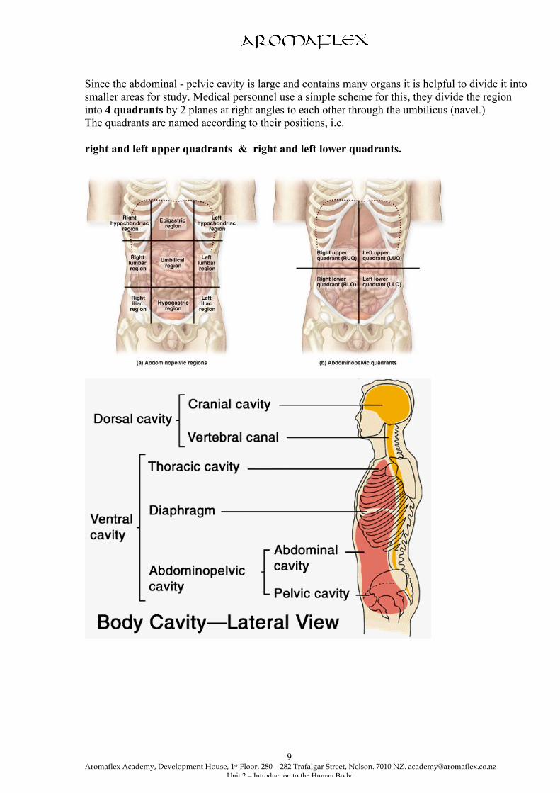
Since the abdominal - pelvic cavity is large and contains many organs it is helpful to divide it into smaller areas for study. Medical personnel use a simple scheme for this, they divide the region into 4 quadrants by 2 planes at right angles to each other through the umbilicus (navel.) The quadrants are named according to their positions, i.e. right and left upper quadrants & right and left lower quadrants.

Aromaflex Academy, Development House, 1st Floor, 280 – 282 Trafalgar Street, Nelson. 7010 NZ. [email protected]
Unit 2 – Introduction to the Human Body
10
CELLS AND TISSUES
Cells: As we have already read cells are the smallest units of living things, and the human body develops from one microscopic cell. Just as bricks and timbers are the structural units of a house, cells are the structural units of all living things. When you define the properties of a cell you are in fact defining the properties of life. Life processes depend on the cell. The cell is the microscopic package that contains all the parts necessary to survive in an ever changing world. Loss of cellular homeostasis underlies virtually every disease we know. The human body has 50 - 60 trillion of these tiny building blocks. Organisation. Chemically, cells are composed of 80% water molecules and 20% organic molecules, all these are made up of carbon, hydrogen, nitrogen and oxygen, and traces of other elements. These elements are found in the air and earth all around us, but in the cell they take on special characteristics of life. Life then relates to the way living matter is organised and carries out metabolic processes, considerations beyond chemical composition. The trillions of cells in the human body include some 200 different types that are diverse in shape, size and function. The function of the individual cells dictates the shape, size and numbers of the various organelles present. For example, red blood cells have no nucleus or organelles but a large surface area for exchange of oxygen and other gases and a shape that enables them to squeeze through the blood capillary walls. Although no one cell type is exactly like all others, cells do have the same basic parts and there are certain functions common to all cells. Characteristics of a cell. Cells possess the characteristics of all living matter. These are: • Ingestion and assimilation. • Respiration. • Growth and repair. • Excretion. • Irritability and activity. (responsiveness.) • Metabolism. • Reproduction. The Structure of a typical cell. As has already been said, no two human cells are exactly the same but they do have the same basic parts:
• a plasma membrane, • cytosol, • organelles, • a nucleus, and • inclusions.
Plasma membrane
Aromaflex Academy, Development House, 1st Floor, 280 – 282 Trafalgar Street, Nelson. 7010 NZ. [email protected]
Unit 2 – Introduction to the Human Body
11
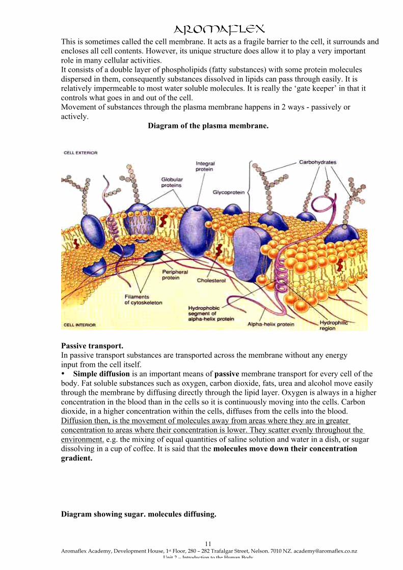
This is sometimes called the cell membrane. It acts as a fragile barrier to the cell, it surrounds and encloses all cell contents. However, its unique structure does allow it to play a very important role in many cellular activities. It consists of a double layer of phospholipids (fatty substances) with some protein molecules dispersed in them, consequently substances dissolved in lipids can pass through easily. It is relatively impermeable to most water soluble molecules. It is really the ‘gate keeper’ in that it controls what goes in and out of the cell. Movement of substances through the plasma membrane happens in 2 ways - passively or actively.
Diagram of the plasma membrane.
Passive transport. In passive transport substances are transported across the membrane without any energy input from the cell itself. • Simple diffusion is an important means of passive membrane transport for every cell of the body. Fat soluble substances such as oxygen, carbon dioxide, fats, urea and alcohol move easily through the membrane by diffusing directly through the lipid layer. Oxygen is always in a higher concentration in the blood than in the cells so it is continuously moving into the cells. Carbon dioxide, in a higher concentration within the cells, diffuses from the cells into the blood. Diffusion then, is the movement of molecules away from areas where they are in greater concentration to areas where their concentration is lower. They scatter evenly throughout the environment. e.g. the mixing of equal quantities of saline solution and water in a dish, or sugar dissolving in a cup of coffee. It is said that the molecules move down their concentration gradient. Diagram showing sugar. molecules diffusing.

Aromaflex Academy, Development House, 1st Floor, 280 – 282 Trafalgar Street, Nelson. 7010 NZ. [email protected]
Unit 2 – Introduction to the Human Body
12
• Facilitated diffusion is a process whereby substances such as glucose needed by the
cell that are lipid in-soluble and too large to pass through the membrane pores, can enter the cell. A protein carrier is needed as a transport vehicle and its some of the proteins in the plasma membrane that act as carriers.
• Osmosis is another means of passive membrane transport for the body cells. Osmosis is the diffusion of a solvent such as water through a selectively permeable Membrane, like the plasma membrane. Osmosis in and out of the cells is occurring all the time as water moves down its concentration gradient.
• Filtration. This is a process by which fluid that contains solutes is forced from a high
pressure area to that of a lower pressure through a membrane or capillary wall. An example of this would be in the kidneys. Here, water and small solutes filter out of the
capillaries where the blood pressure is high, into the kidney tubules where the fluid pressure is lower. Filtration is not very selective and only molecules too large to pass through the membrane are held back.
Active Transport Processes.

Aromaflex Academy, Development House, 1st Floor, 280 – 282 Trafalgar Street, Nelson. 7010 NZ. [email protected]
Unit 2 – Introduction to the Human Body
13
It is called active transport because cellular energy is required from a high energy molecule inside cells, ATP, to do the pumping work across the membrane. This involves movement of a substance through a membrane against a concentration gradient. The substance combines with carrier protein molecules in the membrane. Phagocytosis meaning ‘cell eating’, falls under this heading. Here projections of the cell membrane and cytoplasm, called pseudopods, surround and engulf large particles such as dead body cells or bacteria. The particles are then enclosed within a sac of membrane and enter the cytoplasm where they are digested by enzymes from lysosomes. Only certain cells are capable of this, examples being the white blood cells called neutrophils and monocytes. ( discussed more in the immune system.) Pinocytosis means ‘cell drinking’ which is routinely carried out by most cells, especially those lining the tubules of the kidney and small intestine. Cell drinking enables liquids containing dissolved proteins and fats to be taken in. The plasma membrane folds inwards trapping droplets of extra cellular fluid to form a vesicle which then detaches itself from the intact membrane. The Cytosol. This is the thick, semi fluid forming most of the cell body. It is mostly water and contains many dissolved enzymes, proteins, nutrients and other small molecules. It is inside the plasma membrane and outside the nucleus. It is the major functional area, the site where most cellular activities are accomplished. The organelles, and inclusions are suspended in the cytosol. Cytoplasmic Organelles - meaning little organs. These organelles are the metabolic machinery of the cell. Each of these specialised, cellular compartments performs its own job to maintain the life of the cell. Each has its own little membrane. They are: 1. Mitochondria. These are the “power houses” of the cell. They convert the nutrients taken in
by the cell into energy. Most of the ATP, energy, used by the cell is produced here. Very busy cells such as liver cells and muscle cells have hundreds of mitochondria, whereas lymphocytes have only a few.
2. Ribosomes are small dark staining granules and are the sites of protein synthesis. Necessary genetic information is carried from the nucleus to the ribosomes by a messenger called ribosomal RNA. Some ribosomes float freely in the cytoplasm and the type of protein synthesized here is used within the cell itself and not exported. Other ribosomes attach to another structure within the cell and become the rough endoplasmic reticulum and these proteins are made for export.
3. Endoplasmic Reticulum, or ER, is a series of tubules in the cytoplasm. There are two types; Rough ER is the site of synthesis of proteins like enzymes and hormones which are secreted from cells.
• Smooth ER synthesises specialised proteins such as muscle protein and steroid hormones, and is associated with the detoxification of some drugs and alcohol. The liver cells contain a lot of smooth ER.
4. Golgi Apparatus are tiny flattened sacs stacked like dinner plates. They are concerned with secretory activities of the cell.
5. Lysosomes function as a cell’s demolition site. They produce a variety of enzymes involved in
breaking down molecules which are then exuded from the cell as waste. Lysosomes in white blood cells produce enzymes that digest foreign material. The white blood cells are full of these.

Aromaflex Academy, Development House, 1st Floor, 280 – 282 Trafalgar Street, Nelson. 7010 NZ. [email protected]
Unit 2 – Introduction to the Human Body
14
6. Vacuoles are storage vesicles used for storage of secretions or waste products. 7. Centrosome containing centrioles. This is a region near the nucleus and is essential for cell
division. During this process centrioles generate spindle fibres which are responsible for the separation of chromosomes.
The Nucleus. The nucleus is the cell’s control centre that can be compared to a computer or a board of directors. Without a nucleus a cell cannot divide or synthesise more proteins thus it is destined to die. Every cell in the body has a nucleus except mature red blood cells. While most cells have only one nucleus, some, like skeletal muscle cells, bone destruction cells and some liver cells have many nuclei. The presence of more than one nucleus usually indicates a larger than usual cytoplasmic mass must be regulated. The nucleus is surrounded by, a double membrane penetrated by fairly large pores so that some substances can pass through. Inside the nucleus are small rounded clusters of protein, DNA and RNA which are not surrounded by a membrane, these are called nucleoli ( singular is nucleolus.) DNA is a nucleic acid and it’s where our hereditary information is stored as genes. The nuclear genes are arranged in a single line along structures called chromosomes. Our body cells have 46 chromosomes, 23 inherited from each parent. Cell Inclusions. These are substances produced by cells and stored in the cytosol. Examples include; triglycerides, stored in adipose tissue, adrenal cortex cells and liver cells; glycogen stored in muscle and liver cells; and melanin, a pigment stored in cells of skin, hair and eyes.
A generalised cell.

Aromaflex Academy, Development House, 1st Floor, 280 – 282 Trafalgar Street, Nelson. 7010 NZ. [email protected]
Unit 2 – Introduction to the Human Body
15
Diagram showing cell mitosis.
Cell Reproduction
Aromaflex Academy, Development House, 1st Floor, 280 – 282 Trafalgar Street, Nelson. 7010 NZ. [email protected]
Unit 2 – Introduction to the Human Body
16
Somatic cell division: The frequency with which cell division occurs varies with different types of cell. Cell division is essential for body growth and tissue repair. Cells that continually wear away, such as skin cells and those of the intestinal lining, reproduce themselves almost continuously. Others, such as liver cells, divide more slowly so that the organ maintains the same size. They can however reproduce quickly if the organ is damaged. Cells of the nervous tissue, skeletal and heart muscle lose their ability to divide when they are fully mature. Repairs are made with inelastic scar tissue. Cells multiply by dividing into 2 similar parts, each containing half the nuclear material and cytoplasm of the original cell. In most body cells, cell division or the mitotic phase of the cell life cycle involves 2 events, Mitosis and cytokinesis. • Mitosis, the division of the nucleus, is the series of events that parcel out the replicated DNA
of the mother cell to 2 daughter cells. When the 2 identical sets of chromosomes have moved to the opposite poles of the parent cell, a “waist” forms in the cytoplasm.
• Cytokinesis, the division of the cytoplasm, occurs, the cell divides. The daughter cells are smaller but genetically identical to the mother. These cells grow and carry out normal cell activities until it is their turn to divide.
Reproductive cell division. This is called meiosis and is a somewhat different process of nuclear division that occurs only in the reproductive cells, ova and sperm. These cells are produced with only half the number of genes found in other body cells. When 2 sex cells combine in fertilisation, the full complement of genetic material is restored. There are 23 chromosomes from the mother and 23 from the father. Cells and aging. Cell metabolic rate slows considerably with age. That is they can no longer synthesize the needed materials or divide and reproduce as quickly as in a younger person. Because cell metabolism slows down then naturally the whole body metabolism slows. There are the well known aging characteristics such as greying and loss of hair, wrinkly skin, increased fat deposits and decreased muscle mass etc. One school of thought is that our ‘aging’ processes could be predetermined by our genes. Glucose is the most abundant sugar in the body and is also thought to play an important role in the aging process. Chemically glucose is added to proteins both inside and outside the cell forming irreversible cross links between adjacent protein molecules. With the formation of more and more cross links the aging tissues lose elasticity and become stiff. Free radicals also known as ‘reactive oxygen species’ are really ‘busybody’ chemicals, usually containing oxygen, that rapidly react with other chemicals. These unstable forms of oxygen rove the body damaging proteins, DNA and lipids and generally cause chaos inside the cell. Healthy cells do produce small amounts of antioxidant enzymes for disposal of the free radicals which are produced during normal cellular activities. However, we can get other free radicals from air pollution, radiation and some foods that we eat and a build up can occur as we get older. A diet that includes other antioxidants such as vitamins E & C, beta - carotene, and selenium is helpful in the destruction process. Another theory to do with aging is that the immune system may start to attack the body’s own cells because the identity markers on the cells have changed. This is called an autoimmune response. Interstitial fluid Our cells are constantly bathed in extra cellular fluid called interstitial fluid that is derived from the blood.

Aromaflex Academy, Development House, 1st Floor, 280 – 282 Trafalgar Street, Nelson. 7010 NZ. [email protected]
Unit 2 – Introduction to the Human Body
17
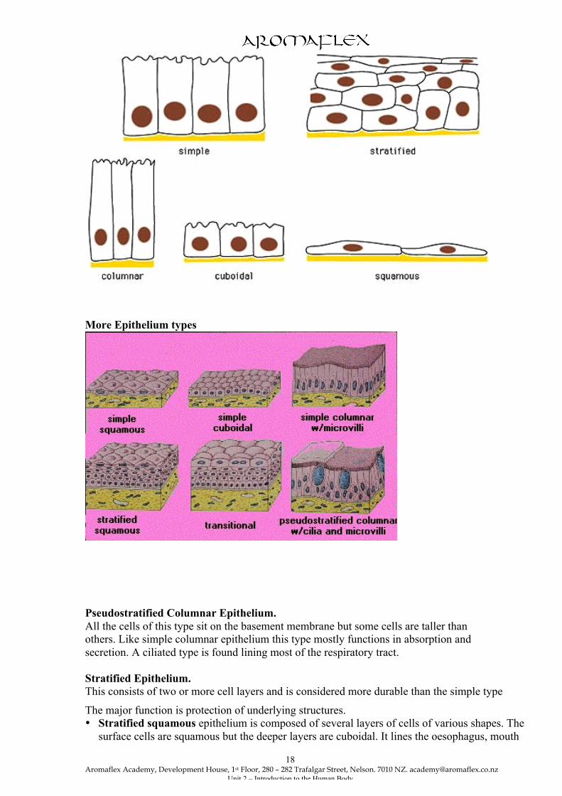
It can be thought of as a rich, nutritious and rather unusual “soup”. It contains thousands of ingredients such as amino acids, sugars, fatty acids, vitamins, hormones, neurotransmitters, salts and waste products. To remain healthy, each cell must take from this fluid the exact amount of each substance it needs at specific times and reject the rest. Selective permeability of the plasma membrane is a characteristic of healthy intact cells. i.e. it allows nutrients to pass through the membrane but keeps undesirable substances out. At the same time its keeping cell proteins and other substances in and allowing wastes to pass out. When the membrane is damaged substances flow in and out freely e.g in the case of burns. Tissues. Tissues are the materials from which all parts of the body are made. Following fertilisation a ball of cells is formed by repeated cell division. Cell differentiation occurs so that cells assume the shape, size and characteristics which will enable them to perform a specific function. Groups of closely associated cells, that are similar in structure, and perform a common or related function, are called tissues. Four main tissue types interweave to form the fabric of the body. These basic tissues and their functions are: • Epithelial tissue - Protection, Secretion, Absorption filtration and Sensory reception. • Connective tissue - Support, Protection, Binding, Insulation, and Transportation. • Muscle tissue - Contractile (able to produce movement.) • Nervous tissue - This has a special function of carrying messages. There are many types of each variety. Epithelial tissue. This group of tissues is found covering the body, lining cavities and tubes, and forming glandular tissue. Its functions include - protection, absorption, secretion, filtration and sensory reception. Epithelium is classified by arrangement of the cells and by cell shape. The cells fit closely together to form continuous sheets. The lower surface of the membrane sits on a basement membrane which is a substance secreted by the cells themselves. Epithelial tissues have no blood supply of their own but depend on that of the underlying connective tissue for oxygen and food. Simple Epithelium. This consists of a single layer of cells and is divided into 4 types. It is usually concerned with filtration secretion and absorption. • Squamous epithelium this is composed of a single layer of flattened cells that fit closely
together like flat stones forming a very smooth membrane. Found lining the heart, blood vessels, lymph vessels and alveoli of the lungs.
• Cuboidal epithelium consists of cube-shaped cells fitting closely together on a basement
membrane. It forms the tubules of the kidneys and is found in some glands and covering the surface of the ovaries.
• Columnar epithelium consists of a single layer of rectangular shaped cells and is found lining the organs of the alimentary tract.
• Ciliated epithelium is formed of columnar cells which have fine hair like processes called cilia. This type is found lining the uterine tubes and most of the respiratory passages.
Types of Epithelium
Aromaflex Academy, Development House, 1st Floor, 280 – 282 Trafalgar Street, Nelson. 7010 NZ. [email protected]
Unit 2 – Introduction to the Human Body
18
More Epithelium types
Pseudostratified Columnar Epithelium. All the cells of this type sit on the basement membrane but some cells are taller than others. Like simple columnar epithelium this type mostly functions in absorption and secretion. A ciliated type is found lining most of the respiratory tract. Stratified Epithelium. This consists of two or more cell layers and is considered more durable than the simple type
The major function is protection of underlying structures. • Stratified squamous epithelium is composed of several layers of cells of various shapes. The
surface cells are squamous but the deeper layers are cuboidal. It lines the oesophagus, mouth
Aromaflex Academy, Development House, 1st Floor, 280 – 282 Trafalgar Street, Nelson. 7010 NZ. [email protected]
Unit 2 – Introduction to the Human Body
19
and pharynx and forms the conjunctiva of the eyes. i.e. areas subject to wear and tear. It is the most common stratified epithelium found in the body.
• Keratinised stratified squamous cells are found on dry surfaces, that is the skin, hair and nails. The surface layer consists of dead epithelial cells to which Keratin, a protective protein, has been added. This forms a tough protective layer that prevents drying of underlying live cells.
• Transitional epithelium is a special sort of stratified squamous epithelium. This is composed of pear shaped cells and is found lining the urinary bladder, the ureters and part of the urethra. It allows for stretching as these organs fill, especially the bladder.
Glandular Epithelium. Glands make and secrete secretions which consist of specialised proteins and a water based fluid. They obtain the needed materials from the blood . There are two types of glands, endocrine which secrete hormones directly into the blood vessels that weave through the glands, and exocrine which empty secretions into a duct on to the epithelial surface. Connective Tissue. Connective tissues are found everywhere in the body. Their functions include binding and support, protection, insulation, and transportation i.e. blood, a fluid connective tissue. All classes of connective tissue consist of living cells surrounded by an extra cellular matrix. Often there are fibres in the matrix for reinforcement and the matrix can be hard, fluid or like a jelly and quite soft. Both the matrix and the fibres are secreted by the connective tissue cells and then secreted to the outside. The fibres vary in type and amount and include collagen fibres, elastic fibres and reticular or fine collagen fibres. Most connective tissues have a very good blood supply but tendons and ligaments are an exception, and cartilages have no blood supply at all. Loose Connective tissue. 1. Areolar or. This forms a very thin transparent tissue which surrounds vessels, nerves and
muscle fibres, function – strength, elasticity & support. The matrix contains all sorts of fibres and is loose and fluid and is ideal for storage of fluid and salts for the surrounding cells to release their waste products and obtain nutrients. This is the most common type of connective tissue in the body.
2. Adipose tissue. This is similar to loose connective tissue but the spaces of the network are filled in with fat cells, it stores triglycerides. Functions – insulation, support & protection. This forms the subcutaneous layer beneath the skin where it aids insulation.
3. Lymphoid or reticular is found in lymph nodes, liver and the spleen. Also binds together smooth muscle cells.
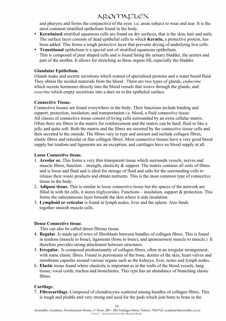
Dense Connective tissue. This can also be called dense fibrous tissue. 4. Regular. Is made up of rows of fibroblasts between bundles of collagen fibres. This is found
in tendons (muscle to bone), ligaments (bone to bone), and aponeuroses( muscle to muscle.) It therefore provides strong attachment between structures.
5. Irregular. Is composed predominantly of collagen fibres, often in an irregular arrangement, with some elastic fibres. Found in periosteum of the bone, dermis of the skin, heart valves and membrane capsules around various organs such as the kidneys, liver, testes and lymph nodes.
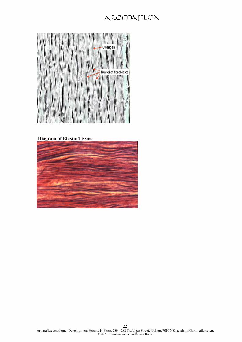
6. Elastic tissue found where elasticity is important as in the walls of the blood vessels, lung tissue, vocal cords, trachea and bronchioles. This type has an abundance of branching elastic fibres.
Cartilage. 7. Fibrocartilage. Composed of chondrocytes scattered among bundles of collagen fibres. This
is tough and pliable and very strong and used for the pads which join bone to bone in the

Aromaflex Academy, Development House, 1st Floor, 280 – 282 Trafalgar Street, Nelson. 7010 NZ. [email protected]
Unit 2 – Introduction to the Human Body
20
slightly movable joints. e.g. between the vertebrae and symphysis pubis. It provides support and fusion because it is the strongest of all three types.
8. Hyaline cartilage. This is the most abundant type found in the body and is quite ‘gristly’ or rubbery. It forms the supporting structures of the larynx, attaches the ribs to the sternum and covers the ends of bones at joints. The foetal skeleton is also made up of hyaline cartilage and is later replaced by bone.
9. Elastic cartilage. This type has chondrocytes in a threadlike arrangement of elastic fibres within the matrix, and provides strength and elasticity and maintains the shape of some organs.
10. Bone. This is a special type of fibrous material and the matrix surrounding the bone cells, (osteocytes), is hardened by deposit of salts such as calcium phosphate. It is the hardest tissue in the body. The fibrous material gives bone its toughness and the mineral matter its rigidity. There are 2 types of bone tissue: • Compact Bone - has a solid appearance and is found in the parts of a bone where extra
strength is required. The matrix here is arranged in concentric rings with the bone cells in the small spaces between. (described in more detail in the skeletal system untit.)
• Cancellous Bone - has a spongy or honeycomb appearance. It is hard and strong but lighter in weight than compact bone so it is found where lightness and strength is important. This consists of a lattice work of bony plates called trabeculae with the spaces in between being filled with red bone marrow. See skeletal system for diagram.
11. Liquid connective tissue. Blood is a connective tissue with a liquid matrix called plasma. The various blood cells are suspended in the plasma. See cardiovascular system for details. Lymph is also a liquid tissue derived from the tissue fluids. See lymphatic system. Diagram of Areola or Loose Connective Tissue.

Aromaflex Academy, Development House, 1st Floor, 280 – 282 Trafalgar Street, Nelson. 7010 NZ. [email protected]
Unit 2 – Introduction to the Human Body
21
Diagram of Adipose Tissue.
Diagram of Fibrous Tissue.
Aromaflex Academy, Development House, 1st Floor, 280 – 282 Trafalgar Street, Nelson. 7010 NZ. [email protected]
Unit 2 – Introduction to the Human Body
22
Diagram of Elastic Tissue.

Aromaflex Academy, Development House, 1st Floor, 280 – 282 Trafalgar Street, Nelson. 7010 NZ. [email protected]
Unit 2 – Introduction to the Human Body
23
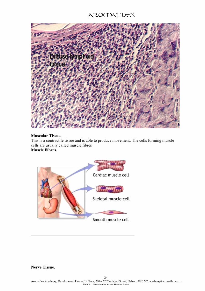
Diagrams of Hyaline cartilage, Fibrous cartilage & Elastic cartilage (found in ear lobes, epiglottis and blood vessel walls) Diagram of Lymphoid Tissue
Aromaflex Academy, Development House, 1st Floor, 280 – 282 Trafalgar Street, Nelson. 7010 NZ. [email protected]
Unit 2 – Introduction to the Human Body
24
Muscular Tissue. This is a contractile tissue and is able to produce movement. The cells forming muscle cells are usually called muscle fibres Muscle Fibres.
Nerve Tissue.
Aromaflex Academy, Development House, 1st Floor, 280 – 282 Trafalgar Street, Nelson. 7010 NZ. [email protected]
Unit 2 – Introduction to the Human Body
25
This has the special function of receiving and transmitting electrical impulses throughout the body. Nervous tissue forms organs of the nervous system. It consists of the nerve cells called neurones, and neuroglia, the supporting cells of the nervous system. These are dealt with in more detail in the Nervous system unit. Membranes. These are made of connective tissue and line the cavities and hollow organs of the body. They secrete lubricating fluids to prevent friction. There are 3 types of membrane found in the body: • Synovial membrane, secretes a thick fluid rather like the white of an egg. • Mucous membrane secrete sticky fluid called mucus. • Serous membrane, made of flattened cells, exudes a watery substance called serum. Oedema is when water logging of the tissues occurs. There are many reasons for this: • A blockage of lymphatic vessels. • An osmotic pressure that is too high. • Damage to capillary walls. • Too high hydrostatic pressure in the capillaries. Tissue repair. This is a process where damaged or destroyed cells are replaced by healthy ones. Three factors can affect this process of wound healing;
• nutrition, proteins and vitamins are particularly necessary, • blood circulation and • age.
Nutrition - is so important for the healing process to occur. Protein is important to make new cell structures along with Vitamins A, B, C, D, E and K. A good blood supply is essential to transport the oxygen, nutrients antibodies and other cells needed to the site of injury, and to remove any debris which might otherwise interfere with the process. The age of a person also greatly affects the rate at which tissue repair is carried out. The younger person usually has a better blood supply and the cells have a faster metabolic rate than an older person. Their nutrition also is often in a better state. Not only do the tissues heal faster but there is less obvious scar tissue. And interestingly enough surgery performed on foetuses leaves no scars at all. The extra cellular composition of tissues such as collagen and elastin also change with aging resulting in slower and less efficient tissue repair processes. Repair process. Tissue repair occurs in two ways, by regeneration and fibrosis. With regeneration the damaged tissues are replaced by the same kind of cells. Epithelial cells such as the skin and mucous membranes have the ability to regenerate well. Fibrosis however involves repair by dense fibrous connective tissue, scar tissue. Scar tissue formation means it is unable to perform the normal functions of the tissue it replaces leading to severe impairment of a particular organ. Tissue injury sets in motion a series of events beginning with the inflammatory response. This allows clotting proteins and other blood cells essential for the drainage and reabsorption of pus to occur. Where a clot is exposed to the air it dries and forms a scab. If tissue injury is slight this might be all that is needed for complete healing to occur. In the case of a more extensive injury the process is more complex and involves the formation of granulation tissue. This type of tissue is full of new fragile blood capillaries that gradually grow into the damaged area. Connective tissue cells called fibroblasts are also manufacturing new collagen fibres for strength. At the same time the new surface epithelium is growing across the granulation tissue beneath the scab which

Aromaflex Academy, Development House, 1st Floor, 280 – 282 Trafalgar Street, Nelson. 7010 NZ. [email protected]
Unit 2 – Introduction to the Human Body
26
will soon detach. This results in a fully regenerated surface epithelium that covers an area of fibrosis, a scar, either invisible or visible. The Integumentary System. The integumentary system is made up of a complex set of organs that serves several functions, mostly protective. These organs are;
• skin, • sweat and oil glands, • hairs and • nails.
The Skin. The skin, also called the integument (meaning covering), is the largest organ in the body. Without our skin we would quickly fall prey to bacteria and die from water and heat loss. The skin varies in thickness from 1.5 to 4.0 mm or more in different parts of the body, and has over 1.7 square metres of surface area. It weighs about 3.2 kg and is twice as heavy as the liver or brain. The skin is semi-permeable. Mineral oils and animal fats, such as lanolin and glycerine are not absorbed by the skin. The Structure of the Skin. It is composed of 2 main regions, the epidermis and the dermis. However, deep to the dermis is the subcutaneous layer which consists of adipose and areola tissues. Fibres from the dermis reach into this layer anchoring the skin to it. In turn the subcutaneous layer attaches to the underlying tissues and organs.. The Epidermis. This is the most superficial layer of the skin and is composed of stratified epithelium. The number of layers vary in thickness in different parts of the body. The epidermis is thickest on the soles of the feet and palms of the hands where there is a large amount of wear and tear. It is thinnest on the lips, which is why the lips appear pinker than the rest of the skin. There are no blood vessels or nerve endings in the epidermis, but the deeper layers are bathed in interstitial fluid, which is drained away as lymph. There are several layers of cells in the epidermis which extend from the deepest germinative layer to the stratum corneum or the horny layer on the surface. The skin showing the main layers of the epidermis.

Aromaflex Academy, Development House, 1st Floor, 280 – 282 Trafalgar Street, Nelson. 7010 NZ. [email protected]
Unit 2 – Introduction to the Human Body
27
A Simple Diagram of the Skin.
• The horny layer consists entirely of flat, thin, non - nucleated dead cells in which the
protoplasm has been replaced by keratin. They are constantly being rubbed off and replaced by cells which originated in the germinative layer and have undergone gradual changes and become flattened as they progress towards the surface. These dead cells, which are composed of the tough protein called keratin, are virtually waterproof and therefore prevent the loss of water from the body surface.
Aromaflex Academy, Development House, 1st Floor, 280 – 282 Trafalgar Street, Nelson. 7010 NZ. [email protected]
Unit 2 – Introduction to the Human Body
28
The average person sheds 18kgs of these skin flakes in a lifetime. We actually have a totally new epidermis every 35- 45 days.
• The germinative layer or growing layer lies beneath the granular layer and it’s very wavy at its
lower edge. If you look at the diagram you can see that this wavy edge helps to bring a larger area of the growing layer into contact with the capillaries in the dermis beneath. These blood vessels supply the oxygen and nutrients required for the production of healthy new skin cells which are forming in this layer all the time. If the general circulation is sluggish, this is reflected in the poor condition of the skin. It is believed that the downward projections in the germinative layer also help stabilise the two layers.
Melanocytes, the specialised cells that produce the pigment melanin, are found in the
deepest layer of the epidermis. Melanin gives the colour to the skin and hair. All humans have roughly the same number of pigment producing cells so individual and racial differences in colouring depend on the amount of melanin secreted. Sunlight darkens existing melanin and promotes its secretion to protect us from the burning effects of the sun’s rays. Prolonged sun exposure causes a melanin build up, a tan.
Despite this protection, excessive sun exposure eventually damages the skin. It causes clumping of the elastin fibres leading to leathery skin, temporarily depresses the immune system and it can alter the DNA of skin cells and in this way lead to skin cancer. Dark skinned people have less skin cancer due to their natural sun screen.
Skin Colour. There are a few pigments that contribute to the skin colour: • Melanin - we have already mentioned, is the only one actually made in the skin. • Haemoglobin - The level of oxygenation of haemoglobin and the amount of blood circulating
in the dermis gives the skin its pink colour. Since Caucasian skin has only small amounts of melanin the epidermis is almost transparent allowing haemoglobin’s colour to show through.
• Carotene - the yellow pigment found in certain plants such as carrots, is a rich source of
Vitamin A. This tends to accumulate in the stratum corneum (horny layer) and in fatty tissue. Its colour is most obvious on the soles of the feet and palms of the hands.
• Any bile pigments - in the blood also give the skin a yellowish colour. The skin colour can also be influenced by emotional stimuli and certain disease states. A Note on absorption through the skin. Certain oils such as essential oils and some carrier oils have a very small molecular structure. This enables them to be absorbed easily through the epidermis, and the pores and hair follicles which open directly on to the skin’s surface. Once the molecules have penetrated the surface they are able to diffuse directly into the blood and lymph capillaries via the tissue fluids. The Dermis. The dermis, the 2nd major skin region, is a strong but flexible connective tissue layer. The cell types found in the dermis are typical of those found in any connective tissue. The semi-fluid matrix is heavily embedded with collagen and elastic fibres. The dermis binds the entire body together like a body stocking. It corresponds to the hide in animals.

Aromaflex Academy, Development House, 1st Floor, 280 – 282 Trafalgar Street, Nelson. 7010 NZ. [email protected]
Unit 2 – Introduction to the Human Body
29
The dermis has two major layers, the thin superficial papillary layer and the deeper reticular layer. It’s the superficial papillary layer that is heavily invested with blood capillaries amongst the loosely woven mat of the connective tissue fibres. As can be seen from the diagram the nipple-like projections indent the overlying epidermis increasing the surface area. Many of these dermal papillae contain capillary loops, pain and touch receptors. The deeper reticular layer accounts for 85% of the dermis and is typical dense connective tissue. It’s the elastin fibres that give the skin its stretch-recoil properties. A layer of adipose tissue lying beneath the dermis is called the subcutaneous tissue or fat. This helps to insulate the body against cold. People from cold climates may accumulate more fat cells in areas that need extra protection e.g. the fatty pads over the eyes of Eskimos. This layer also helps to give the skin a soft and smooth appearance. Extreme stretching of the skin, such as occurs during pregnancy, can tear the dermis. This is indicated by silvery white scars called stretch marks. Short term trauma, such as a burn, can cause a blister. This is the separation of the epidermal and dermal layers by a fluid filled pocket. Structures found in the dermis. All these structures are surrounded and supported by the connective tissues of the dermis. The dermis is richly supplied with: • Nerve endings. There are several varieties of them in the skin, and each reacts to a different
sensory stimulus, e.g. heat - cold - pain - touch. The skin is an important sensory organ through which individuals are aware of their environment.
• Blood vessels. Arterioles form a fine network with capillary branches supplying sweat glands,
sebaceous gland, hair follicles and the dermis. Because the epidermis has no blood supply it obtains its nutrients and oxygen from interstitial fluid derived from the blood vessels here in the dermis.
When we are too hot these capillaries expand to allow more blood to pass through them near the surface of the skin. In this way, some of the heat from the blood is lost and this helps to cool the body. The opposite happens when we are cold, the capillaries shrink.
• Lymphatic vessels. These form a network throughout the dermis and the deeper layers of the
epidermis. • Sweat glands, also called sudoriferous glands. These are distributed over the entire skin
surface except the nipples and parts of the external genitalia, more than 2.5 million per person. There are two types of sweat glands; eccrine and apocrine.
Eccrine are far more numerous especially on the soles of the feet, palms and forehead. Each is a simple coiled tubular gland with the secretory, coiled part lying in the dermis. The duct extends to open on the skin surface as a pore. (see diagram of skin.)
Apocrine sweat glands are largely in the axillary and anogenital areas and they begin to function at puberty. They are larger than eccrine glands and their ducts empty into hair
follicles. Their secretion contains the same basic components as true sweat plus some protein and fatty substances on which bacteria thrive. When the organic molecules are decomposed by the bacteria on the skin it takes on the generally unpleasant odour called ‘body odour’. The role of apocrine glands is not fully understood but they may function as scent glands.
Sweat consists of 99% water and waste products such as urea and salts.
Aromaflex Academy, Development House, 1st Floor, 280 – 282 Trafalgar Street, Nelson. 7010 NZ. [email protected]
Unit 2 – Introduction to the Human Body
30
The major role of sweat glands is to help in the normalising of body temperature by pouring water on to the skin surface when we are too hot. Evaporation of the water into the air helps us to cool down. Interestingly enough, heat induced sweating begins on the forehead and then spreads over the body. Emotionally induced sweating, brought on by fear, embarrassment, or nervousness, usually starts on the palms, soles and axillae before spreading to other parts of the body.
It is when the air is hot and humid that we become conscious of the beads of sweat on the skin. It cannot evaporate fast enough in the already moisture laden air. Most of the time we are not aware of the amount of water lost in sweat because it evaporates continually. In the ear there are modified sweat glands called ceruminous glands which produce waxy secretions. These secretions along with hairs help to protect the ear from foreign bodies. • Sebaceous glands, or oil glands, occur all over the body surface except for the palms and
soles. They produce an oily substance called sebum and open into each hair follicle. The sebum travels up the shaft of the hair and onto the surface of the skin.
Sebaceous glands are also activated at puberty by androgens (sex hormones.) The function of sebum is to lubricate the skin and hair, prevent water loss from the skin and
act as a bactericidal agent. • Arrector pili muscles. These are tiny involuntary muscles with one end attached to
the dermis and the other end attached to a hair follicle. When we are cold or scared these muscles contract and pull the hair into an upright position. Some elevation of the skin occurs around the hair giving rise to the ‘goose pimples’ appearance.
A hair follicle is well supplied with nerve fibres and also richly vascularised. • Hair and hair follicles. Hair serves other mammals by keeping them warm. Our body hair is
far less useful although it covers the whole of the body except the palms of the hands and the soles of the feet, lips, nipples and parts of the external genitalia. There are millions of them, about 100,000 of them in the scalp and another 30,000 in a man’s beard. Hair comes in various shapes and sizes.
Functions.
• Hair on the head helps protect the head from physical trauma, heat loss and sunlight.
• Eyelashes shield the eyes. • Nasal hairs filter large particles like lint and insects from the air we inhale. • Our sparse body hair’s main function is to sense insects on the skin before they sting us.
Each hair on the body has its origin in a duct known as a hair follicle. Structure of hair. Hairs come in all shapes and sizes and are flexible strands produced by hair follicles that consist largely of keratinised cells. The cells are tougher, more durable and do not flake off like those found in typical epidermal cells. The main regions of a hair are the • shaft, which projects from the skin, and • the root, the part embedded in the skin, right into the dermis and the subcutaneous layer. A typical hair consists of a • central medulla, • a cortex and • an outer cuticle. The cuticle provides strength, helps keep the inner layers tightly compacted and neighbouring hairs apart so that the hair doesn’t matt.
Aromaflex Academy, Development House, 1st Floor, 280 – 282 Trafalgar Street, Nelson. 7010 NZ. [email protected]
Unit 2 – Introduction to the Human Body
31
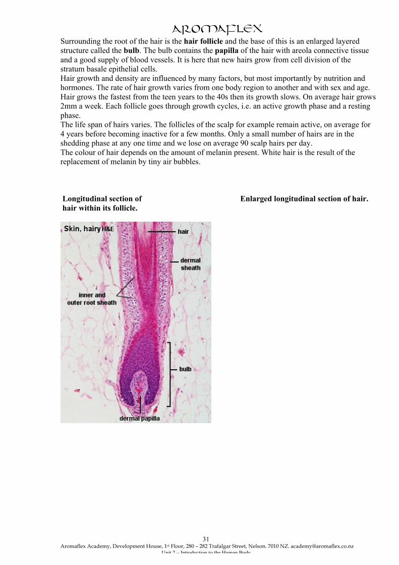
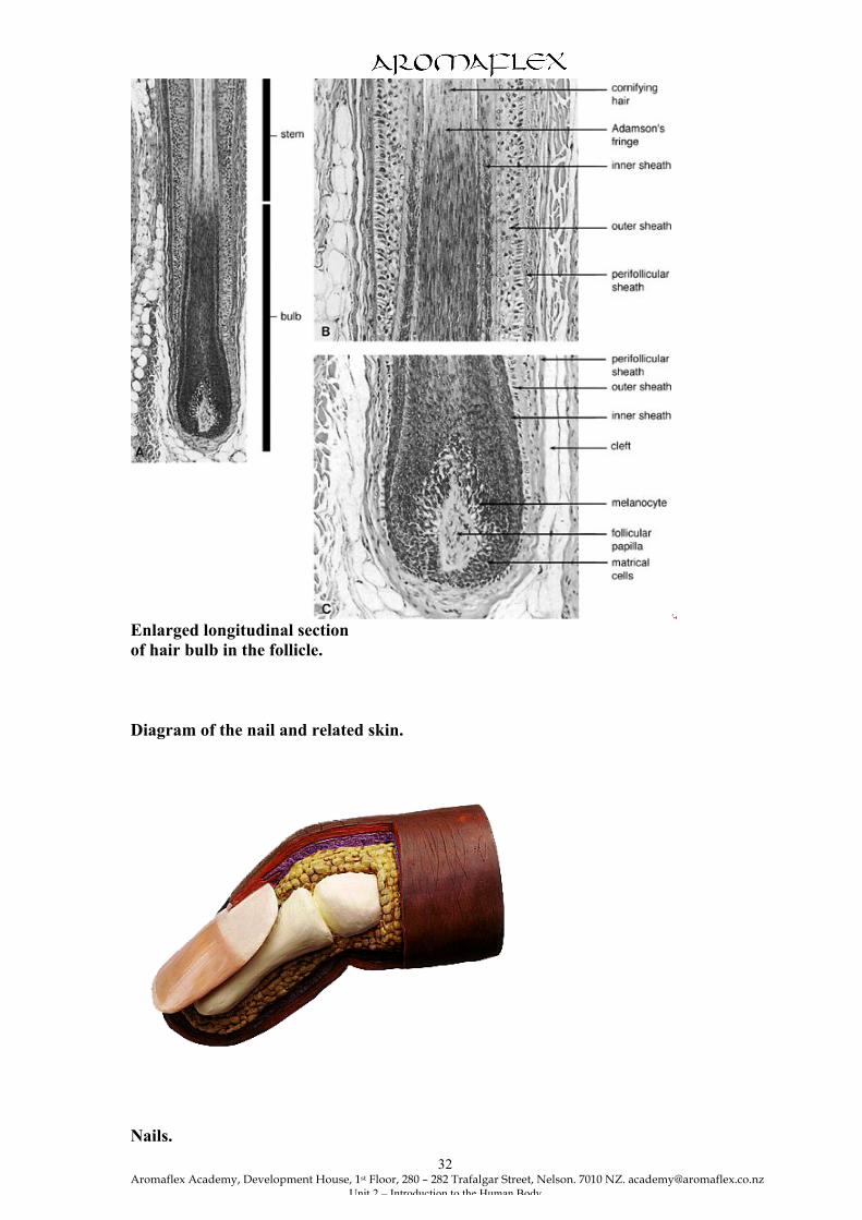
Surrounding the root of the hair is the hair follicle and the base of this is an enlarged layered structure called the bulb. The bulb contains the papilla of the hair with areola connective tissue and a good supply of blood vessels. It is here that new hairs grow from cell division of the stratum basale epithelial cells. Hair growth and density are influenced by many factors, but most importantly by nutrition and hormones. The rate of hair growth varies from one body region to another and with sex and age. Hair grows the fastest from the teen years to the 40s then its growth slows. On average hair grows 2mm a week. Each follicle goes through growth cycles, i.e. an active growth phase and a resting phase. The life span of hairs varies. The follicles of the scalp for example remain active, on average for 4 years before becoming inactive for a few months. Only a small number of hairs are in the shedding phase at any one time and we lose on average 90 scalp hairs per day. The colour of hair depends on the amount of melanin present. White hair is the result of the replacement of melanin by tiny air bubbles. Longitudinal section of Enlarged longitudinal section of hair. hair within its follicle.
Aromaflex Academy, Development House, 1st Floor, 280 – 282 Trafalgar Street, Nelson. 7010 NZ. [email protected]
Unit 2 – Introduction to the Human Body
32
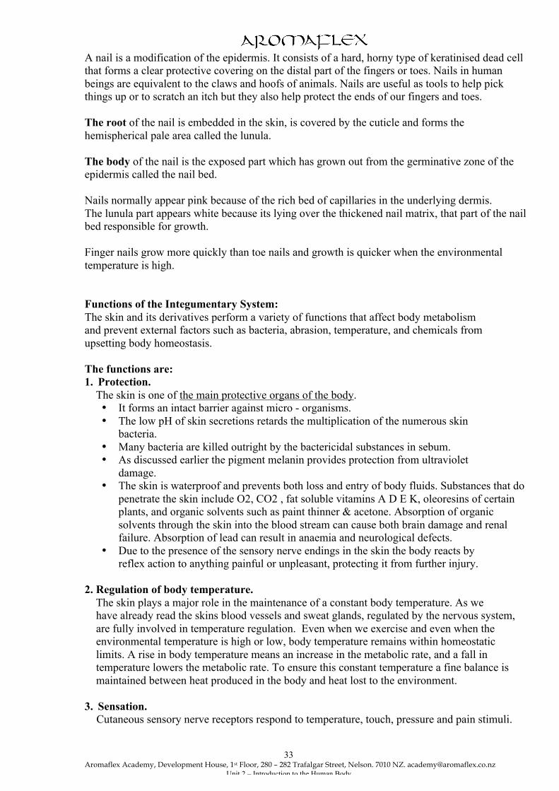
Enlarged longitudinal section of hair bulb in the follicle. Diagram of the nail and related skin.
Nails.
Aromaflex Academy, Development House, 1st Floor, 280 – 282 Trafalgar Street, Nelson. 7010 NZ. [email protected]
Unit 2 – Introduction to the Human Body
33
A nail is a modification of the epidermis. It consists of a hard, horny type of keratinised dead cell that forms a clear protective covering on the distal part of the fingers or toes. Nails in human beings are equivalent to the claws and hoofs of animals. Nails are useful as tools to help pick things up or to scratch an itch but they also help protect the ends of our fingers and toes. The root of the nail is embedded in the skin, is covered by the cuticle and forms the hemispherical pale area called the lunula. The body of the nail is the exposed part which has grown out from the germinative zone of the epidermis called the nail bed. Nails normally appear pink because of the rich bed of capillaries in the underlying dermis. The lunula part appears white because its lying over the thickened nail matrix, that part of the nail bed responsible for growth. Finger nails grow more quickly than toe nails and growth is quicker when the environmental temperature is high. Functions of the Integumentary System: The skin and its derivatives perform a variety of functions that affect body metabolism and prevent external factors such as bacteria, abrasion, temperature, and chemicals from upsetting body homeostasis. The functions are: 1. Protection. The skin is one of the main protective organs of the body.
• It forms an intact barrier against micro - organisms. • The low pH of skin secretions retards the multiplication of the numerous skin
bacteria. • Many bacteria are killed outright by the bactericidal substances in sebum. • As discussed earlier the pigment melanin provides protection from ultraviolet
damage. • The skin is waterproof and prevents both loss and entry of body fluids. Substances that do
penetrate the skin include O2, CO2 , fat soluble vitamins A D E K, oleoresins of certain plants, and organic solvents such as paint thinner & acetone. Absorption of organic solvents through the skin into the blood stream can cause both brain damage and renal failure. Absorption of lead can result in anaemia and neurological defects.
• Due to the presence of the sensory nerve endings in the skin the body reacts by reflex action to anything painful or unpleasant, protecting it from further injury.
2. Regulation of body temperature. The skin plays a major role in the maintenance of a constant body temperature. As we
have already read the skins blood vessels and sweat glands, regulated by the nervous system, are fully involved in temperature regulation. Even when we exercise and even when the environmental temperature is high or low, body temperature remains within homeostatic limits. A rise in body temperature means an increase in the metabolic rate, and a fall in temperature lowers the metabolic rate. To ensure this constant temperature a fine balance is maintained between heat produced in the body and heat lost to the environment.
3. Sensation. Cutaneous sensory nerve receptors respond to temperature, touch, pressure and pain stimuli.
Aromaflex Academy, Development House, 1st Floor, 280 – 282 Trafalgar Street, Nelson. 7010 NZ. [email protected]
Unit 2 – Introduction to the Human Body
34
4. Sectretion. Sebum keeps skin supple and free from cracks and hair soft and pliant. 5. Excretion.
Sweat contains small amounts of nitrogenous wastes and plays a minor role in excretion. However, profuse sweating is an important avenue of water and sodium
chloride loss. 6. Production of Vitamin D. When sunlight bombards the skin Vitamin D is synthesised from cholesterol by specialised epidermal cells. 7. Blood reservoir. The extensive vascular supply of the dermis allows the skin to act as a blood reservoir. 5% of the body’s blood volume can be held in the skin. The process of ageing with regard to the skin. When a baby is born, the skin is covered with vernix caseosa. This is a white cheesy looking substance produced by the sebaceous glands to protect the foetus’s skin from its watery environment. A newborn’s skin is thin. During childhood the skin thickens and more subcutaneous fat is deposited. At puberty sebaceous glands are activated and terminal hairs appear. As old age approaches, the rate of epidermal cell replacement slows, the skin and hair thins, and is more susceptible to injury. The skin glands become less active, and the loss of collagen and elastic fibres and subcutaneous fat leads to wrinkling. Delayed action genes are said to cause greying and balding. The hair and nails grow more slowly too and the decrease in special immune cells in the skin contributes to an impaired immune system. An increase in the size of the melanocytes can cause pigmented blotching, liver spots. As we have already said earlier in this unit aged skin also heals more slowly.
Aromaflex Academy, Development House, 1st Floor, 280 – 282 Trafalgar Street, Nelson. 7010 NZ. [email protected]
Unit 2 – Introduction to the Human Body
35
ANATOMY & PHYSIOLOGY
ASSIGNMENT COVER SHEET Name:
Address:
Telephone Number:
E-Mail Address:
Unit Name and Number:
Date Assignment Submitted:
Questions/Comments from student: Requirements:
• Send in the above cover sheet along with the question sheet. • Enclose a stamped addressed envelope to have your assignment returned. • Answer each question on a separate sheet of paper, with your name at the top of
every page. • All resubmissions need to be sent in with original marked unit. • Make sure your resubmission work is clearly in a different colour, or
highlighted as a “resubmission, attempt 2”, “resubmission, attempt 3” etc.. • Answer the questions in black or blue pen, not pencil. Drawings may be in
pencil. • Submit clearly drawn and clearly labelled diagrams • Answer the questions clearly and in order they are set on the question page. • All questions need to be answered correctly for the unit to pass. • If you don’t understand any questions contact the A&P tutor. • You have as many attempts as you like to get the question right, and to pass the
unit. • Keep a copy of your work
QUESTION PAPER UNIT TWO.

Aromaflex Academy, Development House, 1st Floor, 280 – 282 Trafalgar Street, Nelson. 7010 NZ. [email protected]
Unit 2 – Introduction to the Human Body
36
1. What is Anatomy and Physiology? 2. Define homeostasis, and explain its importance? 3. Define Basal Metabolic Rate and list 4 factors that affect it. For each factor given explain
why BMR is affected. 4. State normal body temperature. Name 3 mechanisms the body uses to lose and gain
heat, giving examples of each method. Please do not use the examples given in the unit.
5. Use the diagram supplied to locate the cavities of the body. 6. Hand draw a typical cell labelling the plasma membrane, cytosol, nucleus and all the
organelles listed in the unit.
7. If the top of a bottle of perfume is taken off and left off, several hours later the aroma will be spread evenly around the room. State why the term ‘diffusion’ is correctly used for the process that occurred in the room.
8. State the four principal types of tissues in the body, and for each type, explain the
function and location of this tissue in the body. For each function give an example of a tissue doing that job in the body.
9. Draw by hand and label a simple diagram of the skin. (not traced from unit) 10. List all the main functions of the Integumentary system. 11. Explain the effects of sunlight exposure to the skin? 12. Describe the structure and functions of hair. 13. Describe the role of the skin in the regulation of body temperature.

Aromaflex Academy, Development House, 1st Floor, 280 – 282 Trafalgar Street, Nelson. 7010 NZ. [email protected]
Unit 2 – Introduction to the Human Body
37