unit 1.1 skeletal system
TRANSCRIPT

Key
Stag
e 4
–B
TEC
Hea
lth
& F
itn
ess
/ S
kele
tal S
yste
m –
HT1
FUNCTION OF THE SKELETON
• Support: the bones are solid and rigid. They keep us upright and hold the rest of the body – the muscles and organs – in place.
• Movement: the skeleton helps the body move by providing anchor points for the muscles to pull against.
• Structural shape and points for attachment: the skeleton gives us our general shape such as height and build. The skeleton also provides anchorage points for the muscles
to attach via tendons, so when muscles contract movement occurs.
• Protection: certain parts of the skeleton enclose and protect the body’s organs from external forces e.g. the brain is inside the cranium. This function is especially important in
activities that involve contact. E.g. rugby, boxing.
• Production of Blood Cells: the bone marrow in long bones and ribs produce red and white blood cells.
• Mineral Storage: bones store several minerals e.g. calcium, which can be released into the blood when needed.
TYPES OF BONES
Flat bones: protect vital organs e.g. cranium
protects your brain, ribs protect heart and lungs
Long bones: enable gross (large) movements e.g.
femur, tibia and fibula in the leg which allow us to
run, humerus, radius and ulna in arm which allows
us to throw a ball
Short bones: enable fine (small) movements e.g.
fingers allowing you to spin a cricket ball
Irregular: vertebrae
Sesamoid: Patella
BONES LOCATED AT JOINTS:
Shoulder = Scapula and Humerus
Elbow = Humerus, Radius, Ulna
Hip = Pelvis, Femur
Knee = Femur, Tibia, Patella
Types of Joint: Ball and Socket Joint
Location in Body: Shoulder and HipType of Movement Allowed by Joint: Flexion,
Extension, Adduction, Abduction, Rotation
Hinge Joint
Location in Body: Knee and ElbowType of Movement Allowed by Joint: Flexion and
Extension
Ligaments
Attaches bone to bone to keep the joint stable e.g. knee when kicking the
ball or restricts movement/prevents movement to stop injury.
Cartilage
Found between bones and prevents friction by stopping the bones from
rubbing together.
Synovial Membrane
Secrets synovial fluid.
Synovial Fluid
Is produced by the synovial membrane and helps lubricate the joint.
Joint Capsule
This is lined with synovial membrane. It encloses the joint making sure
the cartilage and synovial fluid remain in place.
Bursae
Fluid filled sac providing cushion between bones and tendons. This stops
friction at the joint.
Tendons
Attach muscle to bone. When a muscle contracts to move a joint, it is the
tendon which pulls on the bone, keeps muscles/bones stable or holds
join in place.
TYPES OF JOINTS - a place where two bones meet
Fixed - skull and pelvis
Slightly Moveable - spine
Synovial Joints
• Pivot - vertebrae
• Condyloid - wrist
• Saddle - thumb
• Gliding - clavicle
• Ball and Socket - shoulder and hip
• Hinge - knee and elbow
Unit 1.1 Skeletal System
Synovial Joints
Articular
cartilage
Ligament
Synovial
membrane BursaeJoint
capsule

Key
Stag
e 4
–B
TEC
Hea
lth
& F
itn
ess
/ S
kele
tal S
yste
m –
HT1
Flexion and extension
at the shoulder
– The Deltoid causes
flexion at the shoulder
– The Latissimus dorsi
causes extension at the
shoulder
Flexion and extension
at the elbow
– The Biceps cause
flexion at the elbow
– The Triceps cause
extension at the elbow
Flexion and extension
at the knee
– The Hamstrings cause
flexion at the knee
– The Quadriceps cause
extension at the knee
Flexion and extension
at the hip
– The Hip Flexors cause
flexion at the hip
– The Gluteals cause
extension at the hip
Flexion and extension
at the ankle
– The Tibialis Anterior
causes dorsiflexion at
the ankle
– The Gastrocnemius
cause plantar flexion at
the ankle
Rotation
of the Shoulder
– The Rotator Cuff
causes rotation at the
shoulder
Abduction and Adduction
at the shoulder
– The deltoid causes abduction at
the shoulder
– The Pectorals / Latissimus Dorsi
cause adduction at the shoulder
Lordosis: Also called swayback, the spine of a person with lordosis curves significantly inward at the lower back.
Kyphosis: Kyphosis is characterized by an abnormally rounded upper back (more than 50 degrees of curvature).
Scoliosis: A person with scoliosis has a sideways curve to their spine. The curve is often S-shaped or C-shaped.
Unit 1.1 Skeletal System
Ribs
Cranium
Sternum
Vertebrae
Pelvis
Femur
Fibula
Talus
Scapula
Humerus
Tibia
Patella
RadiusUlna
STRUCTURE OF THE SKELETON
AXIAL-Cranium, sternum, ribs and vertebraeAPPENDICULAR- clavicle, scapula, humerus, radius, ulna, carpals, tarsals, pelvis, femur, tibia, fibula, and phalanges.
Phalanges
Phalanges
Cervical
vertebrae
Thoracic
vertebrae
Lumbar
vertebrae
Coccyx
Sacrum
(S1-5)
Atlas (C1)
Axis (C2)
C3
C4
C5
C6
C7
T1
T2
T3
T4
T5
T6
T7
T8
T9
T10
T11
T12
L1
L2
L3
L4
L5
Extension
FlexionFlexion
Extension
180°
60°
0°
Flexion Extension
Flexion Extension
hip
Shoulder abduction Shoulder adduction
Plantar flexion
Dorsiflexion

Patella
Vertebrae
Irregular bones
Scapula
Pelvis
Cranium
Ribs
Flat bones
Long bones
Long bones
Radius
Ulna
Femur
Fibula
TibiaShort bones
Key
Stag
e 4
–B
TEC
Hea
lth
& F
itn
ess
/ S
kele
ton
–H
T1

Key
Stag
e 4
–B
TEC
Hea
lth
& F
itn
ess
/ S
kele
tal S
yste
m –
HT1

Key
Stag
e 4
–B
TEC
Hea
lth
& F
itn
ess
/ M
usc
ula
r Sy
stem
–H
T1
Isotonic ContractionsThese contractions occur when there is movement of the body. The ends of the muscles move closer together to cause the movement.
Isometric ContractionsThis type of contraction takes place when the body is being held in the same position. The length of the muscle during these contractions stays the same length.
Isotonic Concentric Contraction occurs when the muscle shortens e.g. biceps contracting concentrically during the upwards phase of a bicep curl / triceps contracting concentrically during the upwards phase of a press-up
Isotonic Eccentric Contraction occurs when the muscle lengthening (antagonist) is under tension. An eccentric contraction provides the control of a movement on the downward phase and it works to resist the force of gravity e.g. biceps contracting eccentrically when lowering the weight in a bicep curl / triceps contracting eccentrically during the downwards phase of a press-up.
Muscular Contractions
Types Of Muscles
CARDIAC- Found in the heart wall- Oxygen dependent, involuntary- aids blood flow through the heart
SMOOTH- Found in internal organs, digestive tract, blood vessels and lungs.- can work without oxygen, involuntary- aids digestion, helps distribution of blood.
SKELETAL- Found around the body- can work with or without oxygen, works voluntarily.- aids with movement.
• How do MUSCLES WORK?• Muscles can only PULL they cannot push. This means that they must work in pairs to allow parts of the body to move back and forth. THESE PAIRS ARE
CALLED ANTAGONISTIC PAIRS.• Antagonistic Pairs• A muscle must work in partnership with another muscle to allow movement to occur. • The muscle that causes the movement (the pulling muscle) is called the AGONIST or PRIME MOVER. When this muscle contracts in becomes shorter.• During this time the other muscle within this partnership is relaxing. This muscle is called the ANTAGONIST and is lengthening while it relaxes.EXAMPLES:• When we flex our elbow the bicep is the agonist and the tricep is the antagonist. However these roles are reversed when the elbow extends ,with the
tricep becoming the agonist and the bicep becoming the antagonist.
• When dorsiflexion occurs in our ankle the tibialis anterior is the agonist and the gastrocnemius is the antagonist. However these roles are reversed when plantar flexion occurs at the ankle, with the gastrocnemius becoming the agonist and the tibialis anterior becoming the antagonist.
Muscle fibre typesType 1- Slow twitch fibres- red in colour, slow contraction speed, low force, fatigue slowly and uses oxygen..Type 2- fast twitch fibres- white in colour, fast contraction speed, fatigue quickly, contract without oxygen.
HAMSTRINGS QUADRICEPS
Bicep Tricep
HIP FLEXORS GLUTEALS
DELTOID LATISSIMUS DORSI
Antagonistic Muscle Pairs
Unit 1.2 Muscular System

Deltoid
Bicep
Abdominals
Hip flexors
Quadriceps
Tibialis anterior
Pectoral
Trapezius
Triceps
Latissimus dorsi
Gluteals
Hamstrings
Gastrocnemius
Key
Stag
e 4
–B
TEC
Hea
lth
& F
itn
ess
/ M
usc
ula
r Sy
stem
–H
T1

Key
Stag
e 4
–B
TEC
Hea
lth
& F
itn
ess
/ M
usc
ula
r Sy
stem
–H
T1
Key
Stag
e 4
–B
TEC
Hea
lth
& F
itn
ess
/ R
esp
irat
ory
Sys
tem
–H
T1
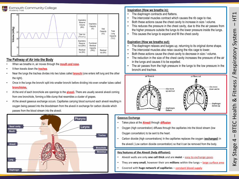
The Pathway of Air into the Body• When we breathe in, air moves through the mouth and nose.
• It then travels down the trachea.
• Near the lungs the trachea divides into two tubes called bronchi (one enters left lung and the other
the right).
• Once in the lungs the bronchi split into smaller bronchi before dividing into even smaller tubes called
bronchioles.
• At the end of each bronchiole are openings to the alveoli. There are usually several alveoli coming
from one bronchiole, forming a little clump that resembles a cluster of grapes.
• At the alveoli gaseous exchange occurs. Capillaries carrying blood surround each alveoli resulting in
oxygen being passed into the bloodstream from the alveoli in exchange for carbon dioxide which
passes from the blood stream into the alveoli.
Inspiration (How we breathe in):
• The diaphragm contracts and flattens.
• The intercostal muscles contract which causes the rib cage to rise.
• Both these actions cause the chest cavity to increase in size / volume.
• This reduces the pressure in the chest cavity, due to this the air passes from
the higher pressure outside the lungs to the lower pressure inside the lungs.
• This causes the lungs to expand and fill the chest cavity
Expiration (How we breathe out):
• The diaphragm relaxes and bulges up, returning to its original dome shape.
• The intercostal muscles also relax causing the ribs cage to lower.
• Both these actions cause the chest cavity to decrease in size / volume.
• The reduction in the size of the chest cavity increases the pressure of the air
in the lungs and causes it to be expelled.
• The air passes from the high pressure in the lungs to the low pressure in the
bronchi and trachea.
Gaseous Exchange
• Takes place at the Alveoli through diffusion
• Oxygen (high concentration) diffuses through the capillaries into the blood stream (low
Oxygen concertation) to be sent to the heart.
• Carbon dioxide (high concentrations) In the capillaries replaces the oxygen (exchanged) in
the alveoli ( Low carbon dioxide concentration) so that it can be removed from the body.
Key features of the Alveoli (help diffusion):
• Alveoli walls are only one cell thick and are moist – easy to exchange gases
• They are very small, however their are millions within the lungs – large surface area
• Covered with huge network of capillaries – constant blood supply
Inspiratory
Reserve
Volume
(IRV)
Tidal Vol
(TV or VT)
Expiratory
Reserve
Vol (ERV)
Residual
Vol (RV)
Inspiratory
Capacity
(IC)
Functional
Residual
Capacity
(FRC)
Vital
Capacity
(VC)
Residual
Vol (RV)
Total
Lung
Capacity
(TLC)
80
37
30
15
0
Vo
lum
e (m
l/kg
)
Nasal cavity
Mouth
Alveoli
Lung
Diaphragm
Trachea
Bronchiole
Bronchus
Larynx
Pharynx

Key
Sta
ge 4
–B
TEC
He
alth
& F
itn
ess
/ C
ircu
lato
ry S
yste
m –
HT1
Key
Sta
ge 4
–B
TEC
He
alth
& F
itn
ess
/ C
ircu
lato
ry S
yste
m –
HT1
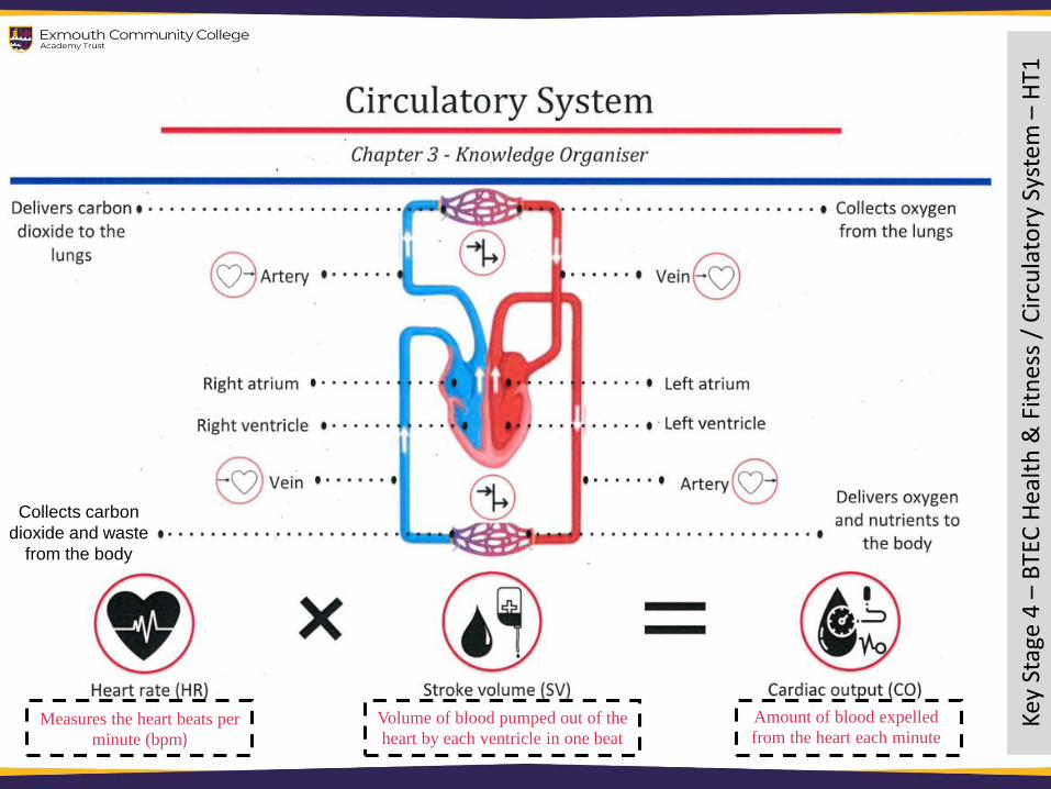
vMeasures the heart beats per
minute (bpm)Volume of blood pumped out of the
heart by each ventricle in one beat
Amount of blood expelled
from the heart each minute
Collects carbon
dioxide and waste
from the body
Key
Stag
e 4
–B
TEC
Hea
lth
& F
itn
ess
/ E
ner
gy S
up
ply
–H
T1
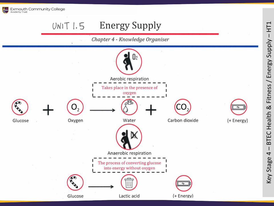
O2 CO2
Key
Stag
e 4
–B
TEC
Hea
lth
& F
itn
ess
/ E
ffec
t o
f Ex
erci
se –
HT1

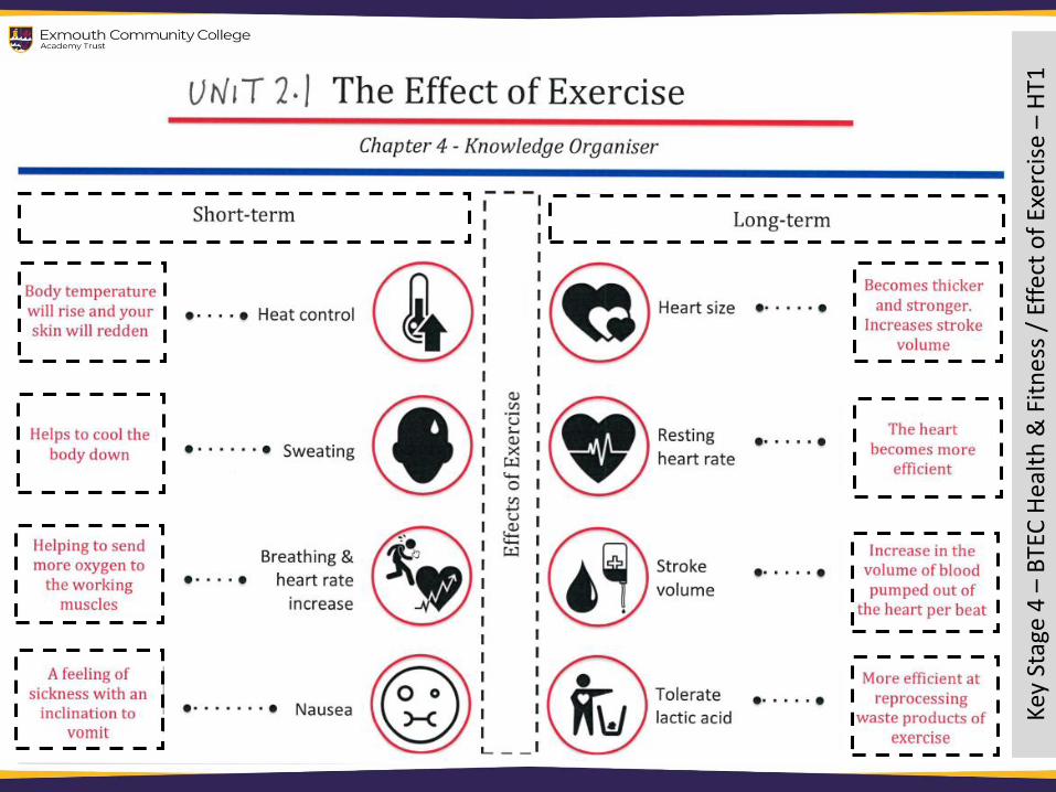
If you are unable to train due to poor health,
your fitness levels can be negatively
affected. However, if you are still able to
train despite ill health, your fitness levels
will not be negatively affected.
FitnessThis relates to your ability
to perform tasks within
your daily life without
becoming overly tired.
HealthHealth is your physical,
mental and social well-
being and not merely the
absence of disease.
Key
Stag
e 4
–B
TEC
Hea
lth
& F
itn
ess
/ C
om
po
nen
ts –
HT1
Health, Fitness and Fitness ComponentsRelationship between Health and Fitness
Maximal StrengthThe maximum force produced in order
to move larger loads
Explosive StrengthForce exerted in one quick
muscular contraction
Static StrengthThe ability to exert against an
immovable object
Dynamic StrengthThe ability to create a force repeatedly
for a long period of time
SpeedThe ability to travel a
certain distance quickly
Components of Fitness and Physical Activity
The different components of fitness which can be improved through physical training are
outlined below. Different sports require different components to perform different skills.
BalanceThe ability to maintain a
stable centre of gravity in
order to avoid falling
AgilityThe ability to rapidly change
direction and body position
Reaction TimeThe ability to quickly
respond to a cue
Cardiovascular EnduranceThe ability to exercise for a
prolonged period of time
StrengthThe ability to
overcome resistance
PowerThe ability to apply
a force quickly
FlexibilityThe ability to quickly
respond to a cue
CoordinationThe ability to time the
movement of the whole
body or individual body
parts in relation to
external or internal cues
Muscular EnduranceThe ability to quickly
respond to a cue
Componentsof Fitness
Key
Stag
e 4
–B
TEC
Hea
lth
& F
itn
ess
/ T
rain
ing –
HT1